hyperactivation of p2x7 receptors as a culprit of covid-19
TRANSCRIPT

Molecular Psychiatry (2021) 26:1044–1059https://doi.org/10.1038/s41380-020-00965-3
REVIEW ARTICLE
Hyperactivation of P2X7 receptors as a culprit of COVID-19neuropathology
Deidiane Elisa Ribeiro 1● Ágatha Oliveira-Giacomelli 1
● Talita Glaser1 ● Vanessa F. Arnaud-Sampaio1●
Roberta Andrejew1● Luiz Dieckmann2
● Juliana Baranova1 ● Claudiana Lameu1● Mariusz Z. Ratajczak3 ●
Henning Ulrich 1
Received: 22 July 2020 / Revised: 4 November 2020 / Accepted: 17 November 2020 / Published online: 16 December 2020© The Author(s), under exclusive licence to Springer Nature Limited 2020
AbstractScientists and health professionals are exhaustively trying to contain the coronavirus disease 2019 (COVID-19) pandemic byelucidating viral invasion mechanisms, possible drugs to prevent viral infection/replication, and health cares to minimizeindividual exposure. Although neurological symptoms are being reported worldwide, neural acute and long-termconsequences of SARS-CoV-2 are still unknown. COVID-19 complications are associated with exacerbated immunoin-flammatory responses to SARS-CoV-2 invasion. In this scenario, pro-inflammatory factors are intensely released into thebloodstream, causing the so-called “cytokine storm”. Both pro-inflammatory factors and viruses may cross the blood–brainbarrier and enter the central nervous system, activating neuroinflammatory responses accompanied by hemorrhagic lesionsand neuronal impairment, which are largely described processes in psychiatric disorders and neurodegenerative diseases.Therefore, SARS-CoV-2 infection could trigger and/or worse brain diseases. Moreover, patients with central nervous systemdisorders associated to neuroimmune activation (e.g. depression, Parkinson’s and Alzheimer’s disease) may presentincreased susceptibility to SARS-CoV-2 infection and/or achieve severe conditions. Elevated levels of extracellular ATPinduced by SARS-CoV-2 infection may trigger hyperactivation of P2X7 receptors leading to NLRP3 inflammasome sti-mulation as a key mediator of neuroinvasion and consequent neuroinflammatory processes, as observed in psychiatricdisorders and neurodegenerative diseases. In this context, P2X7 receptor antagonism could be a promising strategy toprevent or treat neurological complications in COVID-19 patients.
Introduction
Severe acute respiratory syndrome coronavirus 2 (SARS-CoV-2) was first identified in December 2019 as theinfectious agent of coronavirus disease 2019 (COVID-19).
SARS-CoV-2 invades cells using prominent spike protein,which binds to cellular membrane receptors. Host cellreceptors recognized by SARS-CoV-2 spike proteinsinclude angiotensin-converting enzyme 2 (ACE2) [1, 2]and CD147 (basigin) [3, 4] besides involving virus spikeprotein priming/processing by transmembrane serine pro-tease 2 (TMPRSS2) [5]. SARS-CoV-2 enters the cellthrough receptor-mediated endocytosis or receptor-independent entry, as shown for HEK293/hACE2 cells[6]. The challenging question is, whether the infection canoccur through extracellular microvesicles shed frominfected cells. Such mechanism is often called “trojanhorse” and has been proposed for human immunodefi-ciency virus (HIV) [7, 8]. Although neural acute and long-term consequences of SARS-CoV-2 infection are stillunknown, neurological symptoms are being reportedworldwide. Therefore, urgent challenges are to identify,ameliorate, or even eliminate these effects.
These authors contributed equally: Deidiane Elisa Ribeiro, ÁgathaOliveira-Giacomelli
* Henning [email protected]
1 Department of Biochemistry, Institute of Chemistry, University ofSão Paulo, São Paulo, Brazil
2 Department of Psychiatry, Federal University of São Paulo,São Paulo, Brazil
3 Stem Cell Program at the Department of Medicine, University ofLouisville, Kentucky, KY, USA
1234
5678
90();,:
1234567890();,:
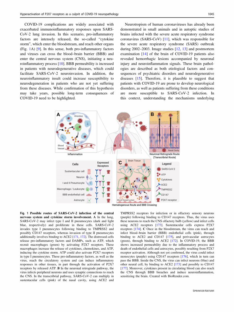
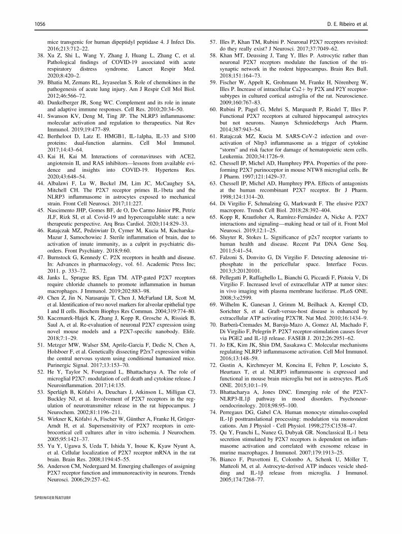
COVID-19 complications are widely associated withexacerbated immunoinflammatory responses upon SARS-CoV-2 lung invasion. In this scenario, pro-inflammatoryfactors are intensely released, the so-called “cytokinestorm”, which enter the bloodstream, and reach other organs(Fig. 1A) [9]. In this sense, both pro-inflammatory factorsand viruses can cross the blood–brain barrier (BBB) andenter the central nervous system (CNS), initiating a neu-roinflammatory process [10]. BBB permeability is increasedin patients with neurodegenerative diseases, which couldfacilitate SARS-CoV-2 neuroinvasion. In addition, theneuroinflammatory insult could increase susceptibility toneurodegeneration in patients who are not yet sufferingfrom these diseases. While confirmation of this hypothesismay take years, possible long-term consequences ofCOVID-19 need to be highlighted.
Neurotropism of human coronaviruses has already beendemonstrated in small animals and in autoptic studies ofbrains infected with the severe acute respiratory syndromecoronavirus (SARS-CoV) [11], which was responsible forthe severe acute respiratory syndrome (SARS) outbreakduring 2002–2003. Image studies [12, 13] and postmortemexamination [14] of the brain of COVID-19 patients alsorevealed hemorrhagic lesions accompanied by neuronalinjury and neuroinflammation signals. These brain pathol-ogies are described as both etiological factors and con-sequences of psychiatric disorders and neurodegenerativediseases [15]. Therefore, it is plausible to suggest thatpatients with COVID-19 are prone to develop neurologicaldisorders, as well as patients suffering from these conditionsare more susceptible to SARS-CoV-2 infection. Inthis context, understanding the mechanisms underlying
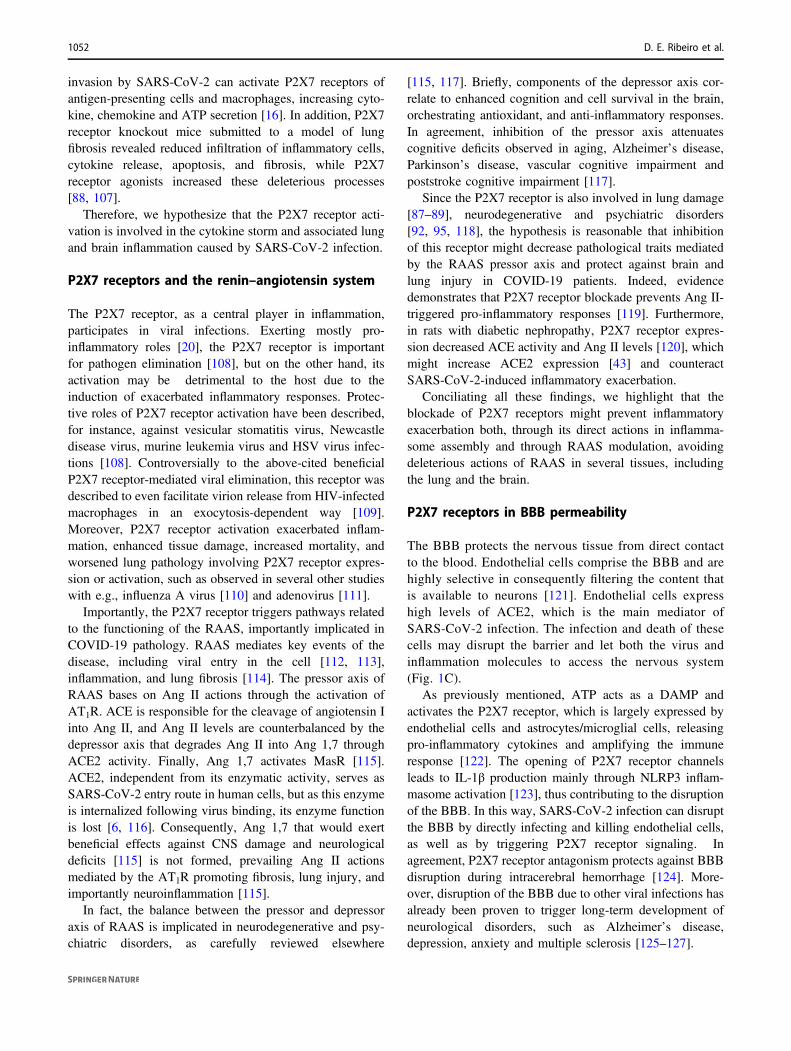
Fig. 1 Possible routes of SARS-CoV-2 infection of the centralnervous system and cytokine storm involvement. A In the lung,SARS-CoV-2 may infect type I and II pneumocytes (dark and lightblue, respectively) and proliferate in these cells. SARS-CoV-2invades type I pneumocytes following binding to TMPRSS2 andpossibly CD147 receptors, whereas invasion of type II pneumocytesadditionally involves binding to ACE2 [171, 172]. The distressed cellsrelease pro-inflammatory factors and DAMPs, such as ATP, whichrecruit macrophages (green) by activating P2X7 receptors. Thesemacrophages increase the release of cytokines, chemokines, and ATP,inducing the cytokine storm. ATP could also activate P2X7 receptorsin type I pneumocytes. These pro-inflammatory factors, as well as thevirus, reach the circulatory system and can induce inflammatoryresponses in other tissues, in part through the activation of P2X7receptors by released ATP. B In the neuronal retrograde pathway, thevirus infects peripheral neurons and uses synaptic connections to reachthe CNS. In the transcribrial pathway, SARS-CoV-2 can multiply insustentacular cells (pink) of the nasal cavity, using ACE2 and
TMPRSS2 receptors for infection or in olfactory sensory neurons(purple) following binding to CD147 receptors. Thus, the virus usesthese neurons to reach the CNS olfactory bulb (yellow) and infect cellsusing ACE2 receptors [173]. Sustentacular cells express P2X7receptors [174]. C Once in the bloodstream, the virus can reach andinfect blood–brain barrier (BBB) endothelial cells (pink), throughbinding to ACE2 and CD147 [175], and perivascular astrocytes(green), through binding to ACE2 [172]. In COVID-19, the BBBshows increased permeability due to the inflammatory process anddeath of endothelial cells and astrocytes, possibly resulting from P2X7receptor activation. Although not yet confirmed, the virus could infectmonocytes (purple) using CD147 receptors [176], which in turn canpass the BBB. Inside the CNS, the virus can infect neurons (blue) andother neural cell, by binding to ACE2 [173] and possibly to CD147[177]. Moreover, cytokines present in circulating blood can also reachthe CNS through BBB breaches and induce neuroinflammation,sensitizing the brain. Created with BioRender.com.
Hyperactivation of P2X7 receptors as a culprit of COVID-19 neuropathology 1045

COVID-19 neuropathology is essential for the developmentof therapeutic strategies.
Hyperactivation of P2X7 receptors is closely related toinflammatory processes since they are stimulated by ATPreleased from distressed cells and induce inflammasomeactivation [16–18]. P2X7 receptors are ATP-gated ionchannels widely expressed in the CNS [19], and theiractivation induced by viral infection leads to molecular(mainly neuroimmune response activation, reactive oxy-gen species (ROS) formation, and glutamate release)and behavioral alterations [17, 20] as well as to mentaldisorders [21].
Based on this, we postulate that both, neuroinvasionthrough the BBB and hyperstimulation of neuroimmuneresponses observed during COVID-19 infection, aremediated by hyperactivation of the P2X7 receptor, possiblythrough NLRP3 inflammasome stimulation. This cascadecould lead to COVID-19-associated psychiatric disordersand neurodegenerative diseases. Thus, P2X7 receptorantagonism would be a promising strategy to prevent ortreat neurological complications in infected patients. Thisconcept will be discussed in detail in our review.
Neurological symptoms in COVID-19
An increasing number of studies reports the manifestationof neurological symptoms in patients with COVID-19. Asreviewed by Pezzini and Padovani, neurological manifes-tations affect between 4.2 and 100% of studied patients:dizziness (prevalent in 7.6–46.1%), headache (5.1–77.1%),impaired consciousness (9.5–64.1%), and seizures(1.2–26%) [22]. Noteworthy, in COVID-19 patients pre-senting at least one neurological symptom, the prevalenceof acute stroke (2.9–76.8%), confusion (14.2–65%), andencephalitis (0–27.9%) stands out. COVID-19 patients alsopresent other nervous system symptoms, such as impairedtaste (10–19.2%) and smell (6–21.7%), dysautonomia(4.3–12%), and acute inflammatory demyelinating poly-neuropathy (0–16,2%) [22].
Together with an acute cerebrovascular disorder, allsymptoms are correlated with the consequences of strokesor micro-strokes [23]. Moreover, reported impaired olfac-tion could be due to CNS invasion or direct damage ofolfactory sensory neurons in the nasal cavity [24].
Psychiatric symptoms are being reported in COVID-19patients, healthcare workers and in the general population.Table 1 summarizes meta-analysis and systematic reviewson these topics. The studies were selected after searchingthe PubMed data base on September 20, 2020 for the terms:“(COVID-19 or SARS-CoV-2 or severe acute respiratorysyndrome coronavirus 2) and (mental health or psycholo-gical health or depression or anxiety or PTSD or PTSS or
post-traumatic stress disorder or post-traumatic stresssymptoms)”.
Although studies on psychiatric symptoms accompany-ing SARS-CoV-2 illness need further investigation, theavailable data indicate that COVID-19 patients mainlypresent depressive symptoms (42–65%), anxiety (37–47%),and PTSS (93–96%). Noteworthy, stress caused by thepandemic situation (e.g., social isolation, fear of infection,and financial instability) may also induce depressivesymptoms (14.6–48.3%), anxiety (6.33–50.9%), and PTSS(7–53.8%) in the general population. The stressful situationof healthcare workers, specially the fear to infect relatives,may lead to depressive symptoms (12.2–26.3%), anxiety(13–29%), and PTSS (3–20.7%) as well.
In this scenario, the following proposals are raised andrequire additional studies: (1) alterations induced by stressexposure (e.g., immune hyperactivation) could both facil-itate SARS-CoV-2 infection as well as aggravate COVID-19 symptoms; (2) neurological symptoms are probably notspecifically related to SARS-CoV-2 invasion, but they are ageneral consequence of infectious disorders (which includesickness behavior), stressful events (pandemic situation),and/or immune response hyperactivation (“cytokinestorm”); (3) genetic and/or environmental factors affect thedevelopment of neurological symptoms, as these responseshighly vary across the population. Clarifying these issueswould improve the understanding on SARS-CoV-2infection and direct the search for treatments.
SARS-CoV-2 neuroinvasion
COVID-19 patients are presenting neurological symptomsworldwide. Although brain analysis of these patients isnot being widely performed, a study detected SARS-CoV-2 in 8 of 21 postmortem brain tissues, based on reversetranscriptase polymerase chain reaction [25]. Moreover,neuroinvasion capability of SARS-CoV-2 was observedin human brain organoids, especially in neuronal cells,such as neural progenitor and radial glial cells, accom-panied by increased cell death [26]. The same study foundpositive staining for SARS-CoV-2 spike protein in thebrain of three COVID-19 patients, with different expres-sion patterns and staining intensities [26]. Neuroinvasionpotential of SARS-CoV-2 was demonstrated in miceexpressing human ACE2 and correlated with increasedmortality independent from respiratory infection [26].SARS-CoV-2 also seems to invade infants’ brains, sinceimmunohistochemical analysis of the postmortem brain ofa 1-year-old infant showed positive staining in the choroidplexus, ventricles and cerebral cortex [27]. Remarkably, acase report demonstrated the presence of SARS-CoV-2viral particles in neural tissue and brain capillary
1046 D. E. Ribeiro et al.
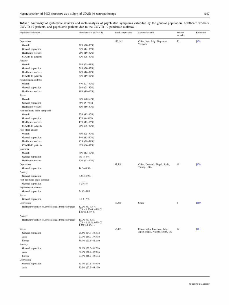
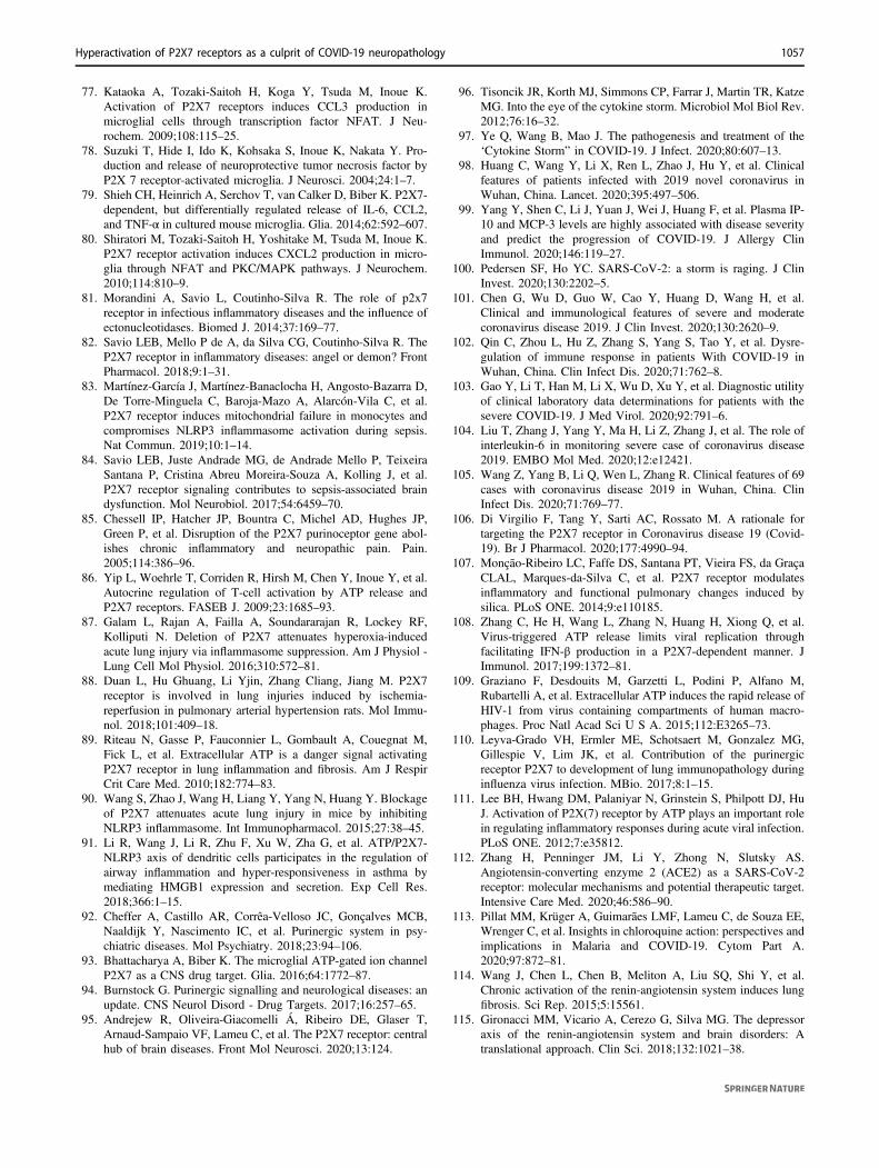
Table 1 Summary of systematic reviews and meta-analysis of psychiatric symptoms exhibited by the general population, healthcare workers,COVID-19 patients, and psychiatric patients due to the COVID-19 pandemic outbreak.
Psychiatric outcome Prevalence % (95% CI) Total sample size Sample location Studiesincluded
Reference
Depression 173,662 China, Iran, Italy, Singapore,Vietnam
50 [178]
Overall 26% (20–33%)
General population 24% (14–36%)
Healthcare workers 25% (19–32%)
COVID-19 patients 42% (28–57%)
Anxiety
Overall 26% (21–31%)
General population 26% (20–32%)
Healthcare workers 24% (16–32%)
COVID-19 patients 37% (19–57%)
Psychological distress
Overall 34% (27–42%)
General population 26% (21–32%)
Healthcare workers 41% (19–65%)
Stress
Overall 34% (20–50%)
General population 36% (5–75%)
Healthcare workers 33% (19–50%)
Post-traumatic stress symptoms
Overall 27% (12–45%)
General population 15% (4–31%)
Healthcare workers 13% (11–16%)
COVID-19 patients 96% (95–97%)
Poor sleep quality
Overall 40% (25–57%)
General population 34% (12–60%)
Healthcare workers 43% (28–59%)
COVID-19 patients 82% (66–92%)
Insomnia
Overall 30% (12–52%)
General population 7% (7–8%)
Healthcare workers 37% (32–42%)
Depression 93,569 China, Denmark, Nepal, Spain,Turkey, USA
19 [179]
General population 14.6–48.3%
Anxiety
General population 6.33–50.9%
Post-traumatic stress disorder
General population 7–53.8%
Psychological distress
General population 34.43–38%
Stress
General population 8.1–81.9%
Depression 17,330 China 8 [180]
Healthcare workers vs. professionals from other areas 12.2% vs. 9.5 %(OR= 1.3246; 95% CI1.0930–1.6053)
Anxiety
Healthcare workers vs. professionals from other areas 13.0% vs. 8.5%(OR= 1.6152; 95% CI1.3283–1.9641)
Stress 63,439 China, India, Iran, Iraq, Italy,Japan, Nepal, Nigeria, Spain, UK
17 [181]
General population 29.6% (24.3–35.4%)
Asia 27.9% (19.7–37.8%)
Europe 31.9% (23.1–42.2%)
Anxiety
General population 31.9% (27.5–36.7%)
Asia 32.9% (28.2–37.9%)
Europe 23.8% (16.2–33.5%)
Depression
General population 33.7% (27.5–40.6%)
Asia 35.3% (27.3–44.1%)
Hyperactivation of P2X7 receptors as a culprit of COVID-19 neuropathology 1047

endothelium of a Parkinson’s disease patient, whichwas associated with the worsening of neurologicalsymptoms [28].
However, the mechanism of SARS-CoV-2 infection ofthe brain is still unknown. Invasion routes of the CNS byother viruses include: (a) the hematogenous route, in which
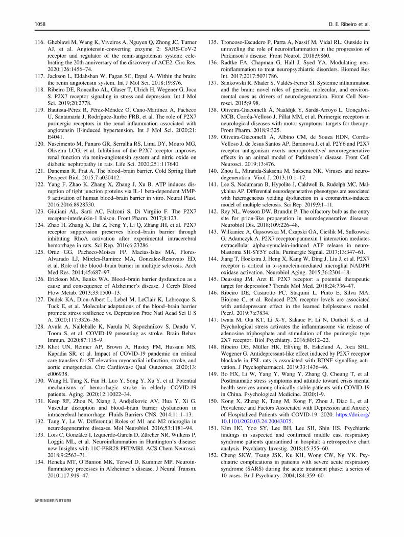
Table 1 (continued)
Psychiatric outcome Prevalence % (95% CI) Total sample size Sample location Studiesincluded
Reference
Europe 32.4% (21.6–45.5%)Psychological distress N.R. N.R. 40 [182]
Healthcare workers exposed to SARS/MERS/COVID-19
37.8% (28.4–48.2%)
Burnout
Healthcare workers exposed to SARS/MERS/COVID-19
34.4% (19.3–53.5%)
Anxiety
Healthcare workers exposed to SARS/MERS/COVID-19
29.0% (14.2–50.3%)
Depressive symptoms
Healthcare workers exposed to SARS/MERS/COVID-19
26.3% (12.5–47.1%)
Post-traumatic stress disorder
Healthcare workers exposed to SARS/MERS/COVID-19
20.7% (13.2–31%)
Anxiety 162,639 Argentina, Brazil, Chile, China,Denmark, Greece, India, Iran,Israel, Italy, Japan, Mexico,Pakistan, Singapore, Spain,Turkey, Vietnam
62 [183]
Overall 33% (28–38%)
General population 32% (25–39%)
General population—Italy 81% (80–83%)
Healthcare workers 26% (18–34%)
Healthcare workers—Singapore 7% (5–9%)
Healthcare workers—Italy 57% (52–63%)
Psychiatric patients with moderate-to-severe anxiety 24% (14–33%)
COVID-19 patients 47% (34–61%)
COVID-19 patients with type 2 diabetes—India 40% (30–50%)
COVID-19 patients with Parkinson’s Disease—Iran 82% (74–88%)
Depression
Overall 28% (23–32%)
General population 27% (22–33%)
General population—Italy 67% (65–69%)
Healthcare workers 25% (17–33%)
Healthcare workers—Singapore 9% (7–12%)
Healthcare workers—China 51% (48–53%)
Psychiatric patients with moderate-to-severedepression
22% (13–32%)
COVID-19 patients—China 65% (51–77%)
Distress
Overall 35% (23–47%)
Stress
Overall 40% (20–60%)
Insomnia
Overall 32% (25–39%)
Post-traumatic stress symptoms/disorders
General population 16% (15–17%)
Healthcare workers 3% (2–4%)
COVID-19 patients 93% (92–95%)
Anxiety 33,062 China, Singapore 12 [184]
Healthcare workers 23.21% (17.77–29.13%)
Male 20.92% (11.86–31.65%)
Female 29.06% (20.21–38.78%)
Depression
Healthcare workers 22.93% (13.16–34.38%)
Male 20.34% (11.57–30.75%)
Female 26.87% (15.39–40.09%)
Insomnia
Healthcare workers 34.32% (27.45–41.54%)
CI Confidence interval, OR Odds ratio, N.R. Not reported
1048 D. E. Ribeiro et al.

viruses use the bloodstream to reach and invade epithelialcells from the BBB or the blood–cerebrospinal fluid barrier,or use leukocytes as a vector to enter the CNS; (b) theneuronal retrograde route, in which viruses invade periph-eral neurons and reach CNS, including the transcribrialroute, using olfactory sensory neurons in the nasal cavity(Fig. 1B) [10].
In the hematogenous route, the virus must be capable ofcrossing the BBB (Fig. 1C). This barrier is composed ofendothelial cells, pericytes, and astrocytes. The restrictedpermeability of the BBB is a reflex of the connectionbetween brain microvascular endothelial cells and tightcell–cell junctions. The BBB is disrupted under inflamma-tory conditions [29, 30]. In the “Trojan horse” mechanismof CNS invasion, infected leukocytes pass the BBB. Thismechanism is observed for HIV, and since SARS-CoV caninfect immune cells, it is likely that SARS-CoV-2 also usesthis route toward the CNS [10]. Viral infection also affectsBBB integrity by different mechanisms, including phos-phorylation of tight junction proteins, disruption of the basallamina or of the actin cytoskeleton, or by invading BBB-epithelial cells and furthermore astrocytes [10, 31].
Evidence of CNS invasion through neuronal retrograderoutes was reported for coronaviruses, such as HCoV-OC43, HEV67 and avian bronchitis virus [32]. Oncerespiratory and digestive tracts of animals were infected,coronaviruses invaded peripheral neurons and passedthrough synaptic connections until they reached medullaryneurons and subsequently other neurons and glial cells ofthe CNS [32]. As the main entry route of SARS-CoV-2 inhumans, cells from the nasal cavity could be susceptible toviral infection and replication. Studies reported that humanolfactory sustentacular cells express both ACE2 andTMPRSS2 virus receptors [33]. Although current literaturedata reports that human olfactory sensory neurons do notexpress these proteins, they express CD147 that could allowSARS-CoV-2 neuroinvasion (Fig. 1) [33, 34]. In fact,SARS-CoV, MERS-CoV, and HCoV-OCR43 were able toinvade the murine CNS using the transcribrial route,infecting olfactory sensory neurons of nasal cavity, andpassing to other neural cells, indicating that SARS-CoV-2could use the same mechanism [24, 35–37].
Inflammation and CNS-related lethality of COVID-19
Severe COVID-19 patients commonly develop the AcuteRespiratory Distress Syndrome [38], which is characterizedby inflammatory injury to the alveoli–capillary membrane,leading to lung over-permeability and increased pulmonaryedema fluid into the airspaces, resulting in the lack ofrespiratory capacity [39]. This overreaction of the innateimmune system against viral infection induces the so-called“cytokine storm”, comprising of: (1) the release of large
amounts of several pro-inflammatory cytokines (interferonsIFNα and IFNγ, interleukins [IL-1β, IL-6, IL-12, IL-18 andIL-33], tumor necrosis factor [TNF]-α, and transforminggrowth factor [TGF]-β) and chemokines (CXCL10,CXCL8, CXCL9, CCL2, CCL3, CCL5); (2) release ofrenin–angiotensin aldosterone system (RAAS) mediatorsand increasing blood levels of angiotensin II (Ang II); andlately (3) amplification of the innate immune systemresponse and activation of its major humoral arm, thecomplement cascade (ComC) [40].
Novel evidence indicates that COVID-19-released med-iators merge on a common pathway, upregulating cytosolicdanger sensing pattern recognition receptor, which is part ofa multiprotein complex of the innate immune system that iscalled inflammasome, and recognizes both pathogen-associated molecular patterns and self-derived danger-associated molecular patterns (DAMPs) or alarmines [41].
Importantly, upon inflammasome protein assembly andactivation, pro-caspase 1 protein is cleaved to functionalcaspase 1, whose main function is the conversion of theinactive and intracellularly stored pro-inflammatory cyto-kines, pro-IL-1β and pro-IL-18, into their active forms thatare released from cells. This release is facilitated by creatinggasdermin D (GSDMD) pore channels in cell membranes.In addition to these two cytokines, gasdermin D channelsalso mediate the release of several biologically activeDAMPs or alarmines, including extracellular ATP, highmobility group protein B1, and S100 calcium-binding pro-teins A8 and A9 (S1008/9a) [42].
There are various inflammasome subtypes. The NLRP3inflammasome protein complex is usually involved in virusinfections and consists of NLRP3 protein, apoptosis-associated speck-like protein containing a CARD (ASC)and pro-caspase-1, and remains in the cytosol in steady-state conditions in an inactive form. Upon activation, itbecomes a multiprotein aggregate composed of severalNLRP3 molecules (speck complexes), each containingNLRP3 protein, ASC, and pro-caspase 1 [41].
We consider the NLRP3 inflammasome as a trigger ofthe cytokine storm, as seen in COVID-19 patients, that maybe induced by P2X7 receptor activation, including in thebrain (Fig. 2). Once SARS-CoV-2 spike protein interactswith ACE2, macrophages/microglia cells can potentiate theimmune response through the cleavage of fragments com-plement component 3a and 5a (C3a and C5a, respectively)and non-lytic C5b-C9 membrane attack complex by ComC,thus activating the NLRP3 inflammasome [41]. Moreover,NLRP3 inflammasome activation during COVID-19 infec-tion is usually triggered by Ang II, which binds to theangiotensin type 1 receptor (AT1R), leading to vessel con-traction and increasing blood pressure. In normal condi-tions, Ang II is converted by the ACE2 receptor intoangiotensin 1–7 (Ang 1–7) [43]. Ang II activates the AT1R,
Hyperactivation of P2X7 receptors as a culprit of COVID-19 neuropathology 1049
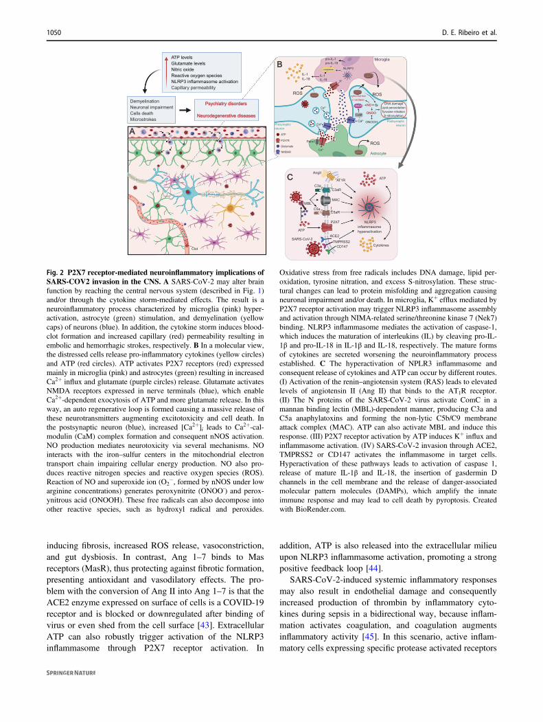
inducing fibrosis, increased ROS release, vasoconstriction,and gut dysbiosis. In contrast, Ang 1–7 binds to Masreceptors (MasR), thus protecting against fibrotic formation,presenting antioxidant and vasodilatory effects. The pro-blem with the conversion of Ang II into Ang 1–7 is that theACE2 enzyme expressed on surface of cells is a COVID-19receptor and is blocked or downregulated after binding ofvirus or even shed from the cell surface [43]. ExtracellularATP can also robustly trigger activation of the NLRP3inflammasome through P2X7 receptor activation. In
addition, ATP is also released into the extracellular milieuupon NLRP3 inflammasome activation, promoting a strongpositive feedback loop [44].
SARS-CoV-2-induced systemic inflammatory responsesmay also result in endothelial damage and consequentlyincreased production of thrombin by inflammatory cyto-kines during sepsis in a bidirectional way, because inflam-mation activates coagulation, and coagulation augmentsinflammatory activity [45]. In this scenario, active inflam-matory cells expressing specific protease activated receptors
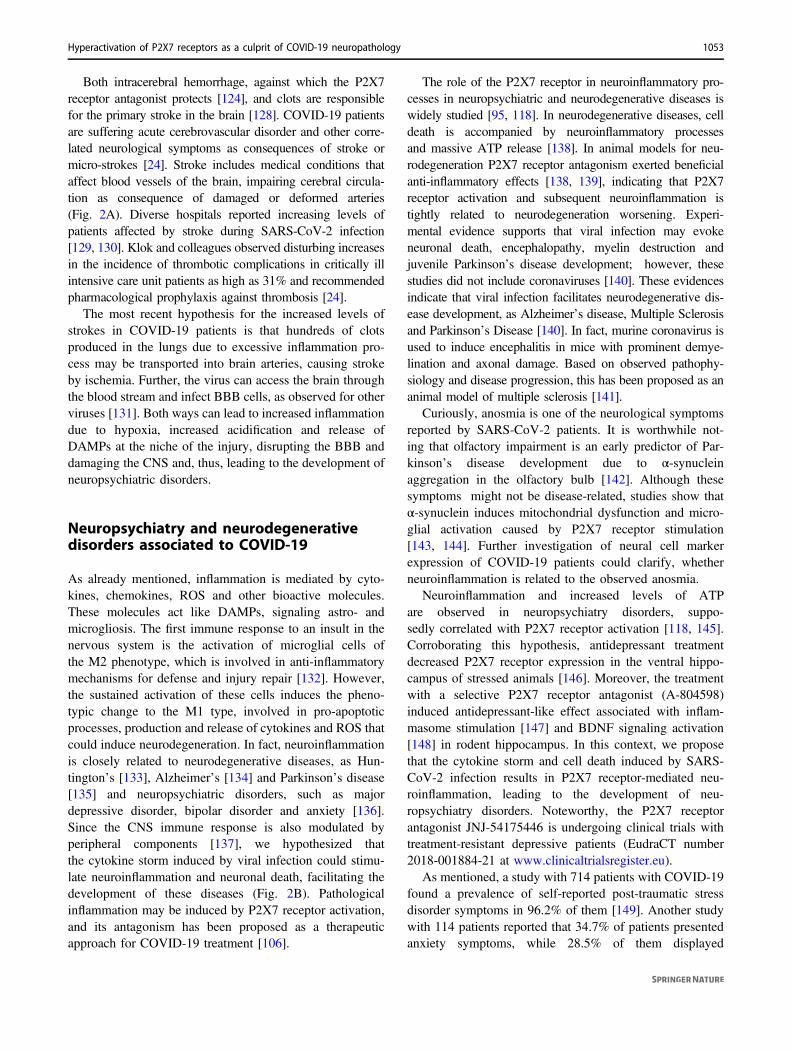
Fig. 2 P2X7 receptor-mediated neuroinflammatory implications ofSARS-COV2 invasion in the CNS. A SARS-CoV-2 may alter brainfunction by reaching the central nervous system (described in Fig. 1)and/or through the cytokine storm-mediated effects. The result is aneuroinflammatory process characterized by microglia (pink) hyper-activation, astrocyte (green) stimulation, and demyelination (yellowcaps) of neurons (blue). In addition, the cytokine storm induces blood-clot formation and increased capillary (red) permeability resulting inembolic and hemorrhagic strokes, respectively. B In a molecular view,the distressed cells release pro-inflammatory cytokines (yellow circles)and ATP (red circles). ATP activates P2X7 receptors (red) expressedmainly in microglia (pink) and astrocytes (green) resulting in increasedCa2+ influx and glutamate (purple circles) release. Glutamate activatesNMDA receptors expressed in nerve terminals (blue), which enableCa2+-dependent exocytosis of ATP and more glutamate release. In thisway, an auto regenerative loop is formed causing a massive release ofthese neurotransmitters augmenting excitotoxicity and cell death. Inthe postsynaptic neuron (blue), increased [Ca2+]i leads to Ca2+-cal-modulin (CaM) complex formation and consequent nNOS activation.NO production mediates neurotoxicity via several mechanisms. NOinteracts with the iron–sulfur centers in the mitochondrial electrontransport chain impairing cellular energy production. NO also pro-duces reactive nitrogen species and reactive oxygen species (ROS).Reaction of NO and superoxide ion (O2
−, formed by nNOS under lowarginine concentrations) generates peroxynitrite (ONOO-) and perox-ynitrous acid (ONOOH). These free radicals can also decompose intoother reactive species, such as hydroxyl radical and peroxides.
Oxidative stress from free radicals includes DNA damage, lipid per-oxidation, tyrosine nitration, and excess S-nitrosylation. These struc-tural changes can lead to protein misfolding and aggregation causingneuronal impairment and/or death. In microglia, K+ efflux mediated byP2X7 receptor activation may trigger NLRP3 inflammasome assemblyand activation through NIMA-related serine/threonine kinase 7 (Nek7)binding. NLRP3 inflammasome mediates the activation of caspase-1,which induces the maturation of interleukins (IL) by cleaving pro-IL-1β and pro-IL-18 in IL-1β and IL-18, respectively. The mature formsof cytokines are secreted worsening the neuroinflammatory processestablished. C The hyperactivation of NPLR3 inflammasome andconsequent release of cytokines and ATP can occur by different routes.(I) Activation of the renin–angiotensin system (RAS) leads to elevatedlevels of angiotensin II (Ang II) that binds to the AT1R receptor.(II) The N proteins of the SARS-CoV-2 virus activate ComC in amannan binding lectin (MBL)-dependent manner, producing C3a andC5a anaphylatoxins and forming the non-lytic C5b/C9 membraneattack complex (MAC). ATP can also activate MBL and induce thisresponse. (III) P2X7 receptor activation by ATP induces K+ influx andinflammasome activation. (IV) SARS-CoV-2 invasion through ACE2,TMPRSS2 or CD147 activates the inflammasome in target cells.Hyperactivation of these pathways leads to activation of caspase 1,release of mature IL-1β and IL-18, the insertion of gasdermin Dchannels in the cell membrane and the release of danger-associatedmolecular pattern molecules (DAMPs), which amplify the innateimmune response and may lead to cell death by pyroptosis. Createdwith BioRender.com.
1050 D. E. Ribeiro et al.
(PAR1–4) bind to thrombin, while the Toll-like receptor 4interacts with fibrin. Supporting these events, patients inparticular at younger age are often diagnosed with strokeand cardiovascular complications.
Recent evidence indicates that the occurrence of psy-chiatric disorders in patients is linked to “sterile” inflam-mation of the brain that may be initiated locally by somestressors affecting nervous tissue or occurs due to a sys-temic inflammation process [17, 46]. This is supported bythe observation that several inflammatory mediators andmarkers are detected in the peripheral blood of patients withpsychiatric and neurodegenerative disorders [17, 46], whichcould worsen prognoses for COVID-19 outcome. More-over, clinical data describe correlations between systemicchronic inflammatory processes and psychiatric disorders.This may also explain, why some reported anti-inflammatory treatment strategies ameliorate neurodegen-eration [17, 46]. In agreement, pathological increase ofAngII-AT1R-mediated activation of NLRP3 inflammasomemay initiate psychosis. We believe that bioactive inflam-matory mediators released during the cytokine storm, suchas extracellular ATP, affect the CNS and may lead to itsimpairment, mainly through P2X7 receptor activation, aswe discuss in the following.
Probable P2X7 receptor roles in COVID-19processes
The P2X7 receptor is widely expressed through the body[47], including in immune [48], lung [49], and CNS cells,mainly microglia and oligodendrocytes [50–52], whereas itsexpression in neurons and astrocytes is still under discus-sion [50, 51, 53–60]. During the progress of infection, ATPrelease may result from the NLRP3 inflammasome responseto COVID-19 infection (i) after virus spike protein inter-action with the virus entry receptor ACE2, (ii) due to ele-vated level of Ang II observed in infected patients, or (iii) inresponse to activated ComC mediators [61]. Consequently,P2X7 receptors are activated, increasing inflammatoryresponses as well as modulating RAAS-related pathwaysand BBB permeability, as we discuss in the following.
P2X7 receptor role in inflammation
The P2X7 receptor is a trimeric ionotropic receptor thatbelongs to the P2X family of purinergic receptors, pre-senting low affinity for ATP (EC50 around 0.3–1.8 mM)and, under prolonged stimulation, can form pores allowingthe passage of large hydrophilic molecules [62–64]. Acti-vation of P2X7 receptor elicits rapid K+ efflux as well asCa2+ and Na+ influx, resulting in the stimulation of severalintracellular intermediators, as PLC, PLA2, PKC, MAPK,
PI3K, ERK1/2 and p38, among others [65]. Consequently,P2X7 receptor activation is associated with numerous cel-lular functions, as plasma membrane blebbing, phosphati-dylserine exposure in the membrane, formation of ROS,interleukin secretion, cell death and proliferation [65, 66].
ATP concentration is present in the nanomolar range inextracellular space of healthy tissues [67]. Conversely, in adisease state as infection or brain disease, extracellular ATPlevels largely increase and activate P2X7 receptors [68–70].Consequently, pore formation results in enhanced release ofATP and establishes a positive feedback in pathologicalconditions. In these circumstances, ATP may act as DAMP,which activates the nuclear transcription factor NF-κBresulting in expression upregulation of pro-IL-1β and pro-IL-18 and the NLRP3 protein [16, 71]. In addition, P2X7receptor activation triggers K+ efflux, a signal required forefficient NLRP3 inflammasome stimulation [72]. Conse-quently, there is an activation of caspase-1 and maturationof IL-1β and IL-18, resulting in pro-inflammatory cytokinerelease [16, 20, 52, 71, 73]. The P2X7 receptor is a majoractivator of the NLRP3 inflammasome in several cell types[16], including macrophages and microglial cells [72, 74–76]. Moreover, P2X7 receptor activation also promotes therelease of other inflammatory mediators such as IL-6, TNF-α, CCL2, CCL3, and CXCL2 [77–80].
In fact, the role of P2X7 receptor as a modulator ofinflammatory response is well-established [16, 70]. It wasalready reported its capacity to modulate acute and chronicinfection [81, 82], inflammatory diseases [82], sepsis[83, 84], neuropathic pain [85], and T-cell activation [86].In addition, P2X7 receptor expression deletion appears to bebeneficial in case of acute lung injury, asthma, lunginflammation, and fibrosis [87–91]. Remarkably, P2X7receptor activation increases neuroinflammatory responses,which have been associated with neurodegenerative dis-eases, psychiatric disorders and stroke [21, 92–95], as fur-ther discussed.
Cytokine storms have been reported in several viralinfections [96], including SARS-CoV-2 [9, 97]. Patientsinfected with SARS-CoV-2 present elevated levels of IL-6,IL-10, IL-1β, INF-γ, CCL2, TNF-α, CXCL10, CCL7, IL-1receptor antagonist and IL-2 receptor, supposedly asso-ciated with disease severity [9, 98–105]. Considering theP2X7 receptor role in inflammation, its activation may beinvolved in the cytokine storm observed in COVID-19 bystimulating NLRP3 inflammasome, overshooting inflam-mation with extensive cytokine release, affecting coagula-tion and leading to diffuse lung edema and infiltration byimmune cells and inflammatory cytokines [106]. As dis-cussed by Di Virgilio et al. [106], many processes asso-ciated with lung impairment, as seen in COVID-19 patients,are mediated by P2X7 receptors [16]. The massiveATP release following lung mononuclear phagocytes
Hyperactivation of P2X7 receptors as a culprit of COVID-19 neuropathology 1051
invasion by SARS-CoV-2 can activate P2X7 receptors ofantigen-presenting cells and macrophages, increasing cyto-kine, chemokine and ATP secretion [16]. In addition, P2X7receptor knockout mice submitted to a model of lungfibrosis revealed reduced infiltration of inflammatory cells,cytokine release, apoptosis, and fibrosis, while P2X7receptor agonists increased these deleterious processes[88, 107].
Therefore, we hypothesize that the P2X7 receptor acti-vation is involved in the cytokine storm and associated lungand brain inflammation caused by SARS-CoV-2 infection.
P2X7 receptors and the renin–angiotensin system
The P2X7 receptor, as a central player in inflammation,participates in viral infections. Exerting mostly pro-inflammatory roles [20], the P2X7 receptor is importantfor pathogen elimination [108], but on the other hand, itsactivation may be detrimental to the host due to theinduction of exacerbated inflammatory responses. Protec-tive roles of P2X7 receptor activation have been described,for instance, against vesicular stomatitis virus, Newcastledisease virus, murine leukemia virus and HSV virus infec-tions [108]. Controversially to the above-cited beneficialP2X7 receptor-mediated viral elimination, this receptor wasdescribed to even facilitate virion release from HIV-infectedmacrophages in an exocytosis-dependent way [109].Moreover, P2X7 receptor activation exacerbated inflam-mation, enhanced tissue damage, increased mortality, andworsened lung pathology involving P2X7 receptor expres-sion or activation, such as observed in several other studieswith e.g., influenza A virus [110] and adenovirus [111].
Importantly, the P2X7 receptor triggers pathways relatedto the functioning of the RAAS, importantly implicated inCOVID-19 pathology. RAAS mediates key events of thedisease, including viral entry in the cell [112, 113],inflammation, and lung fibrosis [114]. The pressor axis ofRAAS bases on Ang II actions through the activation ofAT1R. ACE is responsible for the cleavage of angiotensin Iinto Ang II, and Ang II levels are counterbalanced by thedepressor axis that degrades Ang II into Ang 1,7 throughACE2 activity. Finally, Ang 1,7 activates MasR [115].ACE2, independent from its enzymatic activity, serves asSARS-CoV-2 entry route in human cells, but as this enzymeis internalized following virus binding, its enzyme functionis lost [6, 116]. Consequently, Ang 1,7 that would exertbeneficial effects against CNS damage and neurologicaldeficits [115] is not formed, prevailing Ang II actionsmediated by the AT1R promoting fibrosis, lung injury, andimportantly neuroinflammation [115].
In fact, the balance between the pressor and depressoraxis of RAAS is implicated in neurodegenerative and psy-chiatric disorders, as carefully reviewed elsewhere
[115, 117]. Briefly, components of the depressor axis cor-relate to enhanced cognition and cell survival in the brain,orchestrating antioxidant, and anti-inflammatory responses.In agreement, inhibition of the pressor axis attenuatescognitive deficits observed in aging, Alzheimer’s disease,Parkinson’s disease, vascular cognitive impairment andpoststroke cognitive impairment [117].
Since the P2X7 receptor is also involved in lung damage[87–89], neurodegenerative and psychiatric disorders[92, 95, 118], the hypothesis is reasonable that inhibitionof this receptor might decrease pathological traits mediatedby the RAAS pressor axis and protect against brain andlung injury in COVID-19 patients. Indeed, evidencedemonstrates that P2X7 receptor blockade prevents Ang II-triggered pro-inflammatory responses [119]. Furthermore,in rats with diabetic nephropathy, P2X7 receptor expres-sion decreased ACE activity and Ang II levels [120], whichmight increase ACE2 expression [43] and counteractSARS-CoV-2-induced inflammatory exacerbation.
Conciliating all these findings, we highlight that theblockade of P2X7 receptors might prevent inflammatoryexacerbation both, through its direct actions in inflamma-some assembly and through RAAS modulation, avoidingdeleterious actions of RAAS in several tissues, includingthe lung and the brain.
P2X7 receptors in BBB permeability
The BBB protects the nervous tissue from direct contactto the blood. Endothelial cells comprise the BBB and arehighly selective in consequently filtering the content thatis available to neurons [121]. Endothelial cells expresshigh levels of ACE2, which is the main mediator ofSARS-CoV-2 infection. The infection and death of thesecells may disrupt the barrier and let both the virus andinflammation molecules to access the nervous system(Fig. 1C).
As previously mentioned, ATP acts as a DAMP andactivates the P2X7 receptor, which is largely expressed byendothelial cells and astrocytes/microglial cells, releasingpro-inflammatory cytokines and amplifying the immuneresponse [122]. The opening of P2X7 receptor channelsleads to IL-1β production mainly through NLRP3 inflam-masome activation [123], thus contributing to the disruptionof the BBB. In this way, SARS-CoV-2 infection can disruptthe BBB by directly infecting and killing endothelial cells,as well as by triggering P2X7 receptor signaling. Inagreement, P2X7 receptor antagonism protects against BBBdisruption during intracerebral hemorrhage [124]. More-over, disruption of the BBB due to other viral infections hasalready been proven to trigger long-term development ofneurological disorders, such as Alzheimer’s disease,depression, anxiety and multiple sclerosis [125–127].
1052 D. E. Ribeiro et al.
Both intracerebral hemorrhage, against which the P2X7receptor antagonist protects [124], and clots are responsiblefor the primary stroke in the brain [128]. COVID-19 patientsare suffering acute cerebrovascular disorder and other corre-lated neurological symptoms as consequences of stroke ormicro-strokes [24]. Stroke includes medical conditions thataffect blood vessels of the brain, impairing cerebral circula-tion as consequence of damaged or deformed arteries(Fig. 2A). Diverse hospitals reported increasing levels ofpatients affected by stroke during SARS-CoV-2 infection[129, 130]. Klok and colleagues observed disturbing increasesin the incidence of thrombotic complications in critically illintensive care unit patients as high as 31% and recommendedpharmacological prophylaxis against thrombosis [24].
The most recent hypothesis for the increased levels ofstrokes in COVID-19 patients is that hundreds of clotsproduced in the lungs due to excessive inflammation pro-cess may be transported into brain arteries, causing strokeby ischemia. Further, the virus can access the brain throughthe blood stream and infect BBB cells, as observed for otherviruses [131]. Both ways can lead to increased inflammationdue to hypoxia, increased acidification and release ofDAMPs at the niche of the injury, disrupting the BBB anddamaging the CNS and, thus, leading to the development ofneuropsychiatric disorders.
Neuropsychiatry and neurodegenerativedisorders associated to COVID-19
As already mentioned, inflammation is mediated by cyto-kines, chemokines, ROS and other bioactive molecules.These molecules act like DAMPs, signaling astro- andmicrogliosis. The first immune response to an insult in thenervous system is the activation of microglial cells ofthe M2 phenotype, which is involved in anti-inflammatorymechanisms for defense and injury repair [132]. However,the sustained activation of these cells induces the pheno-typic change to the M1 type, involved in pro-apoptoticprocesses, production and release of cytokines and ROS thatcould induce neurodegeneration. In fact, neuroinflammationis closely related to neurodegenerative diseases, as Hun-tington’s [133], Alzheimer’s [134] and Parkinson’s disease[135] and neuropsychiatric disorders, such as majordepressive disorder, bipolar disorder and anxiety [136].Since the CNS immune response is also modulated byperipheral components [137], we hypothesized thatthe cytokine storm induced by viral infection could stimu-late neuroinflammation and neuronal death, facilitating thedevelopment of these diseases (Fig. 2B). Pathologicalinflammation may be induced by P2X7 receptor activation,and its antagonism has been proposed as a therapeuticapproach for COVID-19 treatment [106].
The role of the P2X7 receptor in neuroinflammatory pro-cesses in neuropsychiatric and neurodegenerative diseases iswidely studied [95, 118]. In neurodegenerative diseases, celldeath is accompanied by neuroinflammatory processesand massive ATP release [138]. In animal models for neu-rodegeneration P2X7 receptor antagonism exerted beneficialanti-inflammatory effects [138, 139], indicating that P2X7receptor activation and subsequent neuroinflammation istightly related to neurodegeneration worsening. Experi-mental evidence supports that viral infection may evokeneuronal death, encephalopathy, myelin destruction andjuvenile Parkinson’s disease development; however, thesestudies did not include coronaviruses [140]. These evidencesindicate that viral infection facilitates neurodegenerative dis-ease development, as Alzheimer’s disease, Multiple Sclerosisand Parkinson’s Disease [140]. In fact, murine coronavirus isused to induce encephalitis in mice with prominent demye-lination and axonal damage. Based on observed pathophy-siology and disease progression, this has been proposed as ananimal model of multiple sclerosis [141].
Curiously, anosmia is one of the neurological symptomsreported by SARS-CoV-2 patients. It is worthwhile not-ing that olfactory impairment is an early predictor of Par-kinson’s disease development due to α-synucleinaggregation in the olfactory bulb [142]. Although thesesymptoms might not be disease-related, studies show thatα-synuclein induces mitochondrial dysfunction and micro-glial activation caused by P2X7 receptor stimulation[143, 144]. Further investigation of neural cell markerexpression of COVID-19 patients could clarify, whetherneuroinflammation is related to the observed anosmia.
Neuroinflammation and increased levels of ATPare observed in neuropsychiatry disorders, suppo-sedly correlated with P2X7 receptor activation [118, 145].Corroborating this hypothesis, antidepressant treatmentdecreased P2X7 receptor expression in the ventral hippo-campus of stressed animals [146]. Moreover, the treatmentwith a selective P2X7 receptor antagonist (A-804598)induced antidepressant-like effect associated with inflam-masome stimulation [147] and BDNF signaling activation[148] in rodent hippocampus. In this context, we proposethat the cytokine storm and cell death induced by SARS-CoV-2 infection results in P2X7 receptor-mediated neu-roinflammation, leading to the development of neu-ropsychiatry disorders. Noteworthy, the P2X7 receptorantagonist JNJ-54175446 is undergoing clinical trials withtreatment-resistant depressive patients (EudraCT number2018-001884-21 at www.clinicaltrialsregister.eu).
As mentioned, a study with 714 patients with COVID-19found a prevalence of self-reported post-traumatic stressdisorder symptoms in 96.2% of them [149]. Another studywith 114 patients reported that 34.7% of patients presentedanxiety symptoms, while 28.5% of them displayed
Hyperactivation of P2X7 receptors as a culprit of COVID-19 neuropathology 1053
depression symptoms [150]. Moreover, COVID-19 relatedpsychiatric symptoms (including anger, anxiety, suicidalideas, hallucinations, insomnia, impaired memory, poorconcentration, time disorientation, fear/panic, pressuredspeech, mood alterations, pessimistic thinking, crying spelland persecutory ideas) have been largely described duringand after the occurrence of other respiratory pandemics,such as MERS [151] and SARS [152–159]. Further indi-cation for psychiatric implications of coronaviruses infec-tions came from a study of Okusaga et al. [160], whoconnected seropositivity for human coronavirus strain L63with mood disorders and suicide attempts. The study couldcome to a limited conclusion, as it would need to be con-ducted with more control and coronavirus-serum positivevolunteers. However, the authors of this study distinguishedbetween two possible scenarios, which might be importantfor the understanding of neuronal effects of COVID-19:(1) the connection between depression and immuneresponses with possible pro-inflammatory interleukinovershooting, which may trigger oxidative stress and neu-roinflammation as part of the causes of psychiatric dis-orders; and (2) viral infection acting as a form of stress.Dysregulated stress and neuroinflammatory responsesmight cause hypothalamic–pituitary–adrenal (HPA)axis dysfunction. Alterations in the HPA axis have beenlinked to mood diseases, and HPA activation has beenconnected with suicide. Hiroi et al. found hyperplasticadrenals in suicide victims, corroborating with suchhypothesis [161]. In line, a small COVID-19 patients’ studyconcluded that preexisting psychiatric disease patientsreported mental symptom worsening following infectionwith the virus [162].
Noteworthy, gene knockout or pharmacological inhibi-tion of P2X7 receptors induced antidepressant-like behaviorin mice exposed to stress, accompanied by HPA axisrestoration [163, 164]. This axis is also activated forcounteracting tissue damage, evoked by cytokine storms, inSARS-CoV-2 infection, as previously reviewed [165].Thus, the HPA axis might provide a connection betweencoronaviruses and psychiatric disorders. Noteworthy, suchmechanism would explain the possible propensity for thedevelopment of mood disorders triggered by COVID-19infection as well as raise the hypothesis that mood disorderpatients might be more prone to severe COVID-19 diseasedevelopment. These patients already carry augmentedinflammation patterns, such as systemic inflammation, orsuffer from a cytokine storm [166] and show enhancedP2X7 receptor expression, as previously discussed. How-ever, a wide variety of responses to SARS-CoV-2 infectionare not yet understood. Further studies are needed to elu-cidate genetic and environmental factors that could affectindividual vulnerability to COVID-19 infection.
Finally, increasing VEGF concentrations, which recruitinflammatory cells into the brain and sustain neuroin-flammation, have been named as target for COVID-19treatment [167]. The COVID-19 entry receptor ACE2activates RAAS for neuroinflammation and VEGF synth-esis by Ang II binding to AT1R (reviewed in ref. [167]).Besides ACE2, aberrant P2X7 receptor activation alsoinduces VEGF release and signaling in the brain [168].Thus, although VEGF involvement in major depressivedisorder is controversially discussed [169, 170], this growthfactor could be involved in the connection betweenCOVID-19, major depressive disorder and purinergicsignaling.
Conclusion
Several lines of evidence have raised the possibility ofneuroinvasion by SARS-CoV-2 (Fig. 1), which may causeshort- or long-term impairment of the CNS. The mainmechanism involved in this scenario is neuroinflammation,a critical process in psychiatric and neurodegenerative dis-ease development. SARS-CoV-2 infection induces a cyto-kine storm that could trigger and/or worseneuroinflammatory processes. Patients with mental dis-orders associated to neuroimmune activation such asdepression, Parkinson’s or Alzheimer’s disease may alsopresent increased susceptibility to SARS-CoV-2 infectionand/or severe disease development. Neuroimmune responsehyperstimulation observed during viral infection and inmental disorders may be mediated by P2X7 receptor acti-vation (Fig. 2). In view of that, we suggest P2X7 receptor asa key mediator of the neuroinflammatory process as apossible consequence of SARS-CoV-2 infection. In thiscontext, P2X7 receptor antagonism could be a promisingstrategy to avoid and treat psychiatric disorders and neu-rodegenerative diseases of COVID-19 patients.
Acknowledgements HU and CL acknowledge grant support from theSão Paulo Research Foundation (FAPESP, Project No. 2015/19128-2and 2018/07366-4). HU acknowledges the National Council for Sci-entific and Technological Development for fellowship support (CNPq,Project No. 306392/2017-8). MZR work was supported by the NIHgrant 2R01 DK074720, the Stella and Henry Hoenig Endowment.DER, AO-G, and TG are grateful for postdoctoral fellowships grantedby FAPESP (Project No. 2018/17504-5, 2019/26852-0, and 2015/13345-1-PD, respectively). VFA-S thanks CNPq for a doctoral fel-lowship (Project No. 141264/2017-9). RA is grateful for a doctoralfellowship from FAPESP (Project No. 2019/24553-5) and JB for thedoctoral fellowship granted by CNPq (Project No. 142137/2019-7).
Compliance with ethical standards
Conflict of interest The authors declare that they have no conflict ofinterest.
1054 D. E. Ribeiro et al.
Publisher’s note Springer Nature remains neutral with regard tojurisdictional claims in published maps and institutional affiliations.
References
1. Walls AC, Park Y-J, Tortorici MA, Wall A, McGuire AT,Veesler D. Structure, function, and antigenicity of the SARS-CoV-2 spike glycoprotein. Cell. 2020;181:281–92.
2. Letko M, Marzi A, Munster V. Functional assessment of cellentry and receptor usage for SARS-CoV-2 and other lineage Bbetacoronaviruses. Nat Microbiol. 2020;5:562–9.
3. Wang K, Chen W, Zhou Y-S, Lian J-Q, Zhang Z, Du P, et al.SARS-CoV-2 invades host cells via a novel route: CD147-spikeprotein. 2020. https://doi.org/10.1101/2020.03.14.988345.
4. Ulrich H, Pillat MM. CD147 as a target for COVID-19 treat-ment: suggested effects of azithromycin and stem cell engage-ment. Stem Cell Rev Rep. 2020;2020:1–7.
5. Hoffmann M, Kleine-Weber H, Schroeder S, Krüger N, HerrlerT, Erichsen S, et al. SARS-CoV-2 cell entry depends on ACE2and TMPRSS2 and is blocked by a clinically proven proteaseinhibitor. Cell. 2020;181:271–80.e8.
6. Ou X, Liu Y, Lei X, Li P, Mi D, Ren L, et al. Characterization ofspike glycoprotein of SARS-CoV-2 on virus entry and itsimmune cross-reactivity with SARS-CoV. Nat Commun.2020;11:1620.
7. Ratajczak J, Wysoczynski M, Hayek F, Janowska-Wieczorek A,Ratajczak MZ. Membrane-derived microvesicles: Important andunderappreciated mediators of cell-to-cell communication. Leu-kemia. 2006;20:1487–95.
8. Rozmyslowicz T, Majka M, Kijowski J, Murphy SL, ConoverDO, Poncz M, et al. Platelet- and megakaryocyte-derivedmicroparticles transfer CXCR4 receptor to CXCR4-null cellsand make them susceptible to infection by X4-HIV. AIDS.2003;17:33–42.
9. Sun X, Wang T, Cai D, Hu Z, Chen J, Liao H, et al. Cytokinestorm intervention in the early stages of COVID-19 pneumonia.Cytokine Growth Factor Rev. 2020;53:38–42.
10. Zubair AS, McAlpine LS, Gardin T, Farhadian S, Kuruvilla DE,Spudich H. Neuropathogenesis and neurologic manifestations ofthe coronaviruses in the age of coronavirus disease 2019: areview. JAMA Neurol. 2020;77:1018–27.
11. Gu J, Gong E, Zhang B, Zheng J, Gao Z, Zhong Y, et al.Multiple organ infection and the pathogenesis of SARS. J ExpMed. 2005;202:415–24.
12. Politi LS, Salsano E, Grimaldi M. Magnetic resonance imagingalteration of the brain in a patient with coronavirus disease 2019(COVID-19) and anosmia. JAMA Neurol. 2020;77:1028–9.
13. Poyiadji N, Shahin G, Noujaim D, Stone M, Patel S, Griffith B.COVID-19-associated acute hemorrhagic necrotizing encepha-lopathy: CT and MRI features. Radiology. 2020;296:E119–20.
14. Reichard R, Kashani K, Boire N, Constantopoulos E, Guo Y,Lucchinetti C. Neuropathology of COVID-19: a spectrum ofvascular and acute disseminated encephalomyelitis (ADEM)-likepathology. Acta Neuropathol. 2020;140:1–6.
15. Ferro JM, Caeiro L, Figueira ML. Neuropsychiatric sequelae ofstroke. Nat Rev Neurol. 2016;12:269–80.
16. Di Virgilio F, Dal Ben D, Sarti AC, Giuliani AL, Falzoni S. TheP2X7 receptor in infection and inflammation. Immunity.2017;47:15–31.
17. Ratajczak MZ, Mack A, Bujko K, Domingues A, Pedziwiatr D,Kucia M, et al. ATP-Nlrp3 inflammasome-complement cascadeaxis in sterile brain inflammation in psychiatric patients and itsimpact on stem cell trafficking. Stem Cell Rev Rep.2019;15:497–505.
18. Ratajczak MZ, Bujko K, Cymer M, Thapa A, Adamiak M,Ratajczak J, et al. The Nlrp3 inflammasome as a “rising star” instudies of normal and malignant hematopoiesis. Leukemia.2020;34:1512–23.
19. Sluyter R. The P2X7 receptor. In: Atassi M, editor. Proteinreviews. Advances in experimental medicine and biology. Sin-gapore: Springer; 2017. p. 17–53.
20. Adinolfi E, Giuliani AL, De Marchi E, Pegoraro A, Orioli E, DiVirgilio F. The P2X7 receptor: a main player in inflammation.Biochem Pharmacol. 2018;151:234–44.
21. Sperlágh B, Illes P. P2X7 receptor: An emerging target in centralnervous system diseases. Trends Pharm Sci. 2014;35:537–47.
22. Pezzini A, Padovani A. Lifting the mask on neurological mani-festations of COVID-19. Nat Rev Neurol. 2020;16:636–44.
23. Klok FA, Kruip MJHA, van der Meer NJM, Arbous MS,Gommers DAMPJ, Kant KM, et al. Incidence of thromboticcomplications in critically ill ICU patients with COVID-19.Thromb Res. 2020;191:145–7.
24. Butowt R, Bilinska K. SARS-CoV-2: olfaction, brain infection,and the urgent need for clinical samples allowing earlier virusdetection. ACS Chem Neurosci. 2020;11:1200–3.
25. Puelles VG, Lütgehetmann M, Lindenmeyer MT, Sperhake JP,Wong MN, Allweiss L, et al. Multiorgan and renal tropism ofSARS-CoV-2 [letter]. N Engl J Med. 2020;383:590–92.
26. Song E, Zhang C, Israelow B, Lu-Culligan A, Prado AV,Skriabine S, et al. Neuroinvasion of SARS-CoV-2 in humanand mouse brain. 2020. https://doi.org/10.1101/2020.06.25.169946.
27. Gomes IC, Karmirian K, Oliveira J, Pedrosa C, Rosman FC,Chimelli L, et al. SARS-CoV-2 infection in the central nervoussystem of a 1-year-old infant submitted to complete autopsy.2020:2020090297. https://doi.org/10.20944/preprints202009.0297.v1.
28. Paniz-Mondolfi A, Bryce C, Grimes Z, Gordon RE, Reidy J,Lednicky J, et al. Central nervous system involvement by severeacute respiratory syndrome coronavirus-2 (SARS-CoV-2). J MedVirol. 2020;92:699–702.
29. Yarlagadda A, Alfson E, Clayton AH. The blood–brain barrierand the role of cytokines in neuropsychiatry. Psychiatry.2009;6:18–22.
30. Pan W, Stone KP, Hsuchou H, Manda VK, Zhang Y, Kastin AJ.Cytokine signaling modulates blood–brain barrier function. CurrPharm Des. 2011;17:3729–40.
31. Spindler KR, Hsu TH. Viral disruption of the blood–brain bar-rier. Trends Microbiol. 2012;20:282–90.
32. Li YC, Bai WZ, Hashikawa T. The neuroinvasive potential ofSARS-CoV2 may play a role in the respiratory failure ofCOVID-19 patients. J Med Virol. 2020;92:552–5.
33. Fodoulian L, Tuberosa J, Rossier D, Landis B, Carleton A,Rodriguez I. SARS-CoV-2 receptor and entry genes areexpressed by sustentacular cells in the human olfactory neuroe-pithelium. 2020. https://doi.org/10.1101/2020.03.31.013268.
34. Brann DH, Tsukahara T, Weinreb C, Lipovsek M, Van denBerge K, Gong B, et al. Non-neuronal expression of SARS-CoV-2 entry genes in the olfactory system suggests mechanismsunderlying COVID-19-associated anosmia. Sci Adv. 2020;6:eabc5801.
35. McCray PB, Pewe L, Wohlford-Lenane C, Hickey M, Manzel L,Shi L, et al. Lethal infection of K18-hACE2 mice infected withsevere acute respiratory syndrome coronavirus. J Virol.2007;81:813–21.
36. Jacomy H, Talbot PJ. Vacuolating encephalitis in mice infectedby human coronavirus OC43. Virology. 2003;315:20–33.
37. Li K, Wohlford-Lenane C, Perlman S, Zhao J, Jewell AK,Reznikov LR, et al. Middle east respiratory syndrome cor-onavirus causes multiple organ damage and lethal disease in
Hyperactivation of P2X7 receptors as a culprit of COVID-19 neuropathology 1055
mice transgenic for human dipeptidyl peptidase 4. J Infect Dis.2016;213:712–22.
38. Xu Z, Shi L, Wang Y, Zhang J, Huang L, Zhang C, et al.Pathological findings of COVID-19 associated with acuterespiratory distress syndrome. Lancet Respir Med.2020;8:420–2.
39. Bhatia M, Zemans RL, Jeyaseelan S. Role of chemokines in thepathogenesis of acute lung injury. Am J Respir Cell Mol Biol.2012;46:566–72.
40. Dunkelberger JR, Song WC. Complement and its role in innateand adaptive immune responses. Cell Res. 2010;20:34–50.
41. Swanson KV, Deng M, Ting JP. The NLRP3 inflammasome:molecular activation and regulation to therapeutics. Nat RevImmunol. 2019;19:477–89.
42. Bertheloot D, Latz E. HMGB1, IL-1alpha, IL-33 and S100proteins: dual-function alarmins. Cell Mol Immunol.2017;14:43–64.
43. Kai H, Kai M. Interactions of coronaviruses with ACE2,angiotensin II, and RAS inhibitors—lessons from available evi-dence and insights into COVID-19. Hypertens Res.2020;43:648–54.
44. Albalawi F, Lu W, Beckel JM, Lim JC, McCaughey SA,Mitchell CH. The P2X7 receptor primes IL-1beta and theNLRP3 inflammasome in astrocytes exposed to mechanicalstrain. Front Cell Neurosci. 2017;11:227.
45. Nascimento JHP, Gomes BF, de O, Do Carmo Júnior PR, PetrizJLF, Rizk SI, et al. Covid-19 and hypercoagulable state: a newtherapeutic perspective. Arq Bras Cardiol. 2020;114:829–33.
46. Ratajczak MZ, Pedziwiatr D, Cymer M, Kucia M, Kucharska-Mazur J, Samochowiec J. Sterile inflammation of brain, due toactivation of innate immunity, as a culprit in psychiatric dis-orders. Front Psychiatry. 2018;9:60.
47. Burnstock G, Kennedy C. P2X receptors in health and disease.In: Advances in pharmacology, vol. 61. Academic Press Inc;2011. p. 333–72.
48. Janks L, Sprague RS, Egan TM. ATP-gated P2X7 receptorsrequire chloride channels to promote inflammation in humanmacrophages. J Immunol. 2019;202:883–98.
49. Chen Z, Jin N, Narasaraju T, Chen J, McFarland LR, Scott M,et al. Identification of two novel markers for alveolar epithelial typeI and II cells. Biochem Biophys Res Commun. 2004;319:774–80.
50. Kaczmarek-Hajek K, Zhang J, Kopp R, Grosche A, Rissiek B,Saul A, et al. Re-evaluation of neuronal P2X7 expression usingnovel mouse models and a P2X7-specific nanobody. Elife.2018;7:1–29.
51. Metzger MW, Walser SM, Aprile-Garcia F, Dedic N, Chen A,Holsboer F, et al. Genetically dissecting P2rx7 expression withinthe central nervous system using conditional humanized mice.Purinergic Signal. 2017;13:153–70.
52. He Y, Taylor N, Fourgeaud L, Bhattacharya A. The role ofmicroglial P2X7: modulation of cell death and cytokine release. JNeuroinflammation. 2017;14:135.
53. Sperlágh B, Köfalvi A, Deuchars J, Atkinson L, Milligan CJ,Buckley NJ, et al. Involvement of P2X7 receptors in the reg-ulation of neurotransmitter release in the rat hippocampus. JNeurochem. 2002;81:1196–211.
54. Wirkner K, Köfalvi A, Fischer W, Günther A, Franke H, Gröger-Arndt H, et al. Supersensitivity of P2X7 receptors in cere-brocortical cell cultures after in vitro ischemia. J Neurochem.2005;95:1421–37.
55. Yu Y, Ugawa S, Ueda T, Ishida Y, Inoue K, Kyaw Nyunt A,et al. Cellular localization of P2X7 receptor mRNA in the ratbrain. Brain Res. 2008;1194:45–55.
56. Anderson CM, Nedergaard M. Emerging challenges of assigningP2X7 receptor function and immunoreactivity in neurons. TrendsNeurosci. 2006;29:257–62.
57. Illes P, Khan TM, Rubini P. Neuronal P2X7 receptors revisited:do they really exist? J Neurosci. 2017;37:7049–62.
58. Khan MT, Deussing J, Tang Y, Illes P. Astrocytic rather thanneuronal P2X7 receptors modulate the function of the tri-synaptic network in the rodent hippocampus. Brain Res Bull.2018;151:164–73.
59. Fischer W, Appelt K, Grohmann M, Franke H, Nörenberg W,Illes P. Increase of intracellular Ca2+ by P2X and P2Y receptor-subtypes in cultured cortical astroglia of the rat. Neuroscience.2009;160:767–83.
60. Rubini P, Pagel G, Mehri S, Marquardt P, Riedel T, Illes P.Functional P2X7 receptors at cultured hippocampal astrocytesbut not neurons. Naunyn Schmiedebergs Arch Pharm.2014;387:943–54.
61. Ratajczak MZ, Kucia M. SARS-CoV-2 infection and over-activation of Nlrp3 inflammasome as a trigger of cytokine“storm” and risk factor for damage of hematopoietic stem cells.Leukemia. 2020;34:1726–9.
62. Chessell IP, Michel AD, Humphrey PPA. Properties of the pore-forming P2X7 purinoceptor in mouse NTW8 microglial cells. BrJ Pharm. 1997;121:1429–37.
63. Chessell IP, Michel AD, Humphrey PPA. Effects of antagonistsat the human recombinant P2X7 receptor. Br J Pharm.1998;124:1314–20.
64. Di Virgilio F, Schmalzing G, Markwardt F. The elusive P2X7macropore. Trends Cell Biol. 2018;28:392–404.
65. Kopp R, Krautloher A, Ramírez-Fernández A, Nicke A. P2X7interactions and signaling—making head or tail of it. Front MolNeurosci. 2019;12:1–25.
66. Sluyter R, Stokes L. Significance of p2x7 receptor variants tohuman health and disease. Recent Pat DNA Gene Seq.2011;5:41–54.
67. Falzoni S, Donvito G, Di Virgilio F. Detecting adenosine tri-phosphate in the pericellular space. Interface Focus.2013;3:20120101.
68. Pellegatti P, Raffaghello L, Bianchi G, Piccardi F, Pistoia V, DiVirgilio F. Increased level of extracellular ATP at tumor sites:in vivo imaging with plasma membrane luciferase. PLoS ONE.2008;3:e2599.
69. Wilhelm K, Ganesan J, Grimm M, Beilhack A, Krempl CD,Sorichter S, et al. Graft-versus-host disease is enhanced byextracellular ATP activating P2X7R. Nat Med. 2010;16:1434–9.
70. Barberà‐Cremades M, Baroja-Mazo A, Gomez AI, Machado F,Di Virgilio F, Pelegrín P. P2X7 receptor‐stimulation causes fevervia PGE2 and IL‐1β release. FASEB J. 2012;26:2951–62.
71. Jo EK, Kim JK, Shin DM, Sasakawa C. Molecular mechanismsregulating NLRP3 inflammasome activation. Cell Mol Immunol.2016;13:148–59.
72. Gustin A, Kirchmeyer M, Koncina E, Felten P, Losciuto S,Heurtaux T, et al. NLRP3 inflammasome is expressed andfunctional in mouse brain microglia but not in astrocytes. PLoSONE. 2015;10:1–19.
73. Bhattacharya A, Jones DNC. Emerging role of the P2X7-NLRP3-IL1β pathway in mood disorders. Psychoneur-oendocrinology. 2018;98:95–100.
74. Perregaux DG, Gabel CA. Human monocyte stimulus-coupledIL-1β posttranslational processing: modulation via monovalentcations. Am J Physiol - Cell Physiol. 1998;275:C1538–47.
75. Qu Y, Franchi L, Nunez G, Dubyak GR. Nonclassical IL-1 betasecretion stimulated by P2X7 receptors is dependent on inflam-masome activation and correlated with exosome release inmurine macrophages. J Immunol. 2007;179:1913–25.
76. Bianco F, Pravettoni E, Colombo A, Schenk U, Möller T,Matteoli M, et al. Astrocyte-derived ATP induces vesicle shed-ding and IL-1β release from microglia. J Immunol.2005;174:7268–77.
1056 D. E. Ribeiro et al.
77. Kataoka A, Tozaki-Saitoh H, Koga Y, Tsuda M, Inoue K.Activation of P2X7 receptors induces CCL3 production inmicroglial cells through transcription factor NFAT. J Neu-rochem. 2009;108:115–25.
78. Suzuki T, Hide I, Ido K, Kohsaka S, Inoue K, Nakata Y. Pro-duction and release of neuroprotective tumor necrosis factor byP2X 7 receptor-activated microglia. J Neurosci. 2004;24:1–7.
79. Shieh CH, Heinrich A, Serchov T, van Calker D, Biber K. P2X7-dependent, but differentially regulated release of IL-6, CCL2,and TNF-α in cultured mouse microglia. Glia. 2014;62:592–607.
80. Shiratori M, Tozaki-Saitoh H, Yoshitake M, Tsuda M, Inoue K.P2X7 receptor activation induces CXCL2 production in micro-glia through NFAT and PKC/MAPK pathways. J Neurochem.2010;114:810–9.
81. Morandini A, Savio L, Coutinho-Silva R. The role of p2x7receptor in infectious inflammatory diseases and the influence ofectonucleotidases. Biomed J. 2014;37:169–77.
82. Savio LEB, Mello P de A, da Silva CG, Coutinho-Silva R. TheP2X7 receptor in inflammatory diseases: angel or demon? FrontPharmacol. 2018;9:1–31.
83. Martínez-García J, Martínez-Banaclocha H, Angosto-Bazarra D,De Torre-Minguela C, Baroja-Mazo A, Alarcón-Vila C, et al.P2X7 receptor induces mitochondrial failure in monocytes andcompromises NLRP3 inflammasome activation during sepsis.Nat Commun. 2019;10:1–14.
84. Savio LEB, Juste Andrade MG, de Andrade Mello P, TeixeiraSantana P, Cristina Abreu Moreira-Souza A, Kolling J, et al.P2X7 receptor signaling contributes to sepsis-associated braindysfunction. Mol Neurobiol. 2017;54:6459–70.
85. Chessell IP, Hatcher JP, Bountra C, Michel AD, Hughes JP,Green P, et al. Disruption of the P2X7 purinoceptor gene abol-ishes chronic inflammatory and neuropathic pain. Pain.2005;114:386–96.
86. Yip L, Woehrle T, Corriden R, Hirsh M, Chen Y, Inoue Y, et al.Autocrine regulation of T‐cell activation by ATP release andP2X7 receptors. FASEB J. 2009;23:1685–93.
87. Galam L, Rajan A, Failla A, Soundararajan R, Lockey RF,Kolliputi N. Deletion of P2X7 attenuates hyperoxia-inducedacute lung injury via inflammasome suppression. Am J Physiol -Lung Cell Mol Physiol. 2016;310:572–81.
88. Duan L, Hu Ghuang, Li Yjin, Zhang Cliang, Jiang M. P2X7receptor is involved in lung injuries induced by ischemia-reperfusion in pulmonary arterial hypertension rats. Mol Immu-nol. 2018;101:409–18.
89. Riteau N, Gasse P, Fauconnier L, Gombault A, Couegnat M,Fick L, et al. Extracellular ATP is a danger signal activatingP2X7 receptor in lung inflammation and fibrosis. Am J RespirCrit Care Med. 2010;182:774–83.
90. Wang S, Zhao J, Wang H, Liang Y, Yang N, Huang Y. Blockageof P2X7 attenuates acute lung injury in mice by inhibitingNLRP3 inflammasome. Int Immunopharmacol. 2015;27:38–45.
91. Li R, Wang J, Li R, Zhu F, Xu W, Zha G, et al. ATP/P2X7-NLRP3 axis of dendritic cells participates in the regulation ofairway inflammation and hyper-responsiveness in asthma bymediating HMGB1 expression and secretion. Exp Cell Res.2018;366:1–15.
92. Cheffer A, Castillo AR, Corrêa-Velloso JC, Gonçalves MCB,Naaldijk Y, Nascimento IC, et al. Purinergic system in psy-chiatric diseases. Mol Psychiatry. 2018;23:94–106.
93. Bhattacharya A, Biber K. The microglial ATP-gated ion channelP2X7 as a CNS drug target. Glia. 2016;64:1772–87.
94. Burnstock G. Purinergic signalling and neurological diseases: anupdate. CNS Neurol Disord - Drug Targets. 2017;16:257–65.
95. Andrejew R, Oliveira-Giacomelli Á, Ribeiro DE, Glaser T,Arnaud-Sampaio VF, Lameu C, et al. The P2X7 receptor: centralhub of brain diseases. Front Mol Neurosci. 2020;13:124.
96. Tisoncik JR, Korth MJ, Simmons CP, Farrar J, Martin TR, KatzeMG. Into the eye of the cytokine storm. Microbiol Mol Biol Rev.2012;76:16–32.
97. Ye Q, Wang B, Mao J. The pathogenesis and treatment of the‘Cytokine Storm” in COVID-19. J Infect. 2020;80:607–13.
98. Huang C, Wang Y, Li X, Ren L, Zhao J, Hu Y, et al. Clinicalfeatures of patients infected with 2019 novel coronavirus inWuhan, China. Lancet. 2020;395:497–506.
99. Yang Y, Shen C, Li J, Yuan J, Wei J, Huang F, et al. Plasma IP-10 and MCP-3 levels are highly associated with disease severityand predict the progression of COVID-19. J Allergy ClinImmunol. 2020;146:119–27.
100. Pedersen SF, Ho YC. SARS-CoV-2: a storm is raging. J ClinInvest. 2020;130:2202–5.
101. Chen G, Wu D, Guo W, Cao Y, Huang D, Wang H, et al.Clinical and immunological features of severe and moderatecoronavirus disease 2019. J Clin Invest. 2020;130:2620–9.
102. Qin C, Zhou L, Hu Z, Zhang S, Yang S, Tao Y, et al. Dysre-gulation of immune response in patients With COVID-19 inWuhan, China. Clin Infect Dis. 2020;71:762–8.
103. Gao Y, Li T, Han M, Li X, Wu D, Xu Y, et al. Diagnostic utilityof clinical laboratory data determinations for patients with thesevere COVID-19. J Med Virol. 2020;92:791–6.
104. Liu T, Zhang J, Yang Y, Ma H, Li Z, Zhang J, et al. The role ofinterleukin-6 in monitoring severe case of coronavirus disease2019. EMBO Mol Med. 2020;12:e12421.
105. Wang Z, Yang B, Li Q, Wen L, Zhang R. Clinical features of 69cases with coronavirus disease 2019 in Wuhan, China. ClinInfect Dis. 2020;71:769–77.
106. Di Virgilio F, Tang Y, Sarti AC, Rossato M. A rationale fortargeting the P2X7 receptor in Coronavirus disease 19 (Covid-19). Br J Pharmacol. 2020;177:4990–94.
107. Monção-Ribeiro LC, Faffe DS, Santana PT, Vieira FS, da GraçaCLAL, Marques-da-Silva C, et al. P2X7 receptor modulatesinflammatory and functional pulmonary changes induced bysilica. PLoS ONE. 2014;9:e110185.
108. Zhang C, He H, Wang L, Zhang N, Huang H, Xiong Q, et al.Virus-triggered ATP release limits viral replication throughfacilitating IFN-β production in a P2X7-dependent manner. JImmunol. 2017;199:1372–81.
109. Graziano F, Desdouits M, Garzetti L, Podini P, Alfano M,Rubartelli A, et al. Extracellular ATP induces the rapid release ofHIV-1 from virus containing compartments of human macro-phages. Proc Natl Acad Sci U S A. 2015;112:E3265–73.
110. Leyva-Grado VH, Ermler ME, Schotsaert M, Gonzalez MG,Gillespie V, Lim JK, et al. Contribution of the purinergicreceptor P2X7 to development of lung immunopathology duringinfluenza virus infection. MBio. 2017;8:1–15.
111. Lee BH, Hwang DM, Palaniyar N, Grinstein S, Philpott DJ, HuJ. Activation of P2X(7) receptor by ATP plays an important rolein regulating inflammatory responses during acute viral infection.PLoS ONE. 2012;7:e35812.
112. Zhang H, Penninger JM, Li Y, Zhong N, Slutsky AS.Angiotensin-converting enzyme 2 (ACE2) as a SARS-CoV-2receptor: molecular mechanisms and potential therapeutic target.Intensive Care Med. 2020;46:586–90.
113. Pillat MM, Krüger A, Guimarães LMF, Lameu C, de Souza EE,Wrenger C, et al. Insights in chloroquine action: perspectives andimplications in Malaria and COVID-19. Cytom Part A.2020;97:872–81.
114. Wang J, Chen L, Chen B, Meliton A, Liu SQ, Shi Y, et al.Chronic activation of the renin-angiotensin system induces lungfibrosis. Sci Rep. 2015;5:15561.
115. Gironacci MM, Vicario A, Cerezo G, Silva MG. The depressoraxis of the renin-angiotensin system and brain disorders: Atranslational approach. Clin Sci. 2018;132:1021–38.
Hyperactivation of P2X7 receptors as a culprit of COVID-19 neuropathology 1057
116. Gheblawi M, Wang K, Viveiros A, Nguyen Q, Zhong JC, TurnerAJ, et al. Angiotensin-converting enzyme 2: SARS-CoV-2receptor and regulator of the renin-angiotensin system: cele-brating the 20th anniversary of the discovery of ACE2. Circ Res.2020;126:1456–74.
117. Jackson L, Eldahshan W, Fagan SC, Ergul A. Within the brain:the renin angiotensin system. Int J Mol Sci. 2018;19:876.
118. Ribeiro DE, Roncalho AL, Glaser T, Ulrich H, Wegener G, JocaS. P2X7 receptor signaling in stress and depression. Int J MolSci. 2019;20:2778.
119. Bautista-Pérez R, Pérez-Méndez O, Cano-Martínez A, PachecoU, Santamaría J, Rodríguez-Iturbe FRB, et al. The role of P2X7purinergic receptors in the renal inflammation associated withangiotensin II-induced hypertension. Int J Mol Sci. 2020;21:E4041.
120. Nascimento M, Punaro GR, Serralha RS, Lima DY, Mouro MG,Oliveira LCG, et al. Inhibition of the P2X7 receptor improvesrenal function via renin-angiotensin system and nitric oxide ondiabetic nephropathy in rats. Life Sci. 2020;251:117640.
121. Daneman R, Prat A. The blood–brain barrier. Cold Spring HarbPerspect Biol. 2015;7:a020412.
122. Yang F, Zhao K, Zhang X, Zhang J, Xu B. ATP induces dis-ruption of tight junction proteins via IL-1 beta-dependent MMP-9 activation of human blood–brain barrier in vitro. Neural Plast.2016;2016:8928530.
123. Giuliani AL, Sarti AC, Falzoni S, Di Virgilio F. The P2X7receptor-interleukin-1 liaison. Front Pharm. 2017;8:123.
124. Zhao H, Zhang X, Dai Z, Feng Y, Li Q, Zhang JH, et al. P2X7receptor suppression preserves blood–brain barrier throughinhibiting RhoA activation after experimental intracerebralhemorrhage in rats. Sci Rep. 2016;6:23286.
125. Ortiz GG, Pacheco-Moises FP, Macias-Islas MA, Flores-Alvarado LJ, Mireles-Ramirez MA, Gonzalez-Renovato ED,et al. Role of the blood–brain barrier in multiple sclerosis. ArchMed Res. 2014;45:687–97.
126. Erickson MA, Banks WA. Blood–brain barrier dysfunction as acause and consequence of Alzheimer’s disease. J Cereb BloodFlow Metab. 2013;33:1500–13.
127. Dudek KA, Dion-Albert L, Lebel M, LeClair K, Labrecque S,Tuck E, et al. Molecular adaptations of the blood–brain barrierpromote stress resilience vs. Depression Proc Natl Acad Sci U SA. 2020;117:3326–36.
128. Avula A, Nalleballe K, Narula N, Sapozhnikov S, Dandu V,Toom S, et al. COVID-19 presenting as stroke. Brain BehavImmun. 2020;87:115–9.
129. Khot UN, Reimer AP, Brown A, Hustey FM, Hussain MS,Kapadia SR, et al. Impact of COVID-19 pandemic on criticalcare transfers for ST-elevation myocardial infarction, stroke, andaortic emergencies. Circ Cardiovasc Qual Outcomes. 2020;13:e006938.
130. Wang H, Tang X, Fan H, Luo Y, Song Y, Xu Y, et al. Potentialmechanisms of hemorrhagic stroke in elderly COVID-19patients. Aging. 2020;12:10022–34.
131. Keep RF, Zhou N, Xiang J, Andjelkovic AV, Hua Y, Xi G.Vascular disruption and blood–brain barrier dysfunction inintracerebral hemorrhage. Fluids Barriers CNS. 2014;11:1–13.
132. Tang Y, Le W. Differential Roles of M1 and M2 microglia inneurodegenerative diseases. Mol Neurobiol. 2016;53:1181–94.
133. Lois C, González I, Izquierdo-García D, Zürcher NR, Wilkens P,Loggia ML, et al. Neuroinflammation in Huntington’s disease:new Insights with 11C-PBR28 PET/MRI. ACS Chem Neurosci.2018;9:2563–71.
134. Heneka MT, O’Banion MK, Terwel D, Kummer MP. Neuroin-flammatory processes in Alzheimer’s disease. J Neural Transm.2010;117:919–47.
135. Troncoso-Escudero P, Parra A, Nassif M, Vidal RL. Outside in:unraveling the role of neuroinflammation in the progression ofParkinson’s disease. Front Neurol. 2018;9:860.
136. Radtke FA, Chapman G, Hall J, Syed YA. Modulating neu-roinflammation to treat neuropsychiatric disorders. Biomed ResInt. 2017;2017:5071786.
137. Sankowski R, Mader S, Valdés-Ferrer SI. Systemic inflammationand the brain: novel roles of genetic, molecular, and environ-mental cues as drivers of neurodegeneration. Front Cell Neu-rosci. 2015;9:98.
138. Oliveira-Giacomelli Á, Naaldijk Y, Sardá-Arroyo L, GonçalvesMCB, Corrêa-Velloso J, Pillat MM, et al. Purinergic receptors inneurological diseases with motor symptoms: targets for therapy.Front Pharm. 2018;9:325.
139. Oliveira-Giacomelli Á, Albino CM, de Souza HDN, Corrêa-Velloso J, de Jesus Santos AP, Baranova J, et al. P2Y6 and P2X7receptor antagonism exerts neuroprotective/ neuroregenerativeeffects in an animal model of Parkinson’s disease. Front CellNeurosci. 2019;13:476.
140. Zhou L, Miranda-Saksena M, Saksena NK. Viruses and neuro-degeneration. Virol J. 2013;10:1–17.
141. Lee S, Nedumaran B, Hypolite J, Caldwell B, Rudolph MC, Mal-ykhina AP. Differential neurodegenerative phenotypes are associatedwith heterogeneous voiding dysfunction in a coronavirus-inducedmodel of multiple sclerosis. Sci Rep. 2019;9:1–11.
142. Rey NL, Wesson DW, Brundin P. The olfactory bulb as the entrysite for prion-like propagation in neurodegenerative diseases.Neurobiol Dis. 2018;109:226–48.
143. Wilkaniec A, Gąssowska M, Czapski GA, Cieślik M, SulkowskiG, Adamczyk A. P2X7 receptor-pannexin 1 interaction mediatesextracellular alpha-synuclein-induced ATP release in neuro-blastoma SH-SY5Y cells. Purinergic Signal. 2017;13:347–61.
144. Jiang T, Hoekstra J, Heng X, Kang W, Ding J, Liu J, et al. P2X7receptor is critical in α-synuclein-mediated microglial NADPHoxidase activation. Neurobiol Aging. 2015;36:2304–18.
145. Deussing JM, Arzt E. P2X7 receptor: a potential therapeutictarget for depression? Trends Mol Med. 2018;24:736–47.
146. Ribeiro DE, Casarotto PC, Staquini L, Pinto E, Silva MA,Biojone C, et al. Reduced P2X receptor levels are associatedwith antidepressant effect in the learned helplessness model.PeerJ. 2019;7:e7834.
147. Iwata M, Ota KT, Li X-Y, Sakaue F, Li N, Dutheil S, et al.Psychological stress activates the inflammasome via release ofadenosine triphosphate and stimulation of the purinergic type2X7 receptor. Biol Psychiatry. 2016;80:12–22.
148. Ribeiro DE, Müller HK, Elfving B, Eskelund A, Joca SRL,Wegener G. Antidepressant-like effect induced by P2X7 receptorblockade in FSL rats is associated with BDNF signalling acti-vation. J Psychopharmacol. 2019;33:1436–46.
149. Bo HX, Li W, Yang Y, Wang Y, Zhang Q, Cheung T, et al.Posttraumatic stress symptoms and attitude toward crisis mentalhealth services among clinically stable patients with COVID-19in China. Psychological Medicine. 2020;1-9.
150. Kong X, Zheng K, Tang M, Kong F, Zhou J, Diao L, et al.Prevalence and Factors Associated with Depression and Anxietyof Hospitalized Patients with COVID-19. 2020. https://doi.org/10.1101/2020.03.24.20043075.
151. Kim HC, Yoo SY, Lee BH, Lee SH, Shin HS. Psychiatricfindings in suspected and confirmed middle east respiratorysyndrome patients quarantined in hospital: a retrospective chartanalysis. Psychiatry Investig. 2018;15:355–60.
152. Cheng SKW, Tsang JSK, Ku KH, Wong CW, Ng YK. Psy-chiatric complications in patients with severe acute respiratorysyndrome (SARS) during the acute treatment phase: a series of10 cases. Br J Psychiatry. 2004;184:359–60.
1058 D. E. Ribeiro et al.

153. Chua SE, Cheung V, McAlonan GM, Cheung C, Wong JWS,Cheung EPT, et al. Stress and psychological impact on SARSpatients during the outbreak. Can J Psychiatry. 2004;49:385–90.
154. Lee AM, Wong JGWS, McAlonan GM, Cheung V, Cheung C,Sham PC, et al. Stress and psychological distress among SARSsurvivors 1 year after the outbreak. Can J Psychiatry. 2007;52:233–40.
155. Sheng B, Cheng SKW, Kwok KL, Ho LL, Chan ELY. Theeffects of disease severity, use of corticosteroids and socialfactors on neuropsychiatric complaints in severe acute respira-tory syndrome (SARS) patients at acute and convalescent phases.Eur Psychiatry. 2005;20:236–42.
156. Cheng SKW, Wong CW, Tsang J, Wong KC. Psychologicaldistress and negative appraisals in survivors of severe acuterespiratory syndrome (SARS). Psychol Med. 2004;34:1187–95.
157. Mak IWC, Chu CM, Pan PC, Yiu MGC, Chan VL. Long-termpsychiatric morbidities among SARS survivors. Gen Hosp Psy-chiatry. 2009;31:318–26.
158. Hong X, Currier GW, Zhao X, Jiang Y, Zhou W, Wei J. Post-traumatic stress disorder in convalescent severe acute respiratorysyndrome patients: a 4-year follow-up study. Gen Hosp Psy-chiatry. 2009;31:546–54.
159. Lam MHB, Wing YK, Yu MWM, Leung CM, Ma RCW, KongAPS, et al. Mental morbidities and chronic fatigue in severeacute respiratory syndrome survivors long-term follow-up. ArchIntern Med. 2009;169:2142–7.
160. Okusaga O, Yolken RH, Langenberg P, Lapidus M, Arling TA,Dickerson FB, et al. Association of seropositivity for influenzaand coronaviruses with history of mood disorders and suicideattempts. J Affect Disord. 2011;130:220–5.
161. Hiroi N, Wong ML, Licinio J, Park C, Young M, Gold PW, et al.Expression of corticotropin releasing hormone receptors type Iand type II mRNA in suicide victims and controls. Mol Psy-chiatry. 2001;6:540–6.
162. Vindegaard N, Eriksen Benros M. COVID-19 pandemic andmental health consequences: systematic review of the currentevidence. Brain Behav Immun. 2020;89:531–42.
163. Farooq RK, Tanti A, Ainouche S, Roger S, Belzung C, CamusV. A P2X7 receptor antagonist reverses behavioural alterations,microglial activation and neuroendocrine dysregulation in anunpredictable chronic mild stress (UCMS) model of depressionin mice. Psychoneuroendocrinology. 2018;97:120–30.
164. Csölle C, Andó RD, Kittel A, Gölöncsér F, Baranyi M, SoproniK, et al. The absence of P2X7 receptors (P2rx7) on non-haematopoietic cells leads to selective alteration in mood-relatedbehaviour with dysregulated gene expression and stress reac-tivity in mice. Int J Neuropsychopharmacol. 2013;16:213–33.
165. Raony Í, de Figueiredo CS, Pandolfo P, Giestal-de-Araujo E,Oliveira-Silva Bomfim P, Savino W. Psycho-Neuroendocrine-Immune Interactions in COVID-19: potential impacts on mentalhealth. Front Immunol. 2020;11:1170.
166. Fleshner M, Frank M, Maier SF. Danger signals and inflam-masomes: stress-evoked sterile inflammation in mood disorders.Neuropsychopharmacology. 2017;42:36–45.
167. Yin XX, Zheng XR, Peng W, Wu ML, Mao XY. Vascularendothelial growth factor (VEGF) as a vital target for braininflammation during the COVID-19 outbreak. ACS ChemNeurosci. 2020;11:1704–5.
168. Kanellopoulos JM, Delarasse C. Pleiotropic roles of P2X7 in thecentral nervous system. Front Cell Neurosci. 2019;13:401.
169. Clark-Raymond A, Meresh E, Hoppensteadt D, Fareed J, Sina-core J, Halaris A. Vascular endothelial growth factor: a potentialdiagnostic biomarker for major depression. J Psychiatr Res.2014;59:22–7.
170. Nowacka MM, Obuchowicz E. Vascular endothelial growthfactor (VEGF) and its role in the central nervous system: a newelement in the neurotrophic hypothesis of antidepressant drugaction. Neuropeptides. 2012;46:1–10.
171. Ziegler CGK, Allon SJ, Nyquist SK, Mbano IM, Miao VN,Tzouanas CN, et al. SARS-CoV-2 receptor ACE2 is aninterferon-stimulated gene in human airway epithelial cells and isdetected in specific cell subsets across tissues. Cell. 2020;181:1016–35.
172. Chen J, Chen Z, Chintagari NR, Bhaskaran M, Jin N, NarasarajuT, et al. Alveolar type I cells protect rat lung epithelium fromoxidative injury. J Physiol. 2006;572:625–38.
173. Regenhardt RW, Bennion DM, Sumners C. Cerebroprotectiveaction of angiotensin peptides in stroke. Clin Sci. 2014;126:195–205.
174. Rotermund N, Schulz K, Hirnet D, Lohr C. Purinergic signalingin the vertebrate olfactory system. Front Cell Neurosci. 2019;13:112.
175. Sameshima T, Nabeshima K, Toole BP, Yokogami K, Okada Y,Goya T, et al. Expression of emmprin (CD147), a cell surfaceinducer of matrix metalloproteinases, in normal human brain andgliomas. Int J Cancer. 2000;88:21–7.
176. Radzikowska U, Ding M, Tan G, Zhakparov D, Peng Y,Wawrzyniak P, et al. Distribution of ACE2, CD147, CD26 andother SARS‐CoV‐2 associated molecules in tissues and immunecells in health and in asthma, COPD, obesity, hypertension, andCOVID‐19 risk factors. Allergy. 2020;75:2829–45.
177. Kanyenda LJ, Verdile G, Martins R, Meloni BP, Chieng J,Mastaglia F, et al. Is cholesterol and amyloid-β stress inducedCD147 expression a protective response? Evidence that extra-cellular cyclophilin a mediated neuroprotection is reliant onCD147. J Alzheimer’s Dis. 2014;39:545–56.
178. Krishnamoorthy Y, Nagarajan R, Saya GK, Menon V. Pre-valence of psychological morbidities among general population,healthcare workers and COVID-19 patients amidst the COVID-19 pandemic: a systematic review and meta-analysis. PsychiatryRes. 2020;293:113382.
179. Xiong J, Lipsitz O, Nasri F, Lui LMW, Gill H, Phan L, et al.Impact of COVID-19 pandemic on mental health in the generalpopulation: a systematic review. J Affect Disord. 2020;277:55–64.
180. da Silva FCT, Neto MLR. Psychological effects caused by theCOVID-19 pandemic in health professionals: a systematicreview with meta-analysis. Prog Neuropsychopharmacol BiolPsychiatry. 2021;104:110062.
181. Salari N, Hosseinian-Far A, Jalali R, Vaisi-Raygani A, Rasoul-poor S, Mohammadi M, et al. Prevalence of stress, anxiety,depression among the general population during the COVID-19pandemic: a systematic review and meta-analysis. Glob Health.2020;16:57.
182. de Pablo GS, Vaquerizo-Serrano J, Catalan A, Arango C,Moreno C, Ferre F, et al. Impact of coronavirus syndromeson physical and mental health of health care workers:systematic review and meta-analysis. J Affect Disord. 2020;275:48–57.
183. Luo M, Guo L, Yu M, Wang H. The psychological and mentalimpact of coronavirus disease 2019 (COVID-19) on medicalstaff and general public—a systematic review and meta-analysis.Psychiatry Res. 2020;291:113190.
184. Pappa S, Ntella V, Giannakas T, Giannakoulis VG, Papoutsi E,Katsaounouc P, et al. Prevalence of depression, anxiety, andinsomnia among healthcare workers during the COVID-19pandemic: a systematic review and meta-analysis. Brain BehavImmun. 2020;88:901–7.
Hyperactivation of P2X7 receptors as a culprit of COVID-19 neuropathology 1059