human postural sway results from frequent, ballistic bias impulses by soleus and gastrocnemius
TRANSCRIPT
J Physiol 564.1 (2005) pp 295–311 295
Human postural sway results from frequent, ballistic biasimpulses by soleus and gastrocnemius
Ian D. Loram1, Constantinos N. Maganaris2 and Martin Lakie1
1Applied Physiology Research Group, School of Sport and Exercise Sciences, University of Birmingham, Birmingham B15 2TT, UK2Institute for Biophysical and Clinical Research into Human Movement, Manchester Metropolitan University, Alsager ST7 2HL, UK
It has been widely assumed for nearly a century, that postural muscles operate in a spring-likemanner and that muscle length signals joint angle (the mechano-reflex mechanism). Here weemploy automated analysis of ultrasound images to resolve calf muscle (soleus and gastro-cnemius) length changes as small as 10 µm in standing subjects. Previously, we have usedbalancing of a real inverted pendulum to make predictions about human standing. Here wetest and confirm these predictions on 10 subjects standing quietly. We show that on average thecalf muscles are actively adjusted 2.6 times per second and 2.8 times per unidirectional swayof the body centre of mass (CoM). These alternating, small (30–300 µm) movements provideimpulsive, ballistic regulation of CoM movement. The timing and pattern of these adjustmentsare consistent with multisensory integration of all information regarding motion of the CoM,pattern recognition, prediction and planning using internal models and are not consistent withcontrol solely by local reflexes. Because the system is unstable, errors in stabilization provide aperturbation which grows into a sway which has to be reacted to and corrected. Sagittal swayresults from this impulsive control of calf muscle activity rather than internal sources (e.g. theheart, breathing). This process is quite unlike the mechano-reflex paradigm. We suggest thatstanding is a skilled, trial and error activity that improves with experience and is automated(possibly by the cerebellum). These results complement and extend our recent demonstrationthat paradoxical muscle movements are the norm in human standing.
(Received 28 September 2004; accepted after revision 15 January 2005; first published online 20 January 2005)Corresponding author I. Loram: Applied Physiology Research Group, School of Sport and Exercise Sciences, Universityof Birmingham, Birmingham B15 2TT, UK. Email: [email protected]
The physiological paradigm of postural maintenance haschanged little for nearly a century (Massion et al. 2004).Sherrington and Magnus established the existence of reflexmechanisms in the spinal cord and brainstem and showedtheir ability in mammals to maintain standing by tonicreflexes, to adjust postural configuration by attitudinalreflexes and to restore disturbances of normal postureby righting reflexes (Sherrington, 1906; Magnus, 1925;Creed et al. 1932). The existence of these mechanisms inmammals generally presents an impressive argument thatin humans the nervous system maintains upright balanceby utilizing these low level reflex systems (Grillner &Wallen, 2004; Massion, 1998). Indeed, most contemporaryanalysis of human standing has proceeded on the basis thatcontrol of the centre of mass (CoM) is largely delegatedto this ancient subsystem (Gurfinkel et al. 1974, 1995;Shadmehr & Arbib, 1992; Fitzpatrick et al. 1992; 1994,1996; Horak & MacPherson, 1996; Winter et al. 1998;Schieppati & Nardone, 1999; Fitzpatrick, 2003) or toa correspondingly simple, reflex-like, negative feedback
system with time delays of up to 200 ms (Peterka, 2000,2002; Masani et al. 2003; Maurer & Peterka, 2004; Peterka& Loughlin, 2004).
More recently, an alternative hypothesis has beendeveloped. This argument, initially proposed by Morassoet al. (1999), suggested that quiet standing is nodifferent from any other form of movement, in that itrequires planning, anticipation and internal models forits accomplishment just like moving a limb in a controlledmanner. In standing, it is not so obvious that this is thecase because the movements of the body and movementsof the muscles are so small. One has a false impression thatthe system is static and that nothing very exciting is goingon. In reality, bipedal human standing is highly precariousand somewhat different from standing in four-leggedmammals. In normal stance the CoM of a four-leggedmammal is almost inevitably within the base of supportand there is a postural requirement to maintain adequatestiffness in the limbs. For human standing, the CoM ishigh, the base of support is small, and the stiffness of the
C© The Physiological Society 2005 DOI: 10.1113/jphysiol.2004.076307

296 I. D. Loram and others J Physiol 564.1
ankle joint is low (Loram & Lakie, 2002b; Casadio et al.2005) which together demands a more advanced systemfor controlling balance and which requires a longer periodof learning before it is mastered. Humans are the onlymammals to sustain bipedal stance as their normal postureand thus it is entirely possible that standing is controlledby those more developed facilities of the nervous systemthat we do not have in common with other mammals.
Morasso et al. (1999) originally based their argument forinternal models on the low value of ankle stiffness derivedfrom the literature of biomechanical measurements.However these measurements were obtained with largeankle perturbations and with subjects seated or holding onto a rail. More recent, ecologically sensitive measurementsof the intrinsic ankle stiffness present during standing haveshown that the mean intrinsic stiffness ranges from 91%(Loram & Lakie, 2002b) to 64% (Casadio et al. 2005)of the load stiffness of the human ‘inverted pendulum’.This stiffness was hypothesized to lie in the series elasticcomponent (SEC) of the calf muscles (Loram & Lakie,2002b), namely the Achilles tendon, aponeuroses and alsothe flexible foot, which together transmit force generatedfrom the ground to the body (Fig. 1). This means that ifthe calf muscles are maintained at constant activation thena person standing with feet side by side will inevitablytopple forwards. The implications of this low stiffness,
Figure 1. The dynamic bias modelThe body is represented by an inverted pendulum with its centre ofmass (CoM) indicated. The gastrocnemius and soleus muscles togetherare represented by the contractile element (CE). These muscles actthrough a spring-like element which connects them to the groundthrough the foot. The total stiffness of this elastic link is representedby K. The system operates by dynamically altering the length of the CEthus altering the position of one end of K. We refer to the length ofthe CE as the bias of the spring. In angular terms, the length of thespring is given by the angle of the CoM relative to the vertical (θ )minus the length of the bias (θ0). Ankle torque is then given byT = K(θ − θ0). All quantities are expressed in angular terms.
spring-like linkage in series with the muscles and the bodywere predicted by an experiment in which an invertedpendulum was manually balanced using a range of stiffnessof series springs (Lakie et al. 2003). The muscle and the loadwere shown to be decoupled: they are not mechanicallyconstrained to do the same thing at the same time. Fromthis experiment, two main predictions emerged: (i) onaverage the muscle movements would be paradoxical, thatis the calf muscles should move in the opposite directionto the stretch imposed by the sway of the body; and (ii)control was achieved by active, dynamic changes in musclelength that regulate the acceleration and change in positionof the CoM. There were predicted to be approximatelytwo to three of these active adjustments in muscle lengthfor each sway of the body and these muscle movementswere predicted to average 120 µm in size for a typicaladult.
The actual movements of the postural muscles duringstanding have only very recently been observed (Loramet al. 2005). We have used a novel non-invasive techniquethat enables us to resolve changes in muscle length as smallas 10 µm and without disturbing the standing process.We used an ultrasound scanner to view the muscles andautomated analysis of the images to compute thecontinuous changes in muscle length. We have alreadyshown that, in accordance with a low stiffness SEC,muscle and body move on average in opposite directions(paradoxical muscle movements) both in exaggeratedvoluntary sways (Loram et al. 2004) and during themuch smaller, involuntary sways of quiet standing (Loramet al. 2005). Here in this paper, we examine the dynamicadjustments in muscle length that are used to controlacceleration and position of the CoM during posturalsway. Specifically we are testing the second prediction ofour inverted pendulum experiment, which is that sagittalstanding is controlled by two to three, small (120 µm),active adjustments in muscle length per sway of thebody. Having confirmed their existence and frequencybandwidth, we describe the way in which these activeadjustments in muscle length can control standing. Wethen examine the source of sagittal instability in quietstance. Having presented our results, we briefly outline theprocesses that might be responsible for the generation ofthese active muscle length adjustments. We suggest thatthe neural controller requires a level of flexibility andsophistication that is higher than has often been supposed.
Methods
Subjects and procedure
Ten healthy subjects, one female and nine male, agedbetween 25 and 50 years, stood quietly, with neither footin front of the other and feet at a normal distance apart.Subjects were asked to stand for six trials of 40 s in
C© The Physiological Society 2005
J Physiol 564.1 Ballistic bias impulses in human standing 297
short succession in which three trials with eyes open werealternated with three trials with eyes closed. The subjectsgave informed consent, and the study was approved bythe local human ethics committee and conformed to theprinciples of the Declaration of Helsinki.
Signal measurement and recording
As reported previously (Loram et al. 2005), combinedankle torque from both legs was measured using apurpose-built foot plate. Surface EMG signal was recordedfrom the left soleus and gastrocnemius medialis, amplified(10 000 ×) and band-pass filtered at 60–500 Hz. All signalswere sampled at 1000 Hz and recorded to 16-bit resolution.The EMG signals were digitally rectified and were eitherpresented raw, or integrated (τ = 200 ms) as appropriate.The position of the body CoM was calculated by filteringthe combined torque signal (Loram & Lakie, 2002b).We also measured ankle angle using a laser range finder(YT25MGV80; Wenglor Sensoric, Germany) that wasmounted on the support surface and reflected off theleft shin. An ultrasound probe (Esaote Biomedica AU5scanner, 7.5 MHz linear-array probe) was fixed alongthe left calf to provide a parasagittal-plane view of theunderlying muscles. Images from the ultrasound scannerwere digitized at 25 frames s−1 using a frame grabber(DT3120; Data Translation) and synchronously recordedon computer using MATLAB software. The method fortracking and calculating changes in muscle length hasalready been reported (Loram et al. 2004, 2005). Fromthe 60 trials, two were subsequently discarded when it wasrealized that the ultrasound scanner had frozen during thetrial.
Methods of data analysis
Coherence analysis. For each trial a coherence spectrum(Schwarzenbach & Gill, 1992) was calculated betweenthe following pairs of signals: (i) CoM angle versussoleus muscle length; (ii) CoM angle versus gastrocnemiusmuscle length; (iii) CoM angle versus soleus integratedEMG activity; (iv) CoM angle versus gastrocnemiusintegrated EMG activity; (v) soleus integrated EMGactivity versus gastrocnemius integrated EMG activity; and(vi) soleus muscle length versus gastrocnemius musclelength. Coherence at different frequencies measures theextent to which two signals x and y are phase locked andis calculated as:
Cxy( f ) = |Pxy( f )|2
Pxx ( f )Pyy( f )
where Pxx is the power spectrum of x and Pxy is the crosspower spectrum of x and y.
Calculating CoM sway and bias movements from thevelocity spectrum. Standing sway is irregular and iscomposed of individual, relatively independent sways. ACoM sway is defined as a unidirectional movement of theCoM and a bias movement is defined as a unidirectionallengthening or shortening movement of the muscle. Themean duration of these movements was determined usingfrequency analysis. We have found that frequency analysisgives a more robust measure of the mean duration than themean value of inter-reversal durations which is dominatedby the more numerous, small amplitude, short durationbias movements. CoM velocity and muscle velocity werecalculated from CoM angle and muscle length usinga REMEZ, FIR, differentiating filter with a pass bandof 12 Hz. The power spectrum of CoM velocity andmuscle velocity were calculated and the mean frequencyfor the postural bandwidth 0–3 Hz was calculatedusing:
f =∑
f Pvv∑
Pvv
where f is the frequency and Pvv is the velocity powerspectrum. This value represents the mean frequency atwhich the CoM or muscle velocity oscillates. During eachvelocity cycle there are two unidirectional CoM sways.Thus the mean CoM sway duration, T , is calculated usingthe formula:
T = 1
2 f
where f is the mean frequency of CoM velocity. Similarly,the mean bias movement duration was calculated usingthe same formula:
s = T × |v|where f is the mean frequency of the muscle velocity. Themean size of sway and bias movement was calculated using:
s = T × |v|where |v| is the mean absolute velocity (speed) of the CoMor muscle length.
The dynamic bias model. The dynamic bias model, shownin Fig. 1, is used as a conceptual aid to interpreting thedata. This model predicts that changes in muscle length(θ 0), CoM angle (θ) and CoM acceleration α are relatedby the following equation,
θ0 = (I/cmgh) × α + ((c – 1)/c) × θ
where I is the moment of inertia of the invertedpendulum, mgh is the toppling torque per unit angle(load stiffness) of the inverted pendulum and c isthe stiffness of the SEC relative to the load stiffness
C© The Physiological Society 2005
298 I. D. Loram and others J Physiol 564.1
(Loram et al. 2005). All quantities, including musclelength, are expressed in angular terms. The linearequivalent can be calculated by dividing by the momentarm of the Achilles tendon.
Quantifying the relative SEC stiffness. The relativestiffness, c, of the SEC was calculated using the normalizedcross correlation between muscle length and CoM angleand using the values of load stiffness and moment of inertiadetermined previously (Loram et al. 2005). The relativeSEC stiffness was calculated for each muscle, soleus andgastrocnemius, and the trial stiffness was calculated as themean of these two values.
Statistical analysis of CoM sway and bias movements.The possible correlation between SEC stiffness and each ofCoM sway duration, CoM sway size, bias duration and biassize was tested using the Pearson correlation coefficient. Aone-way ANOVA was used to test for significant differencesbetween eyes open and eyes closed conditions in thesize and duration of sway and bias movements. To testfor a combined effect of SEC stiffness and the eyesopen/eyes closed condition on the size of bias movements,a MANOVA was used. For all tests, 58 trials from 10subjects were available.
Time-locked averaging of micro falls and bias reversals.Because the events of significance occur irregularly duringstanding sway, we used time-locked averaging to identifythe common features by averaging out randomly occurringchanges. We systematically identified events of interest andaveraged the data 1.5 s either side of those instants. Wewanted to investigate the source of instability in humanstanding and so we focused on destabilizing rises in velocitywhich we call micro falls. These micro falls can be identified
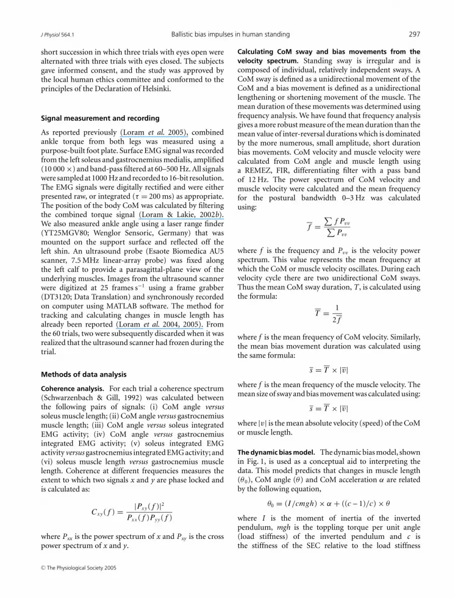
Figure 2. The bandwidth of postural controlA, shows the coherence between the CoMangle and muscle length (continuous line) andbetween CoM angle and EMG activity (•–•).Both soleus and gastrocnemius muscles havebeen averaged together for muscle length andEMG activity. B, shows the coherence betweensoleus EMG and gastrocnemius EMG activity(continuous line) and between soleus musclelength and gastrocnemius muscle length (•–•).In both A and B the lines represent thecombined average of 10 subjects. The dashedlines represent 95% confidence intervals in themean values. Muscle length was sampled at25 Hz and thus the frequency range shown is0–12.5 Hz. A coherence of one means that thetwo signals are perfectly phase locked at thatfrequency. A coherence of zero means that atthat frequency, sinusoids in one signal areinitiated and terminated entirely randomly withrespect to the other signal.
by a rise in velocity which reaches a maximum and whichthen reduces while the subject is moving forwards. So,for our averaging points, we used velocity maxima (zeroacceleration) occurring when the velocity was positive. Wealso wanted to examine the impulsive nature of changes inbias and so we focused on reversals in muscle length. Sofor our averaging points we determined the instants whenthe muscle velocity changed from positive to negative, orfrom negative to positive. To prevent cases arising fromnoise in the velocity record, we low-pass filtered the CoMand muscle velocity with a cut off at 3 Hz and we calculatedthe CoM acceleration by differentiating the CoM velocityusing a REMEZ, FIR differentiating filter with a passband of 3 Hz. Having identified the averaging points, theaveraging process was applied to unsmoothed data.
These instants were averaged for each trial and thesix trials for each subject were averaged to produce asubject average. Finally, the pattern from each of the10 subjects was averaged. We averaged rectified EMGrather than integrated EMG so as not to compromise thetiming information in the EMG signals.
Results
The bandwidth of postural control in standing
Coherence measures the extent to which two signalsmaintain a constant phase relationship. A value of onemeans that the two signals are perfectly phase locked atthat frequency, and a value of zero means that componentsat that frequency are initiated and terminated entirelyrandomly in one signal with respect to the other. Thus,an entirely random fluctuation would have a coherencevalue of zero with any other signal.
Figure 2 shows that during standing balance, inter-action between the CoM and the calf muscles soleus and
C© The Physiological Society 2005

J Physiol 564.1 Ballistic bias impulses in human standing 299
gastrocnemius occurs in the frequency bandwidth 0–3 Hz.Figure 2A shows the coherence between muscle length andthe CoM angle, and between EMG signal and the CoMangle. The Figure shows two distinct regions. Above 3 Hzthere is a region which shows approximately constant lowcoherence. This is due to the presence of some coherentnoise in both signals and it sets the noise floor. Below 3 Hz,the coherences are outside the 95% confidence intervals ofthe noise floor. However, even in the region of 0–3 Hz, thecoherences are relatively low. This indicates that while notentirely random and purposeless, the changes in musclelength and EMG activity are irregular and only partlysynchronized with changes in CoM angle. Muscle lengthis generally more coherent with CoM angle than is EMGactivity. This reflects the mechanical coupling betweenmuscle length and body angle via the Achilles tendon andthe noisier nature of the EMG signal.
Figure 2B shows the coherence between soleus andgastrocnemius for muscle length and for EMG signal.Again there are two distinct regions. Above 3 Hz,there is a region of approximately constant moderatecoherence. These two muscles are mechanically coupledby the Achilles tendon at their distal end, and sonoise like fluctuations in one muscle are inevitablyrecorded in the other muscle. This means that the back-ground coherence (noise floor) is quite high. The twomuscles have a coherence that is greater than the 95%confidence limits of the noise floor in the range 0–3Hz(Fig. 2A) and shorten and lengthen most coherently(0.77) at a frequency of 1.0 Hz. It is interesting that theintegrated EMG activities of these muscles are highlycoherent (0.97) at tonic levels (0–0.2 Hz), but remarkablyincoherent at higher frequencies. Thus the two musclesare activated independently above tonic frequencies andthe synchronous changes in muscle length results almostentirely from the mechanical coupling of the muscles. Inother words an external stretch of one muscle or an activecontraction of one muscle has a synchronous effect on theother muscle. At around 1 Hz, the two muscles effectivelyfunction as a single unit.
Muscle movements and body sways
Frequency analysis was used to examine the overallpattern of body sway and muscle length alteration.Figure 3A shows that alterations in muscle length areconsiderably more frequent than alterations in CoMangle. Muscle lengthening or shortening reverses directionwith a peak frequency of 1 Hz and a mean frequencyof 1.3 Hz whereas body sway reverses direction with apeak frequency of 0.25 Hz and mean frequency of 0.45 Hz(Fig. 3A). These values indicate that reversals in muscleshortening/lengthening are on average 2.8 times morefrequent than reversals in CoM sway.
The SEC is the linkage through which changes inmuscle length and tension exert their effect on the body.We examined the influence of SEC stiffness on bodysway and muscle movement (Fig. 3B–E). Unidirectionalsways of the body are generally a few tenths of a degree(Fig. 3B). This magnitude is not affected by the valueof SEC stiffness (Pearson correlation coefficient, n = 58,P = 0.27), though the sways were larger with the eyesclosed condition (mean, 0.23 deg) compared with theeyes open condition (mean, 0.13 deg) (ANOVA, n = 58,P = 0.014). Unidirectional changes in muscle length rangefrom mean values of 30 µm to 300 µm (Fig. 3C). There isclear, dramatic increase in the size of muscle movementas the stiffness of the SEC decreases (Pearson correlationcoefficient, n = 58, P = 0.00006). In Fig. 3C it looks asthough there may be a combined effect of SEC stiffnessand size of muscle movement separating the eyes openfrom the eyes closed cases; however, this distinction isnot justified statistically (MANOVA, n = 58, P = 0.07).The duration of body sways is generally around 1–1.5 swith no effect from SEC stiffness (Pearson correlationcoefficient, n = 58, P = 0.3) or whether the eyes areopen or closed (ANOVA, n = 58, P = 0.6; Fig. 3D). Theduration of muscle shortening/lengthening is 0.41 ± 0.06 s(mean ± s.d) (Fig. 3E). Unlike the variation in the sizeof muscle movements with SEC stiffness, there is nosuch variation in the duration of muscle movementseither resulting from SEC stiffness (Pearson correlationcoefficient, n = 58, P = 0.7) or from eye closure (ANOVA,n = 58, P = 0.7). This result raises the interesting questionof what limits the control bandwidth of these posturalmuscles.
Illustration of key results usinga representative subject
In order to determine the effect and possible purpose ofthese changes in muscle length it is necessary to examinethe irregular sway pattern and changes of muscle lengthin the time domain.
Figure 4 shows data from a representative subjectstanding with eyes open. This Figure shows all of the keypoints that will be made more clearly by the time-lockedaveraging analysis that follows. The CoM angle variesthrough several tenths of a degree over 20 s and irregularlyreverses direction (Fig. 4A). Within this sway pattern speedis regulated to remain less than 0.3 deg s−1 (Fig. 4B) andacceleration less than ∼1 deg s−2 (Fig. 4C). Muscle lengthis constantly changing (Fig. 4D) with a peak amplitude ofseveral hundred micrometres with both muscles, soleusand gastrocnemius, following a similar pattern. Thechanges in muscle length show clear similarities withthe CoM acceleration (Fig. 4C). As acceleration is ameasure of the lack of balance between ankle torque andthe torque generated on the body by gravity (Loram &
C© The Physiological Society 2005

300 I. D. Loram and others J Physiol 564.1
Lakie, 2002a), this indicates that changes in muscle lengthare associated with changes in balance.
The alternations in muscle length are at a different,higher, frequency than the sway frequency. The lowfrequency drift in muscle length is paradoxical; that is,muscle length increases as the CoM angle decreases. Theseparadoxical changes can be seen most clearly over the first
Figure 3. CoM sway and bias movementsA, shows the velocity power spectrum of CoM angle (continuous line) and muscle length (•–•). The power spectraare calculated relative to their maximal values. Muscle length is the average of soleus and gastrocnemius whichwere very similar across the range shown. The lines represent the combined average of 10 subjects. The dashedlines represent 95% confidence intervals in the mean values. For all 10 subjects, mean values plotted against SECstiffness are shown for sway size (B), bias movement size (C), sway duration (D) and bias movement duration (E).The filled circles are a mean of the three eyes open trials and the crosses are a mean of three eyes closed trials.A sway is defined as a unidirectional movement of the CoM. A bias movement is defined as a unidirectional changein muscle length. Relative stiffness is defined as the stiffness of the SEC divided by the load stiffness of the humaninverted pendulum and has been calculated by the cross correlation between CoM angle and muscle length,averaged for soleus and gastrocnemius muscles (Loram et al. 2005). The mean range and standard deviation ofthe relative stiffness for an individual subject are 0.35 and 0.14, respectively.
8 s. For this subject, the muscle length appears to fluctuatewith some regularity and it is the size more than the timingof muscle movements that is modulated.
In Fig. 4, the asterisks show all the falling cases ofa destabilizing rise in velocity, i.e. where the velocityof the CoM rises to a maximum while the person isswaying forwards. These asterisks mark the averaging
C© The Physiological Society 2005

J Physiol 564.1 Ballistic bias impulses in human standing 301
points for micro falls and they amount to an instantaneousattainment of equilibrium (zero velocity gradients). Here,using the representative subject, we preview the averagedinformation that we will subsequently show in Fig. 7. Thedestabilizing rise in velocity before the asterisk is usuallyclosely preceded by an increase in muscle length and themuscles usually shorten immediately before balance isattained and the speed of the micro fall reduces. Thus theshortening of calf muscles is associated with the regulationof speed and balance at the position of the asterisk. As aresult of this muscle shortening, usually the direction ofsway reverses (e.g. t = 11 s) and sometimes (e.g. t = 8.5,9 and 13.5 s) it does not. For this subject, whole body
Figure 4. Locating micro falls and bias reversalsTime records are shown for a representative subject of CoM angle (A), CoM velocity (B), acceleration (C) and soleus(dashed line) and gastrocnemius (continuous line) muscle length (D). The asterisks identify micro falls when theCoM speed rises to a maximum value while the subject is swaying forwards. All values are shown relative to amean of zero. Positive angle, velocity and acceleration are forwards, away from the vertical. Positive changes inmuscle length indicate lengthening. All quantities are expressed relative to their mean value.
sagittal sway is mechanically both caused and regulated bythe soleus and gastrocnemius muscles.
In the next analysis (Figs 5 and 6), reversals in musclelength are used as averaging points. In this Fig. 4, thesepoints would be located at all the local maxima and minimaof the muscle length records in Fig. 4D.
Time locked averaging of transient changesin muscle length
For individual subjects, Fig. 5 shows the impulsive effect(Fig. 5A) that is associated with muscle shortening (Fig. 5Band C). Time-locked averaging of all minima in muscle
C© The Physiological Society 2005

302 I. D. Loram and others J Physiol 564.1
Figure 5. Averaged bias reversals – variation with subjectFor each subject, all cases of bias reversals from six trials have beenaveraged. An average n = 297 events per subject for six trials. Timezero represents the averaging point which is the local minimum in
length (Fig. 5B and C) shows that the mean transientshortening in gastrocnemius muscle length varies in sizefrom 20 µm to 130 µm for the different subjects. Thechanges in length are smaller for soleus. These transientshortenings are associated with a change in CoM velocity(peak to peak, a–b in Fig. 5) of 0.02–0.15 deg s−1. Animpulse is defined as a change in momentum which in thiscontext is equivalent to a change in velocity of the CoM.Measured from a to b, the duration of these impulses is383 ± 55 ms (subject mean ± s.d.). Thus, amongst all 10subjects, there is little variation in the duration of theirmuscle shortenings (Fig. 5B and C), and the duration ofthe associated impulsive effect (Fig. 5A).
The symmetrical impulsive effect of muscle shortenings(Fig. 6A–D) and muscle lengthenings (Fig. 6E–H) isshown in Fig. 6. More importantly, Fig. 6 reveals howthe transient changes in soleus and gastrocnemius EMGactivity and muscle length are related to the velocityof the CoM (Fig. 6A). Using cross correlation betweenaveraged EMG signal (Fig. 6C, D, G and H) and musclelength (Fig. 6B and F), changes in rectified EMG signalprecede changes in muscle length by 167 ms and 203 msfor soleus and gastrocnemius, respectively. What stimuluscauses the changes in the EMG? The changes in rectifiedEMG signal show a very similar pattern to changes inCoM velocity (Fig. 6A). Could changes in the EMG derivefrom a simple feedback reflection of the velocity signal asrecently proposed (Masani et al. 2003)? Cross correlationbetween averaged EMG signal (Fig. 6C, D, G and H) andaveraged CoM velocity (Fig. 6A and E) show that soleusand gastrocnemius EMG activity leads CoM velocity by58 ms and 28 ms, respectively. However, the duration ofthis lead of EMG activity over CoM velocity is not constant.Moving along the time axis through Fig. 6, EMG activitybecomes progressively further in advance of CoM velocity.Before a, the EMG marginally lags CoM velocity; at a,EMG and velocity are broadly simultaneous; and at bEMG is noticeably ahead of velocity. The completion ofeach impulse is anticipatory of changes in velocity. Thisresult occurs symmetrically for shortening (Fig. 6A–D)and lengthening (Fig. 6E–H) impulses.
Time-locked averaging of micro falls
As we shall show (Fig. 7) as an averaged result forall subjects, micro falls, destabilizing increases in speedwhile swaying forwards (∗ in Fig. 4), are associated
muscle length. A, shows CoM velocity. B and C, show gastrocnemiusand soleus muscle length, respectively. Velocity, angle and musclelength all increase positively. The marker lines, a and b, indicate thebeginning and end of the impulse. The continuous lines indicatesubjects with a SEC stiffness greater than 100%. All quantities areexpressed relative to their mean value.
C© The Physiological Society 2005
J Physiol 564.1 Ballistic bias impulses in human standing 303
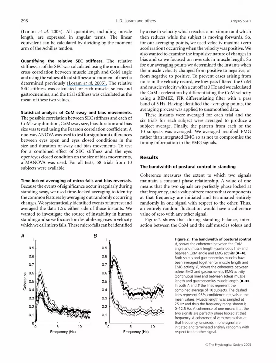
with closely preceding increases in calf muscle length.Regulation of the micro fall, reduction in speed, resultsfrom reacto-predictive modulation of calf muscle activity.The inter-relationship between ankle torque, CoM angle,muscle length and SEC stiffness is excellently explained bythe dynamic bias model.
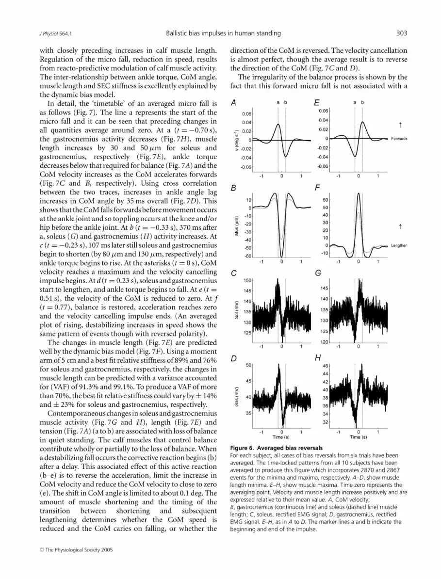
In detail, the ‘timetable’ of an averaged micro fall isas follows (Fig. 7). The line a represents the start of themicro fall and it can be seen that preceding changes inall quantities average around zero. At a (t = −0.70 s),the gastrocnemius activity decreases (Fig. 7H), musclelength increases by 30 and 50 µm for soleus andgastrocnemius, respectively (Fig. 7E), ankle torquedecreases below that required for balance (Fig. 7A) and theCoM velocity increases as the CoM accelerates forwards(Fig. 7C and B, respectively). Using cross correlationbetween the two traces, increases in ankle angle lagincreases in CoM angle by 35 ms overall (Fig. 7D). Thisshows that the CoM falls forwards before movement occursat the ankle joint and so toppling occurs at the knee and/orhip before the ankle joint. At b (t = −0.33 s), 370 ms aftera, soleus (G) and gastrocnemius (H) activity increases. Atc (t = −0.23 s), 107 ms later still soleus and gastrocnemiusbegin to shorten (by 80 µm and 130 µm, respectively) andankle torque begins to rise. At the asterisks (t = 0 s), CoMvelocity reaches a maximum and the velocity cancellingimpulse begins. At d (t = 0.23 s), soleus and gastrocnemiusstart to lengthen, and ankle torque begins to fall. At e (t =0.51 s), the velocity of the CoM is reduced to zero. At f(t = 0.77), balance is restored, acceleration reaches zeroand the velocity cancelling impulse ends. (An averagedplot of rising, destabilizing increases in speed shows thesame pattern of events though with reversed polarity).
The changes in muscle length (Fig. 7E) are predictedwell by the dynamic bias model (Fig. 7F). Using a momentarm of 5 cm and a best fit relative stiffness of 89% and 76%for soleus and gastrocnemius, respectively, the changes inmuscle length can be predicted with a variance accountedfor (VAF) of 91.3% and 99.1%. To produce a VAF of morethan 70%, the best fit relative stiffness could vary by ± 14%and ± 23% for soleus and gastrocnemius, respectively.
Contemporaneous changes in soleus and gastrocnemiusmuscle activity (Fig. 7G and H), length (Fig. 7E) andtension (Fig. 7A) (a to b) are associated with loss of balancein quiet standing. The calf muscles that control balancecontribute wholly or partially to the loss of balance. Whena destabilizing fall occurs the corrective reaction begins (b)after a delay. This associated effect of this active reaction(b–e) is to reverse the acceleration, limit the increase inCoM velocity and reduce the CoM velocity to close to zero(e). The shift in CoM angle is limited to about 0.1 deg. Theamount of muscle shortening and the timing of thetransition between shortening and subsequentlengthening determines whether the CoM speed isreduced and the CoM caries on falling, or whether the
direction of the CoM is reversed. The velocity cancellationis almost perfect, though the average result is to reversethe direction of the CoM (Fig. 7C and D).
The irregularity of the balance process is shown by thefact that this forward micro fall is not associated with a
Figure 6. Averaged bias reversalsFor each subject, all cases of bias reversals from six trials have beenaveraged. The time-locked patterns from all 10 subjects have beenaveraged to produce this Figure which incorporates 2870 and 2867events for the minima and maxima, respectively. A–D, show musclelength minima. E–H, show muscle maxima. Time zero represents theaveraging point. Velocity and muscle length increase positively and areexpressed relative to their mean value. A, CoM velocity;B, gastrocnemius (continuous line) and soleus (dashed line) musclelength; C, soleus, rectified EMG signal; D, gastrocnemius, rectifiedEMG signal. E–H, as in A to D. The marker lines a and b indicate thebeginning and end of the impulse.
C© The Physiological Society 2005

304 I. D. Loram and others J Physiol 564.1
Figure 7. Averaged micro fallsFor each subject, all cases of micro falls from six trials have beenaveraged. The time-locked pattern from all 10 subjects have beenaveraged to produce this Figure which incorporates 1541 events intothe average. A micro fall is a rise in speed of the CoM to a maximumwhile the subject is swaying forwards. Time zero represents theaveraging point which is a velocity maximum while falling forwards.Positive changes in torque represent increases. Positive angle and
previous forward micro fall (Fig. 7B–D). Because theyoccur at random instants, preceding CoM movementaverages zero. This lack of regularity in motion of the CoMreduces the timescale over which prediction of a micro fallcan occur. This raises the question of what informationstimulates the rise in muscle activity at b. By inspection onecan see that the activity of soleus and gastrocnemius doesnot copy the pattern of any one signal, e.g. muscle length,ankle torque, CoM velocity, ankle velocity, CoM angle orankle angle. The pattern of rectified EMG signal is closestto velocity. Overall, using cross correlation, soleus andgastrocnemius rectified EMG activity (Fig. 7G and H)precedes ankle velocity by 53 ms and 38 ms, respectively,and lags CoM velocity by 4 ms and 17 ms, respectively.Between the asterisks (t = 0) and e, the EMG activityis modulated in advance of CoM velocity whichdemonstrates the predictive element to the velocitycancelling impulsive change in muscle length (asterisk tof). Between a and b, gastrocnemius EMG activity decreaseswhich does not reflect the velocity signal. The earliestinformation concerning the destabilizing rise in velocitycomes from CoM acceleration and the simultaneouschanges in muscle length associated with that acceleration.In summary, the pattern of EMG activity is initially reactiveto the loss of balance, and is subsequently predictive of thedamping impulse.
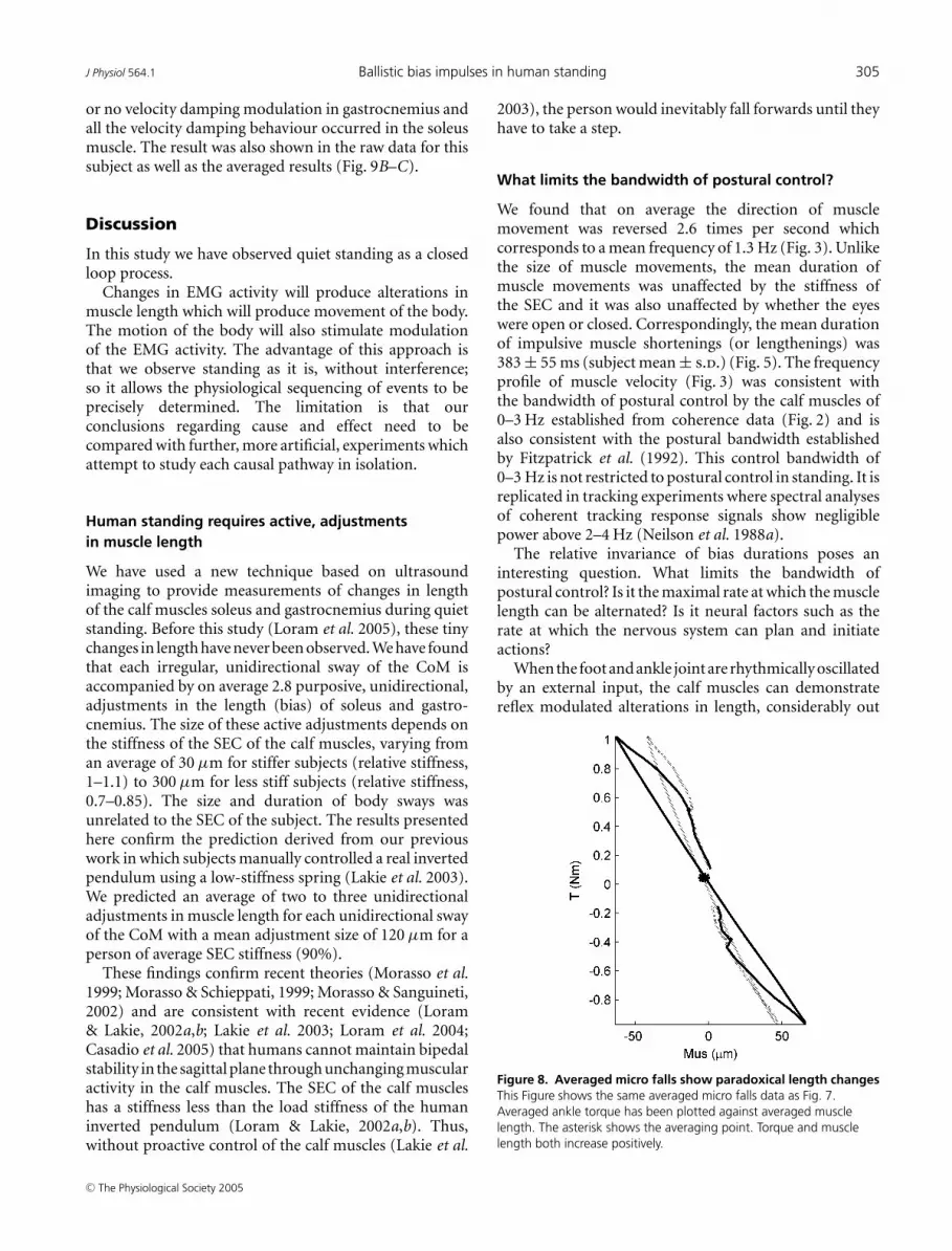
In this process of regulating balance and velocity,muscle length is actively controlled in a counter spring-likemanner, i.e. muscle length decreases as muscle tensionincreases (Fig. 8). This behaviour is the complete oppositeof the spring-like behaviour that has traditionally beenassumed and it can only be achieved by neural modulationof muscle length. This Figure illustrates how changesin muscle length are equivalent to changes in ankletorque.
The averaged changes in muscle length associated with adestabilizing rise in velocity were shown by all 10 subjects(Fig. 9). The subjects with stiffest SEC showed smallerchanges in muscle length and larger destabilizing rises invelocity. For one subject (SEC stiffness > 1), there was little
velocity are forwards from the vertical. Positive changes in musclelength indicate lengthening. All quantities except EMG activity areexpressed relative to their mean value. A, ankle torque from both legs(continuous line) and ankle torque required to balance the CoM(dashed line); B, CoM acceleration; C, CoM velocity (dashed line) andankle joint velocity (continuous line); D, CoM angle (dashed line) andankle angle (continuous line); E, gastrocnemius (continuous line) andsoleus (dashed line) muscle length; F, muscle length predicted by thedynamic bias model, gastrocnemius (continuous line), soleus (dashedline); G, soleus, rectified EMG signal; H, gastrocnemius, rectified EMGsignal. The marker lines indicate the preceding loss of balance (a), theinitial increase in EMG activity (b), the consequent decrease in musclelength (c), the subsequent increase in muscle length (d), thecancellation of CoM velocity (e) and the attainment of zeroacceleration (f).
C© The Physiological Society 2005
J Physiol 564.1 Ballistic bias impulses in human standing 305
or no velocity damping modulation in gastrocnemius andall the velocity damping behaviour occurred in the soleusmuscle. The result was also shown in the raw data for thissubject as well as the averaged results (Fig. 9B–C).
Discussion
In this study we have observed quiet standing as a closedloop process.
Changes in EMG activity will produce alterations inmuscle length which will produce movement of the body.The motion of the body will also stimulate modulationof the EMG activity. The advantage of this approach isthat we observe standing as it is, without interference;so it allows the physiological sequencing of events to beprecisely determined. The limitation is that ourconclusions regarding cause and effect need to becompared with further, more artificial, experiments whichattempt to study each causal pathway in isolation.
Human standing requires active, adjustmentsin muscle length
We have used a new technique based on ultrasoundimaging to provide measurements of changes in lengthof the calf muscles soleus and gastrocnemius during quietstanding. Before this study (Loram et al. 2005), these tinychanges in length have never been observed. We have foundthat each irregular, unidirectional sway of the CoM isaccompanied by on average 2.8 purposive, unidirectional,adjustments in the length (bias) of soleus and gastro-cnemius. The size of these active adjustments depends onthe stiffness of the SEC of the calf muscles, varying froman average of 30 µm for stiffer subjects (relative stiffness,1–1.1) to 300 µm for less stiff subjects (relative stiffness,0.7–0.85). The size and duration of body sways wasunrelated to the SEC of the subject. The results presentedhere confirm the prediction derived from our previouswork in which subjects manually controlled a real invertedpendulum using a low-stiffness spring (Lakie et al. 2003).We predicted an average of two to three unidirectionaladjustments in muscle length for each unidirectional swayof the CoM with a mean adjustment size of 120 µm for aperson of average SEC stiffness (90%).
These findings confirm recent theories (Morasso et al.1999; Morasso & Schieppati, 1999; Morasso & Sanguineti,2002) and are consistent with recent evidence (Loram& Lakie, 2002a,b; Lakie et al. 2003; Loram et al. 2004;Casadio et al. 2005) that humans cannot maintain bipedalstability in the sagittal plane through unchanging muscularactivity in the calf muscles. The SEC of the calf muscleshas a stiffness less than the load stiffness of the humaninverted pendulum (Loram & Lakie, 2002a,b). Thus,without proactive control of the calf muscles (Lakie et al.
2003), the person would inevitably fall forwards until theyhave to take a step.
What limits the bandwidth of postural control?
We found that on average the direction of musclemovement was reversed 2.6 times per second whichcorresponds to a mean frequency of 1.3 Hz (Fig. 3). Unlikethe size of muscle movements, the mean duration ofmuscle movements was unaffected by the stiffness ofthe SEC and it was also unaffected by whether the eyeswere open or closed. Correspondingly, the mean durationof impulsive muscle shortenings (or lengthenings) was383 ± 55 ms (subject mean ± s.d.) (Fig. 5). The frequencyprofile of muscle velocity (Fig. 3) was consistent withthe bandwidth of postural control by the calf muscles of0–3 Hz established from coherence data (Fig. 2) and isalso consistent with the postural bandwidth establishedby Fitzpatrick et al. (1992). This control bandwidth of0–3 Hz is not restricted to postural control in standing. It isreplicated in tracking experiments where spectral analysesof coherent tracking response signals show negligiblepower above 2–4 Hz (Neilson et al. 1988a).
The relative invariance of bias durations poses aninteresting question. What limits the bandwidth ofpostural control? Is it the maximal rate at which the musclelength can be alternated? Is it neural factors such as therate at which the nervous system can plan and initiateactions?
When the foot and ankle joint are rhythmically oscillatedby an external input, the calf muscles can demonstratereflex modulated alterations in length, considerably out
Figure 8. Averaged micro falls show paradoxical length changesThis Figure shows the same averaged micro falls data as Fig. 7.Averaged ankle torque has been plotted against averaged musclelength. The asterisk shows the averaging point. Torque and musclelength both increase positively.
C© The Physiological Society 2005

306 I. D. Loram and others J Physiol 564.1
Figure 9. Averaged micro falls - variation with subjectFor each subject, all cases of micro falls from six trials have beenaveraged. An average n = 159 events per subject for six trials. A microfall is a rise in speed of the CoM to a maximum while the subject isswaying forwards. Time zero represents the averaging point which is avelocity maximum while falling forwards. A, shows CoM velocity.B and C, show gastrocnemius and soleus muscle length, respectively.
of phase with the mechanical input, at up to 8 Hz ormore (Rack et al. 1983; Evans et al. 1983). Thus thenervous system is capable of modulating soleus and gastro-cnemius muscle length at considerably higher frequenciesthan the mean and range (1.3 Hz, 0–3 Hz) that areobserved under postural conditions. This indicates thata longer duration of neural processing is associated witheach muscle length adjustment that we observe. A meanfrequency of 1.3 Hz, indicates a mean time period of∼800 ms which indicates a mean processing time perunidirectional muscle movement of 400 ms (Fig. 3). Thisduration is longer than the reflex initiated response time(65 ms for a monosynaptic reflex; Evans et al. 1983),and is comparable to a human reaction time requiringsome choice and predictive planning (Craik, 1947; Vince,1948).
The finding of modulated responses occurring every400 ms resonates with results from investigations intovisually guided pursuit tracking of a continuously movingtarget. It has been demonstrated that the human operatorbehaves as an ‘intermittent correction servo’ by makingballistic movements at a mean interval of approximately500 ms (Craik, 1947). The intermittency results from apsychological refractory period, equal in duration to thereaction time, during which the operator is unable torespond to a second stimulus, similar to the first (Vince,1948). In continuous tracking, inverse internal models areemployed to transform desired trajectories into motorcommands. There is evidence that the nervous systemrequires a finite time period for planning and it doesnot commence planning a new movement until planningof the old movement has been completed (Neilson et al.1988a,b). We hypothesize that standing balance is a processof this type. The relatively long duration of 400 msallows plenty of opportunity for predictive, planningmechanisms to shape the amplitude and timing of biasadjustments.
At low frequencies, this process resembles a sampled,negative feedback process (Neilson et al. 1988a). Thiswould explain why simple negative feedback circuitscan characterize the mean parameters of standing swaywell (Maurer & Peterka, 2004) even if the instantaneousbalancing process consists of intermittent, ballisticallyexecuted movements. After all, position is regulated inquiet standing, and so is velocity, so is it not inevitable thatthis process can be represented by a proportionaldifferential (PD) controller (Peterka, 2000; 2002; Loramet al. 2001; Masani et al. 2003; Peterka & Loughlin, 2004;Maurer & Peterka, 2004)?
Velocity and muscle length increase positively and are expressedrelative to their mean value. The continuous lines indicate subjectswith a SEC stiffness greater than 100%.
C© The Physiological Society 2005
J Physiol 564.1 Ballistic bias impulses in human standing 307
The correspondence between muscle length andankle torque predicted by the dynamic bias model
Standing is usually studied using a force plate and thecentre of pressure (CoP), the point of application of theground reaction force, is usually recorded. Fluctuationsin CoP forwards and backwards are entirely equivalentto fluctuations in ankle torque. By applying the dynamicbias model (Fig. 1) to the averaged micro falls (Fig. 7F) wehave shown that these averaged, short duration changesin muscle length, ankle torque and CoM angle are verywell explained by this model using a mean SEC stiffnessof 83% relative to the load stiffness of the subject. Thissimple model aids interpretation, in that short durationmuscle movements are equivalent to simultaneous torquefluctuations via the spring-like (Loram et al. 2005) SEC ofthe calf muscles. In individual cases there are other factorsthat influence muscle length, such as ankle co-contraction,varying muscle modulation between the two legs, tendoncreep and tendon hysteresis. These other factors mean thatat any one time, muscle length does not correspond as wellto acceleration and CoM angle as is shown here in theseaveraged plots. But on average these other factors cancelout.
A unidirectional muscle movement is equivalent to achange in torque. Does this mean that the nervous systemis modulating muscle length rather than ankle torque inits control of balance? We don’t know. We also don’t knowwhether there is any real distinction between the two ideas.But we can see that there is a difference between inter-nally and externally generated changes in ankle torque.While a change in muscle length is equivalent to a changein ankle torque according to the stiffness of the SEC(mainly the Achilles tendon), an externally applied changein ankle torque does not necessarily produce the samechange in muscle length. The change in muscle lengthproduced by an external change in torque depends onthe muscle stiffness which may not be at all the samething as the SEC stiffness. Thus by examining changesin muscle length, we gain insight into the working ofthe controlling actuator, that is the internally generatedchanges in torque that are controlled by the nervoussystem.
Because the SEC is not stiff, muscle length is notmechanically constrained to follow ankle angle. And,due to the complexity of the nervous system, changes inmuscle length can be driven by stimuli other than CoMmovement and its linear, time invariant derivatives. Thusmuscle movements can be generated independently ofCoM motions. The dynamic bias model predicts thatmuscle movements cause changes in CoM acceleration.The muscle may alternate between shortening andlengthening while the inertia of the CoM means that CoMmotion need not alternate between rising and falling. Thusthere is no mechanical or control objection to the muscle
velocity alternating independently of, and more frequentlythan, CoM velocity.
The ballistic bias impulse mechanism
The integral of the torque change through the duration ofthe adjustment produces an impulse, effectively a changein velocity, given to the CoM. It is appropriate to think inimpulse terms because the effect of the bias change is givenin a short timescale relative to the motion of the CoM. Asthere are on average 2.8 bias adjustments for each CoMsway, the velocity change is delivered in approximatelyone-third of a unidirectional CoM sway. The changesin muscle activation are delivered in a shorter time still(Fig. 6). As the impulsive effect is discharged by the nervoussystem in a short timescale relative to the effect on CoMposition, and before feedback of the result can be received,this process is properly described as ballistic. For example,after a bias action, the nervous system will not know, andwill have to wait to find out whether or not the direction ofCoM motion will be reversed. Small differences in impulsewill result in completely different motion sequences forthe CoM. Instants when the CoM is finely balanced andmoving at low speed are effectively bifurcation pointswhere alternative small changes in ankle torque couldresult in opposite motions of the CoM (Loram & Lakie,2002a). These bifurcation points create unpredictability inthe motion of the CoM. The delay between the initiation ofa destabilizing rise in velocity and the corrective reaction(Fig. 7) is evidence that these micro falls are not perfectlypredicted. The summated effect of these ballistic bias,impulse actions is regulation of position and velocity.This interpretation is consistent with previous evidenceof the ballistic nature of balance derived from pedalbalancing of a real inverted pendulum (Loram & Lakie,2002a).
It has been previously hypothesized that posturalcontrol of standing operates in an open loop mode overdurations less than approximately 1 s, and in a closedloop mode over durations more than 1 s (Collins &DeLuca, 1993, 1995). This hypothesis was based on theobservation that the direction of motion of the CoMcorrelates positively with itself for durations up to 1 s(persistent motion) and correlates negatively with itself(antipersistent motion) for longer durations. The CoMsway durations that we observe (Fig. 3D) are consistentwith those observed by Collins and DeLuca and our ideasof impulsive, ballistic control are sympathetic to theiridea of open loop/closed loop control. Our observationsof muscle length show that over a sway timescale of 1 s,there are several ballistic-like attempts to control CoMmotion. The existence of bifurcation points which areextremely sensitive to small changes in ankle torque,the unstable nature of the human ‘inverted pendulum’(Loram & Lakie, 2002a) and the time taken to respond to
C© The Physiological Society 2005

308 I. D. Loram and others J Physiol 564.1
unpredicted losses of balance (a to e in Fig. 7) all accountfor the 1 s timescale of persistent motion.
The physiological origin of postural sway
There has been some debate whether standing sway resultsfrom internal perturbations such as breathing or the heartbeat (Sturm, 1980; Conforto et al. 2001; Hodges et al. 2002;Gandevia et al. 2002), whether it results from ‘noise’ fromsome unattributed source (Winter et al. 1998; Peterka,2000; Masani et al. 2003) or whether it results frominaccuracies in the modulation of calf muscle activity(Loram et al. 2001; Loram & Lakie, 2002a). For the10 subjects studied, a very clear answer can be given.On average, the CoM sway is very closely related tofluctuations in muscle length and ankle torque via thedynamic bias model (Figs 1 and 7). This indicates thatmodulation of the ankle musculature largely explains(91–99% VAF, Fig. 7F) control of the CoM. CoM swaysand corresponding acceleratory ankle torques are clearlyrelated to simultaneous fluctuations in calf muscle length.The 370-ms delay between the onset of the loss of balance,and the start of the corrective reaction indicates that onaverage, the destabilization represents a deviation fromwhat was predicted. Because the system is unstable, thepredictive error in torque provides a perturbation whichgrows into a sway which is reacted to and corrected.The correction is itself imperfect and thus the source ofsubsequent sway. This process is consistent with the ideathat sensory, computational and motor noise place limitson the ability of the subject to produce a perfect torqueresponse (Jeka et al. 2004).
Thus, CoM sagittal sway results from fluctuations in calfmuscle activity acting through the low stiffness SEC. Anysway resulting from heart beat or respiration must be muchsmaller than the average sway pattern reported here.
Neural mechanisms of postural control
Time-locked averaging of micro falls (Fig. 7) and changesin muscle length (Fig. 6) indicate that the nervous systemresponds to an increase in CoM velocity with a timedelay of around 370 ms. The latency of a group I stretchreflex is 65 ms (Evans et al. 1983) or 42 ms (Schieppati &Nardone, 1999) and that of the group II, medium latencystretch reflex is 75 ms (Schieppati & Nardone, 1999). Thus,local stretch and other reflexes are ruled out on the basisof timing. As the response pattern of the EMG activitydoes not follow any one signal (e.g. ankle angle, anklevelocity, muscle length, ankle torque) it probably drawson composite sources. In this case, CoM acceleration(a composite signal) and the simultaneous changes incalf muscle length provide the earliest information of theloss of balance. It is well known that the brain will useany information from any meaningful source to solvethe task at hand. Thus the most natural explanation,
consistent with the timing information, is that thenervous system uses all global sources of informationavailable that contribute to knowing the motion of theCoM. This explanation is consistent with the resultsderived from relatively large balance perturbations thatshow that global rather than local sources of proprio-ception are used to control the ankle musculature duringstanding (Bloem et al. 2000; Allum et al. 1998). Thisexplanation is also consistent with research that showsthat integrated information from multiple sense organsis readily combined and reweighted in the maintenance ofbalance (Peterka, 2002; Oie et al. 2002; Peterka & Loughlin,2004).
The nervous system acts predictively during quietstanding as the EMG pattern producing an impulseterminates well before velocity cancellation is complete(Figs 6 and 7). It has been recently proposed that velocityfeedback can explain the predictive modulation of calfmuscle activity (Masani et al. 2003). These authors arguethat the nervous system acts as a PD controller with a timedelay of 100 ms such that the control signal (EMG) reflectsthe CoM velocity and angle signals. Our results showthat the EMG signal controlling muscle impulses generallyanticipates CoM velocity by 58 ms and 28 ms, for soleusand gastrocnemius, respectively (Fig. 6), and when lossesof balance (rather than all impulses) are sampled (Fig. 7),EMG signal anticipates ankle velocity and fractionally lags(4–17 ms) CoM velocity. Given that a short latency reflexrequires about 42 ms (Schieppati & Nardone (1999)), eventhis fractional lag during losses of balance is inconsistentwith the idea that the control signal follows CoM velocityafter a time delay of 100 ms.
Generally, the timescale (400 ms) of the observedimpulses (Fig. 6) and reaction to loss of balance (Fig. 7)means the brain has time to use its own internal modelsof CoM motion and muscle activity to modulate theamplitude and timing of the ballistic bias activity. Arguablythe earliest knowledge the nervous system has of animpending micro fall, comes from the knowledge ofprevious changes in muscle length combined with aninternal model of the effect of those changes in muscleon the motion of the CoM. As the impulses are deliveredballistically, the nervous system will almost certainly refineits internal models with the immediate feedback of resultsthat is received from the velocity signal. This leads tothe speculation that a supervising learning network isutilized. Such updating of internal models is thought tooccur in the cerebellum (Imamizu et al. 1998; Wolpertet al. 1998; Morasso et al. 1999; Kawato, 1999). Thispostulated role of the cerebellum in standing mightexplain why patients with cerebellar ataxia suffer impairedpostural control (Sanguineti et al. 2003). Our inter-pretation identifies standing balance in humans as an auto-mated, trial and error, skilled, learned activity more thana low level reflex process.
C© The Physiological Society 2005

J Physiol 564.1 Ballistic bias impulses in human standing 309
Paradigms of posture
The mechano-reflex understanding of posturalmechanisms has derived much evidence frominvestigations on cats and other quadrupeds. Theanimal stands on limbs which are flexed by the animal’sweight. Collapse is prevented by constant activity of theextensor muscles. An external perturbation on the animalcauses the extensor muscles to be elongated as the jointsflex beyond their set position. The perturbation is resistedby intrinsic mechanical joint impedance supplementedby local reflexes.
In human standing, forward toppling about the anklejoint is resisted by near constant activity in the soleusand gastrocnemius muscles. Much contemporary analysishas extended the mechano-reflex paradigm to humanstanding (Gurfinkel et al. 1974, 1995; Shadmehr &Arbib, 1992; Fitzpatrick et al. 1992, 1994, 1996; Horak& MacPherson, 1996; Winter et al. 1998; Schieppati &Nardone, 1999; Fitzpatrick, 2003; Masani et al. 2003). Acommon view is that in man, as with other mammals,the posture-preserving system is phylogenetically old andoperates relatively autonomously (van Ingen Schenau et al.1996; Massion et al. 2004). If the cerebellum is intimatelyinvolved in the automation of standing balance then theprocess is phylogenetically more recent than previouslythought and if the anticipatory impulsive adjustments ofsoleus and gastrocnemius required for standing are thesame process as the anticipatory postural adjustmentsrequired as a preliminary to general movement, then theposture-preserving system may in fact be much moreintegrated into the movement control scheme than hasbeen recognized.
The evidence presented here shows that in quietstanding, the sagittal motion of the CoM is controlledby an active, impulsive, ballistic, process operatingat a rate of 2.6 modulated actions per secondconsistent with complex sensorimotor integration andpredictive planning. This process is a good candidate forautomation by the cerebellum. The CoM stands nearlyperfectly balanced by the SEC of the calf muscles asa well-sprung, mobile mechanism that is ponderouslyunstable. By delivering alternating impulses via the gastro-cnemius and soleus muscles, the nervous system keeps thevelocity of the CoM low and controls the position of theCoM. As well as controlling CoM position and velocitythe bias adjustments are themselves the major source ofpostural sway. Because their magnitude is rarely if everprecisely correct they act as perturbations which if leftuncompensated, would lead to a fall.
Here is a simple analogy that illustrates the impulsiveballistic nature of the process. Imagine trying to maintaina heavy ball as still as possible on a hillside. The ball iscontrolled by striking it with a bat at a relatively fixedrate. The motion of the ball will be caused by the blows
themselves. It will move sometimes up the hill (because theeffect of the blows are greater than gravity) and sometimesdown the hill (effect of blows less than gravity), but notin any regular way. It can be maintained near the top ofthe hill or near the bottom or at any point in between. Todo this, the batter has to judge the size of each blow. Wesuggest that in essence it is this never ending, trial and errorprocess which has to be carried out in human standing. Theprocess of loss of balance and regaining balance has to berepeatedly solved under the ever changing conditions ofbalance and we suggest that this is a skilled, trial and erroractivity that improves with experience rather than a reflexprocess.
References
Allum JHJ, Bloem BR, Carpenter MG, Hulliger M &Hadders-Algra M (1998). Proprioceptive control of posture:a review of new concepts. Gait Posture 8, 214–242.
Bloem BR, Allum JHJ, Carpenter MG & Honegger F (2000).Is lower leg proprioception essential for triggering humanautomatic postural responses? Exp Brain Res 130,375–391.
Casadio M, Morasso PG & Sanguineti V (2005). Directmeasurement of ankle stiffness during quiet standing:implications for control modelling and clinical application.Gait Posture (in press).
Collins JJ & DeLuca CJ (1993). Open-loop and closed-loopcontrol of posture: a random-walk analysis of centre-of-pressure trajectories. Exp Brain Res 95, 308–318.
Collins JJ & De Luca CJ (1995). The effects of visual input onopen-loop and closed-loop postural control mechanisms.Exp Brain Res 103, 151–163.
Conforto S, Schmid M, Camomilla V, D’Alessio T & CappozzoA (2001). Hemodynamics as a possible internal mechanicaldisturbance to balance. Gait Posture 14, 28–35.
Craik K (1947). Theory of the human operator in controlsystems. I. The operator as an engineering system.Br J Psychol xxxviii, 56–61.
Creed RS, Denny-Brown D, Eccles JC, Liddell EGT &Sherrington CS (1932). Reflex Activity of the Spinal Cord.Clarendon Press, Oxford.
Evans CM, Fellows SJ, Rack PM, Ross HF & Walters DK (1983).Response of the normal human ankle joint to imposedsinusoidal movements. J Physiol 344, 483–502.
Fitzpatrick RC (2003). More pulsating movement. J Physiol551, 4.
Fitzpatrick R, Burke D & Gandevia SC (1996). Loop gain ofreflexes controlling human standing measured with the useof postural and vestibular disturbances. J Neurophysiol 76,3994–4008.
Fitzpatrick RC, Gorman RB, Burke D & Gandevia SC (1992).Postural proprioceptive reflexes in standing human subjects:bandwidth of response and transmission characteristics.J Physiol 458, 69–83.
Fitzpatrick R, Rogers DK & McCloskey DI (1994). Stablehuman standing with lower-limb muscle afferents providingthe only sensory input. J Physiol 480, 395–403.
C© The Physiological Society 2005

310 I. D. Loram and others J Physiol 564.1
Gandevia SC, Butler JE, Hodges PW & Taylor JL (2002).Balancing acts: respiratory sensations, motor control andhuman posture. Clin Exp Pharm Physiol 29, 118–121.
Grillner S & Wallen P (2004). Innate versus learned movements– a false dichotomy? In Brain Mechanisms for IntegrationPosture Movement. Progress in Brain Research, 143, eds MoriS, Stuart DG, Wiesendanger M & Pierce PA, pp. 3–12.
Gurfinkel VS, Ivanenko YP, Levik YS & Babakova IA (1995).Kinesthetic reference for human orthograde posture.Neuroscience 68, 229–243.
Gurfinkel VS, Lipshits MI & Popov KY (1974). Is the stretchreflex the main mechanism in the system of regulation of thevertical posture of man? Biophysics 19, 761–766.
Hodges PW, Gurfinkel VS, Brumagne S, Smith TC & Cordo PC(2002). Coexistence of stability and mobility in posturalcontrol: evidence from postural compensation forrespiration. Exp Brain Res 144, 293–302.
Horak FB & MacPherson JM (1996). Postural orientation andequilibrium. In Handbook of Physiology, Section 12, Exercise.Regulation and Integration of Multiple Systems, ed. Rowell LB& Shepherd JT, pp. 255–292. Oxford University Press,Oxford.
Imamizu H, Uno Y & Kawato M (1998). Adaptive internalmodel of intrinsic kinematics involved in learning an aimingtask. J Exp Psychol Hum Percept Perform 24, 812–829.
Jeka J, Kiemel T, Creath R, Horak F & Peterka R (2004).Controlling human upright posture: velocity information ismore accurate than position or acceleration. J Neurophysiol92, 2368–2379.
Kawato M (1999). Internal models for motor control andtrajectory planning. Curr Opin Neurobiol 9,718–727.
Lakie M, Caplan N & Loram ID (2003). Human balancing ofan inverted pendulum with a compliant linkage: neuralcontrol by anticipatory intermittent bias. J Physiol 551,357–370.
Loram ID, Kelly S & Lakie M (2001). Human balancing of aninverted pendulum: is sway size controlled by ankleimpedance? J Physiol 532, 879–891.
Loram ID & Lakie M (2002a). Human balancing of an invertedpendulum: position control by small, ballistic-like, throwand catch movements. J Physiol 540, 1111–1124.
Loram ID & Lakie M (2002b). Direct measurement of humanankle stiffness during quiet standing: the intrinsicmechanical stiffness is insufficient for stability. J Physiol 545,1041–1053.
Loram ID, Maganaris CN & Lakie M (2004). Paradoxicalmuscle movement in human standing. J Physiol 556,683–689.
Loram ID, Maganaris CN & Lakie M (2005). Active, non-spring-like muscle movements in postural sway: how mightparadoxical changes in muscle length be produced? J Physiol564, 281–293.
Magnus R (1925). The Croonian Lecture: animal posture. ProcR Soc Lond B Biol Sci 98, 339–353.
Masani K, Popovic MR, Nakazawa K, Kouzaki M & Nozaki D(2003). Importance of body sway velocity information incontrolling ankle extensor activities during quiet stance.J Neurophysiol 90, 3774–3782.
Massion J (1998). Postural control systems in developmentalperspective. Neurosci Biobehav Rev 22, 465–472.
Massion J, Alexandrov A & Frolov A (2004). Why and how areposture and movement coordinated? In Brain Mechanismsfor Integration Posture Movement. Progress in Brain Research,143, eds Mori S, Stuart DG, Wiesendanger M & Pierce PA,pp. 13–27.
Maurer C & Peterka RJ (2004). A new interpretation ofspontaneous sway measures based on a simple model ofhuman postural control. J Neurophysiol 93,189–200.
Morasso PG, Baratto L, Capra R & Spada G (1999). Internalmodels in the control of posture. Neural Netw 12,1173–1180.
Morasso PG & Sanguineti V (2002). Ankle muscle stiffnessalone cannot stabilize balance during quiet standing.J Neurophysiol 88, 2157–2162.
Morasso PG & Schieppati M (1999). Can muscle stiffness alonestabilize upright standing? J Neurophysiol 82, 1622–1626.
Neilson PD, Neilson MD & O’Dwyer NJ (1988a). Internalmodels and intermittency: a theoretical account of humantracking behavior. Biol Cybern 58, 101–112.
Neilson PD, O’Dwyer NJ & Neilson MD (1988b). Stochasticprediction in pursuit tracking: an experimental test ofadaptive model theory. Biol Cybern 58, 113–122.
Oie KS, Kiemel T & Jeka JJ (2002). Multisensory fusion.simultaneous re-weighting of vision and touch for thecontrol of human posture. Brain Res Cogn Brain Res 14,164–176.
Peterka RJ (2000). Postural control model interpretation ofstabilogram diffusion analysis. Biol Cybern 82,335–343.
Peterka RJ (2002). Sensorimotor integration in human posturalcontrol. J Neurophysiol 88, 1097–1118.
Peterka RJ & Loughlin PJ (2004). Dynamic regulation ofsensorimotor integration in human postural control.J Neurophysiol 91, 410–423.
Rack PM, Ross HF, Thilmann AF & Walters DK (1983). Reflexresponses at the human ankle: the importance of tendoncompliance. J Physiol 344, 503–524.
Sanguineti V, Morasso PG, Baratto L, Brichetto G, MancardiGL & Solaro C (2003). Cerebellar ataxia: quantitativeassessment and cybernetic interpretation. Hum Mov Sci 22,189–205.
Schieppati M & Nardone A (1999). Group II spindle afferentfibers in humans: their possible role in the reflex control ofstance. In Peripheral and Spinal Mechanisms in the NeuralControl of Movement. Progress in Brain Research 123,ed, Binder MD, pp. 461–472.
Schwarzenbach J & Gill KF (1992). System Modelling andControl, 3rd edn. Edward Arnold, London.
Shadmehr R & Arbib MA (1992). A mathematical-analysis ofthe force-stiffness characteristics of muscles in control of asingle joint system. Biol Cybern 66, 463–477.
Sherrington CS (1906). Integrative Action of the Nervous System,1st edn. Constable, London.
Sturm R (1980). The impact of cardiac activity on triaxiallyrecorded endogenous microvibrations of the body.Eur J Appl Physiol 44, 83–96.
C© The Physiological Society 2005

J Physiol 564.1 Ballistic bias impulses in human standing 311
van Ingen Schenau GJ & van Soest AJ (1996). On theBiomechanical Basis of Dexterity, ed. Latash ML & TurveyMT, pp. 305–338, Lawrence Erlbaum associates, Mahway,NJ, USA.
Vince M (1948). The intermittency of control movements andthe psychological refractory period. Br J Psycholxxxviii, 149–157.
Winter DA, Patla AE, Prince F, Ishac M & Gielo-Perczak K(1998). Stiffness control of balance in quiet standing.J Neurophysiol 80, 1211–1221.
Wolpert DM, Miall RC & Kawato M (1998). Internal models inthe cerebellum. Trends Cogn Sci 2, 338–347.
Acknowledgements
We would like to thank the Leverhulme Trust for their supportof I.D.L. throughout this project.
C© The Physiological Society 2005