human integrin α3β1 regulates tlr2 recognition of lipopeptides from endosomal compartments
TRANSCRIPT

Human Integrin a3b1 Regulates TLR2 Recognition ofLipopeptides from Endosomal CompartmentsMeghan L. Marre1., Tanja Petnicki-Ocwieja2., Alicia S. DeFrancesco2, Courtney T. Darcy2,
Linden T. Hu1,2*
1 Graduate Program in Immunology, Sackler School of Graduate Biomedical Sciences, Tufts University, Boston, Massachusetts, United States of America, 2 Division of
Geographic Medicine and Infectious Diseases, Tufts Medical Center, Boston, Massachusetts, United States of America
Abstract
Background: Toll-like receptor (TLR)-2/TLR1 heterodimers recognize bacterial lipopeptides and initiate the production ofinflammatory mediators. Adaptors and co-receptors that mediate this process, as well as the mechanisms by which theseadaptors and co-receptors function, are still being discovered.
Methodology/Principal Findings: Using shRNA, blocking antibodies, and fluorescent microscopy, we show that U937macrophage responses to the TLR2/1 ligand, Pam3CSK4, are dependent upon an integrin, a3b1. The mechanism for integrina3b1 involvement in TLR2/1 signaling is through its role in endocytosis of lipopeptides. Using inhibitors of endosomalacidification/maturation and physical tethering of the ligand, we show that the endocytosis of Pam3CSK4 is necessary for thecomplete TLR2/1-mediated pro-inflammatory cytokine response. We also show that TLR2/1 signaling from the endosomeresults in the induction of different inflammatory mediators than TLR2/1 signaling from the plasma membrane.
Conclusion/Significance: Here we identify integrin a3b1 as a novel regulator for the recognition of bacterial lipopeptides.We demonstrate that induction of a specific subset of cytokines is dependent upon integrin a3b1-mediated endocytosis ofthe ligand. In addition, we address an ongoing controversy regarding endosomal recognition of bacterial lipopeptides bydemonstrating that TLR2/1 signals from within endosomal compartments as well as the plasma membrane, and thatdownstream responses may differ depending upon receptor localization. We propose that the regulation of endosomalTLR2/1 signaling by integrin a3b1 serves as a mechanism for modulating inflammatory responses.
Citation: Marre ML, Petnicki-Ocwieja T, DeFrancesco AS, Darcy CT, Hu LT (2010) Human Integrin a3b1 Regulates TLR2 Recognition of Lipopeptides fromEndosomal Compartments. PLoS ONE 5(9): e12871. doi:10.1371/journal.pone.0012871
Editor: Roberto F. Speck, University Hospital Zurich, Switzerland
Received April 29, 2010; Accepted August 28, 2010; Published September 22, 2010
Copyright: � 2010 Marre et al. This is an open-access article distributed under the terms of the Creative Commons Attribution License, which permitsunrestricted use, distribution, and reproduction in any medium, provided the original author and source are credited.
Funding: The work was supported by the U.S. National Institutes of Health (R56AI80846, R01-AI-50043 to L.T.H., T32AI07077 to M.L.M, and T32AI007329 to T.P-O.). The Tufts Center for Neuroscience Research microscopy core is funded by the National Institutes of Health (P30 NS047243 to F. Rob Jackson). The funders hadno role in the study design, data collection and analysis, decision to publish, or preparation of the manuscript.
Competing Interests: The authors have declared that no competing interests exist.
* E-mail: [email protected]
. These authors contributed equally to this work.
Introduction
The innate immune response protects the host from microbial
invaders through recognition of specific patterns that are recurrent
either in pathogens or in the signals they create. The toll-like
receptor (TLR) family contains a variety of receptors that recognize
a diverse array of these patterns and activate downstream
inflammatory cascades [1]. Early models of interactions of TLR
signaling proposed simple, direct interactions between TLRs and
their ligands, without the aid of other molecules. It is now
understood that other adaptor molecules and receptors mediate
and alter these interactions resulting in great diversity of responses
to different ligands and pathogens recognized by the same receptor
[2,3,4,5]. A portion of the diversity is generated by the context and
location in which TLRs interact with their ligands [6,7] and may be
further altered by co-stimulation of other pathways that cross talk
with a specific TLR [8,9,10].
Integrins are divalent, cation-dependent, heterodimeric recep-
tors that mediate a variety of cell-cell and cell-extracellular matrix
interactions within host tissues including tissue differentiation, cell
migration, and tumor metastases. Roles for integrins in a variety of
pathogen recognition and host defense mechanisms are increas-
ingly being recognized. One mechanism by which integrins
participate in host defense is by facilitating endocytosis. For
example, endocytosis of bacterial pathogens such as enteropatho-
genic Yersinia species [11] and Staphylococcus aureus [12,13] is
dependent upon b1 integrins. In addition, viruses such as human
cytomegalovirus [14] and Kaposi’s sarcoma-associated herpes
virus [15,16] are endocytosed via interactions with integrin avb3.
Integrins can also participate in host defense through co-
operation with other innate immune receptors such as TLRs.
Several groups have demonstrated a necessary role for integrin
aMb2 (CD11b/CD18) in the induction of an inflammatory
cytokine response to the TLR4 ligand, lipopolysaccharide (LPS)
[8,17,18]. In addition, a recent publication demonstrated a role for
integrin avb3 in the regulation of TLR2/1-mediated responses to a
number of stimuli including the prototypical bacterial lipopeptide,
palmitoyl-3-Cys-Ser-(Lys)4 (Pam3CSK4) [10]. This co-operation
PLoS ONE | www.plosone.org 1 September 2010 | Volume 5 | Issue 9 | e12871
was suggested to be mediated through the interaction of
Pam3CSK4 with vitronectin, the extracellular matrix ligand for
integrin avb3. It was proposed that integrin avb3 mediates the
attachment of Pam3CSK4 to macrophages which could lead to
clustering of the lipopeptide with the TLR2/1 receptor at the cell
surface, thus facilitating signaling.
Integrins also play an important role in the recognition of B.
burgdorferi [19,20,21,22], an organism that expresses a large
number of TLR2 ligands [23,24,25,26]. We have previously
shown that B. burgdorferi expresses ligands for integrin a3b1 [27]
and that integrin a3b1 is important for mediating the inflammatory
response to B. burgdorferi [28]. As a result, we were interested in
determining whether integrin a3b1 may play a role similar to avb3
in mediating TLR2 responses to the organism and to purified
TLR2 ligands. In this study, we show that human macrophage
inflammatory responses to the TLR2/1 ligand Pam3CSK4 require
integrin a3b1. However, the mechanism by which integrin a3b1
regulates TLR2/1 function is not through attachment and
clustering of ligand at the cell surface as proposed for integrin
avb3, but rather through the endocytosis of lipopeptides. We
further demonstrate that this endocytosis is necessary for the
complete response to the lipopeptide. TLR2/1 is classically
described as recognizing ligands and activating signaling pathways
from the plasma membrane. There remains controversy as to
whether TLR2/1 is active within endosomal compartments
[29,30,31,32,33]. In this report, we provide clear evidence using
both chemical inhibitors and physical tethering of TLR2/1 ligands
that recognition of bacterial lipopeptides, both synthetic and in the
context of an intact organism, occurs from within sub-cellular
compartments. Recognition of lipopeptides from within endoso-
mal compartments results in the induction of a different subset
of inflammatory mediators than recognition from the plasma
membrane. Our data provide a new mechanism for the
interactions of integrin and TLR receptors and support for the
emerging concept that localization and context of TLR-mediated
recognition of ligands alters the inflammatory response to a
stimulus.
Results
Integrin a3b1 Mediates the U937 Macrophage Responseto Pam3CSK4
To determine whether integrin a3b1 cooperates with TLR2/1
signaling, we used shRNA to reduce expression of integrin a3 by
73% in U937 macrophage cells (Fig. S1A). Specificity of the
shRNA construct was confirmed by demonstrating that the
shRNA construct did not affect expression of other integrin achains or TLR2 (Fig. S1B). U937 macrophages stably transduced
with either non-targeting, control shRNA or integrin a3-targeting
shRNA were stimulated with the synthetic TLR2/1 ligand
Pam3CSK4 under serum-free conditions. shRNA targeting the
integrin a3 chain reduced the IL-6 response to Pam3CSK4 by 62%
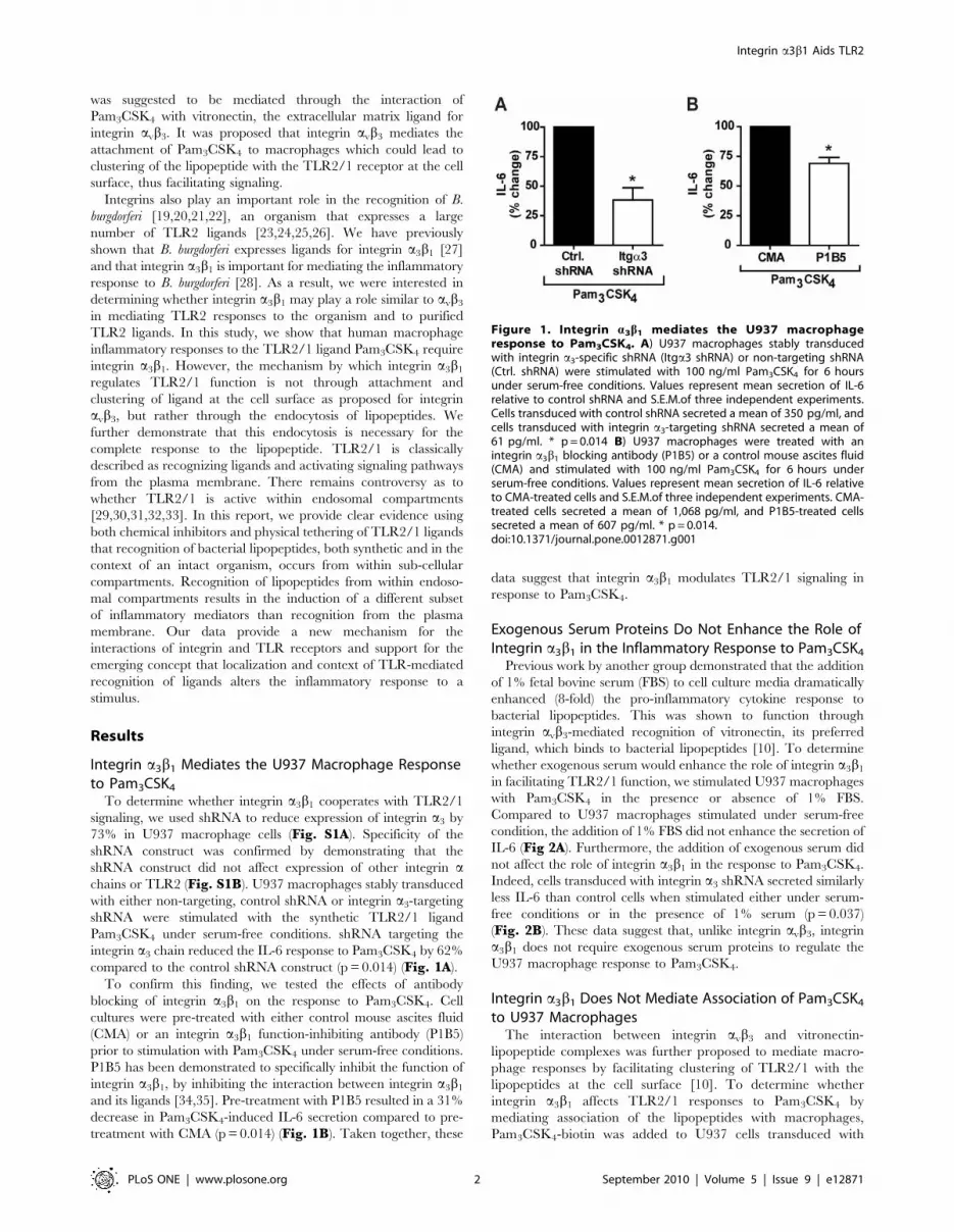
compared to the control shRNA construct (p = 0.014) (Fig. 1A).
To confirm this finding, we tested the effects of antibody
blocking of integrin a3b1 on the response to Pam3CSK4. Cell
cultures were pre-treated with either control mouse ascites fluid
(CMA) or an integrin a3b1 function-inhibiting antibody (P1B5)
prior to stimulation with Pam3CSK4 under serum-free conditions.
P1B5 has been demonstrated to specifically inhibit the function of
integrin a3b1, by inhibiting the interaction between integrin a3b1
and its ligands [34,35]. Pre-treatment with P1B5 resulted in a 31%
decrease in Pam3CSK4-induced IL-6 secretion compared to pre-
treatment with CMA (p = 0.014) (Fig. 1B). Taken together, these
data suggest that integrin a3b1 modulates TLR2/1 signaling in
response to Pam3CSK4.
Exogenous Serum Proteins Do Not Enhance the Role ofIntegrin a3b1 in the Inflammatory Response to Pam3CSK4
Previous work by another group demonstrated that the addition
of 1% fetal bovine serum (FBS) to cell culture media dramatically
enhanced (8-fold) the pro-inflammatory cytokine response to
bacterial lipopeptides. This was shown to function through
integrin avb3-mediated recognition of vitronectin, its preferred
ligand, which binds to bacterial lipopeptides [10]. To determine
whether exogenous serum would enhance the role of integrin a3b1
in facilitating TLR2/1 function, we stimulated U937 macrophages
with Pam3CSK4 in the presence or absence of 1% FBS.
Compared to U937 macrophages stimulated under serum-free
condition, the addition of 1% FBS did not enhance the secretion of
IL-6 (Fig 2A). Furthermore, the addition of exogenous serum did
not affect the role of integrin a3b1 in the response to Pam3CSK4.
Indeed, cells transduced with integrin a3 shRNA secreted similarly
less IL-6 than control cells when stimulated either under serum-
free conditions or in the presence of 1% serum (p = 0.037)
(Fig. 2B). These data suggest that, unlike integrin avb3, integrin
a3b1 does not require exogenous serum proteins to regulate the
U937 macrophage response to Pam3CSK4.
Integrin a3b1 Does Not Mediate Association of Pam3CSK4
to U937 MacrophagesThe interaction between integrin avb3 and vitronectin-
lipopeptide complexes was further proposed to mediate macro-
phage responses by facilitating clustering of TLR2/1 with the
lipopeptides at the cell surface [10]. To determine whether
integrin a3b1 affects TLR2/1 responses to Pam3CSK4 by
mediating association of the lipopeptides with macrophages,
Pam3CSK4-biotin was added to U937 cells transduced with
Figure 1. Integrin a3b1 mediates the U937 macrophageresponse to Pam3CSK4. A) U937 macrophages stably transducedwith integrin a3-specific shRNA (Itga3 shRNA) or non-targeting shRNA(Ctrl. shRNA) were stimulated with 100 ng/ml Pam3CSK4 for 6 hoursunder serum-free conditions. Values represent mean secretion of IL-6relative to control shRNA and S.E.M.of three independent experiments.Cells transduced with control shRNA secreted a mean of 350 pg/ml, andcells transduced with integrin a3-targeting shRNA secreted a mean of61 pg/ml. * p = 0.014 B) U937 macrophages were treated with anintegrin a3b1 blocking antibody (P1B5) or a control mouse ascites fluid(CMA) and stimulated with 100 ng/ml Pam3CSK4 for 6 hours underserum-free conditions. Values represent mean secretion of IL-6 relativeto CMA-treated cells and S.E.M.of three independent experiments. CMA-treated cells secreted a mean of 1,068 pg/ml, and P1B5-treated cellssecreted a mean of 607 pg/ml. * p = 0.014.doi:10.1371/journal.pone.0012871.g001
Integrin a3b1 Aids TLR2
PLoS ONE | www.plosone.org 2 September 2010 | Volume 5 | Issue 9 | e12871

control shRNA or integrin a3-targeting shRNA. After 60 minutes,
the macrophages were fixed, permeabilized, and examined by
immunofluorescent microscopy using an anti-biotin antibody
conjugated to Texas Red (Fig. 3A). The association index was
determined by counting the subset of cells with Pam3CSK4-biotin
associated, and expressing this number as a percentage of the total
number of cells. No decrease in the association of Pam3CSK4-
biotin to cells transduced with integrin a3 targeted shRNA was
observed (Fig. 3A and B). These data suggest that, unlike integrin
avb3, integrin a3b1 is not involved in the association of
Pam3CSK4 with macrophages.
Integrin a3b1 Mediates Endocytosis of Pam3CSK4 in U937Macrophages
Integrins are known to be involved in the internalization of
ligands such as extracellular matrix proteins and pathogens or
their products [36,37]. To determine whether integrin a3b1
participates in endocytosis of Pam3CSK4, we again employed
immunofluorescent methods. In this experiment, U937 macro-
phages were incubated with Pam3CSK4-biotin for 60 minutes,
then fixed and stained with anti-biotin antibodies before (FITC-
labeled) and after (Texas Red-labeled) permeabilization to
distinguish Pam3CSK4-biotin on the surface of cells from that
which had been internalized (Fig. 3C). The endocytic index was
determined by counting the subset of cells to which Pam3CSK4-
biotin molecules attached, and determining the fraction of these
cells that had internalized at least one molecule. Knockdown of
integrin a3 resulted in a 45.9% decrease in internalization of
Pam3CSK4-biotin (p = 0.037) (Fig. 3D). These data demonstrate
that integrin a3b1 participates in the endocytosis of Pam3CSK4.
Pam3CSK4 Induces Signaling Through TLR2/1 fromEndosomal Compartments and Is Internalized ThroughClathrin-Mediated Endocytosis
Having shown that integrin a3b1 mediates uptake of Pam3CSK4
into sub-cellular compartments, we next sought to determine
whether this internalization is important for the inflammatory
response to the ligand. To determine whether TLR2 and
Pam3CSK4 are localized together within the cell, we examined
co-localization by confocal microscopy. Pam3CSK4-rhodamine
was incubated with U937 macrophages for 20 min and subse-
quently fixed and stained with anti-TLR2 antibodies, followed by
a secondary anti-mouse antibody conjugated to Alexa Fluor 488
(Fig. 4). Cells were visualized by confocal microscopy to reveal
Pam3CSK4 and TLR2 intracellular co-localization.
To determine whether intracellular TLR2/1 is able to signal in
response to Pam3CSK4, we pre-treated cells with inhibitors of
endosomal acidification and maturation. We first tested the effects
of the vacuolar-ATP-ase inhibitors concanamycin A and bafilo-
mycin A1. Pre-treatment of cells with these inhibitors resulted in
significant 53% and 37% decreases in IL-6 secretion (p = 0.037)
(Fig. 5A). To further confirm the importance of endosomal
acidification and to rule out a non-specific effect of v-ATPase
inhibitors, we also determined the effects of monensin, an
antibiotic ionophore, which acts as a Na+/K+ antiporter and
inhibits endosomal acidification through a different mechanism.
Pre-treatment with monensin also reduced IL-6 secretion by 38%
(p = 0.037) (Fig. 5B). A caveat to the use of monensin is that it is a
known inhibitor of intracellular protein transport. Although we
used monensin at concentrations that have not been reported to
inhibit protein transport to a significant degree [38], we confirmed
our IL-6 ELISA measurements by examining mRNA transcript
levels. Quantitative reverse transcriptase PCR (qRT-PCR) analysis
of IL-6 transcript confirmed that pre-treatment with monensin
reduced this cytokine 32% in Pam3CSK4 stimulated macrophages
(Fig. S2). To confirm that these inhibitors do not affect the
secretion of IL-6 itself, we pre-treated U937 macrophages with
these inhibitors prior to stimulation with TNF-a, which should not
require processing in endosomal compartments to induce IL-6.
Pre-treatment with either concanamycin A or bafilomycin A1
resulted in no significant change in secretion of IL-6 (data not
shown). Pre-treatment with monensin did result in a decrease in
IL-6 secretion in response to TNF-a. However, the reduction in
IL-6 secretion observed for Pam3CSK4 stimulation was greater
than the decrease observed for TNF-a data not shown). Taken
together, these data suggest that endocytosis and endosomal
Figure 2. Exogenous serum proteins do not enhance the role of integrin a3b1. A) U937 macrophages were stimulated with 100 ng/ml ofPam3CSK4 in the presence or absence of 1% FBS for 6 hours. Values represent mean secretion of IL-6 relative to cells stimulated under serum-freeconditions and S.E.M. of three independent experiments. Cells stimulated under serum-free conditions secreted a mean of 1,048 pg/ml, and cellsstimulated in the presence of 1% FBS secreted a mean of 975 pg/ml. B) U937 macrophages stably transduced with integrin a3-specific shRNA (Itga3shRNA) or non-targeting shRNA (Ctrl. shRNA) were stimulated with 100 ng/ml Pam3CSK4 in the presence or absence of 1% FBS for 6 hours. Valuesrepresent mean secretion of IL-6 relative to control cells and S.E.M. of three independent experiments. Under serum-free conditions, control cellssecreted a mean of 1,048 pg/ml and cells transduced with integrin a3-targeting shRNA secreted a mean of 545 pg/ml. When stimulated in thepresence of 1% FBS, control cells secreted a mean of 974 pg/ml and cells transduced with integrin a3-targeting shRNA secreted a mean of 556 pg/ml.* p = 0.037.doi:10.1371/journal.pone.0012871.g002
Integrin a3b1 Aids TLR2
PLoS ONE | www.plosone.org 3 September 2010 | Volume 5 | Issue 9 | e12871

Figure 3. Integrin a3b1 mediates internalization, but not attachment, of Pam3CSK4. A) U937 macrophages were stably transduced withintegrin a3-targeting shRNA (Itga3 shRNA) or non-targeting shRNA (Ctrl. shRNA), stimulated with 5 mg/ml Pam3CSK4-biotin for 60 minutes, and fixedand stained for immunofluorescent microscopy. Pam3CSK4-biotin was detected by a-biotin antibodies conjugated to Texas Red. Scale bars, 10 mm.Data are representative of three independent experiments. B) The association of Pam3CSK4-biotin to the macrophages was quantified by determiningthe association index (the number of cells associated with Pam3CSK4-biotin divided by total cells). Data represent the mean association index andS.E.M of three independent experiments. The mean association index for control cells was 54.6%, and the mean association index for cells transducedwith integrin a3-targeting shRNA was 60.6%. C) U937 macrophages were stably transduced with integrin a3-targeting shRNA (Itga3 shRNA) or non-targeting shRNA (Ctrl. shRNA) and stimulated with 5 mg/ml Pam3CSK4-biotin for 60 minutes. The cells were fixed and stained for immunofluorescentmicroscopy using a-biotin antibodies before (FITC) or after (Texas Red) permeabilization of the cells. Arrows represent internalized Pam3CSK4-biotin.Scale bars, 10 mm. Data are representative of three independent experiments. D) The endocytosis of Pam3CSK4-biotin was quantified by determiningthe endocytic index (the number of cells with internalized Pam3CSK4-biotin divided by number of cells with Pam3CSK4-biotin associated). Datarepresent the mean endocytic index and S.E.M. of three independent experiments. The mean endocytic index for control cells was 79.3%, and themean endocytic index for cells transduced with integrin a3-targeting shRNA was 42.9%. * p = 0.037.doi:10.1371/journal.pone.0012871.g003
Figure 4. Pam3CSK4 co-localizes with TLR2 intracellularly. U937 macrophages were stimulated with 5 mg/ml Pam3CSK4-rhodamine for 20minutes. The cells were fixed and stained for immunofluorescent microscopy using a-TLR2 antibodies and secondary antibodies conjugated to AlexaFluor 488. Images show one representative Z stack of 0.7 mm thickness. Scale bars, 8.61 m mm.doi:10.1371/journal.pone.0012871.g004
Integrin a3b1 Aids TLR2
PLoS ONE | www.plosone.org 4 September 2010 | Volume 5 | Issue 9 | e12871

acidification are important for the Pam3CSK4-induced IL-6
response.
A previous study has suggested that Pam3CSK4-ovalbumin
conjugates are endocytosed by dendritic cells through a clathrin-
dependent mechanism. This study did not address whether
clathrin-mediated uptake of Pam3CSK4-ovalbumin (OVA) was
through interaction with the lipopeptide or the OVA component
[39]. To determine whether endocytosis of Pam3CSK4 is
dependent on clathrin, we tested the addition of chlorpromazine
(CPZ), an inhibitor of clathrin-mediated endocytosis [40]. CPZ
had a significant effect on the response to Pam3CSK4, reducing
the secretion of IL-6 in U937 macrophages by 49% (p = 0.037)
(Fig. 3C). These data suggest that endocytosis of Pam3CSK4 may
be clathrin-mediated.
Because all chemical inhibitors may have off-target effects, we
further confirmed the importance of endocytosis of Pam3CSK4 in
the secretion of IL-6 by immobilizing Pam3CSK4-biotin to
streptavidin plates to prevent internalization. Pam3CSK4-biotin
was bound to streptavidin plates overnight and washed prior to the
addition of U937 macrophages. As compared to macrophages
stimulated with free Pam3CSK4-biotin, macrophages plated in
wells containing plate-bound Pam3CSK4-biotin secreted 56% less
IL-6 (p = 0.037) (Fig. 6A). In addition, to ascertain if Pam3CSK4-
biotin ‘‘plate-bound’’ versus ‘‘soluble’’ amounts were comparable,
we used a second plate-bound stimulation method. We first
blocked the streptavidin wells with biotin-HRP or control. We
then added U937 macrophages and Pam3CSK4-biotin to blocked
and unblocked wells simultaneously. In this experiment, a
proportion of the Pam3CSK4 in the control-blocked wells would
be expected to bind to streptavidin on the plate, thus reducing the
amount of free lipopeptide for endocytosis. We observed a 48%
decrease in IL-6 production in the unblocked compared to the
blocked wells (p = 0.037) (Fig. 6B). This confirms the role of
endocytosis of Pam3CSK4 in inducing TLR2/1-dependent
pathways from sub-cellular compartments.
TLR2/1 Transduces Signals from the Endosome for theInduction of IFN-a1
Endosomally located TLR2/1 has been shown to induce type I
interferons, specifically IFN-b, in response to viral and bacterial
ligands [32,33]. While multiple studies have shown that TLR2
can activate IFN-b from the endosome [31,32], we did not
observe any induction of IFN-b in U937 macrophage at either
6 hrs or 16 hrs post stimulation (Fig. 7A). However, we sought to
determine whether sub-cellular localization of TLR2 could
induce other type I interferons. We examined the role of
Pam3CSK4 stimulation on induction of IFN-a1, the major IFN-
a subtype elicited by human plasmacytoid dendritic cells (pDCs)
[41]. U937 macrophages were stimulated with Pam3CSK4 for 6
and 16 hours in the presence or absence of the endosomal
acidification inhibitors concanamycin A and monensin. Induction
of IFN-a1 was measured by qRT-PCR. Inhibition of endosomal
acidification had a dramatic effect on the transcription of IFN-a1,
reducing the transcript levels by 84% for concanamycin-treated
cells and 88% for monensin-treated cells (p = 0.037) (Fig. 7B).
These data demonstrate that Pam3CSK4 induces an interferon
response in U937 macrophages, and that this interferon response
requires endocytosis of the ligand.
TLR2 Mediates the Inflammatory Cytokine Response to B.burgdorferi in U937 Macrophages
We have so far demonstrated that integrin a3b1 mediates the
secretion of IL-6 in response to the synthetic TLR2/1 ligand,
Pam3CSK4, by regulating endocytosis of the ligand and facilitating
its recognition by TLR2/1 from within endosomal compartments.
To confirm the role of integrin a3b1 and sub-cellular signaling by
TLR2/1 in the recognition of lipoproteins presented in the context
of a bacterial membrane, we stimulated U937 macrophages with a
bacterium that expresses numerous lipoproteins, B. burgdorferi. It
has previously been reported that TLR2/1 plays the major role in
the induction of the inflammatory response to B. burgdorferi in
macrophages [23,24,25,26]. We first determined the degree to
which TLR2 is responsible for the IL-6 response to B. burgdorferi in
U937 macrophages. Expression of TLR2 mRNA was reduced by
47% in U937 cells by use of an shRNA construct targeting TLR2
mRNA (Fig. S3A). Specificity of the shRNA was confirmed by
demonstrating that the construct did not affect the expression of
other TLRs (Fig. S3B). Decreased expression of TLR2 reduced
the secretion of IL-6 in response to B. burgdorferi by 70% (p = 0.037)
(Fig. 8). These data suggest that signaling through TLR2 is
responsible for the majority of B. burgdorferi-induced IL-6 secretion
in U937 macrophages.
Figure 5. Pam3CSK4 induces signaling through TLR2/1 fromendosomal compartments and is internalized through clathrin-mediated endocytosis. A) U937 macrophages were treated with100 ng/ml concanamycin A (Conc.), 500 mM bafilomycin A1 (Baf.), orcontrol (Ctrl.), and stimulated with 100 ng/ml Pam3CSK4 for 6 hoursunder serum-free conditions. Values represent mean secretion of IL-6relative to control cells and S.E.M. of three independent experiments.Control cells secreted a mean of 670 pg/ml, concanamycin A-treatedcells secreted a mean of 320 pg/ml, and bafilomycin A1-treated cellssecreted a mean of 430 pg/ml. * p = 0.037 B) U937 macrophages weretreated with 1 mM monensin (Mon.) or control (Ctrl.), and stimulatedwith 100 ng/ml Pam3CSK4 for 6 hours under serum-free conditions.Values represent mean secretion of IL-6 relative to control cells andS.E.M. of three independent experiments. Control cells secreted a meanof 670 pg/ml, and monensin-treated cells secreted a mean of 420 pg/ml, * p = 0.037. C) U937 macrophages were treated with 5 mMchlorpromazine (CPZ) or control (Ctrl.) and stimulated with 100 ng/mlPam3CSK4 for 6 hours under serum-free conditions. Values representmean secretion of IL-6 relative to control cells and S.E.M. of threeindependent experiments. Control cells secreted a mean of 670 pg/mland CPZ-treated cells secreted a mean of 340 pg/ml. * p = 0.037.doi:10.1371/journal.pone.0012871.g005
Integrin a3b1 Aids TLR2
PLoS ONE | www.plosone.org 5 September 2010 | Volume 5 | Issue 9 | e12871

Integrin a3b1 Mediates the Inflammatory Response to B.burgdorferi in U937 Macrophages
It has previously been reported that integrin a3b1 may play an
important role in mediating the inflammatory response to B.
burgdorferi in human chondrocyte cell cultures [28]. To determine
whether integrin a3b1 regulates the inflammatory response in a
macrophage model of infection, we tested the effects of integrin a3-
targeting shRNA and antibody blocking of integrin a3b1 on the
cellular response to B. burgdorferi. shRNA targeting the integrin a3
chain reduced the IL-6 response to B. burgdorferi by 47% (p = 0.014)
(Fig. 9A). Pre-treatment with the integrin a3b1 blocking antibody
resulted in a 68% decrease (p = 0.014) in B. burgdorferi-induced IL-6
secretion compared to pre-treatment with CMA (Fig. 9B),
confirming the findings in the shRNA experiments. These data
demonstrate that integrin a3b1 participates in the inflammatory
cytokine response to B. burgdorferi not only in chondrocytes, but
also in macrophages.
Integrin a3b1 Mediates Attachment and Endocytosis of B.burgdorferi by U937 Macrophages
To determine whether integrin a3b1 regulates the inflamma-
tory response to B. burgdorferi by regulating association with the
macrophages and subsequent endocytosis, U937 macrophages
were stably transduced with shRNA targeting integrin a3 or
control prior to stimulation with the spirochetes. At 60 minutes,
the macrophages were fixed and visualized by immunofluores-
cent microscopy using an anti-B. burgdorferi polyclonal antibody
and fluorescently labeled secondary antibodies. Integrin a3-
targeting shRNA did not reduce the association index (Fig. 10Aand B). However, integrin a3 shRNA did inhibit endocytosis of
the organism, decreasing the endocytic index by 53%
(p = 0.037) (Fig. 10A and C). These results suggest that, like
its role in the response to Pam3CSK4, integrin a3b1 regulates
the endocytosis, but not the association, of B. burgdorferi in U937
macrophages.
Induction of Inflammatory Cytokines by B. burgdorferiOccurs Downstream of Endocytosis and EndolysosomalProcessing
To determine whether acidification and endosomal maturation
is important in inflammatory signaling in response to B. burgdorferi,
we tested inhibitors that were used for the above studies with
Pam3CSK4. The addition of either concanamycin A, bafilomycin
A1 or monensin to U937 cells prior to the addition of B. burgdorferi
inhibited induction of IL-6 by 56%, 30% and 40% respectively
(p = 0.037) (Fig. 11A and B). Monensin ELISA data was again
confirmed by qRT-PCR. The IL-6 transcript was reduced 51%
upon monensin pre-treatment of B. burgdorferi-stimulated macro-
phages (Fig. S4). These studies with inhibitors of endosomal
acidification support the concept that endosomal maturation and
bacterial digestion within the endosome are important in eliciting
a full pro-inflammatory host response to B. burgdorferi.
Discussion
The inflammatory response of macrophages to bacteria involves
the engagement of many different receptors both on the cell
surface and in sub-cellular compartments. The mechanisms by
which different receptors interact to mediate inflammation are
only beginning to be understood. Here, we have demonstrated
that integrin a3b1 co-operates with TLR2/1 to facilitate
inflammatory responses to bacterial lipopeptides by macrophages.
Inhibition or knockdown of integrin a3b1 inhibits inflammatory
responses by macrophages to both the prototypic TLR2/1 ligand,
Pam3CSK4, and to live B. burgdorferi, an organism that expresses
numerous TLR2/1 lipoprotein ligands. The mechanism we have
identified is through the role of integrin a3b1 in mediating the
endocytosis of Pam3CSK4 and B. burgdorferi, thus facilitating the
recognition of ligands by TLR2/1 within the endosome. Using
shRNA, blocking antibodies, and fluorescent imaging, we have
clearly demonstrated that Pam3CSK4 is endocytosed by macro-
phages in an integrin a3b1-dependent manner. Using acidification
Figure 6. The IL-6 response is dependent upon internalization of Pam3CSK4. A) Schematic of experiment comparing the inflammatoryresponse in cells stimulated with either soluble Pam3CSK4-biotin or Pam3CSK4-biotin immobilized on streptavidin plates. U937 macrophages werestimulated with either soluble Pam3CSK4-biotin or Pam3CSK4-biotin immobilized on steptavidin plates for 6 hours under serum-free conditions.Values represent mean secretion of IL-6 relative to cells stimulated with soluble Pam3CSK4-biotin and S.E.M. of three independent experiments. Cellsstimulated with soluble Pam3CSK4-biotin secreted a mean of 1,456 pg/ml, and cells stimulated with plate-bound Pam3CSK4-biotin secreted a mean of620 pg/ml. * p = 0.037 B) Schematic of experiment comparing the inflammatory response in cells stimulated with Pam3CSK4-biotin in streptavidinwells either blocked or not with biotin-HRP prior to the addition of cells and Pam3CSK4-biotin simultaneously. U937 macrophages were stimulatedwith soluble Pam3CSK4-biotin in either unblocked streptavidin plates or streptavidin plates blocked with biotin-HRP for 6 hours under serum-freeconditions. Values represent mean secretion of IL-6 relative to cells stimulated in blocked wells and S.E.M. of three independent experiments. Cellsstimulated in blocked wells secreted a mean of 1,690 pg/ml and cells stimulated in unblocked wells secreted a mean of 895 pg/ml. * p = 0.037.doi:10.1371/journal.pone.0012871.g006
Integrin a3b1 Aids TLR2
PLoS ONE | www.plosone.org 6 September 2010 | Volume 5 | Issue 9 | e12871

inhibitors and tethering of lipopeptides, we have shown that
endocytosis is necessary for induction of IL-6 and IFN-a1.
Although another integrin, avb3, has also been reported to
regulate TLR2/1-mediated macrophage responses [10], our data
shows that integrin a3b1 mediates TLR2/1 interactions with its
ligands through a different mechanism. Integrin avb3 was
proposed to mediate attachment of Pam3CSK4 to cell surfaces
through binding of vitronectin attached to the lipopeptide. Our
results differ in that the addition of exogenous serum (which
contains both vitronectin and ligands for integrin a3b1) does not
affect the inflammatory response to Pam3CSK4 in U937
macrophages. In addition, since the majority of our experiments
were performed in serum-free media, we have shown that the
absence of exogenous serum does not affect the requirement for
integrin a3b1 in facilitating Pam3CSK4 induction of IL-6. The fact
that down-regulation of integrin a3 does not decrease attachment
of Pam3CSK4 to the macrophages further supports the case that
integrin a3b1 plays a different role than integrin avb3 in facilitating
TLR2/1 signaling.
Although integrins are being increasingly recognized to be
important mediators of internalization of host factors as well as
bacterial and viral ligands and pathogens [36], this is the first
report demonstrating that an integrin mediates the endocytosis of
synthetic bacterial lipopeptides. Integrin-associated mechanisms of
endocytosis include recruitment of clathrin, caveloin, and dynamin
to the endocytic cup [42] which is consistent with our observations
that chemical inhibition of clathrin also blocks IL-6 induction in
response to Pam3CSK4.
Whether integrin a3b1 plays a similar role in facilitating
responses to live bacterial pathogens was determined by testing
responses to B. burgdorferi, a bacterium characterized by its high
concentration of lipoproteins. We demonstrated that integrin a3b1
is important in the endocytosis of this organism, facilitating the
recognition of borrelial ligands within endolysosomal compart-
Figure 7. TLR2/1 transduces signals from the endosome for theinduction of IFN-a1. A) U937 macrophages were treated with100 ng/ml of Pam3CSK4 under serum-free conditions for the indicatedtimes. Expression of IFN-b was measured by qRT-PCR. Values representmean induction of IFN-b expression relative to control cells and S.E.M.of three independent experiments. B) U937 macrophages were treatedwith 100 ng/ml concanamycin A (Conc.), 1 mM monensisn (Mon.), orcontrol (Ctrl.), and stimulated with 100 ng/ml Pam3CSK4 under serum-free conditions for the indicated times. Expression of IFN-a1 wasmeasured by qRT-PCR. Values represent mean induction of IFN-a1expression relative to control cells and S.E.M. of three independentexperiments. * p = 0.037.doi:10.1371/journal.pone.0012871.g007
Figure 8. TLR2 mediates the inflammatory cytokine responseto B. burgdorferi in U937 macrophages. U937 macrophages werestably transduced with TLR2-specific shRNA (TLR2 shRNA) or non-targeting shRNA (Ctrl. shRNA) and stimulated with B. burgdorferi MOI 10for 6 hours under serum-free conditions. Values represent meansecretion of IL-6 relative to control shRNA and S.E.M. of threeindependent experiments. Cells transduced with control shRNAsecreted a mean of 553 pg/ml, and cells transduced with TLR2-targeting shRNA secreted a mean of 142 pg/ml. * p = 0.037.doi:10.1371/journal.pone.0012871.g008
Figure 9. Integrin a3b1 mediates the inflammatory response toB. burgdorferi in U937 macrophages. A) U937 macrophages werestably transduced with integrin a3-specific shRNA (Itga3 shRNA) or non-targeting shRNA (Ctrl. shRNA), and stimulated with B. burgdorferi MOI10 for 6 hours under serum-free conditions. Values represent meansecretion of IL-6 relative to control shRNA and S.E.M. of threeindependent experiments. Cells transduced with control shRNAsecreted a mean of 560 pg/ml, and cells transduced with integrin a3-targeting shRNA secreted a mean of 310 pg/ml. * p = 0.014 B) U937macrophages were treated with an integrin a3b1 blocking antibody(P1B5) or control mouse ascites fluid (CMA) and stimulated with B.burgdorferi MOI 10 for 6 hours under serum-free conditions. Valuesrepresent mean secretion of IL-6 relative to CMA treated cells and S.E.M.of three independent experiments. CMA-treated cells secreted a meanof 1,500 pg/ml, and P1B5-treated cells secreted a mean of 560 pg/ml.* p = 0.014.doi:10.1371/journal.pone.0012871.g009
Integrin a3b1 Aids TLR2
PLoS ONE | www.plosone.org 7 September 2010 | Volume 5 | Issue 9 | e12871

ments by receptors including TLR2/1. Although we cannot rule
out the involvement of other endosomal receptors (primarily
TLR7 [43]; B. burgdorferi does not activate TLR4 [44] and U937
macrophages are unresponsive to TLR9 ligands [45]), our data
show that TLR2/1 plays the major role in regulating the IL-6
response to B. burgdorferi. We have previously published that
integrin a3b1 mediates TLR2-independent signaling in human
chondrocytes in response to B. burgdorferi stimulation [28]. There
are several possible explanations that would be consistent with our
current findings. First, the function of integrin a3b1 may be
different between cell types. Second, integrin a3b1 may induce
some direct signaling for the induction of inflammatory cytokines,
but the contribution of integrin a3b1 signaling is minor in
comparison to its TLR2-mediated effects. Finally, the primary
contribution of integrin a3b1 may be the endocytosis of B.
burgdorferi or its ligands. This endocytosis may still occur in the
absence of TLR2, with activation of other endosomal TLRs
leading to the induction of inflammatory cytokines.
Our current model for the role of integrin a3b1 in facilitating
TLR2 signaling is shown in figure 12. Because binding of either
Pam3CSK4 or B. burgdorferi to cells is independent of integrin a3b1,
we propose that lipoproteins attach to the cell through a ‘‘tethering
receptor’’. This attachment brings the ligand into proximity with
TLR2/1. We further propose that clustering of Pam3CSK4, the
tethering receptor, and TLR2 initiates inside-out signaling to
activate integrin a3b1, which acts as a ‘‘tickling receptor’’ to
facilitate endocytosis of the receptor complex. There is precedent
that internalization of particles or ligands can involve a series of
receptors that separately mediate attachment and internalization
[46,47,48]. Once localized within the endosome, TLR2/1 recruits
adaptor molecules such as MyD88 that then activate pathways
responsible for induction of inflammatory cytokines.
There has been significant controversy regarding whether
TLR2/1 is active within endosomal compartments. TLR2 is
clearly recruited to endosomal membranes [49], but its ability to
signal from these compartments has been questioned. It has been
suggested that TLR2 can only signal from the plasma membrane
because its adaptor, TIRAP, does not localize to intracellular
compartments [31]. In this model, the TIRAP/MyD88 adaptor
complex dissociates from TLR2 prior to its inclusion in an
endolysosomal membrane, leaving it unable to signal. However,
other studies have shown that signaling defects caused by TIRAP
deficiency can be overcome by higher levels of Pam3CSK4
stimulation [50] and TIRAP deficient mice are still capable
of an inflammatory response to TLR2 ligands [30,51] sug-
gesting that the lack of recruitment of TIRAP to endolysosomes
does not exclude the possibility of TLR2 signaling from these
compartments.
Figure 10. Integrin a3b1 mediates endocytosis of spirochetes. A) U937 macrophages were stably transduced with integrin a3-specific shRNA(Itga3 shRNA) or non-targeting shRNA (Ctrl. shRNA) and stimulated with B. burgdorferi MOI 10 for 60 minutes under serum-free conditions.Endocytosis of spirochetes was determined by immunofluorescent staining before (FITC-labeled) and after (Texas Red-labeled) permeabilization ofthe cells. Arrows indicate internalized spirochetes. Scale bars, 10 mm. Data are representative of three independent experiments. B) The association ofB. burgdorferi with the macrophages was quantified by determining the association index (the number of cells associated with B. burgdorferi dividedby the total number of cells). Data represent the mean association index and S.E.M. of three independent experiments. The mean association indexfor control cells was 51.1%, and the mean association index for cells transduced with integrin a3-targeting shRNA was 62.2%. C) The endocytosis of B.burgdorferi was quantified by determining the endocytic index (the number of cells with B. burgdorferi internalized divided by the number of cellswith B. burgdorferi associated). Data represent the mean endocytic index and S.E.M. of three independent experiments. The mean endocytic index forcontrol cells was 41.8%, and the mean endocytic index for cells transduced with integrin a3-targeting shRNA was 20.1%. * p = 0.037.doi:10.1371/journal.pone.0012871.g010
Integrin a3b1 Aids TLR2
PLoS ONE | www.plosone.org 8 September 2010 | Volume 5 | Issue 9 | e12871

Recently, Barbalat et al. showed that TLR2 signals from
endosomal compartments of specialized mouse inflammatory
monocytes in response to virus, but not in response to Pam3CSK4
[32]. However, a subsequent study by Dietrich et al., which was
published while this manuscript was under review at another
journal, showed that synthetic bacterial lipopeptides can also
signal from endosomal compartments of murine bone marrow-
derived macrophages [33]. Our data, using similar, as well as
different, techniques than in the above reports, also shows that
bacterial lipopeptides are recognized from within endosomes by
TLR2/1, providing strong evidence to resolve this issue.
While our study and the studies by Barbalat et al. and Dietrich
et al. agree that TLR2/1 signals from the endosome, there were
differences seen in the character of the inflammatory response
generated. One study showed that TLR2 signals for IFN-a and
IFN-b induction in response to virus, but not in response to
Pam3CSK4 [32]. In contrast, another study showed that bacterial
TLR2 ligands stimulate the induction of IFN-b [33]. We did not
observe induction of IFN-b in response to Pam3CSK4 in human
U937 macrophages; however, this may be due to differences in cell
type since we did confirm Pam3CSK4-induced IFN-b in murine
bone marrow derived macrophages (data not shown). We also
found that bacterial lipopeptide stimulation can indeed result in
the induction of IFN-a. We found that both B. burgdorferi (data not
shown) and Pam3CSK4 induce mRNA for IFN-a1 in U937
macrophages and that this induction could be almost completely
inhibited by the addition of endosomal acidification inhibitors.
The discrepancy between our data and previous reports which
showed no effect of Pam3CSK4 on IFN-a induction is likely due to
the examination of different subtypes of IFN-a in different cell
types [32,33].
Our study also differs from previous claims that TLR2/1 does
not induce pro-inflammatory cytokines from the endosome
[31,32,33] as we show clear evidence that internalization and
endosomal acidification is necessary for an IL-6 response. It has
been suggested that the involvement of the IFN-b autocrine/
paracrine loop enhances NF-kB-mediated induction of IL-6. It is
unclear how much IFN-b can contribute to the enhancement of
IL-6 production, as studies addressing this point in different cell
types and downstream of different stimuli have produced variable
results [52,53,54,55,56]. In U937 macrophages, our data show no
induction of IFN-b in response to Pam3CSK4, suggesting that IL-6
secretion in our system is not controlled by IFN-b. Therefore, the
decrease we observe in IL-6 production upon treatment with
endosomal acidification inhibitors is likely due to the more classical
endosomal TLR-mediated induction of IL-6 through NF-kB.
Localization of TLR2 has been suggested to generate specificity
in the inflammatory response [32,33]. Although induction of IL-6
by Pam3CSK4 was significantly decreased by endosomal acidifi-
cation inhibitors, the same inhibitors had much less effect on TNF-
a production by U937 macrophages (Fig. S5). This is consistent
with the observations in murine macrophages [33]. Conversely, we
found that the effects of endosomal acidification inhibitors on IFN-
a1 were more pronounced than the effects on IL-6, suggesting a
greater dependence on endosomal signaling for IFN-a1. The fact
that different cytokines induced by Pam3CSK4 are affected
differentially by acidification inhibitors suggests that TLR2/1
responses are likely to be context dependent, in that, signaling
from TLR2/1 localized to the plasma membrane may differ from
signaling activated by TLR2/1 in endosomes. One could
hypothesize that induction of TNF-a occurs primarily from
plasma membrane-localized TLR2/1, that induction of IFN-a1
and IFN-b occurs primarily from endosomally localized TLR2/1,
and that IL-6 may be induced by both plasma and endosomally
localized TLR2/1. The differences in cytokine profiles resulting
from context-dependent TLR2 signaling, as well as the mecha-
nisms by which cellular context alters TLR2/1 signaling, remain
to be determined.
The importance of integrin a3b1 in the recognition of bacterial
lipopeptides and host defense in an in vivo model is unknown. Mice
with integrin a3 deficiency die early after birth and, to our
knowledge, there are no cohorts of human subjects deficient in
integrin a3b1. Patients with leukocyte adhesion deficiency type III
(LAD III) harbor a mutation in the KINDLIN3 gene which
inhibits the activation of members of the b1, b2, and b3 integrin
families [57]. These patients are highly susceptible to multiple
different infections. Whether the increased susceptibility to
infection is caused by loss of integrin a3b1 function specifically
will require further research.
In the model presented in Fig. 12, there are still aspects that will
require further investigation. The identity of the tethering receptor
that binds Pam3CSK4 to the surface of macrophages has not been
identified. It is tempting to speculate that integrin avb3 is
responsible for attachment of Pam3CSK4 to the surface of the
macrophages, since integrin avb3 was previously proposed to
mediate attachment of Pam3CSK4 to cell surfaces [10]. Another
candidate molecule that could serve as a tethering receptor is the
Figure 11. Induction of IL-6 by B. burgdorferi occurs down-stream of endocytosis and endolysosomal processing ofspirochetes. A) U937 macrophages were treated with 100 ng/mlconcanamycin A (Conc.), 500 mM bafilomycin A1 (Baf.), or control (Ctrl.)and stimulated with B. burgdorferi MOI 10 for 6 hours under serum-freeconditions. Values represent mean secretion of IL-6 relative to controlcells and S.E.M. of three independent experiments. Control cellssecreted a mean of 955 pg/ml, concanamycin A-treated cells secreteda mean of 437 pg/ml, and bafilomycin A1-treated cells secreted a meanof 680 pg/ml. * p = 0.037 B) U937 macrophages were treated with 1 mMmonensin (Mon.) or control (Ctrl.), and stimulated with B. burgdorferiMOI 10 for 6 hours under serum-free conditions. Values represent meansecretion of IL-6 relative to control cells and S.E.M. of three independentexperiments. Control cells secreted a mean of 955 pg/ml andmonensin-treated cells secreted a mean of 567 pg/ml. * p = 0.037.doi:10.1371/journal.pone.0012871.g011
Integrin a3b1 Aids TLR2
PLoS ONE | www.plosone.org 9 September 2010 | Volume 5 | Issue 9 | e12871

TLR2 and TLR4 adaptor molecule CD14. CD14 has been shown
to serve as a tethering receptor in other systems [48] and is known
to interact with TLR2/1 in the recognition of Pam3CSK4 [4,5].
Further work will be necessary to determine which receptors serve
as tethering receptors to facilitate integrin a3b1-mediated endocy-
tosis of Pam3CSK4.
Our model also includes the possibility that signaling from TLR2/
1 activated on the plasma membrane results in ‘‘inside-out’’ signaling
for the activation of integrin a3b1. Integrins exist in the plasma
membrane in a state of equilibrium between active and inactive
conformations. The balance between these two states can be shifted
toward the open conformation by signaling pathways, which are
initiated by the ligation of other cellular receptors and ultimately lead
to the activation of the integrin [36]. Prior work in our laboratory has
shown that MyD88 activation is important in mediating endocytosis
of B. burgdorferi through activation of phosphoinositide 3-kinase (PI3-
K) [24,58]. Activation of PI3-K can result in membrane conforma-
tional changes that activate integrins [59]. Further work is required
to determine whether TLR2-mediated signals are important for the
activation of integrin a3b1.
In summary, we have demonstrated three important findings in
this study. First, induction of IL-6 in response to bacterial
lipopeptides is mediated through integrin a3b1. Second, the
complete pro-inflammatory cytokine response to both Pam3CSK4
and B. burgdorferi requires a3b1 integrin-mediated endocytosis and
subsequent maturation of the endolysosomes, demonstrating that
TLR2/1-mediated induction of pro-inflammatory cytokines and
IFN-a1 occur from sub-cellular compartments. And finally,
signaling through TLR2/1 may be context-dependent with
activation of different downstream pathways from the plasma
membrane and from endolysosomal compartments. We therefore
propose a model in which integrin a3b1 mediates the endocytosis
of bacterial lipopeptides, thus facilitating the recognition of these
ligands and subsequent initiation of signaling cascades by
endosomal TLR2/1.
Materials and Methods
Cell Cultures and ReagentsThe human monocyte cell line U937 (American Type Culture
Collection) was maintained in RPMI (Mediatech) with 10% FBS
and 1% penicillin-streptomycin. For all experiments, U937
monocytes were differentiated at a concentration of 56105 cells
per well in 24-well plates with 100 nM phorbol 12-myristate 13-
acetate (Sigma) for 48 hours. All experiments were performed
under serum-free conditions, unless otherwise noted.
The TLR2/1 triacylated lipid ligand, Pam3CSK4 (Invivogen),
biotinylated Pam3CSK4 (Axxora) or Pam3CSK4-rhodamine (In-
vivogen) were resuspended in endotoxin-free water. Pam3CSK4
was used at a concentration of 100 ng/ml to stimulate macro-
phages, and Pam3CSK4-biotin and Pam3CSK4-rhodamine were
used at indicated concentrations.
Clonal isolates of infectious, low passage B. burgdorferi sensu
stricto (strain N40, clone D10E9) were cultured in Barbour-
Stoenner-Kelley (BSK II) medium at 37uC as described [60], and
used at multiplicity of infection (MOI) 10:1.
Figure 12. Model. Free bacterial lipopeptide is bound to host proteins which act as ligands for a ‘‘tethering receptor’’, anchoring Pam3CSK4 to thecell and bringing it into proximity with TLR2 to initiate TLR2-mediated signaling at the cell surface. In addition to the induction of pro-inflammatorycytokines, the activation of cells via TLR2 may contribute to inside-out signaling, causing a shift in integrin a3b1 conformational equilibrium from aninactive (‘‘closed’’) to an active (‘‘open’’) conformation. Once active, integrin a3b1 serves as a tickling receptor, participating in the endocytosis ofPam3CSK4 through clathrin-mediated mechanisms. Upon internalization, Pam3CSK4 and TLR2 co-localize in the endosome. TLR2 signals from thisendosomal compartment for the induction of a second subset pro-inflammatory cytokines such as IL-6 and type I interferons.doi:10.1371/journal.pone.0012871.g012
Integrin a3b1 Aids TLR2
PLoS ONE | www.plosone.org 10 September 2010 | Volume 5 | Issue 9 | e12871

shRNALentiviral plasmid vectors (pLKO.1) encoding non-targeting
shRNA, integrin a3-targeting shRNA, or TLR2-targeting shRNA
(Sigma-Aldrich) were packaged into lentiviruses following the
manufacturer’s instructions. Briefly, HEK293 cells were transfect-
ed with shRNA vectors and packaging vectors (Sigma-Aldrich)
using FuGENE6 (Roche). Supernatants were harvested 48 and
72 hours post-transfection and stored at 280uC. To reduce
expression of target genes in U937 cells, the monocytes were
incubated with control, integrin a3-targeting, or TLR2-targeting
virus for 20 hours at 37uC. The media was then replaced with
fresh RPMI for 24 hours. Positively transduced cells were selected
with 6 mg/ml of puromycin for 24 hours. These cells were then
differentiated as described above. After differentiation, the cells
were harvested in TRIzol, and mRNA was isolated as described
below. From this mRNA, cDNA was synthesized and qRT-PCR
analysis was performed to determine the relative expression of
integrins and TLRs. Of the five different gene-specific shRNA
constructs tested, the data presented in this paper were obtained
with the construct which best reduced expression of the target
mRNA with no impact on other integrins or TLRs. The shRNA
construct targeting integrin a3 was 59 - CCTCTATATTGGG-
TACACGAT -39. The shRNA construct targeting TLR2 was 59-
CCCATGTTACTAGTATTGAAA -39. In each experiment
performed, some cells were examined by qRT-PCR to confirm
the reduction of the expression of target genes.
Inhibitors and Blocking AntibodiesInhibition of endosomal acidification was achieved using
inhibitors of V-ATPase, concanamycin A (100 ng/ml) and
bafilomycin A1 (500 mM) (Sigma) or the ionophore monensin
(1 mM) (Sigma). Clathrin-mediated endocytosis was inhibited with
chlorpromazine (CPZ) (5 mM) (Sigma). Concentrations were
chosen based on prior studies [40,61]. All inhibitors were added
30 minutes prior to stimulation. For CPZ experiments, the media
was replaced at the time of stimulation to remove CPZ.
For blocking antibody experiments, U937 macrophages were
incubated for 2 hours prior to stimulation with 50 mg/ml of
control mouse ascites fluid (NS-1 murine myeloma, Sigma-
Aldrich) or anti-integrin a3b1 monoclonal antibody (P1B5,
Millipore).
ELISASupernatants were collected 6 hours post stimulation. IL-6 and
TNF-a were measured using the DuoSet enzyme linked
immunoabsorbent assay (ELISA) kit (R&D systems) following the
manufacturer’s instructions.
Ligand Tethering ExperimentsHigh sensitivity streptavidin-coated plates (Pierce, Thermo-
Scientific) were coated with 10 mg/ml of Pam3CSK4-biotin in PBS
or control and washed prior to the addition of U937 macrophages.
In control wells, soluble Pam3CSK4-biotin was added after
allowing U937s cells to settle. In experiments with blocked wells,
wells were coated with 20 mg/ml biotin-HRP (Invitrogen) in PBS
or control and washed prior to the simultaneous addition of U937
cells and 10 mg/ml Pam3CSK4-biotin.
Endocytosis AssayFor Pam3CSK4 endocytosis experiments, U937 monocytes stably
transduced with integrin a3-targeting shRNA or control shRNA were
differentiated in wells containing glass coverslips. After differenti-
ation, 5 ug/ml of Pam3CSK4-biotin or Pam3CSK4-rhodamine were
added. After 60 minutes at 37uC for Pam3CSK4-biotin or 20 min
at 37uC for Pam3CSK4-rhodamine, the cells were washed three times
in cold PBS, fixed in 3.7% paraformaldehyde, and stained for
immunofluorescent microscopy.
B. burgdorferi endocytosis experiments were performed according
to a similar protocol. U937 monocytes stably transduced with
integrin a3-targeting shRNA or control shRNA were differentiated
in wells containing glass coverslips. After the addition of B.
burgdorferi, the plates were centrifuged at 3006 g at 4uC for 5
minutes. After 60 minutes at 37uC, the cells were washed three
times in cold PBS, fixed in 3.7% paraformaldehyde, and stained
for immunofluorescent microscopy.
MicroscopyImmunofluorescent microscopy was performed as previously
described [24,58] with modifications. For Pam3CSK4 endocytosis
experiments, cells on coverslips were incubated with an anti-biotin
FITC-conjugated polyclonal goat antibody (Novus Biologicals) at a
1:500 dilution to label extracellular Pam3CSK4-biotin. Cells were
then washed three times for 5 minutes in PBS and permeabilized
with 220uC methanol. Cells were then incubated with anti-biotin
Texas Red-conjugated polyclonal goat antibody (Novus Biologi-
cals) at a 1:500 dilution to label both extracellular and intracellular
Pam3CSK4-biotin. The coverslips were mounted using 49,6-
diamidino-2-phenylindole in Vectashield mounting medium
(Vector Laboratories).
For B. burgdorferi endocytosis experiments, cells on coverslips
were incubated with an anti-B. burgdorferi polyclonal rabbit
antibody (gift from Dr. Allen Steere) at a 1:10,000 dilution, then
washed and incubated with a FITC-conjugated goat anti-rabbit
IgG antibody (Molecular Probes) to stain extracellular bacteria.
Cells were washed three times for 5 minutes in PBS and
permeabilized with 220uC methanol. Cells were again incubated
with anti-B. burgdorferi antibody, followed by a Texas Red-
conjugated goat anti-rabbit IgG antibody (Molecular Probes) to
stain both extracellular and intracellular bacteria. Coverslips were
mounted in Vectashield mounting medium.
Coverslips were examined using a Zeiss Axiolan 2 microscope.
Images were captured with a digital CCD camera (Hamamatsu).
Images were merged using Volocity software (Improvision Inc.).
The association index was determined by dividing the number of
macrophages with at least one Pam3CSK4-biotin molecule or
bacterium associated (either external or internal) by the total
number of macrophages. The endocytic index was determined by
dividing the number of cells that had internalized at least one
molecule or bacteria by the number of cells associated with
molecules or bacteria.
For confocal microscopy studies, cells were permeabilized and
incubated with an anti-TLR2 antibody (clone TLR2.1, Invivogen)
at 1:50 dilution, then washed and incubated with an anti-mouse
Alexa Fluor 488-conjugated secondary antibody to detect
intracellular and extracellular TLR2.
Confocal microscopy was performed at the Tufts Imaging Core
Facility using the Leica TCS SP2 AOBS microscope using the
Argon 488 nm and HeNe 568 nm red diode lasers. For
simultaneous green and red channel imaging, the multitracking
function was utilized and each laser was activated one at a time,
ensuring no cross-talk occurred between the two fluorochromes. Z
stack images of 0.7 mm were captured using the 63X oil objective
and analyzed using the Leica Confocal Software (Leica).
Quantitative PCRCells were collected at 6 hours post infection unless otherwise
indicated. RNA was extracted using TRIzol (Invitrogen) following
Integrin a3b1 Aids TLR2
PLoS ONE | www.plosone.org 11 September 2010 | Volume 5 | Issue 9 | e12871

the manufacturer’s instructions. RNA was resuspended in water
containing RNaseOut (Invitrogen) and treated with DNaseI using
the Turbo DNA-free kit (Ambion). cDNA was synthesized using
the ImPromII kit (Promega) following the manufacturer’s
instructions. Quantification of cDNA was performed by quanti-
tative RT-PCR (iCycler, BioRad) using the iQ SYBR Green
Supermix (BioRad). Cycling parameters were 95uC for 15 minutes
followed by 40 cycles of 95uC for 30 seconds, 60uC for 30 seconds,
and 72uC for 30 seconds. Primers used to measure IL-6, TNF-a,
and b-actin were published previously [28,62]. Human integrin a3
primers, forward: 59-CCCGCTATTATCAGATCATGCC-39,
reverse 59-CAGTAGTATTGGTCCCGAGTCT-39 were gener-
ated by Primer Bank, ID# 6006011a1. Human integrin av
primers, forward: 59-AATGTGACTGGTCTTCTACCCG-39,
reverse 59-ACCACTGATGGGACTTAAATTCC-39 were gen-
erated by Primer Bank, ID# 4504763a2. Human integrin a5
primers, forward: 59-TTCTGGAGTATGCACCCTGC-39, re-
verse 59-TGGTCCACCTAAAACCACACG-39 were generated
by Primer Bank, ID# 4504751a3. Human integrin a6 primers,
forward: 59-TCGGCACAGCAACCTTGAA-39, reverse 59-TT-
GTGAGACTCCTTTTCCAATC-39 were generated by Primer
Bank, ID# 30046796a2. Human TLR2 primers, forward: 59-
CCAGCACACGAATACACAGT-39, reverse 59- CAAATGAA-
GTTATTGCCACC-39. Human TLR4 primers, forward: 59-
TACAAAATCCCCGACAACCTCC-39, reverse 59-GCTGCC-
TAAATGCCTCAGGG-39 were generated by Primer Bank, ID#19924149a1. Human TLR7 primers, forward: 59-GGAACGGG-
TACCAAAATGGTGTTTCCAATGTGG-39, reverse 59-TAA-
TCTGGATCCGACCGTTTCCTTGAACACCTG-39. Human
TLR9 primers, forward: 59-GCGACCAGGCTCCCGAAGG-39,
reverse 59-GTGTCCTTTGCCCACCTGTCTC-39. Human
IFN-a1 primers, forward: 59-GCCTCGCCCTTTGCTTTACT-
39, reverse: 59-CTGTGGGTCTCAGGGAGATCA-39 were gen-
erated by Primer Bank, ID# 13128950a1 [63,64]. Primers used to
measure IFN-b were published previously [65]. Gene expression
was normalized using the DDCt method, where the amount of
target, normalized to an endogenous reference and relative to a
calibrator, is given by 2-DDCt, where Ct is the cycle number of the
detection threshold.
StatisticsFor ELISA and qRT-PCR, the mean percentage of gene
expression relative to control is reported, with error bars
representing the S.E.M. for at least three independent experiments.
For endocytosis experiments, the mean association or endocytosis
index relative to control is reported, with error bars representing the
S.E.M. for three independent experiments. Statistical significance
was determined by Mann-Whitney U analysis.
Supporting Information
Figure S1 shRNA targeting integrin a3 specifically reduces the
expression of integrin a3. A) U937 cells were stably transduced
with integrin a3-targeting shRNA (Itga3 shRNA) or non-targeting
shRNA (Ctrl. shRNA) and analyzed by qRT-PCR. All values are
normalized to b-actin. Values represent mean integrin a3
expression relative to cells transduced with control shRNA and
S.E.M. of three independent experiments. * p = 0.037 B) U937
cells stably transduced with integrin a3-targeting (Itga3 shRNA) or
non-targeting shRNA (Ctrl. shRNA) and analyzed by qRT-PCR.
All values are normalized to b-actin. Values represent mean
expression relative to cells transduced with control shRNA and
S.E.M. of three independent experiments.
Found at: doi:10.1371/journal.pone.0012871.s001 (3.68 MB TIF)
Figure S2 Monensin reduces expression of IL-6 mRNA in
response to Pam3CSK4. U937 cells were treated with 1 mM
monensin (Mon.) or control (Ctrl.), and stimulated with 100 ng/ml
of Pam3CSK4 for 6 hours under serum-free conditions. IL-6
expression was analyzed by qRT-PCR and normalized to b-actin.
Values represent mean transcription of IL-6 relative to control
cells and S.E.M. of three independent experiments. * p = 0.037.
Found at: doi:10.1371/journal.pone.0012871.s002 (1.72 MB
TIF)
Figure S3 shRNA targeting TLR2 specifically reduces the
expression of TLR2. A) U937 cells stably transduced with
TLR2-targeting shRNA (TLR2 shRNA) or non-targeting (Ctrl.
shRNA) constructs were analyzed by qRT-PCR. All values are
normalized to b-actin. Values represent mean TLR2 expression
relative to cells transduced with control shRNA and S.E.M. of
three independent experiments. * p = 0.037 B) U937 cells stably
transduced with TLR2-targeting (TLR2 shRNA) or non-targeting
shRNA (Ctrl. shRNA) and analyzed by qRT-PCR. All values are
normalized to b-actin. Values represent mean TLR expression
relative to cells transduced with control shRNA and S.E.M. of
three independent experiments.
Found at: doi:10.1371/journal.pone.0012871.s003 (3.83 MB
TIF)
Figure S4 Monensin reduces expression of IL-6 mRNA in
response to B. burgdorferi. U937 cells were treated with 1 mM
monensin (Mon.) or control (Ctrl.), and stimulated with B.
burgdorferi at MOI 10 for 6 hours under serum-free conditions.
IL-6 expression was analyzed by qRT-PCR and normalized to b-
actin. Values represent mean transcription of IL-6 relative to
control cells and S.E.M. of three independent experiments.
* p = 0.037.
Found at: doi:10.1371/journal.pone.0012871.s004 (1.72 MB
TIF)
Figure S5 TNF-a secretion requires endosomal maturation to a
lesser degree than IL-6 secretion. A) U937 cells were treated with
100 ng/ml concanamycin A (Conc.), 1 mM monensin (Mon.), or
control (Ctrl.) and stimulated with 100 ng/ml Pam3CSK4 for
6 hours under serum-free conditions. Values represent mean
secretion of TNF-a relative to control cells and S.E.M. of three
independent experiments. Control cells secreted a mean of 860 pg/
ml, concanamycin A-treated cells secreted a mean of 928 pg/ml,
and monensin-treated cells secreted a mean of 567 pg/ml.
* p = 0.037 B) U937 cells were treated with 1 mM monensin
(Mon.) or control (Ctrl.), and stimulated with 100 ng/ml of
Pam3CSK4 for 6 hours under serum-free conditions. TNF-aexpression was analyzed by qRT-PCR and normalized to b-actin.
Values represent mean transcription of TNF-a relative to control
cells and S.E.M. of three independent experiments.
Found at: doi:10.1371/journal.pone.0012871.s005 (3.45 MB
TIF)
Acknowledgments
We thank members of the Hu lab for useful comments and discussion. We
thank the Tufts Center for Neuroscience Research Imaging Core Facility
and Dr. Alenka Lovy-Wheeler for technical help on the confocal
microscope.
Author Contributions
Conceived and designed the experiments: MLM TPO LTH. Performed
the experiments: MLM TPO ASD CTD. Analyzed the data: MLM TPO.
Contributed reagents/materials/analysis tools: LTH. Wrote the paper:
MLM TPO LTH.
Integrin a3b1 Aids TLR2
PLoS ONE | www.plosone.org 12 September 2010 | Volume 5 | Issue 9 | e12871

References
1. Takeuchi O, Akira S (2010) Pattern recognition receptors and inflammation.Cell 140: 805–820.
2. Ferwerda G, Meyer-Wentrup F, Kullberg BJ, Netea MG, Adema GJ (2008)Dectin-1 synergizes with TLR2 and TLR4 for cytokine production in human
primary monocytes and macrophages. Cell Microbiol 10: 2058–2066.
3. Bowdish DM, Sakamoto K, Kim MJ, Kroos M, Mukhopadhyay S, et al. (2009)
MARCO, TLR2, and CD14 are required for macrophage cytokine responses tomycobacterial trehalose dimycolate and Mycobacterium tuberculosis. PLoS
Pathog 5: e1000474.
4. Schwandner R, Dziarski R, Wesche H, Rothe M, Kirschning CJ (1999)Peptidoglycan- and lipoteichoic acid-induced cell activation is mediated by toll-
like receptor 2. J Biol Chem 274: 17406–17409.
5. Yoshimura A, Lien E, Ingalls RR, Tuomanen E, Dziarski R, et al. (1999)
Cutting edge: recognition of Gram-positive bacterial cell wall components by theinnate immune system occurs via Toll-like receptor 2. J Immunol 163: 1–5.
6. McGettrick AF, O’Neill LA (2010) Localisation and trafficking of Toll-likereceptors: an important mode of regulation. Curr Opin Immunol 22: 20–27.
7. Barton GM, Kagan JC (2009) A cell biological view of Toll-like receptor
function: regulation through compartmentalization. Nat Rev Immunol 9:535–542.
8. Kagan JC, Medzhitov R (2006) Phosphoinositide-mediated adaptor recruitmentcontrols Toll-like receptor signaling. Cell 125: 943–955.
9. Kobayashi K, Inohara N, Hernandez LD, Galan JE, Nunez G, et al. (2002)RICK/Rip2/CARDIAK mediates signalling for receptors of the innate and
adaptive immune systems. Nature 416: 194–199.
10. Gerold G, Ajaj KA, Bienert M, Laws HJ, Zychlinsky A, et al. (2008) A Toll-like
receptor 2-integrin beta3 complex senses bacterial lipopeptides via vitronectin.
Nat Immunol 9: 761–768.
11. Isberg RR, Leong JM (1990) Multiple beta 1 chain integrins are receptors for
invasin, a protein that promotes bacterial penetration into mammalian cells. Cell60: 861–871.
12. Dziewanowska K, Patti JM, Deobald CF, Bayles KW, Trumble WR, et al.(1999) Fibronectin binding protein and host cell tyrosine kinase are required for
internalization of Staphylococcus aureus by epithelial cells. Infect Immun 67:4673–4678.
13. Sinha B, Francois PP, Nusse O, Foti M, Hartford OM, et al. (1999) Fibronectin-binding protein acts as Staphylococcus aureus invasin via fibronectin bridging to
integrin alpha5beta1. Cell Microbiol 1: 101–117.
14. Wang X, Huang DY, Huong SM, Huang ES (2005) Integrin alphavbeta3 is acoreceptor for human cytomegalovirus. Nat Med 11: 515–521.
15. Akula SM, Pramod NP, Wang FZ, Chandran B (2002) Integrin alpha3beta1(CD 49c/29) is a cellular receptor for Kaposi’s sarcoma-associated herpesvirus
(KSHV/HHV-8) entry into the target cells. Cell 108: 407–419.
16. Garrigues HJ, Rubinchikova YE, Dipersio CM, Rose TM (2008) Integrin
alphaVbeta3 Binds to the RGD motif of glycoprotein B of Kaposi’s sarcoma-associated herpesvirus and functions as an RGD-dependent entry receptor.
J Virol 82: 1570–1580.
17. Ingalls RR, Arnaout MA, Delude RL, Flaherty S, Savedra R Jr., et al. (1998)The CD11/CD18 integrins: characterization of three novel LPS signaling
receptors. Prog Clin Biol Res 397: 107–117.
18. Perera PY, Mayadas TN, Takeuchi O, Akira S, Zaks-Zilberman M, et al. (2001)
CD11b/CD18 acts in concert with CD14 and Toll-like receptor (TLR) 4 to elicitfull lipopolysaccharide and taxol-inducible gene expression. J Immunol 166:
574–581.
19. Coburn J (2001) Adhesion mechanisms of the Lyme disease spirochete, Borrelia
burgdorferi. Curr Drug Targets Infect Disord 1: 171–179.
20. Coburn J, Barthold SW, Leong JM (1994) Diverse Lyme disease spirochetesbind integrin alpha IIb beta 3 on human platelets. Infect Immun 62: 5559–5567.
21. Coburn J, Leong JM, Erban JK (1993) Integrin alpha IIb beta 3 mediatesbinding of the Lyme disease agent Borrelia burgdorferi to human platelets. Proc
Natl Acad Sci U S A 90: 7059–7063.
22. Coburn J, Magoun L, Bodary SC, Leong JM (1998) Integrins alpha(v)beta3 and
alpha5beta1 mediate attachment of lyme disease spirochetes to human cells.Infect Immun 66: 1946–1952.
23. Hirschfeld M, Kirschning CJ, Schwandner R, Wesche H, Weis JH, et al. (1999)
Cutting edge: inflammatory signaling by Borrelia burgdorferi lipoproteins ismediated by toll-like receptor 2. J Immunol 163: 2382–2386.
24. Shin OS, Isberg RR, Akira S, Uematsu S, Behera AK, et al. (2008) Distinct rolesfor MyD88 and Toll-like receptors 2, 5, and 9 in phagocytosis of Borrelia
burgdorferi and cytokine induction. Infect Immun 76: 2341–2351.
25. Wooten RM, Ma Y, Yoder RA, Brown JP, Weis JH, et al. (2002) Toll-like
receptor 2 is required for innate, but not acquired, host defense to Borreliaburgdorferi. J Immunol 168: 348–355.
26. Wooten RM, Ma Y, Yoder RA, Brown JP, Weis JH, et al. (2002) Toll-like
receptor 2 plays a pivotal role in host defense and inflammatory response toBorrelia burgdorferi. Vector Borne Zoonotic Dis 2: 275–278.
27. Behera AK, Durand E, Cugini C, Antonara S, Bourassa L, et al. (2008) Borreliaburgdorferi BBB07 interaction with integrin alpha3beta1 stimulates production
of pro-inflammatory mediators in primary human chondrocytes. Cell Microbiol10: 320–331.
28. Behera AK, Hildebrand E, Uematsu S, Akira S, Coburn J, et al. (2006)Identification of a TLR-independent pathway for Borrelia burgdorferi-induced
expression of matrix metalloproteinases and inflammatory mediators throughbinding to integrin alpha 3 beta 1. J Immunol 177: 657–664.
29. Nilsen NJ, Deininger S, Nonstad U, Skjeldal F, Husebye H, et al. (2008) Cellulartrafficking of lipoteichoic acid and Toll-like receptor 2 in relation to signaling:
role of CD14 and CD36. J Leukoc Biol 84: 280–291.
30. Cole LE, Laird MH, Seekatz A, Santiago A, Jiang Z, et al. (2010) Phagosomal
retention of Francisella tularensis results in TIRAP/Mal-independent TLR2signaling. J Leukoc Biol 87: 275–281.
31. Kagan JC, Su T, Horng T, Chow A, Akira S, et al. (2008) TRAM couples
endocytosis of Toll-like receptor 4 to the induction of interferon-beta. NatImmunol 9: 361–368.
32. Barbalat R, Lau L, Locksley RM, Barton GM (2009) Toll-like receptor 2 oninflammatory monocytes induces type I interferon in response to viral but not
bacterial ligands. Nat Immunol 10: 1200–1207.
33. Dietrich N, Lienenklaus S, Weiss S, Gekara NO (2010) Murine toll-like receptor
2 activation induces type I interferon responses from endolysosomal compart-ments. PLoS One 5: e10250.
34. Wayner EA, Carter WG (1987) Identification of multiple cell adhesion receptors
for collagen and fibronectin in human fibrosarcoma cells possessing uniquealpha and common beta subunits. J Cell Biol 105: 1873–1884.
35. Takada Y, Wayner EA, Carter WG, Hemler ME (1988) Extracellular matrixreceptors, ECMRII and ECMRI, for collagen and fibronectin correspond to
VLA-2 and VLA-3 in the VLA family of heterodimers. J Cell Biochem 37:385–393.
36. Dupuy AG, Caron E (2008) Integrin-dependent phagocytosis: spreading frommicroadhesion to new concepts. J Cell Sci 121: 1773–1783.
37. Scott MJ, Billiar TR (2008) Beta2-integrin-induced p38 MAPK activation is a
key mediator in the CD14/TLR4/MD2-dependent uptake of lipopolysaccha-ride by hepatocytes. J Biol Chem 283: 29433–29446.
38. Takemura H, Li Z, Ohshika H (1992) Practical usage concentrations ofmonensin have non-specific actions other than as a sodium ionophore in rat
parotid acinar cells. Biochem Pharmacol 44: 1395–1400.
39. Khan S, Bijker MS, Weterings JJ, Tanke HJ, Adema GJ, et al. (2007) Distinct
uptake mechanisms but similar intracellular processing of two different toll-likereceptor ligand-peptide conjugates in dendritic cells. J Biol Chem 282:
21145–21159.
40. Marina-Garcia N, Franchi L, Kim YG, Hu Y, Smith DE, et al. (2009) Clathrin-
and dynamin-dependent endocytic pathway regulates muramyl dipeptide
internalization and NOD2 activation. J Immunol 182: 4321–4327.
41. McKenna K, Beignon AS, Bhardwaj N (2005) Plasmacytoid dendritic cells:
linking innate and adaptive immunity. J Virol 79: 17–27.
42. Caswell PT, Vadrevu S, Norman JC (2009) Integrins: masters and slaves of
endocytic transport. Nat Rev Mol Cell Biol 10: 843–853.
43. Petzke MM, Brooks A, Krupna MA, Mordue D, Schwartz I (2009) Recognition
of Borrelia burgdorferi, the Lyme disease spirochete, by TLR7 and TLR9induces a type I IFN response by human immune cells. J Immunol 183:
5279–5292.
44. Lien E, Sellati TJ, Yoshimura A, Flo TH, Rawadi G, et al. (1999) Toll-likereceptor 2 functions as a pattern recognition receptor for diverse bacterial
products. J Biol Chem 274: 33419–33425.
45. Yamane I, Nishikawa M, Takakura Y (2005) Cellular uptake and activation
characteristics of naked plasmid DNA and its cationic liposome complex inhuman macrophages. Int J Pharm 305: 145–153.
46. Zullig S, Hengartner MO (2004) Cell biology. Tickling macrophages, a seriousbusiness. Science 304: 1123–1124.
47. Hoffmann PR, deCathelineau AM, Ogden CA, Leverrier Y, Bratton DL, et al.
(2001) Phosphatidylserine (PS) induces PS receptor-mediated macropinocytosisand promotes clearance of apoptotic cells. J Cell Biol 155: 649–659.
48. Somersan S, Bhardwaj N (2001) Tethering and tickling: a new role for thephosphatidylserine receptor. J Cell Biol 155: 501–504.
49. Underhill DM, Ozinsky A, Hajjar AM, Stevens A, Wilson CB, et al. (1999) TheToll-like receptor 2 is recruited to macrophage phagosomes and discriminates
between pathogens. Nature 401: 811–815.
50. Kenny EF, Talbot S, Gong M, Golenbock DT, Bryant CE, et al. (2009) MyD88
adaptor-like is not essential for TLR2 signaling and inhibits signaling by TLR3.
J Immunol 183: 3642–3651.
51. Horng T, Barton GM, Flavell RA, Medzhitov R (2002) The adaptor molecule
TIRAP provides signalling specificity for Toll-like receptors. Nature 420:329–333.
52. Fleetwood AJ, Dinh H, Cook AD, Hertzog PJ, Hamilton JA (2009) GM-CSF-and M-CSF-dependent macrophage phenotypes display differential dependence
on type I interferon signaling. J Leukoc Biol 86: 411–421.
53. Galdiero M, Vitiello M, D’Isanto M, Raieta K, Galdiero E (2006) STAT1 and
STAT3 phosphorylation by porins are independent of JAKs but are dependent
on MAPK pathway and plays a role in U937 cells production of interleukin-6.Cytokine 36: 218–228.
54. Gautier G, Humbert M, Deauvieau F, Scuiller M, Hiscott J, et al. (2005) A type Iinterferon autocrine-paracrine loop is involved in Toll-like receptor-induced
interleukin-12p70 secretion by dendritic cells. J Exp Med 201: 1435–1446.
55. Mitani Y, Takaoka A, Kim SH, Kato Y, Yokochi T, et al. (2001) Cross talk of
the interferon-alpha/beta signalling complex with gp130 for effective interleu-kin-6 signalling. Genes Cells 6: 631–640.
Integrin a3b1 Aids TLR2
PLoS ONE | www.plosone.org 13 September 2010 | Volume 5 | Issue 9 | e12871
56. Thomas KE, Galligan CL, Newman RD, Fish EN, Vogel SN (2006)
Contribution of interferon-beta to the murine macrophage response to the
toll-like receptor 4 agonist, lipopolysaccharide. J Biol Chem 281: 31119–31130.
57. Malinin NL, Zhang L, Choi J, Ciocea A, Razorenova O, et al. (2009) A point
mutation in KINDLIN3 ablates activation of three integrin subfamilies in
humans. Nat Med 15: 313–318.
58. Shin OS, Miller LS, Modlin RL, Akira S, Uematsu S, et al. (2009) Downstream
signals for MyD88-mediated phagocytosis of Borrelia burgdorferi can be
initiated by TRIF and are dependent on PI3K. J Immunol 183: 491–498.
59. Katsumi A, Naoe T, Matsushita T, Kaibuchi K, Schwartz MA (2005) Integrin
activation and matrix binding mediate cellular responses to mechanical stretch.
J Biol Chem 280: 16546–16549.
60. Barbour AG (1984) Isolation and cultivation of Lyme disease spirochetes.
Yale J Biol Med 57: 521–525.
61. Herskovits AA, Auerbuch V, Portnoy DA (2007) Bacterial ligands generated in a
phagosome are targets of the cytosolic innate immune system. PLoS Pathog 3:e51.
62. Behera AK, Hildebrand E, Scagliotti J, Steere AC, Hu LT (2005) Induction of
host matrix metalloproteinases by Borrelia burgdorferi differs in human andmurine lyme arthritis. Infect Immun 73: 126–134.
63. Wang X, Seed B (2003) A PCR primer bank for quantitative gene expressionanalysis. Nucleic Acids Res 31: e154.
64. Spandidos A, Wang X, Wang H, Dragnev S, Thurber T, et al. (2008) A
comprehensive collection of experimentally validated primers for PolymeraseChain Reaction quantitation of murine transcript abundance. BMC Genomics
9: 633.65. Zeng H, Goldsmith C, Thawatsupha P, Chittaganpitch M, Waicharoen S, et al.
(2007) Highly pathogenic avian influenza H5N1 viruses elicit an attenuated typei interferon response in polarized human bronchial epithelial cells. J Virol 81:
12439–12449.
Integrin a3b1 Aids TLR2
PLoS ONE | www.plosone.org 14 September 2010 | Volume 5 | Issue 9 | e12871