human b cell-derived lymphoblastoid cell lines constitutively produce fas ligand and secrete...
TRANSCRIPT

ORIGINAL RESEARCH ARTICLEpublished: 02 April 2014
doi: 10.3389/fimmu.2014.00144
Human B cell-derived lymphoblastoid cell linesconstitutively produce Fas ligand and secrete MHCII+FasL+
killer exosomesMatthew W. Klinker 1,2,Vincent Lizzio2,Tamra J. Reed 2, David A. Fox 1,2 and Steven K. Lundy 1,2*1 Graduate Program in Immunology, University of Michigan, Ann Arbor, MI, USA2 Division of Rheumatology, Department of Internal Medicine, University of Michigan, Ann Arbor, MI, USA
Edited by:Sophie Brouard, Centre National de laRecherche Scientifique, France
Reviewed by:Marcella Franquesa, ErasmusMedisch Centrum, NetherlandsIgnacio Anegon, Institut National de laSanté et de la Recherche Médicale,France
*Correspondence:Steven K. Lundy, Division ofRheumatology, Department ofInternal Medicine, University ofMichigan, Room 4043, BSRB, 109Zina Pitcher Place, Ann Arbor, MI48109-2200, USAe-mail: [email protected]
Immune suppression mediated by exosomes is an emerging concept with potentiallyimmense utility for immunotherapy in a variety of inflammatory contexts, including allo-geneic transplantation. Exosomes containing the apoptosis-inducing molecule Fas ligand(FasL) have demonstrated efficacy in inhibiting antigen-specific immune responses uponadoptive transfer in animal models. We report here that a very high frequency of human Bcell-derived lymphoblastoid cell lines (LCL) constitutively produce MHCII+FasL+ exosomesthat can induce apoptosis in CD4+ T cells. All LCL tested for this study (>20 indepen-dent cell lines) showed robust expression of FasL, but had no detectable FasL on thecell surface. Given this intracellular sequestration, we hypothesized that FasL in LCL wasretained in the secretory lysosome and secreted via exosomes. Indeed, we found bothMHCII and FasL proteins present in LCL-derived exosomes, and using a bead-based exo-some capture assay demonstrated the presence of MHCII+FasL+ exosomes among thosesecreted by LCL. Using two independent experimental approaches, we demonstrated thatLCL-derived exosomes were capable of inducing antigen-specific apoptosis in autologousCD4+ T cells. These results suggest that LCL-derived exosomes may present a realisticsource of immunosuppressive exosomes that could reduce or eliminate T cell-mediatedresponses against donor-derived antigens in transplant recipients.
Keywords: lethal exosomes, transplant tolerance, Epstein–Barr virus, microvesicles,T cell apoptosis, regulatory Bcells, killer B cells
INTRODUCTIONAllograft rejection mediated by immune responses to donor-derived antigens remains a significant concern following organtransplantation (1). Alloreactive T cells are thought to be centralto the process of rejection,and most recipients of organ transplantsreceive long-term treatment with immunosuppressive drugs thatglobally suppress T cell responses. The broad immunosuppres-sion mediated by these drugs can lead to increased susceptibilityto infection and reduced cancer surveillance in patients, and there-fore a therapeutic means of targeting alloantigen-specific T cellsin transplant recipients would be a profound advancement overcurrent treatments.
Exosomes are extracellular vesicles approximately 50–90 nmin diameter that are secreted by a variety of immune cells. Inantigen-presenting cells (APC), exosomes originate from the sameintracellular compartment where newly synthesized and recycledMHCII molecules are loaded with peptides derived from endocy-tosed proteins (2). While most reports suggest that the majorityof exosomes released by APC activate T cells, immunosuppressiveAPC-derived exosomes have been described as well. Stimulationof murine bone marrow-derived dendritic cells (BMDC) with IL-10 resulted in the production of exosomes capable of suppressingan immune response in vivo (3). Additionally, BMDC transfectedwith a vector expressing the gene encoding the apoptosis-inducing
molecule Fas ligand (FasL) produced MHCII+FasL+ exosomesthat were able to suppress an immune response in vivo (4).Importantly, the suppression mediated by the MHCII+FasL+ exo-somes was antigen-specific and FasL-dependent. Naturally occur-ring MHCII+FasL+ exosomes have been identified as well, andthese endogenously produced exosomes demonstrated antigen-specific immune suppression upon transfer to recipient mice (5).Immunosuppressive exosomes also were effective in prolonginggraft survival in a cardiac allograft model in rats (6). For the sup-pression of human immune responses, exosomes may representa safer alternative to regulatory cells for immunotherapy becausethe phenotype of exosomes is static, whereas regulatory cells canpotentially differentiate into effector cells after transfer (7). There-fore, a cost-effective and reliable method for producing immuno-suppressive MHCII+FasL+ exosomes is potentially of great valuefor the development of exosome-based immunotherapies.
While FasL is most frequently studied in T cells or naturalkiller (NK) cells, FasL expression by B cells has been reported innumerous conditions (8). B cells expressing FasL were initiallyobserved following stimulation of murine B cells with mitogens(9). Some forms of B cell-derived cancers in humans have beenreported to express FasL, including multiple myeloma, B cellchronic lymphocytic leukemia, and large B cell lymphoma (10–12). FasL-expressing B cells were induced by infection with the
www.frontiersin.org April 2014 | Volume 5 | Article 144 | 1

Klinker et al. B-LCL-derived FasL+ killer exosomes
parasitic worm Schistosoma mansoni in mice, and their increasedfrequency coincided with greater levels of apoptosis in CD4+ Tcells (13). There is also evidence that FasL-expressing B cells mayplay a role in the regulation of autoimmunity and maintainingself-tolerance. Activated B cells expressing FasL and TGFβ havebeen reported to delay the onset of diabetes in non-obese diabetic(NOD) mice, and the frequency of FasL+ B cells is reduced in micewith severe autoimmune arthritis relative to those with mild or noarthritis (14, 15). Mice with a B cell-specific loss of FasL sponta-neously develop autoantibodies despite the fact that T cells in theseanimals are FasL-sufficient, demonstrating that B cell expression ofFasL plays a role in maintaining immune homeostasis (16). Bonemarrow cells treated with the TLR-9 agonist CpG are enrichedfor B cells that express high levels of FasL and protect NOD micefrom type 1 diabetes upon adoptive transfer (17). B cells from Fas-deficient MRL/lpr mice also express high levels of FasL, and killFas-susceptible target cells with an efficiency similar to that of NKcells (18). In a male-to-female transplantation model, transfer ofB cells from wild-type males prior to skin grafting induced toler-ance to H–Y antigen in female recipients, whereas FasL-deficient Bcells were unable to transfer tolerance (19). Taken together, thesestudies demonstrate that FasL production by B cells is potentiallyimportant for suppressing immune responses in many settings,including tolerance of allografts.
In the current study, we report that a high frequency of lym-phoblastoid cell lines (LCL) derived from human peripheral bloodB cells constitutively produce FasL protein. Importantly, all LCL-tested secreted MHCII+FasL+ exosomes, and using two indepen-dent experimental approaches, we demonstrated that LCL-derivedexosomes can induce targeted apoptosis in activated CD4+ Tcells. Therefore, we propose that exosomes produced by a donor-derived LCL may represent a reliable source of alloantigen-specificimmunosuppressive exosomes that could potentially be used totolerize transplant recipients.
MATERIALS AND METHODSPREPARATION OF PERIPHERAL BLOOD MONONUCLEAR CELLSAll donors provided informed consent prior to their participa-tion in this study. Blood was obtained by venipuncture and col-lected into syringes containing sodium heparin. Following a 1:1dilution with un-supplemented RPMI 1640, blood was gently lay-ered onto Histopaque-1077 (Sigma-Aldrich) in 50 mL centrifugetubes. Buffy coats containing peripheral blood mononuclear cells(PBMCs) were collected from tubes following centrifugation at1,200× g for 30 min at 20°C.
CELL LINESLymphoblastoid cell lines were produced according to establishedtechniques for the transformation of B cells by Epstein–Barr virus(EBV) using the non-replicating laboratory strain B95-8 (Amer-ican Type Culture Collection) (20). Cell lines used were derivedfrom either healthy donors and generated in our laboratory, orwere from a collection of LCL derived from monozygotic twinpairs discordant for rheumatoid arthritis (a kind gift from Dr.Joseph Holoshitz, University of Michigan) (21). LCL were main-tained in RPMI 1640 media supplemented with 20% FBS, 2%l-glutamine, 1% penicillin/streptomycin, 1% non-essential amino
acids, and 1% sodium pyruvate. Most cell lines were kept in culturecontinuously for longer than 2 months with no detectable changesin growth, viability, or experimental results. Once or twice perweek, LCL cultures were split 1:3, and kept in a 37°C, 5% CO2
incubator.
EXOSOME ISOLATION AND PREPARATION FROM LCL CULTURESUPERNATANTSExosome-free FBS was produced by centrifuging FBS overnightat 100,000× g to remove any bovine-derived exosomes. Culturesupernatants from LCL cultures were spun at 500× g for 10 minto remove cells, followed by a spin at 10,000× g for at least 1 h toremove large cellular debris and microparticles. Exosomes werethen obtained by centrifugation at 100,000× g for 1–4 h. Theresulting exosome pellets were diluted once with PBS prior toanother 100,000× g centrifugation, after which pellets were re-suspended in a small volume of PBS. Protein concentration wasused as a proxy measure for the amount of exosomes in a givenre-suspension, and was determined by BCA assay. The presence ofmicroparticles with sizes that were consistent with exosomes wasconfirmed by transmission electron microscopy (data not shown).In some cases, supernatant from bulk cultures of LCL were con-centrated using centrifuge tubes equipped with a 100-kDa filterprior to exosome isolation by ultracentrifugation.
IMMUNOBLOTTINGLymphoblastoid cell lines and LCL-derived exosomes were lysedin SDS buffer prior to separation by SDS-PAGE and transferto a PVDF membrane. Membranes were blocked using man-ufacturer recommended buffers specific to each antibody andincubated with polyclonal rabbit anti-FasL IgG (Cell Signaling),mouse anti-HLA-DR (Abcam, clone TAL 14.1), or polyclonal rab-bit anti-β-Actin (Cell Signaling). Antibody binding was detectedwith an anti-rabbit or anti-mouse IgG–HRP secondary antibody(Cell Signaling) and ECL reagent (Thermo Scientific).
FLOW CYTOMETRYPE-conjugated anti-FasL and isotype control antibody wereobtained from Biolegend (clone NOK-1). LCL were incubatedwith anti-CD16/CD32 Fc Block (BD Biosciences) prior to stain-ing and analyzed on a Beckman Coulter FC500 flow cytometer.For intracellular staining, LCL were fixed for 20 min at roomtemperature in 4% PFA, washed three times with PBS, and perme-abilized with 0.5% saponin buffer prior to staining with anti-FasL.For apoptosis staining, annexin-V-FITC (eBioscience) was used toidentify cells in early apoptosis and propidium iodide was used toidentify dead cells. Data were analyzed using Cytobank web-basedsoftware (22) or FlowJo v7.6.5 (Tree Star, Inc.).
DENSITY GRADIENT CENTRIFUGATIONSerial dilutions of iodixanol (OptiPrep; Sigma-Aldrich) were pre-pared with PBS, with densities ranging from 1.03 to 1.27 g/mL.One milliliter of each density fraction was added sequentially toan ultracentrifuge tube so as to maintain a discontinuous gradient.A sample of LCL-derived exosomes in solution was placed on topof the density gradient and centrifuged at 100,000× g for 1 h. Lay-ers were then removed to separate tubes and diluted in PBS, and
Frontiers in Immunology | Alloimmunity and Transplantation April 2014 | Volume 5 | Article 144 | 2

Klinker et al. B-LCL-derived FasL+ killer exosomes
diluted fractions were centrifuged in individual tubes overnightat 100,000× g. Pellets from each density fraction were lysed in anequal amount of SDS buffer, and interrogated for FasL and MHCIIby immunoblot.
EXOSOME BEAD CAPTURE EXPERIMENTSPolystyrene beads (~6.7 µm diameter) coated with streptavidinwere obtained from Corpuscular Inc. or Spherotech Inc. Beadswere coated for 1 h at 20°C with biotinylated antibodies againsthuman HLA-DR (Biolegend, clone L243), or the appropriate iso-type control antibody. After washing, antibody-coated beads wereincubated for 2–3 h with ultracentrifuge-purified exosomes at 4°Cwith gentle agitation. Unbound exosomes were then washed away,and bead-bound exosomes were stained for FasL and subsequentlyanalyzed on a Beckman Coulter FC500 flow cytometer.
EXOSOME-INDUCED APOPTOSIS ASSAYS (TT PEPTIDE)Peripheral blood mononuclear cells were isolated from wholeblood and stimulated for 12 days with an immunodominant pep-tide of tetanus toxoid (TT) (10 µg/mL). This donor had receiveda scheduled booster vaccination against tetanus within 2 monthsof these experiments. CD4+ T cells were separated from PBMCscultures by negative selection by MACS (Miltenyi Biotec) andcultured overnight with exosomes (156 µg total protein/mL forthe experiment shown) derived from an autologous LCL in thepresence or absence of the stimulating peptide. The number ofexosomes used for each experiment was optimized based on theyield from ultracentrifugation and was the same for all wells ofthe experiment. Total exosome protein content measurement wasused to assess the inter-experimental variability and ranged from100 to 250 µg/mL in all experiments. The activity of FasL wasblocked in culture by the addition of 10 µg/mL of anti-FasL anti-body (BioLegend; clone NOK-1). Apoptosis was assessed in Tcells by annexin-V/propidium iodide staining among activated(CD4+CD62Lneg) T cells.
EXOSOME-INDUCED APOPTOSIS ASSAYS (SEA)CD4+ T cells were isolated from whole blood by RosetteSepHuman CD4+ T cell enrichment cocktail (Stem Cell Technolo-gies) and stimulated for 6 days with Staphylococcal enterotoxin A(10 ng/mL; Sigma-Aldrich) in the presence or absence of exosomes(127 µg/mL) purified from an autologous LCL. T cells were thenharvested and apoptosis was assessed by annexin-V/propidiumiodide staining among total CD4+ T cells.
RESULTSHIGH FREQUENCY OF LCL CONSTITUTIVELY PRODUCESINTRACELLULAR FasL PROTEINTo study the regulation and trafficking of FasL in B cells, weobtained several B cell-derived cell lines to screen for FasL expres-sion. Among the cell lines tested were several LCL generated bytransformation of human peripheral blood B cells with the atten-uated B95-8 clone of EBV (23). By immunoblotting for FasL, wefound that all LCL tested (>20 independent lines) displayed robustand constitutive expression of FasL protein (Figure 1A). Expres-sion of FasL in the myeloid leukemia cell line, K562, and the Tcell leukemia line, Jurkat, was not detected. Other B cell-derived
FIGURE 1 | A high frequency of LCL constitutively produce intracellularFasL protein. (A) Equivalent numbers of LCL and control cell lines werelysed and probed for expression of FasL protein by immunoblot. (B) LCLwere fixed and stained for surface FasL, or permeabilized with saponinbuffer for intracellular FasL staining by flow cytometry. An isotype controlantibody was used to control for non-specific binding on the cell surfaceand following cell permeabilization. Results are representative of at leastthree LCL tested.
cell lines such as human B cell lymphomas were only sporadicallyFasL+ (data not shown). We therefore conclude that a high fre-quency of cell lines made by transformation of human B cells withEBV constitutively produce FasL protein.
We next sought to determine the cellular localization of FasL inLCL by flow cytometry. To this end, we stained the surface of LCLcells with anti-FasL antibody or an appropriate isotype controlantibody. Somewhat surprisingly, we found little or no detectableFasL on the surface of all LCL tested (Figure 1B). After fixationand permeabilization, however, we were able to detect intracellularFasL in all LCL tested (Figure 1B). It therefore appears that whileLCL constitutively produce FasL protein, very little FasL is presenton the cell surface under normal culture conditions.
LCL SECRETE EXOSOMES CONTAINING FasL AND MHC CLASS IILymphoblastoid cell lines are known to spontaneously secreteexosomes, but there are no reports of LCL-derived exosomes con-taining FasL (2). Additionally, the secretory lysosome is the defaultdestination for FasL in cells, which possess this compartment (24).We therefore hypothesized that as LCL express robust amounts ofFasL, this FasL is likely to be sorted to the secretory lysosomeand secreted on exosomes. To test this hypothesis, we collectedsupernatants from several independent LCL and isolated exosomesfrom these supernatants using ultracentrifugation. Briefly, cellsand large debris were removed from supernatants by centrifuga-tion at 500× g and 10,000× g, respectively. To pellet exosomes,the cleared supernatants were spun at 100,000× g for 1–4 h. The
www.frontiersin.org April 2014 | Volume 5 | Article 144 | 3

Klinker et al. B-LCL-derived FasL+ killer exosomes
FIGURE 2 | LCL secrete exosomes containing FasL and MHC class II.(A) Cell lysates and spontaneously secreted exosomes were collected fromfour LCL (lines E, P, 2, and 4) and probed for the presence of FasL andHLA-DR by immunoblot. (B) Exosomes from a representative LCL werefloated onto a discontinuous density gradient of iodixanol solution and spunfor 1 h at 100,000×g. Individual layers were removed and diluted with PBS,followed by an overnight centrifugation at 100,000×g. The resulting pelletswere lysed with SDS buffer and probed for FasL by immunoblot.
resulting exosome pellets were lysed with SDS buffer and probedfor expression of FasL and HLA-DR by immunoblot. FasL wasdetectable in the exosome fraction from all LCL tested (Figure 2A).We also confirmed that MHCII molecules were present in LCL-derived exosomes, as we found abundant HLA-DR in the exosomepellets as well (Figure 2A).
The pellet obtained by centrifuging supernatants at 100,000× gcan potentially be contaminated with large soluble complexes orother types of cell debris. It has been reported that exosomes havea characteristic density distinct from other membrane fragmentsor microparticles (2). We therefore centrifuged re-suspended LCL-derived exosomes through a discontinuous density gradient madeby serial dilution of iodixanol in PBS. After spinning for 1 h at100,000× g, each layer was harvested and diluted in PBS, and cen-trifuged again at 100,000× g overnight. The resulting pellets werethen lysed in SDS buffer and probed for the presence of FasL byimmunoblot as in Figure 1A. FasL protein was detected only inthe fraction with a density of 1.16 g/mL (Figure 2B), a densityindicative of exosomes (2). Taken together, these data demon-strate that exosomes containing FasL are constitutively secretedby LCL.
DOUBLE-POSITIVE FasL+MHCII+ EXOSOMES ARE PRESENT AMONGLCL-DERIVED EXOSOMESOur previous experiments demonstrated that both FasL andMHCII protein were present in exosomes secreted by LCL. Whileboth proteins are present on exosomes, it is not clear from theprevious data whether individual exosomes possess both FasLand MHCII (FasL+MHCII+), or whether FasL and MHCII arepresent on distinct subsets of exosomes. Exosomes are too smallto be accurately detected by standard flow cytometry, and somust be linked in aggregate to larger beads for flow cytometric
analysis. We therefore developed an assay to capture exosomes onan antibody-coated bead and stain the captured exosomes withfluorescently conjugated antibodies (Figure 3). Polystyrene beadscoated with streptavidin were incubated with a biotinylated anti-body and washed several times. Antibody-coated beads were thenincubated with exosomes, and after washing away excess exosomes,those bound to the beads were stained with anti-FasL antibody(Figure 3).
To test for the co-localization of MHCII and FasL into thesame exosomes, we harvested exosomes from unstimulated andPMA/ionomycin-stimulated LCL culture supernatants and con-centrated them by centrifugation. Exosomes were then incu-bated with beads coated with anti-MHCII antibody, and stainedwith anti-FasL or isotype control antibodies. As can be seen inFigure 4A, exosomes bound to anti-MHCII-coated beads stainedpositive for the presence of FasL, demonstrating that FasL andMHCII are found on the same LCL-derived exosomes. Stimula-tion with PMA/ionomycin increased the amount of FasL detectedon MHCII+ exosomes in both LCL tested (Figure 4A). Cells fromthese experiments were also lysed and probed for FasL, and bothcell lines had increased cellular FasL protein levels in response toPMA/ionomycin stimulation (Figure 4B). Therefore, LCL secreteMHCII+FasL+ exosomes, and both the production of FasL pro-tein and the release of MHCII+FasL+ exosomes can be increasedby stimulation with PMA/ionomycin.
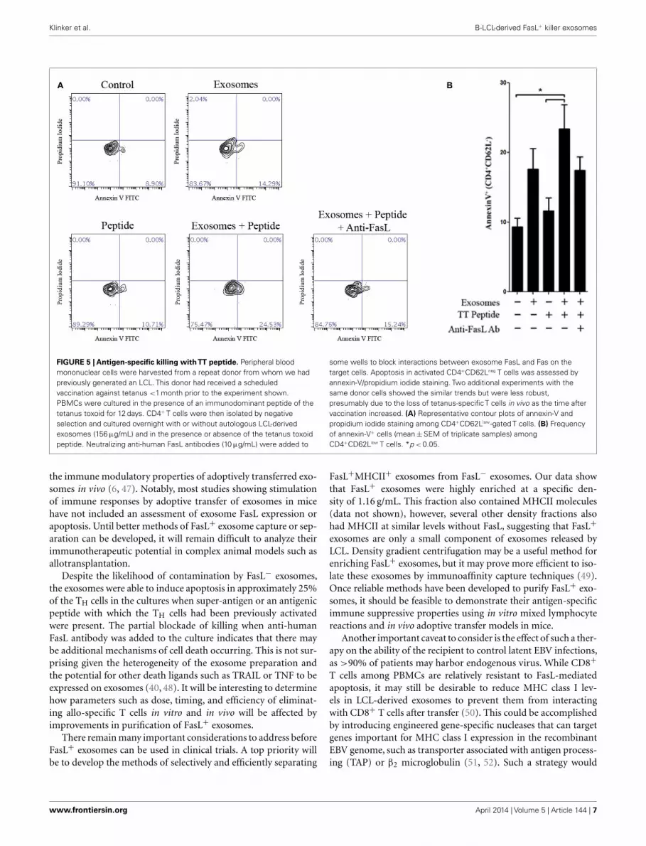
LCL-DERIVED EXOSOMES CAN INDUCE APOPTOSIS IN AUTOLOGOUSCD4+ T CELLSSince LCL-derived exosomes contained a measurable amount ofMHCII+FasL+ exosomes, we hypothesized that activated CD4+ Tcells would be susceptible to exosome-induced apoptosis. To testthis hypothesis,we obtained PBMCs from a healthy donor who hadbeen recently immunized against tetanus and from whom we hadpreviously made an LCL and could collect autologous exosomes.T cells specific for a nominal antigen are rare among peripheralCD4+ T cells, and therefore detecting the capacity of LCL-derivedexosomes to induce peptide antigen-specific apoptosis requiresprior activation to enrich for peptide-specific T cells. PBMCs fromthis donor were cultured with the immunodominant peptide of TTto enrich the CD4+ T cell pool for cells specific to this antigen. After12 days in culture, CD4+ T cells were isolated by negative selec-tion. To demonstrate the antigen-specificity of exosome-mediatedT cell apoptosis, the same TT peptide was introduced in excess toa portion of LCL-derived exosomes to displace peptides alreadypresent on the exosome MHC class II molecule. CD4+ T cellswere then incubated overnight with autologous LCL-derived exo-somes in the presence or absence of the TT peptide. Apoptosis wasassessed by annexin-V/propidium iodide staining among activatedT cells (CD4+CD62Lneg). LCL-derived exosomes in the presenceof TT peptide induced significant levels of apoptosis in CD4+
CD62Lneg T cells (Figure 5). While exosomes that were not loadedwith TT peptide also induced apoptosis above baseline, this differ-ence did not reach statistical significance (Figure 5). The additionof a neutralizing anti-FasL antibody to culture with exosomes andTT peptide appeared to inhibit exosome-induced apoptosis, butthis difference also did not reach statistical significance (Figure 5).Taken together, these data suggest that LCL-derived exosomes can
Frontiers in Immunology | Alloimmunity and Transplantation April 2014 | Volume 5 | Article 144 | 4

Klinker et al. B-LCL-derived FasL+ killer exosomes
FIGURE 3 | Diagram of exosome-bead capture experiments.Exosomes are too small to be accurately detected by standard flowcytometry, and so must be linked in aggregate to larger beads for flowcytometric analysis. Polystyrene beads coated with streptavidin wereincubated with biotin-conjugated anti-HLA-DR or an isotype control
antibody. Beads were then washed and incubated with gentle agitationfor several hours with LCL-derived exosomes. Beads were washed andstained with PE-conjugated anti-FasL or an appropriate isotype antibody.Positive staining for FasL indicates the presence of MHCII+FasL+
exosomes.
induce apoptosis in activated T cells that is antigen-dependent andmay be at least partially mediated by FasL.
To assess the ability of LCL-derived exosomes to induce apop-tosis of T cells that had not previously been exposed to antigen, weemployed the super-antigen, staphylococcal enterotoxin A (SEA),to facilitate interactions between MHC class II and the T cell recep-tor. LCL-derived exosomes were isolated from unstimulated LCLculture supernatant as described above and cultured with freshCD4+ T cells in the presence or absence of SEA. After 6 daysin culture, we assessed apoptosis in CD4+ T cells by annexin-V/propidium iodide staining. In the absence of SEA, LCL-derivedexosomes produced a modest increase in apoptosis in CD4+ T cells(Figure 6). In contrast, in the presence of SEA, exosomes inducedsignificant levels of apoptosis in CD4+ T cells (Figure 6). Similarresults were obtained using an LCL and CD4+ T cells from a secondindependent donor (data not shown). Taken together, these resultssuggest that LCL-derived exosomes can mediate antigen-specifickilling of CD4+ T cells.
DISCUSSIONExosome-mediated immunotherapy for the treatment of inflam-matory disorders is an intriguing concept because MHCII+FasL+
exosomes have demonstrated permanent and precisely focusedsuppression of antigen-specific immune responses in mouse mod-els (4, 5). For the treatment of human inflammatory conditions,exosomes may represent a safer alternative to regulatory cells forimmunotherapy because the phenotype of exosomes is expected
to be static, whereas regulatory cells can potentially differentiateinto effector cells after transfer (7). Although this technique haspromise, a reliable method for producing donor-derived immuno-suppressive exosomes is required before this therapeutic strategycan be developed as a strategy to induce and maintain toleranceafter transplantation. We show here that transformation of humanB cells with EBV results in robust expression of FasL, and thatall LCL tested in this study also produced MHCII+FasL+ exo-somes. Generating LCL from peripheral blood B cells is widelypracticed and a relatively simple process, requiring only minimallaboratory labor and reagents. The resulting transformed B cellscan be grown to high concentrations and stored over long peri-ods of time. Therefore, LCL represent a potentially reliable sourceof immunosuppressive exosomes from any donor that could betherapeutically useful in humans.
B cells expressing FasL are relatively infrequent under mostconditions, and we therefore hypothesized at the outset of thisproject that FasL expression among human B cell-derived tumorlines would be rare as well. While FasL expression was indeedinfrequent among primary B cells and cell lines derived fromother types of B cell cancers, we found that FasL protein waspresent in cell lysates from all LCL tested in this study. This resultwas somewhat surprising as LCL are reportedly susceptible toFasL-induced apoptosis, and LCL have been used extensively asAPC for activating T cells (25–28). These conflicting results canbe explained in part by the fact that unlike endogenous FasL+
B cells in mice, FasL protein is undetectable on the surface of
www.frontiersin.org April 2014 | Volume 5 | Article 144 | 5

Klinker et al. B-LCL-derived FasL+ killer exosomes
FIGURE 4 | LCL secrete MHCII+FasL+ exosomes. (A) Two LCL(designated “LCL-E” and “LCL-P”) were placed into fresh exosome-freemedia in the presence or absence of PMA/ionomycin. After 5 h (top panels)or 24 h (bottom panels), exosomes were isolated from culture supernatantsas described in the section “Materials and Methods.” Purified exosomeswere then incubated with anti-HLA-DR-coated beads as described inFigure 3. Beads were washed and stained with either anti-FasL or anisotype control antibody. Beads were coated with an appropriate isotypecontrol antibody as the capture antibody did not display any FasL staining(data not shown). (B) Cells from above experiment were harvested, lysed,and probed for FasL and MHCII (HLA-DR) by immunoblot.
LCL. Therefore, although LCL produce FasL, this intracellularsequestration makes it unavailable for inducing apoptosis in tar-get cells unless transported to the cell surface or released. In thepresent study, we demonstrate that PMA/ionomycin stimulationtriggers increased production of FasL protein by LCL, as well asthe release of FasL+MHCII+ exosomes. These data suggest thatantigen receptor- and calcium-dependent signaling pathways areinvolved in the regulation of FasL+ exosome transport. Otherstudies have demonstrated that ligation of ICAM-1, B7-H1, or B7-H4 on cultured LCL leads to activation of reactive oxygen species,which in turn cause the translocation of FasL to the cell surfaceand induction of LCL apoptosis, however the release of FasL+
exosomes was not measured (29–31).The high frequency of FasL expression and exosome release
among LCL suggests that FasL may be an important componentof natural EBV infection. LCL are generated experimentally byinfection with the B95-8 strain of EBV, which is a replicationincompetent form of the native γ-1 herpes virus that has onco-genic potential in humans (32). The natural virus persists in mostinfected individuals in latently infected circulating memory B cells(33, 34). Greater than 90% of adults have been infected with
EBV, and although clinical manifestations of infection are gen-erally rare, the transforming properties of the virus can lead toB cell-derived malignancies such as Burkitt’s and Hodgkin lym-phomas (35, 36). LCL generated by infection with EBV maintain alatent viral growth program, expressing at least eight proteins fromthe viral genome (35). Among these proteins is latent membraneprotein 1 (LMP1), a functional mimic of CD40 (37). Signalingof LMP1 differs from that mediated by CD40, as LMP1 signal-ing is constitutive rather than ligand-dependent. Therefore, LCLare essentially in a state of constant CD40 stimulation. We previ-ously observed that mouse B cells stimulated in vitro with CD40Land IL-5 express higher levels of FasL (38), and therefore theCD40-mimicry of LMP1 might potentially explain the constitu-tive production of FasL in LCL. Additionally, stimulation withCD40L has been reported to induce FasL expression in other typesof cells (39–41).
The expression of FasL by LCL and their production ofFasL+/MHCII+ exosomes may have important implications inclinical management of EBV infections. While most people are firstexposed to EBV in infancy, those infected later in life can developacute infectious mononucleosis (AIM) (42). At the height of acuteinfection, T cells are susceptible to Fas-mediated apoptosis, andin vitro infection of PBMCs with EBV leads to elevated levels ofFasL on the surface of B cells (43). Inducing surface expressionof FasL in B cells may therefore be a means of immune evasionemployed by EBV during the lytic cycle (44). Importantly, FasLlocalization appears to differ between the lytic and latent cycles,as FasL in LCL is intracellular whereas FasL can be found on thesurface of B cells during acute infection (43). As the virus transi-tions into a latent state and settles into homeostasis with the hostimmune system, infected B cells may maintain FasL productionbut cease transporting it to the cell surface. This may representa natural mechanism by which LCL can persist without causingsignificant tissue damage while maintaining the ability to ward offelimination by virus-specific T lymphocytes.
We demonstrate here that LCL constitutively produceMHCII+FasL+ exosomes with apoptosis-inducing activity againstCD4+ T cells. Although FasL can be detected in LCL-derived exosomes under normal conditions, stimulation withPMA/ionomycin increased both the amount of MHCII+FasL+
exosomes secreted and FasL production in LCL. However, whileFasL is abundant in LCL cell lysates, it is relatively difficult to detectin exosomes, even after stimulation. In contrast, the expression ofMHC class II was very abundant in exosomes in comparison toFasL. Based on these findings, we hypothesize that LCL may pro-duce a mixture of MHC class II+ exosomes that are either FasL+
or FasL−. It has proven difficult to separate these potential LCL-derived exosome subsets using current techniques (45, 46). Somelimitations were created because anti-human or mouse FasL anti-bodies have been unable to capture exosomes (data not shown),and the beads used for exosome capture have multiple bindingsites that can potentially capture several different exosomes simul-taneously. These are important limitations to the current studybecause there is likely to be a balance of stimulation with andwithout concomitant T cell apoptosis in our experimental modelsystem. The balance of these two forces may explain some seem-ingly contradictory results that have been published regarding
Frontiers in Immunology | Alloimmunity and Transplantation April 2014 | Volume 5 | Article 144 | 6
Klinker et al. B-LCL-derived FasL+ killer exosomes
FIGURE 5 | Antigen-specific killing withTT peptide. Peripheral bloodmononuclear cells were harvested from a repeat donor from whom we hadpreviously generated an LCL. This donor had received a scheduledvaccination against tetanus <1 month prior to the experiment shown.PBMCs were cultured in the presence of an immunodominant peptide of thetetanus toxoid for 12 days. CD4+ T cells were then isolated by negativeselection and cultured overnight with or without autologous LCL-derivedexosomes (156 µg/mL) and in the presence or absence of the tetanus toxoidpeptide. Neutralizing anti-human FasL antibodies (10 µg/mL) were added to
some wells to block interactions between exosome FasL and Fas on thetarget cells. Apoptosis in activated CD4+CD62Lneg T cells was assessed byannexin-V/propidium iodide staining. Two additional experiments with thesame donor cells showed the similar trends but were less robust,presumably due to the loss of tetanus-specificT cells in vivo as the time aftervaccination increased. (A) Representative contour plots of annexin-V andpropidium iodide staining among CD4+CD62Llow-gated T cells. (B) Frequencyof annexin-V+ cells (mean±SEM of triplicate samples) amongCD4+CD62Llow T cells. *p < 0.05.
the immune modulatory properties of adoptively transferred exo-somes in vivo (6, 47). Notably, most studies showing stimulationof immune responses by adoptive transfer of exosomes in micehave not included an assessment of exosome FasL expression orapoptosis. Until better methods of FasL+ exosome capture or sep-aration can be developed, it will remain difficult to analyze theirimmunotherapeutic potential in complex animal models such asallotransplantation.
Despite the likelihood of contamination by FasL− exosomes,the exosomes were able to induce apoptosis in approximately 25%of the TH cells in the cultures when super-antigen or an antigenicpeptide with which the TH cells had been previously activatedwere present. The partial blockade of killing when anti-humanFasL antibody was added to the culture indicates that there maybe additional mechanisms of cell death occurring. This is not sur-prising given the heterogeneity of the exosome preparation andthe potential for other death ligands such as TRAIL or TNF to beexpressed on exosomes (40, 48). It will be interesting to determinehow parameters such as dose, timing, and efficiency of eliminat-ing allo-specific T cells in vitro and in vivo will be affected byimprovements in purification of FasL+ exosomes.
There remain many important considerations to address beforeFasL+ exosomes can be used in clinical trials. A top priority willbe to develop the methods of selectively and efficiently separating
FasL+MHCII+ exosomes from FasL− exosomes. Our data showthat FasL+ exosomes were highly enriched at a specific den-sity of 1.16 g/mL. This fraction also contained MHCII molecules(data not shown), however, several other density fractions alsohad MHCII at similar levels without FasL, suggesting that FasL+
exosomes are only a small component of exosomes released byLCL. Density gradient centrifugation may be a useful method forenriching FasL+ exosomes, but it may prove more efficient to iso-late these exosomes by immunoaffinity capture techniques (49).Once reliable methods have been developed to purify FasL+ exo-somes, it should be feasible to demonstrate their antigen-specificimmune suppressive properties using in vitro mixed lymphocytereactions and in vivo adoptive transfer models in mice.
Another important caveat to consider is the effect of such a ther-apy on the ability of the recipient to control latent EBV infections,as >90% of patients may harbor endogenous virus. While CD8+
T cells among PBMCs are relatively resistant to FasL-mediatedapoptosis, it may still be desirable to reduce MHC class I lev-els in LCL-derived exosomes to prevent them from interactingwith CD8+ T cells after transfer (50). This could be accomplishedby introducing engineered gene-specific nucleases that can targetgenes important for MHC class I expression in the recombinantEBV genome, such as transporter associated with antigen process-ing (TAP) or β2 microglobulin (51, 52). Such a strategy would
www.frontiersin.org April 2014 | Volume 5 | Article 144 | 7

Klinker et al. B-LCL-derived FasL+ killer exosomes
FIGURE 6 | LCL-derived exosomes can induce apoptosis in autologousCD4+ T cells. CD4+ T cells were harvested from a repeat donor from whomwe had previously generated an LCL. CD4+ T cells were incubated withexosomes (127 µg/mL) in the presence or absence of the super-antigen,staphylococcal enterotoxin A (SEA). After 6 days in culture, PBMCs wereharvested and apoptosis was assessed in CD4+ T cells byannexin-V/propidium iodide staining. Data are representative of threeindependent experiments using exosomes from this donor. Similar resultswere obtained using exosomes from a different LCL donor (data notshown). *p < 0.05, **p < 0.01, ***p < 0.001.
expectedly reduce the amount of MHC class I on exosomes, andtherefore reduce the likelihood that LCL-derived exosomes woulddeplete EBV-specific CD8+ T cells in LCL-exosome recipients.CD4+ T cell immunity also appears to be crucial for control-ling the latent EBV infection (53). To overcome the potential lossof virus-specific CD4+ T cells, it may be necessary to vaccinaterecipients of LCL-derived FasL+ exosomes post-transplantationto reestablish immunity against EBV.
Despite the concerns listed above, there are many positiveaspects to using EBV-transformed B cells as a source of tolero-genic exosomes. Generating LCL with B95-8 EBV has proven to bea simple, reliable, and safe method that has already been used toproduce thousands of lines that could serve as a source of FasL+
exosomes. Huge repositories of LCL exist containing many linesthat have been genotyped for MHC class I and class II expres-sion and the high frequency of FasL expression by LCL also makesit feasible to generate de novo LCL from people on organ donorregistries with exact MHC representation. EBV transformationresults in a homogeneous culture of immortalized B cells thatgrow rapidly, can be maintained at high cell densities, and does notrequire cell sorting to remove contaminating cell populations. Exo-somes are reportedly stable in phenotype and can be stored overlong periods of time without significant loss of function (54). Thegenome of EBV is maintained in proliferating LCL as a large epi-some (~167 kb), and techniques for engineering recombinant EBVare well-established (55, 56). The large size of the viral genome willallow for the addition of transgenes to the virus, such as a segmentcontaining the FasL gene under a strong, ubiquitous promoter toensure robust production of FasL. Additionally, recombinant EBV
can be produced containing the coding sequence for tissue-specificalloantigens fused to a lysosomal sorting sequence. Proteins con-taining this sequence are actively sorted to the secretory lysosomewhere they are processed and presented on MHCII molecules (57).Thus, MHCII+FasL+ exosomes produced by such a LCL couldbe engineered to present various epitopes of the alloantigen. Theresulting exosomes could be harvested under sterile conditions bycentrifugation, affinity, or filtration, and either frozen for futureuse or administered directly to the patient.
The utility of LCL-derived FasL+ exosomes to tolerize allo-graft recipients remains to be determined. Other potential uses ofthese immune suppressive exosomes could include treatments forT cell-mediated allergies and autoimmune diseases, since the goalof eliminating antigen-specific T cells is similar in these condi-tions. Alternatively, the development of effective tumor vaccinesusing exosomes, which is currently being intensely studied, maybe dependent on the removal or suppression of FasL+ exosomesfrom the preparation. An important consideration is that exo-somes have the ability to travel relatively far away from the cells thatproduced them, and yet perform many of the same functions thathave been previously attributed to direct cell–cell contact. Muchcontroversy has existed over the functions of soluble forms of FasL,yet until recently there was no distinction made between truly sol-uble FasL and vesicular FasL. The recent developments in the fieldof exosome research should cause a reassessment of what we thinkis known about cellular interactions in the immune system andconsequently to our approaches toward immunotherapy.
REFERENCES1. Liu Z, Fan H, Jiang S. CD4(+) T-cell subsets in transplantation. Immunol Rev
(2013) 252(1):183–91. doi:10.1111/imr.120382. Raposo G, Nijman HW, Stoorvogel W, Liejendekker R, Harding CV, Melief
CJ, et al. B lymphocytes secrete antigen-presenting vesicles. J Exp Med (1996)183(3):1161–72. doi:10.1084/jem.183.3.1161
3. Kim SH, Lechman ER, Bianco N, Menon R, Keravala A, Nash J, et al. Exo-somes derived from IL-10-treated dendritic cells can suppress inflammationand collagen-induced arthritis. J Immunol (2005) 174(10):6440–8.
4. Kim SH, Bianco N, Menon R, Lechman ER, Shufesky WJ, Morelli AE, et al.Exosomes derived from genetically modified DC expressing FasL are anti-inflammatory and immunosuppressive. Mol Ther (2006) 13(2):289–300. doi:10.1016/j.ymthe.2005.09.015
5. Kim SH, Bianco NR, Shufesky WJ, Morelli AE, Robbins PD. MHC class II+exosomes in plasma suppress inflammation in an antigen-specific and Fasligand/Fas-dependent manner. J Immunol (2007) 179(4):2235–41.
6. Pêche H, Renaudin K, Beriou G, Merieau E, Amigorena S, Cuturi MC. Inductionof tolerance by exosomes and short-term immunosuppression in a fully MHC-mismatched rat cardiac allograft model. Am J Transplant (2006) 6(7):1541–50.doi:10.1111/j.1600-6143.2006.01344.x
7. Ohkura N, Kitagawa Y, Sakaguchi S. Development and maintenance of regula-tory T cells. Immunity (2013) 38(3):414–23. doi:10.1016/j.immuni.2013.03.002
8. Lundy SK. Killer B lymphocytes: the evidence and the potential. Inflamm Res(2009) 58(7):345–57. doi:10.1007/s00011-009-0014-x
9. Hahne M, Renno T, Schroeter M, Irmler M, French L, Bornard T, et al. Acti-vated B cells express functional Fas ligand. Eur J Immunol (1996) 26(3):721–4.doi:10.1002/eji.1830260332
10. Villunger A, Egle A, Marschitz I, Kos M, Böck G, Ludwig H, et al. Constitutiveexpression of Fas (Apo-1/CD95) ligand on multiple myeloma cells: a poten-tial mechanism of tumor-induced suppression of immune surveillance. Blood(1997) 90(1):12–20.
11. Tinhofer I, Marschitz I, Kos M, Henn T, Egle A, Villunger A, et al. Differentialsensitivity of CD4+ and CD8+ T lymphocytes to the killing efficacy of Fas (Apo-1/CD95) ligand+ tumor cells in B chronic lymphocytic leukemia. Blood (1998)91(11):4273–81.
Frontiers in Immunology | Alloimmunity and Transplantation April 2014 | Volume 5 | Article 144 | 8

Klinker et al. B-LCL-derived FasL+ killer exosomes
12. Kojima Y, Tsurumi H, Goto N, Shimizu M, Kasahara S, Yamada T, et al. Fas andFas ligand expression on germinal center type-diffuse large B-cell lymphomais associated with the clinical outcome. Eur J Haematol (2006) 76(6):465–72.doi:10.1111/j.1600-0609.2006.00631.x
13. Lundy SK, Lerman SP, Boros DL. Soluble egg antigen-stimulated T helper lym-phocyte apoptosis and evidence for cell death mediated by FasL(+) T andB cells during murine Schistosoma mansoni infection. Infect Immun (2001)69(1):271–80. doi:10.1128/IAI.69.1.271-280.2001
14. Tian J, Zekzer D, Hanssen L, Lu Y, Olcott A, Kaufman DL. Lipopolysaccharide-activated B cells down-regulate Th1 immunity and prevent autoimmune dia-betes in nonobese diabetic mice. J Immunol (2001) 167(2):1081–9.
15. Lundy SK, Fox DA. Reduced Fas ligand-expressing splenic CD5+ B lympho-cytes in severe collagen-induced arthritis. Arthritis Res Ther (2009) 11(4):R128.doi:10.1186/ar2795
16. Mabrouk I, Buart S, Hasmim M, Michiels C, Connault E, Opolon P, et al. Pre-vention of autoimmunity and control of recall response to exogenous antigen byFas death receptor ligand expression on T cells. Immunity (2008) 29(6):922–33.doi:10.1016/j.immuni.2008.10.007
17. Montandon R, Korniotis S, Layseca-Espinosa E, Gras C, Mégret J, Ezine S,et al. Innate pro-B-cell progenitors protect against type 1 diabetes by reg-ulating autoimmune effector T cells. Proc Natl Acad Sci U S A (2013)110(24):E2199–208. doi:10.1073/pnas.1222446110
18. Bonardelle D, Benihoud K, Kiger N, Bobé P. B lymphocytes mediate Fas-dependent cytotoxicity in MRL/lpr mice. J Leukoc Biol (2005) 78(5):1052–9.doi:10.1189/jlb.0904536
19. Minagawa R, Okano S, Tomita Y, Kishihara K, Yamada H, Nomoto K, et al.The critical role of Fas-Fas ligand interaction in donor-specific transfusion-induced tolerance to H-Y antigen. Transplantation (2004) 78(6):799–806.doi:10.1097/01.TP.0000129799.96439.6F
20. Tosato G, Cohen JI. Generation of Epstein-BarrVirus (EBV)-immortalized B celllines. Curr Protoc Immunol (2007) Chapter 7:Unit722. doi:10.1002/0471142735.im0722s76
21. Haas CS, Creighton CJ, Pi X, Maine I, Koch AE, Haines GK, et al. Identification ofgenes modulated in rheumatoid arthritis using complementary DNA microar-ray analysis of lymphoblastoid B cell lines from disease-discordant monozygotictwins. Arthritis Rheum (2006) 54(7):2047–60. doi:10.1002/art.21953
22. Kotecha N, Krutzik PO, Irish JM. Web-based analysis and publication offlow cytometry experiments. Curr Protoc Cytom (2010) Chapter 10:Unit1017.doi:10.1002/0471142956.cy1017s53
23. Miller G, Robinson J, Heston L, Lipman M. Differences between labora-tory strains of Epstein-Barr virus based on immortalization, abortive infec-tion, and interference. Proc Natl Acad Sci U S A (1974) 71(10):4006–10.doi:10.1073/pnas.71.10.4006
24. Blott EJ, Bossi G, Clark R, Zvelebil M, Griffiths GM. Fas ligand is targeted tosecretory lysosomes via a proline-rich domain in its cytoplasmic tail. J Cell Sci(2001) 114(Pt 13):2405–16.
25. de Campos-Lima PO,Torsteinsdóttir S,Cuomo L,Klein G,Sulitzeanu D,MasucciMG. Antigen processing and presentation by EBV-carrying cell lines: cell-phenotype dependence and influence of the EBV-encoded LMP1. Int J Cancer(1993) 53(5):856–62. doi:10.1002/ijc.2910530525
26. Rowe M, Khanna R, Jacob CA, Argaet V, Kelly A, Powis S, et al. Restorationof endogenous antigen processing in Burkitt’s lymphoma cells by Epstein-Barr virus latent membrane protein-1: coordinate up-regulation of pep-tide transporters and HLA-class I antigen expression. Eur J Immunol (1995)25(5):1374–84. doi:10.1002/eji.1830250536
27. Tatsuta T, Cheng J, Mountz JD. Intracellular IL-1beta is an inhibitor of Fas-mediated apoptosis. J Immunol (1996) 157(9):3949–57.
28. Wilson AD, Redchenko I, Williams NA, Morgan AJ. CD4+ T cells inhibit growthof Epstein-Barr virus-transformed B cells through CD95-CD95 ligand-mediatedapoptosis. Int Immunol (1998) 10(8):1149–57. doi:10.1093/intimm/10.8.1149
29. Kim YS, Park GB, Song HK, Hur I, Lee HK, Kang JS, et al. Cross-linking ofCD54 on Burkitt lymphoma cell line Raji and Ramos induces FasL expressionby reactive oxygen species and apoptosis of adjacent cells in Fas/FasL interaction.J Immunother (2007) 30(7):727–39. doi:10.1097/CJI.0b013e31814a69fa
30. Kim YS, Park GB, Lee HK, Song H, Choi IH, Lee WJ, et al. Cross-linking ofB7-H1 on EBV-transformed B cells induces apoptosis through reactive oxygenspecies production, JNK signaling activation, and FasL expression. J Immunol(2008) 181(9):6158–69.
31. Song H, Park G, Kim YS, Hur I, Kim H, Ryu JW, et al. B7-H4 reverse signal-ing induces the apoptosis of EBV-transformed B cells through Fas ligand up-regulation. Cancer Lett (2008) 266(2):227–37. doi:10.1016/j.canlet.2008.02.067
32. Kuppers R. B cells under influence: transformation of B cells by Epstein-Barrvirus. Nat Rev Immunol (2003) 3(10):801–12. doi:10.1038/nri1201
33. Babcock GJ, Decker LL, Volk M, Thorley-Lawson DA. EBV persistence in mem-ory B cells in vivo. Immunity (1998) 9(3):395–404. doi:10.1016/S1074-7613(00)80622-6
34. Joseph AM, Babcock GJ, Thorley-Lawson DA. EBV persistence involves strictselection of latently infected B cells. J Immunol (2000) 165(6):2975–81.
35. Thorley-Lawson DA. Epstein-Barr virus: exploiting the immune system. NatRev Immunol (2001) 1(1):75–82. doi:10.1038/35095584
36. Hislop AD, Taylor GS, Sauce D, Rickinson AB. Cellular responses to viral infec-tion in humans: lessons from Epstein-Barr virus. Annu Rev Immunol (2007)25:587–617. doi:10.1146/annurev.immunol.25.022106.141553
37. Uchida J, Yasui T, Takaoka-Shichijo Y, Muraoka M, Kulwichit W, Raab-Traub N, et al. Mimicry of CD40 signals by Epstein-Barr virus LMP1 in Blymphocyte responses. Science (1999) 286(5438):300–3. doi:10.1126/science.286.5438.300
38. Klinker MW, Reed TJ, Fox DA, Lundy SK. Interleukin-5 supports the expan-sion of Fas ligand-expressing killer B cells that induce antigen-specific apoptosisof CD4(+) T cells and secrete interleukin-10. PLoS One (2013) 8(8):e70131.doi:10.1371/journal.pone.0070131
39. Afford SC, Randhawa S, Eliopoulos AG, Hubscher SG, Young LS, AdamsDH. CD40 activation induces apoptosis in cultured human hepatocytes viainduction of cell surface Fas ligand expression and amplifies Fas-mediatedhepatocyte death during allograft rejection. J Exp Med (1999) 189(2):441–6.doi:10.1084/jem.189.2.441
40. Eliopoulos AG, Davies C, Knox PG, Gallagher NJ, Afford SC, Adams DH,et al. CD40 induces apoptosis in carcinoma cells through activation of cyto-toxic ligands of the tumor necrosis factor superfamily. Mol Cell Biol (2000)20(15):5503–15. doi:10.1128/MCB.20.15.5503-5515.2000
41. Shibaki A, Katz SI. Activation through CD40 ligation induces functionalFas ligand expression by Langerhans cells. Eur J Immunol (2001) 31(10):3006–15. doi:10.1002/1521-4141(2001010)31:10<3006::AID-IMMU3006>3.0.CO;2-L
42. Crawford DH, Macsween KF, Higgins CD, Thomas R, McAulay K, Williams H,et al. A cohort study among university students: identification of risk factors forEpstein-Barr virus seroconversion and infectious mononucleosis. Clin Infect Dis(2006) 43(3):276–82. doi:10.1086/505400
43. Tanner JE, Alfieri C. Epstein-Barr virus induces Fas (CD95) in T cells and Fasligand in B cells leading to T-cell apoptosis. Blood (1999) 94(10):3439–47.
44. Ohshima K, Suzumiya J, Sugihara M, Nagafuchi S, Ohga S, Kikuchi M. CD95(Fas) ligand expression of Epstein-Barr virus (EBV)-infected lymphocytes: apossible mechanism of immune evasion in chronic active EBV infection. PatholInt (1999) 49(1):9–13. doi:10.1046/j.1440-1827.1999.00816.x
45. Johansson SM, Admyre C, Scheynius A, Gabrielsson S. Different types of in vitrogenerated human monocyte-derived dendritic cells release exosomes with dis-tinct phenotypes. Immunology (2008) 123(4):491–9. doi:10.1111/j.1365-2567.2007.02714.x
46. Bobrie A, Colombo M, Krumeich S, Raposo G, Théry C. Diverse subpopula-tions of vesicles secreted by different intracellular mechanisms are present inexosome preparations obtained by differential ultracentrifugation. J ExtracellVesicles (2012) 1:18397. doi:10.3402/jev.v1i0.18397
47. Admyre C, Bohle B, Johansson SM, Focke-Tejkl M, Valenta R, Scheynius A, et al.B cell-derived exosomes can present allergen peptides and activate allergen-specific T cells to proliferate and produce TH2-like cytokines. J Allergy ClinImmunol (2007) 120(6):1418–24. doi:10.1016/j.jaci.2007.06.040
48. Peng P, Yan Y, Keng S. Exosomes in the ascites of ovarian cancer patients:origin and effects on anti-tumor immunity. Oncol Rep (2011) 25(3):749–62.doi:10.3892/or.2010.1119
49. Tauro BJ, Greening DW, Mathias RA, Ji H, Mathivanan S, Scott AM, et al. Com-parison of ultracentrifugation, density gradient separation, and immunoaffinitycapture methods for isolating human colon cancer cell line LIM1863-derivedexosomes. Methods (2012) 56(2):293–304. doi:10.1016/j.ymeth.2012.01.002
50. Suda T, Hashimoto H, Tanaka M, Ochi T, Nagata S. Membrane Fas ligandkills human peripheral blood T lymphocytes, and soluble Fas ligand blocks thekilling. J Exp Med (1997) 186(12):2045–50. doi:10.1084/jem.186.12.2045
www.frontiersin.org April 2014 | Volume 5 | Article 144 | 9

Klinker et al. B-LCL-derived FasL+ killer exosomes
51. de Souza N. Primer: genome editing with engineered nucleases. Nat Methods(2012) 9(1):27. doi:10.1038/nmeth.1848
52. Joung JK, Sander JD. TALENs: a widely applicable technology for targetedgenome editing. Nat Rev Mol Cell Biol (2013) 14(1):49–55. doi:10.1038/nrm3486
53. Münz C, Bickham KL, Subklewe M, Tsang ML, Chahroudi A, Kurilla MG, et al.Human CD4(+) T lymphocytes consistently respond to the latent Epstein-Barrvirus nuclear antigen EBNA1. J Exp Med (2000) 191(10):1649–60. doi:10.1084/jem.191.10.1649
54. Théry C, Amigorena S, Raposo G, Clayton A. Isolation and characterization ofexosomes from cell culture supernatants and biological fluids. Curr Protoc CellBiol (2006) Chapter 3:Unit 322. doi:10.1002/0471143030.cb0322s30
55. Delecluse HJ, Hammerschmidt W. The genetic approach to the Epstein-Barrvirus: from basic virology to gene therapy. Mol Pathol (2000) 53(5):270–9.doi:10.1136/mp.53.5.270
56. Yu Z, Lu J, Yu H, Yan Q, Zuo L, Li G. A precise excision of the complete Epstein-Barr virus genome in a plasmid based on a bacterial artificial chromosome.J Virol Methods (2011) 176(1–2):103–7. doi:10.1016/j.jviromet.2011.06.015
57. Thomson SA, Burrows SR, Misko IS, Moss DJ, Coupar BE, Khanna R. Target-ing a polyepitope protein incorporating multiple class II-restricted viral epi-topes to the secretory/endocytic pathway facilitates immune recognition by
CD4+ cytotoxic T lymphocytes: a novel approach to vaccine design. J Virol(1998) 72(3):2246–52.
Conflict of Interest Statement: The authors declare that the research was conductedin the absence of any commercial or financial relationships that could be construedas a potential conflict of interest.
Received: 01 October 2013; paper pending published: 30 October 2013; accepted: 20March 2014; published online: 02 April 2014.Citation: Klinker MW, Lizzio V, Reed TJ, Fox DA and Lundy SK (2014)Human B cell-derived lymphoblastoid cell lines constitutively produce Fas lig-and and secrete MHCII+FasL+ killer exosomes. Front. Immunol. 5:144. doi:10.3389/fimmu.2014.00144This article was submitted to Alloimmunity and Transplantation, a section of thejournal Frontiers in Immunology.Copyright © 2014 Klinker, Lizzio, Reed, Fox and Lundy. This is an open-access articledistributed under the terms of the Creative Commons Attribution License (CC BY).The use, distribution or reproduction in other forums is permitted, provided the originalauthor(s) or licensor are credited and that the original publication in this journal is cited,in accordance with accepted academic practice. No use, distribution or reproduction ispermitted which does not comply with these terms.
Frontiers in Immunology | Alloimmunity and Transplantation April 2014 | Volume 5 | Article 144 | 10