hla-derived peptides as novel immunosuppressives
TRANSCRIPT

Nephrol Dial Transplant (1997) 12: 865–878 NephrologyDialysis
TransplantationEditorial Comments
HLA-derived peptides as novel immunosuppressives
A. M. Krensky1 and C. Clayberger2
Departments of 1Pediatrics and 2Cardiothoracic Surgery, Stanford University School of Medicine, Stanford, CA, USA
HLA molecules and T lymphocytes and set out toIntroductionmake synthetic peptides corresponding to thesesequences as potential therapeutic reagents [4,5]. WeA basic understanding of the cellular and molecularfirst showed that peptides corresponding to specificmechanisms of organ transplant rejection may provideHLA sequences could block cytotoxic T lymphocytesnew insights into the mechanism of action of existing(CTL) in an allele-specific manner. Synthetic peptidesdrugs and lay the foundation for the development ofcorresponding to residues 98–113 of HLA-A2 spe-novel approaches to immunotherapy. We have focusedcifically inhibited cytolysis directed at HLA–A2, buton the T lymphocyte as a mediator of transplantnot other HLA types [4]. We proceeded to define arejection. T cells recognize foreign HLA on the graftseries of synthetic peptides corresponding to linearand orchestrate various e�ector arms involved in bothsequences of HLA molecules that blocked a variety ofhumoral and cellular immunity [1]. We designed syn-assays both in vitro and in vivo [5–7]. The mostthetic peptides corresponding to structural regions ofinteresting of these peptides corresponded to the a1HLA molecules and used them to modulate thealpha helix of HLA class I molecules (Figure 1). Aimmune response. Over the past decade the use of suchsubgroup of these peptides, corresponding to residuespeptides has progressed from the bench to the bedside60–84 of HLA-Bw4a, inhibited cytotoxicity between[2]. This report will briefly summarize this progress.all donor-recipient pairs in vitro. Furthermore thesehuman sequences induced immunological tolerance inanimal models of transplantation (Figure 3) [8]. BasedThe role of HLA in the alloresponseupon these studies, SangStat Medical Corporation,Menlo Park, CA, USA, licensed the peptide technologyThe human leukocyte-associated (HLA) antigensand developed it under the brandname ‘Allotrap@’. Inencoded in the major histocompatibility complexcollaboration with Jean-Paul Soulillou and colleagues,(MHC) on chromosome 6 in man subserve numerousSangStat showed that the Allotrap@ peptide was non-functions in the immune response, but they were firsttoxic in phase I clinical trials and embarked uponidentified as molecules that direct and restrict trans-phase II trials in France. Recent results from thisplant rejection [3]. Studies of the structure of HLAgroup show that the peptides inhibit natural killing (aclass I (Figure 1) and definition of the structure andsurrogate marker for CTL and cell-mediated immunefunction of the T cell receptor provided new insightsresponses) in vivo in patients who have received other-into the mechanism of HLA recognition, T cell activa-wise standard therapy for renal transplantation [9].tion, and di�erentiation, and the events leading to
The mechanism of action of these peptides is stillacute and subacute transplant rejection [1]. Non-selfunclear. Of interest, however, the functional peptides,HLA molecules are recognized by T cell receptors bylike the immunosuppressive drug deoxyspergualin,both ‘direct’ and ‘indirect’ pathways. In the directbind to members of the heat shock protein 70 familypathway, whole foreign HLA is recognized, while inof chaperones [7]. In addition, these peptides corre-the indirect pathway, non-self HLA peptides arespond to regions of HLA class I molecules recognizedrecognized in the context of self HLA (Figure 2).by natural killer inhibitory receptors, KIR. Thesereceptors send inhibitory signals into natural killercells when they recognize HLA molecules on targetHLA class-I-derived peptidescells [10].
In 1985 we reasoned that linear sequences of HLAmolecules may be important in the interaction between
HLA class-II-derived peptidesCorrespondence and o�print requests to: Alan M. Krensky MD,Chief, Division of Immunology and Transplantation Biology,
Gallon et al. found that synthetic peptides correspond-Department of Pediatrics, Stanford University, Stanford, CA94305–5119, USA. ing to linear sequences corresponding to polymorphic
© 1997 European Renal Association–European Dialysis and Transplant Association

Nephrol Dial Transplant (1997) 12: Editorial Comments866
Fig. 1. Structure of HLA class I showing regions corresponding to immunomodulatory peptides. A, side view and B, top view. Syntheticpeptides corresponding to residues 56–69, 60–84, 98–113, and 222–235 can inhibit T cell function. Potent allele non-specific inhibitionresults with sequences corresponding to the HLA-Bw4a supratypic specificity (residues 75–84 of HLA-B2702 and -B38 ). (Reprinted withpermission from Krensky AM. HLA derived peptides as novel immunosuppressives. Pediatr Res 1995; 38: 275–279.)
regions of mhC class II molecules had similar inhibit-ory e�ects both in vitro and in vivo [11]. They inter-preted their findings in the context of competition fortarget antigens in ‘indirect allorecognition’. Recentlywe described a sequence corresponding to a relativelymonomorphic region of an HLA class II molecule thatinhibits T cell proliferation by blocking cell-cycleprogression. This peptide, derived from the a1 alphahelix of HLA-DQ 03011, inhibits proliferation ofhuman peripheral blood lymphocytes, purified T cells,and cytolysis by CTL in an allele non-specific, dose-responsive manner. Proliferation to interleukin-2(IL-2) was blocked, but signalling through the IL-2receptor was intact. S6 kinase activity, downstream ofthe IL-2 receptor, is inhibited. Like rapamycin [12],this peptide inhibits cdk2 kinase activity by blockingdegradation of the p27 cell cycle inhibitor. However,unlike rapamycin, the DQ peptide is unable to inhibityeast cell growth. Thus a peptide from the same regionof an HLA class II molecule as previously described
Fig. 2. Two molecular mechanisms for allorecognition. Direct allore-cognition involves recognition of foreign HLA with a variety ofpotential peptides present in the antigen presentation groove. Indirectallorecognition, in contrast, involves recognition of specific HLAsequences as peptides in the context of self HLA. (Reprinted withpermission from Krensky AM, Clayberger C. The induction oftolerance to alloantigens using HLA-based synthetic peptides. CurrOpin Immunol 1994; 6: 791–796.)

Nephrol Dial Transplant (1997) 12: Editorial Comments 867
Fig. 3. Survival of rat heterotopic heart allografts. LEW donor hearts were transplanted into the abdomen of ACI recipients and palpateddaily. Rejection coincides with a loss of heart beat. Either (a) intravenous injection or (b) gavage of synthetic peptide B7 (correspondingto residues 75–87 of HLA-B7), with a 5-day course of oral cyclosporin, resulted in long-term graft survival. (Reprinted with permissionfrom Krensky AM, Clayberger C. The induction of tolerance to alloantigens using HLA-based synthetic peptides. Curr Opin Immunol1994; 6: 791–796.)
for class I molecules also has functional e�ects on T peptides are currently being evaluated in clinical trials.lymphocytes, but the two sets of peptides function via In animal models these peptides, given over 1 weekentirely di�erent mechanisms. with cyclosporin alone, induced long-term immunolo-
gical tolerance (Figure 3). They may similarly inducetolerance in humans. The next major hurdle for such
Summary and conclusions tolerogenic drugs, however, is to prove e�cacy inclinical circumstances. Many of the drugs used to treattransplant patients today (steroids, cyclosporin, aza-Over the past decade the use of synthetic peptidesthioprine, mycophenolate mofetil ) may actually inhibitcorresponding to linear sequences of HLA molecules
has progressed from a concept to a reality. These the ‘active’ processes of induction and maintenance of
Nephrol Dial Transplant (1997) 12: Editorial Comments868
GK, Krensky AM. HLA-A2 peptides can regulate cytolysis byimmunological tolerance. Demonstration that newhuman allogeneic T lymphocytes. Nature 1987; 330: 763–765drugs induce tolerance will require e�cacy studies in
6. Clayberger C, Lyu S-C, DeKruy� R, Parham P, Krensky AM.other immune-mediated diseases in which monother- Peptides corresponding to the CD8 and CD4 binding domainsapy is feasible (such as psoriasis), before further of HLA molecules block T lymphocyte immune responses in
vitro. J Immunol 1994; 153: 946–951advances can be made in the induction of transplant7. Noessner E, Naftzger C, Clayberger C, Krensky AM. HLAtolerance. In addition, rapid assays of rejection must
derived peptides which inhibit T cell function bind to membersbe developed in order to reverse tolerance inductionof the heat shock protein 70 family. J Exp Med 1996; 183:
failures without graft damage or loss. Lastly, it will 339–348require heroic physicians, surgeons, and patients to 8. Nisco S, Vriens P, Hoyt G et al. Induction of allograft tolerance
in rats by an HLA class I derived peptide and cyclosporine A.make immunological tolerance a reproducible clinicalJ Immunol 1994; 152: 3786–3792reality.
9. Giral M, Cuturi M-C, Nguyen J-M et al. Decreased cytotoxicactivity of natural killer cells in kidney allograft recipientstreated with human HLA derived peptide. Transplantation (inReferencespress)
10. Colona M, Samaridis J. Cloning of Ig-superfamily members1. Krensky AM, Weiss A, Crabtree G, Davis M, Parham P.associated with HLA-C and HLA-B recognition by NK cells.Mechanisms of disease: T lymphocyte—antigen interactions inScience 1995; 268: 405–408transplant rejection. N Engl J Med 1990; 322: 510–517
11. Gallon L, Watschinger B, Murphy B, Akalin E, Sayegh MH,2. Krensky AM, Clayberger C. HLA derived peptides as a novelCarpenter CB. The indirect pathway of allorecognition. Thestrategy for the prevention of allograft rejection. Exp Opin Investoccurrence of self-restricted T cell recognition of allo-MHCDrugs 1996; 5: 809–818peptides early in acute renal allograft rejection and its inhibition3. Dausset J. Iso-leuco-anticorps. Acta Hematol 1958; 20: 156–166by conventional immunosuppression. Transplantation 1995; 59:4. Parham P, Clayberger C, Zorn SL, Ludwig DS, Schoolnik GK,612–616Krensky AM. Inhibition of alloreactive cytotoxic T lymphocytes
12. Nourse J, Firpo E, Flanagan WM et al. Interleukin-2 mediatedby peptides from the alpha2 domain of HLA-A2. Nature 1987;elimination of the p27Kip1 cyclin-dependent kinase inhibitor325: 625–628
5. Clayberger C, Parham P, Rothbard J, Ludwig DS, Schoolnik prevented by rapamycin. Nature 1994; 372: 570–573
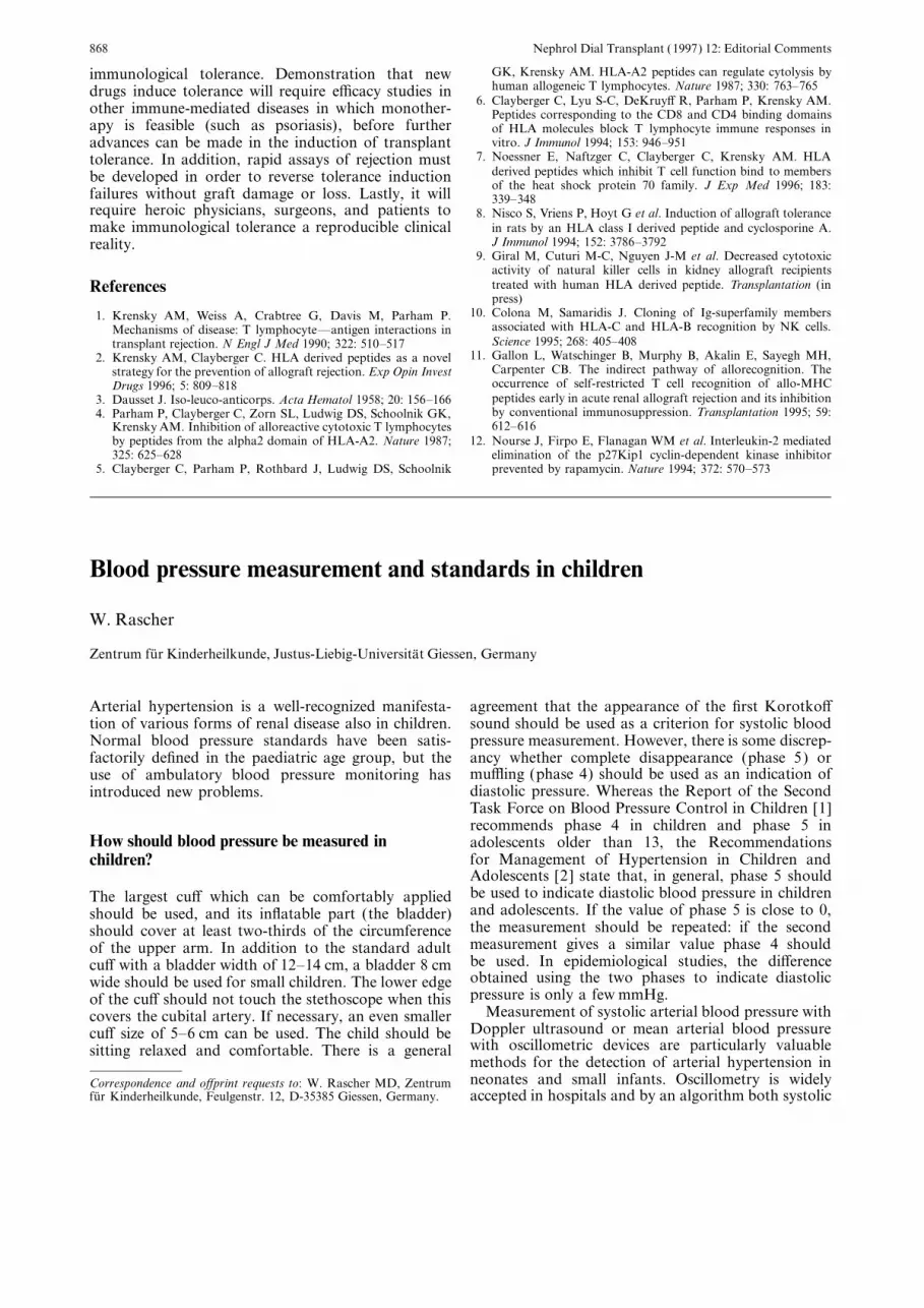
Blood pressure measurement and standards in children
W. Rascher
Zentrum fur Kinderheilkunde, Justus-Liebig-Universitat Giessen, Germany
Arterial hypertension is a well-recognized manifesta- agreement that the appearance of the first Korotko�sound should be used as a criterion for systolic bloodtion of various forms of renal disease also in children.pressure measurement. However, there is some discrep-Normal blood pressure standards have been satis-ancy whether complete disappearance (phase 5) orfactorily defined in the paediatric age group, but themu�ing (phase 4) should be used as an indication ofuse of ambulatory blood pressure monitoring hasdiastolic pressure. Whereas the Report of the Secondintroduced new problems.Task Force on Blood Pressure Control in Children [1]recommends phase 4 in children and phase 5 in
How should blood pressure be measured in adolescents older than 13, the Recommendationsfor Management of Hypertension in Children andchildren?Adolescents [2] state that, in general, phase 5 shouldbe used to indicate diastolic blood pressure in childrenThe largest cu� which can be comfortably appliedand adolescents. If the value of phase 5 is close to 0,should be used, and its inflatable part (the bladder)the measurement should be repeated: if the secondshould cover at least two-thirds of the circumferencemeasurement gives a similar value phase 4 shouldof the upper arm. In addition to the standard adultbe used. In epidemiological studies, the di�erencecu� with a bladder width of 12–14 cm, a bladder 8 cmobtained using the two phases to indicate diastolicwide should be used for small children. The lower edgepressure is only a few mmHg.of the cu� should not touch the stethoscope when this
Measurement of systolic arterial blood pressure withcovers the cubital artery. If necessary, an even smallerDoppler ultrasound or mean arterial blood pressurecu� size of 5–6 cm can be used. The child should bewith oscillometric devices are particularly valuablesitting relaxed and comfortable. There is a generalmethods for the detection of arterial hypertension inneonates and small infants. Oscillometry is widelyCorrespondence and o�print requests to: W. Rascher MD, Zentrum
fur Kinderheilkunde, Feulgenstr. 12, D-35385 Giessen, Germany. accepted in hospitals and by an algorithm both systolic

Nephrol Dial Transplant (1997) 12: Editorial Comments 869
and diastolic pressure are calculated from measured both age and height [10]. However, these Tables arerather di�cult to handle in daily practice.mean arterial pressure in a su�ciently accurate manner.
In very-low-birth-weight neonates larger cu�s are moreappropriate, but hypotension cannot adequately be
New information from ambulatory blood pressuredetected in these infants. Normative oscillometricmeasurements in childrenblood pressure values have been reported in very-low-
birth-weight infants [3] and during the first 5 years oflife [4]. Recent technological advances have enabled Recently, reference values for ambulatory blood pres-non-invasive repetitive blood pressure determinations sure monitoring of children and adolescents have beenover 24 h in subjects performing their normal activities. reported. A working group in Germany has reportedThis new technique of ambulatory blood pressure normative reference values in 1141 children and adoles-monitoring has been introduced as a valuable method cents that allows calculation of percentiles [11].for the evaluation of arterial hypertension in adults as Compared to reference values obtained from casualwell as in children and adolescents [5]. In the paediatric blood pressure measurements [1,8] mean systolic day-age group nearly exclusively oscillometric ambulatory time ambulatory blood pressure rose only moderatelyblood pressure monitors are in use. Whereas blood by height, from 112 to 124 mmHg in boys and frompressure can be measured accurately in mildly active 111 to 120 mmHg in girls, whereas mean diastolicor inactive children, no reliable measurements are daytime blood pressure was 72–74 mmHg, irrespectiveobtained during exercise and vigorous physical activity of height or sex. Small children had systolic as well as[6 ]. Systolic and diastolic blood pressure follow a diastolic daytime ambulatory blood pressure meanstypical circadian rhythm, values being 15–25% lower significantly above normal casual blood pressure atat night than during the day. rest, whereas this e�ect was less pronounced in taller
adolescents. The reason for this discrepancy cannot beexplained on a biological basis (e.g. higher level ofphysical activity throughout the day in younger chil-
What blood pressure is normal in children? dren). Particularly, lack of rise in diastolic bloodpressure during maturation does not correspond to the
A number of epidemiological studies have established findings of casual measurements in a variety of epide-normal blood pressure values in di�erent populations. miological studies. Methodological di�culties (e.g.Labarthe [7] reviewed 88 epidemiological reports on algorithm constructed for adults) might explain thisblood pressure in children from 30 countries: a consist- phenomenon. There was a large decrease of bloodent increase in both systolic and diastolic blood pres- pressure during the night by more than 20% of daytimesure with age was observed in almost all surveys. A pressure.marked increase in average blood pressure with age inchildhood and adolescence may be one aspect of
The diagnosis of hypertension in childrennormal growth and development. Distribution andpercentiles of general populations provide referencesfor blood pressure in growing children: there is a Criteria for the diagnosis of hypertension in adults are
not applicable to children. A value of 140/90 mmHgprogressive rise of casual blood pressure of approxi-mately 1.5 mmHg systolic and 0.7 mmHg diastolic per for casual blood pressure and 135/85 mmHg for day-
time ambulatory blood pressure is generally acceptedyear of age.Combined data from several studies as references as the upper limit of normal blood pressure in adults,
and this might also be true for adolescents. Since afor casual blood pressure has been published fromstudies performed in the United States [1] and in linear relationship exists between height and blood
pressure and between log weight and blood pressure,Europe [8]. The United States data included oneEuropean study, the Brompton study of more than a major blood pressure determinant in childhood is
body size. Therefore it has been recommended that7000 children aged from 4 days to 5 years [9]. Althoughthe Task Force stresses the importance of making blood pressure levels in the growing child should be
related to height.several blood pressure recordings in any individualchild before drawing conclusions about the blood Uncertainties exist with regard to normal blood
pressure range, and there is no agreement on the exactpressure, only the first blood pressure readings madein the 70 000 subjects were used to prepare its stand- definition of hypertension at di�erent ages. The most
helpful criterion for the definition of hypertension inards, because in one of the nine studies just one bloodpressure measurement per subject was available. In an children is the persistence of blood pressure above the
95th percentile. With repeated measurements not 5%attempt to incorporate the important contribution ofbody size into the analysis, the Task Force has added (95th percentile), but even less (about 1%) of the
children show chronic high blood pressure. Whenthe 90th percentile of height and weight for normalchildren at the bottom of the charts. The European ambulatory blood pressure values with oscillometric
devices are the basis to diagnose arterial hypertension,percentile charts have related casual blood pressure toheight rather than to age. Recently, tables for blood separate reference values should be used [11]
(Table 1).pressure in children were published taken into account

Nephrol Dial Transplant (1997) 12: Editorial Comments870
Table 1. Paediatric reference values for ambulatory blood pressure Referencesmonitoring (ABPM) [11]
1. Report of the second task force on blood pressure control inchildren. Pediatrics 1987; 79: 1–25Height (cm) Daytime (8–20) Night-time 24 h
(0–6) 2. Recommendations for management of hypertension in children(Percentile) (Percentile) (Percentile) and adolescents. Clin Exp Hypertens 1986; A8: 901–918
3. Tan KL. Blood pressure in very low birth weight infants in the50 95 50 95 50 95 first 70 days of life. J Pediatr 1988; 112: 266–270
4. Park MK, Menard SM. Normative blood pressure values in thefirst years in an o�ce setting. Am J Dis Child 1989; 143: 860–864ABPM percentiles in boys
5. Bald M, Kubel S, Rascher W. Validity and reliability of 24 h120 112/73 123/85 95/55 104/63 105/65 113/72130 113/73 125/85 96/55 107/65 105/65 117/75 blood pressure monitoring in children and adolescents using a140 114/73 127/85 97/55 110/67 107/65 121/77 portable, oscillometric device. J Hum Hypertens 1994; 8: 363–366150 115/73 129/85 99/56 113/67 109/66 124/78 6. Jacoby AC, Fixler DE, Torres EJ. Limitations of an oscillometric160 118/73 132/85 102/56 116/67 112/66 126/78 ambulatory blood pressure monitor in physically active children.170 121/73 135/85 104/56 119/67 115/67 128/77 J Pediatr 1993; 122: 321–326180 124/73 137/85 107/56 122/67 120/67 130/77 7. Labarthe DR. Epidemiology of primary hypertension in the
young. Clin Exp Hypertens 1986; A8: 495–513ABPM percentiles in girls 8. DeMan SA, Andre JL, Bachmann H et al. Blood pressure in120 111/72 120/84 96/55 107/66 103/65 113/73childhood: pooled findings of six European studies. J Hypertens130 112/72 124/84 97/55 109/66 105/66 117/751991; 9: 109–114140 114/72 126/84 98/55 111/66 108/66 120/76
9. DeSwiet M, Fayers P, Shinebourne EA. Blood pressure survey150 115/73 128/84 99/55 112/66 110/66 122/76in a population of newborn infants. Br Med J 1976; 2: 9–11160 116/73 131/84 100/55 113/66 111/66 124/76
10. Rosner B, Prineas RJ, Loggie JMH, Daniels SR. Blood pressure170 118/74 134/84 101/55 113/66 112/66 124/76nomograms for children and adolescents, by height, sex, and180 120/74 131/84 103/55 114/66 113/66 124/76age. J Pediatr 1993; 123: 871–886
11. Soergel M, Kirschstin M, Busch C, et al. Oscillometric twenty-four hour ambulatory blood pressure measurement in healthychildren and adolescents: a multicenter trial including 1141Note added in proofsubjects. J Pediatr (in press)
12. National High Blood Pressure Education Program WorkingUS authorities have recently revised their guidelines Group on Hypertension Control in Children and Adolescents:and recommended the fifth Korotko� sounds to define Update on the 1987 Task Force Report on High Blood Pressure
in Children and Adolescents. Pediatrics 1996; 98: 649–658diastolic hypertension in children [12].
Intradialytic renal haemodynamics—potential consequences for themanagement of the patient with acute renal failure
M. Manns, M. H. Sigler and B. P. Teehan
Division of Nephrology, Lankenau Hospital, Wynnewood, PA, USA
studies suggest that a haemodynamic instability duringIntroductiontraditional intermittent haemodialysis may haveadverse e�ects on the course and recovery of acuteHypotension is one of the major complications ofrenal failure (ARF ). Less haemodynamic instabilityhaemodialysis. A symptomatic reduction in blood pres-during slow continuous therapies could protect thesure occurs in approximately 20–50% of all acutekidney from further damage and might result indialysis treatments and results in interruption of theenhanced recovery.treatment in about 5–10% of cases. Several procedure-
related and patient-related factors contribute toEvidence for continuing tubular necrosis in acutehaemodynamic instability during dialysis. Among therenal failureprocedure related factors are ultrafiltration-induced
hypovolaemia, leading to a decrease in venous returnConger suggests that repeated episodes of hypotensionand cardiac output, the rapid reduction in plasmaduring dialysis may exacerbate renal injury and delayosmolality, which causes extracellular water to moverecovery from ARF [2]. Renal biopsies obtained 3–4into cells and slow plasma refilling time [1]. Someweeks after the initial insult from patients who su�eredcombat injuries with prolonged renal shutdown uni-Correspondence and o�print requests to: Miles H Sigler, Division of
Nephrology, Lankenau Hospital, Wynnewood, PA 19096, USA. formly revealed areas of fresh tubular necrosis. All

Nephrol Dial Transplant (1997) 12: Editorial Comments 871
patients had required daily or alternate day haemodia- Does renal replacement therapy a�ect the residuallysis, and many dialysis treatments were complicated renal function in acute renal failure?by transient blood pressure reduction to mean valuesas low as 70–90 mmHg, despite an overall stable We have studied the e�ects of intermittent haemodialysisclinical condition. The temporary decline of the blood (IHD) and continuous haemodialysis (CVVHD) on thepressure, which was within the autoregulatory range residual kidney function in critically ill patients withof the healthy kidney, seemed to be the only explana- acute renal failure [7]. The data has been publishedtion for the presence of these new ischaemic lesions. only in abstract form. Twenty-seven patients wereSolez compared renal biopsies in 50 patients with treated with intermittent haemodialysis and 16 patientspersistent acute renal failure with biopsies from seven with continuous haemodialysis using polysulphonepatients who had recently recovered from ARF [3]. membranes in each group. Creatinine clearance, ureaThe biopsies were taken 3–4 weeks after the onset of clearance, urine volume, and fractional excretion raterenal failure, at a time when regeneration of the for sodium were measured for 3 h before dialysis, duringdamaged tubular epithelium should have been com- dialysis and for 3 h after intermittent haemodialysis.pleted. Morphological analysis of the biopsies showed The same parameters were determined for 3 h before,necrosis of the tubular epithelial cells and loss of the 0–3 h and 4–6 h during continuous haemodialysis.PAS-positive brush border in the proximal tubules, Virtually all renal function parameters changed sig-which were significantly more severe in the ARF group
nificantly during and after intermittent haemodialysis,than in the recovery group. Both of these histologicalindicating an acute worsening of residual kidney func-changes could contribute to renal function failure intion. Creatinine clearance, which represents an estima-ARF by permitting passive backflow of tubular filtrate tion of the glomerular filtration rate (GFR), declinedand by causing decreased tubular reabsorption ofsignificantly intra- and postdialysis, when compared tosodium, resulting in stimulation of renin release andpredialysis (Table 1). However, as GFR falls, enhanceda�erent arteriolar vasoconstriction [3]. Tubular caststubular creatinine secretion leads to an up to twofoldseemed to play no role in the maintenance of renaloverestimation of GFR by the creatinine clearance.failure. The data suggests that necrosis of tubularThe variability of tubular urea reabsorption, whichepithelial cells is a continuing process, not necessarilydepends on the degree of sodium and water reabsorp-limited to the original lesion, and contributing to, iftion, causes urea clearance to underestimate GRF bynot causing, continued renal failure and prolonged30–50%. The mean of creatinine clearance and ureaATN. These observations may be explained by theclearance, which may be a closer estimation of trueresults of the animal experiments, described below.glomerular filtration rate in patients with moderate toadvanced renal insu�ciency [8], was also substantially
The post-ischaemic kidney in acute renal failure lower during and after dialysis compared to predialysis.However, if serum creatinine falls during dialysis below
Animal experiments designed to evaluate possible rela- the saturation limit of the secretory mechanism,tionships between alterations in renal haemodynamics changes in creatinine clearance after dialysis may notand ongoing damage of the surviving or recovering necessarily reflect changes in GFR. In this case, thenephrons showed an impaired ability to autoregulate declining serum creatinine during dialysis would causerenal blood flow in post-ischaemic ARF. decreased tubular creatinine secretion, which in turnHaemorrhagic reduction in renal perfusion pressure would falsely suggest a decreased GFR. Two studieswithin the autoregulatory range was associated with investigated the e�ects of dialysis on creatinine secre-paradoxical vasoconstriction (rather than vasodilat- tion in chronic renal failure and the importance ofation), leading to a substantial decrease in renal blood creatinine clearance as an estimate of GFR in ARF.flow, further reduction in creatinine clearance and The first study showed an increased creatinine secretionhistological evidence of recurrent ischemic injury [4].It was shown that the post-ischaemic kidney exhibits Table 1. E�ects of dialysis on residual renal function in acute and
chronic renal failurean increased sensitivity to renal nerve stimulation withexaggerated catecholamine release from nerve endings[5]. Furthermore, the ability to autoregulate renal Creatinine Inulin Urine FeNa
clearance clearance volume (%)blood flow was significantly improved after renal(ml/min) (ml/min) (ml/min)denervation [4]. In addition, the vasodilatory capacity
of the post-ischaemic renal vessels seemed to be dimin-−7% −10% −12%Manns [7], ARF,ished, due to decreased or lost endothelium derived
CVVHDrelaxing factor activity [6]. These results suggest thatManns [7], ARF, −25% −50% −46%hypotensive episodes during intermittent haemodialysis IHD
may result in decreased renal perfusion pressure in the Yeh [11], CRF, +5% −21% −43%IHD onlypost-ischaemic kidney. If autoregulation of the post-dialysisischaemic kidney is impaired, reduced renal perfusiononly UF −4% −25% −30%pressure might induce paradoxical vasoconstriction
Schuck [9], CRF, −3% −35%with severely diminished renal blood flow and recurrent IHDischemic injury.

Nephrol Dial Transplant (1997) 12: Editorial Comments872
from 1.09 to 2.01 mmol/min 12 h after dialysis [9 ]. The the first 6 h of CVVHD in comparison to IHD andfurther comparisons using comparable dialysis pre-authors propose the hypothesis that dialysis eliminatesscriptions in regard to urea clearance, solute removalsubstances which inhibit tubular creatinine secretion.and volume removal are necessary to confirm thisThe second study reports a good correlation betweenadvantage. Further research should also focus on thecreatinine clearance and inulin clearance in ARF (r=question of whether dialysis-induced hypoperfusion is0.93) [14]. We believe therefore that the observedlimited to the kidney or whether it also a�ects othersignificant decline in creatinine clearance and ureaorgan systems adversely. Our study was not designedclearance during and following intermittent haemodia-to investigate possible di�erences between both treat-lysis suggests a marked decrease of the glomerularment modalities in terms of mortality. The fact thatfiltration rate.CVVHD is associated with less change in residualThere are several explanations for these observa-renal function does not necessarily translate intotions. First, the MAP fell during IHD by 7% from aimproved morbidity or mortality.baseline of 84 mmHg. The correlation between the
percentage decrease in creatinine clearance and the fallin MAP during dialysis (r=0.72, P<0.001) provides Referencesevidence that renal hypoperfusion during and following
1. Zucchelli P, Santoro A. Dialysis-induced hypotension. A freshdialysis might aggravate renal damage. Secondly, dia- look at pathophysiology. Blood Purif 1993; 11: 85–98lysis removes urea from the blood, which in turn 2. Conger JD. Does Hemodialysis delay recovery from acute renal
failure? Semin Dial 1990; 3: 146–148reduces the urinary osmotic drive [10]. The decrease3. Solez L, Morel-Maroger L, Sraer J. The morphology of acutein urine output after dialysis in chronic renal failure is
tubular necrosis in man. Analysis of 57 renal biopsies andthe result of reduced solute load with enhanced tubular comparison with the glycerol model. Medicine (Baltimore) 1979;reabsorption (Table 1) [11,12]. In our study of ARF 58: 362–367
4. Kelleher SP, Robinette JB, Conger JD. E�ect of hemorrhagicthe decrease in BUN during dialysis did not correlatereduction in blood pressure on recovery from acute renal failure.with the fall in urine output or the fall in creatinineKidney Int 1987; 31: 725–730clearance. This makes it unlikely that reduced urinary 5. Kelleher SP, Robinette JB, Conger JD. Sympathetic nervous
osmotic drive during and after dialysis played a signi- system in the loss of autoregulation in acute renal failure. AmJ Physiol 1984; 246: F379–F386ficant role in the observed changes [13]. This is in
6. Conger JD, Robinette JB, Schrier RW. Smooth muscle calciumagreement with the results from Rahman and Conger,and endothelium-derived relaxing factor in the abnormal vascu-which demonstrate that the residual GRF is the prim- lar responses in acute renal failure. J Clin Invest 1988; 82:
ary determinant of the urine flow rate in ARF [14]. 532–5377. Manns M, Sigler MH, Teehan BP. Advantages of continuousIn comparison with the observed changes during
veno-venous hemodialysis in acute renal failure. J Am Socintermittent haemodialysis, renal function parameters Nephrol 1995; 6: 470 (abstract)such as creatinine clearance, urine volume, and frac- 8. Perrone RD, Steinman TI, Beck GJ et al. and The Modificationtional excretion of sodium were significantly less a�ec- of Diet in Renal Disease Study. Utility of radioisotopic filtration
markers in chronic renal insu�ciency: Simultaneous comparisonted during continuous haemodialysis (Table 1) [7].of 125I-iothalamate, 169Yb-DTPA, 99mTc-DTPA and inulin. AmHaemodynamic instability did not occur during J Kidney Dis 1990; 16: 224–235
CVVHD despite significantly lower baseline blood 9. Schuck O, Taplan V, Nadvornikova H. The e�ect of haemodia-lysis on tubular secretion of creatinine in residual nephrons.pressure before initiation of continuous haemodialysis.Nephrol Dial Transplant 1990; 5: 549–550This improved haemodynamic stability during
10. Bankir L, Ahloulay M, Bouby N et al. Is the process of urinaryCVVHD might be the explanation for the better preser- urea concentration responsible for a high glomerular filtrationvation of the residual kidney function. It also makes rate? J Am Soc Nephrol 1993; 4: 1091–1103
11. Yeh BP, Tomko DJ, Stacy WK. Factors influencing sodium andit less likely that, despite the use of biocompatiblewater excretion in uremic man. Kidney Int 1975; 7: 103–110membranes in both groups, blood–membrane inter-
12. Feinfeld DA, Danovitch GM. Factors a�ecting urine volume inactions such as activation of the humoral pathways chronic renal failure. Am J Kidney Dis 1987; 10: 231–235and the cellular blood elements might have influenced 13. Sigler MH, Manns M, Teehan BP. Does intermittent hemodia-
lysis a�ect the residual kidney function in acute renal failure?renal function [15,16].J Am Soc Artif Int Organs [Suppl] 1995; 41: 73 (abstract)
14. Rahman SN, Conger JD. Glomerular and tubular factors inurine flow rates of acute renal failure patients. Am J Kidney DisConclusion1994; 23: 788–793
15. Schulman G, Hakim R. Hemodialysis membrane bioincompat-Renal hypoperfusion during intermittent haemodia- ibility in acute renal failure. Adv Renal Replace Ther 1994;
1: 75–82lysis might delay tubular regeneration in postischaemic16. Sigler MH, Manns M. Membranes and devices used in continu-ARF. Continuous forms of renal replacement therapy
ous renal replacement therapy. Semin Dial 1996; 9: 98–105have several advantages in the setting of critically ill 17. Bellomo R, Farmer M, Wright C, Parkin G, Boyce N. Treatmentpatients with acute renal failure [17,18]. Improved of sepsis-associated severe acute renal failure with continuous
hemodiafiltration. Clinical experience and comparison with con-haemodynamic stability during continuous haemodia-ventional dialysis. Blood Purif 1995; 13: 246–254lysis protects the kidney from further damage and 18. Van Bommel EFH. Are continuous therapies superior to inter-
might result in enhanced recovery. However, in our mittent haemodialysis for acute renal failure on the intensivecare unit? Nephrol Dial Transplant 1995; 10: 311–314study [7] dialysis e�ciency was substantially less during

Nephrol Dial Transplant (1997) 12: 873–876 873
Renal replacement therapy in the elderly: an old problem with youngsolutions
Nuahd Ismail
Division of Nephrology, Vanderbilt University School of Medicine, Nashville, TN37232–2372, USA
Therapy of end-stage renal disease (ESRD) in the patient on haemodialysis (HD) is frequently accept-able. The 5-year patient survival presently rangeselderly is characterized by tremendous challenges. It
also raises ethical and socioeconomic concerns: ethical between 20–40% [3]. Nevertheless, multiple studieshave drawn di�erent conclusions based upon the periodissues because an expensive treatment is being o�ered
to a population with limited life-expectancy, and of evaluation, type of patient, and study design. Anearly study [4] of 157 patients with a mean age of 75socioeconomic because of the progressive increase in
the proportion of elderly requiring ESRD therapy years and multiple comorbidities at initiation of dialysis(17.2% of patients had diabetes, 35% had coronaryworldwide [1]. According to the US Renal Data
System (RDS), 1995 Annual Data Report, the median disease, 10% had cerebrovascular disease, 18% hadchronic obstructive pulmonary disease, and 22% peri-age at incidence of ESRD has reached 62–63 years in
the US, Japan, Germany, France and Italy [2]. Indeed, pheral vascular disease) found 3- and 5-year survivalrates of 47% and 22%, respectively. Despite thesegeriatric ESRD will be an important topic for 21st
Century physicians to face. In the U.S., it is projected comorbidities, most of the patients were doing well,had social contacts (90%), spent time outdoors (86%)that 60% of the ESRD population will be over 65
years in the year 2000. and ranked high on Karnovsky scale (73% of patientswith scale >80%). In addition, 40% perceived theirhealth to be better than 70-year old subjects, while
Factors to be considered in o�ering renal only 25% regarded their health as worse. An acceptablereplacement therapy to elderly patients with ESRD subjective quality of life and degree of rehabilitation
have also been noted in other reports [4–8]. In theWhen making decisions about initiating, withholding, Battelle Human A�airs Research Center Study [8] ofor terminating dialysis in the elderly or the appro- 859 dialysis and transplant patients, 70% of the elderlypriateness of o�ering renal transplantation to these dialysis patients (comprising 16% of the total numbersindividuals, several important points must be consid- of the elderly dialysis) fell in the top four categories ofered: (1) The elderly are a ‘heterogeneous’ group of overall functional status using the Karnovsky index.persons united by ‘chronologic’, but not necessarily It has been proposed that the regular visit to the‘biologic’, age (physiologic, psychologic, or social func- dialysis unit and mixing with younger patients may acttioning). The latter (biologic age) has a variable expres- as a beneficial social stimulus in elderly patients [6].sion depending on the pace of ageing as well as the However, the importance of this social e�ect is broughtimpact of comorbid pathologic conditions whose into question by the observation in one study thatnumber and severity increases with age. (2) When those elderly subjects treated by home HD had theanalysing results of renal replacement therapy (RRT) highest quality of life, as assessed by life satisfaction,in the elderly, health care professionals must recognize well-being index, and psychological a�ect [9].the importance of not only ‘absolute’ survival time ofthese individuals, but also the ‘quality’ of that survival.
Factors impacting on quality of lifeStated otherwise, the goals of RRT in the elderlyis to add ‘life to years’, not simply ‘years to life’.
Two more recent studies painted a much grimmerFurthermore, while there might be little disagreementpicture of the life of elderly dialysis patients [10,11],on the definition of quality of life, the yardstick for itsespecially when ESRD is caused by diabetes. It ismeasurement has not been straightforward. (3) Whenimportant, however, to appreciate the flaws in therenal transplantation is being considered for the eld-design of these studies. These include basing the dataerly, a dilemma exists if it is ‘fair’ to waste an invaluableon recollection in one study, not realizing the fullresource (the cadaver kidney) to an elderly individualbenefits of erythropoietin, and not addressing dialysiswith a limited life-expectancy. i.e. send a functioningadequacy. In addition, the conclusions may not applygraft to the grave with the elderly.to patients who do not live in the inner city. Thesestudies, however, demonstrate the impact that poverty,
Survival of the elderly on dialysis illiteracy, and other urban troubles can have on thelikelihood of rehabilitation. Therefore, negative resultssuch as these should not provide ammunition for thoseDespite complex comorbid and psychosocial condi-
tions, survival and the quality of life in the elderly who wish to institute age-based rationing of HD, but

Nephrol Dial Transplant (1997) 12: Editorial Comments874
rather, among other things, to examine the critical morbidity and mortality are limited, however, by smallsample size or the possibility of selection bias amongissue of rehabilitation and most importantly early
implementation of measures that might improve func- CAPD patients.tional status whenever it is significantly impaired.
A recent study suggested that racial di�erences mayTransplantation in the elderly—an under utilizeda�ect the outcome of elderly dialysis patients [12].opportunity?Black elderly patients were found to have higher sur-
vival rates (57 vs 38% at 3 years) and a better qualityof life than white patients. The following factors may Over the last decade, the e�cacy and safety of renal
transplantation in the elderly has been demonstratedhave contributed to these di�erences: (1) blacks spentmore time on dialysis per week than whites, (2) whites in several published reports [22–26]. One-year patient
survival now ranges between 80–90% and 5-yearhad more dialysis-accelerated atherosclerosis, cardio-vascular morbidity, and other comorbid conditions at patient survival for cadaveric renal transplantation
(CRT) has increased to 70%. ‘Functional’ graft sur-3 years and (3) blacks may be more likely to die of‘competing risks’ before reaching kidney failure; as a vival has increased in parallel, averaging 80% at one
year and 55–60% at 5 years. The increasing success ofresult, those who survive to chronic dialysis mayconstitute a ‘hardier group’. organ transplantation in the elderly, coupled with a
chronic persistent shortage in cadaver donors haveIn the aggregate, therefore, prolonged survival inthe elderly on HD is feasible. Furthermore, the elderly created an ethical dilemma for physicians regarding
the allocation of a scarce resource (the cadaver kidney)can adapt to a complicated treatment regimen andactivities, and may be more compliant than younger to the elderly with limited-life expectancy.
Ageing is accompanied by senescence of the immunepatients [13]. However, malnutrition and the potentialfor under dialysis, as well as depression and withdrawal system [27]. The T-lymphocyte population undergoes
major alterations with age resulting in a decline in cell-from dialysis are serious complications in the elderlydialysis patient [14]. mediated cytotoxicity. T-cells of elderly do not recog-
nize self-MHC. These changes lead to down regulationof the T-cell activation cascade, with less rejection
Dialysis technique and the elderly episodes to organ transplantation. On the other hand,these changes also lead to a decline in cell-mediatedimmunity against viral, bacterial, and fungal infections.The new synthetic membranes and the full benefits of
erythropoietin hopefully will decrease the incidence of B-cell function is unimpaired in the elderly; however,reduced antibody response to foreign antigens declinesthese complications in the elderly dialysis patient. Most
importantly, delivery of ‘adequate’ dose of dialysis because of impaired CD4+ helper function. This trade-o� (decreased rejection and increased vulnerability( KT/V>1.4 ) is critical to the elderly. Five- and 10-
year survival rates of 69% and 64%, respectively, were to infection) necessitates modification of immuno-suppressive therapy in the elderly.achieved in patients older than 64 years in one of the
longest studies of high dialysis doses (KT/V=1.67) Patient death with a functioning allograft accountsfor the majority of graft loss in older transplantreported from France by Laurent et al. [15].
Similarly, the outcome of chronic peritoneal dialysis patients [22–26 ]. In several studies, older patients hada graft loss rate of nearly 50% as a consequence of(CPD) in the elderly is also acceptable, with 5-year
survival rates of 20–30% [3,16–19]. CPD is thus an death compared to a rate of 15% in younger patients.Since graft loss in the elderly is related primarily toalternate dialysis modality for the elderly and may
confer some advantages such as absence for vascular patient death (and not immunologic graft-loss), andthe two main causes of mortality following transplanta-access and the promotion of independence and a sense
of self-worth. Also the judicious use of CPD in nursing tion are cardiovascular disease and infection, it followsthat comprehensive preoperative evaluation of elderlyhomes, the use of home helpers, and the development
of Adult Health Care Centers may enable physicians patients is imperative to improve patient and thus graftsurvival. Similarly, the goal of immunosuppressionto o�er this modality to more elderly patients.
Although many comparisons have been made of mor- protocols in the elderly should be to reduce the risk ofinfection without causing rejection and consequenttality between HD and CAPD for the ESRD popula-
tion as a whole, only few specific studies have addressed high-dose immunosuppression, infection, and graftloss. Since the elderly have a degree of immune incom-the elderly subgroup. Overall, in terms of outcome,
there is little evidence to suggest that one modality is petence and decreased hepatic enzyme activity (especi-ally the P450 enzyme system), they require lesssuperior for the elderly ESRD patients [20]. At least
in the long-term, technique survival appears to be aggressive immunotherapy.Recent data from USRDS and from UNOS havegreater in HD-treated patients. Two recent epidemiol-
ogical studies from the US suggest that HD may be revealed that recipients of older kidneys (donor age56–65) on average have lower graft survivals and lowerassociated with less mortality in the elderly diabetic
(in the USRDS case-mix severity study, RR of death GFR’s, without a�ecting patient survival [28]. Thesedecrements in graft function are magnified at a donorwas 1.25 for elderly diabetic on CAPD compared to
HD) [21,22]. All of the available studies comparing age above 60, and are reflected more in long-term

Nephrol Dial Transplant (1997) 12: Editorial Comments 875
Fig. 1. Algorithm for selection of treatment modality for Geriatric ESRD. CRT=Cadaveric renal transplantation, LRT=living-relatedtransplantation. Adapted from Ismail et al., Am J Kidney Disease 1994; 23: 1–15.
survival (after 3 years) than in 1-year graft survival. elderly <70 years old (and possibly <75) free ofrecent or metastatic malignancy, active infection, severeThe increased (but acceptable) risk of graft loss permit
the conclusion that the donor pool should be expanded extrarenal disease (cardiac, pulmonary, hepatic) ormental or psychiatric illnesses should be consideredto routinely include kidneys up to donor age of 60
years, that routine peri-harvest renal core biopsies for renal transplantation (Figure 1). (2) Expandingthe donor pool to include older donors (56–65) is ashould be used to evaluate the integrity of renal
anatomy in donors between 60–65 years, and that suitable strategy if judiciously applied (excluding suchkidneys when serum creatinine is >1.8–2.0 mg/dl ).older kidneys might be harvested for particular use in
older recipients when 20–30 year graft survival are not Such older kidneys may be appropriately matched toolder recipients. (3) Elderly patients should receiveas important as in younger recipients. This age match-
ing of donor and recipient is gaining support both as extensive information about ESRD therapy modalities.They should be able to select the modality that maxim-a fair and a physiologically fair process [28, 29].
Further support for age matching comes from a recent izes their quality of life (including psychosocial adapta-tion). They should also be considered for rehabilitationreport that kidneys from older donors survived for
longer in older patients than in younger patients [30]. programs.Finally, living-related transplantation may confer
some benefit (compared to CRT ) in graft survival; itsmajor limitation, however, remains the finding of a Referencessuitable living donor [31].
1. Mignon F, Michel C, Mentre F, Viron B. Worldwide demo-graphics and future trends of the management of renal failurein the elderly. Kidney Int 1993; 43 (suppl. 41): S18–S26Conclusions 2. US Renal Data system, USRDS 1995 Annual Data Report, theNational Institutes of Health, National Institute of Diabetesand digestive and Kidney Diseases, Bethesda, MD, April 1995In summary, (1) elderly patients should not be denied
3. Ismail N, Hakim RM, Oreopoulos DG, Patrikarea A. Renaleither transplantation or dialysis on the basis of age replacement therapies in the elderly: Part 1. Hemodialysis andper se. Irrespective of age, they should be o�ered chronic peritoneal dialysis. Am J Kidney Dis 1993; 22: 759–782
4. Westlie L, Umen A, Nestrud S, Kjellstrand CM. Mortality,dialysis if there is no contraindication (senile dementia,morbidity, and life satisfaction in the very old dialysis patient.metastatic cancer, and advanced liver disease). DialysisASAIO Trans 1984; 30: 21–30however should not be used merely to prolong the
5. Tapson JS, Rodger RSC, Mansy H, Elliot RS, Ward MK,dying-process, nor should dialysis facilitate a longer Wilkinson R. Renal replacement therapy in patients aged overlife of a quality considered unacceptable. When there 60 years. Postgrad Med J 1987; 63: 1071–1077
6. Walls J. Dialysis in the elderly. Some UK experience. Adv Peritis doubt about chances of recovery from a severeDial 1990; 6(suppl ): 82–85underlying illness, a ‘trial’ of dialysis may be o�ered
7. Schaefer K, Asmus G, Quellhorst E, Pauls A, von Herrath D.to an elderly patient (several weeks and perhaps few Optimum dialysis treatment for patients over 60 years withmonths). Withdrawal of dialysis later would be prefer- primary renal disease. Survival data and clinical results from
242 patients treated either by hemodialysis or hemofiltration.able to withholding it. A ‘biologically’ well maintained
Nephrol Dial Transplant (1997) 12: Editorial Comments876
Proc Eur Dial Transplant Assoc Eur Renal Assoc 1985; 21: 20. Benevent D, Benzakour M, Peyronnet P, Legarde C, Leroux-Robert C, Charmes JP. Comparison of continuous ambulatory510–523peritoneal dialysis and hemodialysis in the elderly. Adv Perit8. Battelle Human A�airs Research Centers. Seattle, WA., NationalDial 1990; 6(Suppl ): 68–71Kidney Dialysis and Kidney Transplantation Study, 1985
21. Nelson CB, Port FK, Guize KE. Dialysis patient survival:9. Evans RW, Manninem DL, Garrison LD, Hart LG, Blagg CR,Evaluation of CAPD versus HD techniques. Perit Dial Int 1992;Gutman RA, Hull AR, Lowrie EG. The quality of life of12: 144patients with end-stage renal disease. N Engl J Med 1985;
22. Bloembergen WE, Nelson CB, Port FK. Outcomes of CAPD312: 553–559versus hemodialysis in the elderly. In: D. G. Oreopoulos, M. F.10. Ifudu O, Mayers J, Matthew J et al. Dismal rehabilitation inMichelis and S. Herschorn (eds) Nephrology and Urology in thegeriatric inner city hemodialysis patients. JAMA 1994; 271:Aged Patient. Kluwer Academic Publishers, The Netherlands.29–331993; pp 251–26111. Byrne C, Vernon P, Cohen J. E�ect if age and diagnosis on
23. Ismail N, Hakim RM, Helderman JH. Renal replacement therap-survival of older patients beginning chronic dialysis. JAMAies in the elderly: Part II. Renal transplantation. Am J Kidney1994; 271: 34–36Dis 1994; 23: 1–1512. Kutner NG, Fielding B, Brogan D. Quality of life for elderly
24. Tesi RJ, Elkhammas EA, Davies EA et al. Renal transplantationdialysis patients. E�ects related to race and treatment modality.in older people. The Lancet 1994; 343: 461–464In Oreopoulos DG, Michaelis MF, Herschorn S (eds)
25. Cole EH. Renal transplantation in the elderly: The TorontoNephrology and Urology in the Aged Patient. Dordrecht, Kluwerexperience. In: Oreopoulos D. G., Michelis M. F. andAcademic, 1993, pp 263–272Herschorn S. (eds) Nephrology and Urology in the Aged Patient,13. Walker PJ, Ginn HE, Johnson HK et al. Long-term hemodialysisKluwer Academic Press, The Netherlands, 1993; pp 349–355for patients over 50. Geriatrics 1976; 31: 55
26. Cameron JS, Compton F, Ko�man G, Bewick M.14. Latos DL. Chronic dialysis in patients over age 65. J Am Soc Transplantation in elderly recipients. Geriatr Nephrol Urol (InNephrol 1996; 7: 637–646 press)15. Laurent G, Calemard E, Charra B. Long dialysis: A review of 27. Ismail N. Changes in immune function with aging. In: B. D.
15 years in one center. Proc Eur Dial Transplant 1983; 20: Rose (ed.). UpToDateA Wellesley, MA, USA. 1996122–134 28. Alexander JW, Bennett LE, Green TJ. E�ect of donor age on
16. Gentile DE and the Geriatric Advisory Committee: Peritoneal outcome of kidney transplantation. A two-year analysis ofdialysis in geriatric patients: A survey of clinical practice. Adv transplants reported to the United Network for Organ SharingPerit Dial 1990; 6(suppl 6 ): 29–32 Registry. Transplantation 1994; 57: 871–876
17. Posen GA, Fenton SSA, Arbus GS et al. The Canadian 29. Morris GE, Jamieson NV. Small J et al. Cadaveric renalexperience with peritoneal dialysis in the elderly. Adv Perit Dial transplantation in elderly recipients: Is it worthwhile? Nephrol1990; 6(Suppl ): 47–50 Dial Transplant 1991; 6: 887–892
18. Nolph KD, Lindblad AS, Novak JW, Steinberg SM. Experiences 30. Cecka M, Terasaki P. Optimal use for older donor kidney.with the elderly in the National CAPD Registry. Adv Perit Dial Transpl Proc (In press)1990; 6(Suppl ): 33–38 31. Hayashi T, Koga S, Higashi Y et al. Living-related renal
19. Gokal R. CAPD in the elderly European and UK experience. transplantation for elderly donors (older than 66 years of age).Transpl Proc 1995; 27: 984–985Adv Perit Dial 1990; 6 (Suppl): 38–40
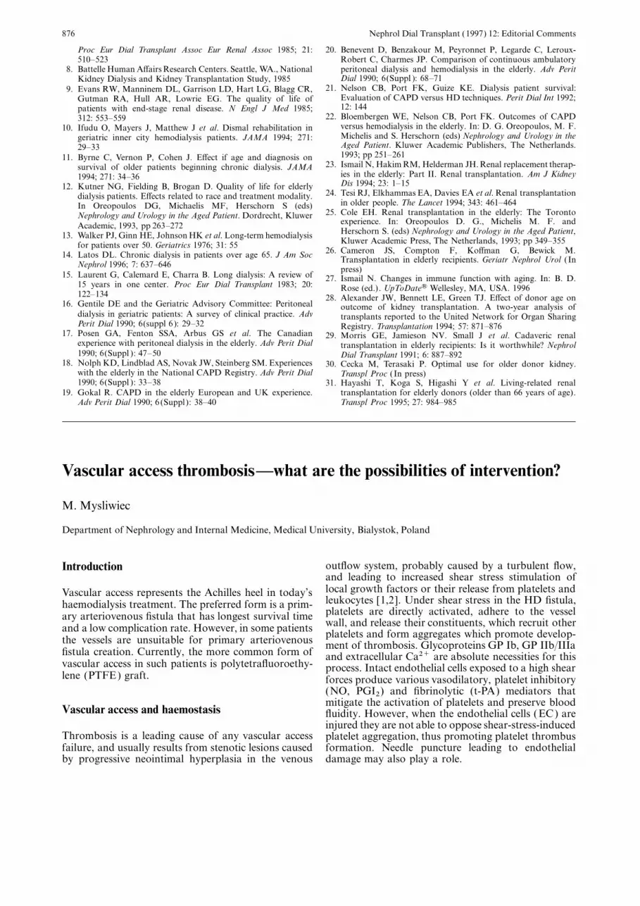
Vascular access thrombosis—what are the possibilities of intervention?
M. Mysliwiec
Department of Nephrology and Internal Medicine, Medical University, Bialystok, Poland
outflow system, probably caused by a turbulent flow,Introductionand leading to increased shear stress stimulation oflocal growth factors or their release from platelets andVascular access represents the Achilles heel in today’sleukocytes [1,2]. Under shear stress in the HD fistula,haemodialysis treatment. The preferred form is a prim-platelets are directly activated, adhere to the vesselary arteriovenous fistula that has longest survival timewall, and release their constituents, which recruit otherand a low complication rate. However, in some patients platelets and form aggregates which promote develop-the vessels are unsuitable for primary arteriovenous ment of thrombosis. Glycoproteins GP Ib, GP IIb/IIIa
fistula creation. Currently, the more common form of and extracellular Ca2+ are absolute necessities for thisvascular access in such patients is polytetrafluoroethy- process. Intact endothelial cells exposed to a high shearlene (PTFE) graft. forces produce various vasodilatory, platelet inhibitory
(NO, PGI2) and fibrinolytic (t-PA) mediators thatmitigate the activation of platelets and preserve blood
Vascular access and haemostasis fluidity. However, when the endothelial cells (EC) areinjured they are not able to oppose shear-stress-induced
Thrombosis is a leading cause of any vascular access platelet aggregation, thus promoting platelet thrombusfailure, and usually results from stenotic lesions caused formation. Needle puncture leading to endothelial
damage may also play a role.by progressive neointimal hyperplasia in the venous

Nephrol Dial Transplant (1997) 12: Editorial Comments 877
An obstruction to venous outflow typically occurs Is there evidence for genetic predisposition?within 5–6 cm of the arteriovenous anastomosis in afistula and within 2–3 cm of the venous anastomosis Resistance to activated protein C (APC) caused by aof the graft. It is more proximal in 15–20% of cases. single point mutation in the factor V gene (factor VHypertrophied venous valves and sites of intimal Leiden) was recently identified as a thrombophilictrauma from old temporary catheters in central veins defect which is strongly associated with deep venousmay lead to their stenoses and disturb outflow of blood thrombosis and recurrent idiopathic thromboembol-from the distal access. ism. However, a heterozygous carrier status for
factor V Leiden does not appear to be a risk factorfor vascular access thrombosis in haemodialysisCan one identify the patient at high risk? patients, possibly due to a high functional APC activ-ity, which is very close to or within the normal range
There is a higher risk of blood access failure in patients [6]. It cannot, however, be excluded that a homozygouswith severe vascular endothelial injury, a useful marker factor V mutation represents an increased risk forof which is a high plasma thrombomodulin [3]. shunt thrombosis.Predisposing factors are diabetes mellitus, hypotension,hypoalbuminaemia, anticardiolipin antibodies, hyper-
Thrombosis of vascular access—non-surgicalhomocysteinaemia, and increased serum levels oflipoprotein (a). options
In a 2-year, cross-sectional, prospective pilot studyin 30 non-diabetic haemodialysis patients with primary A treatment of access dysfunction requires a combinedarteriovenous fistula in 11 (37%), a progressive stenosis medical, surgical, and radiologic approach. The prim-of the venous circuit, complicated by thrombosis in ary non-surgical therapy is angioplasty. Thrombolyticthree of them was found [1]. Compared with 19 therapy may be tried as well. Valji et al. [7] modifiedpatients without fistula dysfunction, they had higher thrombolysis by a placement of two specially designedserum levels of monocyte chemoattractant protein-1 catheters with multiple side holes into the thrombus,(MCP-1) and interleukin-6 (Il-6), two cytokines that oriented in opposite directions. Urokinase in a dose ofregulate the proliferation of vascular smooth muscle 250 000–500 000 units is then injected in small pulsescells (VSCM). In addition they had hyperinsulinaemia, as the catheters are advanced and withdrawn. A 98%hyperlipidaemia (high triglycerides and the ratio for success rate of that ‘pulsed-spray pharmacomechanicalcholesterol/HDL cholesterol ), and increased plasma thrombolysis’ was reported. Newer techniques arelevels of tissue plasminogen activator inhibitor (PAI-I) intravascular stents and directional atherectomy [8].and factor VII.
The role of anticoagulants and platelet inhibitorsIs there a role for serotonin and cytokines?
Anticoagulants and antiplatelet agents may be used inpatients with recurrent graft thrombosis but alwaysSerotonin (5HT) released from dense granules of plate-
lets may also stimulate thickening of the blood vessel after ruling out an anatomic cause for thrombosis. Anattempt to identification of all contributing factors towall in several ways. By amplifying the release of other
aggregatory agents, 5HT facilitates ongoing platelet thrombosis including hypercoagulable state should bepursued.aggregation and thus the release of growth factors, in
particular platelet-derived growth factor (PDGF). There are no prospective, randomized studies todemonstrate a beneficial e�ect of vitamin K antagonistsEven at low concentrations serotonin stimulates dir-
ectly the mitogenesis of vascular smooth muscle cells in preventing graft thrombosis. Of many antiplateletdrugs acetylsalicylic acid (ASA) has been most studied.in culture. In vascular smooth muscle cells it signific-
antly potentiates the mitogenesis induced by PDGF. It induces irreversible inactivation of platelet cyclo-oxygenase. Although, none of the trials achievedThe 5HT2 receptor antagonist, ketanserin, besides
lowering the arterial blood pressure, exerts a significant acceptable statistical significance, most of the studiesinvolving aspirin, alone or combined with dipyridamol,antiproliferative e�ects on vascular smooth muscle
cells [4]. Growth of vascular smooth muscle cells is displayed trends toward prevention of fistula throm-bosis [9]. Therefore if there are no contraindicationsalso promoted by thrombin and angiotensin II.
In haemodialysis patients the increased concentra- ASA may be used for prevention of both threatenedand recurrent fistula thrombosis. Enteric-coated ASAtion of IL-1 beta and TNF alpha may induce endothel-
ial cells to increase prothrombotic activities including in a dose of 150–325 mg once a day after breakfastmay be recommended.production of IL-6 and PAI-1, expression of intercellu-
lar adhesion molecule-1 ( ICAM-1) and vascular cell ASA does not inhibit ADP, thrombin, serotonin,and PAF-induced platelet aggregation. These medi-adhesion molecule-1 (VCAM-1) [5]. The enhanced
circulating levels of VCAM-1 and ICAM-1 probably ators are generated and accumulate at sites of endothel-ial injury and vascular stenoses and they may beare the result of activation of endothelial cells and
increased leukocyte/endothelial cells interactions. responsible for reduction in blood flow in these

Nephrol Dial Transplant (1997) 12: Editorial Comments878
patients. In addition cyclo-oxygenase inhibition may Don’t forget medical common senseshift arachidonate metabolism toward non-prostaglandin metabolites (e.g. lipo-oxygenases) that Prevention of fistula thrombosis should include alsomight promote intimal hyperplasia. such simple measures as keeping the patient and
Ticlopidine inhibits aggregation induced by ADP, extremity warm, avoiding hypotension, maintainingcollagen and thrombin by preventing exposure of fib- adequate hydration, discouraging smoking, avoidingrinogen receptors GP IIb/IIIa on the platelet mem- excessive pressure over needle sites postdialysis, andbrane, which is a common pathway of the platelet self control of the access several times a day.aggregation. It acts only in vivo and requires severaldays of oral administration before achieving its fullantiplatelet e�ect. Probably its platelet-bound metabol- Referencesites are active as antiplatelet agents. Additionally,ticlopidine stimulates early fibrinolysis possibly due to 1. De Marchi S, Falleti E, Giacomello R et al. Risk factor for
vascular disease and arteriovenous fistula dysfunction in hemodi-an inhibition of the release of PAI-1 from plateletsalysis patients. J Am Soc Nephrol 1996; 7: 1169–1177[10]. Ticlopidine has theoretical advantage over ASA
2. Sukhatme VP. Vascular access stenosis. Prospects for preventionas it does not inhibit PGI2 synthesis in the vascular and therapy. Kidney Int 1996; 49: 1161–1174endothelium and is an antioxidant. Its ability to inhibit 3. Taniguchi Y, Yorioka N, Oda H et al. Plasma thrombomodulin:
usefulness as a blood access failure marker in hemodialysisfibrinogen binding to platelets supports its use in allpatients. Nephron 1996; 73: 91–93patients, but especially in those who cannot take or 4. Geerling RA, de Bruin RWF, Scheringa M et al. Ketanserin
have failed to respond to ASA. It appears to be a reduces graft arteriosclerosis after allogenic aorta transplantationin rats. J Cardiovasc Pharmacol 1996; 27: 307–311useful agent for prevention of fistula thrombosis.
5. Vaziri ND, Gonzales EC, Wang J et al. Blood coagulation,Ticlopidine may be combined with ASA [11].fibrinolytic, and inhibitory proteins in end stage renal disease.The frequency of unwanted e�ects of ticlopidine is E�ect of hemodialysis. Am J Kidney Dis 1994; 23: 828–835
slightly higher than that of ASA. They include: dia- 6. Fodinger M, Mannhalter C, Pabinger I et al. Resistance toactive protein C (APC): mutation at ARG506 of coagulationrrhoea, nausea, gastrointestinal upset, bleeding, macro-factor V and vascular access thrombosis in haemodialysispapular or urticariar rash and, in about 2%,patients. Nephrol Dial Transplant 1996; 11: 668–672neutropenia. They tend to occur early, within 2–3 7. Valji K, Bookstein JJ, Roberts AC et al. Pulse-spray pharmaco-
months of the therapy. No recent study has been mechanical thrombolysis of thrombosed hemodialysis accessgrafts: long-term experience and comparison of original andconducted but several studies were performed oncurrent techniques. Am J Roentgenol 1995; 164: 1495–1500ESRD patients in the 1980s showing that statistically
8. Kovalik EC, Newman GE, Suhocki P et al. Correction of centralfewer patients su�ered haemodialysis fistula throm- venous stenoses: Use of angioplasty and vascular Wallstents.bosis in the ticlopidine-treated groups [9]. A dose of Kidney Int 1994; 45: 1171–1181
9. Antiplatelet Trialists’ Collaboration. Collaborative overview ofticlopidine is 2×250 mg during meals.randomized trial of antiplatelet therapy-II. Maintenance ofThere are some new antiplatelet agents: ticlopidinevascular graft or arterial patency by antiplatelet therapy. Br
analogues (PCR 4099 and clopidogrel ), P2T purinocep- Med J 1994; 308: 159–168tor antagonists (ARL 66096), GPIIb/IIIa antagonists 10. Gryglewski RJ, Korbut R, Swies J et al. Thrombolytic action
of ticlopidin: possible mechanism. Eur J Pharmacol 1996; 308:(MK-852, SB-214857, pentamidine) and the most61–67promising GP IIb/IIIa antibodies (7E3), but prospect- 11. Markert T, Bertsch G, Langenfeld H et al. Elektive koronare
ive clinical studies are needed to prove their e�ect- implantation eines neu entwickelten stents ohne klassische anti-koagulation. Dtsch Med Wachenschr 1996; 121: 1213–1219iveness in protection of fistula thrombosis.