hepatoprotective effects of swimming exercise against d-galactose-induced senescence rat model
TRANSCRIPT
Hindawi Publishing CorporationEvidence-Based Complementary and Alternative MedicineVolume 2013, Article ID 275431, 9 pageshttp://dx.doi.org/10.1155/2013/275431
Research ArticleHepatoprotective Effects of Swimming Exercise againstd-Galactose-Induced Senescence Rat Model
Chi-Chang Huang,1 Wen-Dee Chiang,2 Wen-Ching Huang,3 Chih-Yang Huang,4,5
Mei-Chich Hsu,6 and Wan-Teng Lin7
1 Graduate Institute of Sports Science, College of Exercise and Health Sciences, National Taiwan Sport University,Taoyuan 33301, Taiwan
2Department of Food Science, Tunghai University, 181, Section 3, Taichung Kan Road, Taichung 40704, Taiwan3Graduate Institute of Athletics and Coaching Science, National Taiwan Sport University, Taoyuan 33301, Taiwan4Graduate Institute of Basic Medical Science, China Medical University, Taichung 40402, Taiwan5Department of Health and Nutrition Biotechnology, Asia University, Taichung 41354, Taiwan6Department of Sports Medicine, Kaohsiung Medical University, Kaohsiung 80708, Taiwan7Department of Hospitality, Tunghai University, Taichung 40704, Taiwan
Correspondence should be addressed to Wan-Teng Lin; [email protected]
Received 18 February 2013; Accepted 11 May 2013
Academic Editor: Kenji Watanabe
Copyright © 2013 Chi-Chang Huang et al. This is an open access article distributed under the Creative Commons AttributionLicense, which permits unrestricted use, distribution, and reproduction in any medium, provided the original work is properlycited.
This study investigateswhether a 12-week swimming exercise training can prevent liver damage or senescence associated biomarkersin an experimental aging model in rats. Twenty-three male Sprague-Dawley rats were divided into four groups: vehicle treatmentwith sedentary control (C, 𝑛 = 6), aging induction with sedentary (A, 𝑛 = 6), vehicle treatment with swimming exercise (SW,𝑛 = 5), and aging induction with swimming exercise (A + SW, 𝑛 = 6). Rats in groups A and AS received intraperitoneal d-galactoseinjections (150mg/kg/day) for 12 weeks to induce aging. Rats in groups SW and A + SW were subjected to swimming exercisetraining for 12 weeks. Body weight, liver weight, epididymal fat mass, blood biochemistry, and liver pathology were performed atthe end of the experiment. Hepatic senescence protein markers such as 𝛽-galactosidase, p53, and p21, as well as the inflammatorymediator, IL-6, were examined.The d-galactose-treated rats exhibited increases in AST and 𝛾-GT plasma levels and 𝛽-galactosidaseprotein expression compared to the control group. Swimming exercise significantly reduced BW, epididymal fatmass, 𝛾-GT activity,and p53, p21, and IL-6 protein levels compared to the aging group.These results suggest that a 12-week swimming exercise programsuppresses senescence markers and downregulates inflammatory mediator in the liver tissues of d-galactose-induced aging rats.
1. Introduction
Chronic liver diseases are an important health issue world-wide, especially liver fibrosis [1]. Liver fibrosis results fromprophase risks including chronic liver inflammation, fattyliver, and hepatic virus infection. The obese population hashigher proportional fatty liver than the normal population.The fat accumulation in the liver causes abnormal proteinexpression of p53 and p21, which are important cell cycle reg-ulator signaling pathways for senescence- or apoptosis-asso-ciated gene expressions [2]. Fatty liver increases pro-inflammatory cytokines expression and results in steato-hepatitis, which can easily induce further liver fibrosis and
cirrhosis processes. Currently, exercise is not only an integralcomponent of a healthy lifestyle at weight loss, but alsoan independent benefit of exercise in non-alcoholic fattyliver disease and age-related loss in physical function in theelderly population has been demonstrated [3, 4]. Given thepaucity of current treatment options on fatty liver and aging,exercise intervention could be expected to provide a low-costtherapy for disorders characterized by fatty liver and agingpopulation.
d-Galactose is a kind of restoration hexoses that can pro-vide energy and substances for the physiological require-ments of macromolecular synthesis. d-galactose can react
2 Evidence-Based Complementary and Alternative Medicine
with the amine groups in free amino acids to cause nonen-zymatic glycation, resulting in the accumulation of largeadvanced glycated protein end-products (AGE) [5]. TheseAGEs could combine with the receptors for advanced glyca-tion end-products (RAGE) expressed on the peripheral vas-cular surface as well as internal organs such as the lungs, liver,and kidneys. This could induce tissue senescence, diabetes-associated inflammation, and immune system imbalance [6].Excessive d-galactose administration produces a large num-ber of free radicals, such as superoxide anions ( ∙O
2
−), reac-tive oxygen species (ROS), for example, hydrogen peroxide[7]. This oxidative stress causes mitochondrial dysfunctionand destroys the cell structure and eventually the collapse ofantioxidant mechanisms. Oxidative stress is the main factorthat causes aging and even may shorten animal lifespan[8]. In d-galactose induced aging models the animals willproduce oxidative stress, inflammation and malondialde-hyde as well as a decrease in the total antioxidation statusand superoxide dismutase [9, 10]. In previous studies, thisd-galactose-induced aging model triggered the brain to re-duce learning and memory [11] exhibited neuronal damage[12], as well as aging damage to the cardiovascular system,kidneys, and liver [9, 13, 14].
Oxidative pressure causes DNA lesions, p53 activation,and related genes expression is the main risk causing cellularsenescence [15]. Cellular senescence is positively related toorgan aging [16]. In liver tissues, cellular senescence couldresult ultimately in cirrhosis [17]. In the senescence pro-cess, several regulators arrest cell proliferation. p53 playsan important role in maintaining DNA integrity and keepsthe cell cycle in the G1 phase [18]. p53 could be highlyexpressed under irradiation or free radical pressure to arrestthe cell cycle from repairing DNA. If the repair systemcannot completely fix mutations, the cells will be pushedinto apoptosis [19]. p53 can activate the downstream p21protein which inhibits the cyclin-CDK complex to stop cellproliferation [20].
The 𝛽-galactosidase (𝛽-gal) gene has higher expressionas well as the associated enzyme activity in aged cells [21].This important indicator has a 3- to 5-fold higher expres-sion in aged cells than in preaged cells. Studies have foundthat the liver tissue of patients with cirrhosis of the livercontains large amounts of 𝛽-gal [17]. 𝛽-Gal is often usedas a biochemical marker to identify cells in the agingstage. Previous studies demonstrated that d-galactose couldinduce aging by increasing oxidative stress and reducingantioxidant defense mechanisms in vivo, causing liver agingand functional decline [22]. However, no studies have todate focused on the aging indicator, 𝛽-gal protein expression,or enzymatic activity in the liver under the d-galactose-induced aging model. Therefore, this study investigates thesenescence marker protein levels associated with liver tissuesand the preventive effects of regular exercise intervention in ad-galactose-induced aging rat model.
2. Materials and Methods
2.1. Chemicals and Antibodies. d-Galactose was purchasedfrom Sigma-Aldrich Chemical Co. (St. Louis, MO, USA).
Primary antibodies against 𝛽-gal from Abcam (MA, USA)were used inWestern blotting. All other antibodies includingp53, p21, IL-6, 𝛼-tubulin and second antibodies, and inter-nal control 𝛼-tubulin were from Santa Cruz Biotechnology(Santa Cruz, CA, USA).
2.2. Animals, Treatment, and Swimming Protocol. Seven-week-old male Sprague-Dawley rats were purchased fromBioLASCO (A Charles River Licensee Corporation, Yi-Lan,Taiwan). All animal experiments conformed to the guide-lines of the Institutional Animal Care and Use Committee(IACUC) of Tunghai University. This study was conductedunder the IACUC-98-27 protocol and approved by theIACUC ethics committee. The rats were raised for one-weekbefore the experiments to adapt to the environment anddiet. All animals were given a standard laboratory diet (no.5001; PMI Nutrition International, Brentwood, MO, USA)and distilled water ad libitum and individually housed in aroom maintained at 24 ± 2∘C and humidity 55 ± 10% with a12-h light-dark cycle during the one-week adaptation period.Afterward, the rats were randomly divided into four groups:(1) vehicle treatment with sedentary control (C, 𝑛 = 6), (2)aging inductionwith sedentary control (A, 𝑛 = 6), (3) vehicletreatment with swimming exercise (SW, 𝑛 = 5), and (4)aging induction with a 12-week swimming exercise (A + SW,𝑛 = 6). The d-galactose was administrated by intraperitoneal(i.p.) injection with 150mg/kg BW to groups A and A + SW.Equal volumes of vehicle (0.9% saline) were i.p. administeredto groups C and SW. The d-galactose i.p. injections wereadministrated every day for 12 weeks to accelerate senescenceinduction. The swimming exercise protocol was conducted5 times/week for 60min/time for 12-week duration as previ-ously described with some modifications [23].The rats swamindividually in a water sink with an area of 60 × 90 cm, 50 cmdepth, with the water temperature kept at 35 ± 1∘C.
2.3. Immunoblotting. Western blot analysis followed a pre-vious report [24]. Protein content was measured using theBradford method (Bio-Rad). Proteins were resolved using 5–20% gradient SDS-PAGE and then immunoblotted using anenhanced chemiluminescence assay (ECL; Perkin Elmer LifeScience, Inc., USA) and image retrieval using the Fuji LAS-4000. The intensities were quantified using Alpha Ease FCsoftware (Alpha Innotech software Inc., CA, USA).
2.4. TUNEL Assay. Terminal deoxynucleotidyl transferase-mediated dUTPnick end labeling (TUNEL) assaywas used todetect in situ apoptotic cells according to the manufacturer’sprotocol (Chemicon/Millipore, Temecula, CA, USA). Afterdeparaffinizing and rehydration pretreatments, the sectionswere immersed in 0.3% H
2
O2
for 30 minutes at roomtemperature to inactivate endogenous peroxidase activity.After rinsing with PBS, the sections were incubated withProteinase K (2mg/mL)/Tris buffer (10mM, pH = 8) so-lution at room temperature for 15min to enhance the per-meability. After rinsing with PBS two times the sections wereincubated in 0.1% Triton-100 (0.1% Sodium citrate) 4∘ for8 minutes and then incubated with blocking buffer (Tris-HCl 0.1M pH 7.5, 3% BSA and 20% normal bovine serum)
Evidence-Based Complementary and Alternative Medicine 3
for 1 h. The sections were rinsed in PBS twice, immersedin TUNEL Buffer (enzyme solution and label solution, 1 : 9)with 37∘C in the dark and rinsed again with PBS. Sampleswere counterstained with DAPI to determine the nucleilocalization.
2.5. Determination of Blood Biochemistry. Blood sampleswere collected from the rat abdominal aortas with indicatedtreatments after 12 weeks. The plasma was prepared bycentrifugation at 1,500×g for 15min at 4∘C. The biochemicalvariables for liver injury indicators including AST, ALT, and𝛾-GT activities were analyzed by an autoanalyzer (Hitachi7060, Hitachi, Tokyo, Japan).
2.6. Gross and Histological Liver Evaluation. Liver tissueswere fixed in 10% formalin, then embedded in paraffin, andcut into 4 𝜇m thick slices. Tissue sections were stained withHematoxylin and Eosin (H&E) or Masson trichrome andexamined using a light microscope equipped with a CCDcamera (BX-51, Olympus, Tokyo, Japan) as per our previousreport [24].
2.7. Statistical Analysis. All data are represented as mean ±SEM. To evaluate the differences among the groups studied,data were analyzed using one-way ANOVA with the Statisti-cal Analysis System (SAS Institute, Cary, NC, USA). 𝑃 < 0.05was considered statistically significant.
3. Results
3.1. BodyWeight, Epididymal Fat Pad Mass, and Liver Weight.Morphological data from each experimental group are sum-marized in Table 1. There were no significant differences inthe initial body and liws thedafdfverweights among groupsC,A, SW and A + SW. Furthermore, d-galactose did not causesignificant changes in the final body, liver and epididymalfat pad weights, compared to the control group. After 12-weeks swimming training there was a significant decreasein the final body weight in the SW and A + SW groups, by11.5% (𝑃 = 0.0460) and 11.8% (𝑃 = 0.0331), respectively,compared to group A. There was also a significant decreasein the epididymal fat pad mass in the SW and A + SW groupsby 47.0% (𝑃 = 0.0119) and 43.2% (𝑃 = 0.0180), respectively,compared to group A. We also observed that there was onlya slight decrease in the liver weight in the A + SW group by17.8% (𝑃 = 0.0558), compared to group A.
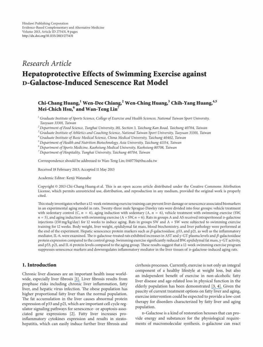
3.2. Swimming Exercise Protects Rats against d-Galactose-Induced Liver Dysfunction. Figure 1 shows that the plasmaAST activity in the groups C, A, SW, and A + SWwere 88.4 ±2.7, 120.0 ± 15.4, 108.4 ± 12.7, and 109.3 ± 7.0, respectively.There was only a significant increase in the AST level in groupA by 1.4-fold (𝑃 = 0.0421), compared to the control group.TheplasmaALT activity in groupsC,A, SW, andA+SWwere42.3 ± 2.5, 47.3 ± 2.5, 39.5 ± 1.2, and 41.1 ± 2.6, respectively.There was only a significant decrease in the ALT level in theSW group by 16.4% (𝑃 = 0.0351) and a slight decrease in the
Table 1: General characteristics of the experimental groups.
Characteristics C A SW A + SWInitial BW (g) 234 ± 3 232 ± 4 234 ± 4 235 ± 2Final BW (g) 527 ± 17ab 549 ± 20b 486 ± 30a 484 ± 15a
Epididymalfat pad (g) 4.60 ± 0.49b 5.25 ± 0.47b 2.78 ± 0.43a 2.98 ± 0.39a
Liver (g) 13.2 ± 0.6 13.9 ± 0.9 12.6 ± 1.2 11.7 ± 0.4Values aremeans± SEM.C: vehicle treatmentwith sedentary control (𝑛 = 6);andA: aging inductionwith sedentary control (𝑛 = 6); SW: vehicle treatmentwith swimming exercise (𝑛 = 5); A + SW: aging induction with a 12-weekswimming exercise (𝑛 = 6). Data represent mean ± SEM. Values that havea different superscript letter (a, b) differ significantly with each other (𝑃 <0.05).
A + SW group by 13.1% (𝑃 = 0.0729), respectively, comparedto group A.
The plasma 𝛾-GT activity in groups C, A, SW, and A +SWwere 0.92 ± 0.13, 2.00 ± 0.16, 1.12 ± 0.23, and 1.47 ± 0.11,respectively. Group A showed a significant increase in the 𝛾-GT level by 2.2-fold (𝑃 < 0.0001), compared to the controlgroup. After 12-week swimming training, we found that theplasma 𝛾-GT levels significantly decreased in groups SW andA + SW by 44.0% (𝑃 = 0.0010) and 26.7% (𝑃 = 0.0234),respectively, compared to group A.
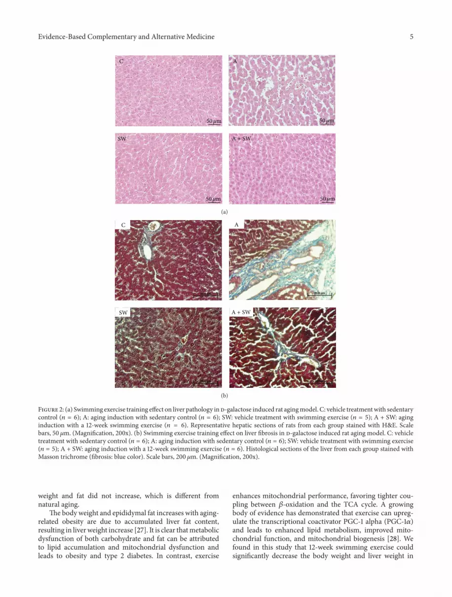
The histology of hepatic structure is shown in Figure 2(a).To evaluate whether d-galactose treatment could induceaging-like changes in the liver, the pathology examinationof liver tissues from rats in each group was measured. Thehepatic cords arranged loosely; dilatation of sinusoid, hepa-tocytes vacuolation, and multi focal necrosis were observedin the d-galactose-treated animals compared to those inthe control group. According to the grading and score oflesions, these pathological changes were almost completelyameliorated by 12-weeks swimming training (Table 2).
3.3. Swimming Exercise Protects Rats against d-Galactose-Induced Liver Fibrosis. d-Galactose also caused significantinterstitial collagen deposition, as demonstrated by Mas-son’s trichrome staining. This fibrosis was also significantlyreduced in d-galactose-treated rats subjected to swimmingtraining for 12 weeks (Figure 2(b) and Table 2).
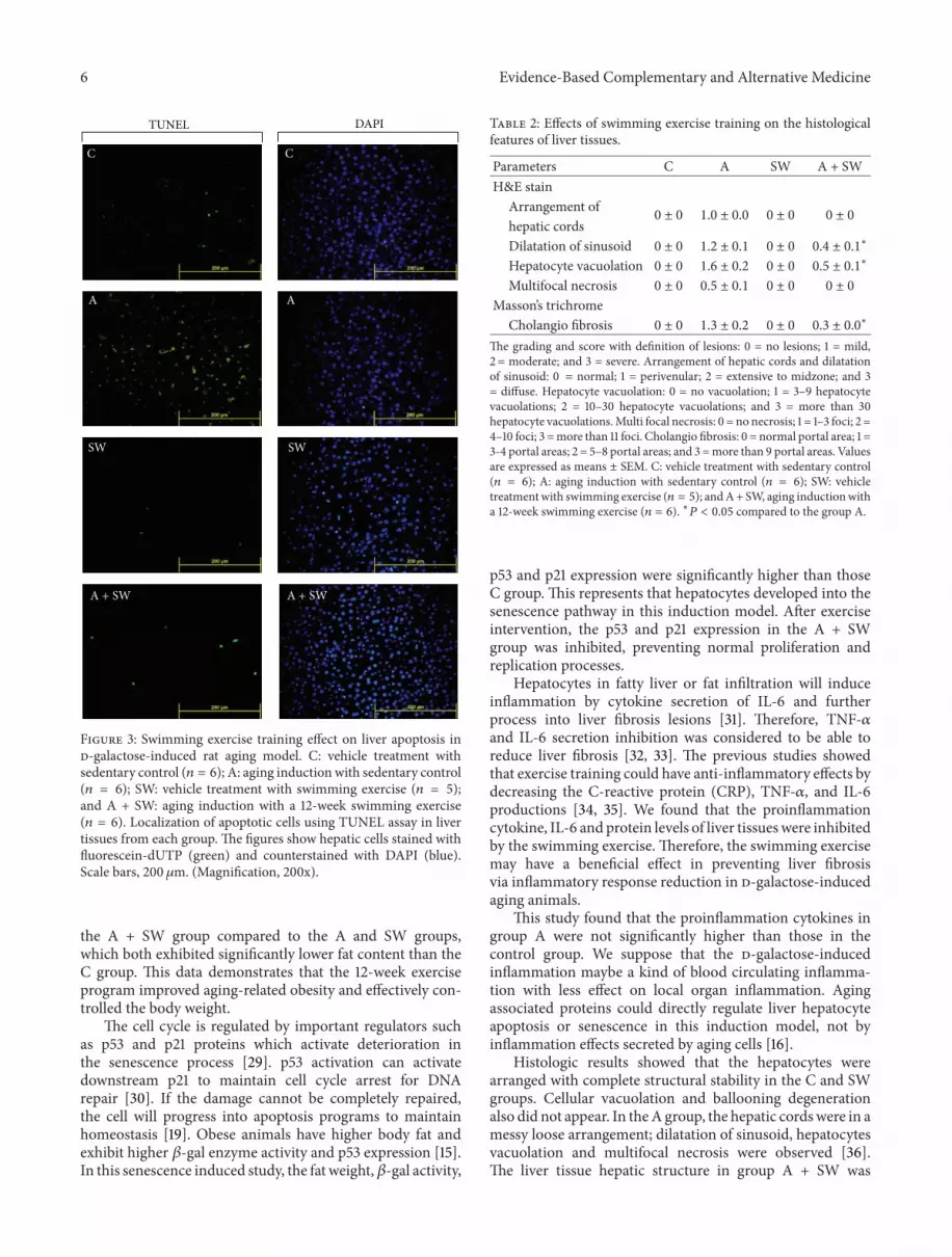
3.4. Swimming Exercise Protects Rats against d-Galactose-Induced Liver Apoptosis. As shown in Figure 3, d-galactosecaused significant apoptosis in the rat liver tissues, as demon-strated by TUNEL assay, compared with the control group(C). This apoptosis was also significantly reduced in d-galactose-treated rats subjected to swimming training for 12weeks.
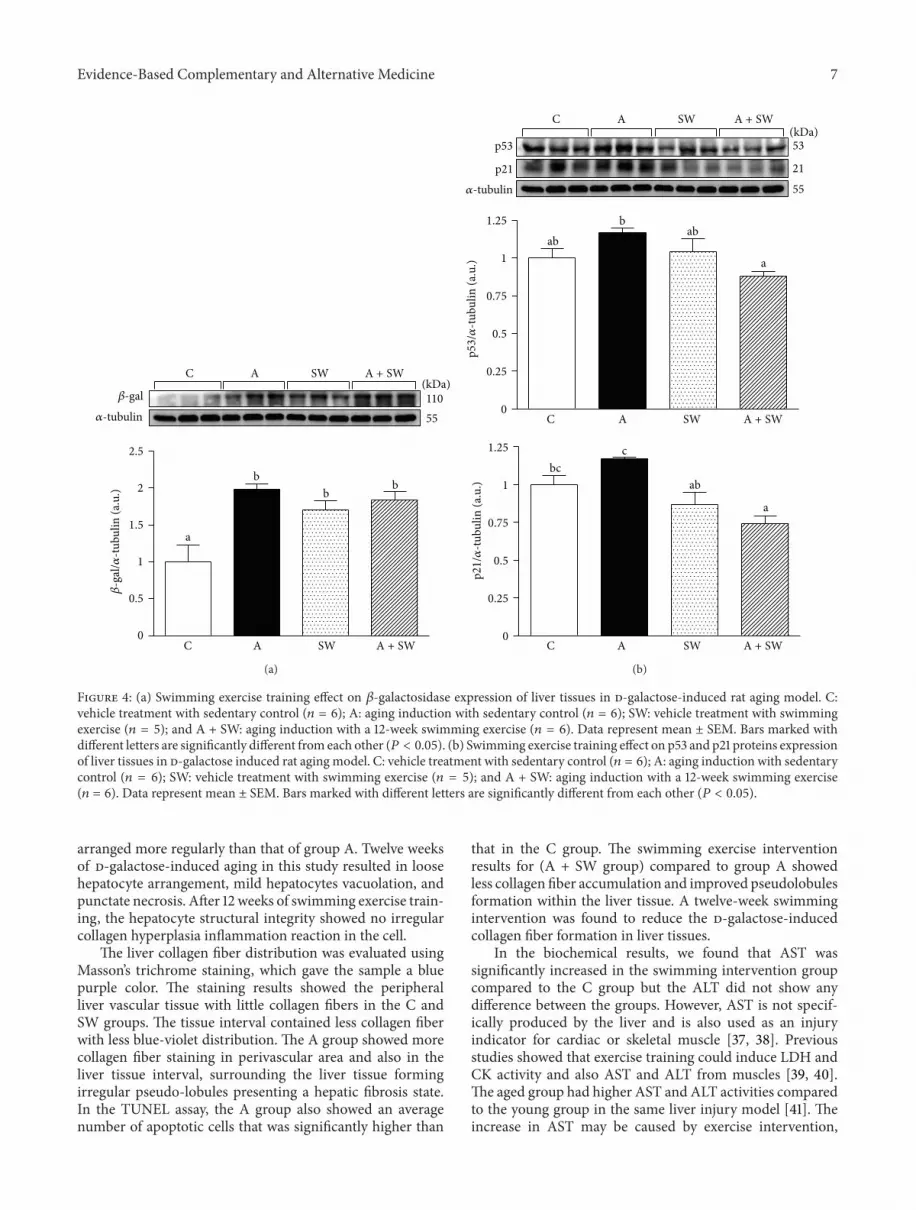
3.5. Effects of Swimming Exercise on Protein Expression ofHepatic 𝛽-Gal, p53, p21, and IL-6 in d-Galactose-Treated Rats.Figure 4(a) shows that the hepatic 𝛽-gal protein expression inthe groups C, A, SW, and A + SWwere 1.00±0.23, 1.98±0.08,1.70 ± 0.13, and 1.84 ± 0.11, respectively. We found that the𝛽-gal levels of liver tissues significantly increased in groups A(𝑃 = 0.0014), SW (𝑃 = 0.0093) and A + SW (𝑃 = 0.0035) by
4 Evidence-Based Complementary and Alternative Medicine
150
125
75
0
50
25
100
C A SW A + SW
AST
(U/L
)b
a
abab
(a)
60
50
30
0
20
10
40
C A SW A + SW
ALT
(U/L
)
b
aab ab
(b)
2.5
2
1.5
0
1
0.5
𝛾-G
T (U
/L)
c
C A SW A + SW
b
a
ab
(c)
Figure 1: Swimming exercise training effect on plasma AST (a), ALT (b), and 𝛾-GT (c) enzymatic activity in d-galactose-induced rat agingmodel. C: vehicle treatment with sedentary control (𝑛 = 6); A: aging induction with sedentary control (𝑛 = 6); SW: vehicle treatment withswimming exercise (𝑛 = 5); and A + SW: aging induction with a 12-week swimming exercise (𝑛 = 6). Data represent mean ± SEM. Barsmarked with different letters are significantly different from each other (𝑃 < 0.05).
2.0-, 1.7- and 1.8-fold, respectively, as compared to the groupC.
Figure 4(b) shows that the hepatic p53 protein expressionin groups C, A, SW, and A + SW were 1.00 ± 0.06, 1.17 ±0.03, 1.04 ± 0.09, and 0.88 ± 0.03, respectively. There was aslight increase in the p53 level in the A group compared tothe control group (𝑃 = 0.0842). After 12-week swimmingtraining we found a significant decrease in the p53 expressionin liver tissues in groups A + SW by 24.9% (𝑃 = 0.0090)compared to group A. The hepatic p21 protein expressionin groups C, A, SW, and A + SW were 1.00 ± 0.06, 1.17 ±0.01, 0.87 ± 0.08, and 0.74 ± 0.05, respectively. After 12-weekswimming training there was a significant decrease in the p21expression in liver tissues in groups SW, andA+ SWby 25.3%(𝑃 = 0.0065) and 36.8% (𝑃 = 0.0007), respectively, comparedto group A.
As shown in Figure 5, the hepatic IL-6 protein expressionin the groups C, A, SW, and A + SW were 1.00 ± 0.06, 1.00 ±0.02, 0.71 ± 0.07, and 0.73 ± 0.10, respectively. After 12-weekswimming trainingwe found a significant decrease in the IL-6expression in liver tissues in groups SWandA+ SWby 28.6%(𝑃 = 0.0182) and 27.4% (𝑃 = 0.0225), respectively, comparedto groups C and A.
4. Discussion
𝛽-Gal has higher enzyme activity and expression in agedorgans, and it is also an important indicator in aged cells[21]. The d-galactose-induced aging model could increasethe oxidative pressure and inflammation, causing senescenceinjury. This senescence-induced model could result in adecline in cognitive function in the brain and cardiovasculardamage [11, 13], but there are no studies focused on theliver 𝛽-gal activity on long-term d-galactose administration.This study showed a 12-week d-galactose induction of Aand A + SW groups could significantly induced higher 𝛽-galexpression, which means that liver is in senescence status.
An increase in body fat often occurs in old age. Theobese have a higher chance of suffering from nonalcoholicsteatohepatitis than those with standard weight [25]. There-fore, obesity with age increases the risk for liver fibrosis.However, comparison between the C and A groups showedno significant difference in body weight and epididymal fatat the end of the experiment. The occurrence of naturalaging could be regulated by many factors or physiologicaleffects [26], including glycation oxidative injury from this d-galactose induction model. This may explain why the body
Evidence-Based Complementary and Alternative Medicine 5
C
SW
A
50𝜇m50𝜇m
50𝜇m 50𝜇m
A + SW
(a)
C A
SW A + SW
(b)
Figure 2: (a) Swimming exercise training effect on liver pathology in d-galactose induced rat agingmodel. C: vehicle treatmentwith sedentarycontrol (𝑛 = 6); A: aging induction with sedentary control (𝑛 = 6); SW: vehicle treatment with swimming exercise (𝑛 = 5); A + SW: aginginduction with a 12-week swimming exercise (𝑛 = 6). Representative hepatic sections of rats from each group stained with H&E. Scalebars, 50 𝜇m. (Magnification, 200x). (b) Swimming exercise training effect on liver fibrosis in d-galactose induced rat aging model. C: vehicletreatment with sedentary control (𝑛 = 6); A: aging induction with sedentary control (𝑛 = 6); SW: vehicle treatment with swimming exercise(𝑛 = 5); A + SW: aging induction with a 12-week swimming exercise (𝑛 = 6). Histological sections of the liver from each group stained withMasson trichrome (fibrosis: blue color). Scale bars, 200 𝜇m. (Magnification, 200x).
weight and fat did not increase, which is different fromnatural aging.
The body weight and epididymal fat increases with aging-related obesity are due to accumulated liver fat content,resulting in liverweight increase [27]. It is clear thatmetabolicdysfunction of both carbohydrate and fat can be attributedto lipid accumulation and mitochondrial dysfunction andleads to obesity and type 2 diabetes. In contrast, exercise
enhances mitochondrial performance, favoring tighter cou-pling between 𝛽-oxidation and the TCA cycle. A growingbody of evidence has demonstrated that exercise can upreg-ulate the transcriptional coactivator PGC-1 alpha (PGC-1𝛼)and leads to enhanced lipid metabolism, improved mito-chondrial function, and mitochondrial biogenesis [28]. Wefound in this study that 12-week swimming exercise couldsignificantly decrease the body weight and liver weight in
6 Evidence-Based Complementary and Alternative Medicine
TUNEL DAPI
C C
A A
SW SW
A + SWA + SW
Figure 3: Swimming exercise training effect on liver apoptosis ind-galactose-induced rat aging model. C: vehicle treatment withsedentary control (𝑛 = 6); A: aging induction with sedentary control(𝑛 = 6); SW: vehicle treatment with swimming exercise (𝑛 = 5);and A + SW: aging induction with a 12-week swimming exercise(𝑛 = 6). Localization of apoptotic cells using TUNEL assay in livertissues from each group. The figures show hepatic cells stained withfluorescein-dUTP (green) and counterstained with DAPI (blue).Scale bars, 200 𝜇m. (Magnification, 200x).
the A + SW group compared to the A and SW groups,which both exhibited significantly lower fat content than theC group. This data demonstrates that the 12-week exerciseprogram improved aging-related obesity and effectively con-trolled the body weight.
The cell cycle is regulated by important regulators suchas p53 and p21 proteins which activate deterioration inthe senescence process [29]. p53 activation can activatedownstream p21 to maintain cell cycle arrest for DNArepair [30]. If the damage cannot be completely repaired,the cell will progress into apoptosis programs to maintainhomeostasis [19]. Obese animals have higher body fat andexhibit higher 𝛽-gal enzyme activity and p53 expression [15].In this senescence induced study, the fat weight,𝛽-gal activity,
Table 2: Effects of swimming exercise training on the histologicalfeatures of liver tissues.
Parameters C A SW A + SWH&E stain
Arrangement of0 ± 0 1.0 ± 0.0 0 ± 0 0 ± 0
hepatic cordsDilatation of sinusoid 0 ± 0 1.2 ± 0.1 0 ± 0 0.4 ± 0.1∗
Hepatocyte vacuolation 0 ± 0 1.6 ± 0.2 0 ± 0 0.5 ± 0.1∗
Multifocal necrosis 0 ± 0 0.5 ± 0.1 0 ± 0 0 ± 0
Masson’s trichromeCholangio fibrosis 0 ± 0 1.3 ± 0.2 0 ± 0 0.3 ± 0.0
∗
The grading and score with definition of lesions: 0 = no lesions; 1 = mild,2 = moderate; and 3 = severe. Arrangement of hepatic cords and dilatationof sinusoid: 0 = normal; 1 = perivenular; 2 = extensive to midzone; and 3= diffuse. Hepatocyte vacuolation: 0 = no vacuolation; 1 = 3–9 hepatocytevacuolations; 2 = 10–30 hepatocyte vacuolations; and 3 = more than 30hepatocyte vacuolations.Multi focal necrosis: 0 = no necrosis; 1 = 1–3 foci; 2 =4–10 foci; 3 =more than 11 foci. Cholangio fibrosis: 0 = normal portal area; 1 =3-4 portal areas; 2 = 5–8 portal areas; and 3 =more than 9 portal areas. Valuesare expressed as means ± SEM. C: vehicle treatment with sedentary control(𝑛 = 6); A: aging induction with sedentary control (𝑛 = 6); SW: vehicletreatmentwith swimming exercise (𝑛 = 5); andA+ SW, aging inductionwitha 12-week swimming exercise (𝑛 = 6). ∗𝑃 < 0.05 compared to the group A.
p53 and p21 expression were significantly higher than thoseC group. This represents that hepatocytes developed into thesenescence pathway in this induction model. After exerciseintervention, the p53 and p21 expression in the A + SWgroup was inhibited, preventing normal proliferation andreplication processes.
Hepatocytes in fatty liver or fat infiltration will induceinflammation by cytokine secretion of IL-6 and furtherprocess into liver fibrosis lesions [31]. Therefore, TNF-𝛼and IL-6 secretion inhibition was considered to be able toreduce liver fibrosis [32, 33]. The previous studies showedthat exercise training could have anti-inflammatory effects bydecreasing the C-reactive protein (CRP), TNF-𝛼, and IL-6productions [34, 35]. We found that the proinflammationcytokine, IL-6 and protein levels of liver tissueswere inhibitedby the swimming exercise. Therefore, the swimming exercisemay have a beneficial effect in preventing liver fibrosisvia inflammatory response reduction in d-galactose-inducedaging animals.
This study found that the proinflammation cytokines ingroup A were not significantly higher than those in thecontrol group. We suppose that the d-galactose-inducedinflammation maybe a kind of blood circulating inflamma-tion with less effect on local organ inflammation. Agingassociated proteins could directly regulate liver hepatocyteapoptosis or senescence in this induction model, not byinflammation effects secreted by aging cells [16].
Histologic results showed that the hepatocytes werearranged with complete structural stability in the C and SWgroups. Cellular vacuolation and ballooning degenerationalso did not appear. In theA group, the hepatic cordswere in amessy loose arrangement; dilatation of sinusoid, hepatocytesvacuolation and multifocal necrosis were observed [36].The liver tissue hepatic structure in group A + SW was
Evidence-Based Complementary and Alternative Medicine 7
11055
2.5
2
1.5
0
1
0.5
b
a
bb
𝛽-g
al/𝛼
-tubu
lin (a
.u.)
𝛽-gal
𝛼-tubulin
C A SW A + SW
C A SW A + SW(kDa)
(a)
53
55
21
1.25
1
0
0.5
0.75
0.25
1.25
1
0
0.5
0.25
0.75
p53
/𝛼-tu
bulin
(a.u
.)p2
1/𝛼
-tubu
lin (a
.u.)
p53
𝛼-tubulin
p21
C A SW A + SW
C A SW A + SW
C A SW A + SW(kDa)
b
c
a
abab
a
abbc
(b)
Figure 4: (a) Swimming exercise training effect on 𝛽-galactosidase expression of liver tissues in d-galactose-induced rat aging model. C:vehicle treatment with sedentary control (𝑛 = 6); A: aging induction with sedentary control (𝑛 = 6); SW: vehicle treatment with swimmingexercise (𝑛 = 5); and A + SW: aging induction with a 12-week swimming exercise (𝑛 = 6). Data represent mean ± SEM. Bars marked withdifferent letters are significantly different from each other (𝑃 < 0.05). (b) Swimming exercise training effect on p53 and p21 proteins expressionof liver tissues in d-galactose induced rat aging model. C: vehicle treatment with sedentary control (𝑛 = 6); A: aging induction with sedentarycontrol (𝑛 = 6); SW: vehicle treatment with swimming exercise (𝑛 = 5); and A + SW: aging induction with a 12-week swimming exercise(𝑛 = 6). Data represent mean ± SEM. Bars marked with different letters are significantly different from each other (𝑃 < 0.05).
arranged more regularly than that of group A. Twelve weeksof d-galactose-induced aging in this study resulted in loosehepatocyte arrangement, mild hepatocytes vacuolation, andpunctate necrosis. After 12 weeks of swimming exercise train-ing, the hepatocyte structural integrity showed no irregularcollagen hyperplasia inflammation reaction in the cell.
The liver collagen fiber distribution was evaluated usingMasson’s trichrome staining, which gave the sample a bluepurple color. The staining results showed the peripheralliver vascular tissue with little collagen fibers in the C andSW groups. The tissue interval contained less collagen fiberwith less blue-violet distribution. The A group showed morecollagen fiber staining in perivascular area and also in theliver tissue interval, surrounding the liver tissue formingirregular pseudo-lobules presenting a hepatic fibrosis state.In the TUNEL assay, the A group also showed an averagenumber of apoptotic cells that was significantly higher than
that in the C group. The swimming exercise interventionresults for (A + SW group) compared to group A showedless collagen fiber accumulation and improved pseudolobulesformation within the liver tissue. A twelve-week swimmingintervention was found to reduce the d-galactose-inducedcollagen fiber formation in liver tissues.
In the biochemical results, we found that AST wassignificantly increased in the swimming intervention groupcompared to the C group but the ALT did not show anydifference between the groups. However, AST is not specif-ically produced by the liver and is also used as an injuryindicator for cardiac or skeletal muscle [37, 38]. Previousstudies showed that exercise training could induce LDH andCK activity and also AST and ALT from muscles [39, 40].The aged group had higher AST and ALT activities comparedto the young group in the same liver injury model [41]. Theincrease in AST may be caused by exercise intervention,

8 Evidence-Based Complementary and Alternative Medicine
55
IL-621.528
bb
a a
1.25
1
0
0.5
0.25
0.75
C A SW A + SW
C A SW A + SW
𝛼-tubulin
(kDa)
IL-6
/𝛼-tu
bulin
(a.u
.)
Figure 5: Swimming exercise training effect on IL-6 expressionin liver tissues in d-galactose induced rat aging model. C: vehicletreatment with sedentary control (𝑛 = 6); A: aging induction withsedentary control (𝑛 = 6); SW: vehicle treatment with swimmingexercise (𝑛 = 5); A + SW: aging induction with a 12-week swimmingexercise (𝑛 = 6). Data represent mean ± SEM. Bars marked withdifferent letters are significantly different fromeach other (𝑃 < 0.05).
not by liver disease including inflammation, fatty liver, orfibrosis.The 𝛾-GT activity in this senescence inductionmodelwas significantly increased after 12 weeks of d-galactoseinduction, and the increase in 𝛾-GT activity is a risk factorfor chronic liver disease formation.
In the results from this d-galactose-induced, senescencerat model showed that the liver could be induced to expressthe senescence indicator, 𝛽-gal, and pathological appearanceof liver specimens from d-galactose-treated animals. Theswimming exercise intervention could prevent the liverpathological changes associated with age-related liver injury,as well as the inflammatory cytokine and cell cycle-associatedregulators involved in senescence progression. In a futurestudy, we could investigate the preventive or therapeuticeffects of different types of exercise using this chemical-induced aged rat model. Many factors, both extrinsic andintrinsic, cause different tissues to age; therefore, the naturalsenescence process should be a more reliable model to studythe preventive or therapeutic effects of exercise intervention.In parallel, more research is also needed to better understandthe different markers affected by the specific aging tissueprogressions and the role of exercise in the management oftarget tissue senescence.
Funding
This Research Grant was funded by the National ScienceCouncil, Taiwan (NSC99-2410-H029-059-MY2) and an Insti-tutional Grant from Tunghai University for Global Re-search and Education on Environment and Society (GREEnS004-3), Taiwan.
Conflict of Interests
All authors have no conflict of interests with respect to thedata collected and procedures used within this study.
Authors’ Contribution
C.-C. Huang and M.-C. Hsu share the first authorship.
Acknowledgments
The authors thank Miss Yu-Wei Lin for her help in animalexperiments. The authors are grateful to veterinarian Dr.Chien-Chao Chiu for his expert assistance in histologicalexamination. They also thank Joel A. Newson for his Englishlanguage editing of this paper.
References
[1] R. Bataller andD. A. Brenner, “Liver fibrosis,” Journal of ClinicalInvestigation, vol. 115, no. 2, pp. 209–218, 2005.
[2] T. Minamino, M. Orimo, I. Shimizu et al., “A crucial role foradipose tissue p53 in the regulation of insulin resistance,”NatureMedicine, vol. 15, no. 9, pp. 1082–1087, 2009.
[3] S. E. Keating, D. A. Hackett, J. George, and N. A. Johnson,“Exercise and non-alcoholic fatty liver disease: a systematicreview and meta-analysis,” Journal of Hepatology, vol. 57, no. 1,pp. 157–166, 2012.
[4] M. E. Cress, D. M. Buchner, K. A. Questad, P. C. Esselman, B.J. DeLateur, and R. S. Schwartz, “Exercise: effects on physicalfunctional performance in independent older adults,” Journalsof Gerontology A, vol. 54, no. 5, pp. M242–M248, 1999.
[5] C. F. Chen, S. Y. Lang, P. P. Zuo, N. Yang, X. Q. Wang, and C.Xia, “Effects of d-galactose on the expression of hippocampalperipheral-type benzodiazepine receptor and spatial memoryperformances in rats,” Psychoneuroendocrinology, vol. 31, no. 7,pp. 805–811, 2006.
[6] A. C. Kaliora and G. V. Z. Dedoussis, “Natural antioxidantcompounds in risk factors for CVD,” Pharmacological Research,vol. 56, no. 2, pp. 99–109, 2007.
[7] J. Lu, Y. L. Zheng, L. Luo, D. M. Wu, D. X. Sun, and Y. J.Feng, “Quercetin reverses d-galactose induced neurotoxicity inmouse brain,” Behavioural Brain Research, vol. 171, no. 2, pp.251–260, 2006.
[8] X. Song, M. Bao, D. Li, and Y. M. Li, “Advanced glycation in D-galactose induced mouse aging model,” Mechanisms of Ageingand Development, vol. 108, no. 3, pp. 239–251, 1999.
[9] S. H. Fan, Z. F. Zhang, Y. L. Zheng et al., “Troxerutin protectsthemouse kidney fromd-galactose-caused injury through anti-inflammation and anti-oxidation,” International Immunophar-macology, vol. 9, no. 1, pp. 91–96, 2009.
[10] Z. J. Yi, Y. R. Fu, M. Li, K. S. Gao, and X. G. Zhang, “Effectof LTA isolated from bifidobacteria on d-galactose-inducedaging,” Experimental Gerontology, vol. 44, no. 12, pp. 760–765,2009.
[11] A. Kumar, A. Prakash, and S. Dogra, “Centella asiatica attenu-ates d-galactose-induced cognitive impairment, oxidative andmitochondrial dysfunction in mice,” International Journal ofAlzheimer’s Disease, vol. 2011, Article ID 347569, 9 pages, 2011.
[12] J. Lu, D. M. Wu, Y. L. Zheng, B. Hu, and Z. F. Zhang,“Purple sweet potato color alleviates D-galactose-induced brain
Evidence-Based Complementary and Alternative Medicine 9
aging in old mice by promoting survival of neurons via PI3Kpathway and inhibiting cytochrome C-mediated apoptosis,”Brain Pathology, vol. 20, no. 3, pp. 598–612, 2010.
[13] M. Buemi, L. Nostro, C. Aloisi, V. Consentini, M. Criseo,and N. Frisina, “Kidney aging: from phenotype to genetics,”Rejuvenation Research, vol. 8, no. 2, pp. 101–109, 2005.
[14] S. C. Ho, J. H. Liu, and R. Y. Wu, “Establishment of the mimeticaging effect in mice caused by D-galactose,” Biogerontology, vol.4, no. 1, pp. 15–18, 2003.
[15] S. Furukawa, T. Fujita,M. Shimabukuro et al., “Increased oxida-tive stress in obesity and its impact on metabolic syndrome,”Journal of Clinical Investigation, vol. 114, no. 12, pp. 1752–1761,2004.
[16] U. Herbig, M. Ferreira, L. Condel, D. Carey, and J. M. Sedivy,“Cellular senescence in aging primates,” Science, vol. 311, no.5765, p. 1257, 2006.
[17] S. U. Wiemann, A. Satyanarayana, M. Tsahuridu et al., “Hepa-tocyte telomere shortening and senescence are general markersof human liver cirrhosis,” FASEB Journal, vol. 16, no. 9, pp. 935–942, 2002.
[18] K. H. Vousden and D. P. Lane, “p53 in health and disease,”Nature ReviewsMolecular Cell Biology, vol. 8, no. 4, pp. 275–283,2007.
[19] R. A. Schwartzman and J. A. Cidlowski, “Mechanism of tissue-specific induction of internucleosomal deoxyribonucleic acidcleavage activity and apoptosis by glucocorticoids,” Endocrinol-ogy, vol. 133, no. 2, pp. 591–599, 1993.
[20] M. S. Sheikh and A. J. Fornace Jr., “Role of p53 family membersin apoptosis,” Journal of Cellular Physiology, vol. 182, no. 2, pp.171–181, 2000.
[21] G. P. Dimri, X. Lee, G. Basile et al., “A biomarker that identifiessenescent human cells in culture and in aging skin in vivo,”Proceedings of the National Academy of Sciences of the UnitedStates of America, vol. 92, no. 20, pp. 9363–9367, 1995.
[22] K. V. Anand, M. S. Mohamed Jaabir, P. A. Thomas, and P.Geraldine, “Protective role of chrysin against oxidative stressin D-galactose-induced aging in an experimental rat model,”Geriatrics & Gerontology International, vol. 12, no. 4, pp. 741–750, 2012.
[23] K. J. Hart, J. M. Shaw, E. Vajda, M. Hegsted, and S. C. Miller,“Swim-trained rats have greater bone mass, density, strength,and dynamics,” Journal of Applied Physiology, vol. 91, no. 4, pp.1663–1668, 2001.
[24] T. J. Ho, C. C. Huang, C. Y. Huang, and W. T. Lin, “Fasudil,a Rho-kinase inhibitor, protects against excessive enduranceexercise training-induced cardiac hypertrophy, apoptosis andfibrosis in rats,” European Journal of Applied Physiology, vol. 112,no. 8, pp. 2943–2955, 2012.
[25] S. Bellentani, G. Saccoccio, F. Masutti et al., “Prevalence of andrisk factors for hepatic steatosis in northern Italy,” Annals ofInternal Medicine, vol. 132, no. 2, pp. 112–117, 2000.
[26] J. G. Eriksson, T. Forsen, J. Tuomilehto, P. D. Winter, C.Osmond, and D. J. P. Barker, “Catch-up growth in childhoodand death from coronary heart disease: Longitudinal Study,”British Medical Journal, vol. 318, no. 7181, pp. 427–431, 1999.
[27] Q. Ding, C. Ash, T. Mracek, B. Merry, and C. Bing, “Caloricrestriction increases adiponectin expression by adipose tissueand prevents the inhibitory effect of insulin on circulatingadiponectin in rats,”The Journal of Nutritional Biochemistry, vol.23, no. 8, pp. 867–874, 2012.
[28] B. K. Pedersen and M. A. Febbraio, “Muscles, exercise andobesity: skeletal muscle as a secretory organ,” Nature ReviewsEndocrinology, vol. 8, no. 8, pp. 457–465, 2012.
[29] J. Campisi and F. d’Adda di Fagagna, “Cellular senescence: whenbad things happen to good cells,”Nature ReviewsMolecular CellBiology, vol. 8, no. 9, pp. 729–740, 2007.
[30] R. A. Weinberg, “The retinoblastoma protein and cell cyclecontrol,” Cell, vol. 81, no. 3, pp. 323–330, 1995.
[31] G. Son, Y. Iimuro, E. Seki, T. Hirano, Y. Kaneda, and J. Fujimoto,“Selective inactivation of NF-𝜅B in the liver using NF-𝜅B decoysuppresses CCl
4
-induced liver injury and fibrosis,” AmericanJournal of Physiology—Gastrointestinal and Liver Physiology,vol. 293, no. 3, pp. G631–G639, 2007.
[32] S. W. Luckey and D. R. Petersen, “Activation of Kupffer cellsduring the course of carbon tetrachloride-induced liver injuryand fibrosis in rats,” Experimental andMolecular Pathology, vol.71, no. 3, pp. 226–240, 2001.
[33] C. A. Rivera, B. U. Bradford, K. J. Hunt et al., “Attenuation ofCCl4
-induced hepatic fibrosis by GdCl3
treatment or dietaryglycine,” American Journal of Physiology—Gastrointestinal andLiver Physiology, vol. 281, no. 1, pp. G200–G207, 2001.
[34] B. J. Nicklas, F. C.Hsu, T. J. Brinkley et al., “Exercise training andplasma C-reactive protein and interleukin-6 in elderly people,”Journal of the American Geriatrics Society, vol. 56, no. 11, pp.2045–2052, 2008.
[35] N. E. Silverman, B. J. Nicklas, and A. S. Ryan, “Addition ofaerobic exercise to a weight loss program increases BMD,with an associated reduction in inflammation in overweightpostmenopausal women,” Calcified Tissue International, vol. 84,no. 4, pp. 257–265, 2009.
[36] Y. F. Liu, B. S. Zha, H. L. Zhang et al., “Characteristic geneexpression profiles in the progression from liver cirrhosis tocarcinoma induced by diethylnitrosamine in a rat model,”Journal of Experimental and Clinical Cancer Research, vol. 28,no. 1, article no. 107, 2009.
[37] H. Nyblom, U. Berggren, J. Balldin, and R. Olsson, “HighAST/ALT ratio may indicate advanced alcoholic liver diseaserather than heavy drinking,”Alcohol and Alcoholism, vol. 39, no.4, pp. 336–339, 2004.
[38] H. Nyblom, E. Bjornsson, M. Simren, F. Aldenborg, S. Almer,and R. Olsson, “The AST/ALT ratio as an indicator of cirrhosisin patients with PBC,” Liver International, vol. 26, no. 7, pp. 840–845, 2006.
[39] J. Pettersson, U. Hindorf, P. Persson et al., “Muscular exercisecan cause highly pathological liver function tests in healthymen,” British Journal of Clinical Pharmacology, vol. 65, no. 2, pp.253–259, 2008.
[40] S. Chevion, D. S. Moran, Y. Heled et al., “Plasma antioxidantstatus and cell injury after severe physical exercise,” Proceedingsof the National Academy of Sciences of the United States ofAmerica, vol. 100, no. 9, pp. 5119–5123, 2003.
[41] N. Gagliano, F. Grizzi, and G. Annoni, “Mechanisms of agingand liver functions,” Digestive Diseases, vol. 25, no. 2, pp. 118–123, 2007.