“hematopoietic and antioxidant activities of gold nanoparticle synthesized by aqueous extract of...
TRANSCRIPT
ARTIC
LECopyright © 2014 by American Scientific Publishers
All rights reserved.
Printed in the United States of America
Advanced Science,Engineering and Medicine
Vol. 6, pp. 1–7, 2014(www.aspbs.com/asem)
Hematopoietic and Antioxidant Activities of GoldNanoparticle Synthesized by Aqueous Extract ofFenugreek (Trigonella foenum-graecum) SeedSourav Ghosh, Jayeeta Sengupta, Poulami Datta, and Antony Gomes∗
Laboratory of Toxinology and Experimental Pharmacodynamics, Department of Physiology,University of Calcutta, Kolkata 700009, India
Fenugreek (Trigonella foenum-graecum) seed has been used for long time as a hematopoietic and antioxidantagent in traditional therapeutics. In the present study, aqueous fenugreek seed extract (FSE) was used to syn-thesize gold nanoparticle (FG-GNP) and its physico-chemical characteristics were carried out by UV-Vis spectra,fluorescent activity, dynamic light scattering and transmission electron microscopy. UV-Vis spectra showed themaximum absorbance of GNP at 535 nm. It showed fluorescence quenching activity when compared with FSE.Its hydrodynamic diameter was 170.6 nm with poly-dispersity index 0.286. Transmission electron microscopyshowed that the shape of GNP was nearly spherical with 5–20 nm size. Hematopoietic and antioxidant proper-ties of GNP were studied in experimental anemic Swiss male albino mice. Anemia was induced in animals byblood loss at regular interval. FG-GNP treatment significantly increased blood hemoglobin, total count of RBC,hematocrit value, serum iron concentration and decreased total iron binding capacity in experimental anemicmice. Treatment with FG-GNP increased the antioxidant parameters such as superoxide dismutase, catalaseand reduced glutathione activity significantly (P < 0.05) and caused no change in lipid peroxidation. Furtherstudies regarding molecular effects of FG-GNP are warranted.
KEYWORDS: Gold Nanoparticle, Green Synthesis, Anti-Anemia, Anti-Oxidant, Fenugreek Seed.
1. INTRODUCTIONGold nanoparticle is a potential molecule for biomedicalapplication due to its environmentally benign nature. Theyhave unique properties for their surface plasmon resonanceand a high surface area to volume ratio. Although theyare used in different perspectives, they may exert severeadverse effects on biological systems.1 Conventional chem-ical and physical methods can produce mono-dispersedgold nanoparticles, but they are not safe as they producetoxic by-products and solvents.2 So concern lies to developnon-toxic, environmentally safe procedures for synthesisof gold nanoparticle. ‘Green’ synthesis of nanoparticleis an emerging field of nanoparticle research which usesenvironmentally benign substances like herbal laef extract,seed extract, fungi, bacteria etc. These green nanoparticlesoffer numerous benefits in pharmaceutical and biomedicalapplications.3 Although fenugreek seed extract has alreadybeen used to produce green gold nanoparticle,4 its detailedbiological activity is not done till date.
∗Author to whom correspondence should be addressed.Email: [email protected]: 9 October 2013Accepted: 14 October 2013
Fenugreek is an annual plant of the family Fabaceae.Fenugreek seeds have been used for a long time as aspice and to treat several diseases. Charred fenugreekseeds have been recovered from Iraq (radiocarbon dat-ing to 4000 BC) and desiccated seeds have also foundin the tomb of Tutankhamen.5 There is mention of fenu-greek seed as a natural healer in different folk and triballiteratures. In Ayurveda, fenugreek seed extract has beenmentioned as an effective blood tonic, which helps incuring anemia during menstruation in women. Ibrahiumand Hegazi (2009) showed that the biscuits made fromfenugreek seed flour increased iron bioavailability, bloodhemoglobin, hematocrit and serum iron content in exper-imental animal model.6 Mahmud et al. (2012) showedthat fenugreek leaf extract and seed extract caused signif-icant increase in hematological and biochemical parame-ters including blood hemoglobin, hematocrit, total count ofRBC, serum total iron binding capacity, serum protein andminerals (iron and zinc) in animal model when comparedto anemia control group.7 A clinical study in females ofchild bearing age indicated that fenugreek seed extract hasgood beneficial effect to raise blood hemoglobin.8 Fenu-greek seed extract has already shown to have antioxidantactivities. Naidu et al. (2011) showed that the extracts of
Adv. Sci. Eng. Med. 2014, Vol. 6, No. xx 2164-6627/2014/6/001/007 doi:10.1166/asem.2014.1511 1
Hematopoietic and Antioxidant Activities of GNP Synthesized by Aqueous Extract of Fenugreek (Trigonella foenum-graecum) Seed Ghosh et al.
ARTIC
LEfenugreek husk, seed and endosperm exhibited 72%, 64%and 56% antioxidant activity respectively by free radicalscavenging method.9 They also mentioned the presenceof saponin, protein, polyphenols etc. in fenugreek seed toexhibit antioxidant activity. Previous works ensured therole of antioxidant in the pathogenesis of iron deficiencyanemia.10 WHO declared iron deficiency anemia as oneof the most important contributing factors to the globalburden of diseases.11 Present remedies available to treatiron deficiency may cause severe adverse effects includingnausea, diarrhea, vomiting, anaphylaxis etc. Hematopoieticand antioxidant properties of fenugreek seed along withgold nanoparticle may reduce the severity of anemia, andmay act as an alternate ‘nano-herboceutical’ therapy.The objective of the present study was to synthe-
size gold nanoparticle by ‘green’ method using aqueousfenugreek seed extract (FSE), to establish its physico-chemical properties, and to evaluate its hematopoieticas well as antioxidant properties in experimental micemodel.
2. MATERIALS AND METHODS2.1. Fenugreek Seed ExtractFenugreek seed was collected from M/s united Chemicaland Allied Products, Kolkata, India. 100 gm of finely pow-dered seed was mixed with 1000 ml of distilled water andwas kept at 4–8 �C for 24 hours. It was then filtered andthe filtrate was collected. The filtrate was centrifuged at8000 rpm for 1 hour. The dry weight of the supernatantwas measured and finally used for the experiments. Thisaqueous fenugreek seed extract (FSE) was kept at 2–8 �Cfor further use.
2.2. Preparation of Gold Nanoparticle (GNP)from Fenugreek Seed Extract
Milli-Q water was heated to about 80 �C and stirred at400 rpm. 2.5 mg of HAuCl4 was added to it with con-tinuous stirring. Then 9 ml of FSE having 200 mg ofdry weight was added to it. It was stirred continuouslyfor 25 minutes. After the formation of gold nanoparticle(GNP), it was centrifuged at 20000 rpm for 30 min andre-suspended in Milli-Q water.
2.3. Characterization of GNPMaximum absorbance (�max� of GNP was measured at therange between 200 nm to 800 nm using Shimadzu UV-1800 spectrophotometer. The fluorescence activity of FSEand GNP was measured after exciting FG-GNP at 280 nmby Perkin-Elmer LS55 fluorimeter. Dynamic light scatter-ing was done to determine the hydrodynamic diameter ofGNP by using Beckmann Coulter Delsa Nano C TM. Zetapotential was determined to show the stabilization of GNP.The size, shape and morphology of GNP were determinedby transmission electron microscope image by using JEOLJEM 2100.
2.4. Animal UsedMale Swiss albino mice (20 ± 2 gm) of 8–9 weeksage were purchased from authorized animal suppliersof the University of Calcutta. All experimental proto-cols described in this study were approved by the ani-mal ethical committee, University of Kolkata (approvalno. IAEC/Revised Proposal/AG-01/2012, dt. 01.02.2013).The animals were maintained as per the guideline of theCommittee for the Purpose of Control and Supervision ofExperiments on Animal (CPCSEA), Government of India.Mice were kept in appropriate cages in a well ventilatedroom with controlled atmosphere (12–12 hour dark-lightcycle, humidity 50± 15%, and temperature 26± 3 �C).All the animals were provided with standard food andwater ad libitum. An acclimatization period of 7 days wasallowed before the commencement of the experiment. Ani-mals were taken in 5 groups (n = 6): Group 1 (Controlgroup), Group 2 (Anemia control group), Group 3 (Stan-dard drug group, iron-sucrose), Group 4 (FSE treatmentgroup) and Group 5 (FG-GNP treatment group).
2.5. Anemia Induction and Treatment ScheduleIron deficiency anemia (IDA) was induced in mice as statedearlier.12 Blood was lost from all mice groups in three alter-nate days (0, 2nd and 4th day) and the lost volume wasreplenished by injecting (i.p.) same amount of 0.9% NaClsolution. From 5th to 15th day the animals of group 3, 4 and5 were treated with standard drug iron-sucrose, FSE andFG-GNP respectively. In group 3, Iron-sucrose was given atthe dose of 1.5 mg/kg/i.p. route. In group 4, FSE was givenat a dose of 100 mg/kg/p.o. route. In group 5, treatmentdose of GNP was 0.25 mg/kg/p.o. route. At 16th day, bloodwas collected from all animals and their hematological andantioxidant parameters were evaluated.
2.6. Hematological ParametersBlood hemoglobin percentage from each group of ani-mals was measured by cyano-hemoglobin method.13 Totalcount of RBC was tested by using hemocytometer. Hema-tocrit value was determined by Wintrobe tube method.13
Serum iron concentration and total iron binding capac-ity (TIBC) were estimated using biochemical kit (Labkit,India).
2.7. Antioxidant ParametersHeparinised blood was centrifuged for 10 min at 3000 rpmand 1% solution of packed cell volume was used as RBClysate. From this lysate, catalase (�M/mg hemoglobin)was estimated according to Beers and Sizers (1952).14
Reduced glutathione (expressed as �M/mg protein),15
superoxide dismutase (IU/mg protein)16 and lipid perox-idase (MDA/mg protein)17 were estimated from serum.Serum protein was estimated by the method of Lowry(1951). Protein was estimated by Lowry et al. (1951) toexpress the antioxidants.18
2 Adv. Sci. Eng. Med., 6, 1–7, 2014
Ghosh et al. Hematopoietic and Antioxidant Activities of GNP Synthesized by Aqueous Extract of Fenugreek (Trigonella foenum-graecum) Seed
ARTIC
LE
2.8. Statistical AnalysisStatistical significance was evaluated by one way anal-ysis of variance (ANOVA) and P < 0�05 was consideredstatistically significant. Values were expressed as mean±standard error of mean (n= 6).
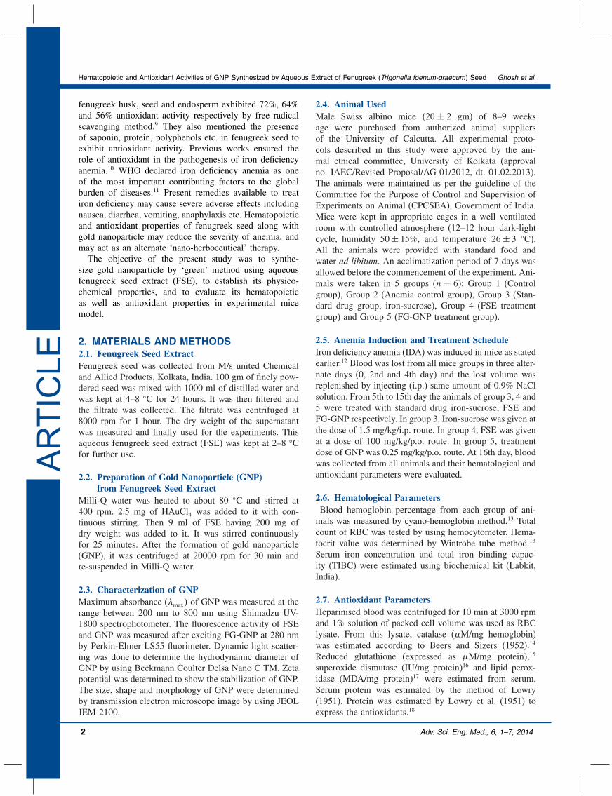
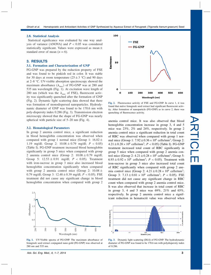
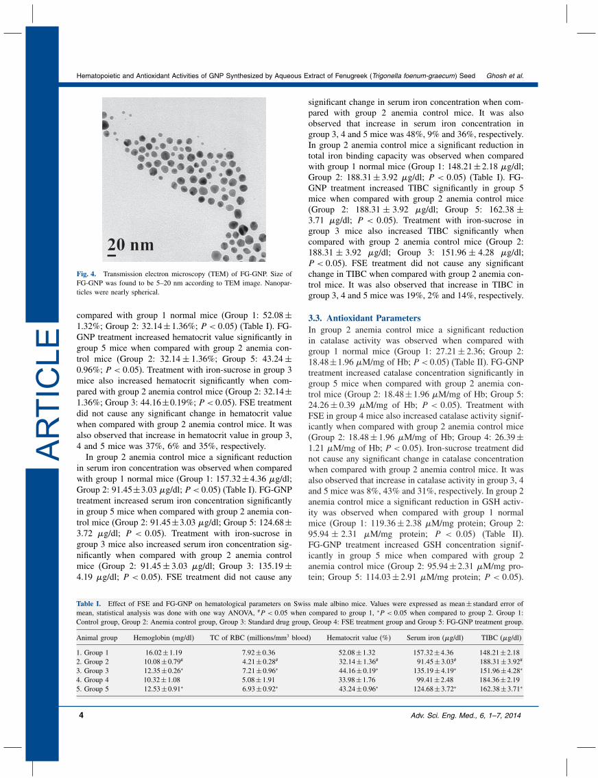
3. RESULTS3.1. Formation and Characterization of GNPFG-GNP was prepared by the reduction property of FSEand was found to be pinkish red in color. It was stablefor 30 days at room temperature (25±3 �C) and 90 daysat 2–8 �C. UV-visible absorption spectroscopy showed themaximum absorbance (�max� of FG-GNP was at 280 and535 nm wavelength (Fig. 1). At excitation wave length of280 nm (which was the �max of FSE), fluorescent activ-ity was significantly quenched after the formation of GNP(Fig. 2). Dynamic light scattering data showed that therewas formation of monodispersed nanoparticles. Hydrody-namic diameter of GNP was found to be 170.6 nm withpoly-dispersity index 0.286 (Fig. 3). Transmission electronmicroscopy showed that the shape of FG-GNP was nearlyspherical with particle size of 5–20 nm (Fig. 4).
3.2. Hematological ParametersIn group 2 anemia control mice, a significant reductionin blood hemoglobin concentration was observed whencompared with group 1 normal mice (Group 1: 16.02±1.19 mg/dl; Group 2: 10.08 ± 0.79 mg/dl; P < 0�05)(Table I). FG-GNP treatment increased blood hemoglobinsignificantly in group 5 mice when compared with group2 anemia control mice (Group 2: 10.08± 0.79 mg/dl;Group 5: 12.53 ± 0.91 mg/dl; P < 0�05). Treatmentwith iron-sucrose in group 3 mice also increased bloodhemoglobin concentration significantly when comparedwith group 2 anemia control mice (Group 2: 10.08±0.79 mg/dl; Group 3: 12.48±0.39 mg/dl; P < 0�05). FSEtreatment did not cause any significant change in bloodhemoglobin concentration when compared with group 2
0
0.5
1
1.5
2
2.5
250
350
450
550
650
750
850
Abs
orba
nce
Wavelength (nm)
Fig. 1. UV-Visible spectra of FG-GNP. The maximum absorbance offenugreek seed extract conjugated nano-gold (FG-GNP) was observed at280 nm and 535 nm.
Fig. 2. Fluorescence activity of FSE and FG-GNP. In curve 1, it wasfound that native fenugreek seed extract had significant fluorescent activ-ity. After formation of nanoparticle (FG-GNP) as in curve 2, there wasquenching of fluorescence activity.
anemia control mice. It was also observed that bloodhemoglobin concentration increase in group 3, 4 and 5mice was 23%, 2% and 24%, respectively. In group 2anemia control mice a significant reduction in total countof RBC was observed when compared with group 1 nor-mal mice (Group 1: 7.92±0.36×10⁶ cells/mm3; Group 2:4.21±0�28×106 cells/mm3; P < 0�05) (Table I). FG-GNPtreatment increased total count of RBC significantly ingroup 5 mice when compared with group 2 anemia con-trol mice (Group 2: 4�21±0�28×106 cells/mm3; Group 5:6.93± 0�92× 106 cells/mm3; P < 0�05). Treatment withiron-sucrose in group 3 mice also increased total countof RBC significantly when compared with group 2 ane-mia control mice (Group 2: 4.21± 0�28× 106 cells/mm3;Group 3: 7.13± 0.81× 10⁶ cells/mm3; P < 0�05). FSEtreatment did not cause any significant change in RBCcount when compared with group 2 anemia control mice.It was also observed that increase in total count of RBCin group 3, 4 and 5 mice was 69%, 21% and 65%,respectively. In group 2 anemia control mice a signif-icant reduction in hematocrit value was observed when
Fig. 3. Dynamic light scattering (DLS) of FG-GNP. The hydrodynamicdiameter of FG-GNP was found to be 170.6 nm with polydispersity indexof 0.286.
Adv. Sci. Eng. Med., 6, 1–7, 2014 3
Hematopoietic and Antioxidant Activities of GNP Synthesized by Aqueous Extract of Fenugreek (Trigonella foenum-graecum) Seed Ghosh et al.
ARTIC
LE
Fig. 4. Transmission electron microscopy (TEM) of FG-GNP. Size ofFG-GNP was found to be 5–20 nm according to TEM image. Nanopar-ticles were nearly spherical.
compared with group 1 normal mice (Group 1: 52.08±1.32%; Group 2: 32.14±1.36%; P < 0�05) (Table I). FG-GNP treatment increased hematocrit value significantly ingroup 5 mice when compared with group 2 anemia con-trol mice (Group 2: 32.14± 1.36%; Group 5: 43.24±0.96%; P < 0�05). Treatment with iron-sucrose in group 3mice also increased hematocrit significantly when com-pared with group 2 anemia control mice (Group 2: 32.14±1.36%; Group 3: 44.16±0.19%; P < 0�05). FSE treatmentdid not cause any significant change in hematocrit valuewhen compared with group 2 anemia control mice. It wasalso observed that increase in hematocrit value in group 3,4 and 5 mice was 37%, 6% and 35%, respectively.In group 2 anemia control mice a significant reduction
in serum iron concentration was observed when comparedwith group 1 normal mice (Group 1: 157.32±4.36 �g/dl;Group 2: 91.45±3.03 �g/dl; P < 0�05) (Table I). FG-GNPtreatment increased serum iron concentration significantlyin group 5 mice when compared with group 2 anemia con-trol mice (Group 2: 91.45±3.03 �g/dl; Group 5: 124.68±3.72 �g/dl; P < 0�05). Treatment with iron-sucrose ingroup 3 mice also increased serum iron concentration sig-nificantly when compared with group 2 anemia controlmice (Group 2: 91.45± 3.03 �g/dl; Group 3: 135.19±4.19 �g/dl; P < 0�05). FSE treatment did not cause any
Table I. Effect of FSE and FG-GNP on hematological parameters on Swiss male albino mice. Values were expressed as mean± standard error ofmean, statistical analysis was done with one way ANOVA, #P < 0�05 when compared to group 1, ∗P < 0�05 when compared to group 2. Group 1:Control group, Group 2: Anemia control group, Group 3: Standard drug group, Group 4: FSE treatment group and Group 5: FG-GNP treatment group.
Animal group Hemoglobin (mg/dl) TC of RBC (millions/mm3 blood) Hematocrit value (%) Serum iron (�g/dl) TIBC (�g/dl)
1. Group 1 16.02±1.19 7.92±0.36 52.08±1.32 157.32±4.36 148.21±2.182. Group 2 10.08±0.79# 4.21±0.28# 32.14±1.36# 91.45±3.03# 188.31±3.92#
3. Group 3 12.35±0.26∗ 7.21±0.96∗ 44.16±0.19∗ 135.19±4.19∗ 151.96±4.28∗
4. Group 4 10.32±1.08 5.08±1.91 33.98±1.76 99.41±2.48 184.36±2.195. Group 5 12.53±0.91∗ 6.93±0.92∗ 43.24±0.96∗ 124.68±3.72∗ 162.38±3.71∗
significant change in serum iron concentration when com-pared with group 2 anemia control mice. It was alsoobserved that increase in serum iron concentration ingroup 3, 4 and 5 mice was 48%, 9% and 36%, respectively.In group 2 anemia control mice a significant reduction intotal iron binding capacity was observed when comparedwith group 1 normal mice (Group 1: 148.21±2.18 �g/dl;Group 2: 188.31± 3.92 �g/dl; P < 0�05) (Table I). FG-GNP treatment increased TIBC significantly in group 5mice when compared with group 2 anemia control mice(Group 2: 188.31 ± 3.92 �g/dl; Group 5: 162.38 ±3.71 �g/dl; P < 0�05). Treatment with iron-sucrose ingroup 3 mice also increased TIBC significantly whencompared with group 2 anemia control mice (Group 2:188.31 ± 3.92 �g/dl; Group 3: 151.96 ± 4.28 �g/dl;P < 0�05). FSE treatment did not cause any significantchange in TIBC when compared with group 2 anemia con-trol mice. It was also observed that increase in TIBC ingroup 3, 4 and 5 mice was 19%, 2% and 14%, respectively.
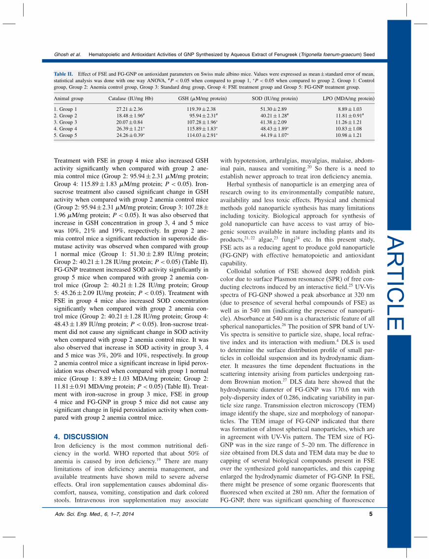
3.3. Antioxidant ParametersIn group 2 anemia control mice a significant reductionin catalase activity was observed when compared withgroup 1 normal mice (Group 1: 27.21± 2.36; Group 2:18.48±1.96 �M/mg of Hb; P < 0�05) (Table II). FG-GNPtreatment increased catalase concentration significantly ingroup 5 mice when compared with group 2 anemia con-trol mice (Group 2: 18.48±1.96 �M/mg of Hb; Group 5:24.26± 0.39 �M/mg of Hb; P < 0�05). Treatment withFSE in group 4 mice also increased catalase activity signif-icantly when compared with group 2 anemia control mice(Group 2: 18.48± 1.96 �M/mg of Hb; Group 4: 26.39±1.21 �M/mg of Hb; P < 0�05). Iron-sucrose treatment didnot cause any significant change in catalase concentrationwhen compared with group 2 anemia control mice. It wasalso observed that increase in catalase activity in group 3, 4and 5 mice was 8%, 43% and 31%, respectively. In group 2anemia control mice a significant reduction in GSH activ-ity was observed when compared with group 1 normalmice (Group 1: 119.36± 2.38 �M/mg protein; Group 2:95.94 ± 2.31 �M/mg protein; P < 0�05) (Table II).FG-GNP treatment increased GSH concentration signif-icantly in group 5 mice when compared with group 2anemia control mice (Group 2: 95.94±2.31 �M/mg pro-tein; Group 5: 114.03± 2.91 �M/mg protein; P < 0�05).
4 Adv. Sci. Eng. Med., 6, 1–7, 2014
Ghosh et al. Hematopoietic and Antioxidant Activities of GNP Synthesized by Aqueous Extract of Fenugreek (Trigonella foenum-graecum) Seed
ARTIC
LE
Table II. Effect of FSE and FG-GNP on antioxidant parameters on Swiss male albino mice. Values were expressed as mean±standard error of mean,statistical analysis was done with one way ANOVA, #P < 0�05 when compared to group 1, ∗P < 0�05 when compared to group 2. Group 1: Controlgroup, Group 2: Anemia control group, Group 3: Standard drug group, Group 4: FSE treatment group and Group 5: FG-GNP treatment group.
Animal group Catalase (IU/mg Hb) GSH (�M/mg protein) SOD (IU/mg protein) LPO (MDA/mg protein)
1. Group 1 27.21±2.36 119.39±2.38 51.30±2.89 8.89±1.032. Group 2 18.48±1.96# 95.94±2.31# 40.21±1.28# 11.81±0.91#
3. Group 3 20.07±0.84 107.28±1.96∗ 41.38±2.09 11.26±1.214. Group 4 26.39±1.21∗ 115.89±1.83∗ 48.43±1.89∗ 10.83±1.085. Group 5 24.26±0.39∗ 114.03±2.91∗ 44.19±1.07∗ 10.98±1.21
Treatment with FSE in group 4 mice also increased GSHactivity significantly when compared with group 2 ane-mia control mice (Group 2: 95.94±2.31 �M/mg protein;Group 4: 115.89± 1.83 �M/mg protein; P < 0�05). Iron-sucrose treatment also caused significant change in GSHactivity when compared with group 2 anemia control mice(Group 2: 95.94±2.31 �M/mg protein; Group 3: 107.28±1.96 �M/mg protein; P < 0�05). It was also observed thatincrease in GSH concentration in group 3, 4 and 5 micewas 10%, 21% and 19%, respectively. In group 2 ane-mia control mice a significant reduction in superoxide dis-mutase activity was observed when compared with group1 normal mice (Group 1: 51.30± 2.89 IU/mg protein;Group 2: 40.21±1.28 IU/mg protein; P < 0�05) (Table II).FG-GNP treatment increased SOD activity significantly ingroup 5 mice when compared with group 2 anemia con-trol mice (Group 2: 40.21± 1.28 IU/mg protein; Group5: 45.26±2.09 IU/mg protein; P < 0�05). Treatment withFSE in group 4 mice also increased SOD concentrationsignificantly when compared with group 2 anemia con-trol mice (Group 2: 40.21±1.28 IU/mg protein; Group 4:48.43±1.89 IU/mg protein; P < 0�05). Iron-sucrose treat-ment did not cause any significant change in SOD activitywhen compared with group 2 anemia control mice. It wasalso observed that increase in SOD activity in group 3, 4and 5 mice was 3%, 20% and 10%, respectively. In group2 anemia control mice a significant increase in lipid perox-idation was observed when compared with group 1 normalmice (Group 1: 8.89± 1.03 MDA/mg protein; Group 2:11.81±0.91 MDA/mg protein; P < 0�05) (Table II). Treat-ment with iron-sucrose in group 3 mice, FSE in group4 mice and FG-GNP in group 5 mice did not cause anysignificant change in lipid peroxidation activity when com-pared with group 2 anemia control mice.
4. DISCUSSIONIron deficiency is the most common nutritional defi-ciency in the world. WHO reported that about 50% ofanemia is caused by iron deficiency.19 There are manylimitations of iron deficiency anemia management, andavailable treatments have shown mild to severe adverseeffects. Oral iron supplementation causes abdominal dis-comfort, nausea, vomiting, constipation and dark coloredstools. Intravenous iron supplementation may associate
with hypotension, arthralgias, mayalgias, malaise, abdom-inal pain, nausea and vomiting.20 So there is a need toestablish newer approach to treat iron deficiency anemia.Herbal synthesis of nanoparticle is an emerging area of
research owing to its environmentally compatible nature,availability and less toxic effects. Physical and chemicalmethods gold nanoparticle synthesis has many limitationsincluding toxicity. Biological approach for synthesis ofgold nanoparticle can have access to vast array of bio-genic sources available in nature including plants and itsproducts,21�22 algae,23 fungi24 etc. In this present study,FSE acts as a reducing agent to produce gold nanoparticle(FG-GNP) with effective hematopoietic and antioxidantcapability.Colloidal solution of FSE showed deep reddish pink
color due to surface Plasmon resonance (SPR) of free con-ducting electrons induced by an interactive field.25 UV-Visspectra of FG-GNP showed a peak absorbance at 320 nm(due to presence of several herbal compounds of FSE) aswell as in 540 nm (indicating the presence of nanoparti-cle). Absorbance at 540 nm is a characteristic feature of allspherical nanoparticles.26 The position of SPR band of UV-Vis spectra is sensitive to particle size, shape, local refrac-tive index and its interaction with medium.4 DLS is usedto determine the surface distribution profile of small par-ticles in colloidal suspension and its hydrodynamic diam-eter. It measures the time dependent fluctuations in thescattering intensity arising from particles undergoing ran-dom Brownian motion.27 DLS data here showed that thehydrodynamic diameter of FG-GNP was 170.6 nm withpoly-dispersity index of 0.286, indicating variability in par-ticle size range. Transmission electron microscopy (TEM)image identify the shape, size and morphology of nanopar-ticles. The TEM image of FG-GNP indicated that therewas formation of almost spherical nanoparticles, which arein agreement with UV-Vis pattern. The TEM size of FG-GNP was in the size range of 5–20 nm. The difference insize obtained from DLS data and TEM data may be due tocapping of several biological compounds present in FSEover the synthesized gold nanoparticles, and this cappingenlarged the hydrodynamic diameter of FG-GNP. In FSE,there might be presence of some organic fluorescents thatfluoresced when excited at 280 nm. After the formation ofFG-GNP, there was significant quenching of fluorescence
Adv. Sci. Eng. Med., 6, 1–7, 2014 5
Hematopoietic and Antioxidant Activities of GNP Synthesized by Aqueous Extract of Fenugreek (Trigonella foenum-graecum) Seed Ghosh et al.
ARTIC
LE
activity which is a conformation for bio-conjugation.28 Incase of fluorescence activity, quenching of energy trans-fer occurs at a close distance (upto 5 nm) between flu-orophore and metal nanoparticles. Here the influence ofmetal nanoparticle decreases the fluorescence intensity ofthe fluorophore which is proportional to the cube of thedistance.29
In iron deficiency, blood hemoglobin level becomes lowdue to inadequate amount of iron in blood. Iron is requiredto synthesize hemoglobin. Previous study suggested thatgold nanoparticle produced from citrate-reduction methodincreased blood hemoglobin significantly at a high doseof 10 mg/kg.1 This high dose was also showed to havesevere adverse effects on experimental animal. But in thisstudy, FG-GNP treatment increased blood hemoglobin atonly 0.25 mg/kg dose. It was also seen that percentageincrease of hemoglobin concentration after FG-GNP treat-ment was better than standard drug or FSE treatment. Totalcount of RBC and hematocrit value was also increased sig-nificantly in FG-GNP group compared to anemic controlmice. Here also FG-GNP treatment was better to increasetotal RBC count and hematocrit value than FSE treatment.From these, it can easily be said that FG-GNP had sig-nificant hematopoietic activity, and it was better than FSEtreatment alone.Transferrin, a glycoprotein present in serum, binds
with serum iron and helps in its transportation. Thistransferrin-bound circulating iron is measured throughdetermining serum iron concentration. Total iron bindingcapacity (TIBC) measures the maximum amount of ironthat transferrin can carry.30 Every molecule of transferrincan carry up to 2 iron atom in ferric (Fe3+) form. Thus,TIBC can indirectly measure the concentration of transfer-rin in serum. In iron deficiency anemia, serum iron concen-tration is significantly low, which leads to produce moretransferrin in liver to maximize the use of little amount ofiron present in serum. FG-GNP treatment exhibited bet-ter increase in serum iron concentration and decrease inTIBC than standard drug treatment or FSE treatment whencompared with anemia control group.Reactive oxygen species (ROS) is generated when cel-
lular prooxidant levels increase and antioxidant levelsdecrease, leading to oxidative stress in animal. Iron defi-ciency anemia causes stress in body leading to produc-tion of ROS. Reduced glutathione (GSH) is an antioxi-dant which reduces free radicals and itself oxidized to formGS-SG.31 Here GSH in anemia control group decreasedsignificantly when compared with normal group, suggest-ing the increased production of oxidative stress. FG-GNPtreatment caused increased GSH activity. Superoxide dis-mutase (SOD) scavenges superoxide anion into hydrogenperoxide.31 Catalase eliminates toxic peroxides in cell andforms non-toxic water and oxygen.32 Thus, SOD and cata-lase decrease free radicals produced due to stress. As therewas significant reduction of SOD and catalase level anemiacontrol group when compared with normal group, there was
increased production of ROS. FG-GNP and FSE treatmentincreased SOD and catalase activity. However, FG-GNPtreatment was better to increase SOD and catalase activitythan FSE treatment. Lipid peroxidation in cell oxidizes cellmembrane, leading to membrane rupture and cell lysis.33
Significant lipid peroxidation activity was seen in anemiacontrol group when compared with normal group. Thisindicates that, there was significant peroxidation of plasmamembrane in iron deficiency anemia, and after FG-GNPtreatment membrane peroxidation was not reduced, indicat-ing that there was no effect of FG-GNP on LPO activity.Several bioactive compounds (saponin, coumarin etc.)
present in fenugreek seed9 are shown to have antianemicand antioxidant properties. Antioxidants are often reduc-ing agents that can reduce gold salt into gold nanoparticle.These bioactive compounds may cap the nanoparticle andmake it stable. Along with gold nanoparticle, these com-pounds exert their hemapoietic effects more significantlyand their antioxidative role helps to reduce the pathogene-sis of iron deficiency anemia. Further studies on the molec-ular action of green nanoparticle in the management ofanemia are in progress.
5. CONCLUSIONGreen synthesis of metal nanoparticles has been a studysubject owing to its effective rather than the detrimentalroles showed by conventional chemical/physical particlesynthesis techniques. In the present study, it is clearly evi-dent that FG-GNP has a edge over the GNP synthesizedby other methods due to its effective therapeutic value as ahematopoietic and antioxidant agent. This study thereforeopens a new dimension in the therapeutic application ofgreen nanoparticle in the management of iron deficiencyanemia.
DisclosureThe authors declare no conflict of interest.
Acknowledgment: The authors thank UGC for provid-ing partial financial support and UGC-BSR fellowship toAG (Professor Antony Gomes).
References and Notes1. J. Sengupta, P. Datta, H. K. Patra, A. K. Dasgupta, and A. Gomes,
J. Nanosci. Nanotechnol. 13, 1660 (2013).2. D. B. Warheit, C. M. Sayes, K. L. Reed, and K. A. Swain, Pharma-
col. Ther. 120, 35 (2008).3. V. Parashar, R. Parashar, B. Sharma, and A. C. Pandey, Dig.
J. Nanomater. Bios. 4, 45 (2009).4. S. A. Aromal and D. Philip, Spectrochim. Acta A Mol. Biomol. Spec-
trosc. 97, 1 (2012).5. D. Zohary and M. Hopf (eds.), Domestication of Plants in the Old
World, Third edn., Oxford University Press, Oxford, London (2000),p. 122.
6. M. I. Ibrahium and A. I. Hegazy, World J. Agr. Sci. 5, 769 (2009).7. N. Y. Mahmoud, R. H. Salem, and A. A. Mater, Acad. J. Nutr. 1, 1
(2012).
6 Adv. Sci. Eng. Med., 6, 1–7, 2014
Ghosh et al. Hematopoietic and Antioxidant Activities of GNP Synthesized by Aqueous Extract of Fenugreek (Trigonella foenum-graecum) Seed
ARTIC
LE
8. M. Doshi, A. Mirza, B. Umarji, and R. Karambelkar, Biomed. Res.23, 47 (2012).
9. M. M. Naidu, B. N. Shyamala, J. Pura Naik, G. Sulochanamma, andP. Srinivas, LWT—Food Science and Technology 44, 456 (2011).
10. M. Aslan, M. Horoz, and H. Çelik, Turk. J. Hematol. 28, 42(2011).
11. World Health Organization (ed.), The World Health Report 2002:Reducing risks, promoting healthy life, World Health Organization,Geneva (2002).
12. K. B. Jones, D. W. Anderson, and G. D. Longmore, I. Orthop.J. 25, 129 (2005).
13. K. L. Mukherjee (ed.), Medical Laboratory Technology, TataMcGraw-Hill Publishing Company Limited, New Delhi (2004),p. 235.
14. R. F. Beers and I. W. Sizer, J. Biol. Chem. 195, 133 (1952).15. G. L. Ellman, Biochem. Biophys. 82, 70 (1959).16. W. A. Pryor, Fed. Proc. 32, 1862 (1973).17. J. A. Buege and S. D. Aust, Methods Enzymol. 52, 302 (1978).18. O. H. Lowry, N. J. Rosebrough, A. L. Farr, and R. J. Randall, J. Biol.
Chem. 193, 265 (1951).19. World Health Organization, (ed.), Iron Deficiency Anaemia: Assess-
ment, Prevention, and Control, A guide for programme managers,Geneva (2001).
20. T. D. J. Wimbley and D. Y. Graham, Therap. Adv. Gastroenterol.4, 177 (2011).
21. J. Huang, Q. Li, D. Sun, Y. Lu, Y. Su, X. Yang, H. Wang, Y. Wang,W. Shao, N. He, J. Hong, and C. Chen, Nanotechnology 18, 1 (2007).
22. S. P. Chandran, M. Chaudhary, R. Pasricha, A. Ahmad, andM. Sastry, Biotechnol. Progr. 22, 577 (2006).
23. G. Singaravelu, J. Arockiyamari, V. G. Kumar, and K. Govindaraju,Colloids Surf. B Biointerfaces 57, 97 (2007).
24. Z. Sadowski, I. H. Maliszewska, B. Grochowalska, I. Polowczyk,and T. Kozlecki, Materials Science—Poland 26, 419 (2008).
25. F. Toderas, M. Baia, L. Baia, and S. Astilean, Nanotechnology18, 255702 (2007).
26. V. A. Markel, V. M. Shalaev, E. B. Stechel, W. Kim, and R. L.Armstrong, Phys. Rev. B Condens Matter. 53, 2425 (1996).
27. M. Kaszuba, D. McKnight, M. T. Connah, F. K. McNeil-Watson,and U. Nobbmann, J. Nanopart. Res. 10, 823 (2008).
28. P. Joshi, V. Shewale, R. Pandey, V. Shanker, S. Hussain, and S. P.Karna, Phys. Chem. Chem. Phys. 13, 476 (2011).
29. C. D. Geddes and J. R. Lakowicz, J. Fluoresc. 12, 121 (2002).30. E. Beutler, M. A. Lichtman, B. S. Coller, T. J. Kipps, and
U. Seligsohn (eds.), Williams Hematology, International 6th edn.,McGraw Hill, USA (2001).
31. M. Isler, N. Delibas, M. Guclu, F. Gultekin, R. Sutcu, M. Bahceci,and A. Kosar, Croat. Med. J. 43, 16 (2002).
32. E. Beutler and R. K. Blaisdell, J. Clin. Invest. 37, 833 (1958).33. J. Acharya, N. A. Punchard, J. A. Taylor, R. P. Thompson, and T. C.
Pearson, Eur. J. Haematol. 47, 287 (1991).
Adv. Sci. Eng. Med., 6, 1–7, 2014 7