hebbian plasticity requires compensatory processes on
TRANSCRIPT
Hebbian plasticity requires compensatory processes on multipletimescales
Friedemann Zenke1,∗and Wulfram Gerstner2
January 16, 2017
1) Dept. of Applied Physics, Stanford University, Stanford, CA 94305, USAorcid.org/0000-0003-1883-644X2) Brain Mind Institute, School of Life Sciences and School of Computer and CommunicationSciences, Ecole Polytechnique Fédérale de Lausanne, CH-1015 Lausanne EPFL, Switzerlandorcid.org/0000-0002-4344-2189
This article is published as:Zenke, F., and Gerstner, W. (2017). Hebbian plasticity requires compensatory processes onmultiple timescales. Phil. Trans. R. Soc. B 372, 20160259. DOI: 10.1098/rstb.2016.0259http://rstb.royalsocietypublishing.org/content/372/1715/20160259
Abstract
We review a body of theoretical and experimental research on Hebbian and homeostaticplasticity, starting from a puzzling observation: While homeostasis of synapses found in exper-iments is a slow compensatory process, most mathematical models of synaptic plasticity userapid compensatory processes. Even worse, with the slow homeostatic plasticity reported inexperiments, simulations of existing plasticity models cannot maintain network stability unlessfurther control mechanisms are implemented. To solve this paradox, we suggest that in ad-dition to slow forms of homeostatic plasticity there are rapid compensatory processes whichstabilize synaptic plasticity on short timescales. These rapid processes may include heterosy-naptic depression triggered by episodes of high postsynaptic firing rate. While slower forms ofhomeostatic plasticity are not sufficient to stabilize Hebbian plasticity, they are important forfine-tuning neural circuits. Taken together we suggest that learning and memory rely on anintricate interplay of diverse plasticity mechanisms on different timescales which jointly ensurestability and plasticity of neural circuits.
Keywords: Hebbian plasticity, homeostasis, rapid compensatory processes, heterosynaptic plas-ticity, synaptic scaling, metaplasticity∗Author for correspondence ([email protected])
1
Introduction
Homeostasis refers to a family of compensatory processes at different spatial and temporal scaleswhose objective is to maintain the body, its organs, the brain, or even individual neurons in thebrain in a dynamic regime where they function optimally. A well-known example is the homeostaticregulation of body temperature in mammals, maintained at about 37 degrees Celsius independentof weather condition and air temperature. In neuroscience, homeostasis or homeostatic plasticityoften refers to a compensatory process that stabilizes neural firing rates. In a classic experiment,cultured neurons that normally fire at, say 5Hz, change their firing rate after a modulation ofthe chemical conditions in the culture, but eventually return to their target rate of 5Hz duringthe following 24 hours [1]. Thus, the experimentally best-studied form of homeostasis acts on atimescale of hours to days. This slow form of homeostatic plasticity manifests itself as the rescalingof the efficacy or weight of all afferent synapses onto a single neuron by a fixed fraction, for instance0.78. This phenomenon is called “synaptic scaling” [1].
Mathematical models of neural networks often make use of compensatory processes similar tosynaptic scaling to stabilize firing rates in the presence of Hebbian plasticity. Hebbian plasticityis a form of synaptic plasticity which is induced by and further amplifies correlations in neuronalactivity. It has been observed in many brain areas and can be induced quickly on a timescale ofseconds to minutes. Its effect, however, is often long-lasting. It can last hours, days and possiblya lifetime. Due to these properties Hebbian plasticity is widely assumed to be the neural basisof associative long-term memory [2–4]. Moreover, Hebbian learning is thought to be the basis ofdevelopmental changes such as receptive field development [5–9].
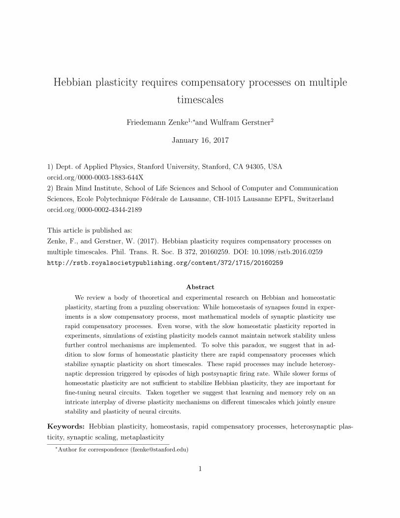
However, Hebbian plasticity alone leads to a positive feedback loop in which correlations of pre-and postsynaptic firing drive potentiation of synapses that increase postsynaptic rates and corre-lations further, which is unstable. To avoid pathological run-away dynamics of neural activity inmathematical models, it is necessary to add appropriate constraints to plasticity models [10, 11]. Atypical example of such a constraint is the normalization or rescaling of the sum of afferent synap-tic weights: When the weight of one synaptic connection increases, weights of other connectionsonto the same neuron are algorithmically decreased to keep the total input constant or close to theoptimal target regime. At a first glance, this form of multiplicative normalization [10] seems virtu-ally identical to homeostatic “synaptic scaling” introduced above. However, these two mechanismsare fundamentally distinct because they act on vastly different timescales. While normalization inmodels typically takes place on a timescale of seconds or less [10, 12–14], in biology the effects ofsynaptic scaling manifest themselves only after hours [15, 16]. A similar observation holds for home-ostatic metaplasticity, which exists on timescales ranging from some tens of minutes to days (Fig. 1;[17, 18]). Moreover, the difference between experimental data and models cannot be explained bya simple rescaling of time in the models, because the problem persists for quantitative plasticitymodels which capture the time course of biological data.
2
instantaneous second minute hour day
Synaptic scaling experiments
Priming experiments Sensory deprivation
†: Based on Kaneko et al. (2008)
ModelsvonderMalsburg(1973)
MillerandMacKay
(1994)
Lazaretal.(2009)
*
Litwin-Kum
arandDoiron(2014)
*
Clopath
etal.(2010)
*
ElBoustanietal.(2012)
*
Gjorgjieva
etal.(2011)
Jedlicka
etal.(2015)
Benuskova
andAbraham
(2007)
vanRossumetal.(2000)
Zenke
etal.(2013)
*
P�sterandGerstner(2006)
Toyoizumiet
al.(2014)
†
Experiments
Huang
etal.(1992)
Aotoetal.(2008)
Ibataetal.(2008)
ChristieandAbraham
(1992)
Greenhilletal.(2015)
Turrigianoet
al.(1998)
Liet
al.(2014)
Goelet
al.(2006)
Figure 1: The timescales of synaptic scaling or metaplasticity are faster in models thanreported in experiments. Here we plot the timescale of either synaptic scaling or homeostaticmetaplasticity as used in influential modeling studies (gray). For comparison we plot the typicalreadout time for experimental studies on synaptic scaling and metaplasticity (red). Publicationssuffixed with * describe network models as opposed to the other studies which relied on singleneurons. Note that the model marked with † by Toyoizumi et al. [23] is an interesting case whichhas both RCPs and a slow form of homeostasis. Here we aligned it according to its homeostatictimescale.
However, this difference in timescales may challenge the popular view that in biology Hebbianplasticity is constrained through homeostatic plasticity [16, 19–22]. The algorithmic normalizationof synaptic weights every second is not the same mechanism as the biological rescaling of synapticweights over hours. Although, in the theoretical literature, a rapid stabilizing mechanism is typicallycalled “homeostatic”, here we will refer to this class of control mechanisms as Rapid CompensatoryProcesses (RCPs). The term “homeostatic plasticity” is in the following reserved for slow negativefeedback processes on the timescale of hours or days — a terminology that seems consistent with theavailable experimental literature [15, 16]. In this review we focus on this discrepancy of timescalesand ask which biologically plausible processes could constrain Hebbian plasticity. Specifically, wewill try to answer the following questions: Why do we need RCPs to stabilize Hebbian plasticity?How fast do these processes have to be — hours, minutes, seconds or less? Which mechanisms couldfill this role in Hebbian learning? Moreover, what are the consequences of fast control mechanismson memory formation and recall in network models? And finally, if RCPs are a requirement, what isthe role of slower forms of negative feedback implemented by known forms of homeostatic plasticity?
Models of Synaptic Plasticity
Synaptic plasticity exists across different timescales. For instance, synaptic changes induced by asequence of four presynaptic spikes in rapid sequence typically decay within a few hundred millisec-onds [42–44] and are called short-term plasticity. The rapid decay implies that the changes are not
3
useful for long-term memory formation, but more likely involved in gain control [42].Other forms of plasticity induced by classic induction protocols [45–47], can have long-term
effects on the timescale of hours or more. Long-term plasticity is therefore potentially useful formemory formation [2]. We remind the reader that the induction of long-term plasticity can be asfast as seconds, but the induced changes persist for much longer. Depending on the direction ofsynaptic change, we speak of long-term potentiation (LTP) or long-term depression (LTD).
Under suitable conditions the changes induced by a protocol of LTP or LTD are further con-solidated after about an hour [48–50]. These effects are often referred to as late-phase long-termplasticity. In the rest of the paper we focus on plasticity induction and the early phase of long-termplasticity and neglect consolidation and maintenance.
The diverse effects of long-term plasticity can be cast into a mathematical framework whichdescribes the change of synaptic efficacy over time. Apart from a few notable exceptions [23, 51–54], the vast majority of models of long-term plasticity assume a one-dimensional synaptic statespace which represents the synaptic efficacy or weight wij of a synapse from neuron j to neuron i[5, 8, 9, 33, 55–65]. The evolution wij is then characterized by the differential equation
dwijdt
= G (1)
in which the function G, often called the “learning rule”, is a member of an infinite dimensionalfunction space G, the space of all possible learning rules. This space is strongly constrained if weonly focus on plausible learning rules, which are the rules in agreement with experimental findings.
For example, classical stimulation protocols for LTP [45–47], LTD [66, 67], or spike-timing-dependent plasticity (STDP) [68–70], combine the activation of a presynaptic neuron, or a presy-naptic pathway, with an activation, depolarization, or chemical manipulation of the postsynapticneurons, to induce synaptic changes. In models this is typically formalized by stating that G onlydepends on quantities which are locally accessible to the synapse. It is customary to assume thatthe main locally accessible variables include: (i) the current synaptic state wij ; (ii) the activityprej of the presynaptic neuron; and (iii) the state posti of the postsynaptic neuron [64, 71, 72].Thus we can write dwij
dt = G(wij ,posti,prej). Additionally, G could also depend on low dimensionalinformation carried by chemical signals such as neuromodulators (see Frémaux and Gerstner [73]for a review).
Most published learning rules G can be written as the linear sum of different terms in whicheach term can be interpreted as a specific manifestation of plasticity. These terms act together toexplain the measured outcome in plasticity experiments. Let us explain the most common ones
4
using the following example learning rule:
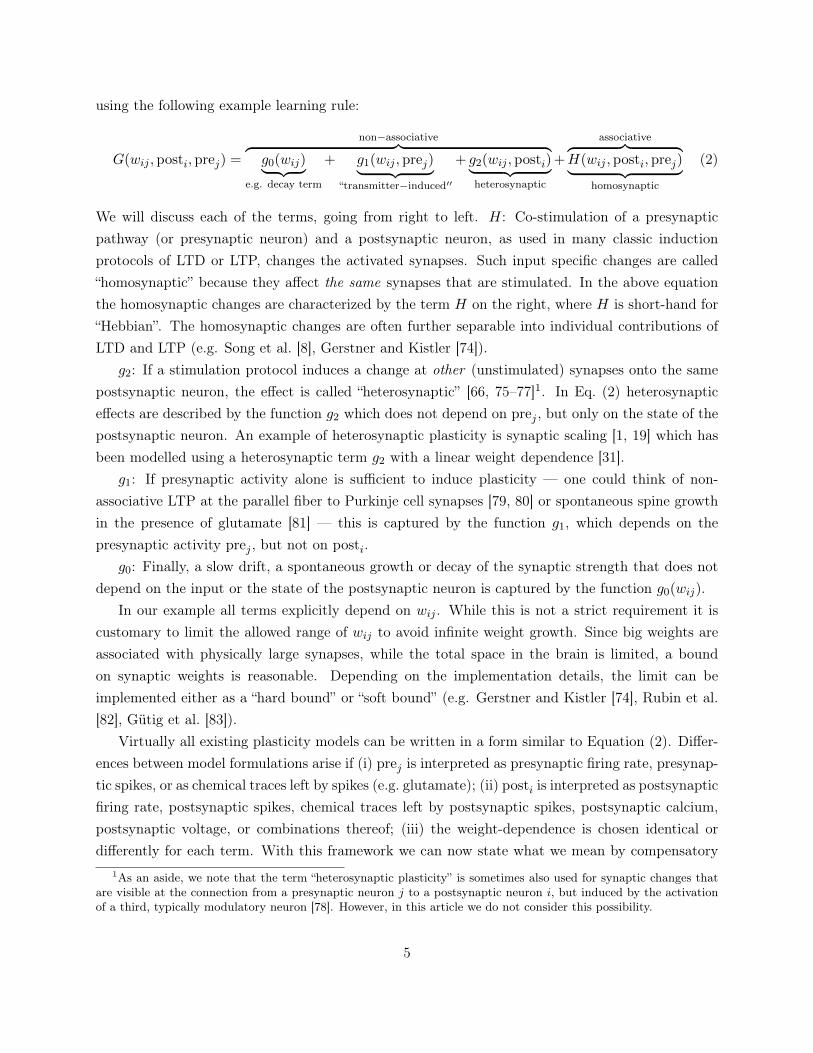
G(wij ,posti, prej) =
non−associative︷ ︸︸ ︷g0(wij)︸ ︷︷ ︸
e.g. decay term
+ g1(wij , prej)︸ ︷︷ ︸“transmitter−induced′′
+ g2(wij ,posti)︸ ︷︷ ︸heterosynaptic
+
associative︷ ︸︸ ︷H(wij ,posti, prej)︸ ︷︷ ︸
homosynaptic
(2)
We will discuss each of the terms, going from right to left. H: Co-stimulation of a presynapticpathway (or presynaptic neuron) and a postsynaptic neuron, as used in many classic inductionprotocols of LTD or LTP, changes the activated synapses. Such input specific changes are called“homosynaptic” because they affect the same synapses that are stimulated. In the above equationthe homosynaptic changes are characterized by the term H on the right, where H is short-hand for“Hebbian”. The homosynaptic changes are often further separable into individual contributions ofLTD and LTP (e.g. Song et al. [8], Gerstner and Kistler [74]).
g2: If a stimulation protocol induces a change at other (unstimulated) synapses onto the samepostsynaptic neuron, the effect is called “heterosynaptic” [66, 75–77]1. In Eq. (2) heterosynapticeffects are described by the function g2 which does not depend on prej , but only on the state of thepostsynaptic neuron. An example of heterosynaptic plasticity is synaptic scaling [1, 19] which hasbeen modelled using a heterosynaptic term g2 with a linear weight dependence [31].
g1: If presynaptic activity alone is sufficient to induce plasticity — one could think of non-associative LTP at the parallel fiber to Purkinje cell synapses [79, 80] or spontaneous spine growthin the presence of glutamate [81] — this is captured by the function g1, which depends on thepresynaptic activity prej , but not on posti.
g0: Finally, a slow drift, a spontaneous growth or decay of the synaptic strength that does notdepend on the input or the state of the postsynaptic neuron is captured by the function g0(wij).
In our example all terms explicitly depend on wij . While this is not a strict requirement it iscustomary to limit the allowed range of wij to avoid infinite weight growth. Since big weights areassociated with physically large synapses, while the total space in the brain is limited, a boundon synaptic weights is reasonable. Depending on the implementation details, the limit can beimplemented either as a “hard bound” or “soft bound” (e.g. Gerstner and Kistler [74], Rubin et al.[82], Gütig et al. [83]).
Virtually all existing plasticity models can be written in a form similar to Equation (2). Differ-ences between model formulations arise if (i) prej is interpreted as presynaptic firing rate, presynap-tic spikes, or as chemical traces left by spikes (e.g. glutamate); (ii) posti is interpreted as postsynapticfiring rate, postsynaptic spikes, chemical traces left by postsynaptic spikes, postsynaptic calcium,postsynaptic voltage, or combinations thereof; (iii) the weight-dependence is chosen identical ordifferently for each term. With this framework we can now state what we mean by compensatory
1As an aside, we note that the term “heterosynaptic plasticity” is sometimes also used for synaptic changes thatare visible at the connection from a presynaptic neuron j to a postsynaptic neuron i, but induced by the activationof a third, typically modulatory neuron [78]. However, in this article we do not consider this possibility.
5
processes and address the question why we need them to be fast.
Why do we need rapid compensatory processes to stabilize Hebbianplasticity?
Intuitively, synaptic plasticity that is useful for memory formation must be sensitive to the presentactivation pattern of the pre- and postsynaptic neuron. Following Hebb’s idea of learning and cellassembly formation, the synaptic changes should make the same activation pattern more likely tore-appear in the future, to allow contents from memory to be retrieved. However, the re-appearanceof the same pattern will induce further synaptic plasticity. This forms an unstable positive feedbackloop. Anybody who was sitting in the audience when the positive feedback loop between thespeaker’s microphone and the loudspeaker resulted in an unpleasant shriek, knows what this means.In many cases an unstable system can be made stable by adding sensible control mechanisms [84]which are thus typically integrated in theoretically motivated plasticity models.
Let us now consider one such classic example of a learning rule. To that end we consider Oja’srule [57]
dwijdt
= G = η(xjyi − wijy2
i
)(3)
where η is a small constant called learning rate. Since Oja’s rule corresponds to a specific choiceof G in Equations (1) and (2), let us highlight the relation. First, in Oja’s rule the presynapticactivity prej is characterized by the presynaptic rate xj and the state of the postsynaptic neuronposti by its firing rate yi. Second, and with this in mind, we can now identify two terms on theright-hand side of Equation (3). Oja’s rule contains a Hebbian term H = ηxjyi which does nothave any weight dependence as well as a heterosynaptic term g2 = −ηwijy2
i which comes with anegative sign and is linear in the weight. Following our convention from above (Eq. 2) we call theterm heterosynaptic because it acts on all synapses, even those that do not receive presynapticstimulation. For simplicity, and following the tradition [57] we combine Oja’s rule with a linearneuron model yi =
∑j wijxj .
It is quite intuitive to see how stability arises in this model. As the synaptic weights wij grow,due to the Hebbian term, the firing rate yi of the postsynaptic neuron increases and therefore theinfluence of the negative heterosynaptic term gets stronger. Because the heterosynaptic term has asuperlinear dependence on yi, it is guaranteed to “catch up” with the Hebbian term eventually. Itcan be shown that for a linear neuron model and sufficiently small η, Oja’s rule ensures that theweights converge such that ~wi = (wi1, ..., wiN ) aligns with the first principal component of the datax, while the squared sum of all afferent weights remains normalized [57].
We interpret the heterosynaptic term in Oja’s rule as RCP: First, it is rapid because it respondsinstantaneously to activity fluctuations in yi. Second, it is compensatory because it ensures stabilityby effectively enforcing a constraint on the afferent synaptic weights [10, 57]. Biologically, such a
6
0.75
1
1.25
1.5
0 5 10
0.75
1
1.25
1.5
0 5 10
Act
ivity
[au]
Time [η−1]
yiyi
Act
ivity
[au]
Time [η−1]
a
b
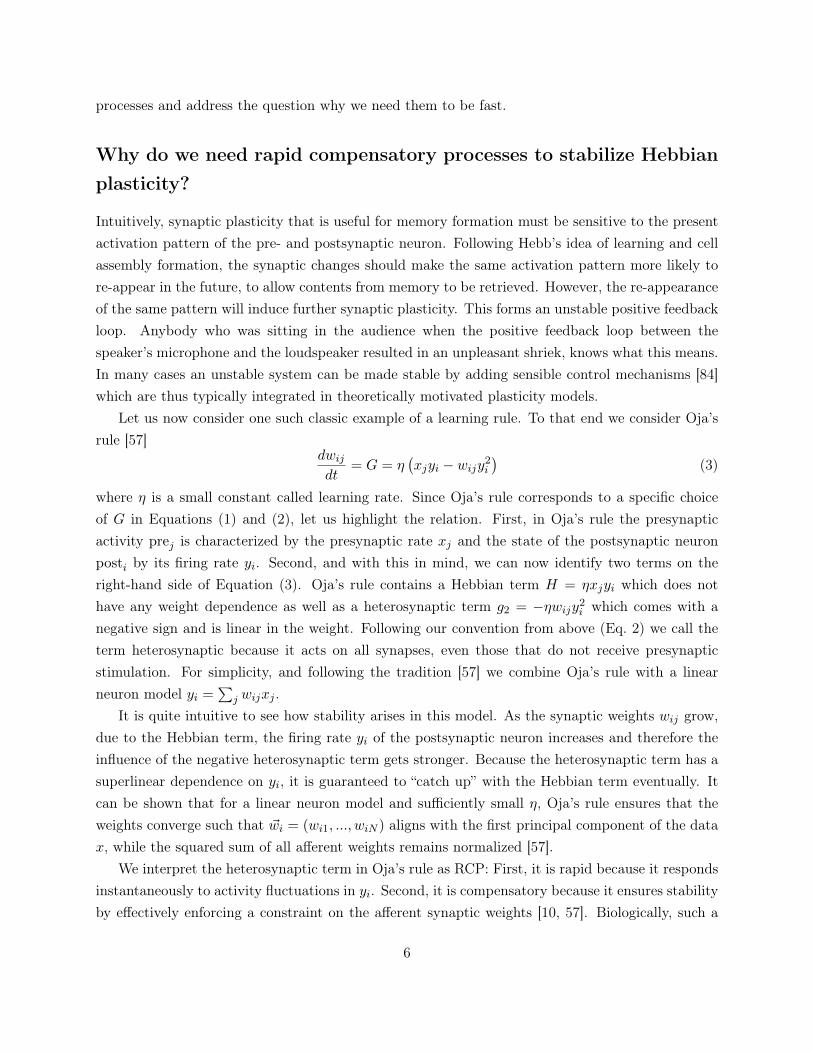
Figure 2: Illustration of a rapid andslow compensatory processes in avariation of Oja’s rule. (a) Here weused a fast filter time constant τ = 0.1η−1
(cf. Eq. (4)) and plot the output firingrate yi (solid) and the delayed estimate yi(dashed). (b) Same as in a), but with τ =2η−1. Model: We simulated dw
dt = xy−wy2,τ dydt = y − y and y = wx with x ≡ 1.
heterosynaptic effect could be obtained, for instance, when synapses have to compete for a sharedresource [57, 85] or send chemical signals to each other [86].
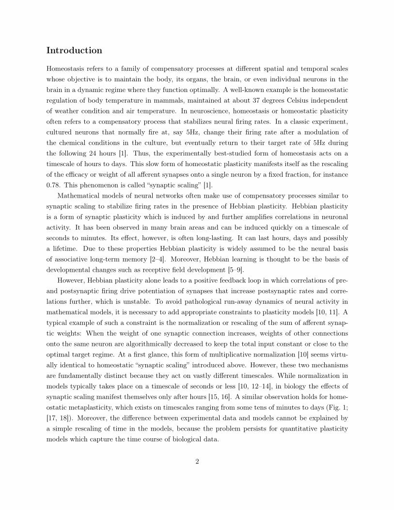
One could now ask if we really need this compensatory processes to be rapid. Could we notsimply replace the instantaneous firing rate yi in the heterosynaptic term by a slower variable?The timescale of the slow variable could be related in a biological system to the time necessaryto estimate the firing rate from, e.g. calcium concentration and translate these into metaplasticchanges in the learning rule. To illustrate the general idea by a concrete example, we take Oja’srule, as in Equation (3), except that, in the heterosynaptic term, we replace y2
i by y2i , where yi is a
low-pass filtered version of the postsynaptic rate
τydyidt
= yi − yi . (4)
If we choose τy = 1ms (for a fixed η of, e.g. η−1 = 10ms), the processes g2 = −ηwij y2i would still be
considered as rapid (Fig. 2a), but if we choose τy = 1h, it would be considered as slow. When thecompensatory processes is too slow, positive feedback induced by the Hebbian term is prone to takeover and oscillations (Fig. 2b) or even run-away dynamics arise. This is why we generally want thecompensatory processes to be rapid.
The same problematic has also been demonstrated nicely in the Bienenstock-Cooper-Munro(BCM) model [5]:
τdwijdt
= ηxjφ (yi, yi) (5)
7
−0.4
0
0.4
0.8
1.2
θLTD
LTP
Chang
ein
synaptic
efficacy
Postsynaptic activation
a
−1
−0.5
0
0.5
1y = y = κ
Chang
ein
synaptic
efficacy
Postsynaptic activation
y < κy = κy > κ
b
Figure 3: Most plasticity models canreproduce the notion of a plasticitythreshold reported in experiments.(a) The change in synaptic efficacy in manyplasticity models is a function of variablesrelated to postsynaptic activation. The pa-rameter θ is fixed and marks the transitionpoint (“threshold”) between LTP and LTD.(b) Schematic illustration of the action ofthe homeostatic moving threshold θ(t) inthe BCM model [5]. When the average y islarger than the target value κ, θ(t) shifts tohigher values. Likewise, θ(t) shifts to lowervalues when y is too low. For y = y = κchanges in synaptic efficacy are naught.
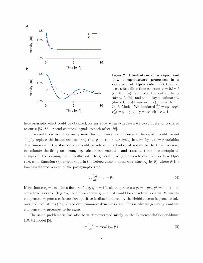
where φ is a nonlinear function with a characteristic shape characterized by a threshold θ betweenLTP and LTD (Fig. 3a), consistent with some induction protocols [70, 87]. The threshold θ dependson the moving average yi over past neuronal activity (Fig. 3b) where yi is defined in Equation (4).This is the reason why the model is said to have a “sliding” threshold.
To ensure stability, the BCM model requires two independent assumptions. First, the slidingthreshold has to be a superlinear function of yi [5]. A standard choice is [88]
θ(t) =y2i
κ(6)
where κ is the “target rate” to which the moving average of the postsynaptic firing rate shouldconverge. Second, τy cannot be “too large” compared to τ , because otherwise oscillations or run-away activity occur [23, 32, 88]. In fact, the ratio τy
τ determines the stability of the model.Oscillations and instabilities are generic to many nonlinear systems and not limited to the
above models. Control theory enables theoreticians to identify parameter ranges that lead to stablebehavior and avoid instabilities [31, 84, 89]. The control theoretic analysis of several plasticitymodels relying on moving averages of the postsynaptic firing rate shows that the response timescaleof the compensatory processes is constrained from above [23, 32, 88, 90]. In other words, theresponse time of the firing rate control has to be “relatively fast” compared to Hebbian plasticity.But how fast is fast enough? Is it seconds, hours or days?
8
How fast do compensatory processes have to be?
Because time can be rescaled arbitrarily in the above model, a quantitative answer to the questioncan only be given for a specific combination of neuronal, network and plasticity model parametersonce units of time are calibrated with biological data. In other words, we need to put a numericalvalue on τ to set the timescale of τy. To fix a timescale2 one can thus use any quantitative plasticitymodel which has been fitted to experimental data in combination with plausible spiking neuronmodels embedded into a spiking neural network with a biologically inspired activity state.
Such an analysis was done in Zenke et al. [32] using the plasticity model by Pfister and Gerstner[33], combined with negative feedback via either a sliding threshold or synaptic scaling. The criticaltimescale τcrit was determined as the value τy (Eq. (4)) above which a recurrent neural network,initially tuned to a low activity asynchronous state [91, 92], generates run-away activity. Usingsimulations and mean field theory τcrit was found to be on the order of seconds to minutes. Thus,the negative feedback needs to be too rapid to be linked to the known experiments of homeostaticsynaptic plasticity reviewed in Figure 1.
Several remarks are in order. First, although signatures of the stereotypical activity dependenceof the BCM model (Fig. 3a) are also present in STDP data and captured by many modern plasticitymodels [33, 61–63, 65, 93], the existence of a sliding threshold mechanisms is still a topic of ongoingdebate. However, we have shown analytically, and confirmed in simulations, that the instabilitythat arises through slow feedback in the BCM model is virtually identical to the situation in whichthe sliding threshold in Eq. (5) is replaced by a fixed threshold and instead synaptic scaling is addedto the model [32]. Additionally, the analysis suggests that similar temporal requirements hold foran entire family of plasticity models with an explicit rate dependence (see Yger and Gilson [90] fora review). Note, however, that additional instabilities can arise in the case of synaptic scaling [89].
Second, the critical timescale τcrit depends not only on the plasticity model, but also on multipleparameters of the neuron and network model. Moreover, the results showed a strong dependenceon background firing rate which was comparatively high in the Zenke et al. [32] model (∼3Hz). Onthe other hand, robust stability is only possible if the actual value of τy is chosen much smaller thanτcrit. The precise value of the critical timescale has therefore to be taken with care: We believe thatany published numerical value for τcrit may be off by a factor of 5 or 10 (because of uncertainty inchoices of neuronal and network parameters), but it is unlikely to be off by a factor 100. In any case,despite the remaining uncertainty, these numerical results suggest that most experimental forms ofhomeostatic plasticity are too slow to stabilize Hebbian plasticity as captured by current modelsand that RCPs are required to maintain stability.
A recent voltage based plasticity model by Jedlicka et al. [29] corroborates the above findings.2Strictly speaking, the notion of a “timescale” is meaningless for a nonlinear differential equation like Eq. (5), it
is only defined for linear dynamical systems. The notion can be rescued, however, in the vicinity of a fixed pointaround which the system can be linearized. We refer the interested reader to the Supplementary Material where weprovide additional information on this important, but technical issue.
9
By fitting their model with a rapid sliding threshold to in vivo data from dentate gyrus, the authorsfind τy ≈ 12s which allows them to speculate that the sliding threshold could be linked to CaMKIIinactivation.
Interestingly, Toyoizumi et al. [23] arrive at qualitatively similar conclusions by carefully analysingthe different phases of synaptic dynamics following monocular deprivation (MD) [39]. Specifically,they find that a fast sliding threshold guarantees stability, but provides a poor fit to experimentaldata, whereas as slow sliding threshold compromises stability altogether. Consequently, they sug-gest a model in which LTP and LTD saturate quickly to attain steady states. Additionally, a slowform of homeostatic plasticity is required to capture the data (cf. Fig. 1), but is no longer requiredto provide stability. In their model LTP and LTD saturate due to soft weight bounds. However, themodel does not crucially depend on this point and would presumably also work with other RCPs.
Finally, these findings are in good empirical agreement with many existing simulation studies ofplastic network models (Fig. 1) — in each of these, a rapid homeostatic control mechanisms on atimescale of seconds to minutes was implemented to maintain stability [14, 25, 27, 28, 33, 63, 94].
We can summarize our insights as follows. The fact that Hebbian plasticity has to be appropri-ately constrained through stabilizing mechanisms to avoid run-away activity is well known. Classicmodels such as Oja’s rule or the BCM model, for example, explicitly include appropriate mech-anisms based on the postsynaptic firing rate as an indicator and driver of stabilizing processes.However, the fact that these processes have to be rapid in absolute terms, only becomes apparentwhen units of time are fixed to a biologically meaningful timescale. Moreover, RCPs need to beeven more rapid in large recurrent network models, because a large number of plastic synapsestends to amplify instabilities unless the learning rate is scaled with the inverse number of synapses.Accumulating evidence suggests that biological forms of LTP and LTD have to be accompanied byRCPs which operate on timescales of seconds to minutes and are thus orders of magnitude fasterthan most known forms of homeostatic plasticity (cf. Fig. 1). This answers the questions as to whyRCPs are needed and gives us first upper limits on the intrinsic timescale of RCPs to successfullystabilize LTP and LTD. However, do we want RCPs to be a rapid version of homeostatic plasticitywith a single set point? We will now discuss this question in some more detail, before we turn topotential mechanisms.
Functional consequences of enforcing constraints on short timescales
Homeostatic mechanisms are typically interpreted as negative feedback processes [22], which relyon an error signal to maintain a given control variable of a dynamical system at designated targetvalues, or set points. Many control systems have a single set point. For instance, an autopilot triesto maintain a single given course at any given time. Similarly, most models of homeostatic plasticityhave a single target value, such as the average postsynaptic firing rate (see κ in the BCM modelEqs. (5) and (6)). Suppose we are dealing with a fast negative feedback processes, what are the
10
TimeSlow hom., single target
RCP, single target
RCP, target range
Inpu
tAct
ivity
Target
Act
ivity
Act
ivity
Limits
a
b
c
d
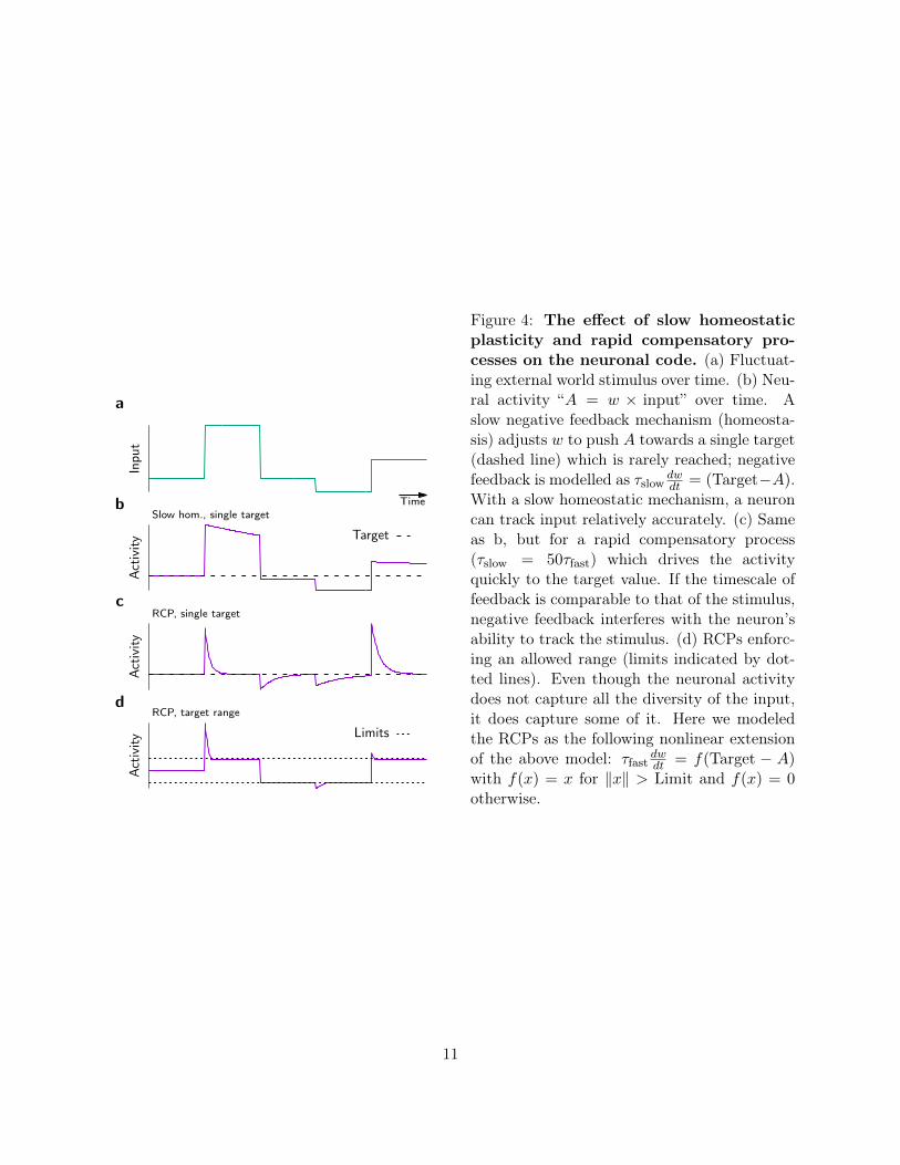
Figure 4: The effect of slow homeostaticplasticity and rapid compensatory pro-cesses on the neuronal code. (a) Fluctuat-ing external world stimulus over time. (b) Neu-ral activity “A = w × input” over time. Aslow negative feedback mechanism (homeosta-sis) adjusts w to push A towards a single target(dashed line) which is rarely reached; negativefeedback is modelled as τslow
dwdt = (Target−A).
With a slow homeostatic mechanism, a neuroncan track input relatively accurately. (c) Sameas b, but for a rapid compensatory process(τslow = 50τfast) which drives the activityquickly to the target value. If the timescale offeedback is comparable to that of the stimulus,negative feedback interferes with the neuron’sability to track the stimulus. (d) RCPs enforc-ing an allowed range (limits indicated by dot-ted lines). Even though the neuronal activitydoes not capture all the diversity of the input,it does capture some of it. Here we modeledthe RCPs as the following nonlinear extensionof the above model: τfast
dwdt = f(Target − A)
with f(x) = x for ‖x‖ > Limit and f(x) = 0otherwise.
11
functional consequences for plasticity and circuit dynamics? To do so, we focus on commonly foundforms of firing rate homeostasis (FRH) with a single firing rate set point [15, 16, 95].
Neurons encode information in changes of their electrical activity levels. For instance, subsetsof simple cells in the visual system fire spikes in response to specific edge-like features in the visualfield [96]; cells in higher brain areas respond with high specificity to complex concepts and remainquiescent when the concept they are coding for is not brought to mind [97–99]; and finally certainneurons respond selectively with elevated firing rates over extended periods during working memorytasks [100–102]. The ability of neurons to selectively indicate through periods of strong activity thepresence of specific features in the input or specific concepts in working memory is an importantcondition for computation.
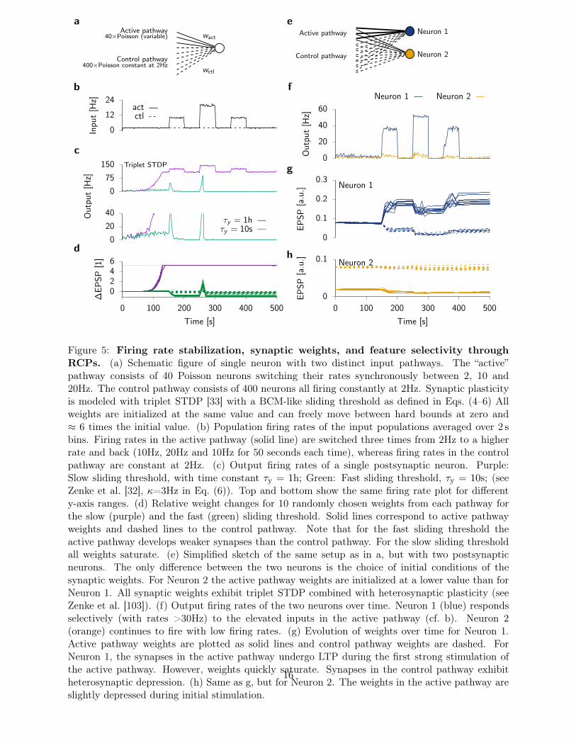
Is the notion of a single set point compatible with the task of neurons to selectively respondto stimulation? If negative feedback control of firing rates is slow (e.g. synaptic homeostasis),neuronal firing can deviate substantially from the mean firing rates during short times and thusencode information (Fig. 4a,b). However, we have strong reasons to believe that slow homeostaticcontrol mechanism cannot stabilize the ravaging effects of Hebbian plasticity. So what can wesay about a putative RCP? If it were to act like FRH, but on a short timescale (e.g. seconds tominutes), neural codes based on neuronal activity become problematic because synaptic plasticitystarts to suppress activity fluctuations which could be carrying important information (Fig. 4c).For example, if the RCP has a timescale of two seconds, rapid stimuli that change on a timescaleof 0.5 seconds would be transmitted as a rate signal of the postsynaptic neuron while stimulisustained for more than 5 seconds would be suppressed by compensatory synaptic changes. Evenmore alarmingly, certain forms of homosynaptic plasticity, like the BCM [5] or the triplet STDP[33] model endowed with a rapid sliding threshold, not only suppress high activity periods, but also“unlearn” previously acquired selectivity and erase memories (Figs. 4a,c and 5a–d). Therefore rapidcompensatory processes which enforce a single set point are hardly desirable from a functional pointof view. Thus the requirement of fast negative feedback control over Hebbian plasticity with a singleset point poses a problem in itself.
It is important to appreciate that this problem arises from the combination of a single targetwith the requirement to implement negative feedback on a short timescale. Fortunately, most formsof homeostatic plasticity are slow (cf. Fig. 1). Thus, known homeostatic mechanisms do not interferewith neuronal coding. For RCPs not to interfere with coding either, it thus seems important thatthey do not enforce a single set point constraint on postsynaptic activity. Nevertheless, these RCPshave to prevent run-away activity.
There could be at least one simple solution to this conundrum [23, 103]. Suppose there are two,or more, set points enforced by two or more RCPs. For instance, one RCP could activate above acertain activity threshold and ensure that neuronal activity does not exceed this threshold. Similarly,a second mechanisms could activate below another lower activity threshold. The combined action
12
of the two mechanisms enforces neural activity to stay within an allowed range, but still permitssubstantial firing rate fluctuations inside that range (Fig. 4d).
When such a pair of RCP is combined with a form of Hebbian plasticity which has its plasticitythreshold within the limits of the allowed activity regime, the neural activity of the compoundsystem naturally becomes multistable for prolonged stimulation with the same stimulus. Withinthe allowed range no RCP is active, but Hebbian plasticity is intrinsically unstable. Thus, for astimulus sustained longer than the timescale of the RCP and Hebbian learning, any value of thepostsynaptic rate within the allowed region will lead to either LTP or LTD until the system reachesthe limits at which either RCP rapidly intervenes by undoing any excess LTP or LTD from there on.The compound system exhibits therefore two, potentially stimulus dependent, stable equilibriumpoints, one at low and one at high activity.
Let us apply these considerations to two different systems, viz., a sensory system and a memorysystem. To be concrete, we assume that in either system the timescales of both LTP inductionand RCP are two seconds. In the sensory system, each neuron will respond in a graded manner toshort stimuli (say, with a duration of half a second) because synapses hardly change during a singlestimulus duration. However, the repeated stimulation with different stimuli will cause long-lastingweight changes. The location of the high-activity fixed point depends on the stimulus ensembleused during stimulation. Moreover, if we drive the neuron with a single sustained stimulus, thehigh-activity fixed point adjusts on the timescale of a few seconds and reflects the value of theinput.
The case of a memory system was considered in [103]. Suppose the high-activity fixed pointcorresponds to a memory retrieval state, while the low-activity equilibrium is associated with aquiescent memory which is not being recalled. Because both states are stable, it is irrelevantwhether the memory is recalled every other minute or once a year. Importantly, this is differentfrom models with rapid FRH, which might require neuronal activity to regularly turn on and off tosatisfy the constraint. An example for this is the network model by Litwin-Kumar and Doiron [14]in which inhibitory synaptic plasticity (ISP) acts as rapid FRH with a single activity fixed point.
We can therefore answer the third of the questions raised in the introduction: The functionalconsequences of rapid control mechanism with a single set point are that neurons lose the flexibilitythat is necessary for coding. The consequences are therefore undesirable and the proposed solution isto design RCPs that allow for several set points or a target range of permissible activity. In the nextsections we will first discuss common ways to constrain unbounded weight growth and explain whythey are insufficient to provide stability, before we turn to potential candidate plasticity mechanismswhich could act as RCP. Finally, we show an example of a spiking network model based on theseprinciples which forms and recalls memories encoded in cell assemblies.
13
Potential mechanisms
To stabilize Hebbian plasticity, any RCP at the synaptic, dendritic, neuronal or network level canbe considered. Due to temporal and spatial constraints of the biological substrate it seems mostlikely, however, that the fastest mechanisms are implemented as close to the synapse as possible.
At the synaptic level, excessive growth has traditionally been limited by soft or hard boundson individual synaptic weights or other choices of explicit weight dependence of the Hebbian andheterosynaptic terms in Eq. (2) [8, 10, 31, 65, 82, 83, 104, 104–107]. For example, to avoid bimodalweight distributions, which sometimes arise in competitive models, but are not observed in biology,a range of multiplicative plasticity models [31, 82, 83, 104, 105, 108], with an appropriate choice ofthe weight-dependence of H in Eq. (2), have been devised. However, bounds on individual synapticweights only impose an implicit constraint on the postsynaptic activity. To see this, consider apermissible range of individual synaptic strength of, say, ±50% around the initial efficacy, whichseems not uncommon for plasticity induction protocols. However, by setting this range we do notautomatically exclude the situation in which all synapses increase their efficacy by 50% which wouldin all likelihood correspond to pathological activity levels.
To avoid such run-away activity, plasticity has to ensure that not all synapses are potentiated ordepressed equally. Rather there should be some form of competition which ensures that when oneset of synapses is potentiated other synapses are depressed by a certain amount. While some degreeof competition can be seen in STDP models, in which presynaptic spikes compete in time to elicit apostsynaptic spike [8, 58, 109, 110], this competition is generally weak [31] and without additionalconstraints, activity levels still succumb to run-away effects with detrimental consequences in recur-rent neural networks [14, 32, 103]. Robust competition, for instance, through a BCM-like threshold[5] or explicit constraints on the sum of weights [10], are therefore of paramount importance forplasticity models.
In summary, there exist multiple mechanisms to limit growth of individual synaptic weights.However, to achieve robust synaptic competition and stability of output firing rates, more explicitactivity constraints are required, as exemplified in the BCM model, or through explicit heterosynap-tic interactions, similar to Oja’s rule (cf. Eq. (3)). We have already argued that these constraintsneed to be enforced rapidly. We now ask what possible mechanisms at the neuronal or networklevel could achieve that.
At the network level RCPs might be implemented by inhibition and ISP which could influenceplasticity at excitatory synapses either directly or indirectly. Some theoretical forms ISP are knownto implement a rapid form of FRH for individual neurons [14, 111]. With accumulating experimentalevidence for ISP [95, 112, 113] it therefore seems likely that synaptic inhibition influences plasticityof excitatory synapses at least indirectly through changes in activity. However, in experiments thetimescale of FRH mediated through ISP appears to be relatively slow [95] and it remains to be seenwhether biological forms of ISP can act as RCP or whether they have a rather homeostatic role.
14
However, in some cases, inhibition without ISP can have a stabilizing effect. Lim et al. [114] haverecently demonstrated that this can indeed lead to stability of certain forms of Hebbian plasticity.Moreover, inhibition can also directly affect and regulate excitatory plasticity [115]. Particularlyinteresting in this context are results by Delgado et al. [116] who observed total-conductance-dependent changes of the STDP curve depending on excitatory and inhibitory background input.Their results suggests that increased, but balanced, excitatory and inhibitory input biases theSTDP window towards LTD and can thus act as a RCP. Delgado et al. [116] demonstrated this in asingle-neuron feed-forward model, but it is not yet known whether these results generalize to largernetworks.
At the neuronal level, algorithmic normalization of afferent synaptic weights is a commonlyused mechanism to stabilize Hebbian plasticity in network models while simultaneously allowingstructure formation [9, 10, 13, 14]. While such rapid and precise scaling at the neuronal level hasbeen criticized as biologically implausible [5], an “approximate” scaling could potentially be achievedthrough heterosynaptic plasticity at the dendritic level [117].
Heterosynaptic plasticity has moved back in the focus recently, because of its potential roleas RCP [20, 21, 86, 103, 118]. Importantly, some forms of heterosynaptic plasticity are fast, andprovide primarily negative feedback in response to high postsynaptic activity levels [20, 119] or inthe presence of strong LTP on a dendritic segment [86]. This is reminiscent of Oja’s rule (Eq. (3))and seems well suited to counteract run-away LTP. In contrast to Oja’s rule, these heterosynapticchanges are induced by bursts of postsynaptic activity which implies that the quadratic term y2
i inEq. (3) should be replaced by a term that is triggered either by firing rates yi above some threshold[118] or by a higher power such as y4
i [103].In models which also show run-away LTD at low activities (e.g. [33, 63]), an additional RCP is
needed which either saturates or counteracts LTD. Possible forms of plasticity include, but are notlimited to, transmitter-induced plasticity, homeostatic scaling-up or spontaneous spine formation.
In the following section we review a plasticity model which combines Hebbian plasticity withtwo RCPs that enable more than a single set point of neuronal activity. We also discuss, in thecontext of the model, the potential role of additional slow homeostatic mechanisms.
Learning and recall in a recurrent spiking network model
We now discuss a learning rule which combines a plausible model of Hebbian plasticity with twoadditional RCPs [103]. For sensible combinations this compound model does not suffer from the run-away effects of purely Hebbian plasticity and exhibits intrinsic multistability instead (cf. Fig. 4d).
The basic logic of multistable plasticity can be summarized as follows. At high activity levelsa rapid form of heterosynaptic plasticity limits run-away LTP and creates synaptic competition.Similarly, at low activity levels an unspecific form of plasticity which only depends on presynapticactivity prevents run-away LTD. The well-orchestrated interplay between these adversarial plasticity
15
Active pathway40×Poisson (variable)
Control pathway400×Poisson constant at 2Hz
wact
wctl
a
0
12
24In
put
[Hz]
actctl
b
0
75
150 Triplet STDP
0
20
40Out
put
[Hz]
τy = 1hτy = 10s
c
0246
0 100 200 300 400 500
∆EP
SP[1
]
Time [s]
d
Neuron 1
Neuron 2
Active pathway
Control pathway
e
0
20
40
60
0
0.1
0.2
0.3Neuron 1
0
0.1
0 100 200 300 400 500
Neuron 2
Out
put
[Hz]
Neuron 1 Neuron 2
EPSP
[a.u
.]EP
SP[a
.u.]
Time [s]
f
g
h
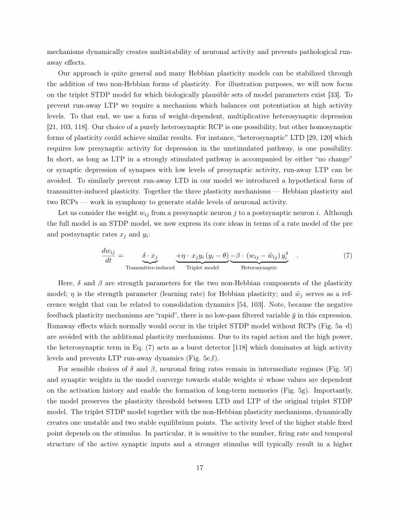
Figure 5: Firing rate stabilization, synaptic weights, and feature selectivity throughRCPs. (a) Schematic figure of single neuron with two distinct input pathways. The “active”pathway consists of 40 Poisson neurons switching their rates synchronously between 2, 10 and20Hz. The control pathway consists of 400 neurons all firing constantly at 2Hz. Synaptic plasticityis modeled with triplet STDP [33] with a BCM-like sliding threshold as defined in Eqs. (4–6) Allweights are initialized at the same value and can freely move between hard bounds at zero and≈ 6 times the initial value. (b) Population firing rates of the input populations averaged over 2 sbins. Firing rates in the active pathway (solid line) are switched three times from 2Hz to a higherrate and back (10Hz, 20Hz and 10Hz for 50 seconds each time), whereas firing rates in the controlpathway are constant at 2Hz. (c) Output firing rates of a single postsynaptic neuron. Purple:Slow sliding threshold, with time constant τy = 1h; Green: Fast sliding threshold, τy = 10s; (seeZenke et al. [32], κ=3Hz in Eq. (6)). Top and bottom show the same firing rate plot for differenty-axis ranges. (d) Relative weight changes for 10 randomly chosen weights from each pathway forthe slow (purple) and the fast (green) sliding threshold. Solid lines correspond to active pathwayweights and dashed lines to the control pathway. Note that for the fast sliding threshold theactive pathway develops weaker synapses than the control pathway. For the slow sliding thresholdall weights saturate. (e) Simplified sketch of the same setup as in a, but with two postsynapticneurons. The only difference between the two neurons is the choice of initial conditions of thesynaptic weights. For Neuron 2 the active pathway weights are initialized at a lower value than forNeuron 1. All synaptic weights exhibit triplet STDP combined with heterosynaptic plasticity (seeZenke et al. [103]). (f) Output firing rates of the two neurons over time. Neuron 1 (blue) respondsselectively (with rates >30Hz) to the elevated inputs in the active pathway (cf. b). Neuron 2(orange) continues to fire with low firing rates. (g) Evolution of weights over time for Neuron 1.Active pathway weights are plotted as solid lines and control pathway weights are dashed. ForNeuron 1, the synapses in the active pathway undergo LTP during the first strong stimulation ofthe active pathway. However, weights quickly saturate. Synapses in the control pathway exhibitheterosynaptic depression. (h) Same as g, but for Neuron 2. The weights in the active pathway areslightly depressed during initial stimulation.
16
mechanisms dynamically creates multistability of neuronal activity and prevents pathological run-away effects.
Our approach is quite general and many Hebbian plasticity models can be stabilized throughthe addition of two non-Hebbian forms of plasticity. For illustration purposes, we will now focuson the triplet STDP model for which biologically plausible sets of model parameters exist [33]. Toprevent run-away LTP we require a mechanism which balances out potentiation at high activitylevels. To that end, we use a form of weight-dependent, multiplicative heterosynaptic depression[21, 103, 118]. Our choice of a purely heterosynaptic RCP is one possibility, but other homosynapticforms of plasticity could achieve similar results. For instance, “heterosynaptic” LTD [29, 120] whichrequires low presynaptic activity for depression in the unstimulated pathway, is one possibility.In short, as long as LTP in a strongly stimulated pathway is accompanied by either “no change”or synaptic depression of synapses with low levels of presynaptic activity, run-away LTP can beavoided. To similarly prevent run-away LTD in our model we introduced a hypothetical form oftransmitter-induced plasticity. Together the three plasticity mechanisms — Hebbian plasticity andtwo RCPs — work in symphony to generate stable levels of neuronal activity.
Let us consider the weight wij from a presynaptic neuron j to a postsynaptic neuron i. Althoughthe full model is an STDP model, we now express its core ideas in terms of a rate model of the preand postsynaptic rates xj and yi:
dwijdt
= δ · xj︸ ︷︷ ︸Transmitter-induced
+η · xjyi (yi − θ)︸ ︷︷ ︸Triplet model
−β · (wij − wij) y4i︸ ︷︷ ︸
Heterosynaptic
. (7)
Here, δ and β are strength parameters for the two non-Hebbian components of the plasticitymodel; η is the strength parameter (learning rate) for Hebbian plasticity; and wj serves as a ref-erence weight that can be related to consolidation dynamics [54, 103]. Note, because the negativefeedback plasticity mechanisms are “rapid”, there is no low-pass filtered variable y in this expression.Runaway effects which normally would occur in the triplet STDP model without RCPs (Fig. 5a–d)are avoided with the additional plasticity mechanisms. Due to its rapid action and the high power,the heterosynaptic term in Eq. (7) acts as a burst detector [118] which dominates at high activitylevels and prevents LTP run-away dynamics (Fig. 5e,f).
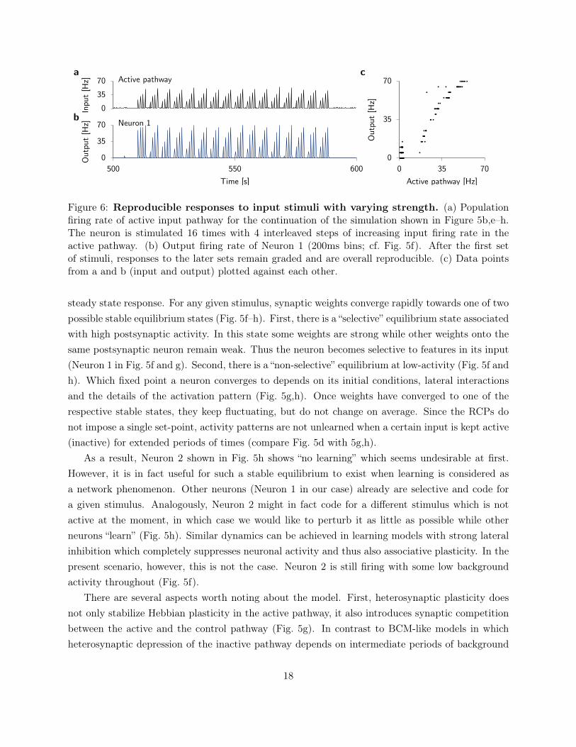
For sensible choices of δ and β, neuronal firing rates remain in intermediate regimes (Fig. 5f)and synaptic weights in the model converge towards stable weights w whose values are dependenton the activation history and enable the formation of long-term memories (Fig. 5g). Importantly,the model preserves the plasticity threshold between LTD and LTP of the original triplet STDPmodel. The triplet STDP model together with the non-Hebbian plasticity mechanisms, dynamicallycreates one unstable and two stable equilibrium points. The activity level of the higher stable fixedpoint depends on the stimulus. In particular, it is sensitive to the number, firing rate and temporalstructure of the active synaptic inputs and a stronger stimulus will typically result in a higher
17
0
35
70 Active pathway
0
35
70
500 550 600
Neuron 1
0
35
70
0 35 70
Inpu
t[H
z]O
utpu
t[H
z]
Time [s]
Out
put
[Hz]
Active pathway [Hz]
a
b
c
Figure 6: Reproducible responses to input stimuli with varying strength. (a) Populationfiring rate of active input pathway for the continuation of the simulation shown in Figure 5b,e–h.The neuron is stimulated 16 times with 4 interleaved steps of increasing input firing rate in theactive pathway. (b) Output firing rate of Neuron 1 (200ms bins; cf. Fig. 5f). After the first setof stimuli, responses to the later sets remain graded and are overall reproducible. (c) Data pointsfrom a and b (input and output) plotted against each other.
steady state response. For any given stimulus, synaptic weights converge rapidly towards one of twopossible stable equilibrium states (Fig. 5f–h). First, there is a “selective” equilibrium state associatedwith high postsynaptic activity. In this state some weights are strong while other weights onto thesame postsynaptic neuron remain weak. Thus the neuron becomes selective to features in its input(Neuron 1 in Fig. 5f and g). Second, there is a “non-selective” equilibrium at low-activity (Fig. 5f andh). Which fixed point a neuron converges to depends on its initial conditions, lateral interactionsand the details of the activation pattern (Fig. 5g,h). Once weights have converged to one of therespective stable states, they keep fluctuating, but do not change on average. Since the RCPs donot impose a single set-point, activity patterns are not unlearned when a certain input is kept active(inactive) for extended periods of times (compare Fig. 5d with 5g,h).
As a result, Neuron 2 shown in Fig. 5h shows “no learning” which seems undesirable at first.However, it is in fact useful for such a stable equilibrium to exist when learning is considered asa network phenomenon. Other neurons (Neuron 1 in our case) already are selective and code fora given stimulus. Analogously, Neuron 2 might in fact code for a different stimulus which is notactive at the moment, in which case we would like to perturb it as little as possible while otherneurons “learn” (Fig. 5h). Similar dynamics can be achieved in learning models with strong lateralinhibition which completely suppresses neuronal activity and thus also associative plasticity. In thepresent scenario, however, this is not the case. Neuron 2 is still firing with some low backgroundactivity throughout (Fig. 5f).
There are several aspects worth noting about the model. First, heterosynaptic plasticity doesnot only stabilize Hebbian plasticity in the active pathway, it also introduces synaptic competitionbetween the active and the control pathway (Fig. 5g). In contrast to BCM-like models in whichheterosynaptic depression of the inactive pathway depends on intermediate periods of background
18
activity in between stimuli [29, 88], here the heterosynaptic depression happens simultaneouslyto LTP induction (Fig. 5g). Second, although the learning rule effectively implements a rapidredistribution of synaptic weights reminiscent of synaptic scaling, it is still a fully local learningrule which only depends on information which is available at the synapse (cf. Eq. (7)). Third,although the learning rule effectively implements bistable dynamics for each stimulus, the “selective”equilibrium level remains stimulus dependent, which allows the neuron to respond in a graded andreproducible way to input stimuli of varying intensity (Fig. 6). Fourth, in general the referenceweight w is not fixed, but follows its own temporal dynamics on a slower timescale (≈ 20min
and more). Such slow complex synaptic dynamics are essential to capture experiments on synapticconsolidation [50, 54, 121], but could similarly be used to model slow forms of homeostatic plasticity[23].
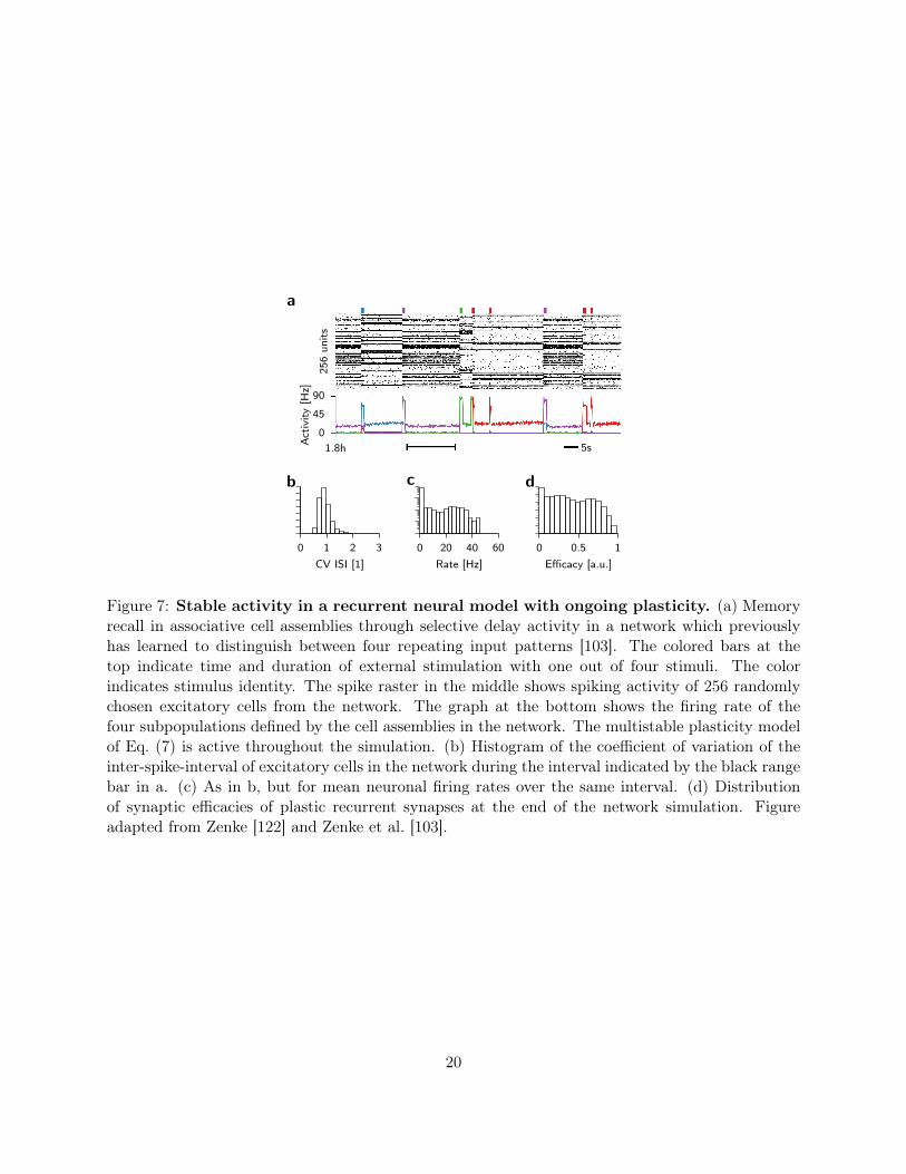
Finally, the stability properties of the learning rule in Eq. (7) are not limited to simple feed-forward circuits, but generalize to more realistic scenarios. Specifically, the combination of het-erosynaptic and Hebbian plasticity enables stable on-line learning and recall of cell assemblies inlarge spiking neural networks (Fig. 7a,b; [103]). Stationary firing rates in the model depend on theconnectivity pattern and the spiking statistics of active inputs. In a recurrent network, however,output spike trains pose as inputs to other neurons. As a nontrivial consequence, stationary so-lutions of the network state exhibit firing rate distributions which are uni-modal and long-tailed(Fig. 7c,d). Individual neuronal firing rates only stabilize under stationary conditions. If the ratesare non stationary, for instance due to the inclusion of additional adaptation processes in the neuronmodel, rates in the model drift on behavioral timescales (see Zenke et al. [103] for details).
In summary, orchestrating Hebbian plasticity and RCPs on comparable timescales dynamicallygenerates multistability. This reconciles the experimentally observed fast induction of synapticplasticity with stable synaptic dynamics and stability of learning and memory at the single neuronlevel as well as in large networks. However, there are a few caveats with this approach which wewill discuss in the following.
Problems of RCPs at the single neuron level
Consider a population of neurons with plastic synapses which follow intrinsically stable plasticitydynamics such as the ones described in the last section. To encode and process information effi-ciently, neuronal populations need to create internal representations of the external world. Doingthis efficiently requires the response to be sparse across the population. In other words, only a sub-set of neurons should respond for each stimulus. Moreover, different stimuli should evoke responsesfrom different subsets of neurons within the population to avoid that all stimuli look “the same” tothe neural circuit. Finally, individual neurons should respond sparsely over time. Imagine a neuronwhich is active for all possible stimuli. It would be as uninformative as one which never responds toany of the inputs. Therefore, to represent and process information in neural populations efficiently,
19
0
45
90
1.8h 5s
256
units
Act
ivity
[Hz]
a
0 1 2 3
CV ISI [1]
b
0 20 40 60
Rate [Hz]
c
0 0.5 1
Efficacy [a.u.]
d
Figure 7: Stable activity in a recurrent neural model with ongoing plasticity. (a) Memoryrecall in associative cell assemblies through selective delay activity in a network which previouslyhas learned to distinguish between four repeating input patterns [103]. The colored bars at thetop indicate time and duration of external stimulation with one out of four stimuli. The colorindicates stimulus identity. The spike raster in the middle shows spiking activity of 256 randomlychosen excitatory cells from the network. The graph at the bottom shows the firing rate of thefour subpopulations defined by the cell assemblies in the network. The multistable plasticity modelof Eq. (7) is active throughout the simulation. (b) Histogram of the coefficient of variation of theinter-spike-interval of excitatory cells in the network during the interval indicated by the black rangebar in a. (c) As in b, but for mean neuronal firing rates over the same interval. (d) Distributionof synaptic efficacies of plastic recurrent synapses at the end of the network simulation. Figureadapted from Zenke [122] and Zenke et al. [103].
20
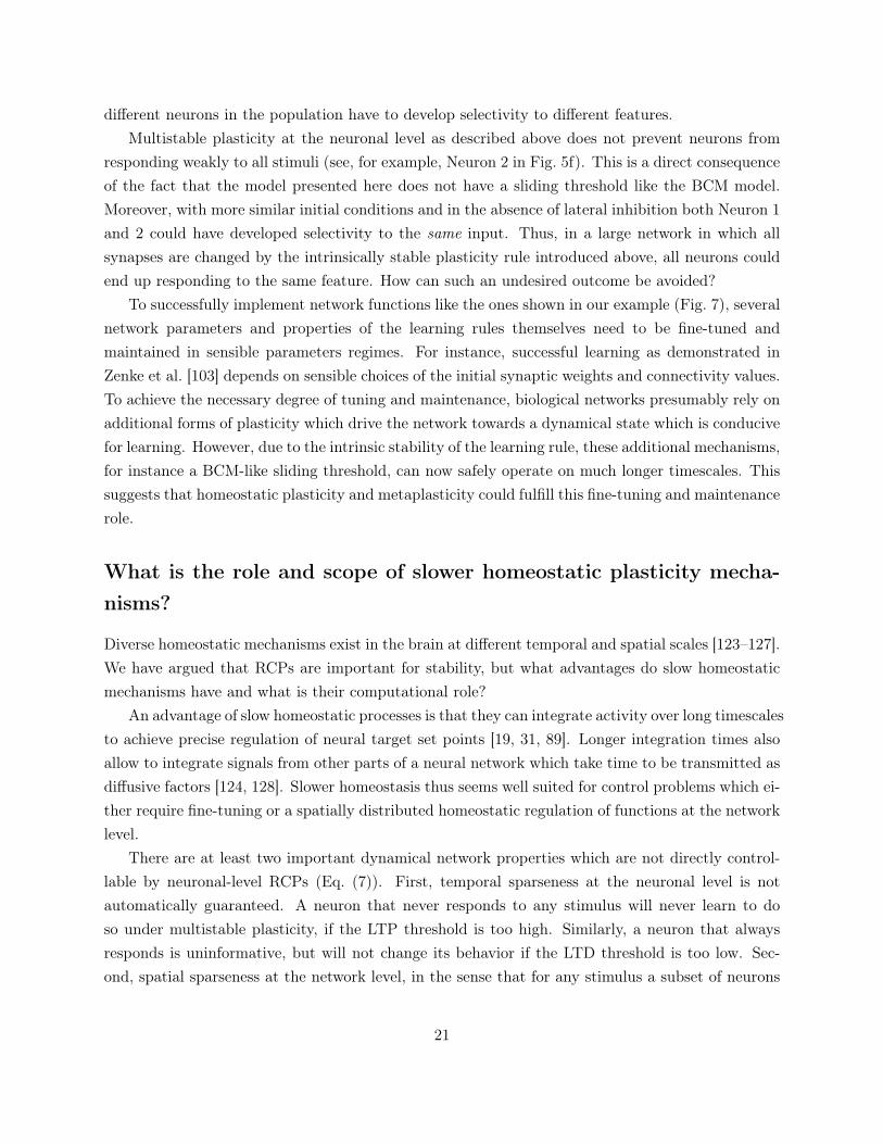
different neurons in the population have to develop selectivity to different features.Multistable plasticity at the neuronal level as described above does not prevent neurons from
responding weakly to all stimuli (see, for example, Neuron 2 in Fig. 5f). This is a direct consequenceof the fact that the model presented here does not have a sliding threshold like the BCM model.Moreover, with more similar initial conditions and in the absence of lateral inhibition both Neuron 1and 2 could have developed selectivity to the same input. Thus, in a large network in which allsynapses are changed by the intrinsically stable plasticity rule introduced above, all neurons couldend up responding to the same feature. How can such an undesired outcome be avoided?
To successfully implement network functions like the ones shown in our example (Fig. 7), severalnetwork parameters and properties of the learning rules themselves need to be fine-tuned andmaintained in sensible parameters regimes. For instance, successful learning as demonstrated inZenke et al. [103] depends on sensible choices of the initial synaptic weights and connectivity values.To achieve the necessary degree of tuning and maintenance, biological networks presumably rely onadditional forms of plasticity which drive the network towards a dynamical state which is conducivefor learning. However, due to the intrinsic stability of the learning rule, these additional mechanisms,for instance a BCM-like sliding threshold, can now safely operate on much longer timescales. Thissuggests that homeostatic plasticity and metaplasticity could fulfill this fine-tuning and maintenancerole.
What is the role and scope of slower homeostatic plasticity mecha-nisms?
Diverse homeostatic mechanisms exist in the brain at different temporal and spatial scales [123–127].We have argued that RCPs are important for stability, but what advantages do slow homeostaticmechanisms have and what is their computational role?
An advantage of slow homeostatic processes is that they can integrate activity over long timescalesto achieve precise regulation of neural target set points [19, 31, 89]. Longer integration times alsoallow to integrate signals from other parts of a neural network which take time to be transmitted asdiffusive factors [124, 128]. Slower homeostasis thus seems well suited for control problems which ei-ther require fine-tuning or a spatially distributed homeostatic regulation of functions at the networklevel.
There are at least two important dynamical network properties which are not directly control-lable by neuronal-level RCPs (Eq. (7)). First, temporal sparseness at the neuronal level is notautomatically guaranteed. A neuron that never responds to any stimulus will never learn to doso under multistable plasticity, if the LTP threshold is too high. Similarly, a neuron that alwaysresponds is uninformative, but will not change its behavior if the LTD threshold is too low. Sec-ond, spatial sparseness at the network level, in the sense that for any stimulus a subset of neurons
21
responds, is also not automatically guaranteed. Lateral inhibition is a suitable candidate to decorre-late responses of different neurons in a network, but, as excitatory synapses change during learning,the strength of lateral inhibition needs to be co-regulated.
The problem of temporal sparseness can be solved by any mechanism which ensures that aneuron which has been completely silent for very long, eventually “gets a chance” to reach anactivity level above the LTP threshold. This can be achieved by either lowering the thresholdas in the BCM theory [5, 88, 103] or by slowly increasing the gain of either the neuron itself orthe excitatory synapses through other forms of slow homeostatic plasticity [23, 129, 130]. Finally,similar homeostatic effects could be achieved by dis-inhibition through the action of neuron specificISP [112] or by decreasing the response of inhibitory neurons [40, 131]. Conversely, a neuron thatis uninformative because it is always active could decrease its response to some stimuli by theopposite action of one or several of the homeostatic mechanisms mentioned above, such as increasedinhibition, reduced excitation, or reduced excitability.
While it is conceivable that mechanisms addressing the issue of temporal sparseness could actlocally at the neuronal level, it is clear that enforcing spatially sparse activity at the population levelcan only be achieved in a non-local manner. A common approach to guarantee spatial sparsenessin models, is to include lateral inhibition, as done in subspace learning algorithms [132, 133], sparsecoding paradigms [134, 135] or models of associative memory [14, 103, 136–140]. However, achievingappropriate levels of inhibition can be difficult, especially if excitatory synaptic weights are notstatic, but change over time and on a per neuron basis [111]. To solve this task in biologicalnetworks, ISP would be a natural candidate. However, most existing models of ISP are purely localand tune inhibition on a per neuron level [111, 112, 141]. More specifically, ISP acts as a neuronalRCP which rapidly drives firing rates to a single stable set point (cf. Fig. 4; Vogels et al. [111]).To achieve a certain level of spatial sparseness through any form of homeostatic plasticity, requiresa signal with a wider scope which encodes network activity [124, 128]. Using such a signal it isthen possible to modulate plasticity [73]. For example, in Zenke et al. [103] ISP is modulated by alow-pass filtered signal which encodes network activity. As a result, the combination of intrinsicallymultistable plasticity at excitatory synapses and ISP, ensures that recurrent inhibition is tuned toa level where only one cell assembly can be active at any given time. Importantly, this homeostaticmechanism does not have to be permanently active. For instance, once the inhibitory feedbackwithin the model is tuned to the “sweet spot” at which the network can operate, ISP can be turnedoff safely without impairing stability. Similarly, it seems likely that some forms of homeostaticplasticity could be dormant for most of the time and spring into action only during the initial phaseof development [142] or when an extreme external manipulation changes the network dynamics [1].
We are thus able to answer the final question from the introduction as follows: Slow homeostaticmechanisms tune parameters of plasticity rules and neurons to enable efficient use of the availableresources in networks. For example, for the sake of efficiency, no neuron should be never active;
22
0
0.2
0.4
0.6
Plasticity protocol Time
Syna
ptic
stat
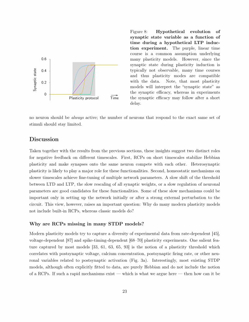
eFigure 8: Hypothetical evolution ofsynaptic state variable as a function oftime during a hypothetical LTP induc-tion experiment. The purple, linear timecourse is a common assumption underlyingmany plasticity models. However, since thesynaptic state during plasticity induction istypically not observable, many time coursesand thus plasticity modes are compatiblewith the data. Note, that most plasticitymodels will interpret the “synaptic state” asthe synaptic efficacy, whereas in experimentsthe synaptic efficacy may follow after a shortdelay.
no neuron should be always active; the number of neurons that respond to the exact same set ofstimuli should stay limited.
Discussion
Taken together with the results from the previous sections, these insights suggest two distinct rolesfor negative feedback on different timescales. First, RCPs on short timescales stabilize Hebbianplasticity and make synapses onto the same neuron compete with each other. Heterosynapticplasticity is likely to play a major role for these functionalities. Second, homeostatic mechanisms onslower timescales achieve fine-tuning of multiple network parameters. A slow shift of the thresholdbetween LTD and LTP, the slow rescaling of all synaptic weights, or a slow regulation of neuronalparameters are good candidates for these functionalities. Some of these slow mechanisms could beimportant only in setting up the network initially or after a strong external perturbation to thecircuit. This view, however, raises an important question: Why do many modern plasticity modelsnot include built-in RCPs, whereas classic models do?
Why are RCPs missing in many STDP models?
Modern plasticity models try to capture a diversity of experimental data from rate-dependent [45],voltage-dependent [87] and spike-timing-dependent [68–70] plasticity experiments. One salient fea-ture captured by most models [33, 61, 63, 65, 93] is the notion of a plasticity threshold whichcorrelates with postsynaptic voltage, calcium concentration, postsynaptic firing rate, or other neu-ronal variables related to postsynaptic activation (Fig. 3a). Interestingly, most existing STDPmodels, although often explicitly fitted to data, are purely Hebbian and do not include the notionof a RCPs. If such a rapid mechanisms exist — which is what we argue here — then how can it be
23

that existing plasticity models without them can quantitatively capture the data from experiments?There are presumably three main reasons for this. First, STDP experiments typically manipulate
a single pathway, either by stimulating a presynaptic neuron or a bundle of presynaptic axons.Sometimes a designated control pathway (i.e. a second presynaptic neuron) is missing, or, if it isnot missing, the effect size in the control pathway is considered as weak. However, from a theoreticalperspective, we expect that heterosynaptic effects caused by stimulation of one presynaptic pathwayare weak when measured at only one “control” synapse; a weak change at individual synapsescould still have a strong accumulated effect over thousands of synapses. Therefore even weakheterosynaptic plasticity could act as a strong RCP [21, 103, 118].
Second, in an STDP experiment with 60 repetitions of pre-post-pairs, the total activation ofthe postsynaptic neuron is still in a reasonable regime. Therefore it is unclear whether the “burst-detector” for heterosynaptic plasticity would be triggered [21, 103, 118].
Third, experiments typically rely on repeated pre- and postsynaptic activation. Moreover, duringthe induction protocol, synaptic efficacy changes are usually not observable. Plasticity models arethus fitted to pairs of initial and final synaptic strength. However, the unobserved intermediatesynaptic dynamics during a standard LTP induction protocol could be qualitatively very different(Fig. 8), but are obscured in experiments by measurement artifacts as well as short-term plasticityriding on top of the induced Hebbian LTP. These differences in the dynamics contain the answers toquestions such as: Is the final synaptic strength stable or would it increase further with additionalpairings? Is there a threshold of number of pairings that needs to be reached for an all or nothingeffect?
Because the detailed internal dynamics of synapses during induction are not known, differentplasticity models make different assumptions about the saturation of weights. Due to the limitedamount of experimental data, it is possible to construct a diversity of different models which are allconsistent with the data. For instance, the Zenke et al. [103] model discussed in this paper is basedon the triplet STDP model, and therefore consistent with existing STDP data, but it includesadditional non-Hebbian RCPs. Although the presence of these added processes is important fornetwork stability, their overall contribution to simulated STDP protocols is negligible. So, how canone verify or falsify the existence of RCPs experimentally?
How can we further constrain plasticity models by experiments?
There are multiple ways in which synaptic plasticity models could be constrained better throughadditional data. In the past, a large body of research has focused on homosynaptic associativeplasticity, also called Hebbian plasticity, using pairing experiments with various protocols such asSTDP. Here, we argue that heterosynaptic plasticity as well as transmitter-induced plasticity orsimilar stabilizing plasticity mechanisms are as important as Hebbian plasticity due to their crucialrole for network stability.
24
Heterosynaptic plasticity and heterosynaptic effects mediated through metaplasticity [29, 120,143] are promising candidates to stabilize Hebbian plasticity models against run-away LTP [21, 32,103, 118]. While heterosynaptic plasticity has been observed in various experiments [20, 117], aconclusive picture and data-driven models are still scarce. Is it possible to measure the timescale,frequency dependence, and weight-dependence of neuron-wide heterosynaptic depression by manip-ulating the stimulation of the postsynaptic neuron? Does “pure” heterosynaptic plasticity exist inthe absence of presynaptic activity or is a slight activation of the presynaptic pathway always nec-essary to induce changes [120]? Another important question for the interpretation of heterosynapticplasticity is whether it causes mostly synaptic depression similar to LTD or if it rather prevents oreven resets early LTP through depotentiation at the unstimulated pathway [144]. Finally, the roleof heterosynaptic metaplasticity [143] remains largely elusive.
Transmitter-induced plasticity is important in models and might be present in many experiments,even though it has not been reported as such. Here, we have interpreted transmitter-inducedplasticity as a potentially weak form of long-term potentiation that is caused by presynaptic firingin the absence of postsynaptic activity. Why is this form of plasticity important? Suppose youhave a network of neurons firing at low activity, so that any given neuron can be considered aweakly active postsynaptic neuron. Since low activity typically induces LTD, many plastic networksimulations have the tendency to fall silent. To compensate for this theorists have either introducedlower bounds on synaptic weights or added weak LTP triggered by presynaptic activity [103, 114].How realistic are these assumptions?
Direct experimental evidence for such terms would, for instance, be the growth of synapticefficacy during low activity “pre only” stimulation. Such a term would manifest as a systematicpositive drift of baseline in an experiment and could thus be easily interpreted as an unwantedinstability [145, 146]. From a theoretical standpoint, the importance of such a term — even if onlyweak — makes it an interesting target for future studies.
Finally, transmitter-induced plasticity could be replaced by a growth term without explicit presy-naptic dependence. A plausible candidate for such a mechanism would for instance be spontaneousspine-growth in the vicinity of a presynaptic axon. However, whether or not these rates would beon the correct timescale to compensate LTD effects requires further theoretical investigation.
Consolidation of synapses is summarized in the present model by a reference weight w [54,103]. Simulations predict that synaptic consolidation renders synapses inert against heterosynapticplasticity. Intuitively, the measured synaptic weights become “sticky” and are always attracted backto their momentary stable state, i.e. weak or strong. This prediction requires future experimentalclarification.
The path towards saturation of synaptic weights during a pairing experiment (Fig. 8) is vitalto building better plasticity models. Virtually any information which helps theorists to constrainhow the synaptic weight increases would be helpful. Importantly, this also includes any information
25
about conditions (or experimental protocols) which do not induce plasticity, despite the fact thateither the presynaptic or the postsynaptic neuron or both have been activated.
Conclusion
One of the most striking differences between plasticity models and experimental data concernstheir timescales. Hebbian plasticity can be induced within seconds to minutes [45, 68, 69, 87]. Insimulated network models, a similarly fast form of Hebbian plasticity leads to run-away activitywithin seconds, unless Hebbian plasticity is complemented with RCPs. Here, “rapid” means thatthese changes need to take effect after seconds or at most a few minutes [32]. This, however, is muchfaster than homeostatic plasticity observed in experiments. One of the most extensively studiedforms of homeostasis in experiments is synaptic scaling [1] which proportionally scales synapses upor down when the network activity is too low or too high respectively. However, even the fastestknown forms of scaling take hours to days to cause measurable changes to synaptic weights (Fig. 1;[15, 35, 36]).
This apparent difference of timescales between RCPs required for stability in models and exper-imental results is a challenge for current theories [23, 32, 118, 147]. To reconcile plasticity modelsand stability in networks of simulated neurons, we need to reconsider models of Hebbian plasticityand how they are fitted to data.
In most plasticity induction experiments neither the time course of the manipulated synapticstate nor the precise changes of other synapses are observable during stimulation. Quantitativemodels of synaptic plasticity thus make minimal assumptions about these unobserved temporaldynamics and generally ignore heterosynaptic effects entirely. In other words, missing experimentaldata makes it possible to build different models which all capture the existing experimental data,but make different assumptions about the unobserved dynamics. Importantly, some of these modelsbecome intrinsically stable [10, 57, 118] or even multistable [23, 103]. In most situations these modelscan be interpreted as compound models consisting of Hebbian plasticity and forms of RCPs whichonly rely on quantities that are locally known to the synapse, i.e. the pre- postsynaptic activityas well as its own synaptic weight. Although such local forms of plasticity can solve the problemof stability at a neuronal level, in practice, most network models require additional fine-tuningof parameters to achieve plausible activity levels across a network of neurons. This role can befulfilled by slow homeostatic mechanisms which act on timescales of hours or days, consistent withexperimental data on homeostatic plasticity.
In summary, several theoretical arguments suggest that Hebbian plasticity is intrinsically stabi-lized on short timescales by RCPs, likely to be implemented as heterosynaptic plasticity, or networkwide negative feedback mechanisms. Slow forms of homeostatic plasticity, on the other hand, setthe stage for stable learning. This hypothesis will now have to stand the test of time. It will thus bean important challenge for the coming years to go beyond homosynaptic Hebbian plasticity and to
26
gain a more complete understanding of its interactions with a diversity of compensatory processesacross timescales.
Funding statement
WG was supported for this work by the European Research Council under grant agreement number268689 (MultiRules) and by the European Community’s Seventh Framework Program under grantno. 604102 (Human Brain Project). FZ was supported by the SNSF (Swiss National ScienceFoundation).
References
[1] Gina G. Turrigiano, Kenneth R. Leslie, Niraj S. Desai, Lana C. Rutherford, and Sacha B.Nelson. Activity-dependent scaling of quantal amplitude in neocortical neurons. Nature, 391(6670):892–896, February 1998. doi: 10.1038/36103.
[2] Donald O. Hebb. The Organization of Behavior: A Neuropsychological Theory. Wiley & SonsNew York, 1949.
[3] R. G. M. Morris, E. Anderson, G. S. Lynch, and M. Baudry. Selective impairment of learningand blockade of long-term potentiation by an N-methyl-D-aspartate receptor antagonist, AP5.Nature, 319(6056):774–776, February 1986. doi: 10.1038/319774a0.
[4] S. J. Martin, P. D. Grimwood, and R. G. M. Morris. Synaptic Plasticity and Memory: AnEvaluation of the Hypothesis. Annu Rev Neurosci, 23(1):649–711, 2000. doi: 10.1146/annurev.neuro.23.1.649.
[5] EL Bienenstock, LN Cooper, and PW Munro. Theory for the development of neuron selectiv-ity: orientation specificity and binocular interaction in visual cortex. J Neurosci, 2(1):32–48,January 1982.
[6] R. Linsker. Self-organization in a perceptual network. Computer, 21:105–117, 1988.
[7] Brian S. Blais, N. Intrator, H. Shouval, and Leon N. Cooper. Receptive Field Formation inNatural Scene Environments: Comparison of Single-Cell Learning Rules. Neural Computation,10(7):1797–1813, October 1998. doi: 10.1162/089976698300017142.
[8] Sen Song, Kenneth D. Miller, and L. F. Abbott. Competitive Hebbian learning throughspike-timing-dependent synaptic plasticity. Nat Neurosci, 3(9):919–926, September 2000. doi:10.1038/78829.
27
[9] Carlos S. N. Brito and Wulfram Gerstner. Nonlinear Hebbian Learning as a Unifying Principlein Receptive Field Formation. PLOS Comput Biol, 12(9):e1005070, September 2016. doi:10.1371/journal.pcbi.1005070.
[10] Kenneth D Miller and David JC MacKay. The role of constraints in Hebbian learning. NeuralComput, 6(1):100–126, 1994.
[11] L. F. Abbott and Sacha B. Nelson. Synaptic plasticity: taming the beast. Nat Neurosci, 3:1178–1183, 2000. doi: 10.1038/81453.
[12] Alfonso Renart, Pengcheng Song, and Xiao-Jing Wang. Robust Spatial Working Memorythrough Homeostatic Synaptic Scaling in Heterogeneous Cortical Networks. Neuron, 38(3):473–485, May 2003. doi: 10.1016/S0896-6273(03)00255-1.
[13] Ila R Fiete, Walter Senn, Claude Z H Wang, and Richard H R Hahnloser. Spike-time-dependent plasticity and heterosynaptic competition organize networks to produce long scale-free sequences of neural activity. Neuron, 65(4):563–576, February 2010. doi: 10.1016/j.neuron.2010.02.003.
[14] Ashok Litwin-Kumar and Brent Doiron. Formation and maintenance of neuronal assembliesthrough synaptic plasticity. Nat Commun, 5, November 2014. doi: 10.1038/ncomms6319.
[15] Gina G. Turrigiano. The Self-Tuning Neuron: Synaptic Scaling of Excitatory Synapses. Cell,135(3):422–435, October 2008. doi: 10.1016/j.cell.2008.10.008.
[16] Alanna J. Watt and Niraj S. Desai. Homeostatic plasticity and STDP: keeping a neuron’s coolin a fluctuating world. Front Synaptic Neurosci, 2:5, 2010. doi: 10.3389/fnsyn.2010.00005.
[17] Wickliffe C. Abraham. Metaplasticity: Tuning synapses and networks for plasticity. Nat RevNeurosci, 9(5):387–387, May 2008. doi: 10.1038/nrn2356.
[18] Ada X. Yee, Yu-Tien Hsu, and Lu Chen. A metaplastic view of the interaction betweenhomeostatic and Hebbian plasticity. Phil Trans R Soc B, (accepted), 2016.
[19] Gina G Turrigiano and Sacha B Nelson. Hebb and homeostasis in neuronal plasticity. CurrOpin Neurobiol, 10(3):358–364, June 2000. doi: 10.1016/S0959-4388(00)00091-X.
[20] Marina Chistiakova, Nicholas M. Bannon, Maxim Bazhenov, and Maxim Volgushev. Heterosy-naptic Plasticity Multiple Mechanisms and Multiple Roles. Neuroscientist, 20(5):483–498,October 2014. doi: 10.1177/1073858414529829.
[21] Marina Chistiakova, Nicholas M. Bannon, Jen-Yung Chen, Maxim Bazhenov, and MaximVolgushev. Homeostatic role of heterosynaptic plasticity: models and experiments. FrontComput Neurosci, 9:89, 2015. doi: 10.3389/fncom.2015.00089.
28
[22] Gina G. Turrigiano. The Dialectic of Hebb and Homeostasis. Phil Trans R Soc B, (accepted),2016.
[23] Taro Toyoizumi, Megumi Kaneko, Michael P. Stryker, and Kenneth D. Miller. Modeling theDynamic Interaction of Hebbian and Homeostatic Plasticity. Neuron, 84(2):497–510, October2014. doi: 10.1016/j.neuron.2014.09.036.
[24] Christoph von der Malsburg. Self-organization of orientation sensitive cells in the striatecortex. Kybernetik, 14(2):85–100, December 1973.
[25] Andreea Lazar, Gordon Pipa, and Jochen Triesch. SORN: A Self-Organizing Recurrent NeuralNetwork. Front Comput Neurosci, 3, October 2009. doi: 10.3389/neuro.10.023.2009.
[26] Claudia Clopath, Lars Büsing, Eleni Vasilaki, and Wulfram Gerstner. Connectivity reflectscoding: a model of voltage-based STDP with homeostasis. Nat Neurosci, 13(3):344–352,March 2010. doi: 10.1038/nn.2479.
[27] Sami El Boustani, Pierre Yger, Yves Frégnac, and Alain Destexhe. Stable Learning in Stochas-tic Network States. J Neurosci, 32(1):194 –214, January 2012. doi: 10.1523/JNEUROSCI.2496-11.2012.
[28] Julijana Gjorgjieva, Claudia Clopath, Juliette Audet, and Jean-Pascal Pfister. A tripletspike-timing–dependent plasticity model generalizes the Bienenstock–Cooper–Munro rule tohigher-order spatiotemporal correlations. Proc Natl Acad Sci U S A, 108(48):19383 –19388,November 2011. doi: 10.1073/pnas.1105933108.
[29] Peter Jedlicka, Lubica Benuskova, and Wickliffe C. Abraham. A Voltage-Based STDP RuleCombined with Fast BCM-Like Metaplasticity Accounts for LTP and Concurrent “Heterosy-naptic” LTD in the Dentate Gyrus In Vivo. PLoS Comput Biol, 11(11):e1004588, November2015. doi: 10.1371/journal.pcbi.1004588.
[30] Lubica Benuskova and Wickliffe Abraham. STDP rule endowed with the BCM sliding thresh-old accounts for hippocampal heterosynaptic plasticity. J Comput Neurosci, 22(2):129–133,2007. doi: 10.1007/s10827-006-0002-x.
[31] M. C. W. van Rossum, G. Q. Bi, and G. G. Turrigiano. Stable Hebbian Learning from SpikeTiming-Dependent Plasticity. J Neurosci, 20(23):8812–8821, December 2000.
[32] Friedemann Zenke, Guillaume Hennequin, and Wulfram Gerstner. Synaptic Plasticity inNeural Networks Needs Homeostasis with a Fast Rate Detector. PLoS Comput Biol, 9(11):e1003330, November 2013. doi: 10.1371/journal.pcbi.1003330.
29
[33] Jean-Pascal Pfister and Wulfram Gerstner. Triplets of Spikes in a Model of Spike Timing-Dependent Plasticity. J Neurosci, 26(38):9673–9682, September 2006. doi: 10.1523/JNEUROSCI.1425-06.2006.
[34] Y Y Huang, A Colino, D K Selig, and R C Malenka. The influence of prior synaptic activityon the induction of long-term potentiation. Science, 255(5045):730–733, February 1992.
[35] Jason Aoto, Christine I. Nam, Michael M. Poon, Pamela Ting, and Lu Chen. SynapticSignaling by All-Trans Retinoic Acid in Homeostatic Synaptic Plasticity. Neuron, 60(2):308–320, October 2008. doi: 10.1016/j.neuron.2008.08.012.
[36] Keiji Ibata, Qian Sun, and Gina G. Turrigiano. Rapid Synaptic Scaling Induced by Changes inPostsynaptic Firing. Neuron, 57(6):819–826, March 2008. doi: 10.1016/j.neuron.2008.02.031.
[37] Brian R. Christie and Wickliffe C. Abraham. Priming of associative long-term depression inthe dentate gyrus by theta frequency synaptic activity. Neuron, 9(1):79–84, July 1992. doi:10.1016/0896-6273(92)90222-Y.
[38] Stuart David Greenhill, Adam Ranson, and Kevin Fox. Hebbian and Homeostatic PlasticityMechanisms in Regular Spiking and Intrinsic Bursting Cells of Cortical Layer 5. Neuron, 88(3):539–552, November 2015. doi: 10.1016/j.neuron.2015.09.025.
[39] Megumi Kaneko, David Stellwagen, Robert C. Malenka, and Michael P. Stryker. TumorNecrosis Factor-αMediates One Component of Competitive, Experience-Dependent Plasticityin Developing Visual Cortex. Neuron, 58(5):673–680, June 2008. doi: 10.1016/j.neuron.2008.04.023.
[40] Lu Li, Melanie A. Gainey, Joseph E. Goldbeck, and Daniel E. Feldman. Rapid homeostasisby disinhibition during whisker map plasticity. PNAS, 111(4):1616–1621, January 2014. doi:10.1073/pnas.1312455111.
[41] Anubhuthi Goel, Bin Jiang, Linda W. Xu, Lihua Song, Alfredo Kirkwood, and Hey-KyoungLee. Cross-modal regulation of synaptic AMPA receptors in primary sensory cortices by visualexperience. Nat Neurosci, 9(8):1001–1003, August 2006. doi: 10.1038/nn1725.
[42] Larry F Abbott, J A Varela, K Sen, and S B Nelson. Synaptic depression and cortical gaincontrol. Science, 275(5297):220–224, January 1997.
[43] Misha V. Tsodyks and Henry Markram. The neural code between neocortical pyramidalneurons depends on neurotransmitter release probability. PNAS, 94(2):719–723, January 1997.
[44] Misha Tsodyks, Klaus Pawelzik, and Henry Markram. Neural Networks with DynamicSynapses. Neural Comput, 10(4):821–835, 1998. doi: 10.1162/089976698300017502.
30
[45] T. V. P. Bliss and T. Lømo. Long-Lasting Potentiation of Synaptic Transmission in theDentate Area of the Anaesthetized Rabbit Following Stimulation of the Perforant Path. JPhysiol, 232(2):331–356, July 1973.
[46] Robert C. Malenka and and Roger A. Nicoll. Long-Term Potentiation–A Decade of Progress?Science, 285(5435):1870–1874, September 1999. doi: 10.1126/science.285.5435.1870.
[47] John Lisman. Long-term potentiation: outstanding questions and attempted synthesis. PhilTrans R Soc Lond B, 358(1432):829–842, April 2003. doi: 10.1098/rstb.2002.1242.
[48] Uwe Frey and Richard G. M. Morris. Synaptic tagging: implications for late maintenanceof hippocampal long-term potentiation. Trends Neurosci, 21(5):181–188, May 1998. doi:10.1016/S0166-2236(97)01189-2.
[49] Klaus G. Reymann and Julietta U. Frey. The late maintenance of hippocampal LTP: Require-ments, phases, ‘synaptic tagging’, ‘late-associativity’ and implications. Neuropharmacology,52(1):24–40, January 2007. doi: 10.1016/j.neuropharm.2006.07.026.
[50] Roger L. Redondo and Richard G. M. Morris. Making memories last: the synaptic taggingand capture hypothesis. Nat Rev Neurosci, 12(1):17–30, January 2011. doi: 10.1038/nrn2963.
[51] Stefano Fusi, Patrick J. Drew, and Larry F. Abbott. Cascade models of synaptically storedmemories. Neuron, 45(4):599–611, February 2005. doi: 10.1016/j.neuron.2005.02.001.
[52] Subhaneil Lahiri and Surya Ganguli. A memory frontier for complex synapses. In Advancesin Neural Information Processing Systems, volume 26, pages 1034–1042, Tahoe, USA, 2013.Curran Associates, Inc.
[53] Rui Ponte Costa, Robert C. Froemke, Per Jesper Sjöström, and Mark C. W. van Rossum.Unified pre- and postsynaptic long-term plasticity enables reliable and flexible learning. eLifeSciences, page e09457, August 2015. doi: 10.7554/eLife.09457.
[54] Lorric Ziegler, Friedemann Zenke, David B. Kastner, and Wulfram Gerstner. Synaptic Con-solidation: From Synapses to Behavioral Modeling. J Neurosci, 35(3):1319–1334, January2015. doi: 10.1523/JNEUROSCI.3989-14.2015.
[55] Stephen Grossberg. On learning of spatiotemporal patterns by networks with ordered sensoryand motor components. 1. Excitatory components of the cerebellum. Stud Appl Math, pages105–132, 1969.
[56] David J Willshaw and Christoph Von Der Malsburg. How patterned neural connections canbe set up by self-organization. Proc R Soc Lond B Biol Sci, 194(1117):431–445, 1976.
31
[57] Erkki Oja. Simplified neuron model as a principal component analyzer. J Math Biol, 15(3):267–273, 1982. doi: 10.1007/BF00275687.
[58] Richard Kempter, Wulfram Gerstner, and J. van Hemmen. Hebbian learning and spikingneurons. Phys Rev E, 59(4):4498–4514, April 1999. doi: 10.1103/PhysRevE.59.4498.
[59] Stefano Fusi, Mario Annunziato, Davide Badoni, Andrea Salamon, and Daniel J. Amit. Spike-Driven Synaptic Plasticity: Theory, Simulation, VLSI Implementation. Neural Comput, 12(10):2227–2258, 2000. doi: 10.1162/089976600300014917.
[60] Walter Senn, Henry Markram, and Misha Tsodyks. An Algorithm for Modifying Neurotrans-mitter Release Probability Based on Pre- and Postsynaptic Spike Timing. Neural Comput, 13(1):35–67, January 2001. doi: 10.1162/089976601300014628.
[61] Harel Z. Shouval, Mark F. Bear, and Leon N. Cooper. A Unified Model of NMDA Receptor-Dependent Bidirectional Synaptic Plasticity. Proc Natl Acad Sci U S A, 99(16):10831–10836,August 2002. doi: 10.1073/pnas.152343099.
[62] Jonathan E. Rubin, Richard C. Gerkin, Guo-Qiang Bi, and Carson C. Chow. Calcium TimeCourse as a Signal for Spike-Timing–Dependent Plasticity. J Neurophysiol, 93(5):2600–2613,May 2005. doi: 10.1152/jn.00803.2004.
[63] Claudia Clopath and Wulfram Gerstner. Voltage and spike timing interact in STDP – aunified model. Front Synaptic Neurosci, 2:25, 2010. doi: 10.3389/fnsyn.2010.00025.
[64] Abigail Morrison, Markus Diesmann, and Wulfram Gerstner. Phenomenological models ofsynaptic plasticity based on spike timing. Biol Cybern, 98(6):459–478, June 2008. doi: 10.1007/s00422-008-0233-1.
[65] Michael Graupner and Nicolas Brunel. Calcium-based plasticity model explains sensitivity ofsynaptic changes to spike pattern, rate, and dendritic location. Proc Natl Acad Sci U S A,109(10):3991–3996, March 2012. doi: 10.1073/pnas.1109359109.
[66] G. S. Lynch, T. Dunwiddie, and V. Gribkoff. Heterosynaptic depression: a postsynapticcorrelate of long-term potentiation. Nature, 266(5604):737–739, April 1977. doi: 10.1038/266737a0.
[67] W. B. Levy and O. Steward. Temporal contiguity requirements for long-term associativepotentiation/depression in the hippocampus. Neuroscience, 8(4):791–797, April 1983. doi:10.1016/0306-4522(83)90010-6.
[68] Henry Markram, Joachim Lübke, Michael Frotscher, and Bert Sakmann. Regulation of Synap-tic Efficacy by Coincidence of Postsynaptic APs and EPSPs. Science, 275(5297):213 –215,January 1997. doi: 10.1126/science.275.5297.213.
32
[69] Guo-Qiang Bi and Mu-Ming Poo. Synaptic Modifications in Cultured Hippocampal Neurons:Dependence on Spike Timing, Synaptic Strength, and Postsynaptic Cell Type. J Neurosci,18(24):10464 –10472, December 1998.
[70] Per Jesper Sjöström, Gina G Turrigiano, and Sacha B Nelson. Rate, Timing, and Cooper-ativity Jointly Determine Cortical Synaptic Plasticity. Neuron, 32(6):1149–1164, December2001. doi: 10.1016/S0896-6273(01)00542-6.
[71] T. H. Brown, A. M. Zador, Z. F. Mainen, and B. J. Claiborne. Hebbian modifications inhippocampal neurons. In M. Baudry and J. L. Davis, editors, Long–term potentiation., pages357–389. MIT Press, Cambridge, London, 1991.
[72] Wulfram Gerstner, Werner M Kistler, Richard Naud, and Liam Paninski. Neuronal dynam-ics: from single neurons to networks and models of cognition. Cambridge University Press,Cambridge, 2014.
[73] Nicolas Frémaux and Wulfram Gerstner. Neuromodulated Spike-Timing-Dependent Plastic-ity, and Theory of Three-Factor Learning Rules. Front Neural Circuits, page 85, 2016. doi:10.3389/fncir.2015.00085.
[74] Wulfram Gerstner and Werner M. Kistler. Mathematical formulations of Hebbian learning.Biol Cybern, 87(5-6):404–415, December 2002. doi: 10.1007/s00422-002-0353-y.
[75] W. C. Abraham and G. V. Goddard. Asymmetric relationships between homosynaptic long-term potentiation and heterosynaptic long-term depression. Nature, 305(5936):717–719, Oc-tober 1983. doi: 10.1038/305717a0.
[76] Thomas H. Brown and Sumantra Chattarji. Hebbian Synaptic Plasticity: Evolution of theContemporary Concept. In Professor Eytan Domany, Professor Dr J. Leo van Hemmen, andProfessor Klaus Schulten, editors, Models of Neural Networks, Physics of Neural Networks,pages 287–314. Springer New York, 1994.
[77] Maxim Volgushev, Leon L. Voronin, Marina Chistiakova, and Wolf Singer. Relations BetweenLong-term Synaptic Modifications and Paired-pulse Interactions in the Rat Neocortex. Euro-pean Journal of Neuroscience, 9(8):1656–1665, August 1997. doi: 10.1111/j.1460-9568.1997.tb01523.x.
[78] Craig H. Bailey, Maurizio Giustetto, Yan-You Huang, Robert D. Hawkins, and Eric R. Kandel.Is Heterosynaptic modulation essential for stabilizing hebbian plasiticity and memory. NatRev Neurosci, 1(1):11–20, October 2000. doi: 10.1038/35036191.
33
[79] Paul A Salin, Robert C Malenka, and Roger A Nicoll. Cyclic AMP Mediates a PresynapticForm of LTP at Cerebellar Parallel Fiber Synapses. Neuron, 16(4):797–803, April 1996. doi:10.1016/S0896-6273(00)80099-9.
[80] Varda Lev-Ram, Scott T. Wong, Daniel R. Storm, and Roger Y. Tsien. A new form ofcerebellar long-term potentiation is postsynaptic and depends on nitric oxide but not cAMP.PNAS, 99(12):8389–8393, June 2002. doi: 10.1073/pnas.122206399.
[81] Hyung-Bae Kwon and Bernardo L. Sabatini. Glutamate induces de novo growth of functionalspines in developing cortex. Nature, 474(7349):100–104, June 2011. doi: 10.1038/nature09986.
[82] Jonathan Rubin, Daniel D. Lee, and H. Sompolinsky. Equilibrium Properties of TemporallyAsymmetric Hebbian Plasticity. Phys. Rev. Lett., 86(2):364–367, January 2001. doi: 10.1103/PhysRevLett.86.364.
[83] R. Gütig, R. Aharonov, S. Rotter, and Haim Sompolinsky. Learning Input Correlationsthrough Nonlinear Temporally Asymmetric Hebbian Plasticity. J Neurosci, 23(9):3697–3714,May 2003.
[84] Karl Johan Aström and Richard M. Murray. Feedback Systems: An Introduction for Scientistsand Engineers. Princeton University Press, April 2010.
[85] Vincent Delattre, Daniel Keller, Matthew Perich, Henry Markram, and Eilif Benjamin Muller.Network-timing-dependent plasticity. Front. Cell. Neurosci., page 220, 2015. doi: 10.3389/fncel.2015.00220.
[86] Won Chan Oh, Laxmi Kumar Parajuli, and Karen Zito. Heterosynaptic Structural Plasticityon Local Dendritic Segments of Hippocampal CA1 Neurons. Cell Reports, 10(2):162–169,January 2015. doi: 10.1016/j.celrep.2014.12.016.
[87] A. Artola, S. Bröcher, and W. Singer. Different voltage-dependent thresholds for inducinglong-term depression and long-term potentiation in slices of rat visual cortex. Nature, 347(6288):69–72, September 1990. doi: 10.1038/347069a0.
[88] Leon N Cooper, Nathan Intrator, Brian S Blais, and Harel Z Shouval. Theory of CorticalPlasticity. World Scientific, New Jersey, April 2004.
[89] Daniel Harnack, Miha Pelko, Antoine Chaillet, Yacine Chitour, and Mark C.W. van Rossum.Stability of Neuronal Networks with Homeostatic Regulation. PLoS Comput Biol, 11(7):e1004357, July 2015. doi: 10.1371/journal.pcbi.1004357.
[90] Pierre Yger and Matthieu Gilson. Models of Metaplasticity: A Review of Concepts. Front.Comput. Neurosci, page 138, 2015. doi: 10.3389/fncom.2015.00138.
34
[91] Nicolas Brunel. Dynamics of sparsely connected networks of excitatory and inhibitory spikingneurons. J Comput Neurosci, 8(3):183–208, May 2000.
[92] Tim P. Vogels, Kanaka Rajan, and Larry F. Abbott. Neural network dynamics. Annu RevNeurosci, 28:357–376, January 2005. doi: 10.1146/annurev.neuro.28.061604.135637.
[93] A Artola and W Singer. Long-term depression of excitatory synaptic transmission and itsrelationship to long-term potentiation. Trends Neurosci, 16(11):480–487, November 1993.
[94] Pierre Yger and Kenneth D. Harris. The Convallis Rule for Unsupervised Learning in CorticalNetworks. PLoS Comput Biol, 9(10):e1003272, October 2013. doi: 10.1371/journal.pcbi.1003272.
[95] Melanie A. Gainey and Daniel E. Feldman. Multiple shared mechanisms for homeostaticplasticity in rodent somatosensory and visual cortex. Phil Trans R Soc B, (accepted), 2016.
[96] D. H. Hubel and T. N. Wiesel. Receptive fields, binocular interaction and functional archi-tecture in the cat’s visual cortex. J Physiol, 160(1):106–154.2, January 1962.
[97] Nikos K Logothetis and David L Sheinberg. Visual Object Recognition. Annual Review ofNeuroscience, 19(1):577–621, 1996. doi: 10.1146/annurev.ne.19.030196.003045.
[98] C. P. Hung, G. Kreiman, T. Poggio, and J. J. DiCarlo. Fast Readout of Object Identity fromMacaque Inferior Temporal Cortex. Science, 310:863 – 866, 2005.
[99] R. Quian Quiroga, L. Reddy, G. Kreiman, C. Koch, and I. Fried. Invariant visual represen-tation by single neurons in the human brain. Nature, 435(7045):1102–1107, June 2005. doi:10.1038/nature03687.
[100] Yasushi Miyashita. Neural correlate of visual associative long-term memory in the primatetemporal cortex. Nature, 335(27):817–820, October 1988.
[101] S. Funahashi, C. J Bruce, and P. S Goldman-Rakic. Mnemonic Coding of Visual Space in theMonkey’s Dorsolateral Prefrontal Cortex. J Neurophysiol, 61(2):331–349, February 1989.
[102] Fraser A. W. Wilson, Séamas P. Ó Scalaidhe, and Patricia S. Goldman-Rakic. Dissociationof Object and Spatial Processing Domains in Primate Prefrontal Cortex. Science, 260(5116):1955–1958, June 1993.
[103] Friedemann Zenke, Everton J. Agnes, and Wulfram Gerstner. Diverse synaptic plasticitymechanisms orchestrated to form and retrieve memories in spiking neural networks. NatCommun, 6, April 2015. doi: doi:10.1038/ncomms7922.
35
[104] Werner M. Kistler and J. Leo Hemmen. Modeling synaptic plasticity in conjunction with thetiming of pre-and postsynaptic action potentials. Neural Computation, 12(2):385–405, 2000.
[105] Abigail Morrison, Ad Aertsen, and Markus Diesmann. Spike-timing-dependent plasticity inbalanced random networks. Neural Comput, 19(6):1437–67, June 2007. doi: 10.1162/neco.2007.19.6.1437.
[106] Guillaume Hennequin, Wulfram Gerstner, and Jean-Pascal Pfister. STDP in Adaptive NeuronsGives Close-To-Optimal Information Transmission. Front Comput Neurosci, 4, December2010. doi: 10.3389/fncom.2010.00143.
[107] Christian Tetzlaff, Christoph Kolodziejski, Marc Timme, and Florentin Wörgötter. Analysisof synaptic scaling in combination with Hebbian plasticity in several simple networks. FrontComput Neurosci, 6:36, 2012. doi: 10.3389/fncom.2012.00036.
[108] Matthieu Gilson, Anthony N. Burkitt, David B. Grayden, Doreen A. Thomas, and J. LeoHemmen. Emergence of network structure due to spike-timing-dependent plasticity in recur-rent neuronal networks. I. Input selectivity–strengthening correlated input pathways. BiolCybern, 101(2):81–102, June 2009. doi: 10.1007/s00422-009-0319-4.
[109] R Kempter, W Gerstner, and J L van Hemmen. Intrinsic stabilization of output rates byspike-based Hebbian learning. Neural Comput, 13(12):2709–2741, December 2001. doi: 10.1162/089976601317098501.
[110] Baktash Babadi and L. F. Abbott. Stability and Competition in Multi-spike Models of Spike-Timing Dependent Plasticity. PLOS Comput Biol, 12(3):e1004750, March 2016. doi: 10.1371/journal.pcbi.1004750.
[111] Tim P. Vogels, Henning Sprekeler, Friedemann Zenke, Claudia Clopath, and Wulfram Gerst-ner. Inhibitory Plasticity Balances Excitation and Inhibition in Sensory Pathways and MemoryNetworks. Science, 334(6062):1569–1573, December 2011. doi: 10.1126/science.1211095.
[112] Tim P Vogels, Robert C Froemke, Nicolas Doyon, Matthieu Gilson, Julie S Haas, Robert Liu,Arianna Maffei, Paul Miller, Corette Wierenga, Melanie A Woodin, Friedemann Zenke, andHenning Sprekeler. Inhibitory Synaptic Plasticity - Spike timing dependence and putativenetwork function. Front Neural Circuits, 7(119), 2013. doi: 10.3389/fncir.2013.00119.
[113] James A. D’Amour and Robert C. Froemke. Inhibitory and Excitatory Spike-Timing-Dependent Plasticity in the Auditory Cortex. Neuron, 86(2):514–528, April 2015. doi:10.1016/j.neuron.2015.03.014.
[114] Sukbin Lim, Jillian L. McKee, Luke Woloszyn, Yali Amit, David J. Freedman, David L.Sheinberg, and Nicolas Brunel. Inferring learning rules from distributions of firing rates in
36
cortical neurons. Nat Neurosci, advance online publication, November 2015. doi: 10.1038/nn.4158.
[115] Lang Wang and Arianna Maffei. Inhibitory Plasticity Dictates the Sign of Plasticity at Ex-citatory Synapses. J. Neurosci., 34(4):1083–1093, January 2014. doi: 10.1523/JNEUROSCI.4711-13.2014.
[116] J. Y. Delgado, J. F. Gomez-Gonzalez, and N. S. Desai. Pyramidal Neuron Conductance StateGates Spike-Timing-Dependent Plasticity. J Neurosci, 30(47):15713–15725, November 2010.doi: 10.1523/JNEUROSCI.3068-10.2010.
[117] Sébastien Royer and Denis Paré. Conservation of total synaptic weight through balancedsynaptic depression and potentiation. Nature, 422(6931):518–522, April 2003. doi: 10.1038/nature01530.
[118] Jen-Yung Chen, Peter Lonjers, Christopher Lee, Marina Chistiakova, Maxim Volgushev, andMaxim Bazhenov. Heterosynaptic Plasticity Prevents Runaway Synaptic Dynamics. J Neu-rosci, 33(40):15915–15929, October 2013. doi: 10.1523/JNEUROSCI.5088-12.2013.
[119] Maxim Volgushev, Jen-Yung Chen, Vladimir Ilin, Roman Goz, Marina Chistiakova, andMaxim Bazhenov. Partial Breakdown of Input Specificity of STDP at Individual SynapsesPromotes New Learning. J. Neurosci., 36(34):8842–8855, August 2016. doi: 10.1523/JNEUROSCI.0552-16.2016.
[120] Wickliffe C. Abraham, Barbara Logan, Amy Wolff, and Lubica Benuskova. “Heterosynaptic”LTD in the Dentate Gyrus of Anesthetized Rat Requires Homosynaptic Activity. J Neuro-physiol, 98(2):1048–1051, August 2007. doi: 10.1152/jn.00250.2007.
[121] Uwe Frey and Richard G. M. Morris. Synaptic tagging and long-term potentiation. Nature,385(6616):533–6, March 1997. doi: 10.1038/385533a0.
[122] Friedemann Zenke. Memory formation and recall in recurrent spiking neural networks. PhDthesis, École polytechnique fédérale de Lausanne EPFL, Lausanne, Switzerland, 2014.
[123] Eve Marder and Jean-Marc Goaillard. Variability, compensation and homeostasis in neuronand network function. Nat Rev Neurosci, 7(7):563–574, July 2006. doi: 10.1038/nrn1949.
[124] Gina G. Turrigiano. Homeostatic Synaptic Plasticity: Local and Global Mechanisms forStabilizing Neuronal Function. Cold Spring Harb Perspect Biol, 4(1):a005736, January 2012.doi: 10.1101/cshperspect.a005736.
[125] Alex Williams, Timothy O’Leary, and Eve Marder. Homeostatic Regulation of NeuronalExcitability. Scholarpedia, 8(1):1656, 2013. doi: 10.4249/scholarpedia.1656.
37
[126] Graeme W. Davis. Homeostatic Signaling and the Stabilization of Neural Function. Neuron,80(3):718–728, October 2013. doi: 10.1016/j.neuron.2013.09.044.
[127] Julijana Gjorgjieva, Guillaume Drion, and Eve Marder. Computational implications of bio-physical diversity and multiple timescales in neurons and synapses for circuit performance.Current Opinion in Neurobiology, 37:44–52, April 2016. doi: 10.1016/j.conb.2015.12.008.
[128] Yann Sweeney, Jeanette Hellgren Kotaleski, and Matthias H. Hennig. A Diffusive Homeo-static Signal Maintains Neural Heterogeneity and Responsiveness in Cortical Networks. PLoSComput Biol, 11(7):e1004389, July 2015. doi: 10.1371/journal.pcbi.1004389.
[129] Gaël Daoudal and Dominique Debanne. Long-Term Plasticity of Intrinsic Excitability: Learn-ing Rules and Mechanisms. Learn Mem, 10(6):456–465, November 2003. doi: 10.1101/lm.64103.
[130] Gina Turrigiano. Too Many Cooks? Intrinsic and Synaptic Homeostatic Mechanismsin Cortical Circuit Refinement. Annu Rev Neurosci, 34(1):89–103, 2011. doi: 10.1146/annurev-neuro-060909-153238.
[131] David R. C. House, Justin Elstrott, Eileen Koh, Jason Chung, and Daniel E. Feldman. ParallelRegulation of Feedforward Inhibition and Excitation during Whisker Map Plasticity. Neuron,72(5):819–831, December 2011. doi: 10.1016/j.neuron.2011.09.008.
[132] A. Hyvärinen, J. Karhunen, and E. Oja. Independent Component Analysis. Wiley, New York,2001.
[133] Cengiz Pehlevan and Dmitri B. Chklovskii. A Hebbian/Anti-Hebbian Network Derivedfrom Online Non-Negative Matrix Factorization Can Cluster and Discover Sparse Features.arXiv:1503.00680 [cs, q-bio, stat], March 2015.
[134] Bruno A. Olshausen and David J. Field. Emergence of simple-cell receptive field propertiesby learning a sparse code for natural images. Nature, 381(6583):607–609, June 1996. doi:10.1038/381607a0.
[135] Paul D. King, Joel Zylberberg, and Michael R. DeWeese. Inhibitory Interneurons DecorrelateExcitatory Cells to Drive Sparse Code Formation in a Spiking Model of V1. J. Neurosci., 33(13):5475–5485, March 2013. doi: 10.1523/JNEUROSCI.4188-12.2013.
[136] D. J. Willshaw, O. P. Bunemann, and H. C. Longuet-Higgins. Non-holographic associativememory. Nature, 222:960–962, 1969.
[137] W. A. Little and Gordon L. Shaw. Analytic study of the memory storage capacity of a neuralnetwork. Math Biosci, 39(3):281–290, June 1978. doi: 10.1016/0025-5564(78)90058-5.
38
[138] John J. Hopfield. Neural networks and physical systems with emergent collective computa-tional abilities. Proc Natl Acad Sci U S A, 79(8):2554, 1982.
[139] Daniel J. Amit, Hanoch Gutfreund, and H. Sompolinsky. Storing Infinite Numbers of Patternsin a Spin-Glass Model of Neural Networks. Phys Rev Lett, 55(14):1530–1533, September 1985.doi: 10.1103/PhysRevLett.55.1530.
[140] Daniel J. Amit and Nicolas Brunel. Model of global spontaneous activity and local structuredactivity during delay periods in the cerebral cortex. Cereb Cortex, 7(3):237–252, May 1997.
[141] Florence I. Kleberg, Tomoki Fukai, and Matthieu Gilson. Excitatory and inhibitory STDPjointly tune feedforward neural circuits to selectively propagate correlated spiking activity.Front Comput Neurosci, 8:53, 2014. doi: 10.3389/fncom.2014.00053.
[142] Gina G. Turrigiano and Sacha B. Nelson. Homeostatic plasticity in the developing nervoussystem. Nat Rev Neurosci, 5(2):97–107, February 2004. doi: 10.1038/nrn1327.
[143] Sarah R. Hulme, Owen D. Jones, Clarke R. Raymond, Pankaj Sah, and Wickliffe C. Abra-ham. Mechanisms of heterosynaptic metaplasticity. Phil Trans R Soc B, 369(1633):20130148,January 2014. doi: 10.1098/rstb.2013.0148.
[144] Qiang Zhou, Huizhong W. Tao, and Mu-Ming Poo. Reversal and Stabilization of SynapticModifications in a Developing Visual System. Science, 300(5627):1953–1957, June 2003. doi:10.1126/science.1082212.
[145] M.e. Gilbert and C.m. Mack. Field potential recordings in dentate gyrus of anesthetizedrats: Stability of baseline. Hippocampus, 9(3):277–287, January 1999. doi: 10.1002/(SICI)1098-1063(1999)9:3<277::AID-HIPO7>3.0.CO;2-H.
[146] John Th. Rick and Norton W. Milgram. Instability of Dentate Gyrus Field Potentials inAwake and Anesthetized Rats. Hippocampus, 9(3):333–339, January 1999. doi: 10.1002/(SICI)1098-1063(1999)9:3<333::AID-HIPO12>3.0.CO;2-2.
[147] Zhihua Wu and Yoko Yamaguchi. Conserving total synaptic weight ensures one-trial sequencelearning of place fields in the hippocampus. Neural Networks, 19(5):547–563, June 2006. doi:10.1016/j.neunet.2005.06.048.
39