haematological characterization of loach misgurnus anguillicaudatus: comparison among diploid,...
TRANSCRIPT
y, Part A 147 (2007) 1001–1008www.elsevier.com/locate/cbpa
Comparative Biochemistry and Physiolog
Haematological characterization of loach Misgurnus anguillicaudatus:Comparison among diploid, triploid and tetraploid specimens
Zexia Gao a, Weimin Wang a,⁎, Khalid Abbas a, Xiaoyun Zhou a, Yi Yang b, James S. Diana c,Hanping Wang d, Huanling Wang a, Yang Li a, Yuhua Sun a
a College of Fishery, Key Lab of Agricultural Animal Genetics, Breeding and Reproduction of Ministry of Education,Huazhong Agricultural University, Wuhan, Hubei, 430070, PR China
b Aquaculture and Aquatic Resources Management, School of Environment, Resources and Development, Asian Institute of Technology,P.O. Box 4, Klong Luang, Pathum Thani, 12120, Thailand
c School of Natural Resources and Environment, University of Michigan, Ann Arbor, MI 48109-1115, USAd Aquaculture Genetics and Breeding Laboratory, Ohio State University South Centers, 1864 Shyville Road, Piketon, Ohio 45661, USA
Received 10 January 2007; received in revised form 6 March 2007; accepted 7 March 2007Available online 14 March 2007
Abstract
The purpose of this study was to determine whether diploid, triploid and tetraploid loach (Misgurnus anguillicaudatus) differed in terms oftheir main haematological and physiological characteristics. Diploid and tetraploid fish were produced by crossing of natural diploids (2n×2n) andnatural tetraploids (4n×4n), respectively. Triploid fish were produced by hybridization between diploid males and tetraploid females. The bloodcells were significantly larger in polyploids, and the volumetric ratios of erythrocytes and leucocytes (thrombocyte and neutrophil) in tetraploids,triploids and diploids were consistent with the ploidy level ratio of 4:3:2. No significant differences were observed in haematocrit amongpolyploids. The erythrocyte count decreased with increased ploidy level, while total haemoglobin, mean cell volume, mean cellular haemoglobincontent, and mean cell haemoglobin concentration all increased with increase in ploidy level. Erythrocyte osmotic brittleness declined inpolyploids so that polyploid erythrocytes were more resistant to osmotic stress than diploid ones. Overall, loach with higher ploidy levels showedevidence of some advantages in haematological characteristics.© 2007 Elsevier Inc. All rights reserved.
Keywords: Blood cells; Haematology; Misgurnus anguillicaudatus; Ploidy level
1. Introduction
The oriental weather loach Misgurnus anguillicaudatus(Cypriniformes; Cobitidae), is a freshwater teleost that inhabitsstreams, ditches and rice paddy fields, preferably with a softmuddy bottom (Man and Hodgkiss, 1981). This species iswidely distributed in Japan, Korea, Taiwan and eastern coasts ofAsian continent from the Amur River to North Vietnam (Saitoh,1989). The loach, for a long time, had been employed astraditional Chinese medicine in folk remedies for treatment ofhepatitis, osteomyeitis, carbuncles, inflammations and cancers,as well as for restoration to health in debilities caused by various
⁎ Corresponding author. Tel.: +86 27 87282113; fax: +86 27 87282114.E-mail address: [email protected] (W. Wang).
1095-6433/$ - see front matter © 2007 Elsevier Inc. All rights reserved.doi:10.1016/j.cbpa.2007.03.006
pathogens and aging (Qin et al., 2002). In Japan, loach has beencited as a main aquaculture species with a high commercialvalue for a long time. In China, loss of natural ecosystems hascaused a major decline in natural production of the loach andartificial culture of loach has been widely developed mainly forcommercial export to Korea.
As to the loach, diploid individuals (2n=50) are common inwild populations of Japan (Zhang and Arai, 1999a) and a smallnumber of tetraploids (4n=100) have been found among speci-mens obtained from a fish dealer (Arai et al., 1991). In China,populations of both diploid (2n=50) and tetraploid (4n=100)loaches have been recorded (Yin et al., 2005). Such naturaltetraploid loaches are very useful in producing triploids bycrossing them with normal diploids, which is much simpler thanchromosomal manipulation. Mass production of triploids by
1002 Z. Gao et al. / Comparative Biochemistry and Physiology, Part A 147 (2007) 1001–1008
hybridization between diploids and tetraploids has not suc-ceeded due to technical difficulties of artificial tetraploidization(Arai, 2001), except for a few salmonids (Chourrout et al.,1986; Chourrout and Nakayama, 1987), Misgurnus mizolepis(Nam and Kim, 2004) and Megalobrama amblycephala (Zouet al., 2004). In triploid loaches that were produced byhybridization using natural tetraploids, males were sterile butfemales laid both large-sized triploid and small-sized haploideggs (Matsubara et al., 1995; Zhang and Arai, 1996). Suchreproductive characteristics are different from those observed inartificial triploids induced by the inhibition of the second polarbody release. These artificial triploids showed sterility infemales but produced aneuploid spermatozoa in males (Zhangand Arai, 1999b). Surprisingly, the occurrence of natural trip-loid individuals was documented by Ojima and Takaii (1979)who found triploid specimens with 75 chromosomes in naturalpopulations. In China, no natural triploid loach has beenreported.
There have been intensive studies of the loach with the majorfocus on chromosome set manipulation, polyploidy, gynogen-esis and genetics (Arai, 2003). In haematological studies, onlymorphology of peripheral blood cells was reported in Chinesespecies, with no evaluation of ploidy level (Xiao et al., 2001).Polyploidy is always associated with changes in cell morphol-ogy and physiology (Purdom, 1993; Benfey, 1999), which caninfluence the ecological fitness of individuals. Comparison ofhaematological indices of diploid and triploid fish have beenperformed in salmonids (Benfey and Sutterlin, 1984a,b; Benfeyet al., 1984; Small and Benfey, 1987; Cogswell et al., 2001),sturgeons (Palíková et al., 1999; Flajšhans and Vajcová, 2000),Ictalurus punctatus (Wolters et al., 1982), Ctenopharyngodonidella and Hypophthalmichthys nobilis (Beck and Biggers,1983), Tinca tinca (Flajšhans, 1997; Svobodová et al., 1998),and Umbrina cirrosa (Ballarin et al., 2004). The variable oferythrocyte size of diploids and triploids (Sezaki et al., 1977;Benfey et al., 1984; Flajšhans, 1997; Svobodová et al., 1998) isfrequently used as an index to demonstrate ploidy level in somefish species, such as Cobitis biwa (Sezaki et al., 1988), Cobitistaenia (Boron, 1994) and Salmo salar (Benfey et al., 1984).
The purpose of this study was to compare some haematolo-gical parameters and morphological features of peripheral bloodcells among different cytotypes (diploid, triploid and tetraploid)of loach. This work was done as a preliminary investigation onthe effect of ploidy on physiology of the loach. The specificobjective of this work was to investigate major blood con-stituents in diploid, triploid and tetraploid loach with a view toprovide information about possible adaptive physiological andbehavioural interactions during rearing and husbandry practices.
2. Material and methods
2.1. Biological material and production of triploids
Naturally diploid and tetraploid loaches (M. anguillicauda-tus) were collected from waters near Zhijiang and Wuhan city,Hubei province, China, respectively. During May 2006, triploidloaches were produced by hybridization between diploid male
and tetraploid female loach, following Zhang and Arai (1996).Through cross, 2n×2n and 4n×4n, we got the diploid andtetraploid progeny, respectively. The ploidy level of diploid andtetraploid loach was determined by flow cytometry using themethods adopted by Zhang and Arai (1996) prior to breedingexperiments. Briefly, red blood cells taken by caudal arterypuncture were stained with propidium iodide (Sigma, USA),and DNA contents of erythrocytic nuclei were measured using aflow cytometer (Becton Dickinson FACS Calibur). Then theprogeny with different ploidy level were reared in 9 m3 tanksusing similar water conditions. Water temperature was keptfrom 20 to 26 °C and mean dissolved oxygen concentration wasclose to saturation level (mean: 8.5±0.5 mg/l). The fish werefed with rotifers during the early stage and later with the moistlypelleted feed which was made with powdery feed. Specimenswere periodically sampled and their ploidy was determined bythe flow cytometric assessment of the nuclear DNA content inerythrocytes.
2.2. Sampling
Sampling was conducted during October 2006. Specimensof 6 months age with an average body weight of 6.29 g (rangedfrom 4.37 to 8.63 g) and body length of 9.56 cm (ranged from9.9 to 12.4 cm) were used in the experiments. Fish were leftundisturbed and fasting for 2 days prior to sampling. Thespecimens were anaesthetized with MS222 (Sandoz) (100 mg/L), and the peripheral blood was collected under sterile con-ditions by puncture of the caudal vein with a heparin-coated 23-gauge needle attached to a 2.5 ml syringe.
2.3. Haematological indices and light microscopy
Red blood cell counts were estimated immediately aftersampling using standard haematological techniques (Dacie andLewis, 2001). In particular, erythrocyte counts were performedon diluted blood samples (1:200 dilution in Dacie's fluid) using aNeubauer haemocytometer. Blood smears, 2 for each fish, wereprepared using a pinpoint amount of blood. Air-dried smearswere fixed in absolute methanol and stained with Wright–Giemsa (WG) fluid. The stained smears were observed andphotographed under light microscopy with a video cameralinked to computer image analysis software (Motic ImagesAdvanced 3.2, USA). Different leucocytes were countedsimultaneously and 200 total cells (RBC+WBC) per slidewere counted twice. One hundred cells of each type were mea-sured with computer image analyses software, including thelength (a) and width (b) of the cell and nucleus. The volume (V)of both the cell and its nucleus were computed using the fol-lowing formula for ellipsoids or oblate spheroids (Benfey andSutterlin, 1984a,b): V=4 /3×π (a / 2)× (b / 2)2.
Two 50 μL haematocrit tubes per blood sample were filledwith blood and kept refrigerated (4 °C) in an upright positionuntil centrifuged (5 min at 12,000 ×g), then haematocrit mea-sured directly from the tubes. One blood sample was refrig-erated for later haemoglobin (Hb) evaluation. Total bloodhaemoglobin content was determined according to the method
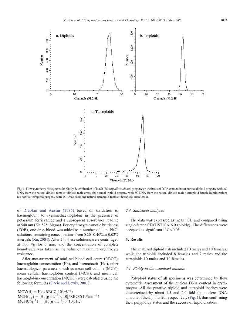
Fig. 1. Flow cytometry histograms for ploidy determination of loach (M. anguillicaudatus) progeny on the basis of DNA content in (a) normal diploid progeny with 2CDNA from the natural diploid female×diploid male cross; (b) normal triploid progeny with 3C DNA from the natural diploid male×tetraploid female hybridization;(c) normal tetraploid progeny with 4C DNA from the natural tetraploid female×tetraploid male cross.
1003Z. Gao et al. / Comparative Biochemistry and Physiology, Part A 147 (2007) 1001–1008
of Drabkin and Austin (1935) based on oxidation ofhaemoglobin to cyanmethaemoglobin in the presence ofpotassium ferricyanide and a subsequent absorbance readingat 540 nm (Kit 525, Sigma). For erythrocyte osmotic brittleness(EOB), one drop blood was added to a number of 1 ml NaClsolutions, containing concentrations from 0.20–0.40% at 0.02%intervals (Xu, 2004). After 2 h, these solutions were centrifugedat 500 ×g for 5 min, and the concentration of completehemolysate was taken as the value of maximum erythrocyteresistance.
After measurement of total red blood cell count (RBCC),haemoglobin concentration (Hb), and haematocrit (Hct), otherhaematological parameters such as mean cell volume (MCV),mean cellular haemoglobin content (MCH), and mean cellhaemoglobin concentration (MCHC) were calculated using thefollowing formulas (Dacie and Lewis, 2001):
MCVðflÞ ¼ Hct=RBCCð106lL−1ÞMCHðpgÞ ¼ ½Hbðg dL−1 � 10�=RBCCð106mm−1ÞMCHCðg−1Þ ¼ ½Hbðg dL−1Þ � 10�=Hct:
2.4. Statistical analyses
The data was expressed as mean±SD and compared usingsingle-factor STATISTICA 6.0 (ploidy). The differences wereaccepted as significant if Pb0.05.
3. Results
The analyzed diploid fish included 10 males and 10 females,while the triploids included 8 females and 2 males and thetetraploids 10 males and 10 females.
3.1. Ploidy in the examined animals
Polyploid status of all specimens was determined by flowcytometric assessment of the nuclear DNA content in eryth-rocytes. All the putative triploid and tetraploid loaches werecharacterised by about 1.5 and 2.0 fold the nuclear DNAamount of the diploid fish, respectively (Fig. 1), thus confirmingtheir polyploidy status and the success of triploidization.
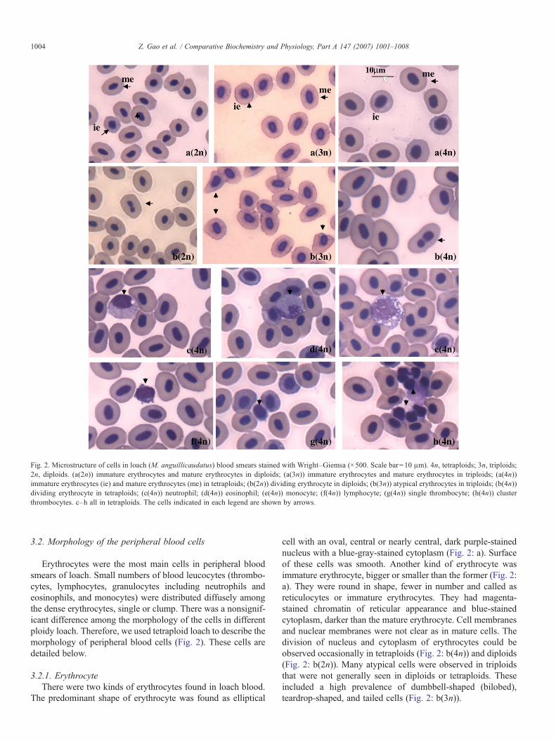
Fig. 2. Microstructure of cells in loach (M. anguillicaudatus) blood smears stained with Wright–Giemsa (×500. Scale bar=10 μm). 4n, tetraploids; 3n, triploids;2n, diploids. (a(2n)) immature erythrocytes and mature erythrocytes in diploids; (a(3n)) immature erythrocytes and mature erythrocytes in triploids; (a(4n))immature erythrocytes (ie) and mature erythrocytes (me) in tetraploids; (b(2n)) dividing erythrocyte in diploids; (b(3n)) atypical erythrocytes in triploids; (b(4n))dividing erythrocyte in tetraploids; (c(4n)) neutrophil; (d(4n)) eosinophil; (e(4n)) monocyte; (f(4n)) lymphocyte; (g(4n)) single thrombocyte; (h(4n)) clusterthrombocytes. c–h all in tetraploids. The cells indicated in each legend are shown by arrows.
1004 Z. Gao et al. / Comparative Biochemistry and Physiology, Part A 147 (2007) 1001–1008
3.2. Morphology of the peripheral blood cells
Erythrocytes were the most main cells in peripheral bloodsmears of loach. Small numbers of blood leucocytes (thrombo-cytes, lymphocytes, granulocytes including neutrophils andeosinophils, and monocytes) were distributed diffusely amongthe dense erythrocytes, single or clump. There was a nonsignif-icant difference among the morphology of the cells in differentploidy loach. Therefore, we used tetraploid loach to describe themorphology of peripheral blood cells (Fig. 2). These cells aredetailed below.
3.2.1. ErythrocyteThere were two kinds of erythrocytes found in loach blood.
The predominant shape of erythrocyte was found as elliptical
cell with an oval, central or nearly central, dark purple-stainednucleus with a blue-gray-stained cytoplasm (Fig. 2: a). Surfaceof these cells was smooth. Another kind of erythrocyte wasimmature erythrocyte, bigger or smaller than the former (Fig. 2:a). They were round in shape, fewer in number and called asreticulocytes or immature erythrocytes. They had magenta-stained chromatin of reticular appearance and blue-stainedcytoplasm, darker than the mature erythrocyte. Cell membranesand nuclear membranes were not clear as in mature cells. Thedivision of nucleus and cytoplasm of erythrocytes could beobserved occasionally in tetraploids (Fig. 2: b(4n)) and diploids(Fig. 2: b(2n)). Many atypical cells were observed in triploidsthat were not generally seen in diploids or tetraploids. Theseincluded a high prevalence of dumbbell-shaped (bilobed),teardrop-shaped, and tailed cells (Fig. 2: b(3n)).
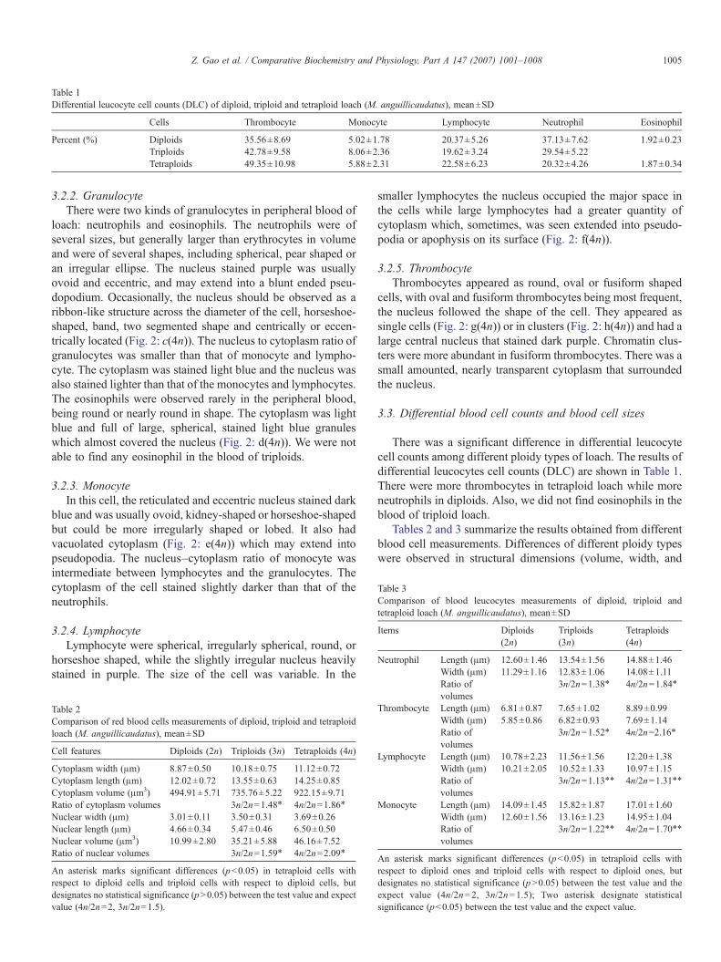
Table 1Differential leucocyte cell counts (DLC) of diploid, triploid and tetraploid loach (M. anguillicaudatus), mean±SD
Cells Thrombocyte Monocyte Lymphocyte Neutrophil Eosinophil
Percent (%) Diploids 35.56±8.69 5.02±1.78 20.37±5.26 37.13±7.62 1.92±0.23Triploids 42.78±9.58 8.06±2.36 19.62±3.24 29.54±5.22Tetraploids 49.35±10.98 5.88±2.31 22.58±6.23 20.32±4.26 1.87±0.34
Table 3Comparison of blood leucocytes measurements of diploid, triploid andtetraploid loach (M. anguillicaudatus), mean±SD
Items Diploids(2n)
Triploids(3n)
Tetraploids(4n)
Neutrophil Length (μm) 12.60±1.46 13.54±1.56 14.88±1.46Width (μm) 11.29±1.16 12.83±1.06 14.08±1.11
1005Z. Gao et al. / Comparative Biochemistry and Physiology, Part A 147 (2007) 1001–1008
3.2.2. GranulocyteThere were two kinds of granulocytes in peripheral blood of
loach: neutrophils and eosinophils. The neutrophils were ofseveral sizes, but generally larger than erythrocytes in volumeand were of several shapes, including spherical, pear shaped oran irregular ellipse. The nucleus stained purple was usuallyovoid and eccentric, and may extend into a blunt ended pseu-dopodium. Occasionally, the nucleus should be observed as aribbon-like structure across the diameter of the cell, horseshoe-shaped, band, two segmented shape and centrically or eccen-trically located (Fig. 2: c(4n)). The nucleus to cytoplasm ratio ofgranulocytes was smaller than that of monocyte and lympho-cyte. The cytoplasm was stained light blue and the nucleus wasalso stained lighter than that of the monocytes and lymphocytes.The eosinophils were observed rarely in the peripheral blood,being round or nearly round in shape. The cytoplasm was lightblue and full of large, spherical, stained light blue granuleswhich almost covered the nucleus (Fig. 2: d(4n)). We were notable to find any eosinophil in the blood of triploids.
3.2.3. MonocyteIn this cell, the reticulated and eccentric nucleus stained dark
blue and was usually ovoid, kidney-shaped or horseshoe-shapedbut could be more irregularly shaped or lobed. It also hadvacuolated cytoplasm (Fig. 2: e(4n)) which may extend intopseudopodia. The nucleus–cytoplasm ratio of monocyte wasintermediate between lymphocytes and the granulocytes. Thecytoplasm of the cell stained slightly darker than that of theneutrophils.
3.2.4. LymphocyteLymphocyte were spherical, irregularly spherical, round, or
horseshoe shaped, while the slightly irregular nucleus heavilystained in purple. The size of the cell was variable. In the
Table 2Comparison of red blood cells measurements of diploid, triploid and tetraploidloach (M. anguillicaudatus), mean±SD
Cell features Diploids (2n) Triploids (3n) Tetraploids (4n)
Cytoplasm width (μm) 8.87±0.50 10.18±0.75 11.12±0.72Cytoplasm length (μm) 12.02±0.72 13.55±0.63 14.25±0.85Cytoplasm volume (μm3) 494.91±5.71 735.76±5.22 922.15±9.71Ratio of cytoplasm volumes 3n/2n=1.48⁎ 4n/2n=1.86⁎
Nuclear width (μm) 3.01±0.11 3.50±0.31 3.69±0.26Nuclear length (μm) 4.66±0.34 5.47±0.46 6.50±0.50Nuclear volume (μm3) 10.99±2.80 35.21±5.88 46.16±7.52Ratio of nuclear volumes 3n/2n=1.59⁎ 4n/2n=2.09⁎
An asterisk marks significant differences (pb0.05) in tetraploid cells withrespect to diploid cells and triploid cells with respect to diploid cells, butdesignates no statistical significance (pN0.05) between the test value and expectvalue (4n/2n=2, 3n/2n=1.5).
smaller lymphocytes the nucleus occupied the major space inthe cells while large lymphocytes had a greater quantity ofcytoplasm which, sometimes, was seen extended into pseudo-podia or apophysis on its surface (Fig. 2: f(4n)).
3.2.5. ThrombocyteThrombocytes appeared as round, oval or fusiform shaped
cells, with oval and fusiform thrombocytes being most frequent,the nucleus followed the shape of the cell. They appeared assingle cells (Fig. 2: g(4n)) or in clusters (Fig. 2: h(4n)) and had alarge central nucleus that stained dark purple. Chromatin clus-ters were more abundant in fusiform thrombocytes. There was asmall amounted, nearly transparent cytoplasm that surroundedthe nucleus.
3.3. Differential blood cell counts and blood cell sizes
There was a significant difference in differential leucocytecell counts among different ploidy types of loach. The results ofdifferential leucocytes cell counts (DLC) are shown in Table 1.There were more thrombocytes in tetraploid loach while moreneutrophils in diploids. Also, we did not find eosinophils in theblood of triploid loach.
Tables 2 and 3 summarize the results obtained from differentblood cell measurements. Differences of different ploidy typeswere observed in structural dimensions (volume, width, and
Ratio ofvolumes
3n/2n=1.38⁎ 4n/2n=1.84⁎
Thrombocyte Length (μm) 6.81±0.87 7.65±1.02 8.89±0.99Width (μm) 5.85±0.86 6.82±0.93 7.69±1.14Ratio ofvolumes
3n/2n=1.52⁎ 4n/2n=2.16⁎
Lymphocyte Length (μm) 10.78±2.23 11.56±1.56 12.20±1.38Width (μm) 10.21±2.05 10.52±1.33 10.97±1.15Ratio ofvolumes
3n/2n=1.13⁎⁎ 4n/2n=1.31⁎⁎
Monocyte Length (μm) 14.09±1.45 15.82±1.87 17.01±1.60Width (μm) 12.60±1.56 13.16±1.23 14.95±1.04Ratio ofvolumes
3n/2n=1.22⁎⁎ 4n/2n=1.70⁎⁎
An asterisk marks significant differences (pb0.05) in tetraploid cells withrespect to diploid ones and triploid cells with respect to diploid ones, butdesignates no statistical significance (pN0.05) between the test value and theexpect value (4n/2n=2, 3n/2n=1.5); Two asterisk designate statisticalsignificance (pb0.05) between the test value and the expect value.
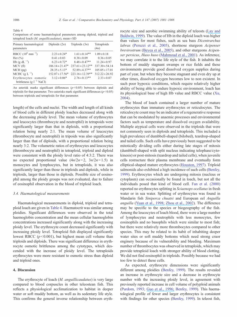
Table 4Comparison of some haematological parameters among diploid, triploid andtetraploid loach (M. anguillicaudatus), mean±SD
Primary haematologicalparameters
Diploids (2n) Triploids (3n) Tetraploids(4n)
RBCC (106 mm−3) 2.23±0.24⁎ 1.61±0.14⁎⁎ 1.09±0.18Hct (%) 0.41±0.03 0.38±0.09 0.36±0.05Hb (g dL−1) 6.23±0.72⁎ 8.48±0.47⁎⁎ 11.24±0.97MCV (fl) 186.14±21.47⁎ 237.61±23.12⁎⁎ 337.30±54.14MCH (pg) 28.35±5.13⁎ 52.89±4.37⁎⁎ 105.05±17.01MCHC (g L−1) 152.07±17.56⁎ 223.16±12.39⁎⁎ 312.22±26.92Erythrocytes osmoticbrittleness (g l−1 NaCl)
3.12±0.06⁎ 2.76±0.13⁎⁎ 2.33±0.07
An asterisk marks significant differences (pb0.05) between diploids andtriploids for that parameter. Two asterisks mark significant differences (pb0.05)between triploids and tetraploids for that parameter.
1006 Z. Gao et al. / Comparative Biochemistry and Physiology, Part A 147 (2007) 1001–1008
length) of the cells and nuclei. The width and length of all kindsof blood cells in different ploidy loaches decreased along withthe decreasing ploidy level. The mean volume of erythrocytesand leucocytes (thrombocyte and neutrophil) in tetrapoids weresignificantly larger than that in diploids, with a proportionalrelation being nearly 2:1. The mean volume of leucocytes(thrombocyte and neutrophil) in tripoids was also significantlylarger than that of diploids, with a proportional relation beingnearly 3:2. The volumetric ratios of erythrocytes and leucocytes(thrombocyte and neutrophil) in tetraploid, triploid and diploidwere consistent with the ploidy level ratio of 4:3:2. There wasno expected proportional value (4n/2n=2, 3n/2n=1.5) inmonocytes and lymphocytes, but in tetraploids, it was alsosignificantly larger than those in triploids and diploids, while intriploids, larger than those in diploids. Possible size of eosino-phil among the ploidy groups was not evaluated, due to failureof eosinophil observation in the blood of triploid loach.
3.4. Haematological measurements
Haematological measurements in diploid, triploid and tetra-ploid loach are given in Table 4. Haematocrit was similar amongploidies. Significant differences were observed in the totalhaemoglobin concentration and the mean cellular haemoglobinconcentrations increased significantly along with the increasingploidy level. The erythrocyte count decreased significantly withincreasing ploidy level. Tetraploid fish displayed significantlylowest RBCC (pb0.001), but highest mean cell volume thantriploids and diploids. There was significant difference in eryth-rocyte osmotic brittleness among the cytotypes, which des-cended with the increase of ploidy level. The tetraploidserythrocytes were more resistant to osmotic stress than diploidand triploid ones.
4. Discussion
The erythrocyte of loach (M. anguillicaudatus) is very largecompared to blood corpuscles in other teleostean fish. Thisreflects a physiological acclimatization to habitat in deeperwater or soft muddy bottom, as well as its sedentary life style.This confirms the general inverse relationship between eryth-
rocyte size and aerobic swimming ability of teleosts (Lay andBaldwin, 1999). The value of Hb in the diploid loach was higherthan values for most fishes, such as sea bass Dicentrarchuslabrax (Peruzzi et al., 2005), shortnose sturgeon Acipenserbrevirostrum (Beyea et al., 2005), and other sturgeons Acipen-ser persicus, Huso huso (Mahmoud et al., 2001). As inference,we may correlate it to the life style of the fish. It inhabits thebottom of muddy stagnant swamps or rice fields and theseenvironments provide good dissolved oxygen supplies duringpart of year, but when they become stagnant and even dry up atother times, dissolved oxygen becomes low to non existent. Insuch poor hypoxic conditions, which require relatively higherability of being able to endure hypoxic environment, loach hasits physiological base of high Hb value and RBCC value (Xu,2004).
The blood of loach contained a larger number of matureerythrocytes than immature erythrocytes or reticulocytes. Thereticulocyte count may be an indicator of a regenerative responsethat can be modulated by anaemic processes and environmentalfactors such as temperature and dissolved oxygen availability.Multiple atypical cells were observed in the triploids that werenot commonly seen in diploids and tetraploids. This included ahigh prevalence of dumbbell-shaped (bilobed), teardrop-shapedand tailed cells. Such cells have previously been suggested to bemitotically dividing cells either during late stages of mitosis(dumbbell-shaped with split nucleus indicating telophase/cyto-kinesis) or post-mitosis (teardrop and tailed cells), when juvenilecells restructure their plasma membrane and eventually formelliptical-shaped mature erythrocytes (Houston, 1997). Triploidsalmonids also exhibited a high incidence of such cells (Benfey,1999). Erythrocytes which are undergoing mitosis (nucleus orcytoplasm) can occasionally be found in loach, but not all theindividuals posed that kind of blood cell. Fan et al. (2000)reported no erythrocytes splitting in Sciaenops ocellatus in freshwater or in sea water. Splitting of erythrocytes was found inMandarin fish Siniperca chuatsi and European eel Anguillaanguilla (Yuan et al., 1998; Zhou et al., 2002). The differencemay be specific to the species or biogeography of the fish.Among the leucocytes of loach blood, there were a large numberof lymphocytes and neutrophils with less monocytes, feweosinophils and no basophils which were similar to most fish;but there were relatively more thrombocytes compared to otherspecies. This may be related to its habit of inhabiting deeperwater sites or soft muddy bottoms which need strong cruorenginery because of its vulnerability and bleeding. Maximumnumber of thrombocytes was observed in tetraploids, which mayprovide tetraploid loach with stronger ability of blood clotting.We did not find eosinophil in triploids. Possibly because we hadtoo few to detect these cells.
As expected, erythrocyte dimensions were significantlydifferent among ploidies (Benfey, 1999). The results revealedan increase in erythrocyte size and a decrease in erythrocytenumber with the increasing ploidy level, in agreement withpreviously reported increase in cell volume of polyploid animals(Purdom, 1993; Guo et al., 1996; Benfey, 1999). This haema-tological profile of fewer and larger erythrocytes is consistentwith findings for other species (Benfey, 1999). In teleost fish,
1007Z. Gao et al. / Comparative Biochemistry and Physiology, Part A 147 (2007) 1001–1008
association of increase in erythrocyte size with polyploidy hasbeen reported and measurement of red blood cell dimensions hasbeen proposed as a rapid and inexpensive assay for triploidydetermination (Krasznai et al., 1984; Ueno, 1984; Sezaki et al.,1988, 1991; Yamamoto and Iida, 1994; Benfey, 1999). In ourstudy, the volumetric ratio of erythrocytes in tetraploids, triploidsand diploids was consistent with the ploidy level ratio of 4:3:2.Therefore, variable sizes of erythrocytes in diploids, triploids andtetraploids could be used for determination of ploidy level inloach species. In spite of these variations in blood cell size, nodifferences in the haematocrit value was observed amongdiploids, triploids and tetraploids, as the increase in volume ofpolyploidy cells was counterbalanced by the decrease in cellcounts. Although this phenomenon is poorly understood, it hasbeen shown for most tissues and cells and allows maintenance ofnormal size of triploid organs and individuals (Benfey, 1999).
We found that total blood haemoglobin concentration in-creased in the blood of polyploids containing significantly fewererythrocytes. The average volume of red blood cells was greaterin polyploids, such that the mean cellular haemoglobin contentin erythrocytes of polyploids was significantly greater than thatin diploids, and the mean cellular haemoglobin concentrationswas also significant greater than that in diploids. Reported valuesfor total Hb and MCHC concentrations in diploid and polyploidfish are not consistent, whereas the mean cellular haemoglobincontent is commonly reported to be higher in polyploids than indiploids (Benfey, 1999). Ballarin et al. (2004) reported nodifferences in total Hb but higher MCH values in triploid shidrum U. cirrosa as compared to their diploid counterparts.Peruzzi et al. (2005) reported that total Hb concentration wasreduced in the blood of triploids, while the average volume ofred blood cells and the mean cellular haemoglobin content inerythrocytes was significant greater in triploids, and the meancellular haemoglobin concentrations were equivalent in sea bassD. labrax. These results were similar to values for silver cruciancarp Carassius auratus (Vetešník et al., 2006). Beyea et al.(2005) reported total blood haemoglobin concentration andcellular haemoglobin concentration did not differ significantlybetween diploids and triploids but cellular haemoglobin contentwas elevated in triploids as a result of the significantly greatersize of triploid erythrocytes in shortnose sturgeon A. breviros-trum. In salmonids, despite some contrasting results on totalblood haemoglobin levels and blood-oxygen carrying capacity,triploid fish were found to be similar to diploids in their overalloxygen-consumption rates and swimming performances undernormal or stress conditions (Stillwell and Benfey, 1995). We didnot find any report in line with our results that total Hb con-centration was increased in the blood along with the increase inploidy level, although the MCH is commonly reported to behigher in polyploids. The differences may be specific to thespecies or biogeography of the fish due to the different livinghabit. Tetraploid loach is a natural existent species. Naturalsurviving condition makes tetraploids more resistant to hypoxicconditions with high Hb value and they grow more quickly (Yinet al., 2005).
Erythrocyte osmotic brittleness is the physiological index ofshowing the characteristic of erythrocytes, related to membra-
nous fluidity. Polyploid erythrocytes were more resistant toosmotic stress than diploid ones in our study, which was con-sistent with findings for other species (Ballarin et al., 2004). Thehypothesis of a reduced exchange of polyploid cells with theexternal environment fits the observation of a lower osmoticfragility of tetraploid erythrocytes with respect to triploid anddiploid ones.
This is the first study to report baseline haematologicalstatus of diploid, triploid and tetraploid loach M. anguillicau-datus. Based on the results, it can be concluded that the higherploidy level loach may have some advantage with respect tomaximum oxygen carrying capacity of the blood and morethrombocytes inducing better ability for blood clotting. Furtherstudies considering performance of different ploidy loachrelated experimental challenges should provide a betterunderstanding of the comparative adaptability of diploid,triploid and tetraploid loach.
Acknowledgement
This research is a component of the Aquaculture Collabo-rative Research Support Program (ACRSP), supported by theUS Agency for International Development and by contributionsfrom the University of Michigan, the Asian Institute ofTechnology, and the Huazhong Agricultural University. TheACRSP accession number is 1326.
References
Arai, K., 2001. Genetic improvement of aquaculture finfish species bychromosome manipulation techniques in Japan. Aquaculture 197, 205–228.
Arai, K., 2003. Genetics of the loach, Misgurnus anguillicaudatus, recentprogress and perspective. Folia Biol. 51, 107–117.
Arai, K., Matsubara, K., Suzuki, R., 1991. Karyotype and erythrocyte size ofspontaneous tetraploidy and triploidy in the loach Misgurnus anguillicau-datus. Nippon Suisan Gakkaishi 57, 2157–2172.
Ballarin, L., Dall'Oro, M., Bertotto, D., Libertini, A., Francescon, A., Barbaro,A., 2004. Haematological parameters in Umbrina cirrosa (Teleostei,Sciaenidae), a comparison between diploid and triploid specimens. Comp.Biochem. Physiol. A 138, 45–51.
Beck, M.L., Biggers, C.J., 1983. Erythrocyte measurements of diploid andtriploid Ctenopharyngodon idella×Hypopthalmichthys nobilis hybrids.J. Fish Biol. 22, 497–502.
Benfey, T.J., 1999. The physiology and behaviour of triploid fishes. Rev. Fish.Sci. 7, 39–67.
Benfey, T.J., Sutterlin, A.M., 1984a. The haematology of triploid landlockedAtlantic salmon (Salmo salar L.). J. Fish Biol. 24, 333–338.
Benfey, T.J., Sutterlin, A.M., 1984b. Oxygen utilization by triploid landlockedAtlantic salmon (Salmo salar L.). Aquaculture 42, 69–73.
Benfey, T.J., Sutterlin, A.M., Thompson, R.J., 1984. Use of erythrocytemeasurements to identify triploid salmonids. Can. J. Fish Aquat. Sci. 41,980–984.
Beyea, M.M., Benfey, T.J., Kieffer, J.D., 2005. Hematology and stressphysiology of juvenile diploid and triploid shortnose sturgeon (Acipenserbrevirostrum). Fish Physiol. Biochem. 31, 303–313.
Boron, A., 1994. Use of erythrocyte measurements to detect natural triploids ofspined loach Cobitis taenia (L.). Cytobios 78, 197–202.
Chourrout, D., Nakayama, T., 1987. Chromosome studies of progenies oftetraploid female rainbow trout. Theor. Appl. Genet. 74, 687–692.
Chourrout, D., Guyomard, R., Houdebine, L., 1986. High efficiency genetransfer in rainbow trout (Salmo gairdncri) by micro-injection into eggcytoplasm. Aquaculture 51, 143–150.
1008 Z. Gao et al. / Comparative Biochemistry and Physiology, Part A 147 (2007) 1001–1008
Cogswell, A.T., Benfey, T.J., Sutterlin, A.M., 2001. The hematology of diploidand triploid transgenic Atlantic salmon (Salmo salar). Fish Physiol.Biochem. 24, 271–277.
Dacie, J.V., Lewis, S.M., 2001. PracticalHaematology, 9th ed. Churchill Livingstone,London. 633 pp.
Drabkin, D.L., Austin, J.H., 1935. Spectrophotometric studies, II. preparationfrom washed blood cells; nitric oxide haemoglobin and sulphaemoglobin.J. Biol. Chem. 112, 51–65.
Fan, R.Q., Jiang, M., Ru, S.G., 2000. Preliminary study on the changes ofultrastructure of the blood cell of Sciaenops ocellatus under differentosmotic pressure. Mar. Sci. 24, 48–53.
Flajšhans, M., 1997. A model approach to distinguish diploid and triploid fishby means of computer-assisted image analysis. Acta Vet. Brno 66,101–110.
Flajšhans, M., Vajcová, V., 2000. Odd ploidy levels in sturgeons suggest abackcross of interspecific hexaploid sturgeon hybrids to evolutionarilytetraploid and/or octaploid parental species. Folia Zool. 49, 133–138.
Guo, X., De Brosse, G.A., Allen Jr., S.K., 1996. All-triploid Pacific oysters(Crassostrea gigas Thunberg) produced by mating tetraploids and diploids.Aquaculture 142, 149–161.
Houston, A.H., 1997. Review, are the classical hematological variablesacceptable indicators of fish health? Trans. Am. Fish. Soc. 126, 879–893.
Krasznai, Z., Marián, T., Jeney, Z., Jeney, G., Zsigri, A., 1984. Effect of triploidyon the blood cell size of hybrid grass carp. Aquacult. Hung. (Szarvas) 4,17–24.
Lay, P.A., Baldwin, J., 1999. What determines the size of teleost erythrocytes?Correlations with oxygen transport and nuclear volume. Fish Physiol.Biochem. 20, 31–35.
Mahmoud, B., Rezvan, K., Donskaya, P., 2001. A comparative study of somehematological features in young reared sturgeons (Acipenser persicus andHuso huso). Fish Physiol. Biochem. 24, 135–140.
Man, S.H., Hodgkiss, I.J. (Eds.), 1981. Hong Kong Freshwater Fishes. TheWishing Printing Company, The Urban Council, Hong Kong, pp. 39.
Matsubara, K., Arai, K., Suzuki, R., 1995. Survival potential and chromosomesof progeny of triploid and pentaploid females in the loach Misgurnusanguillicaudatus. Aquaculture 131, 37–48.
Nam, Y.K., Kim, D.S., 2004. Ploidy status of progeny from the crosses betweentetraploid males and diploid females in mud loach (Misgurnus mizolepis).Aquaculture 236, 575–582.
Ojima, Y., Takaii, A., 1979. The occurrence of spontaneous polyploid in theJapanese common loach, Misgurnus anguillicaudatus. Proc. Jpn. Acad. 55,487– 491.
Palíková, M., Mareš, J., Jirásek, J., 1999. Characteristics of leukocytes andthrombocytes of selected sturgeon species form intensive breeding. ActaVet. Brno. 68, 259–264.
Peruzzi, S., Varsamos, S., Chatain, B., Fauvel, C., Menu, B., Falguière, J.C.,Sévère, A., Flik, G., 2005. Haematological and physiological characteristicsof diploid and triploid sea bass, Dicentrarchus labrax L. Aquaculture 244,359–367.
Purdom, C.E., 1993. Genetic and Fish Breeding. Chapman & Hall, London.Qin, C.G., Huang, K.X., Xu, H.B., 2002. Protective effect of polysaccharides
from the loach on the in vitro and in vivo peroxidative damage ofhepatocyte. J. Nutr. Biochem 13, 592–597.
Saitoh, K., 1989. Asian pond loach. In: Kawanabe, H., Mizuno, N. (Eds.),Freshwater Fishes of Japan. Yamakei Pub, Tokyo, pp. 382–385 (InJapanese).
Sezaki, K., Kobayasi, H., Nakamura, M., 1977. Size of erythrocytes in thediploid and triploid specimens of Carassius auratus langsdorfi. Jpn. J.Ichthyol. 24, 135–140.
Sezaki, K., Watabe, S., Hashimoto, K., 1988. Haematological parameters anderythrocyte enzyme activities associated with increase in ploidy status of thespinous loach, Cobitis biwae Jordan and Snyder. J. Fish Biol. 32, 149–150.
Sezaki, K., Watabe, K., Tsukamoto, K., Hashimoto, K., 1991. Effects of increasein ploidy status on respiratory function of ginbuna, Carassius auratusLangsdorfi (Cyprinidae). Comp. Biochem. Physiol. A 99, 123–127.
Small, S.A., Benfey, T.J., 1987. Cell size in triploid salmon. J. Exp. Zool. 241,339–342.
Stillwell, E.J., Benfey, T.J., 1995. Haemoglobin level, metabolic rate andswimming performance in triploid brook trout (Salvelinus fontinalis).Aquaculture 137, 355–358.
Svobodová, Z., Kolářová, J., Flajšhans, M., 1998. The first findings of thedifferences in complete blood count between diploid and triploid tench,Tinca tinca L. Acta Vet. Brno 67, 243–248.
Ueno, K., 1984. Induction of triploid carp and their haematological characteristics.Jpn. J. Genet. 59, 585–591.
Vetešník, L., Halaèka, K., Lusková, V., Lusk, S., 2006. Erythrocyte profile ofdiploid and triploid silver crucian carp (Carassius auratus). Acta Vet. Brno75, 203–207.
Wolters, W.R., Chrisman, C.L., Libey, G.S., 1982. Erythrocyte nuclearmeasurements of diploid and triploid channel catfish, Ictalurus punctatus(Rafinesque). J. Fish Biol. 20, 253–258.
Xiao, Y.J., Jin, Y.P., Zhang, G.F., Zhang, Q.J., 2001. Observation on the peripheralblood cells morphology of Misgurnus anguillicaudatus. J. Fujian Teach.Univ. (Natural Science) 17 (4), 93–96 (in Chinese with English abstract).
Xu, W.Y., 2004. Relationship between haemotological indices & sexuality inMisgurnus anguillicaudatus. Fish. Sci. 23 (8), 15–17 (in Chinese withEnglish abstract).
Yamamoto, A., Iida, T., 1994. Hematological characteristics of triploid rainbowtrout. Fish Pathol. 29, 239–243.
Yin, J., Zhao, Z.S., Chen, X.Q., Li, Y.Q., Zhu, L.Y., 2005. Karyotypecomparison of diploid and tetraploid loach, Misgurnus anguillicanudatus.Acta Hydrobiol. Sin. 29 (4), 469–472 (in Chinese with English abstract).
Yuan, S.Q., Zhang, Y.A., Yao, W.J., 1998. Micro and ultrastructure of peripheralblood cells of the Mandarin fish, Siniperca chuatsi (Basilewsky). ActaHyrobiol. Sin. 22, 39–47 (in Chinese with English abstract).
Zhang, Q., Arai, K., 1996. Flow cytometry for DNA contents of somatic cellsand spermatozoa in the progeny of natural tetraploid loach. Fish. Sci. 62,870–877.
Zhang, Q., Arai, K., 1999a. Distribution and reproductive capacity of naturaltriploid individuals and occurrence of unreduced eggs as a cause ofpolyploidization in the loach, Misgurnus anguillicaudatus. Ichthyol. Res.46, 153–161.
Zhang, Q., Arai, K., 1999b. Aberrant meioses and viable aneuploid progeny ofinduced triploid loach (Misgurnus anguillicaudatus) when crossed to naturaltetraploids. Aquaculture 175, 63–76.
Zhou, Y., Guo,W.G., Yang, Z.G., Zhou, X.H., Zhang, K., Wen, X.H., Wang, T.D.,2002. Microstructure and Ultrastructure of the peripheral blood cells ofEuropean eel (Anguilla anguilla). Acta Zool. Sin. 48, 393–401 (in Chinesewith English abstract).
Zou, S.M., Li, S.F., Cai, W.Q., Zhao, J.L., Yang, H.Y., 2004. Establishment offertile tetraploid population of blunt snout bream Megalobrama amblyce-phala. Aquaculture 238, 155–164.