growth hormone receptor gene expression in the skeletal
TRANSCRIPT
HAL Id: hal-00900574https://hal.archives-ouvertes.fr/hal-00900574
Submitted on 1 Jan 2005
HAL is a multi-disciplinary open accessarchive for the deposit and dissemination of sci-entific research documents, whether they are pub-lished or not. The documents may come fromteaching and research institutions in France orabroad, or from public or private research centers.
L’archive ouverte pluridisciplinaire HAL, estdestinée au dépôt et à la diffusion de documentsscientifiques de niveau recherche, publiés ou non,émanant des établissements d’enseignement et derecherche français ou étrangers, des laboratoirespublics ou privés.
Growth hormone receptor gene expression in theskeletal muscle of normal and double-muscled bovines
during foetal developmentAnne Listrat, Jean-François J.-F. Hocquette, Brigitte Picard, François
Ménissier, Jean Djiane, Hélène Jammes
To cite this version:Anne Listrat, Jean-François J.-F. Hocquette, Brigitte Picard, François Ménissier, Jean Djiane, etal.. Growth hormone receptor gene expression in the skeletal muscle of normal and double-muscledbovines during foetal development. Reproduction Nutrition Development, EDP Sciences, 2005, 45(4), pp.393-403. �10.1051/rnd:2005036�. �hal-00900574�
393Reprod. Nutr. Dev. 45 (2005) 393–403© INRA, EDP Sciences, 2005DOI: 10.1051/rnd:2005036
Original article
Growth hormone receptor gene expression in the skeletal muscle of normal and double-muscled
bovines during foetal development
Anne LISTRATa*, Jean François HOCQUETTEa, Brigitte PICARDa, François MÉNISSIERb, Jean DJIANEc, Hélène JAMMESc
a INRA, Unité de Recherche sur les Herbivores, Equipe Croissance et Métabolismes du Muscle, Theix, 63122 Saint-Genès-Champanelle, France
b INRA, Station de Génétique Quantitative et Appliquée, Domaine de Vilvert, 78352 Jouy-en-Josas Cedex, France
c INRA, Neurobiologie de l’Olfaction et de la Prise Alimentaire, Bâtiment des Biotechnologies,
78352 Jouy-en-Josas Cedex, France
(Received 13 August 2004; accepted 29 December 2004)
Abstract – The expression of the growth hormone receptor (GHR) gene was investigated insemitendinosus muscle during bovine foetal development in both normal and double-muscledCharolais foetuses which differ with respect to muscle development. Northern-blot analysis of foetalmuscle RNA preparations with a GHR cDNA probe identified the 4.5 kb GHR mRNA as early as130 days post-conception. In double-muscled animals, the expression of GHR mRNA increasedfrom 130 to 210 days of gestation while it stayed stable in normal ones. It was significantly higher(P < 0.05) in double-muscled foetuses compared to normal ones from the second third of gestation.Northern-blot analysis of foetal muscle RNA preparations from both genotypes with a β-actin cDNAprobe, revealed lower β-actin gene expression in double-muscled foetuses than in normal ones,suggesting a delay in the differentiation of muscle cells. In situ hybridisation revealed the localisationof specific GHR mRNA in muscle cells at all gestation stages analysed (130, 170, 210 days post-conception) but not in connective tissue surrounding the muscle cells. At the adult stage, thehybridisation signal was also very high and observed in muscle cells only. These results show theontogeny of GHR mRNA in bovine muscle and demonstrate a difference between normal and double-muscled animals.
muscle / bovine / receptor / growth hormone / in situ hybridisation
1. INTRODUCTION
Some bovine breeds exhibit muscularhypertrophy which is under the control ofthe myostatin gene. In double-muscled ani-mals, this gene, which is deleted in its cod-ing sequence, has been identified by Grobet
et al. [1]. The hypertrophy of some superficialmuscles, which results from the presence ofinactive myostatin, is the consequence of anincrease in the total fibre number [2] due tohyperplasia [3, 4]. Picard et al. [5, 6] showed,in vivo and in vitro, that there was a variabledelay in contractile differentiation of the
* Corresponding author: [email protected]
Article published by EDP Sciences and available at http://www.edpsciences.org/rnd or http://dx.doi.org/10.1051/rnd:2005036
394 A. Listrat et al.
muscle of double-muscled (DM) animals.This delay occurred during the last third offoetal life. This delay could be due to effectsof insulin like growth factors (IGF), mainlyIGF-II. Indeed, these growth factors areknown to play a role in muscle differentia-tion [7]. In addition, in a previous study, weshowed that IGF-II mRNA were present inlarge amounts in foetal muscle cells [8].
In growing animals, growth hormone(GH) is well known for its effects on growthof skeletal muscle and for its metabolicactions. In the foetus, although the concen-tration of growth hormone (GH) in the cir-culation is high, it is established that it doesnot have a significant growth-regulatoryfunction. For instance, anencephalic humanfoetuses [9] and hypophysectomised foetusesof numerous other species [10–12] appearto grow almost normally although theyhave little or no circulating immunoreac-tive GH. Nevertheless, a number of foetaltissues including skeletal muscle have beenshown to respond to GH stimulation in vitroand in vivo [13–16] by an increase in pro-tein synthesis and in myosin heavy chainexpression at the mRNA level implying thepresence of a functional GH receptor(GHR) during foetal development. Theontogeny of GHR gene expression in thefoetuses of several species has been ana-lysed by Northern-blot and RNAse protec-tion assays. In foetal sheep, a major 4.4 kbtranscript was detected [17] as early as day51 of gestation in the liver, kidney, lung,heart, muscle and placenta. Its levelincreased slightly with advancing gesta-tion. In the bovine foetus, Lucy et al. [18]reported the presence of GHR mRNA inseveral tissues, including the muscle andliver. In addition, both GHR mRNA andGHR protein have been detected in bovineembryos [19]. However, to our knowledge,ontogeny studies have never been con-ducted in cattle up till now.
Some authors showed that, on the onehand, a relationship exists between myosta-tin expression and GH action [20] and, on theother hand, between myostatin and IGF II
expression [21]. One of the objectives of thepresent study was to investigate the ontog-eny of GHR mRNA and to compare it to thatof IGF-II [8] and myostatin [22] mRNApreviously described in the same samples inour laboratory. In addition, in order to betterunderstand the regulation of the differenti-ation of muscle fibres, we chose an hyper-trophied muscle in double-muscled animals,the semitendinosus (ST) muscle. We deter-mined the relative changes of GHR geneexpression in double-muscled ST muscle,compared to normal ST muscle by North-ern-blot and in situ hybridisation. This maybe of high biological importance due to theassociation between myostatin expressionand GH action [20].
2. MATERIALS AND METHODS
2.1. Animals
DM embryos of strain INRA95 [23]were transplanted into Charolais/Salerscrossbreed cattle at a mean age of 2 years.Seven DM foetuses (2 at 130 days post-con-ception [d pc], 2 at 170 d pc, 3 at 210 d pc)were obtained from the transplantations.Their development was compared with11 Charolais × Charolais/Salers (CS) foe-tuses (4 at 130 d pc, 3 at 170 d pc, 4 at210 d pc) lacking the DM genotype, andproduced by artificial insemination (the fullgestational period for cattle is 280 days).Foetuses were produced at the INRAResearch Centre and all the animals wereslaughtered and samples were collected incompliance with ethical guidelines for ani-mal care in this same centre (Theix, France).
2.2. Samples
Semitendinosus (ST) muscle and liversamples were taken from all foetuses. AdultST muscle samples were taken from 5 maturecows (3 years of age after birth) and usedas controls for in situ hybridisation. Tissuesamples were removed immediately afterexsanguination of the dam. Samples for
GH receptor mRNA and muscle development 395
in situ hybridisation were frozen in liquidnitrogen cooled isopentane then transferredto liquid nitrogen. Tissue samples collectedfor total RNA preparation were directly fro-zen in liquid nitrogen. All samples werestored at –80 °C.
2.3. Total RNA preparation
Total RNA was extracted according tothe method of Puissant and Houdebine [24].After extraction, RNA was precipitated bythe addition of 5.5 vol of 4 M LiCl, kept at4 °C for 2 h and recovered by centrifugation(5000 × g, 20 min, 4 °C). The pellet was dis-solved in 2 vol of 10 mM Tris-HCl pH 7.5,1 mM EDTA and 0.5% SDS. After oneadditional extraction by phenol:isoamylicalcohol-chloroform (vol·vol–1), the aque-ous phase was precipitated in the presenceof 300 mM sodium acetate and 2.5 vol ofethanol. The RNA was stored in this pre-cipitated form at –20 °C, until quantifica-tion by optical density measurements andanalysis by Northern-blot.
2.4. Probes
A 600 bp fragment of bovine GHRcDNA (bGHR) was obtained from bovinehepatic mRNA using reverse transcriptionand a polymerase chain reaction (RT-PCR).Specific primers were derived from thebGHR cDNA previously published [25].The sequence of the amplified fragment,coding the extracellular-, transmembrane-and part of the intracellular- domains ofbGHR, was verified by sequencing (AppliedBiosystems DNA sequencing system, Fos-ter city, USA), and was identical to the onepreviously published. This fragment wascloned in the pGEM18 plasmid.
2.5. Northern-blot analysis.
The RNA samples (20 µg) of both nor-mal and double-muscled animals weredenatured in 2.2 M formaldehyde, 50% for-mamide (5 min, 65 °C) and size-separatedby electrophoresis in the same gel in agar-
ose (1.5%), 2.2 M formaldehyde in 10 mMsodium phosphate buffer. RNA were trans-ferred to the Zeta probe membrane (Biorad)by capillary blotting overnight under highionic strength (10 × SSC = 1.5 M sodiumchloride, 0.15 M sodium citrate). The bGHRDNA fragment and a mouse β-cDNA probe(1 kb) were labelled with α[32P]-dCTP(3000 Ci·mmol–1) (ICN, France) to a spe-cific activity of 108 cpm·µg–1 DNA usinga random priming kit (Roche). Used as acontrol, a rat 18S ribosomal oligonucle-otide probe was labelled at the 5’end withγ [32P]-ATP using polynucleotide kinase asdescribed by Hocquette et al. [26]. Mem-branes of muscle mRNA were first hybrid-ised with the 32P GHR cDNA probe, thenthey were stripped off (boiling in 0.5% SDS),before re-hybridisation with the 32P labelled18S ribosomal oligonucleotide, then withthe 32P β-actin cDNA probe. Autoradio-grams were obtained by exposure to Amer-sham Hyperfilm with two amplified screens,at –80 °C. Image analysis was performed onautoradiograms using the ChemiImager5000 and the AlphaEase FluorChem soft-ware (Alpha Innotech Corporation, SanLeandro, California, USA).
2.6. In situ hybridisation
The procedure for in situ hybridisationused was similar to that reported by Lyonset al. [27] with slight modifications.Briefly, frozen cross-sections of muscleand liver tissues were made using a 2800Frigocut microtome (Reichert-Jung, Lahn-technik, Germany) and mounted on gelatin-coated (0.2 % w/v gelatin) slides. Foetalliver sections served as positive controls.The sections were fixed in 4% paraformal-dehyde phosphate buffered saline solutionfollowed by dehydration with absolute eth-anol. Sections were stored, at –80 °C.Before hybridisation, the sections wererehydrated, treated with proteinase K(Sigma), fixed and acetylated with trieth-anolamine/acetic anhydride, washed anddehydrated. High specific-activity bGHRantisense RNA probes were transcribed
396 A. Listrat et al.
from the linearised plasmid pGEM18 con-taining bGHR DNA fragment using [35S]-UTP (specific activity > 1000 Ci·mmole–1),(Amersham) and a SP6/T7 transcription kit(Roche). Sense strand cRNA probes weretranscribed in the opposite direction undersimilar conditions. The resulting cRNAprobes were reduced to < 100 nucleotidesusing alkali hydrolysis and separated by gelfiltration on a sephadex G-50 column (Phar-macia). Tissue cross sections, under coverslips, were hybridised in a humid chamberwith 7.5 × 10⁸ cpm·mL–1 labelled bGHRcRNA probe in 50% deionised formamide,0.3 M NaCl, 20 mM Tris-HCl, pH 7.4,5 mM EDTA, 10 mM NaPO4, 10% dextransulfate, 1× Denhardt solution, 100 mM dithi-othreitol (DTT), 100 µg·mL–1 salmon spermDNA at 42 °C. Control slides were proc-essed in an identical manner and probedwith the corresponding sense GHR ribo-probe. After hybridisation, the sections werewashed once for 30 min at 50 °C with 5× SSCcontaining 10 mM DTT then washed at65 °C for 20 min with 50% formamide,2× SSC, 10 mM DTT. Section slides werethen rinsed 10 min. at 37 °C in 1 M Tris-HCl(pH 7.5) with 2× SCC, dehydrated and a66% v/v LM1 nuclear track emulsion (Amer-sham) was applied. The slides were exposedfor one week in light-tight boxes in the pres-ence of desiccant at 4 °C. Photographic devel-opment was performed using Ilford Phenisol(Ciba-Geigy).
2.7. Statistical analysis
All experiments were carried out in trip-licate and all data were tested by varianceanalysis using the General Linear ModelProcedure of the Statistical Analysis Sys-tem [28]. Firstly, data of 18S and of β-actinmRNA were analysed by testing the effectof age and genotype and the interactionbetween these two parameters. For β-actinand for GHR, the statistical analysis wasperformed with different approaches:(a) analysis of crude mRNA levels alone,(b) with β-actin or (c) with 18S RNA levelsintroduced as covariables in the statistical
model as described by Hocquette andBrandstetter [29].
After analysing the results, we chose touse 18S RNA levels as a covariable to ana-lyse GHR and β-actin mRNA levels. In thesame model, we tested the effects of age andgenotype and the effect of their interaction.All data were expressed as means ± SEM.
3. RESULTS
3.1. Developmental expression of 18S, β-actin and of GHR genes
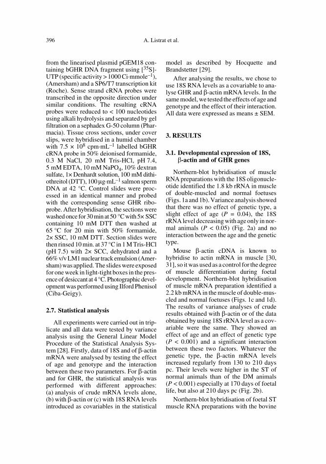
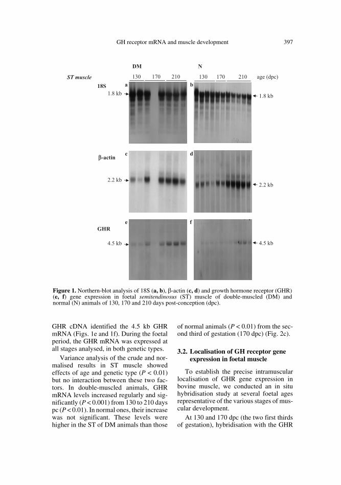
Northern-blot hybridisation of muscleRNA preparations with the 18S oligonucle-otide identified the 1.8 kb rRNA in muscleof double-muscled and normal foetuses(Figs. 1a and 1b). Variance analysis showedthat there was no effect of genetic type, aslight effect of age (P = 0.04), the 18SrRNA level decreasing with age only in nor-mal animals (P < 0.05) (Fig. 2a) and nointeraction between the age and the genetictype.
Mouse β-actin cDNA is known tohybridise to actin mRNA in muscle [30,31], so it was used as a control for the degreeof muscle differentiation during foetaldevelopment. Northern-blot hybridisationof muscle mRNA preparation identified a2.2 kb mRNA in the muscle of double-mus-cled and normal foetuses (Figs. 1c and 1d).The results of variance analyses of cruderesults obtained with β-actin or of the dataobtained by using 18S rRNA level as a cov-ariable were the same. They showed aneffect of age and an effect of genetic type(P < 0.001) and a significant interactionbetween these two factors. Whatever thegenetic type, the β-actin mRNA levelsincreased regularly from 130 to 210 dayspc. Their levels were higher in the ST ofnormal animals than of the DM animals(P < 0.001) especially at 170 days of foetallife, but also at 210 days pc (Fig. 2b).
Northern-blot hybridisation of foetal STmuscle RNA preparations with the bovine
GH receptor mRNA and muscle development 397
GHR cDNA identified the 4.5 kb GHRmRNA (Figs. 1e and 1f). During the foetalperiod, the GHR mRNA was expressed atall stages analysed, in both genetic types.
Variance analysis of the crude and nor-malised results in ST muscle showedeffects of age and genetic type (P < 0.01)but no interaction between these two fac-tors. In double-muscled animals, GHRmRNA levels increased regularly and sig-nificantly (P < 0.001) from 130 to 210 dayspc (P < 0.01). In normal ones, their increasewas not significant. These levels werehigher in the ST of DM animals than those
of normal animals (P < 0.01) from the sec-ond third of gestation (170 dpc) (Fig. 2c).
3.2. Localisation of GH receptor gene expression in foetal muscle
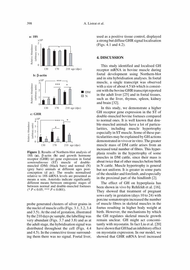
To establish the precise intramuscularlocalisation of GHR gene expression inbovine muscle, we conducted an in situhybridisation study at several foetal agesrepresentative of the various stages of mus-cular development.
At 130 and 170 dpc (the two first thirdsof gestation), hybridisation with the GHR
Figure 1. Northern-blot analysis of 18S (a, b), β-actin (c, d) and growth hormone receptor (GHR)(e, f) gene expression in foetal semitendinosus (ST) muscle of double-muscled (DM) andnormal (N) animals of 130, 170 and 210 days post-conception (dpc).
398 A. Listrat et al.
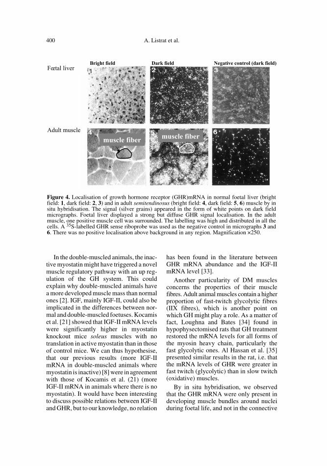
probe generated clusters of silver grains inthe nuclei of muscle cells (Figs. 3.1, 3.2, 3.4and 3.5). At the end of gestation, illustratedby the 210 days pc sample, the labelling wasvery abundant (Figs. 3.7 and 3.8) and as atthe adult stage, the hybridisation signal wasdistributed throughout the cell (Figs. 4.4and 4.5). In the connective tissue surround-ing them there was no signal. Foetal liver,
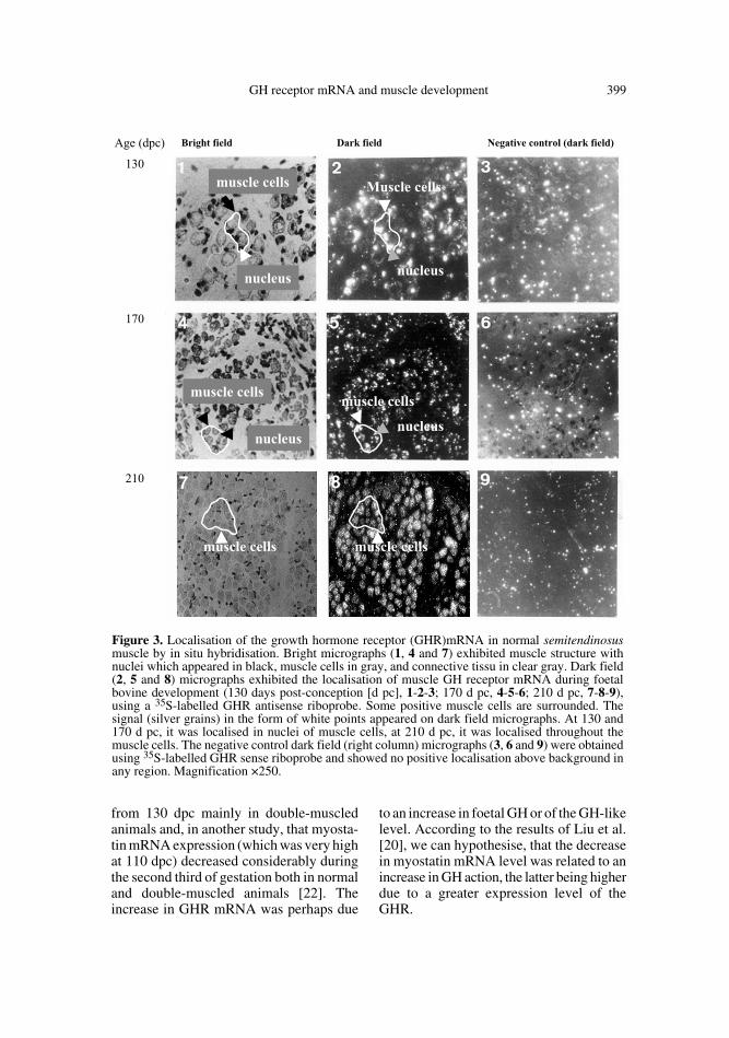
used as a positive tissue control, displayeda strong but diffuse GHR signal localisation(Figs. 4.1 and 4.2).
4. DISCUSSION
This study identified and localised GHreceptor mRNA in bovine muscle duringfoetal development using Northern-blotand in situ hybridisation analyses. In foetalmuscle, a single transcript was observedwith a size of about 4.5 kb which is consist-ent with the bovine GHR transcript reportedin the adult liver [25] and in foetal tissues,such as the liver, thymus, spleen, kidneyand brain [32].
In this study, we demonstrate a higherGH receptor gene expression in the ST ofdouble-muscled bovine foetuses comparedto normal ones. It is well known that dou-ble-muscled animals have a lot of particu-larities, including muscle hypertrophyespecially in ST muscle. Some of these par-ticularities may be explained by GH actionsdemonstrated in vivo or in vitro. The greatermuscle mass of DM cattle arises from anincreased total number of fibres. This hyper-plasia results in the hypertrophy of somemuscles in DM cattle, since their mass isabout twice that of other muscles before birthin N cattle. Muscle hypertrophy is generalbut not uniform. It is greater in some partsof the shoulder and forelimb, and especiallyin the proximal part of the hindlimb [2].
The effect of GH on hyperplasia hasbeen shown in vivo by Rehfeldt et al. [16].They showed that treatment of pregnantsows early in gestation (days 10 to 24) withporcine somatotropin increased the numberof muscle fibres in skeletal muscles in thefoetus resulting in higher body weight atbirth. However, the mechanisms by whichthe GH regulates skeletal muscle growthremain unclear. GH might act concomi-tantly with myostatin. In fact Liu et al. [20]have shown that GH had an inhibitory effecton myostatin expression. In our model, weshowed that GHR mRNA level increased
Figure 2. Results of Northern-blot analysis of18S (a), β-actin (b) and growth hormonereceptor (GHR) (c) gene expression in foetalsemitendinosus (ST) muscle of double-muscled (DM) (black bars) and normal (N)(grey bars) animals at different ages post-conception (d pc). The results normalisedrelative to 18S mRNA levels are presented asmeans ± sem. Asterisks indicate significantlydifferent means between ontogenic stages ofbetween normal and double-muscled foetuses(* P < 0.05; *** P < 0.001).
GH receptor mRNA and muscle development 399
from 130 dpc mainly in double-muscledanimals and, in another study, that myosta-tin mRNA expression (which was very highat 110 dpc) decreased considerably duringthe second third of gestation both in normaland double-muscled animals [22]. Theincrease in GHR mRNA was perhaps due
to an increase in foetal GH or of the GH-likelevel. According to the results of Liu et al.[20], we can hypothesise, that the decreasein myostatin mRNA level was related to anincrease in GH action, the latter being higherdue to a greater expression level of theGHR.
Figure 3. Localisation of the growth hormone receptor (GHR)mRNA in normal semitendinosusmuscle by in situ hybridisation. Bright micrographs (1, 4 and 7) exhibited muscle structure withnuclei which appeared in black, muscle cells in gray, and connective tissu in clear gray. Dark field(2, 5 and 8) micrographs exhibited the localisation of muscle GH receptor mRNA during foetalbovine development (130 days post-conception [d pc], 1-2-3; 170 d pc, 4-5-6; 210 d pc, 7-8-9),using a 35S-labelled GHR antisense riboprobe. Some positive muscle cells are surrounded. Thesignal (silver grains) in the form of white points appeared on dark field micrographs. At 130 and170 d pc, it was localised in nuclei of muscle cells, at 210 d pc, it was localised throughout themuscle cells. The negative control dark field (right column) micrographs (3, 6 and 9) were obtainedusing 35S-labelled GHR sense riboprobe and showed no positive localisation above background inany region. Magnification ×250.
400 A. Listrat et al.
In the double-muscled animals, the inac-tive myostatin might have triggered a novelmuscle regulatory pathway with an up reg-ulation of the GH system. This couldexplain why double-muscled animals havea more developed muscle mass than normalones [2]. IGF, mainly IGF-II, could also beimplicated in the differences between nor-mal and double-muscled foetuses. Kocamiset al. [21] showed that IGF-II mRNA levelswere significantly higher in myostatinknockout mice soleus muscles with notranslation in active myostatin than in thoseof control mice. We can thus hypothesise,that our previous results (more IGF-IImRNA in double-muscled animals wheremyostatin is inactive) [8] were in agreementwith those of Kocamis et al. (21) (moreIGF-II mRNA in animals where there is nomyostatin). It would have been interestingto discuss possible relations between IGF-IIand GHR, but to our knowledge, no relation
has been found in the literature betweenGHR mRNA abundance and the IGF-IImRNA level [33].
Another particularity of DM musclesconcerns the properties of their musclefibres. Adult animal muscles contain a higherproportion of fast-twitch glycolytic fibres(IIX fibres), which is another point onwhich GH might play a role. As a matter offact, Loughna and Bates [34] found inhypophysectomised rats that GH treatmentrestored the mRNA levels for all forms ofthe myosin heavy chain, particularly thefast glycolytic ones. Al Hassan et al. [35]presented similar results in the rat, i.e. thatthe mRNA levels of GHR were greater infast twitch (glycolytic) than in slow twitch(oxidative) muscles.
By in situ hybridisation, we observedthat the GHR mRNA were only present indeveloping muscle bundles around nucleiduring foetal life, and not in the connective
Figure 4. Localisation of growth hormone receptor (GHR)mRNA in normal foetal liver (brightfield: 1, dark field: 2, 3) and in adult semitendinosus (bright field: 4, dark field: 5, 6) muscle by insitu hybridisation. The signal (silver grains) appeared in the form of white points on dark fieldmicrographs. Foetal liver displayed a strong but diffuse GHR signal localisation. In the adultmuscle, one positive muscle cell was surrounded. The labelling was high and distributed in all thecells. A 35S-labelled GHR sense riboprobe was used as the negative control in micrographs 3 and6. There was no positive localisation above background in any region. Magnification ×250.
GH receptor mRNA and muscle development 401
tissue surrounding the fibres. This resultwas in agreement with that obtained byMertani and Morel [36] in rats and byMertani et al. [37] in humans. In the adultmuscle, the signal was marked and homog-enous not around the nuclei, but equally dis-tributed throughout the muscle fibres. In theadult muscle, the GHR mRNA labeling wasunexpected compared with the low specificGH binding observed in microsomial mus-cle preparations from other species [34, 38].
To verify if the GHR mRNA were trans-lated in an active GHR protein, weattempted to realise binding of 125I oGHlabelled both on muscle and liver microso-mal preparation and on muscle sectionsfrom normal and double-muscled foetuses,but we failed (results not shown). So, weobserved an apparent opposition betweenmuscular GHR mRNA amounts and mus-cular GH specific binding. Both in adult andfoetal muscle, other authors have observedthe same contradiction as we did [7, 36, 37,39]. Only a few studies in adult and foetalpig skeletal muscle [40, 41] and in bovineembryos [19] were able to detect a specificbinding of GH. By autoradiographic stud-ies, the GH specific binding was located inthe outer margins of the adult rat muscle andin local regions of the perimysial space[42]. The GH binding was restricted tosmall areas and could explain the very lowGH binding observed if total muscle mem-branes were used. Another hypothesis toexplain the lack of GH binding is that, in thefoetus, another molecule is able to bind toGH receptor. For instance, sheep foetalliver is able to bind both 125I-oGH (ovineGH) and ovine Placental Lactogen (oPL)[43].
In contrast to the higher GHR expressionin the double-muscled animals compared tothe normal ones, the β-actin gene expres-sion was lower. This result was consistentwith the pattern of expression of myosinisoforms observed by immunocytochemi-cal detection, indicating the presence ofmore mature myosins in the muscles of nor-mal than of double-muscled foetuses at the
same gestational stage [6]. These observa-tions confirm a delay in the degree of dif-ferentiation of double-muscled foetuses.
In conclusion, this study shows that dou-ble-muscled foetuses can be used to inves-tigate the potential and direct mitogenicrole of GH via its specific receptor in mus-cular tissue and offers a good model forunderstanding the transcriptional regula-tion of the bovine GH receptor gene duringdevelopment. The major characteristics ofdouble-muscled foetuses are a higher levelof GHR expression at the end of gestationcompared to normal animals, concomitantwith a low myostatin expression and ahigher level of IGF-II mRNA expressionthan in the normal genotype.
REFERENCES
[1] Grobet L, Royo L, Poncelet D, Pirottin D,Brouwers B, Riquet J, Schoeberlein A, DunnerS, Menissier F, Masabanda J, Fries R, HansetR, Georges M. A deletion in the bovinemyostatin gene causes the double-muscledphenotype in cattle. Nat Genet 1997, 17:71–74.
[2] Dumont BL. Carcass composition and musclestructure of hypertrophied animals. In: Mus-cular hypertrophy of genetic origin and its useto improve beef production. In: King JWB,Ménissier F (Eds), Muscle Hypertrophy ofGenetic Origin and its Use to Improve BeefProduction, The Hague: Martinus NijhoffPublishers, Curr Topics Vet Anim Sci 1982,16: 111–133.
[3] Quinn LS, Ong LD, Roeder RA. Paracrinecontrol of myoblast proliferation and differ-entiation by fibroblasts. Dev Biol 1990, 140:8–9.
[4] Gerrard DE, Judge MD. Induction of myob-last proliferation in L6 myoblast cultures byfoetal serum of double-muscled and normalcattle. J Anim Sci 1993, 71: 1464–1470.
[5] Picard B, Depreux F, Geay Y. Muscle differ-entiation of normal and double-muscledbovine foetal myoblasts in primary culture.Basic Appl Myol 1998, 8: 197–203.
[6] Picard B, Gagnière H, Robelin J, Geay Y.Comparison of the foetal development ofmuscle in normal and double-muscled cattle.J Muscle Res Cell Motil 1995, 16: 626–639.
402 A. Listrat et al.
[7] Florini JR, Ewton DZ, Coolican SA. Growthhormone and the insulin growth factor systemin myogenesis. Endocr Rev 1996, 17: 481–517.
[8] Listrat A, Belair L, Picard B, Djiane J, JammesH. Insulin-like growth factor-II messengerribonucleic acids (IGF-II) expression duringskeletal muscle development of double mus-cled and normal bovine foetuses. Reprod NutDev 1999, 39: 113–124.
[9] Chard T. Hormonal control of growth in thehuman foetus. J Endocrinol 1989, 123: 3–9.
[10] Heggestard CB, Wells LJ. Experiments on thecontribution of somatotrophin to prenatalgrowth in the rat. Acta Anat 1965, 12: 348–349.
[11] Jost A. Anterior pituitary function in foetallife. The Pituitary Gland 1966, 2: 329–323.
[12] Gluckman PD. The role of pituitary hor-mones, growth factors and insulin in the reg-ulation of foetal growth. In: Clarke JR (Ed),Oxford Reviews of Reproductive Biology.Clarendon Press-Oxford, 1986, p 1–60.
[13] Strain AJ, Hills DJ, Swenne I, Milner RDG.Regulation of DNA synthesis in human foetalhepatocytes by placental lactogen, growthhormone and IGF1/SmC. J Cell Physiol 1987,132: 33–40.
[14] Swenne I, Hills DJ, Strain AJ, Milner RDG.Growth hormone regulation of SmC/IGF1production and DNA replication in foetal ratislets in tissue culture. Diabetes 1987, 36:288–294.
[15] Slootweg MC, Van Buhl-Offers SC, Hermann-Erlee MPM, Van der Meer JM, Duursma SA.Growth hormone is mitogenic for foetalmouse osteoblasts but not for undifferentiatedbone cells. J Endocrinol 1988, 116: R11–R13.
[16] Rehfeldt C, Fiedler I, Weikard R, Kanitz E,Ender K. It is possible to increase skeletalmuscle fibre number in utero. Biosci Reprod1993, 13: 213–220.
[17] Adams TE, Baker L, Fiddes RJ, Brandon MR.The sheep growth hormone receptor: Molec-ular cloning and ontogeny of mRNA expres-sion in the liver. Mol Cell Endocrinol 1990,73: 135–145.
[18] Lucy MC, Boyd CK, Koenigsfeld AT, OkamuraCS. Expression of somatotropin receptor mes-senger ribonucleic acid in bovine tissues. JDairy Sci 1998, 81: 1889–1895.
[19] Kölle S, Stojkovic M, Prelle K, Waters M,Wolf E, Sinowatz F. Growth hormone (GH)/GH receptor expression and GH-mediatedeffects during early bovine embryogenesis.Biol Reprod 2001, 64: 1826–1834.
[20] Liu W, Thomas SG, Asa SL, Gonzales-Cadavid N, Bhasin S, Ezzat S. Myostatin is askeletal muscle target of growth hormone ana-bolic action. J Clin Endocrinol 2003, 88:5490–5496.
[21] Kocamis H, Gahr SA, Batelli L, Hubbs AF,Killerfer J. IGF-I, IGF-II and IGF-receptor-1transcript and IGF-II protein expression inmyostatin knockout mice tissues. MuscleNerve 2002, 26: 55–63.
[22] Deveaux V, Picard B., Bouley J, Cassar-Malek I. Localisation of myostatin expressionduring bovine embryogenesis in vivo and invitro. Reprod Nutr Dev 2003, 43: 527–542.
[23] Ménissier F. Advantages of using double-muscled sires in crossbreeding and the selec-tion of a specialised double-muscled sire linein France. In: King JWB, Ménissier F (Eds),Muscle Hypertrophy of Genetic Origin and itsUse to Improve Beef Production, The Hague,Martinus Nijhoff Publishers, Curr Topics VetAnim Sci 1982, 16: 480–536.
[24] Puissant C, Houdebine LM. An improvement ofthe single-step method of RNA isolation byacid guanidinium-thiocyanate-phenol-chloro-form extraction. Biotechniques 1990, 8: 148–149.
[25] Hauser SD, McGrath MF, Collier RJ, KriviGG. Cloning and in vivo expression of bovinegrowth hormone receptor mRNA. Mol CellEndocrinol 1990, 72: 187–200.
[26] Hocquette JF, Graulet B, Castiglia-DelavaudC, Bornes F, Lepetit N, Ferré P. Insulin-sen-sitive glucose transporter transcript levels incalf muscles assessed with a bovine Glut4cDNA fragment. Int J Biochem Cell Biol1996, 28: 795–806.
[27] Lyons GE, Ontell M, Cox R, Sassoon D,Buckingham M. The expression of myosingenes in the developing skeletal muscle in themouse embryo. J Cell Biol 1990, 111: 1465–1476.
[28] SAS. SAS/STAT guide for personal compu-ter. 1996.
[29] Hocquette JF, Brandstetter AM. Commonpractice in molecular biology may introducestatistical bias and misleading biologicalinterpretation. J Nutr Biochem 2002, 13: 370–377.
[30] Vandekerckhove J, Weber K. The completeamino acid sequence of actins from bovineaorta, bovine heart, bovine fast skeletal mus-cle and rabbit slow skeletal muscle. Differen-tiation 1979, 14: 123–133.
[31] Alonso S, Minty A, Bourlet Y, BuckinghamM. Comparison of three actin-coding sequencesin the mouse; Evolutionary Relationshipsbetween the actin genes of Warm-bloodedvertebrates. J Mol Evol 1986, 23: 11–22.
GH receptor mRNA and muscle development 403
[32] Scott P, Kessler MA, Schuler LA. Molecularcloning of the bovine prolactin receptor anddistribution of prolactin and growth hormonereceptor transcripts in foetal and utero-placen-tal tissues. Mol Cell Endo 1992, 89: 47–58.
[33] Forhead AJ, Li J, Gilmour RS, Dauncey MJ,Fowden AL. Thyroid hormones and themRNA of the GH receptor and IGFs in skel-etal muscle of foetal sheep. Am J PhysiolEndocrinol Metab 2002, 282: E80–E86.
[34] Loughna PT, Bates PC. Interaction betweengrowth hormone and nutrition in hypophy-sectomised rats: skeletal muscle myosin heavychain mRNA levels. Biochem Biophys ResComm 1994, 198: 97–102.
[35] Al Hassan C, Desplanches D, Mayet-SornetM-H, Raccurt M, Jegou S, Morel G. Growthhormone receptor expression in atrophyingmuscle fibres of rats. Endocrinology 2003,144: 3692–3697.
[36] Mertani HC, Morel G. In situ gene expressionof growth hormone (GH) receptor and GHbinding protein in adult male rat tissues. MolCell Endocrinol 1995, 109: 47–61.
[37] Mertani HC, Delehaye-Zervas MC, MartiniJF, Postel-Vinay MC, Morel G. Localisationof growth hormone receptor messenger RNAin human tissues. Endocrine 1995, 3: 135–142.
[38] Posner BI, Kelly PA, Shiu RPC, Friesen HG.Studies of insulin, growth hormone and prol-actin binding: tissue distribution, species var-iation and characterization. Endocrinology1974, 98: 645–652.
[39] Jennische E, Anderson GL. Expression of GHreceptor mRNA in regenerating skeletal mus-cle of normal and hypophysectomyzed rats.An in situ hybridisation study. Acta Endocri-nol 1991, 125: 595–602.
[40] Schnoebelen-Combes S, Louveau I, Postel-Vinay MC, Bonneau M. Ontogeny of GHreceptor and GH-binding protein in the pig. JEndocrinol 1996, 148: 249–255.
[41] Louveau I, Etherton TD. Characterization ofsomatotropin binding sites in pig skeletalmuscle. J Anim Sci 1992, 70: 1801–1805.
[42] Zanelli J, Ball C, Ayling C, Sutcliffe N, StenningB, Moreland B, Schulster D. Long-termadministration of human growth hormone tothe rat induces 125I labelled-growth hormonebinding sites in skeletal muscle. J Endo 1989,121: Suppl 1, abstract 44.
[43] Klempt M, Bingham B, Breier BH, BaumbachWR, Gluckman PD. Tissue distribution andontogeny of growth hormone receptor mes-senger ribonucleic acid and ligand binding tohepatic tissue in the midgestation sheep foe-tus. Endocrinology 1993, 132: 1071–1077.
To access this journal online: www.edpsciences.org