graphene-based mri contrast agent
TRANSCRIPT
© 2013 Kanakia et al, publisher and licensee Dove Medical Press Ltd. This is an Open Access article which permits unrestricted noncommercial use, provided the original work is properly cited.
International Journal of Nanomedicine 2013:8 2821–2833
International Journal of Nanomedicine
Physicochemical characterization of a novel graphene-based magnetic resonance imaging contrast agent
Shruti Kanakia1
Jimmy D Toussaint1
Sayan Mullick Chowdhury1
Gaurav Lalwani1
Tanuf Tembulkar1
Terry Button1,2
Kenneth R Shroyer3
William Moore2
Balaji Sitharaman1
1Department of Biomedical Engineering, 2Department of Radiology, 3Department of Pathology, Stony Brook University, Stony Brook, NY, USA
Correspondence: Balaji Sitharaman Department of Biomedical Engineering, Bioengineering Building, Rm #115, Stony Brook University, Stony Brook, NY 11794-5281, USA Tel +1 631 632 1810 Email [email protected]
Abstract: We report the synthesis and characterization of a novel carbon nanostructure-based
magnetic resonance imaging contrast agent (MRI CA); graphene nanoplatelets intercalated with
manganese (Mn2+) ions, functionalized with dextran (GNP-Dex); and the in vitro assessment of
its essential preclinical physicochemical properties: osmolality, viscosity, partition coefficient,
protein binding, thermostability, histamine release, and relaxivity. The results indicate that, at
concentrations between 0.1 and 100.0 mg/mL, the GNP-Dex formulations are hydrophilic, highly
soluble, and stable in deionized water, as well as iso-osmolar (upon addition of mannitol) and iso-
viscous to blood. At potential steady-state equilibrium concentrations in blood (0.1–10.0 mg/mL),
the thermostability, protein-binding, and histamine-release studies indicate that the GNP-Dex
formulations are thermally stable (with no Mn2+ ion dissociation), do not allow non-specific protein
adsorption, and elicit negligible allergic response. The r1 relaxivity of GNP-Dex was 92 mM−1s−1
(per-Mn2+ ion, 22 MHz proton Larmor frequency); ∼20- to 30-fold greater than that of clinical
gadolinium (Gd3+)- and Mn2+-based MRI CAs. The results open avenues for preclinical in vivo
safety and efficacy studies with GNP-Dex toward its development as a clinical MRI CA.
Keywords: manganese, dextran, preclinical, physicochemical properties, relaxivity, graphene,
magnetic resonance imaging, contrast agent
IntroductionMagnetic resonance imaging (MRI) is a central whole-body imaging technique used
to visualize anatomical structures in biomedical research and clinical medicine. The
application of MRI contrast agents (CAs) has enabled improvements in diagnostic
confidence as a result of better contrast achieved by shortening the characteristic T1 or
T2 relaxation time of in vivo water protons. Since the 1980s, researchers have developed
a variety of clinical MRI CAs that are complexes of paramagnetic metal ions such as
gadolinium (Gd3+), manganese (Mn2+), or iron (Fe2+).1,2 The Gd3+ chelate-based T1 MRI
CAs currently dominate the market (.95% market share) and, in the USA, approxi-
mately 45% of the 28 million clinical MRI procedures use MRI CAs.3 Recently, the
US Food and Drug Administration (FDA) restricted the clinical use of Gd3+ chelate-
based T1 MRI CAs for patients affected by renal failure.4,5 Thus, there is a need for a
T1 MRI CA that is safer and more efficacious than clinical Gd3+ chelate-based agents,
and agents employing Mn2+ ions have been proposed as possible alternatives.2
The two-dimensional carbon nanostructure graphene has attracted a great deal of
attention for biomedical applications owing to its unique physicochemical properties.6
Recently, we thoroughly and systematically investigated the structural, chemical, mag-
netic, and relaxometric properties of a novel Mn2+ intercalated graphene nanostructure
known as “graphene oxide nanoplatelets.”7 Our results indicate that these nanoparticles
Dovepress
submit your manuscript | www.dovepress.com
Dovepress 2821
O R I G I N A L R E S E A R C h
open access to scientific and medical research
Open Access Full Text Article
http://dx.doi.org/10.2147/IJN.S47062
International Journal of Nanomedicine 2013:8
are disc-shaped with diameters ∼20–40 nm and thickness
∼2–3 nm, paramagnetic, and their suspensions (0.1–1.0 mg/
mL concentrations) in biocompatible Pluronic® F-127 (1
wt%; # P-3000MP, Life Technologies, Carlsbad, CA, USA)
solutions at clinically relevant magnetic fields (eg, 1.5 Tesla)
show an order of magnitude or greater r1 relaxivity7 (relaxiv-
ity r1 or r
2 is a measure of the efficacy of an MRI CA, and
is defined as the change in relaxation rate [inverse of T1 or
T2 relaxation time] per unit concentration of the MRI CA1)
than clinical Gd3+-based (eg, Magnevist™, Bayer Schering
Pharma, Berlin-Wedding, Germany) or Mn2+ chelate-based
(eg, Teslascan [GE Healthcare, Little Chalfont, UK]) MRI
CAs.8 The high relaxivity may allow the development of
MRI CAs that show the same clinical MRI performance
at substantially lower dosages, and could also allow their
development for advanced applications such as MRI CAs for
extended-residence-intravascular, tissue (organ)-specific, and
molecular imaging. However, to translate this promising MRI
CA into the clinic, the hydrophobic graphene nanoplatelets
need to be water solubilized at much higher concentrations
(in the range of tens of mg/mL) with suitable biocompatible
moieties, and their stability needs to be ensured at these high
concentrations in biological media or blood. Additionally,
in vitro and in vivo preclinical assessment of the water-soluble
graphene oxide nanoplatelet formulation’s physicochemical
characteristics, toxicity, and efficacy is necessary according
to the US FDA guidelines.9–11
As a first step in their preclinical development, we have
synthesized and characterized highly water-soluble Mn2+
intercalated graphene nanoplatelets non-covalently func-
tionalized with the natural polymer dextran (hereafter called
GNP-Dex), and have assessed eight key in vitro physico-
chemical properties (osmolality, viscosity, partition coef-
ficient, protein binding, thermostability, histamine release,
relaxivity, and in vitro phantom MRI) required by the US
FDA during an investigational new drug application of MRI
CAs for approval to perform first-in-human trials.9–11
MethodsSynthesis of GNP-DexThe Mn2+ intercalated graphene oxide nanoplatelets (GNPs)
were prepared according to the previously described method.7
The GNPs were non-covalently functionalized with dextran
(technical grade, MW 10,000 Da; Pharmacosmos, Hobaek,
Denmark) to synthesize GNP-Dex as follows. GNPs and
dextran were mixed in distilled deionized (DDI) water at a
1:10 weight ratio, and bath sonicated for 30 minutes followed
by the addition of ammonium hydroxide (NH4OH). The
mixture was then stirred at 95°C for 3 hours. Next, the par-
ticles were centrifuged at 1,000 rpm for 15 minutes, and the
supernatant was carefully transferred into Falcon™ tubes (BD
Biosciences, San Jose, CA, USA) to obtain water-soluble
GNP-Dex. The supernatant was lyophilized and the solid
powder resuspended in DDI water at desired concentrations.
The Mn2+ ion concentration in GNP-Dex was measured by
inductively coupled plasma mass spectrometry (ICP-MS)
and Mn2+ weight percent in the GNP-Dex was measured to
be 0.064%. Mannitol (M8129, Sigma-Aldrich, St Louis, MO,
USA) was added to the GNP-Dex solutions to regulate the
osmolality within the range of blood (290–320 mOsm/kg).
Characterization of the GNP-DexTransmission electron microscopyTransmission electron microscopy (TEM) was performed using
a JEOL JEM-2100F high-resolution Transmission Electron
Microscope (Tokyo, Japan) at the Center for Functional Nano-
materials, Brookhaven National Laboratory, Upton, New York,
USA. The GNP-Dex solutions (1 mg/mL) were sonicated for
1 minute then centrifuged at 5,000 rpm for 5 minutes. A drop
(10 µL) of the resulting supernatant was placed on TEM grids
(300 mesh size, holey lacey carbon, Ted Pella, Inc, Redding, CA,
USA), dried, and imaged at 200 kV accelerating voltage.
Atomic force microscopyAfter sonication and centrifugation (5,000 rpm, 5 minutes),
the GNP-Dex solutions were spin coated at 3,000 rpm for
5 minutes on freshly cleaved silicon wafers (Ted Pella, Inc.).
A V-shaped cantilever of frequency fc = 145–230 kHz,
L = 225 µm, W = 40 µm, tip radius ,10 nm, and spring con-
stant k = 20–95 N/m (ACL-10, Applied NanoStructures, Inc.,
Mountain View, CA, USA) were used. Samples were imaged
using a NanoSurf® EasyScan 2 FlexAFM (NanoScience Instru-
ments, Inc., Phoenix, AZ, USA) operating in tapping mode
under ambient conditions (50% relative humidity, 25°C).
Thermogravimetric analysisThermogravimetric analysis (TGA) was performed on GNPs,
dextran (MW 10,000 Da), and GNP-Dex using a Perkin-
Elmer Diamond 500 (Waltham, MA, USA) instrument at
Brookhaven National Laboratory. The samples were heated
from 50°C to 700°C at a heating rate of 10°C/min under an
airflow of 100 mL/min.
Elemental analysisThe GNP-Dex samples were analyzed by ICP-MS
(Finnigan™ ELEMENT 2, Thermo Fisher Scientif ic,
submit your manuscript | www.dovepress.com
Dovepress
Dovepress
2822
Kanakia et al
International Journal of Nanomedicine 2013:8
Waltham, MA, USA) to determine the concentration of Mn2+
ions. For the ICP analysis, liquid GNP-Dex samples (of
known concentration) were treated with concentrated nitric
acid (HNO3) and carefully heated to obtain a solid residue.
Next, this residue was treated with 30% H2O
2 and heated
again to remove any carbonaceous material. The remaining
non-carbonaceous solid residue was dissolved in 2% HNO3
and analyzed by ICP-MS.
Physicochemical properties assessmentOsmolalityThe osmolality of 300 µL GNP-Dex solutions at concentra-
tions 100.0, 50.0, 20.0, 10.0, and 0.4 mg/mL were measured
using a freezing point depression osmometer (model no 3D3,
Advanced Instruments, Norwood, MA, USA). Mannitol, an
osmotic diuretic agent, was added to adjust their osmolality
to obtain values similar to that of blood.
ViscosityThe viscosity of the GNP-Dex formulations at concentrations
100.0, 50.0, 20.0, 10.0, and 0.4 mg/mL with sample volume
of 700 µL were measured using a rotating spindle viscometer
(DV-I Prime Digital Viscometer, Brookfield Engineering,
Middleboro, MA, USA) at 37°C.
Partition coefficientThe partition coeff icient was measured using the
well-established flask-shaking method.12 Briefly, 100 µL of
20 mg/mL GNP-Dex solution was added to a Falcon tube that
contained 1-octanol/DDI water mixture (1 mL each phase,
n = 3). The two phases were thoroughly mixed by vigorously
shaking the Falcon tube for ∼30 seconds. The Falcon tube was
then kept still for 120 minutes at room temperature to allow
the two phases to separate. Aliquots of the aqueous phase
were removed after 145, 205, and 255 minutes. The concen-
tration of the nanoparticles in aqueous phase was determined
by ultraviolet–visible (UV-Vis) spectrophotometry using an
Evolution 300 UV-Vis Spectrophotometer (Thermo Fisher Sci-
entific) at 254 nm wavelength. The partition coefficient (Pow
)
was calculated as logarithm of the ratio of the GNP-Dex con-
centrations in octanol to water phase as follows: log Pow
= log
([nanoparticles] octanol/[nanoparticles] DDI water).
Protein bindingProtein binding was studied using an MB 74-1610 Micro-
Equilibrium Dialyzer™ (Harvard Apparatus, Holliston,
MA, USA). The protein human serum albumin (HSA)
and GNP-Dex (500 µL each) at concentrations of 0.1, 1.0,
and 10.0 mg/mL (n = 3 per concentration) were pipetted into
two separate chambers of the dialyzer. The two chambers
were partitioned with a cellulose acetate membrane (100 kDa,
molecular weight cut off). The dialysis chamber was allowed
to equilibrate at 37°C in an incubator for 24 hours. The
samples from each chamber were pipetted out at the end of
24 hours. The equilibrated concentration of HSA in both the
compartments was quantified by colorimetry using bicin-
choninic acid (BCA) assay (BCA Pierce® BCA Protein Assay
Kit, Thermo Fisher Scientific) at 562 nm, using a plate reader
(Infinite® M200, Tecan Group Ltd, Morrisville, NC, USA).
ThermostabilityThe thermal stability of the intercalated Mn2+ ions in the
GNP-Dex formulations was characterized at physiological
temperature (37°C), and compared with the concentration
of free Mn2+ ions in the formulation at room temperature
(25°C). One milliliter of each of the 20, 50, and 100 mg/mL
concentration GNP-Dex solutions was incubated at 25°C and
37°C for 24 hours, followed by centrifugation at 7,000 rpm
for 10 minutes. The supernatants were analyzed for the pres-
ence of free Mn2+ ions using sodium bismuthate (NaBiO3)
assay.7 A standard optical absorbance versus concentration
curve was prepared with known concentrations of potassium
permanganate (KMnO4), which has distinctive pink color,
using UV-Vis spectrophotometry at 578 nm. This standard
curve was used to obtain the unknown concentration of per-
manganate (MnO4−) ions produced by the NaBiO3 reaction
by measuring the absorbance values at 578 nm. This value
allowed the determination of the concentration of free Mn2+
ions in the supernatant of the GNP-Dex formulations.
histamine releaseHistamine release was measured using a histamine
enzyme-linked immunosorbent assay kit (#IB89128,
Immuno-Biological Laboratories Inc, Minneapolis, MN,
USA) on heparinized whole human blood (10761 WB-SH-
FI, BioChemed Services, Inc, Winchester, VA, USA). The
assay was performed according to the protocol provided by
the supplier. GNP-Dex solutions at concentrations of 0.1,
1.0, and 10.0 mg/mL (200 µL) and control (200 µL) (pro-
vided with the kit) were first incubated with blood (200 µL)
at 37°C for 60 minutes. The samples were centrifuged at
700× g for 10 minutes and 50 µL of supernatant from each
sample was transferred into a reaction plate (provided with
the kit) for acylation. In the reaction plate, the samples were
incubated with 25 µL of acylation reagent (to convert any
released histamine into N-acylhistamine) for 45 minutes
submit your manuscript | www.dovepress.com
Dovepress
Dovepress
2823
Graphene-based MRI contrast agent
International Journal of Nanomedicine 2013:8
at room temperature. Acylated controls and test solutions
(GNP-Dex) (25 µL each) were incubated with histamine
antiserum (100 µL) in histamine microtiter strips (provided
with the kit, with solid-phase histamine bound to the wells)
overnight at 4°C. The following day, after discarding the
contents of the well, and rinsing the well with wash buffer
(provided with the kit), the wells were treated with 100 µL
of enzyme conjugate at room temperature for 30 minutes.
Next, the contents of the wells were again discarded, and each
well was rinsed with wash buffer. A total of 100 µL of stop
solution (provided with the kit) was added in the wells, and
the absorbance readings were taken at 450 nm with reference
wavelength of 630 nm, using an enzyme-linked immunosor-
bent assay reader (Infinite M200, Tecan Group Ltd).
RelaxivityThe longitudinal (T
1) relaxation time of 250 µL GNP-Dex
solutions at different concentrations (0.4, 10.0, 20.0, 50.0,
and 100.0 mg/mL) was measured at room temperature using
a relaxometer (iSpin-NMRTM; SpinCore Technologies, Inc.,
Gainesville, FL, USA) with a 0.47 T (21.42 MHz resonance
frequency) magnetic field. Samples were placed in a clean
8 mm diameter nuclear magnetic resonance tube (Norell,
Inc., Landisville, NJ, USA). The T1 measurements were made
using an inversion recovery sequence, where inversion time
was varied in 12 steps between 0.5 and 5,000.0 milliseconds.
The plot of relaxation rate (1/T1 in s−1, y-axis) versus concen-
tration (mM of Mn2+, x-axis) was fit to a linear least-square
regression line. The slope of this line provided the relaxivity
r1 value of the GNP-Dex. As described earlier, the Mn2+ ion
concentration was measured by ICP-MS.
Magnetic resonance (MR) phantom imagingThe MRI phantoms were prepared at GNP-Dex concentrations
of 7.800, 3.900, 1.900, 0.780, 0.390, and 0.015 mg/mL in DDI
water. Dextran solution (4.68 mg/mL) and DDI water were used
as controls. T1-weighted MRI using spin-echo sequence was
performed using a 1.5 T clinical GE Healthcare scanner (car-
ried out at Stony Brook University Hospital, Stony Brook, NY,
USA). The slice thickness was set to 3 mm, in plane resolution
2.56 pixel/mm, acquisition time 115.2 seconds and number of
averages was 1. The echo time (TE) and repetition time (TR)
were set at 10 and 800 milliseconds, respectively. The field of
view was set at 100 × 100 mm and the flip angle was 90°.
Results and discussionThe overall objectives of the studies reported were to syn-
thesize and characterize water-soluble graphene nanoplatelet
formulations that are stable at high concentrations in biologi-
cal media and blood, and to characterize key in vitro physico-
chemical properties of the water-soluble GNPs based on US
FDA guidelines and investigations performed on other MRI
CAs during their translation into clinic.9–13 The hydrophobic
GNPs were non-covalently functionalized with dextran, since
previous reports show that it imparts good water solubility
to graphene nanoparticles.14 Additionally, dextran has been
used for a number of biomedical applications, including the
development of the experimental Gd3+-based15 and clinical
iron oxide-based MRI CAs.16 Furthermore, dextran is non-
fouling,17 could prevent nonspecific interactions and may
prolong blood circulation half-life in vivo15 – essential fea-
tures for potential advanced MRI applications such as blood
pool imaging. Concentrations up to 100 mg/mL were used in
the studies since future in vivo preclinical safety and efficacy
studies would require the administration of GNP-Dex at
high dosages (up to possibly 500 mg/kg body weight of the
animal) to determine the lethal and therapeutic dosages. Our
estimates (see Calculation of the dose in the Supplementary
materials section) indicate that to achieve these in vivo dos-
ages, stock nanoparticle solutions with concentration as high
as 100 mg/mL will be needed. For some physicochemical
characterization studies, concentrations between 0.1 and
10.0 mg/mL were used, which correspond to their steady-state
equilibrium concentration in the blood (volume = 12–13 mL)
of a rodent (weight ∼200 g) after the first pass, if the GNP-Dex
formulations are injected intravenously at dosages between
1 and 500 mg/kg.
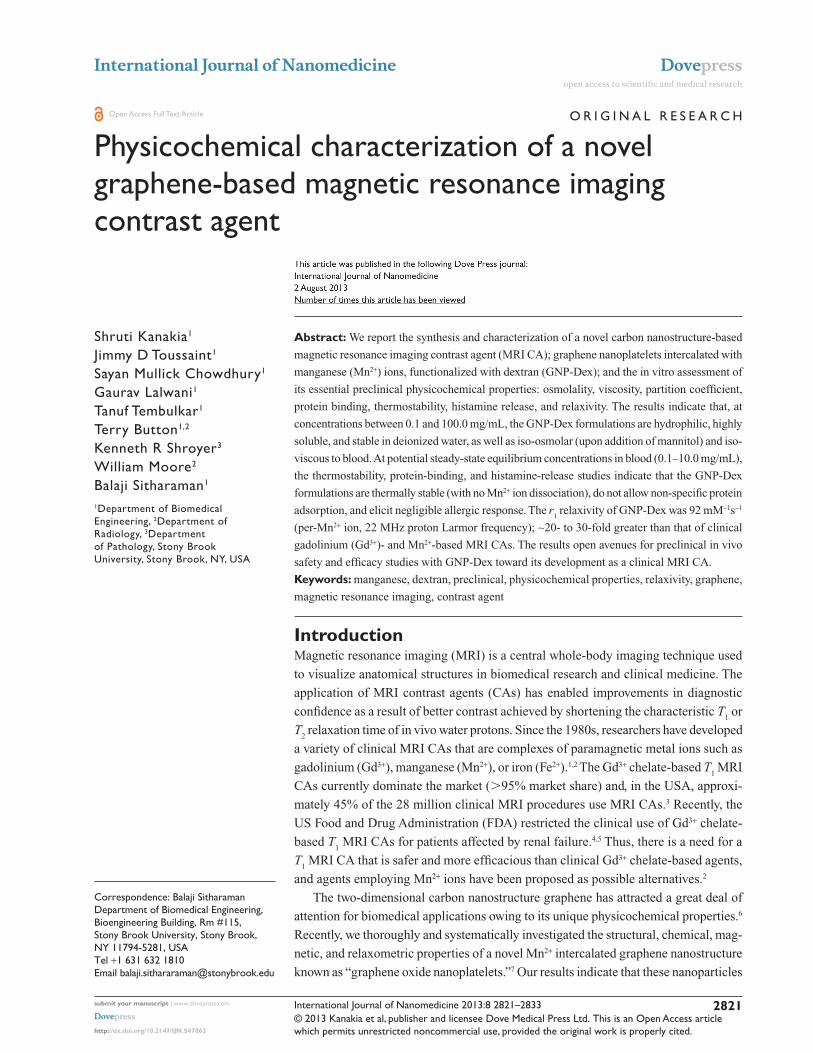
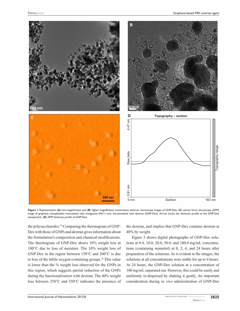
Figure 1 shows the TEM and atomic force microscopy
(AFM) images of GNP-Dex, and provides the formulation’s
structural and morphological information. The low- and
high-resolution TEM images (Figure 1A and B) show that
GNP-Dex are disc-shaped nanoparticles with a coil-like
dextran structure uniformly coating the GNPs. The size of the
GNP-Dex complex, determined by the analysis of the TEM
and the AFM images (Figure 1C and D) was ∼100–120 nm.
Further, the thickness of the GNP-Dex complex determined
by AFM was ∼3–4 nm.
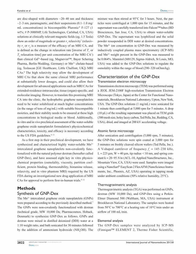
Figure 2 shows the TGA spectrum of the GNP-Dex
formulation. Also included for comparison are TGA spectra
of GNPs and dextran. The TGA spectra of the GNPs show
a weight loss of 10% between 10°C and 150°C due to the
loss of adsorbed moisture. The weight loss of 20% between
150°C and 200°C can be attributed to the pyrolysis of
carboxyl or hydroxyl groups.18 The dextran curve exhib-
its a 80%–90% weight loss between 300°C and 350°C,
which is the characteristic decomposition temperature of
submit your manuscript | www.dovepress.com
Dovepress
Dovepress
2824
Kanakia et al
International Journal of Nanomedicine 2013:8
the polysaccharides.19 Comparing the thermogram of GNP-
Dex with those of GNPs and dextran gives information about
the formulation’s composition and chemical modifications.
The thermogram of GNP-Dex shows 10% weight loss at
100°C due to loss of moisture. The 10% weight loss of
GNP-Dex in the region between 150°C and 200°C is due
to loss of the labile oxygen-containing groups.18 This value
is lower than the % weight loss observed for the GNPs in
this region, which suggests partial reduction of the GNPs
during the functionalization with dextran. The 40% weight
loss between 250°C and 350°C indicates the presence of
the dextran, and implies that GNP-Dex contains dextran at
40% by weight.
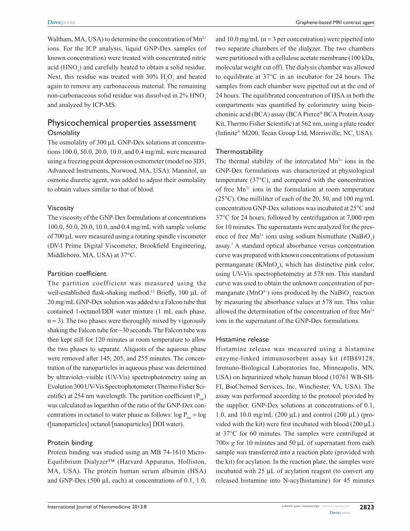
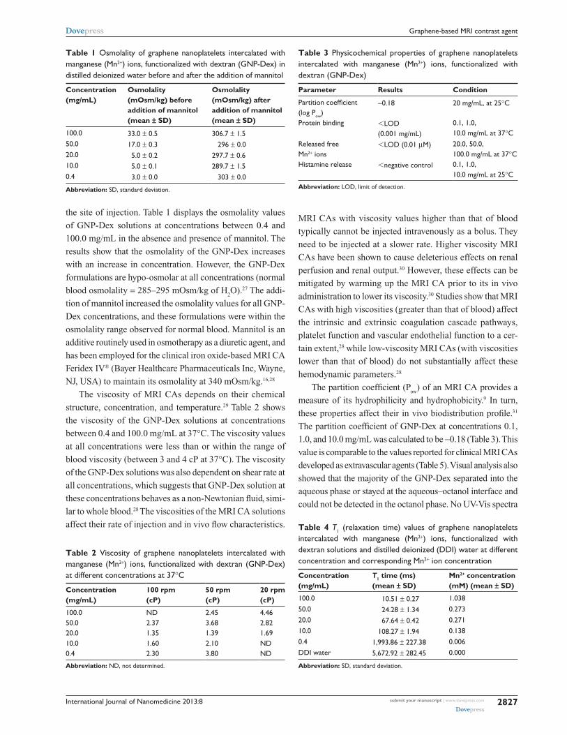
Figure 3 shows digital photographs of GNP-Dex solu-
tions at 0.4, 10.0, 20.0, 50.0, and 100.0 mg/mL concentra-
tions (containing mannitol) at 0, 2, 4, and 24 hours after
preparation of the solutions. As is evident in the images, the
solutions at all concentrations were stable for up to 4 hours.
At 24 hours, the GNP-Dex solution at a concentration of
100 mg/mL separated out. However, this could be easily and
uniformly re-dispersed by shaking it gently. An important
consideration during in vivo administration of GNP-Dex
Figure 1 Representative (A) low-magnification and (B) higher magnification transmission electron microscope images of GNP-Dex; (C) atomic force microscope (AFM) image of graphene nanoplatelets intercalated with manganese (Mn2+) ions, functionalized with dextran (GNP-Dex). Arrow tracks the thickness profile of the GNP-Dex nanoparticle. (D) AFM thickness profile of GNP-Dex.
0 nm Section
Topography – sectionD
183 nm
Top
ogra
phy
rang
e
Raw
dat
a−3
.81
nm4.
47 n
m
submit your manuscript | www.dovepress.com
Dovepress
Dovepress
2825
Graphene-based MRI contrast agent
International Journal of Nanomedicine 2013:8
able as stock solutions for in vivo administration. The higher
stability of the formulations in water can be attributed to
the uniform coating of dextran on GNPs, which counteracts
the hydrophobic forces exerted by the GNPs that can lead
to aggregation.
To the best of our knowledge, this is the highest level of
solubility that has been achieved for any water-solubilized
graphene nanoparticle. An MRI CA needs to exhibit excel-
lent solubility even at high concentrations to achieve the
dose required for bolus injection (the clinical dose of a Gd3+
chelate-based MRI CA can be between 1 and 100 mg/kg21).
Additionally, an important consideration is that the water-
solubilizing moieties need to be biocompatible. Different
covalent and non-covalent functionalization approaches have
been employed to water-solubilize graphene nanoparticles.6
Graphene nanoparticles have been covalently functional-
ized with carboxylic acid, hydroxyl,22 poly-L-lysine,23 and
non-covalently functionalized with synthetic polymer
polyethylene glycol6 or Pluronic F-127.7 The maximum
solubility reported for these functionalization approaches
has been between 1 and 2 mg/mL. Recently, graphene
nanoparticles have been covalently conjugated with the
natural polymer dextran with a maximum water solubility of
1 mg/mL.14,24,25 At the concentration of 1 mg/mL, the volume
needed to be injected for in vivo small animal safety stud-
ies to achieve the above dosages (1–100 mg/kg) would be
up to 1.56%–156.25% of the total blood volume (estimated
based on a 200 g rat with total blood volume of ∼12.8 mL;
see Calculation of the dose in the Supplementary materials
section) or 1.4%–140.0% of the total blood volume (5 L
of an adult human weighing 70 kg; see the Supplementary
materials section), which is not feasible at higher doses. The
GNP-Dex formulations not only show substantially greater
solubility than the other approaches but also stability in physi-
ologically relevant fluids. Additionally, at high concentrations
(eg, 50–100 mg/mL), GNP-Dex can be injected at ∼4%–8%
of the rat blood volume to achieve bolus dosages between
50 and 500 mg/kg, which is feasible.
Tables 1–4 show the results of assessments of the key
physicochemical properties, osmolality, viscosity, partition
coefficient, protein binding, histamine release, and relaxivity.
Osmolality of an MRI CA is an important property that
depends on the solute concentration. Adverse effects of
contrast media with low or high osmolality (compared with
blood osmolality) include changes in the circulatory system
such as vasodilation, vasoconstriction, crenation of red
blood cells, and release of vasopressin.9,26 Clinically, these
changes manifest as sensations of warmth, heat, and pain at
0 100
120
100
80
60
40
20
0200 300Sample temperature °C
Wei
gh
t %
400 500 600
GNP
Dextran
GNP-Dex
700
Figure 2 Representative thermogravimetric analysis (TGA) curve of graphene nanoplatelets, dextran, and graphene nanoplatelets intercalated with manganese (Mn2+) ions, functionalized with dextran (GNP-Dex).
Figure 3 Digital images of vials containing graphene nanoplatelets intercalated with manganese (Mn2+) ions, functionalized with dextran (GNP-Dex) in water (with mannitol) at 100.0, 50.0, 20.0, 10.0, 0.4 mg/mL concentrations at (A) 0 hour (h), (B) 2 hours, (C) 4 hours, and (D) 24 hours after preparing the solutions.
formulations is that they should remain stable long enough
for the dose to be poured out and injected.20 In general, set-
tling is considered a significant issue if the solute cannot be
easily and uniformly re-dispersed.20 The results indicate that,
provided the GNP-Dex formulations at various concentra-
tions are prepared fresh and administered immediately, or
solutions (especially at very high concentrations) kept for
an extended period of time are gently shaken, they are suit-
submit your manuscript | www.dovepress.com
Dovepress
Dovepress
2826
Kanakia et al
International Journal of Nanomedicine 2013:8
MRI CAs with viscosity values higher than that of blood
typically cannot be injected intravenously as a bolus. They
need to be injected at a slower rate. Higher viscosity MRI
CAs have been shown to cause deleterious effects on renal
perfusion and renal output.30 However, these effects can be
mitigated by warming up the MRI CA prior to its in vivo
administration to lower its viscosity.30 Studies show that MRI
CAs with high viscosities (greater than that of blood) affect
the intrinsic and extrinsic coagulation cascade pathways,
platelet function and vascular endothelial function to a cer-
tain extent,28 while low-viscosity MRI CAs (with viscosities
lower than that of blood) do not substantially affect these
hemodynamic parameters.28
The partition coefficient (Pow
) of an MRI CA provides a
measure of its hydrophilicity and hydrophobicity.9 In turn,
these properties affect their in vivo biodistribution profile.31
The partition coefficient of GNP-Dex at concentrations 0.1,
1.0, and 10.0 mg/mL was calculated to be −0.18 (Table 3). This
value is comparable to the values reported for clinical MRI CAs
developed as extravascular agents (Table 5). Visual analysis also
showed that the majority of the GNP-Dex separated into the
aqueous phase or stayed at the aqueous–octanol interface and
could not be detected in the octanol phase. No UV-Vis spectra
Table 1 Osmolality of graphene nanoplatelets intercalated with manganese (Mn2+) ions, functionalized with dextran (GNP-Dex) in distilled deionized water before and after the addition of mannitol
Concentration (mg/mL)
Osmolality (mOsm/kg) before addition of mannitol (mean ± SD)
Osmolality (mOsm/kg) after addition of mannitol (mean ± SD)
100.0 33.0 ± 0.5 306.7 ± 1.550.0 17.0 ± 0.3 296 ± 0.020.0 5.0 ± 0.2 297.7 ± 0.610.0 5.0 ± 0.1 289.7 ± 1.50.4 3.0 ± 0.0 303 ± 0.0
Abbreviation: SD, standard deviation.
Table 2 Viscosity of graphene nanoplatelets intercalated with manganese (Mn2+) ions, functionalized with dextran (GNP-Dex) at different concentrations at 37°C
Concentration (mg/mL)
100 rpm (cP)
50 rpm (cP)
20 rpm (cP)
100.0 ND 2.45 4.4650.0 2.37 3.68 2.8220.0 1.35 1.39 1.6910.0 1.60 2.10 ND0.4 2.30 3.80 ND
Abbreviation: ND, not determined.
Table 3 Physicochemical properties of graphene nanoplatelets intercalated with manganese (Mn2+) ions, functionalized with dextran (GNP-Dex)
Parameter Results Condition
Partition coefficient (log Pow)
−0.18 20 mg/mL, at 25°C
Protein binding ,LOD (0.001 mg/mL)
0.1, 1.0, 10.0 mg/mL at 37°C
Released free Mn2+ ions
,LOD (0.01 µM) 20.0, 50.0, 100.0 mg/mL at 37°C
histamine release ,negative control 0.1, 1.0, 10.0 mg/mL at 25°C
Abbreviation: LOD, limit of detection.
Table 4 T1 (relaxation time) values of graphene nanoplatelets intercalated with manganese (Mn2+) ions, functionalized with dextran solutions and distilled deionized (DDI) water at different concentration and corresponding Mn2+ ion concentration
Concentration (mg/mL)
T1 time (ms) (mean ± SD)
Mn2+ concentration (mM) (mean ± SD)
100.0 10.51 ± 0.27 1.03850.0 24.28 ± 1.34 0.27320.0 67.64 ± 0.42 0.27110.0 108.27 ± 1.94 0.1380.4 1,993.86 ± 227.38 0.006DDI water 5,672.92 ± 282.45 0.000
Abbreviation: SD, standard deviation.
the site of injection. Table 1 displays the osmolality values
of GNP-Dex solutions at concentrations between 0.4 and
100.0 mg/mL in the absence and presence of mannitol. The
results show that the osmolality of the GNP-Dex increases
with an increase in concentration. However, the GNP-Dex
formulations are hypo-osmolar at all concentrations (normal
blood osmolality = 285–295 mOsm/kg of H2O).27 The addi-
tion of mannitol increased the osmolality values for all GNP-
Dex concentrations, and these formulations were within the
osmolality range observed for normal blood. Mannitol is an
additive routinely used in osmotherapy as a diuretic agent, and
has been employed for the clinical iron oxide-based MRI CA
Feridex IV® (Bayer Healthcare Pharmaceuticals Inc, Wayne,
NJ, USA) to maintain its osmolality at 340 mOsm/kg.16,28
The viscosity of MRI CAs depends on their chemical
structure, concentration, and temperature.29 Table 2 shows
the viscosity of the GNP-Dex solutions at concentrations
between 0.4 and 100.0 mg/mL at 37°C. The viscosity values
at all concentrations were less than or within the range of
blood viscosity (between 3 and 4 cP at 37°C). The viscosity
of the GNP-Dex solutions was also dependent on shear rate at
all concentrations, which suggests that GNP-Dex solution at
these concentrations behaves as a non-Newtonian fluid, simi-
lar to whole blood.28 The viscosities of the MRI CA solutions
affect their rate of injection and in vivo flow characteristics.
submit your manuscript | www.dovepress.com
Dovepress
Dovepress
2827
Graphene-based MRI contrast agent
International Journal of Nanomedicine 2013:8
of the GNP-Dex in octanol phase could be obtained, indicat-
ing that the nanoparticles, if present, were below the detection
limit of the instrument. The results indicate that most of the
GNP-Dex nanoparticles were hydrophilic, and preferentially
accumulate in the aqueous phase, and a small fraction were
amphiphilic, accumulating at the water–octanol interface.
The propensity of MRI CAs to bind to protein could affect
their targeting capability, pharmacokinetics, and relaxivity.9,31
Equilibrium dialysis of an MRI CA with the protein HSA
followed by BCA protein assay is one of the most widely-
accepted methods for studying protein–drug interaction.32,33
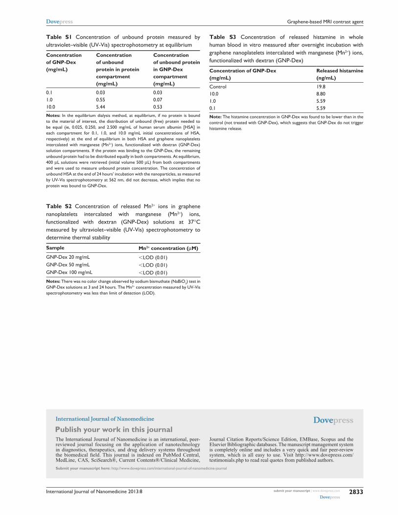
The results (Table S1) of the protein binding, summarized in
Table 4, show that HSA protein was not bound to the GNP-
Dex formulations. The lack of protein binding onto GNP-Dex
can be attributed to the non-fouling property of dextran,17 and
suggests that it could prevent nonspecific interactions.
Thermal stability of the intercalated Mn2+ ions is a key
physiochemical property. It was important to determine if the
intercalated Mn2+ ions dissociate from the GNP-Dex nanopar-
ticles and were present as free Mn2+ ions under physiological
temperatures. The Mn2+ ion, a natural cellular constituent,
functions as a regulatory cofactor for enzymes and recep-
tors.2 The normal daily dietary requirement for manganese is
2.7–7.0 mg,34 and the normal serum level is 0.1 µM.35 How-
ever, prolonged exposure to high concentrations of Mn2+ ions
has been reported to be toxic, and results in “manganism,” a
Parkinson-like neurological disorder, and cardiac dysfunc-
tion.2,36 Therefore, it was critical to investigate whether the
intercalated Mn2+ ions, at the physiological temperature of
37°C, dissociate from GNP-Dex nanoparticles. The thermal
stability experiments (Table S2 and summarized in Table 4)
with GNP-Dex solutions, for 3 or 24 hours, at 20, 50, and
100 mg/mL concentrations, show no presence of free Mn2+
ions (limit of detection [LOD] = 0.01 µM) at 37°C. The results
indicate that Mn2+ ions present in the GNP-Dex formulation
are stable at least short-term (24 hours) at simulated physi-
ological temperature. Typically, clinical first pass or blood pool
CAs have been reported to have average blood half-lives of
1.5 hours, and .95% of the CA is excreted in 24 hours.37 Thus,
taken together with this information, the reported results sug-
gest that provided the in vivo blood half-life and the elimination
rate of the GNP-Dex formulations are similar, the intercalated
Mn2+ ions should be stable within the graphene nanoplatelets
in vivo. However, the long-term (.24 hours) in vitro and
in vivo thermal stability of these formulations still needs to be
determined before a firm conclusion can be drawn.
It is well documented that MRI CAs can evoke adverse
allergic reactions with symptoms that can include urticaria
or hives-like inflammation as well as swelling and reddening
of the tissues, watery eyes, runny nose, migraine headaches,
nausea, vomiting, laryngospasm, and bronchospasm.38–40
Histamine, a naturally occurring biomolecule found in vivo,
is released as a response to allergic reactions40 and is consid-
ered a good marker to evaluate the propensity of an MRI CA
to elicit an allergic response. Our results show no significant
release of histamine at GNP-Dex concentrations of 0.1, 1.0,
and 10.0 mg/mL compared with the control (Table S3). These
results are encouraging since histamine can also stimulate
fibroblast proliferation and collagen production, and can
play an important role in skin fibrosis – a major symptom
associated with nephrogenic systemic fibrosis.40 Studies on
Gd3+- and Mn2+-chelate MRI CAs indicate that, at physiologi-
cally relevant plasma concentrations (0.1–10.0 mM), they
do not exhibit a propensity to release histamine.40 At higher
dosages (50–150 mM), Gd3+-chelate complexes have been
shown to release histamine in vitro.40
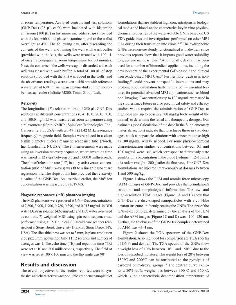
Relaxivity is an important measure of efficacy for T1
MRI CAs.1 Our previous results show that the r2/r
1 ratio
Table 5 Comparison of the physicochemical properties of graphene nanoplatelets intercalated with manganese (Mn2+) ions, functionalized with dextran (GNP-Dex) with US Food and Drug Administration (FDA)-approved magnetic resonance imaging contrast agent (MRI CA)
US FDA-approved MRI CA
Osmolality (mOsm/kg of water)
Viscosity (cP) at 37°C
Partition coefficient (log Pow)
Protein binding Relaxivity (r1) mM-1s-1 at 0.47 T8
Prohance41 630 1.3 −3.6800 None 3.1Multihance®42 1,970 5.3 0.0016 ,5% 4.2Magnevist®43 1,960 2.9 −5.4000 None 3.4Omniscan™46 789 1.4 −2.1000 None 3.5Teslascan44 290 0.7 −5.6200 Fodipir chelate (negligible),
free Mn2+ ions (27%)1.5
Optimark™45 1,110 2.0 −8.2200 None 4.2GNP-Dex 290 2.4 −0.1800 None 92
Notes: ProHance, Bracco Diagnostics Inc. Princeton, NJ, USA; MultiHance®, Bracco Diagnostics Inc.; Magnevist®, Bayer Schering Pharma, Berlin-Wedding, Germany; OmniscanTM, GE Healthcare, Little Chalfont, UK; Teslascan, GE Healthcare; OptimarkTM, Mallinckrodt Inc., St Louis, MO, USA.
submit your manuscript | www.dovepress.com
Dovepress
Dovepress
2828
Kanakia et al
International Journal of Nanomedicine 2013:8
for water-solubilized GNP solutions is 2.2, which is in the
range reported for Mn2+- or Gd3+-chelate complexes (r2/r
1
∼1.0–2.5) T1 MRI CAs.7,8 The ratio is also lower than the
iron oxide-functionalized graphene nanoparticles (r2/r
1 ∼10)
T2 MRI CAs.7 Thus, the GNP-Dex formulations would be
more suitable as a T1 MRI CA. Table 4 shows T
1 relaxation
time at 0.47 T (21.42 MHz proton Larmor frequency) at
different doses of GNP-Dex. Also included are the Mn2+ ion
concentrations (in µM) for each GNP-Dex dose. The relaxiv-
ity value calculated from the plot (Figure 4A) of 1/T1 versus
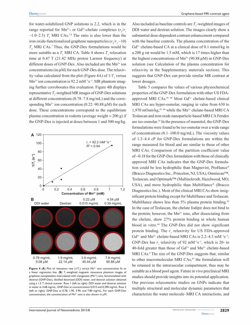
Mn2+ ion concentration is 92.2 mM−1s−1. MR phantom imag-
ing further corroborates this evaluation. Figure 4B displays
representative T1-weighted MR images of GNP-Dex solutions
at different concentrations (0.78–7.8 mg/mL) and the corre-
sponding Mn2+ ion concentration (0.22–90.88 µM) for each
dose. These concentrations correspond to the equilibrium
plasma concentration in rodents (average weight = 200 g) if
the GNP-Dex is injected at doses between 1 and 500 mg/kg.
Also included as baseline controls are T1-weighted images of
DDI water and dextran solution. The images clearly show a
substantial dose-dependent contrast enhancement compared
with the baseline controls. The plasma concentration of the
Gd3+ chelate-based CA at a clinical dose of 0.1 mmol/kg in
a 200 g rat would be 1.5 mM, which is 17 times higher than
the highest concentrations of Mn2+ (90.88 µM) in GNP-Dex
solution (see Calculation of the plasma concentration for
relaxivity in the Supplementary materials section). This
suggests that GNP-Dex can provide similar MR contrast at
lower dosages.
Table 5 compares the values of various physiochemical
properties of the GNP-Dex formulation with other US FDA-
approved MRI CAs.41–46 Most Gd3+ chelate-based clinical
MRI CAs are hyper-osmolar, ranging in value from 630 to
1,970 mOsm/kg,41–46 while the Mn2+ chelate-based MRI CA
Teslascan and iron oxide nanoparticle-based MRI CA Feridex
are iso-osmolar.16 In the presence of mannitol, the GNP-Dex
formulations were found to be iso-osmolar over a wide range
of concentrations (0.1–100.0 mg/mL). The viscosity values
of 1.3–4.4 cP for GNP-Dex formulations are within the
range measured for blood and are similar to those of other
MRI CAs. Comparison of the partition coefficient value
of −0.18 for the GNP-Dex formulation with those of clinically
approved MRI CAs indicates that the GNP-Dex formula-
tion could be less hydrophilic than Magnevist, ProHance®
(Bracco Diagnostics Inc., Princeton, NJ, USA), Omniscan™,
Teslascan, and Optimark™ (Mallinckrodt, Hazelwood, MO,
USA), and more hydrophilic than MultiHance® (Bracco
Diagnostics Inc.). Most of the clinical MRI CAs show insig-
nificant protein binding except for MultiHance and Teslascan.
MultiHance shows less than 5% plasma protein binding.42
In the case of Teslascan, the chelate fodipir does not bind to
the protein; however, the Mn2+ ions, after dissociating from
the chelate, show 27% protein binding in whole human
blood in vitro.44 The GNP-Dex did not show significant
protein binding. The r1 relaxivity for US FDA-approved
Gd3+ and Mn2+ chelate-based MRI CAs is 2.2–4.5 mM−1s−1;
GNP-Dex has r1 relaxivity of 92 mM−1s−1, which is 20- to
40-fold greater than those of Gd3+ and Mn2+ chelate-based
MRI CAs.8 The size of the GNP-Dex suggests that, similar
to other macromolecular MRI CAs,47 the formulation will
be retained in the intravascular compartment, thus may be
suitable as a blood pool agent. Future in vivo preclinical MRI
studies should provide insights into its potential application.
Our previous relaxometric studies on GNPs indicate that
multiple structural and molecular dynamic parameters that
characterize the water molecule–MRI CA interactions, and
00
20
40
60
1/T
1 (s
−1)
80
100
120
0.2 0.4Concentration of Mn2+ (mM)
0.6
r1 = 92.2 mM−1s−1
R2 = 0.94
0.8 1 1.2
A
Figure 4 (A) Plot of relaxation rate (1/T1) versus Mn2+ ions concentration fit to a linear regression line. (B) T1-weighted magnetic resonance phantom images of graphene nanoplatelets intercalated with manganese (Mn2+) ions, functionalized with dextran (GNP-Dex), distilled deionized (DDI) water, and dextran solution obtained using a 1.5 T clinical scanner. Row 1 (left to right): DDI water and dextran solution in water at 4.68 mg/mL, GNP-Dex at concentrations 0.015 and 0.390 mg/mL. Row 2 (left to right): GNP-Dex at 0.78, 1.90, 3.90, and 7.80 mg/mL. For each GNP-Dex concentration, the concentration of Mn2+ ions is also shown in µM.
DDI water Dextran0.22 µM
0.015 mg/mL4.54 µM
0.39 mg/mL
7.8 mg/mL90.88 µM
3.9 mg/mL45.44 µM
1.9 mg/mL22.14 µM
0.78 mg/mL9.08 µM
B
submit your manuscript | www.dovepress.com
Dovepress
Dovepress
2829
Graphene-based MRI contrast agent
International Journal of Nanomedicine 2013:8
affect the T1 relaxation mechanism, are modulated and are
responsible for the observed increase in the relaxivity.48 The
parameters include the number of water molecules (q) that
can simultaneously coordinate with the Mn2+ ions intercalated
within the GNPs, the residence lifetime (τM
) of the water
molecules coordinated on the Mn2+ ions, and the rotational
correlation time (τR) of the GNPs.
Over the past 10 years, carbon nanostructures such
as gadofullerenes (represented as Gd@C60
, Gd@C80
, and
Gd@C82
) and gadonanotubes (represented as Gd@US-
tubes, where “US-tubes” means “ultra-short single-walled
carbon nanotubes”) that encapsulate Gd3+ metal ions have
been proposed as T1 CAs for MRI.1,49 Even though these
MRI CAs show higher relaxivity compared with clinical
MRI CAs, none has translated into clinical use. Recent
reports also show that administration of clinical Gd3+-based
MRI CAs (especially Omniscan) to patients with severe
acute or chronic renal insufficiency (glomerular filtration
rate ,30 mL/min/1.73 m2), significantly increases their risk
for developing nephrogenic systemic fibrosis.4 As a result
of these findings, the US FDA has restricted their use in
patients suffering from renal insufficiency and instructed
manufacturers to include mandatory box warnings and
disclose contraindications for this high-risk group on the
product labeling of all Gd3+-based MRI CAs.5 Recently,
manganese has received attention as a possible alternative to
gadolinium. Manganese, a natural cellular constituent resem-
bling Ca2+, which often functions as a regulatory cofactor for
enzymes and receptors, was one of first elements reported
suitable as a CA for MRI.2 Manganese toxicity is rare, and
has only been reported following long-term exposure to
high concentrations, which has resulted in neurological
symptoms.2 The two Mn2+-based agents – Teslascan and
an oral CA containing manganese (II) chloride (Bracco
Diagnostics Inc.) – that were approved for clinical use have
been discontinued. The synthesis and characterization of
the highly water-soluble Mn2+ ion intercalated GNP-Dex
formulations, along with the positive results of their key in
vitro physiochemical properties opens avenues for further in
vitro and in vivo preclinical studies to assess their suitability
as a next-generation T1 MRI CA with a safety and efficacy
profile that can overcome the outlined limitations.
ConclusionThe GNP-Dex formulation shows very high solubility (up to
100 mg/mL) and stability in deionized water and biological
buffers. The osmolality of GNP-Dex is lower than that of
blood, and can be adjusted to be similar to blood by adding
the diuretic mannitol. The viscosities of the GNP-Dex
formulations were within the range of blood viscosity. At
physiological temperatures (37°C), GNP-Dex was structur-
ally stable and the intercalated Mn2+ ions did not dissociate
for up to 24 hours. Most of the GNP-Dex nanoparticles
were hydrophilic, and a small fraction was amphiphilic. At
potential therapeutic dosages, GNP-Dex showed very little or
no interaction with blood protein HSA, suggesting that they
prevent nonspecific adsorption of proteins. Moreover, the for-
mulations did not trigger histamine release in whole human
blood, suggesting that they are unlikely to evoke allergic
responses. The high r1 relaxivity (92 mM−1s−1) of GNP-Dex
allowed significant contrast enhancement of T1-weighted
MRI phantoms even at modest (nM) concentrations of Mn2+
ions. The in vitro results show that the physicochemical
properties of the GNP-Dex were comparable to or better
than those of clinically approved MRI CAs. The studies open
avenues for further in vitro and in vivo preclinical studies to
translate this promising T1 MRI CA into clinical use.
AcknowledgmentsThis work was sponsored by the Wallace H Coulter Foun-
dation. The authors thank Dr David Hirschberg for his help
with performing ICP-MS and Ms Shelagh Zegers for her
help with lyophilization.
DisclosureThe authors declare no conflicts of interest in this work.
References1. Sitharaman B, Wilson LJ. Gadofullerenes and gadonanotubes: a new
paradigm for high-performance magnetic resonance imaging contrast agent probes. J Biomed Nanotechnol. 2007;3(4):342–352.
2. Pan D, Caruthers SD, Senpan A, Schmieder AH, Wickline SA, Lanza GM. Revisiting an old friend: manganese-based MRI contrast agents. Wiley Interdiscip Rev Nanomed Nanobiotechnol. 2011;3(2):162–173.
3. Ananta JS, Godin B, Sethi R, et al. Geometrical confinement of gadolinium-based contrast agents in nanoporous particles enhances T1 contrast. Nat Nanotechnol. 2010;5(11):815–821.
4. Broome DR. Nephrogenic systemic fibrosis associated with gadolinium based contrast agents: a summary of the medical literature reporting. Eur J Radiol. 2008;66(2):230–234.
5. Bayer HealthCare Pharmaceuticals Inc, Bracco Diagnostics Inc, GE Healthcare, Mallinkrodit Inc Important Drug Warning for Gadolinium-based Contrast Agents [letter to healthcare professionals]. Bayer HealthCare Pharmaceuticals Inc, Bracco Diagnostics Inc, GE Healthcare, Mallinkrodit Inc; 2007. Available from: http://www.fda.gov/downloads/safety/medwatch/safetyinformation/safetyalertsforhumanmedicalproducts/ucm154532.pdf. Accessed June 18, 2013.
6. Feng L, Liu Z. Graphene in biomedicine: opportunities and challenges. Nanomedicine (Lond). 2011;6(2):317–324.
7. Paratala BS, Jacobson BD, Kanakia S, Francis LD, Sitharaman B. Physicochemical characterization, and relaxometry studies of micro-graphite oxide, graphene nanoplatelets, and nanoribbons. PLoS One. 2012;7(6):e38185.
submit your manuscript | www.dovepress.com
Dovepress
Dovepress
2830
Kanakia et al
International Journal of Nanomedicine 2013:8
8. Rohrer M, Bauer H, Mintorovitch J, Requardt M, Weinmann HJ. Comparison of magnetic properties of MRI contrast media solutions at dif-ferent magnetic field strengths. Invest Radiol. 2005;40(11):715–724.
9. Vogler H, Platzek J, Schuhmann-Giampieri G, et al. Pre-clinical evaluation of gadobutrol: a new, neutral, extracellular contrast agent for magnetic resonance imaging. Eur J Radiol. 1995;21(1):1–10.
10. ICH. Safety pharmacology studies for human pharmaceuticals. Geneva: International Conference on Harmonisation; 2000. Available from: http://www.ich.org/fileadmin/Public_Web_Site/ICH_Products/Guidelines/ Safety/S7A/Step4/S7A_Guideline.pdf. Accessed July 15, 2013.
11. US FDA. Developing medical imaging drug and biological products part 1: conducting safety assessments. Rockville: US Food and Drug Adminsitration; 2004. Available from: http://www.fda.gov/downloads/Drugs/.../Guidances/ucm071600.pdf. Accessed July 15, 2013.
12. OECD. OECD guidelines for the testing of chemicals. Paris: Organi-sation for Economic Co-operation and Development; 2000. Available from: http://www.oecd.org/chemicalsafety/testing/2731134.pdf. Accessed July 15, 2013.
13. Runge VM. Safety of approved MR contrast media for intravenous injection. J Magn Reson Imaging. 2000;12(2):205–213.
14. Zhang S, Yang K, Feng L, Liu Z. In vitro and in vivo behaviors of dextran functionalized graphene. Carbon. 2011;49(12):4040–4049.
15. Sirlin CB, Vera DR, Corbeil JA, Caballero MB, Buxton RB, Mattrey RF. Gadolinium-DTPA-dextran: a macromolecular MR blood pool contrast agent. Academic Radiology. 2004;11(12):1361–1369.
16. Feridex IV (ferumoxides injectable solution) [package insert]. Wayne, NJ: Bayer Healthcare Pharmaceuticals Inc; 2007. Available from: http://www.amagpharma.com/documents/products/pdfs/feridex_insert.pdf. Accessed June 18, 2013.
17. Martwiset S, Koh AE, Chen W. Nonfouling characteristics of dextran-containing surfaces. Langmuir. 2006;22(19):8192–8196.
18. Stankovich S, Dikin DA, Piner RD, et al. Synthesis of graphene-based nanosheets via chemical reduction of exfoliated graphite oxide. Carbon. 2007;45(7):1558–1565.
19. Tang M, Dou H, Sun K. One-step synthesis of dextran-based stable nano-particles assisted by self-assembly. Polymer. 2006;47(2):728–734.
20. Zatz JL. Physical stability of suspensions. J Soc Cosmet Chem. 1985;36(6):393–411.
21. Tweedle MF. The ProHance story: the making of a novel MRI contrast agent. Eur Radiol. 1997;7 Suppl 5:225–230.
22. Hummers WS Jr, Offeman RE. Preparation of graphitic oxide. J Am Chem Soc. 1958;80(6):1339.
23. Shan C, Yang H, Han D, Zhang Q, Ivaska A, Niu L. Water-soluble graphene covalently functionalized by biocompatible poly-L-lysine. Langmuir. 2009;25(20):12030–12033.
24. Liu Z, Robinson JT, Sun X, Dai H. PEGylated nanographene oxide for delivery of water-insoluble cancer drugs. J Am Chem Soc. 2008;130(33): 10876–10877.
25. Kim YK, Kim MH, Min DH. Biocompatible reduced graphene oxide prepared by using dextran as a multifunctional reducing agent. Chem Commun (Camb). 2011;47(11):3195–3197.
26. Deray G, Jacobs C. Are low osmolality contrast media less nephrotoxic? Nephrol Dial Transplant. 1996;11(6):930–931.
27. American Society of Health-System Pharmacists. Quick Guide to Contrast Media. Bethesda, MD: Contrast Media and Medication Management Standard Resource Center; 2010.
28. Pugh ND. Haemodynamic and rheological effects of contrast media: the role of viscosity and osmolality. Eur Radiol. 1996;6 Suppl 2: S13–S15.
29. Voeltz MD, Nelson MA, McDaniel MC, Manoukian SV. The important properties of contrast media: focus on viscosity. J Invasive Cardiol. 2007;19(3):1A–9A.
30. Lancelot E, Idée JM, Laclédère C, Santus R, Corot C. Effects of two dimeric iodinated contrast media on renal medullary blood perfusion and oxygenation in dogs. Invest Radiol. 2002;37(7):368–375.
31. Caravan P. Protein-targeted gadolinium-based magnetic resonance imaging (MRI) contrast agents: design and mechanism of action. Acc Chem Res. 2009;42(7):851–862.
32. van Liempd S, Morrison D, Sysmans L, Nelis P, Mortishire-Smith R. Development and validation of a higher-throughput equilibrium dialysis assay for plasma protein binding. J Lab Autom. 2011;16(1):56–67.
33. Rimkus G, Bremer-Streck S, Grüttner C, Kaiser WA, Hilger I. Can we accurately quantify nanoparticle associated proteins when constructing high-affinity MRI molecular imaging probes? Contrast Media Mol Imaging. 2011;6(3):119–125.
34. Watts DL. The nutritional relationships of manganese. J Orthomol Med. 1990;5(4):219–222.
35. Sullivan JF, Blotcky AJ, Jetton MM, Hahn HK, Burch RE. Serum levels of selenium, calcium, copper magnesium, manganese and zinc in various human diseases. J Nutr. 109(8):1432–1437.
36. Aschner M, Aschner JL. Manganese neurotoxicity: cellular effects and blood-brain barrier transport. Neurosci Biobehav Rev. 1991;15(3): 333–340.
37. Morcos SK, Thomsen HS, Webb JA; Contrast Media Safety Committee of the European Society of Urogenital Radiology (ESUR). Dialysis and contrast media. Eur Radiol. 2002;12(12):3026–3030.
38. Prince MR, Zhang H, Zou Z, Staron RB, Brill PW. Incidence of immediate gadolinium contrast media reactions. AJR Am J Roentgenol. 2011;196(2):W138–W143.
39. Dillman JR, Ellis JH, Cohan RH, Strouse PJ, Jan SC. Allergic-like breakthrough reactions to gadolinium contrast agents after corticosteroid and antihistamine premedication. AJR Am J Oroentgenol. 2008;190(1): 187–190.
40. Kuefner MA, Feurle J, Uder M, Bautz W, Schwelberger HG. Influence of magnetic resonance contrast media on the activity of histamine inactivating enzymes. Acad Radiol. 2009;16(3):358–362.
41. ProHance® (gadoteridol) injection, solution [product label]. Princeton, NJ: Bracco Diagnostics Inc; nd. Available from: http://dailymed.nlm.nih.gov/dailymed/lookup.cfm?setid=778aee03-07d4c-481a-be8a-a75db0702f5a. Accessed June 18, 2013.
42. MultiHance – gadobenate dimeglumine injection, solution [highlights of prescribing information]. Princeton, NJ: Bracco Diagnostics Inc; nd. Available from: http://dailymed.nlm.nih.gov/dailymed/lookup.cfm?setid=e56fd303-309e0c-490b-8f31-64fc267b4df5. Accessed June 18, 2013.
43. Magnevist® (brand of gadopentetate dimeglumine) [package insert]. Wayne, NJ: Bayer HealthCare Pharmaceuticals Inc; 2011. Avail-able from: http://labeling.bayerhealthcare.com/html/products/pi/Magnevist_PI.pdf. Accessed June 18, 2013.
44. Teslascan data sheet [webpage on the Internet]. Portland: RxList Inc.; 2009. Available from: http://www.rxlist.com/teslascan-drug.htm. Accessed July 15, 2013.
45. Optimark [package insert]. Hazelwood, MO: Mallinckrodt; nd. Available from: http://www.mallinckrodt.com/webforms/threecolumn.aspx?id=497. Accessed June 18, 2013.
46. Omniscan™ (gadodiamide) injection [product label]. Princeton, NJ: GE Healthcare; 2011. http://dailymed.nlm.nih.gov/dailymed/lookup.cfm?setid=1e9a37e2-f28a-4373-bf0f-3e9b60f42d8a. Accessed July 15, 2013.
47. Kobayashi H, Brechbiel MW. Dendrimer-based macromolecular MRI contrast agents: characteristics and application. Mol Imaging. 2003;2(1):1–10.
48. Yang K, Hu L, Ma X, et al. Multimodal imaging guided photothermal therapy using functionalized graphene nanosheets anchored with magnetic nanoparticles. Adv Mater. 2012;24(14):1868–1872.
49. Sitharaman B, Wilson LJ. Gadonanotubes as new high-performance MRI contrast agents. Int J Nanomedicine. 2006;1(3):291–295.
submit your manuscript | www.dovepress.com
Dovepress
Dovepress
2831
Graphene-based MRI contrast agent
International Journal of Nanomedicine 2013:8
Supplementary materialsCalculation of the doseIf the concentration of the MRI CA solution is 1 mg/mL,
and it is required to be intravenously administered at a dose
of 100 mg/kg in a 200 g rat, the CA injection volume (v) is
calculated as follows:
v (mL) × 1 mg/mL = 100 mg/kg × 200 g
v (mL) × 1 mg/mL = (100 mg/1,000 g) × 200 g
v (mL) × 1 mg/mL = 20 mg
v (mL) = 20 mg/1 mg/mL
v (mL) = 20 mL.
The total circulating blood volume in a 200 g rat has been
assumed to be ∼12.8 mL. Therefore, the injected volume
percent (%) (P) is calculated as:
P% = (20 mL/12.8 mL) × 100%
P (%) = 156.25%.
Similarly, if the concentration of the MRI CA solution is
1 mg/mL, and it is required to be intravenously administered
at a dose of 1 mg/kg in a 200 g rat, the CA injection volume
(v) is calculated as:
v (mL) × 1 mg/mL = 1 mg/kg × 200 g
v (mL) × 1 mg/mL = (1 mg/1,000 g) × 200 g
v (mL) × 1 mg/mL = 2 mg
v (mL) = 2 mg/1 mg/mL
v (mL) = 2 mL.
The injected volume percent (%) (P) can be calculated as:
12.8 mL × P (%) = 2 mL × 100%
P (%) = (2 mL/12.8 mL) × 100%
P (%) = 1.56%.
In humans, the total circulating blood volume in a 70 kg
adult human is 5 L. Therefore, if the concentration of the
MRI CA solution is 1 mg/mL, and a 100 mg/kg dose has to
be administered in a 70 kg human, the injection volume (v)
of CA can be calculated as:
v (mL) × 1 mg/mL = 100 mg/kg × 70,000 g
v (mL) × 1 mg/mL = (100 mg/1,000 g) × 70,000 g
v (mL) × 1 mg/mL = 7,000 mg
v (mL) = 7,000 mg/1 mg/mL
v (mL) = 7,000 mL.
The injected volume percent (%) (P) can be calculated as:
5,000 mL × P (%) = 7,000 mL × 100%
P (%) = (7,000 mL/5,000 mL) × 100%
P (%) = 140%.
Similarly, if the concentration of the MRI CA solution is
1 mg/mL, and it is required to be intravenously administered
at a dose of 1 mg/kg in a 70 kg human, the CA injection
volume (v) is calculated as:
v (mL) × 1 mg/mL = 1 mg/kg × 70,000 g
v (mL) × 1 mg/mL = (1 mg/1,000 g) × 70,000 g
v (mL) × 1 mg/mL = 7 mg
v (mL) = 7 mg/1 mg/mL
v (mL) = 7 mL.
The injected volume percent (%) (P) can be calculated as:
5,000 mL × P (%) = 7 mL × 100%
P (%) = (7 mL/5,000 mL) × 100%
P (%) = 1.4%.
If the concentration of the MRI CA solution is 100 mg/mL,
and it is required to be intravenously administered at a dose
of 500 mg/kg in a 200 g rat, the CA injection volume (v) is
calculated as:
v (mL) × 100 mg/mL = 500 mg/kg × 200 g
v (mL) × 100 mg/mL = (500 mg/1,000 g) × 200 g
v (mL) × 100 mg/mL = 100 mg
v (mL) = 100 mg/100 mg/mL
v (mL) = 1 mL.
The total circulating blood volume in a 200 g rat has been
assumed to be ∼12.8 mL.
Thus, the injected volume percent (%) (P) can be calcu-
lated as follows:
12.8 mL × P (%) = 1 mL × 100%
P (%) = (1 mL/12.8 mL) × 100%
P (%) = 7.8.
Calculation of the plasma concentration for relaxivityAt a 0.1 mmol/kg clinical dose, the total amount of MRI CA
that will be injected in a 200 g rat will be 0.1 mmol/kg ×
0.2 kg = 0.02 mmol.
With a total circulating blood volume of ∼12.8 mL,
the plasma concentration of Gd3+ CA can be calculated as:
0.02 mmol/12.8 mL = 0.001562 mmol/mL = 1,562 µM.
As determined by ICP-MS, the Mn2+ in GNP-Dex by
weight is 0.064%. Therefore, the weight (z) (mg) of Mn2+
ions in 1 mL of 7.8 mg/mL solution of GNP-Dex can be
calculated as:
100 mg × z (mg) = 7.8 mg × 0.064 mg
z (mg) = (7.8 mg/100 mg) × 0.064 mg
z (mg) = 0. 005 mg.
Converting mass of Mn2+ into moles (molecular weight
[MW] of Mn2+ 54.93):
0.5 mg = (0.005 × 10⁻³ g/54.93 g/mole) = 9.08 × 10⁻⁸
moles.
Thus, 7.8 mg/mL GNP-Dex solution has 9.08 × 10⁻⁸
moles/mL or 90.80 µM of Mn2+ ions. This value is 17 times
lower than the plasma concentration of Gd3+ chelate-based
CA (1,562 µM) at clinical dose.
submit your manuscript | www.dovepress.com
Dovepress
Dovepress
2832
Kanakia et al
International Journal of Nanomedicine
Publish your work in this journal
Submit your manuscript here: http://www.dovepress.com/international-journal-of-nanomedicine-journal
The International Journal of Nanomedicine is an international, peer-reviewed journal focusing on the application of nanotechnology in diagnostics, therapeutics, and drug delivery systems throughout the biomedical field. This journal is indexed on PubMed Central, MedLine, CAS, SciSearch®, Current Contents®/Clinical Medicine,
Journal Citation Reports/Science Edition, EMBase, Scopus and the Elsevier Bibliographic databases. The manuscript management system is completely online and includes a very quick and fair peer-review system, which is all easy to use. Visit http://www.dovepress.com/ testimonials.php to read real quotes from published authors.
International Journal of Nanomedicine 2013:8
Table S1 Concentration of unbound protein measured by ultraviolet–visible (UV-Vis) spectrophotometry at equilibrium
Concentration of GNP-Dex (mg/mL)
Concentration of unbound protein in protein compartment (mg/mL)
Concentration of unbound protein in GNP-Dex compartment (mg/mL)
0.1 0.03 0.031.0 0.55 0.0710.0 5.44 0.53
Notes: In the equilibrium dialysis method, at equilibrium, if no protein is bound to the material of interest, the distribution of unbound (free) protein needed to be equal (ie, 0.025, 0.250, and 2.500 mg/mL of human serum albumin [hSA] in each compartment for 0.1, 1.0, and 10.0 mg/mL initial concentrations of hSA, respectively) at the end of equilibrium in both hSA and graphene nanoplatelets intercalated with manganese (Mn2+) ions, functionalized with dextran (GNP-Dex) solution compartments. If the protein was binding to the GNP-Dex, the remaining unbound protein had to be distributed equally in both compartments. At equilibrium, 400 µL solutions were retrieved (initial volume 500 µL) from both compartments and were used to measure unbound protein concentration. The concentration of unbound hSA at the end of 24 hours’ incubation with the nanoparticles, as measured by UV-Vis spectrophotometry at 562 nm, did not decrease, which implies that no protein was bound to GNP-Dex.
Table S2 Concentration of released Mn2+ ions in graphene nanoplatelets intercalated with manganese (Mn2+) ions, functionalized with dextran (GNP-Dex) solutions at 37°C measured by ultraviolet–visible (UV-Vis) spectrophotometry to determine thermal stability
Sample Mn2+ concentration (µM)
GNP-Dex 20 mg/mL ,LOD (0.01)GNP-Dex 50 mg/mL ,LOD (0.01)GNP-Dex 100 mg/mL ,LOD (0.01)
Notes: There was no color change observed by sodium bismuthate (NaBiO3) test in GNP-Dex solutions at 3 and 24 hours. The Mn2+ concentration measured by UV-Vis spectrophotometry was less than limit of detection (LOD).
Table S3 Concentration of released histamine in whole human blood in vitro measured after overnight incubation with graphene nanoplatelets intercalated with manganese (Mn2+) ions, functionalized with dextran (GNP-Dex)
Concentration of GNP-Dex (mg/mL)
Released histamine (ng/mL)
Control 19.810.0 8.801.0 5.590.1 5.59
Note: The histamine concentration in GNP-Dex was found to be lower than in the control (not treated with GNP-Dex), which suggests that GNP-Dex do not trigger histamine release.
submit your manuscript | www.dovepress.com
Dovepress
Dovepress
Dovepress
2833
Graphene-based MRI contrast agent