gm1 gangliosides stimulate neuronal reorganization and reduce rotational asymmetry after...
TRANSCRIPT

Exp Brain Res (1985) 60:27-37 _'mental BraJn Research �9 Springer-Verlag 1985
GM1 gangliosides stimulate neuronal reorganization and reduce rotational asymmetry after hemitransections of the nigro-striatal pathway
B.A. Sabel 1'3, G.L. Dunbar 1, W.M. Butler a, and D. G. Stein 1'2
1 Department of Psychology, Clark University, Worcester, MA 01610, USA 2 Department of Neurology, University of Massachusetts Medical Center, Worcester, MA 01605, USA
Summary. The effects of monosialoganglioside (GM1) injections on neuronal reorganization and behavioral recovery were studied in rats with unilat- eral transections of the nigro-striatal pathway. In Experiment 1, animals were treated daily with injec- tions of saline or GM1 for not more than 14 days. At 2 days after surgery, GMl-treated animals exhibited less amphetamine-induced rotational asymmetry than did saline treated counterparts. This difference was still apparent at day 12, but vanished at post- operative day 39. Apomorphine-induced rotational asymmetry was equal in both groups at day 15, but by day 42, asymmetries increased in saline controls while remaining unchanged in GMl-treated animals. Rats were killed at either post-operative days 3, 15, or 45 after having received injections of horseradish peroxidase (HRP) into the denervated caudate nucleus. The number of neurons labelled by retro- grade HRP-transport were counted in the ipsilateral substantia nigra pars compacta (iSNc), ipsilateral ventral tegmental area (iVTA), frontal cortex, and in the contralateral substantia nigra pars compacta (cSNc). Anterograde transport was also examined in the ipsilateral substantia nigra pars reticulata (iSNr). A significant loss of retrograde labelling in iSNc and iVTA was observed for both groups at post-operative day 3. At day 15, however, GMl-treated animals showed more labelling in these structures as well as in the cSNc. At 45 days after surgery comparable labelling was seen in both lesion groups. The total area of anterograde HRP-labelling in the iSNr signifi- cantly increased over time, with no differences between treatment groups. In Experiment 2, rats given the same hemitransections as in Experiment 1,
3 Present address: Department of Psychology and Brain Science, E25-634, Massachusetts Institute of Technology, Cambridge, MA 02139, USA
Offprint requests to: D. G. Stein, Department of Psychology, Clark University, Worcester, MA 01610, USA
were treated with daily injections of saline or GM1 for 14 days, and then received unilateral injections of 6-hydroxydopamine into the iSNc and iVTA. Nine days later, brain tissue was stained for examination of anterograde degeneration. Significantly more degenerating axons and terminals were found in the caudate nucleus of GMl-treated rats than in saline- treated controls. We propose that the early reduction of behavioral deficits :may be related to a ganglioside- induced reduction of secondary degeneration or edema. The effect of gangliosides on later behavioral recovery is to accelerate neuronal reorganization. This reorganization probably involves terminal pro- liferation of ascending, intact striatal afferents spared by the hemitransection.
Key words: Gangliosides - Brain lesions - Behavioral recovery - Neuronal reorganization
Introduction
Gangliosides are endogenous glycosphingolipids of neuronal membranes (Fishman and Brady 1976) which play an important role in nervous system repair. When injected systemically, ganglioside mix- tures or pure monosialoganglioside (GM1) or their metabolic products cross the blood-brain-barrier in small amounts (Orlando et al. 1979; Tettamanti et al. 1981) and reduce be]havioral impairments in brain damaged animals. This was observed after unilateral lesions of the nigro-striatal pathway (Toffano et al. 1983) and entorhinal cortex (Karpiak 1983) as well as after bilateral lesions of the caudate nucleus (Sabel et al. 1983, 1984b) and the mediofrontal cortex (Sabel et al. 1984c).
Despite these encouraging behavioral findings, little is known about the morphological basis of
28
cut
. . . . . . !
' . ' . ' . '2:~
5 4 3 2 1 0 - 1 - 2 - 3 - 4 - 5 - 6 - 7 - 8 - g - 1 0 - 1 1 - 1 2
mm from bregma
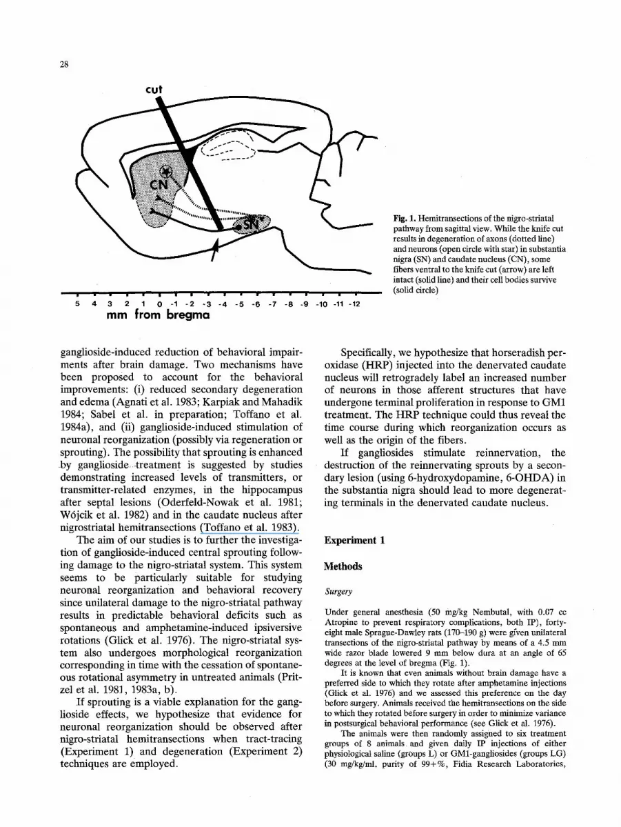
Fig. 1. Hemitransections of the nigro-striatal pathway from sagittal view. While the knife cut results in degeneration of axons (dotted line) and neurons (open circle with star) in substantia nigra (SN) and caudate nucleus (CN), some fibers ventral to the knife cut (arrow) are left intact (solid line) and their cell bodies survive (solid circle)
ganglioside-induced reduction of behavioral impair- ments after brain damage. Two mechanisms have been proposed to account for the behavioral improvements: (i) reduced secondary degeneration and edema (Agnati et al. 1983; Karpiak and Mahadik 1984; Sabel et al. in preparation; Toffano et al. 1984a), and (ii) ganglioside-induced stimulation of neuronal reorganization (possibly via regeneration or sprouting). The possibility that sprouting is enhanced by ganglioside.-treatment is suggested by studies demonstrating increased levels of transmitters, or transmitter-related enzymes, in the hippocampus after septal lesions (Odeffeld-Nowak et al. 1981; Wrjcik et al. 1982) and in the caudate nucleus after nigrostriatal hemitransections (Toffano et al. 1983).
The aim of our studies is to further the investiga- tion of ganglioside-induced central sprouting follow- ing damage to the nigro-striatal system. This system seems to be particularly suitable for studying neuronal reorganization and behavioral recovery since unilateral damage to the nigro-striatal pathway results in predictable behavioral deficits such as spontaneous and amphetamine-induced ipsiversive rotations (Glick et al. 1976). The nigro-striatal sys- tem also undergoes morphological reorganization corresponding in time with the cessation of spontane- ous rotational asymmetry in untreated animals (Prit- zel et al. 1981, 1983a, b).
If sprouting is a viable explanation for the gang- lioside effects, we hypothesize that evidence for neuronal reorganization should be observed after nigro-striatal hemitransections when tract-tracing (Experiment 1) and degeneration (Experiment 2) techniques are employed.
Specifically, we hypothesize that horseradish per- oxidase (HRP) injected into the denervated candate nucleus will retrogradely label an increased number of neurons in those afferent structures that have undergone terminal proliferation in response to GM1 treatment. The HRP technique could thus reveal the time course during which reorganization occurs as well as the origin of the fibers.
If gangliosides stimulate reinnervation, the destruction of the reinnervating sprouts by a secon- dary lesion (using 6-hydroxydopamine, 6-OHDA) in the substantia nigra should lead to more degenerat- ing terminals in the denervated caudate nucleus.
Experiment 1
Methods
Surgery
Under general anesthesia (50 mg/kg Nembutal, with 0.07 cc Atropine to prevent respiratory complications, both IP), forty- eight male Sprague-Dawley rats (170-190 g) were gfven unilateral transeetions of the nigro-striatal pathway by means of a 4.5 mm wide razor blade lowered 9 mm below dura at an angle of 65 degrees at the level of bregma (Fig. 1).
It is known that even animals without brain damage have a preferred side to which they rotate after amphetamine injections (Glick et al. 1976) and we assessed this preference on the day before surgery. Animals received the hemitransections on the side to which they rotated before surgery in order to minimize variance in postsurgical behavioral performance (see Glick et al. 1976).
The animals were then randomly assigned to six treatment groups of 8 animals, and given daily IP injections of either physiological saline (groups L) or GMl-gangliosides (groups LG) (30 mg/kg/ml, purity of 99+%, Fidia Research Laboratories,

29
Fig. 2. A Representative example of horseradish peroxidase (HRP) injected into the head of the caudate nucleus. B After the hemitransection, HRP is transported by spared axons located vent- ral to the cut. This photomicrograph was taken from an area that corresponds to the one indicated with an arrow in Fig. i. HRP molecules are seen here as black grains. C HRP-transport results in label- ling of neurons in the ipsilateral substantia nigra pars compacta (iSNc) and ventral tcgmental area (iVTA) as well as in the contralateral substantia nigra pars com- pacta (cSNc)

30
Abano Terme, Italy) and survived for 3 (L3, LG3), 15 (L15, LG15) or 45 (L45, LG45) days. The treatment was started immediately after surgery and continued for 2 more days in groups L3 and LG3 and 13 days in all other groups. An additional six rats received only sham surgery with daffy injections of saline and were sacrificed on postoperative day 15. The animals' identities were coded to avoid experimenter testing bias.
Behavioral assessment
Both spontaneous and drug-induced rotational behavior were measured in a rotometer similar to the one described by Unger- stedt (1971). Spontaneous rotational behavior was assessed for an average of 16.3 (+ 0.2 SEM) h at various postsurgical intervals. Rotations induced by d-amphetamine sulfate (2 mg/kg, Sigma, IP) were measured once before surgery and twice after surgery for 2 h during the light cycle (two days after the hemitransection and 3-6 days before the animals were killed). Apomorphine- (1 mg/kg, Sigma, IP) induced rotations were measured only once for 2 h on day 15 (groups L15 and LG15) or day 42 (groups L45 and LG45).
A neurological test battery designed to quantify sensory and motor deficits was given to all animals in the 15 day survival group at 4 day intervals and in the 45 day groups at 6--9 day intervals after surgery. The examination included tests of equilibrium (climb-up and bar test), grasping reflex, limb placing, proprioceptive lateral hopping, righting from the supine position, and muscle tone. We also quantified the animals' ability to pull up from fore-limb suspension with rating scores of 0 to 4, and observed the rats for 2 rain in a 50 x 50 cm open field with a grid floor (Marshall and Teitelbanm 1974; Tupper and Wallace 1980).
Histology
The day before the animals were killed they were anesthetized and injected with 0.05 pl of a 10% solution of horseradish peroxidase conjugated to wheat germ aggiutinin (HRP, Sigma, Grade VI) into the head of the denervated caudate nucleus (Fig. 2a). The stereotaxic coordinates were: 1.5 mm anterior to bregma, 3.0 mm lateral to the midline suture, and 5 mm below dura. Approxi- mately 24 h later, the animals were perfused transcardially with isotonic saline, followed by Karnovsky's solution and cold sucrose- buffer as described by Mesulam (1978). After the brains were stored in sucrose buffer for several days, they were cut coronally at 40 lxm thickness on a freezing microtome, processed according to the tetramethylbenzidine (TMB) procedure (Mesulam 1978) and counterstained with neutral red. In this manner, every other section at the level of the substantia nigra and every sixth section of the remaining brain were processed.
When injected into the caudate nucleus, HRP is picked up by synaptic terminals and transported via retrograde axonal transport to the neurons of structures projecting to the caudate. Using an Olympus light microscope, neurons containing retrogradely trans- ported HRP were counted in three representative sections from the center of the substantia nigra pars compacta and the ventral tegmental area of Tsal in the hemisphere ipsilateral to the lesion and HRP-injection (iSNc and iVTA) (Fig. 2c). These brain areas are known to project to the caudate nucleus (Graybiel and Ragsdale 1979). Sparsely distributed labelled neurons in the contralateral substantia nigra pars compacta (cSNc) were counted in all processed sections. In addition, we determined the extent of anterogradely transported HRP in the ipsilateral substantia nigra pars reticulata (iSNr). This was done by projecting the substantia nigra with the aid of an overhead microprojector (at 45x magnification) and then tracing the perimeter of the area contain- ing anterogradely transported HRP. From these tracings, the area
d-Amphetamine induced rotations
I00
8O
o 6O
4O
[ 20 i 2 "12 39
Days after hernitransection
Fig. 3. Mean and standard error of mean (S.E.M.) of d- amphetamine sulfate-induced rotational asymmetry in hemitran- Sected rats with (solid bar) or without GMl-treatment (open bar), measures were taken for 2 h following the injection on different days after surgery (star: p < 0.05)
was calculated with a Graphics Tablet �9 of an Apple-II-plus computer. The volume of labelled tissue was computed by multiplying the average area of the sections by the distance separating the first and last sections. For purposes of comparison, we also: (i) counted neurons in three representative sections of the frontal cortex as described by Sabel and Stein (1981); (ii) determined the size of the remaining nigro-striatal pathway showing evidence of HRP-transport (at 53x magnification) (Fig. 2b); and (lii) determined the size of the HRP injection in the candate nucleus (expressed as percent of candate nucleus stained, 23 x magnification) in one representative slide from the middle of the locus of injection. To avoid experimenter bias, all histological analyses were done without knowledge of the animals' group identity.
Results
Stat is t ical c o m p a r i s o n s w e r e m a d e wi th p a r a m e t r i c tests (e .g. o n e - w a y ana lys i s of v a r i a n c e o r t r e n d analys is ) a n d subsequent post hoc c o m p a r i s o n s ( D u n - ne t t ' s t - tes t ) . I f t he a s s u m p t i o n s of t h e p a r a m e t r i c tes t we re n o t fu l f i l led , M a n n - W h i t n e y ' s U - t e s t was u s e d for g r o u p c o m p a r i s o n s .
Behavioral performance
A f t e r su rge ry , all a n i m a l s e x h i b i t e d s p o n t a n e o u s ro t a t i ons t o w a r d t h e l e s ion s ide ( ips ivers ive ro ta - t ions) . I n t he r o t o m e t e r , all g r o u p s s h o w e d a s t eady r e d u c t i o n o f a s y m m e t r i c b e h a v i o r ( ca l cu l a t ed as ips ivers ive m i n u s c o n t r a v e r s i v e r o t a t i o n s ) a t a n ea r ly s tage of t e s t ing w i th n o a p p a r e n t effect o f G M 1 - t r e a t m e n t . Th i s b e h a v i o r a l i m p r o v e m e n t m a y b e a n exp re s s ion of pos t - su rg ica l r e c o v e r y , b u t m a y also ref lec t h a b i t u a t i o n to t he tes t s i t ua t i on .
80 e-
.o 7o o ~ 6o
~ 5 0
I,. 4 0
�9 ~ 3o
21) 0
lO 6 Z
Apomorphine-induced rotations
15 42 Days after hemitransection
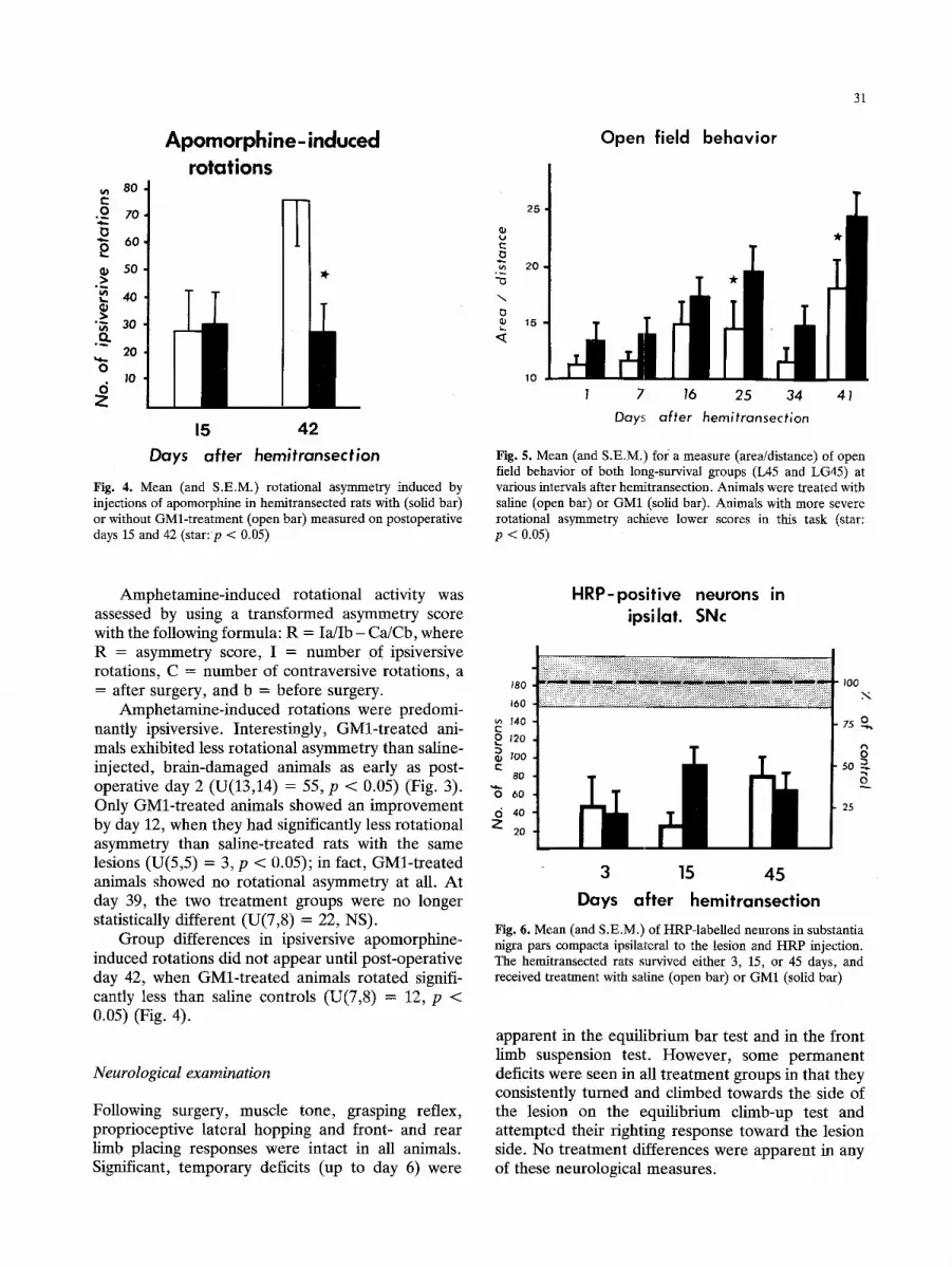
Fig. 4. Mean (and S.E.M.) rotational asymmetry induced by injections of apomorphine in hemitransected rats with (solid bar) or without GMl-treatment (open bar) measured on postoperative days 15 and 42 (star: p < 0.05)
t~
OJ
25
20
15
Open field behavior
31
10
1 7 16 25 34 41
Days a f te r hemi t ransect ion
Fig. 5. Mean (and S.E.M.) foi a measure (area/distance) of open field behavior of both long-survival groups (L45 and LG45) at various intervals after hemitransection. Animals were treated with saline (open bar) or GM1 (solid bar). Animals with more severe rotational asymmetry achieve lower scores in this task (star: p < 0.05)
Amphetamine- induced rotational activity was assessed by using a t ransformed asymmetry score with the following formula: R = Ia/Ib - Ca/Cb, where R = asymmetry score, I = number of ipsiversive rotations, C = number of contraversive rotations, a = after surgery, and b = before surgery.
Amphetamine- induced rotations were predomi- nantly ipsiversive. Interestingly, GMl - t r ea t ed ani- mals exhibited less rotational asymmetry than saline- injected, brain-damaged animals as early as post- operative day 2 (U(13,14) = 55, p < 0.05) (Fig. 3). Only GMl- t r ea t ed animals showed an improvement by day 12, when they had significantly less rotational asymmetry than saline-treated rats with the same lesions (U(5,5) = 3, p < 0.05); in fact, GMl- t r ea t ed animals showed no rotational asymmetry at all. At day 39, the two t rea tment groups were no longer statistically different (U(7,8) = 22, NS).
Group differences in ipsiversive apomorphine- induced rotations did not appear until post-operat ive day 42, when GMl- t r ea t ed animals rotated signifi- cantly less than saline controls (U(7,8) = 12, p < 0.05) (Fig. 4).
Neurological examination
Following surgery, muscle tone, grasping reflex, proprioceptive lateral hopping and front- and rear limb placing responses were intact in all animals. Significant, t emporary deficits (up to day 6) were
HRP-positive neurons in ipsilat. SNc
180
160
u~ 140
~ 120
100 t'-
80
"6 6o d 4o
z 20
100 .~,
7s o
8 so
25
3 15 45 Days after hemitransection
Fig. 6. Mean (and S.E.M.) of HRP-labelled neurons in substantia nigra pars compacta ipsilateral to the lesion and HRP injection. The hemitransected rats survived either 3, 15, or 45 days, and received treatment with saline (open bar) or GM1 (solid bar)
apparent in the equilibrium bar test and in the front limb suspension test. However , some permanent deficits were seen in all t rea tment groups in that they consistently turned and climbed towards the side of the lesion on the equilibrium climb-up test and at tempted their righting response toward the lesion side. No t reatment differences were apparent in any of these neurological measures.
32
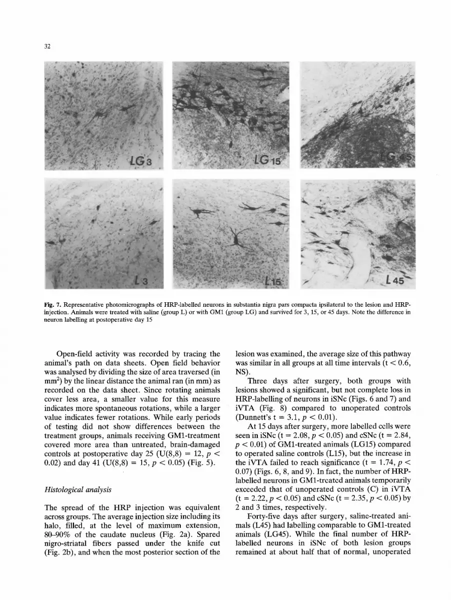
Fig. 7. Representative photomicrographs of HRP-labelled neurons in substantia nigra pars compacta ipsilateral to the lesion and HRP- injection. Animals were treated with saline (group L) or with GM1 (group LG) and survived for 3, 15, or 45 days. Note the difference in neuron labelling at postoperative day 15
Open-field activity was recorded by tracing the animal's path on data sheets. Open field behavior was analysed by dividing the size of area traversed (in mm 2) by the linear distance the animal ran (in mm) as recorded on the data sheet. Since rotating animals cover less area, a smaller value for this measure indicates more spontaneous rotations, while a larger value indicates fewer rotations. While early periods of testing did not show differences between the treatment groups, animals receiving GMl-treatment covered more area than untreated, brain-damaged controls at postoperative day 25 (U(8,8) = 12, p < 0.02) and day 41 (U(8,8) = 15, p < 0.05) (Fig. 5).
Histological analysis
The spread of the HRP injection was equivalent across groups. The average injection size including its halo, filled, at the level of maximum extension, 80-90% of the caudate nucleus (Fig. 2a). Spared nigro-striatal fibers passed under the knife cut (Fig. 2b), and when the most posterior section of the
lesion was examined, the average size of this pathway was similar in all groups at all time intervals (t < 0.6, NS).
Three days after surgery, both groups with lesions showed a significant, but not complete loss in HRP-labelling of neurons in iSNc (Figs. 6 and 7) and iVTA (Fig. 8) compared to unoperated controls (Dunnett 's t = 3.1, p < 0.01).
At 15 days after surgery, more labelled cells were seen in iSNc (t = 2.08, p < 0.05) and cSNc (t = 2.84, p < 0.01) of GMl-treated animals (LG15) compared to operated saline controls (L15), but the increase in the iVTA failed to reach significance (t = 1.74, p < 0.07) (Figs. 6, 8, and 9). In fact, the number of HRP- labelled neurons in GMl-treated animals temporarily exceeded that of unoperated controls (C) in iVTA (t = 2.22, p < 0.05) and cSNc (t = 2.35, p < 0.05)by 2 and 3 times, respectively.
Forty-five days after surgery, saline-treated ani- mals (L45) had labelling comparable to GMl-treated animals (LG45). While the final number of HRP- labelled neurons in iSNc of both lesion groups remained at about half that of normal, unoperated

HRP-positive neurons in ipsilat. VTA
100 200
.,,<
150 o
o
�9 O ~-. 50 100
d 50 Z
I0
3 15 45
Days after hemitransection
Fig. 8. Mean number (and S.E.M.) of HRP-labelled neurons in the ipsilateral ventral tegmental area. Open bar: saline treatment; solid bar: GM1 treatment
33
Volume of anterograde HRP in i SNr
200 ' O3
E 100 '
[••L•••••••••••••••••••••••••••••••••••••••••••••••••••••••••••••••••••••••••••••••••••••••••••••••••••••••••••••••••••••••••••••••••••••••••••••;••••••••••••••••••••••••••••••••••:•••••••••••••••••••••••••••••••••••••• loo
8 3 0 0 ,
so
25
3 15 45
Days after hemitransection
Fig. 10. Mean volume (and S.E.M.) of tissue in the ipsilateral substantia nigra pars reticulata containing anterogradely trans- ported HRP (open bar: saline treatment; solid bar: GM1 treat- ment)
o
6 Z
HRP-positive neurons in contralat. SNc
3OO 5,
4 ' o 200
s, Z
2 100
1
3 15 45
Days after hemitransection
Fig. 9. Mean number (and S.E.M.) of HRP-labelled neurons in the contralateral substantia nigra pars compacta. Open bar: saline treatment; solid bar: GM1 treatment. Note that, on average, there are very few labelled cells in this structure
animals (t > 3.0, p < 0.01), all three groups (C, L45, and LG45) had a comparable number of labelled neurons in iVTA.
The extent of anterogradely transported HRP in iSNr (Fig. 10) significantly increased over time (linear trend analyses; group L: F(1,15) = 5.65, p < 0.05; group LG: F(1,18) = 7.22, p < 0.05), with no significant differences between the treatment groups. In addition, neuron counts in the frontal cortex did not reveal any significant differences among the groups.
Experiment 2
The Fink-Heimer technique, when used in conjunc- tion with a secondary lesion, permits the visualization of newly sprouted fibers in a denervated structure. Specifically, if gangliosides stimulate the formation of new fiber connections, their subsequent destruc- tion by 6-OHDA injections into the ipsilateral sub- stantia nigra and ventral tegmental area would lead to more terminal degeneration in the denervated caudate nucleus.
Methods
Six male Sprague-Dawley rats (170-190 g) received the same unilateral hemitransections of the nigro-striatai pathway and treatment with saline (n = 3) or GM1 (n = 3) for 14 days as described in Experiment 1. On postoperative day 15, animals were anesthetized and given two unilateral, 5 ~1 injections of 6- hydroxydopamine (6-OHDA, 2 mg/ml of saline containing 0.2% ascorbic acid) on the side of the lesion. One injection was given into the substantia nigra pars compacta (iSNc) (6.0 mm posterior to bregma, 2.0 mm lateral to the midline suture, and 7.5 mm below dura), and the other injection was given into the ventral tegmental area (iVTA) (A: -6.0, L: 0.8, V: -8.3). The neurotoxin 6-OHDA is known to destroy dopaminergic neurons selectively (Ungerstedt 1971).
Nine days after the 6-OHDA injection, the rats were perfused transcardially with 0.9% saline followed by 10% formalin in saline solution. Brains were removed and stored in a 30% sucrose-10% formalin solution for about one week. After the brains were cut at 40 ~tm on a freezing microtome, tissue sections were mounted on slides and processed with the Fink-Heimer technique (Firl et al. 1980).
34
Results
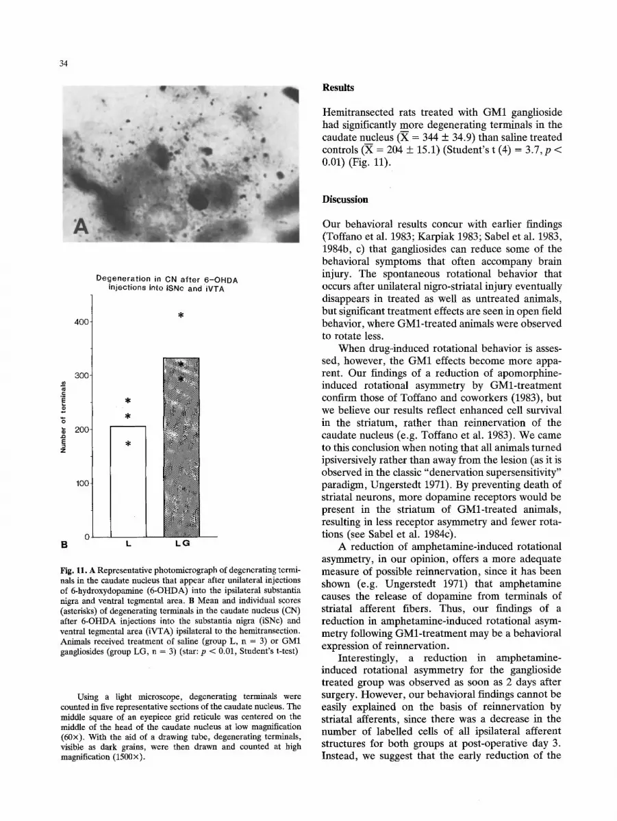
Hemitransected rats treated with GM1 ganglioside had significantly more degenerating terminals in the caudate nucleus (X = 344 + 34.9) than saline treated controls (X = 204 + 15.1) (Student's t (4) = 3.7, p < 0.01) (Fig. 11).
Degeneration in CN after 6-OHDA injections into iSNc and iVTA
400
300
200
100
B L L G
Fig. 11. A Representative photomicrograph of degenerating termi- nals in the caudate nucleus that appear after unilateral injections of 6-hydroxydopamine (6-OHDA) into the ipsilateral substantia nigra and ventral tegmental area. B Mean and individual scores (asterisks) of degenerating terminals in the caudate nucleus (CN) after 6-OHDA injections into the substantia nigra (iSNc) and ventral tegmental area (iVTA) ipsilateral to the hemitransection. Animals received treatment of saline (group L, n = 3) or GM1 gangliosides (group LG, n = 3) (star: p < 0.01, Student's t-test)
Using a light microscope, degenerating terminals were counted in five representative sections of the caudate nucleus. The middle square of an eyepiece grid reticule was centered on the middle of the head of the caudate nucleus at low magnification (60x). With the aid of a drawing tube, degenerating terminals, visible as dark grains, were then drawn and counted at high magnification (1500x).
Discussion
Our behavioral results concur with earlier findings (Toffano et al. 1983; Karpiak 1983; Sabel et al. 1983, 1984b, c) that gangliosides can reduce some of the behavioral symptoms that often accompany brain injury. The spontaneous rotational behavior that occurs after unilateral nigro-striatal injury eventually disappears in treated as well as untreated animals, but significant t reatment effects are seen in open field behavior, where GMl- t rea ted animals were observed to rotate less.
When drug-induced rotational behavior is asses- sed, however, the GM1 effects become more appa- rent. Our findings of a reduction of apomorphine- induced rotational asymmetry by GMl- t rea tment confirm those of Toffano and coworkers (1983), but we believe our results reflect enhanced cell survival in the striatum, rather than reinnervation of the caudate nucleus (e.g. Toffano et al. 1983). We came to this conclusion when noting that all animals turned ipsiversively rather than away from the lesion (as it is observed in the classic "denervation supersensitivity" paradigm, Ungerstedt 1971). By preventing death of striatal neurons, more dopamine receptors would be present in the striatum of GMl- t rea ted animals, resulting in less receptor asymmetry and fewer rota- tions (see Sabel et al. 1984c).
A reduction of amphetamine-induced rotational asymmetry, in our opinion, offers a more adequate measure of possible reinnervation, since it has been shown (e.g. Ungerstedt 1971) that amphetamine causes the release of dopamine from terminals of striatal afferent fibers. Thus, our findings of a reduction in amphetamine-induced rotational asym- metry following GMl- t rea tment may be a behavioral expression of reinnervation.
Interestingly, a reduction in amphetamine- induced rotational asymmetry for the ganglioside treated group was observed as soon as 2 days after surgery. However , our behavioral findings cannot be easily explained on the basis of reinnervafion by striatal afferents, since there was a decrease in the number of labelled cells of all ipsilateral afferent structures for both groups at post-operative day 3. Instead, we suggest that the early reduction of the
35
behavioral impairment may relate to ganglioside- induced changes in some of the "secondary" conse- quences of lesions that take place soon after the injury occurs. For example, it has been shown that gangliosides reduce cerebral edema (Karpiak and Mahadik 1984) as well as the extent of cell death, anterograde and retrograde degeneration that invari- able occur after brain injury (Agnati et al. 1983; Sabel et al., in preparation; Toffano et al. 1984a).
Amphetamine-induced rotational asymmetry at post-operative day 12 and 39, however, apparently does coincide with morphological changes at later survival times (at post-lesion days 15 and 45). Here, the number of labelled neurons correspond in time with the cessation of rotational behavior. At post- operative day 15, for example, significantly more labelling is observed in the iSNc, while the cell numbers in the iVTA and cSNc temporarily increase above normal levels of ganglioside-treated animals. These increases are paralleled in time by a temporary absence of rotational asymmetry after amphetamine- injections. Saline-treated controls still rotate signifi- cantly more at day 15, which also parallels the small number of retrogradely labelled neurons.
The temporary increase of cell labelling to levels 2-3 times above normal in ganglioside-treated ani- mals (Figs. 8 and 9) may be related to a "hypertrophy response" seen in developing (Land and Lund 1979) and aging brains (Buell and Coleman 1979).
A subsequent reduction of this "hypertrophy response" at post-operative day 45 again has a corresponding behavioral effect: an increase in amphetamine-induced rotational asymmetry in GM1- treated animals. This later reversal of structural reorganization (and behavioral recovery) may be due to the fact that treatment was terminated at day 14 or it may indicate a late onset, post-lesion "stabilization response" of the brain.
The disappearance of newly formed connections would add another dimension to neural reorganiza- tion after ganglioside treatment. Also in untreated, but brain-damaged animals such a phenomenon has been reported repeatedly in regrowing cerebral catecholamine neurons (Katzman et al. 1971; Bj6rk- lund and Stenevi 1971), interhemispheric nigro- striatal projections (Pritzel et al. 1983b), and spinal cord fibers (Bernstein et al. 1974). In agreement with our results, this later "dying back" of newly formed terminals was always observed after at least 30 days following the lesions. Thus, our observations may indicate a late onset terminal retraction.
It seems that interhemispheric nigro-striatal fi- bers participate in the reorganization. These and other interhemispheric connections were suggested to be involved in recovery from behavioral asymmet-
ries (Pritzel et al. 1981, 1983a, b; Sabel et al. 1984a). Both the proliferation of crossed projections as demonstrated by the HRP-technique, and the cessa- tion of behavioral asymmetries noted by others (Pritzel et al. 1981, 1983a, b) was observed later than one week after brain damage and are in agreement with our results. This is a time period typical for spontaneous sprouting in the untreated organism (Cotman and Nadler 1978). The sparse interhemis- pheric connections may be an important example of neural reorganization, but being few in number (they only comprise 1-5% of the ipsilaterally existing afferents), their importance for behavioral recovery is probably limited.
It would appear that the ipsilateral fibers spared by the lesion and which arise from the substantia nigra and ventral tegmental area are more important for behavioral recovery. Few fibers are left intact by the lesion, but by post-operative day 15 the number of fibers in the ganglioside-treated animals increases to 2-3 times above control levels. This finding was substantiated by our anterograde degeneration analysis, where the destruction of the new connec- tions led to significantly more terminal degeneration in the caudate nucleus of GMl-treated animals (Experiment 2) ....
A comparable increase in the number of HRP- labelled neurons is not observed in untreated, but brain-damaged animals until day 45. The late increase of cell labelling in saline-treated animals reaches the level maintained by the GMl-treated animals by day 45 and this suggests that gangliosides accelerate reorganization of the brain that would occur even without treatment.
Although it could be argued that increased HRP- labelling is due to an increase in neuronal activity and axonal transport properties, the following arguments make this possibility less tenable: (i) labelling of cells in the frontal cortex, a structure also projecting to the caudate nucleus, and the area of anterogradely transported HRP in iSNr were not significantly different between brain-damaged groups, (ii) The "halo" of HRP diffusion in the caudate is comparable in all animals, and (iii) no differences in cell labelling are seen on postoperative day 3, when animals had already received 3 full days of GM1 treatment.
Furthermore, the observations made in the two experiments described in this paper support the notion that ganglioside treatment stimulates the rate of terminal proliferation due to sprouting: (i)signifi- cantly more degenerating terminals are found in the caudate nucleus after destruction of the new fibers (Experiment 2); (ii) dopamine levels rise in the denervated caudate nucleus as indicated by the amphetamine-induced rotational behavior (Experi-

36
ment 1) and by biochemical studies (Toffano et al. 1983); (iii) cell labelling, with the exception of the short survival interval, closely corresponds in time with amphetamine-induced rotational behavior; and (iv) a morphological "hypertrophy" is paralleled in time by a temporary, complete cessation of rotational asymmetry. Although the size of the nigro-striatal pathway seen in coronal sections appeared about the same in all animals, the possibility still exists that gangliosides may actually promote central regenera- tion.
At the present time, we favor the conclusion that gangliosides stimulate the rate of reinnervation of the denervated caudate nucleus following nigro-striatal hemitransections. Although it appears that the ipsi- lateral, ascending pathways arising from the substan- tia nigra and ventral tegmental area may be primarily responsible for behavioral recovery at later post- operative stages, our data also indicate that gang- lioside-induced reinnervation may also involve cros- sed nigro-striatal projections. These findings are in agreement with those by Toffano et al. (1984b) who recently reported that the effects of GM1 treatment (on TH activity) disappear when lesions are presum- ably too extensive to allow for any survival of spared neurons and their fibers.
Along these lines it is interesting to note that gangliosides did not induce any sprouting after total lesions of the septohippocampal pathway (Fass and Ramirez 1984). This has led us to suggest that the effects of gangliosides on inducing neuronal reorgani- zation via sprouting may generally be dependent on the presence of partial rather than total lesions (Sabel et al. 1985).
In conclusion, gangliosides may reduce be- havioral impairment after nigro-striatal injury via at least two mechanisms: (i) they may reduce the severity of secondary postlesion events such as secon- dary degeneration (Agnati et al. 1983; Sabel et al., in preparation; Toffano et al. 1984a) and/or edema (Karpiak and Mahadik 1984) immediately following brain damage and (ii) they may stimulate a sprouting response at later postoperative periods of spared neurons and their axons. Additional research using other neuroanatomical techniques, such as electron microscopy or amino acid autoradiography, is needed to further delineate the mechanisms under- lying ganglioside-induced reduction of behavioral impairments following brain damage.
Acknowledgements. We thank Dr. B. Fass for his critical reading of the manuscript. This work was supported in part by USAMRDC contract no. DAMD 17-82-C-2205, and funds from Fidia Research Laboratories and Clark University.
Note added in proof. Li and eoworkers (1985) have recently replicated our finding that GM1 reduces amphetamine-induced rotations following nigro-striatal hemitransections as early as 2 days after surgery. Furthermore, they were able to demonstrate that there may be "a critical time period when GM1 treatment leads to the greatest reduction in functional impairment". A significant deficit-reduction was only apparent when GMl-injec- tions were given 0-2 h after surgery, but not at 4 h or later. Li YS, Rapport MM, Karpiak SE (1985) Acute effects of GM1 ganglioside on CNS injury: assessment of dosing schedule for optimal functional response. Soc Neurosci Abstr (in press)
References
Agnati LF, Fuxe K, Calza L, Benfenati F, Cavicchioli L, Toffano G, Goldstein M (1983) Gangliosides increase the survival of lesioned nigral dopamine neurons and favour the recovery of dopaminergic synaptic function in striatum of rats by colla- teral sprouting. Acta Physiol Scand 119:347-363
Bernstein JJ, Gelderd JB, Bernstein ME (1974) Alteration of neuronal synaptic complement during regeneration and axonal sprouting of rat spinal cord. Exp Neurol 44:470-482
Bj6rklund A, Stenevi U (1971) Growth of central catecholamine neurones into smooth mucle grafts in the rat mesencephalon. Brain Res 31:1-20
Buell SJ, Coleman PD (1979) Dendritic growth in the aged human brain and failure of growth in senile dementia. Science 206: 854-856
Cotman CW, Nadler JV (1978) Reactive synaptogenesis in the hippocampus. In: Cotman CW (ed) Neuronal Plasticity. Raven Press, New York, pp 227-271
Fass B, Ramirez JJ (1984) Effects of ganglioside treatments on lesion-induced behavioral impairments and sprouting in the CNS. J Neurosci Res 12:445-458
Firl A, Mufson EJ, Stein DG (1980) Silver impregnation of pre- mounted neural tissue. Soc Neurosci Abstr, Cincinnatti, Ohio
Fishman PH, Brady RO (1976) Biosynthesis and function of gangliosides. Science 194:906-915
Glick SD, Jerussi TP, Fleisher LN (1976) Turning in circles: the neuropathology of rotation. Life Sci 18:889-896
Graybiel AM, Ragsdale CW (1979) Fiber connections of the basal ganglia. In: Cuenod M, Kreutzberg GW, Bloom FE (eds) Development and chemical specificity of neurons. Elsevier, Amsterdam, pp 239-283
Karpiak SE (1983) Ganglioside treatment improves recovery of alternation behavior following unilateral entorhinal cortex lesions. Exp Neurol 81:330-339
Karpiak SE, Mahadik SP (1984) Reduction of cerebral edema with GM1 ganglioside. J Neurosci Res 12:485-492
Katzman R, Bj6rklund A, Owman CH, Stenevi U, West KA (1971) Evidence for regenerative axon sprouting of central catecholamine neurons in the rat mesencephalon following electrolytic lesions. Brain Res 25:579-596
Land PW, Lund RD (1979) Development of the rat's uncrossed retinotectal pathway and its relationship to plasticity studies. Science 205:698-700
Marshall JF, Teitelbaum P (1974) Further analysis of sensory inattention following lateral hypothalamic damage in rats. J Comp Physiol Psychol 86:375-395
Mesulam MM (1978) Tetramethyl benzidine for horseradish- peroxidase neurohistochemistry: a non-carcinogenic blue reaction-product with superior sensitivity for visualizing neural afferents and efferents. J Histochem Cytochem 26: 106-117

37
Oderfeld-Nowak B, W6jcik M, Ulas J, Potempska A (1981) Effects of chronic ganglioside treatment on recovery proces- ses in hippocampus after brain lesions in rats. In: Rapport MM, Gorio A (eds) Gangliosides in neurological and neuromuscular function, development, and repair. Raven Press, New York, pp 197-209
Orlando P, Cocciante G, Ippolito G, Massari P, Roberti S, Tettamanti G (1979) The fate of tritium labelled GM1 ganglioside injected in mice. Pharmacol Res Commun 11: 759-773
Pritzel M, Huston JP (1981) Neural and behavioral plasticity: crossed nigro-thalamic projections following unilateral sub- stantia nigra lesions. Behav Brain Res 3:393-399
Pritzel M, Huston JP (1983a) Behavioral and neural plasticity following unilateral brain lesions. In: Myslobodsky MS (ed) Hemisyndromes: psychobiology, neurology, Academic Press, New York, pp 27-68
Pritzel M, Huston JP, Sarter M (1983b) Behavioral and neuronal reorganization after unilateral substantia nigra lesions: evi- dence for increased interhemispheric nigrostriatal projec- tions. Neuroscience 9:879-888
Sabel BA, Stein DG (1981) Extensive loss of subcortical neurons in the aging rat brain. Exp Neurol 73:507-516
Sabel BA, Slavin MD, Stein DG (1983) Enhancement of behavioral recovery from bilateral caudate lesions by gang- liosides. Soc Neurosci Abstr: 243.9
Sabel BA, Pritzel M, Morgan S, Huston JP (1984a) Interhemis- pheric nigro-thalamic projections and behavioral recovery following unilateral motor and sensory restriction. Exp Neurol 83:4%61
Sabel BA, Slavin MD, Stein DG (1984b) GMl-ganglioside treat- ment facilitates behavioral recovery from bilateral brain damage. Science 225:340-342
Sabel BA, Dunbar GL, Stein DG (1984c) Gangliosides minimize behavioral deficits and enhance structural repair after brain damage. J Neurosci Res 12:429-443
Sabel BA, Dunbar GL, Fass B, Stein DG (1985) Gangliosides, neuroplasticity, and behavioral recovery after brain damage. In: Will B, Schmitt P, Dalrymple-Alford JC (eds) Brain plasticity, learning and memory. New York, Plenum Press, pp 481-493
Tettamanti G, Venerando B, Roberti S, Chigorno V, Sonnino S, Ghidoni R, Orlando P, Massari P (1981) The fate of exogenously administered brain gangliosides. In: Rapport MM, Gorio A (eds) Gangliosides in neurological and neuromuscular function, development, and repair. New York, Raven Press, pp 225-240
Toffano G, Savoini GE, Moroni F, Lombardi MG, Calza L, Agnati LF (1983) GM1 ganglioside stimulates the regenera- tion of dopaminergic neurons in the central nervous system. Brain Res 261:163-166
Toffano G, Savoini GE, Moroni F, Lombardi G, Calza L, Agnati LF (1984a) Chronic GM1 ganglioside treatment reduces dopamine cell body degeneration in the substantia nigra after unilateral hemitransection in rat. Brain Res 296:233-239
Toffano G, Savoini G, Aporti F, Calzolari S, Consolazione A, Maura G, Marchi M, Raiteri M, Agnati LF (1984b) The functional recovery of damaged brain: the effects of GM1 monosialoganglioside. J Neurosci Res 12:397-408
Tupper DE, Wallace RB (1980) Utility of the neurological examination in rats. Acta Neurobiol Exp 40:99%1003
Ungerstedt U (1971) Striatal dopamine release after amphetamine or nerve degeneration revealed by rotational behaviour. Acta Physiol Scand Supp 367:49-68
W6jcik M, Ulas J, Oderfeld-Nowak B (1982) The stimulating effects of ganglioside injections on the recovery of choline acetyltransferase and acetylcholinesterase activities in the hippocampus of the rat after septal lesions. Neuroscience 7: 495-499
Received September 11, 1984 / Accepted April 15, 1985