glycyrrhizin represses total parenteral nutrition-associated
TRANSCRIPT
Int. J. Mol. Sci. 2013, 14, 12563-12580; doi:10.3390/ijms140612563
International Journal of
Molecular Sciences ISSN 1422-0067
www.mdpi.com/journal/ijms
Article
Glycyrrhizin Represses Total Parenteral Nutrition-Associated Acute Liver Injury in Rats by Suppressing Endoplasmic Reticulum Stress
Jai-Jen Tsai 1,2, Hsing-Chun Kuo 3, Kam-Fai Lee 4 and Tung-Hu Tsai 2,5,6,*
1 Division of Gastroenterology, Department of Medicine, National Yang-Ming University Hospital,
I-Lan 26042, Taiwan; E-Mail: [email protected] 2 Institute of Traditional Medicine, School of Medicine, National Yang-Ming University,
Taipei 11221, Taiwan 3 Department of Nursing, Chronic Diseases and Health Promotion Research Center,
Chang Gung University of Science and Technology Chiayi Campus, Chiayi 61363, Taiwan;
E-Mail: [email protected] 4 Department of Pathology, Chang Gung Memorial Hospital at Chiayi, Chiayi 61363, Taiwan;
E-Mail: [email protected] 5 Graduate Institute of Acupuncture Science, China Medical University, Taichung 40402, Taiwan 6 Department of Education and Research, Taipei City Hospital, Taipei 10341, Taiwan
* Author to whom correspondence should be addressed; E-Mail: [email protected];
Tel.: +886-2-2826-7115; Fax: +886-2-2822-5044.
Received: 22 April 2013; in revised form: 5 June 2013 / Accepted: 6 June 2013 /
Published: 14 June 2013
Abstract: Total parenteral nutrition (TPN) is an artificial way to support daily nutritional
requirements by bypassing the digestive system, but long-term TPN administration may
cause severe liver dysfunction. Glycyrrhizin is an active component of licorice root that has
been widely used to treat chronic hepatitis. The aim of this study is to investigate the
hepatoprotective effect of glycyrrhizin on TPN-associated acute liver injury in vivo. Liver
dysfunction was induced by intravenous infusion of TPN at a flow rate of 20 mL/kg/h for
three h in Sprague Dawley rats. The rats were pretreated with Glycyrrhizin (1, 3 and
10 mg/kg intravenously). After receiving TPN or saline (control group) for three h, the rats
were sacrificed, blood samples were collected for biochemical analyses and liver tissue
was removed for histopathological and immunohistochemical examination. We found that
aspartate aminotransferase (AST), alanine aminotransferase (ALT), total bilirubin (TB) and
triglyceride (TG) levels were significantly increased in the TPN group without glycyrrhizin
OPEN ACCESS
Int. J. Mol. Sci. 2013, 14 12564
pretreatment and decreased in the glycyrrhizin-pretreated TPN group in a dose-dependent
manner. The stained liver sections showed that glycyrrhizin relieved acute liver injury. The
upregulation of serum protein biomarkers of reactive nitrogen species, including nitrotyrosine
and inducible NO synthase (iNOS), were attenuated by glycyrrhizin pretreatment. Levels
of endoplasmic reticulum (ER) stress factors, such as phosphorylation of JNK1/2, p38
MAPK and CHOP, were decreased by glycyrrhizin pretreatment. In summary, our results
suggest that glycyrrhizin decreases TPN-associated acute liver injury factors by suppressing
endoplasmic reticulum stress and reactive nitrogen stress.
Keywords: total parenteral nutrition (TPN); endoplasmic reticulum (ER); glycyrrhizin
1. Introduction
Total parenteral nutrition (TPN) is a method for providing nutrients, such as glucose, amino acids,
lipids, added vitamins and dietary minerals, to patients who are unable to sustain adequate nutrition by
standard enteral means, namely, those who suffer from gastrointestinal disorders. However, long-term
TPN administration increases the risks of hyperglycemia, hepatic inflammation, steatosis, insulin
resistance and reduced immune responses [1,2]; among these, hepatitis is one of the most frequent
complications [3,4]. Although several factors have been associated with a high risk of chronic
TPN-induced hepatitis [5], such as enteral feeding history, septic events, length of intestinal resection
and prematurity/low birth weight, its pathogenic mechanism is not fully understood [6,7].
Previous studies demonstrated three possible routes of pathogenesis for chronic TPN-induced
hepatitis: direct toxic effect of detergent-like bile salts [8–14], increasing oxidative stress [15–20] and
hepatocyte apoptosis [21–23]. According to Loff et al. and Shamir et al., injury to liver acinar Zone 3,
which is composed of hepatocytes located around the hepatic venule that are mainly involved in the
production of bile salt, was found in TPN related hepatitis in animal models [12,13]. TPN-related
hepatic injury in Zone 3 might be a typical feature for the direct toxic effect of the bile salt [13,14].
Moreover, TPN is an important source of oxidants and peroxides that are derived mainly from the
reduction of dissolved oxygen by electron donors, such as vitamin C, amino acids and lipids [16].
Peroxide-induced oxidative stress from TPN can trigger pro-inflammatory cytokines and activate
inflammatory cytokine cascades [15,16]. Cytokines can be negatively regulated by Suppressors of
Cytokines Signaling (SOCS), which is a protein that is highly upregulated in response to pro-inflammatory
cytokines [18,19]. In addition, hepatocyte apoptosis has been observed at 24 h after TPN administration in
a mouse model and is correlated with the increased expression of Fas, the pro-apoptotic factor Bad and
the anti-apoptotic factor Bcl-xl [21]. Katz’s group also observed that administering TPN to mouse
for 14 days increased hepatocyte apoptosis in bowel resection mouse [24]. Hepatocytes are very
susceptible to endoplasmic reticulum (ER) stress, and ER-stressed hepatocytes are more susceptible to
tumor necrosis factor-α-induced cell death and apoptosis [25].
Recently, glycyrrhizin has been reported to exhibit hepatoprotective effects in the treatment of viral
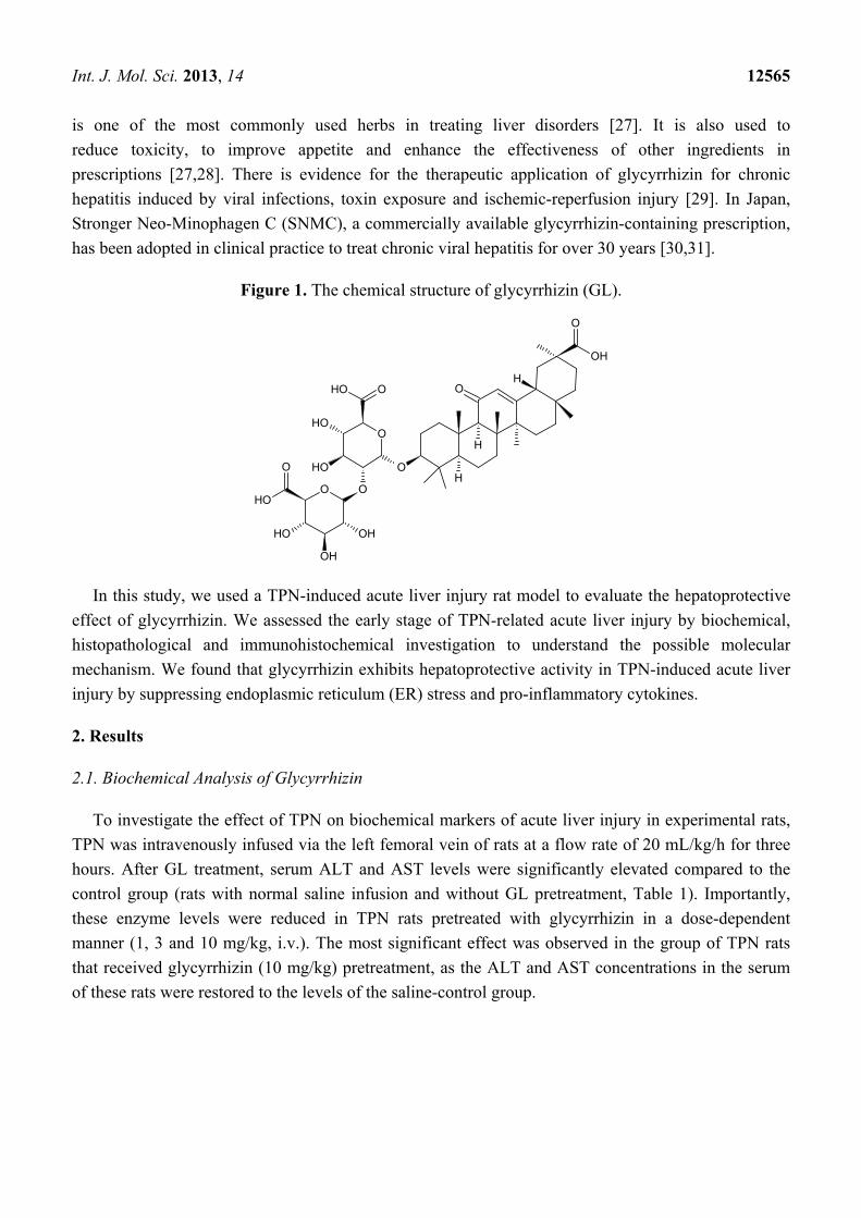
hepatitis [26]. Glycyrrhizin (GL; Figure 1) is an active compound isolated from the root of licorice
(Glycyrrhiza glabra; Glycyrrhiza radix; Chinese name: Gancao). In traditional Chinese medicine, licorice
Int. J. Mol. Sci. 2013, 14 12565
is one of the most commonly used herbs in treating liver disorders [27]. It is also used to
reduce toxicity, to improve appetite and enhance the effectiveness of other ingredients in
prescriptions [27,28]. There is evidence for the therapeutic application of glycyrrhizin for chronic
hepatitis induced by viral infections, toxin exposure and ischemic-reperfusion injury [29]. In Japan,
Stronger Neo-Minophagen C (SNMC), a commercially available glycyrrhizin-containing prescription,
has been adopted in clinical practice to treat chronic viral hepatitis for over 30 years [30,31].
Figure 1. The chemical structure of glycyrrhizin (GL).
O
O
O
O
OO
O
O
OH
OH
OH
OH
OH
OH
OH
OH
H
H
H
In this study, we used a TPN-induced acute liver injury rat model to evaluate the hepatoprotective
effect of glycyrrhizin. We assessed the early stage of TPN-related acute liver injury by biochemical,
histopathological and immunohistochemical investigation to understand the possible molecular
mechanism. We found that glycyrrhizin exhibits hepatoprotective activity in TPN-induced acute liver
injury by suppressing endoplasmic reticulum (ER) stress and pro-inflammatory cytokines.
2. Results
2.1. Biochemical Analysis of Glycyrrhizin
To investigate the effect of TPN on biochemical markers of acute liver injury in experimental rats,
TPN was intravenously infused via the left femoral vein of rats at a flow rate of 20 mL/kg/h for three
hours. After GL treatment, serum ALT and AST levels were significantly elevated compared to the
control group (rats with normal saline infusion and without GL pretreatment, Table 1). Importantly,
these enzyme levels were reduced in TPN rats pretreated with glycyrrhizin in a dose-dependent
manner (1, 3 and 10 mg/kg, i.v.). The most significant effect was observed in the group of TPN rats
that received glycyrrhizin (10 mg/kg) pretreatment, as the ALT and AST concentrations in the serum
of these rats were restored to the levels of the saline-control group.
Int. J. Mol. Sci. 2013, 14 12566
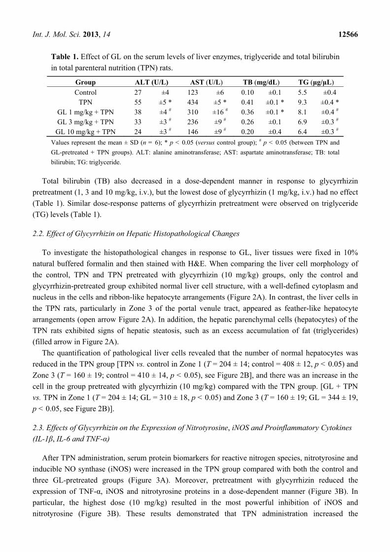
Table 1. Effect of GL on the serum levels of liver enzymes, triglyceride and total bilirubin
in total parenteral nutrition (TPN) rats.
Group ALT (U/L) AST (U/L) TB (mg/dL) TG (μg/μL)
Control 27 ±4 123 ±6 0.10 ±0.1 5.5 ±0.4 TPN 55 ±5 * 434 ±5 * 0.41 ±0.1 * 9.3 ±0.4 *
GL 1 mg/kg + TPN 38 ±4 # 310 ±16 # 0.36 ±0.1 * 8.1 ±0.4 # GL 3 mg/kg + TPN 33 ±3 # 236 ±9 # 0.26 ±0.1 6.9 ±0.3 #
GL 10 mg/kg + TPN 24 ±3 # 146 ±9 # 0.20 ±0.4 6.4 ±0.3 #
Values represent the mean ± SD (n = 6); * p < 0.05 (versus control group); # p < 0.05 (between TPN and
GL-pretreated + TPN groups). ALT: alanine aminotransferase; AST: aspartate aminotransferase; TB: total
bilirubin; TG: triglyceride.
Total bilirubin (TB) also decreased in a dose-dependent manner in response to glycyrrhizin
pretreatment (1, 3 and 10 mg/kg, i.v.), but the lowest dose of glycyrrhizin (1 mg/kg, i.v.) had no effect
(Table 1). Similar dose-response patterns of glycyrrhizin pretreatment were observed on triglyceride
(TG) levels (Table 1).
2.2. Effect of Glycyrrhizin on Hepatic Histopathological Changes
To investigate the histopathological changes in response to GL, liver tissues were fixed in 10%
natural buffered formalin and then stained with H&E. When comparing the liver cell morphology of
the control, TPN and TPN pretreated with glycyrrhizin (10 mg/kg) groups, only the control and
glycyrrhizin-pretreated group exhibited normal liver cell structure, with a well-defined cytoplasm and
nucleus in the cells and ribbon-like hepatocyte arrangements (Figure 2A). In contrast, the liver cells in
the TPN rats, particularly in Zone 3 of the portal venule tract, appeared as feather-like hepatocyte
arrangements (open arrow Figure 2A). In addition, the hepatic parenchymal cells (hepatocytes) of the
TPN rats exhibited signs of hepatic steatosis, such as an excess accumulation of fat (triglycerides)
(filled arrow in Figure 2A).
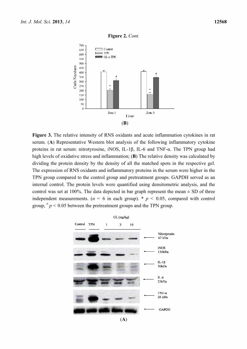
The quantification of pathological liver cells revealed that the number of normal hepatocytes was
reduced in the TPN group [TPN vs. control in Zone 1 (T = 204 ± 14; control = 408 ± 12, p < 0.05) and
Zone 3 (T = 160 ± 19; control = 410 ± 14, p < 0.05), see Figure 2B], and there was an increase in the
cell in the group pretreated with glycyrrhizin (10 mg/kg) compared with the TPN group. [GL + TPN
vs. TPN in Zone 1 (T = 204 ± 14; GL = 310 ± 18, p < 0.05) and Zone 3 (T = 160 ± 19; GL = 344 ± 19,
p < 0.05, see Figure 2B)].
2.3. Effects of Glycyrrhizin on the Expression of Nitrotyrosine, iNOS and Proinflammatory Cytokines
(IL-1β, IL-6 and TNF-α)
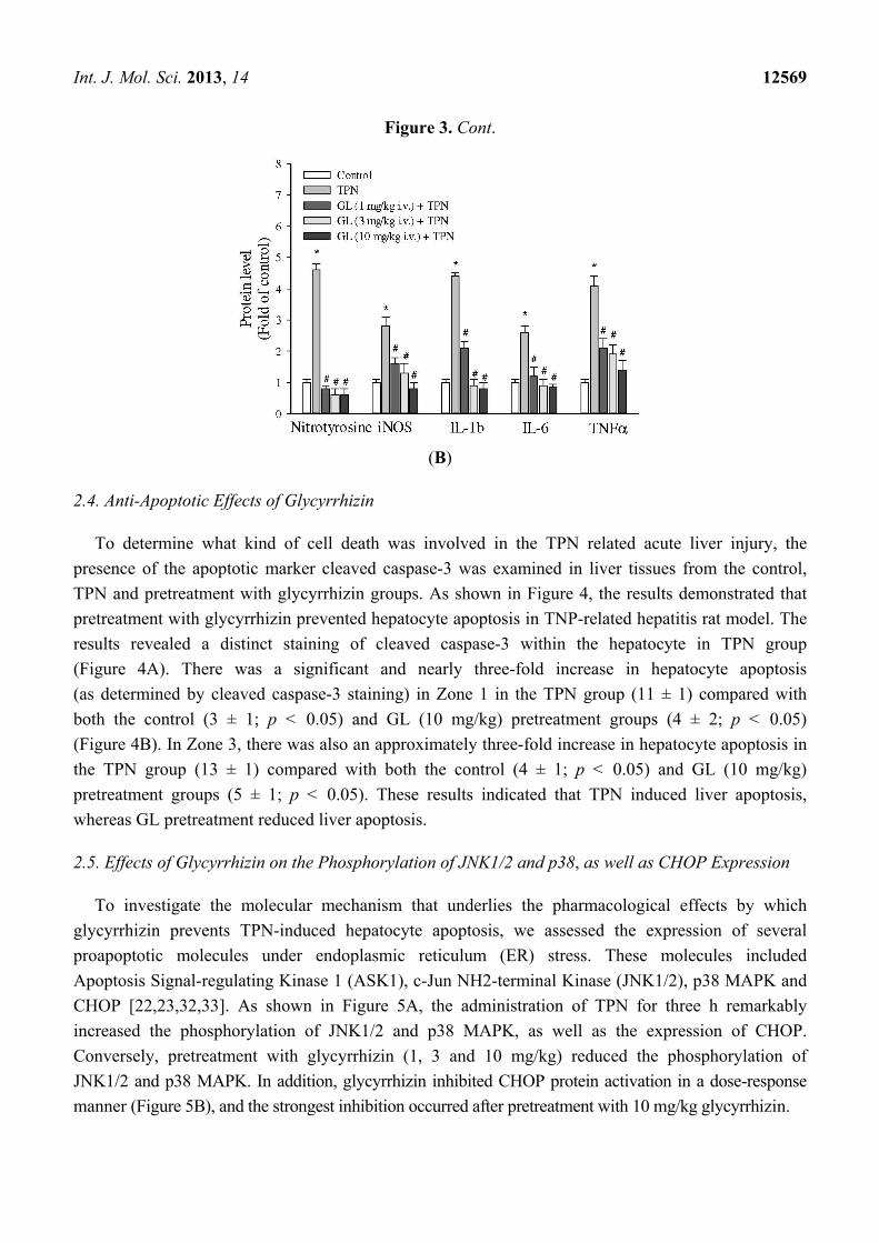
After TPN administration, serum protein biomarkers for reactive nitrogen species, nitrotyrosine and
inducible NO synthase (iNOS) were increased in the TPN group compared with both the control and
three GL-pretreated groups (Figure 3A). Moreover, pretreatment with glycyrrhizin reduced the
expression of TNF-α, iNOS and nitrotyrosine proteins in a dose-dependent manner (Figure 3B). In
particular, the highest dose (10 mg/kg) resulted in the most powerful inhibition of iNOS and
nitrotyrosine (Figure 3B). These results demonstrated that TPN administration increased the

Int. J. Mol. Sci. 2013, 14 12567
production of proinflammatory cytokines, including IL-1β, IL-6 and TNF-α, and these increases were
inhibited by pre-treatment with glycyrrhizin.
Figure 2. Histological examination of the effect of glycyrrhizin (GL) on total parenteral
nutrition (TPN) rats. The livers were H&E-stained to examine the portal (zone) and
terminal hepatic venules (Zone 3). (A) Saline infusion control (I); TPN infusion without
treatment (II); and GL pretreatment (10 mg/kg) before TPN infusion (III) staining of Zone
1 (left column) and Zone 3 (right column) of the liver. Liver pathology in the control group
exhibited normal hepatocyte structure and arrangement. The TPN non-treated group
exhibited damaged liver structure, as indicated by monocyte infiltration (open arrowhead),
steatosis (filled arrow) and the presence of necrotic cells (open arrow). Pretreatment with
GL (10 mg/kg) inhibited hepatocyte damage; (B) Quantitative evaluation of pathological
examinations. Normal hepatocytes were counted from 10 random fields (200× magnification)
of each liver sample and the values represent the mean ± SD of six rats. * p < 0.05,
compared with control group, # p < 0.05 between pretreatment groups and TPN group.
(A)
Int. J. Mol. Sci. 2013, 14 12568
Figure 2. Cont.
(B)
Figure 3. The relative intensity of RNS oxidants and acute inflammation cytokines in rat
serum. (A) Representative Western blot analysis of the following inflammatory cytokine
proteins in rat serum: nitrotyrosine, iNOS, IL-1β, IL-6 and TNF-α. The TPN group had
high levels of oxidative stress and inflammation; (B) The relative density was calculated by
dividing the protein density by the density of all the matched spots in the respective gel.
The expression of RNS oxidants and inflammatory proteins in the serum were higher in the
TPN group compared to the control group and pretreatment groups. GAPDH served as an
internal control. The protein levels were quantified using densitometric analysis, and the
control was set at 100%. The data depicted in bar graph represent the mean ± SD of three
independent measurements. (n = 6 in each group). * p < 0.05, compared with control
group, # p < 0.05 between the pretreatment groups and the TPN group.
(A)
Int. J. Mol. Sci. 2013, 14 12569
Figure 3. Cont.
(B)
2.4. Anti-Apoptotic Effects of Glycyrrhizin
To determine what kind of cell death was involved in the TPN related acute liver injury, the
presence of the apoptotic marker cleaved caspase-3 was examined in liver tissues from the control,
TPN and pretreatment with glycyrrhizin groups. As shown in Figure 4, the results demonstrated that
pretreatment with glycyrrhizin prevented hepatocyte apoptosis in TNP-related hepatitis rat model. The
results revealed a distinct staining of cleaved caspase-3 within the hepatocyte in TPN group
(Figure 4A). There was a significant and nearly three-fold increase in hepatocyte apoptosis
(as determined by cleaved caspase-3 staining) in Zone 1 in the TPN group (11 ± 1) compared with
both the control (3 ± 1; p < 0.05) and GL (10 mg/kg) pretreatment groups (4 ± 2; p < 0.05)
(Figure 4B). In Zone 3, there was also an approximately three-fold increase in hepatocyte apoptosis in
the TPN group (13 ± 1) compared with both the control (4 ± 1; p < 0.05) and GL (10 mg/kg)
pretreatment groups (5 ± 1; p < 0.05). These results indicated that TPN induced liver apoptosis,
whereas GL pretreatment reduced liver apoptosis.
2.5. Effects of Glycyrrhizin on the Phosphorylation of JNK1/2 and p38, as well as CHOP Expression
To investigate the molecular mechanism that underlies the pharmacological effects by which
glycyrrhizin prevents TPN-induced hepatocyte apoptosis, we assessed the expression of several
proapoptotic molecules under endoplasmic reticulum (ER) stress. These molecules included
Apoptosis Signal-regulating Kinase 1 (ASK1), c-Jun NH2-terminal Kinase (JNK1/2), p38 MAPK and
CHOP [22,23,32,33]. As shown in Figure 5A, the administration of TPN for three h remarkably
increased the phosphorylation of JNK1/2 and p38 MAPK, as well as the expression of CHOP.
Conversely, pretreatment with glycyrrhizin (1, 3 and 10 mg/kg) reduced the phosphorylation of
JNK1/2 and p38 MAPK. In addition, glycyrrhizin inhibited CHOP protein activation in a dose-response
manner (Figure 5B), and the strongest inhibition occurred after pretreatment with 10 mg/kg glycyrrhizin.

Int. J. Mol. Sci. 2013, 14 12570
Figure 4. Immunohistochemical analysis of cleaved caspase-3 staining in rat livers from
different liver zonal areas. TPN versus GL pretreatment group (A) Pretreatment GL
(10 mg/kg) prior to TPN administration. The TPN group (I) and GL pretreatment group (II)
animals received an equal volume of solvent. Group (I) normal saline 1 mL, i.v. bolus
before TPN administration; pretreatment group (II) GL (10 mg/kg) in normal saline 1 mL,
i.v. bolus before TPN administration. Representative photographs after immunostaining
with antibodies against cleaved caspase-3; (B) Quantitative analysis of immunohistochemical
staining of cleaved caspase-3. Average integrated optical density (AIOD) of the positively
stained area was evaluated from three randomly selected observation fields in each liver
section. The data are expressed as the mean ± SD (n = 6 per group). * p < 0.05, compared
with the control group, # p < 0.05 between pretreatment groups and TPN group.
(A)
(B)
Int. J. Mol. Sci. 2013, 14 12571
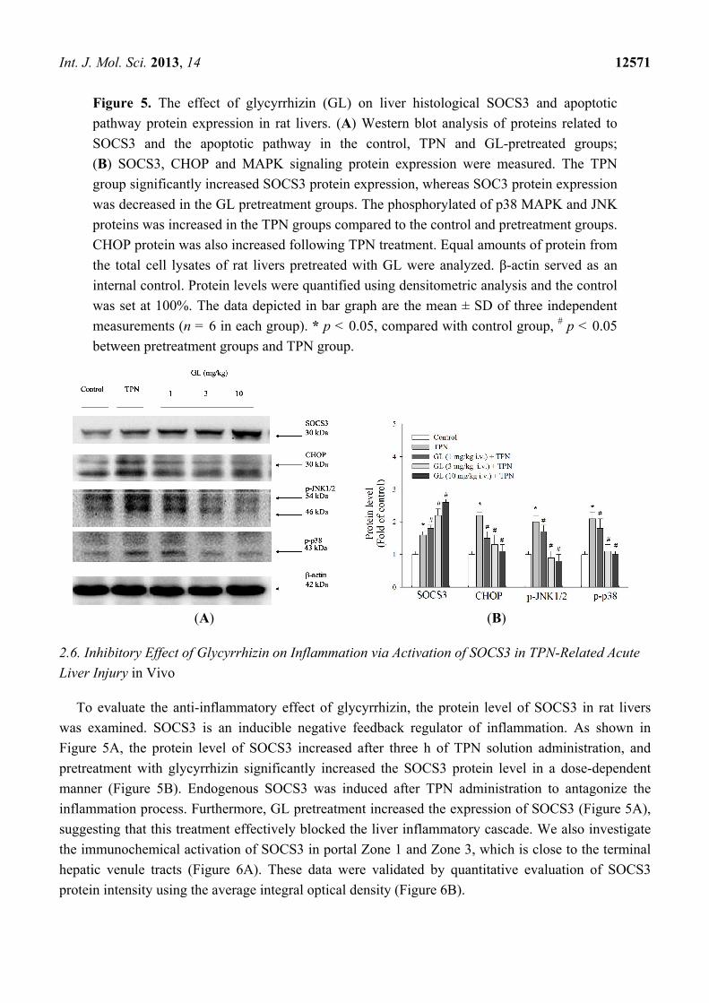
Figure 5. The effect of glycyrrhizin (GL) on liver histological SOCS3 and apoptotic
pathway protein expression in rat livers. (A) Western blot analysis of proteins related to
SOCS3 and the apoptotic pathway in the control, TPN and GL-pretreated groups;
(B) SOCS3, CHOP and MAPK signaling protein expression were measured. The TPN
group significantly increased SOCS3 protein expression, whereas SOC3 protein expression
was decreased in the GL pretreatment groups. The phosphorylated of p38 MAPK and JNK
proteins was increased in the TPN groups compared to the control and pretreatment groups.
CHOP protein was also increased following TPN treatment. Equal amounts of protein from
the total cell lysates of rat livers pretreated with GL were analyzed. β-actin served as an
internal control. Protein levels were quantified using densitometric analysis and the control
was set at 100%. The data depicted in bar graph are the mean ± SD of three independent
measurements (n = 6 in each group). * p < 0.05, compared with control group, # p < 0.05
between pretreatment groups and TPN group.
(A) (B)
2.6. Inhibitory Effect of Glycyrrhizin on Inflammation via Activation of SOCS3 in TPN-Related Acute
Liver Injury in Vivo
To evaluate the anti-inflammatory effect of glycyrrhizin, the protein level of SOCS3 in rat livers
was examined. SOCS3 is an inducible negative feedback regulator of inflammation. As shown in
Figure 5A, the protein level of SOCS3 increased after three h of TPN solution administration, and
pretreatment with glycyrrhizin significantly increased the SOCS3 protein level in a dose-dependent
manner (Figure 5B). Endogenous SOCS3 was induced after TPN administration to antagonize the
inflammation process. Furthermore, GL pretreatment increased the expression of SOCS3 (Figure 5A),
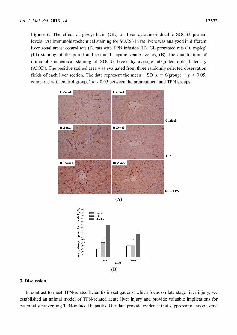
suggesting that this treatment effectively blocked the liver inflammatory cascade. We also investigate
the immunochemical activation of SOCS3 in portal Zone 1 and Zone 3, which is close to the terminal
hepatic venule tracts (Figure 6A). These data were validated by quantitative evaluation of SOCS3
protein intensity using the average integral optical density (Figure 6B).
Int. J. Mol. Sci. 2013, 14 12572
Figure 6. The effect of glycyrrhizin (GL) on liver cytokine-inducible SOCS3 protein
levels. (A) Immunohistochemical staining for SOCS3 in rat livers was analyzed in different
liver zonal areas: control rats (I); rats with TPN infusion (II); GL-pretreated rats (10 mg/kg)
(III) staining of the portal and terminal hepatic venues zones; (B) The quantitation of
immunohistochemical staining of SOCS3 levels by average integrated optical density
(AIOD). The positive stained area was evaluated from three randomly selected observation
fields of each liver section. The data represent the mean ± SD (n = 6/group). * p < 0.05,
compared with control group, # p < 0.05 between the pretreatment and TPN groups.
(A)
(B)
3. Discussion
In contrast to most TPN-related hepatitis investigations, which focus on late stage liver injury, we
established an animal model of TPN-related acute liver injury and provide valuable implications for
essentially preventing TPN-induced hepatitis. Our data provide evidence that suppressing endoplasmic
Int. J. Mol. Sci. 2013, 14 12573
reticulum stress and reactive nitrogen stress may have hepatoprotective effects and reduce the
development of TPN related hepatitis.
As shown in Table 1, ALT, AST and triglyceride levels were elevated in rat serum 3 h after TPN
administration, and pretreatment with GL (1, 3 and 10 mg/kg) normalized these levels. Figure 2A
shows the presence of severe hepatocyte steatosis in the TPN group. The liver is the main organ for
glucose disposal in the normal animals, which received 45% TPN-derived glucose [34]. The
overconsumption of glucose is associated with the increased accumulation of triglycerides in
hepatocytes. As shown in Figure 2A, glycyrrhizin improved the extent of accumulation. However, the
difference in total bilirubin between the GL pretreatment group (1 mg/kg) and the TPN group was not
significantly different, suggesting that an adequate therapeutic dose was achieved, with only the ALT
and AST concentrations being affected, but not total bilirubin levels (Table 1). Based on the observed
biochemical and pathological data, we successfully established a model of acute liver injury using TPN
in rats, finding that liver injury could be prevented by pretreatment with glycyrrhizin in a dose-dependent
manner. Similarly, Shamir (1993) and Zahavi (2000) [13,14] showed a significant reduction in bile
flow and bile salt secretion in rats after a two hour infusion of TPN at a rate of 10 mL/kg/h. The above
reports allowed for us to speculate that short-term TPN infusion (2 h) at an infusion rate of 10 mL/kg/h
can lead to bile stasis. Moreover, these previous studies also partially explain why the short-term
(3 h at a rate of 20 mL/kg/h) TPN infusion used in the present study elevated liver function.
According to a previous study, the hepatoprotective effect of glycyrrhizin in viral hepatitis
patients is due to its cytoprotective effect via the suppression of TNF-α-induced cytotoxicity and
inhibition of immune-mediated cytotoxicity against hepatocytes [35–37]. In addition, glycyrrhizin has
anti-inflammatory effects and attenuates the inflammatory response by inhibiting nuclear factor κB
(NF-κB) and PI3K [38]. Our data in Figure 3 and Table 1 show that TPN administration induced
systemic pro-inflammatory cytokine production and hepatitis. Glycyrrhizin can decrease the production of
these cytokines and improve TPN-related acute liver injury.
The overproduction of nitric oxide and its derivatives have been implicated as a cause of tissue
damage in response to inflammation. Pro-inflammatory mediators, such as NO, which is generated by
iNOS and nitrotyrosine, are considered to be indicators of the reaction during the inflammatory
response [39,40]. iNOS expression and the nitrosylation of tyrosine are induced in many different cell
types in response to endotoxins and cytokines, such as gamma interferon, interleukin 1 and tumor
necrosis factor alpha [41–43]. Thus, iNOS and nitrotyrosine serve as indicators of reactive nitrogen
stress generated by NO [43]. In the present study, (Figure 3), we demonstrated that treatment with
glycyrrhizin effectively inhibited NO production by suppressing the expression of iNOS and
nitrotyrosine. These data suggest that the therapeutic effect by which glycyrrhizin treats TPN-related
acute liver injury is partially due to the inhibition of NO production via the inhibition of iNOS, TNF-α,
iNOS and nitrotyrosine protein levels, and pretreatment with glycyrrhizin (10 mg/kg) resulted in the
most powerful inhibition of iNOS and nitrotyrosine.
Cytokines mediate various biological processes, including inflammation, apoptosis, necrosis and
fibrosis [44,45]. Increased pro-inflammatory cytokines has been demonstrated in animal model of
TPN [46,47]. These findings are consistent with our data, as GL decreased the production of IL-1β,
IL-6 and TNF-α in a dose-dependent manner (Figure 3). The SOCS3 protein is highly upregulated in
response to pro-inflammatory cytokines (i.e., IFN-γ, interleukin IL-1β and IL-6) [18]. SOCS3 is
Int. J. Mol. Sci. 2013, 14 12574
recognized to be the main modulator for the negative regulation of the cytokine-JAK-STAT pathway
and, thus, can induce anti-inflammatory processes [20]. Previous investigations also demonstrated the
SOCS3 is related to the severity of liver inflammation, and it plays a protective role in the
cytokine-mediated inflammatory process by inhibiting IL-6-medicated STAT3 activation [48,49].
Figure 6 shows that endogenous SOCS3 increased after TPN administration as a negative regulator of
TPN-related acute liver injury, and pretreatment with glycyrrhizin (Figure 5A,B) increased SOCS3
protein expression to enhance the negative regulation TPN-related acute liver injury. These findings
indicate that the inhibitory effects of glycyrrhizin on hepatitis may be due to alterations in SOCS3
expression. In summary, our study demonstrated that glycyrrhizin-inducted expression of SOCS3
contributes to the inhibition of inflammation and reactive nitrogen stress (RNS) production.
Infusions containing lipids and glucose activate stress kinases (JNK1/2 and p38 MAPK) and
increase ER stress in rats [50]. ER stress is known to induce apoptosis by the induction of CHOP,
which is a protein important for ER stress-related hepatitis [51,52]. Figure 4A,B shows that caspase-3
cleavage is higher after TPN administration, and it is well established that the caspases are the
executioners of apoptosis [53]. These results demonstrate that glycyrrhizin pretreatment significantly
inhibits the phosphorylation of JNK and p38 MAPK, as well as CHOP protein activation, thereby
suppressing hepatocyte apoptosis in TPN rats.
4. Experimental Section
4.1. Chemicals and Reagents
Glycyrrhizin (Figure 1), Reactive Nitrogen Species (RNS) scavengers (including nitrotyrosine and
anti-iNOS), CHOP, a Janus kinase (JNK) inhibitor (SP600125) and a p38 inhibitor (SB203580) were
purchased from Sigma (St. Louis, MO, USA). Total parenteral nutrition solution Kabiven™ was
obtained from Fresenius Kabi AB (Uppsala, Sweden). Mouse monoclonal antibodies against GAPDH,
β-actin, pro-inflammatory anti-TNF-α, IL-1β, IL-6 and nitrotyrosine were purchased from Santa Cruz
Biotechnology (Santa Cruz, CA, USA). Rabbit antibodies against human SOCS3, cleaved-caspase-3,
phospho-p38 MAPK (Thr180/Tyr182), phospho-JNK1/2 (Thr183/Tyr185), cdk1Tyr15 and a mouse
monoclonal antibody against cdk1 were purchased from Cell Signaling Technology (Beverly, MA,
USA). The TdT-mediated dUTP Nick End Labeling (TUNEL) kits were purchased from Roche
(Mannheim, Germany). SDS, NP-40, sodium deoxycholate and protease inhibitor cocktails were
purchased from Sigma (St. Louis, MO, USA).
4.2. Animal Experiments and Drug Treatment
Adult male Sprague-Dawley rats weighing 200 ± 20 g were obtained from the National Yang-Ming
University Animal Center, Taipei, Taiwan. The rats were pathogen-free and had free access to food
(Laboratory Rodent Diet 5001, PMI Nutrition International LLC, Brentwood, MO, USA) and water.
The rats were housed with a 12 h light and 12 h dark cycle. All of the experimental protocols involving
animals were reviewed and approved by the Institutional Animal Care and Use Committee (IACUC
number: 981106) of National Yang-Ming University. Rats were anesthetized with urethane (1 g/kg,
i.p.). All of the animals were equipped with the left femoral vein for solution or drug administration.
Int. J. Mol. Sci. 2013, 14 12575
The rats were intravenously infused with TPN via the left femoral artery at a flow rate of
20 mL/kg/h for 3 h to induce acute liver injury. Normal saline (0.9% sodium chloride) was used as the
vehicle for the control group. Different doses of glycyrrhizin (1, 3 or 10 mg/kg) were administered
intravenously before TPN infusion.
Before TPN administration, glycyrrhizin was dissolved in 1 mL normal saline and given as an i.v.
bolus, whereas the TPN group received an equal volume of normal saline (1 mL via i.v. bolus). The
total energy of Kabiven® was 1038 kcal [11% glucose, 885 mL; amino acids/electrolytes (Vamin 18),
300 mL; and 20% fat emulsion (Intralipid), 255 mL]. Each rat received a total infusion volume of
12 mL TPN at a rate of 20 mL/kg/h for three h. A typical 200 mg rat received approximately
8.6 kcal of energy.
Every group had six animals. The blood samples were collected through left femoral vein
immediately and stored in heparin-coated capillary tubes until liver enzyme profiling and biochemical
analyses. The liver tissue was collected, washed with normal saline and then fixed in 10% neutral
buffered formalin for histopathological examinations.
4.3. Biochemical Analysis
The blood samples were collected from the animals and centrifuged at 3000 rpm for 15 min. The
plasma samples were used for the following biochemical analyses of aspartate aminotransferase
(AST), alanine aminotransferase (ALT), total bilirubin (TB) and triglyceride (TG) levels. All of these
analyses were performed using the Hitachi 902 Automatic Analyzer with adapted reagents from
Roche (Boehringer, Mannheim, Germany).
4.4. Histopathological Evaluation
The liver tissues were fixed in 10% buffered formalin, embedded in paraffin and cut into 4 μm-thick
slides transversely from portal Zone 1 to Zone 3, which is close to the terminal of hepatic venule
tract, for hematoxylin and eosin (H & E) staining (Tokyo, Japan). The rest of tissues were stored at
−80 °C. The histopathological changes in liver cell morphology were examined using a BX51 light
microscope (Olympus, Tokyo, Japan) at high power (200× magnification) for each slide. For
quantitative purposes, two portal zones were randomly chosen in each slide and photographed using an
Image-pro Plus medical image analysis system. The cell numbers were recorded. The normal
hepatocytes were counted from 10 fields that were randomly chosen in liver samples from each group
under 200× magnification. The means numbers of normal cells were calculated per microscope field
from six animals in each group.
4.5. Immunohistochemical Assay
Immunohistochemistry (IHC) staining was performed using a biotinylated secondary antibody
(Vectastain Universal Elite ABC Kit, Burlingame, CA, USA). Monoclonal rabbit antibodies against
human SOCS3 and cleaved-caspase 3 were diluted at a ratio of 1:100. The primary antibodies were
omitted for the negative controls. For three slides, the cytoplasm stained brown and was scored
positive. The expression of SOCS3 and cleaved-caspase 3 was quantitatively evaluated using an
Int. J. Mol. Sci. 2013, 14 12576
Olympus C × 31 microscope with the Image-pro Plus medical image analysis system. The digital
images were captured using a digital camera (Canon A640, Tokyo, Japan). The positive area and
optical density (OD) of SOCS3 and cleaved-caspase 3-positive cells were determined by measuring
three randomly selected microscopic fields (400× magnification) for each slide. The IHC index was
defined as average integral optical density (AIOD) (AIOD = positive area × OD/total area).
4.6. Western Blot Analysis
After sacrifice, the rats’ livers were rapidly removed, disassociated and homogenized in ice-cold
lysis buffer (1% NP-40, 0.5% sodium deoxycholate, 0.1% sodium dodecyl sulfate and a protease
inhibitor mixture comprised of phenylmethylsulfonyl fluoride, aprotinin and sodium orthovanadate).
The total cell lysate (50 μg of proteins) was separated by the SDS-polyacrylamide gel electrophoresis
(PAGE) (12% running and 4% stacking) and analyzed using the designated antibodies. The
Western-Light Chemiluminescent detection system (Bio-Rad, Hercules, CA, USA) was used to detect
the signals, as previously described by Chiu et al. [54].
4.7. Statistical Analyses
The data are expressed as the mean ± standard deviation (SD) of three independent experiments,
and all of the data were analyzed using one-way analysis of variance (ANOVA) with Bonferroni
correction. The data were analyzed using the SAS software statistical package “SigmaPlot” version 9.0
(SAS Institute Inc., Cary, NC, USA).
5. Conclusions
Taken together, our data provide new information on the molecular mechanisms by which
glycyrrhizin inhibits inflammation. Namely, GL acts by reducing iNOS, TNF-α, IL-1β, IL-6 and
nitrotyrosine levels through the activation of SOCS3. In addition, glycyrrhizin was found to have
anti-apoptotic effects on TPN-related acute liver injury in rats through the inhibition of JNK1/2 and
p38 MAPK phosphorylation and inactivation of the CHOP protein, thereby reducing ER stress. The
inhibitory effect of glycyrrhizin on ER stress and reactive nitrogen species production is one possible
mechanism by which glycyrrhizin prevents TPN-related liver injury. Overall, we demonstrated a novel
animal model for early stage of TPN related acute liver injury and elucidated the potential therapeutic
effects of glycyrrhizin on TPN related hepatitis. Nevertheless, glycyrrhizin therapy for chronic
parenteral nutrition-related hepatitis warrants further investigation.
Acknowledgments
Funding for this study was provided in part by research grants from the National Science Council
(NSC99-2113-M-010-001-MY3, NSC99-2628-B-010-008-MY3) Taiwan; the Tomorrow Medical
Foundation, Taiwan; Chang Gung Memorial Hospital or Chang Gung University of Science and
TechnologyChiayi Campus (grants CZRPG880253 and CMRPF6A0072). We thank Tim Baker and
Bessy Hung for editing this manuscript.
Int. J. Mol. Sci. 2013, 14 12577
Conflict of Interest
The authors declare no conflicts of interest.
References
1. Chance, W.T.; Sheriff, S.; Dayal, R.; Friend, L.A.; Thomas, I.; Balasubramaniam, A. The role of
polyamines in glucagon-like peptide-2 prevention of TPN-induced gut hypoplasia. Peptides 2006,
27, 883–892.
2. Stoll, B.; Horst, D.A.; Cui, L.; Chang, X.; Ellis, K.J.; Hadsell, D.L.; Suryawan, A.; Kurundkar, A.;
Maheshwari, A.; Davis, T.A.; et al. Chronic parenteral nutrition induces hepatic inflammation,
steatosis, and insulin resistance in neonatal pigs. J. Nutr. 2010, 140, 2193–2200.
3. Raman, M.; Allard, J.P. Parenteral nutrition related hepato-biliary disease in adults. Appl. Physiol.
Nutr. Metable 2007, 32, 646–654.
4. Sandhu, I.S.; Jarvis, C.; Everson, G.T. Total parenteral nutrition and cholestasis. Clin. Liver Dis.
1999, 3, 489–508.
5. Kumpf, V.J. Parenteral nutrition-associated liver disease in adult and pediatric patients. Nutr.
Clin. Pract. 2006, 21, 279–290.
6. Cavicchi, M.; Beau, P.; Crenn, P.; Degott, C.; Messing, B. Prevalence of liver disease and
contributing factors in patients receiving home parenteral nutrition for permanent intestinal
failure. Ann. Intern. Med. 2000, 132, 525–532.
7. Luman,W.; Shaffer, J.L. Prevalence, outcome and associated factors of deranged liver function
tests in patients on home parenteral nutrition. Clin. Nutr. 2002, 21, 337–343.
8. Attili, A.F.; Angelico, M.; Cantafora, A.; Alvaro, D.; Capocaccia, L. Bile acid-induced liver toxicity:
Relation to the hydrophobic-hydrophilic balance of bile acids. Med. Hypotheses. 1986, 19, 57–69.
9. Tomar, B.S. Hepatobiliary abnormalities and parenteral nutrition. Indian J. Pediatr. 2000, 67,
695–701.
10. Brinkman, A.S.; Murali, S.G.; Hitt, S.; Solverson, P.M.; Holst, J.J.; Ney, D.M. Enteral nutrients
potentiate glucagon-like peptide-2 action and reduce dependence on parenteral nutrition in a rat
model of human intestinal failure. Am. J. Physiol. Gastrointest Liver Physiol. 2012, 303, 610–622.
11. Jain, A.K.; Stoll, B.; Burrin, D.G.; Holst, J.J.; Moore, D.D. Enteral bile acid treatment improves
parenteral nutrition-related liver disease and intestinal mucosal atrophy in neonatal pigs. Am. J.
Physiol. Gastrointest Liver Physiol. 2012, 302, 218–224.
12. Loff, S.; Kranzlin, B.; Moghadam, M.; Dzakovic, A.; Wessel, L.; Back, W.; Hosie, S.; Wirth, H.;
Waag, K.L. Parenteral nutrition-induced hepatobiliary dysfunction in infants and prepubertal
rabbits. Pediatr. Surg. Int. 1999, 15, 479–482.
13. Shamir, R.; Zahavi, I.; Bar-Sever, Z.; Heckelman, B.; Marcus, H.; Dinari, G. Total parenteral
nutrition-associated cholestasis after selective damage to acinar zone 3 hepatocytes by
bromobenzene in the rat. Life Sci. 1993, 52, 371–376.
14. Zahavi, I.; Rosezki, O.; Stolkart, Y.; Shamir, R.; Heckelman, B.; Marcus, H.; Dinari, G. The
effect of cisapride on total parenteral nutrition-associated cholestasis in rats. Isr. Med. Assoc. J.
2000, 2, 91–93.
Int. J. Mol. Sci. 2013, 14 12578
15. Hong, L.; Wang, X.; Wu, J.; Cai, W. Mitochondria-initiated apoptosis triggered by oxidative
injury play a role in total parenteral nutrition-associated liver dysfunction in infant rabbit model.
J. Pediatr. Sur. 2009, 4, 1712–1718.
16. Laborie, S.; Lavoie, J.C.; Chessex, P. Paradoxical role of ascorbic acid and riboflavin in solutions
of total parenteral nutrition: Implication in photoinduced peroxide generation. Pediatr. Res. 1998,
43, 601–606.
17. Knowles, H.; Li, Y.; Perraud, A.L. The TRPM2 ion channel, an oxidative stress and metabolic
sensor regulating innate immunity and inflammation. Immunol. Res. 2013, 55, 241–248.
18. Egwuagu, C.E.; Yu, C.R.; Zhang, M.; Mahdi, R.M.; Kim, S.J.; Gery, I. Suppressors of cytokine
signaling proteins are differentially expressed in Th1 and Th2 cells: Implications for Th cell
lineage commitment and maintenance. J. Immunol. 2002, 168, 3181–3187.
19. Yu, C.R.; Mahdi, R.M.; Ebong, S.; Vistica, B.P.; Chen, J.; Guo, Y.; Gery, I.; Egwuagu, C.E. Cell
proliferation and STAT6 pathways are negatively regulated in T cells by STAT1 and suppressors
of cytokine signaling. J. Immunol. 2004, 173, 737–746.
20. Yoshimura, A.; Suzuki, M.; Sakaguchi, R.; Hanada, T.; Yasukawa, H. SOCS, Inflammation, and
autoimmunity. Front. Immunol. 2012, 3, 20.
21. Tazuke, Y.; Drongowski, R.A.; Btaiche, I.; Coran, A.G.; Teitelbaum, D.H. Effects of lipid
administration on liver apoptotic signals in a mouse model of total parenteral nutrition (TPN).
Pediatr. Surg. Int. 2004, 20, 224–228.
22. Ferri, K.F.; Kroemer, G. Organelle-specific initiation of cell death pathways. Nat. Cell. Biol.
2001, 3, 255–263.
23. Wang, H.C.; Huang, W.; Lai, M.D.; Su, I.J. Hepatitis B virus pre-S mutants, endoplasmic
reticulum stress and hepatocarcinogenesis. Cancer Sci. 2006, 97,683–688.
24. Katz, M.S.; Thatch, K.A.; Schwartz, M.Z. Dose variation of hepatocyte growth factor and its
effects on an animal model of TPN-induced liver injury. J. Surg. Res. 2010, 163, 294–298.
25. Liu, J.; Ren, F.; Cheng, Q.; Bai, L.; Shen, X.; Gao, F.; Busuttil, R.W.; Kupiec-Weglinski, J.W.;
Zhai, Y. Endoplasmic reticulum stress modulates liver inflammatory immune response in the
pathogenesis of liver ischemia and reperfusion injury. Transplantation 2012, 94, 211–217.
26. Ashfaq, U.A.; Masoud, M.S.; Nawaz, Z.; Riazuddin, S. Glycyrrhizin as antiviral agent against
Hepatitis C Virus. J. Transl. Med. 2011, 9, 112.
27. Liao, H.L.; Ma, T.C.; Li, Y.C.; Chen, J.T.; Chang, Y.S. Concurrent use of corticosteroids with
licorice-containing TCM preparations in Taiwan: A National Health Insurance Database study.
J. Altern. Complement. Med. 2010, 16, 539–544.
28. Asl, M.N.; Hosseinzadeh, H. Review of pharmacological effects of Glycyrrhiza sp. and its
bioactive compounds. Phytother. Res. 2008, 22, 709–724.
29. Del Prete, A.; Scalera, A.; Iadevaia, M.D.; Miranda, A.; Zulli, C.; Gaeta, L.; Tuccillo, C.;
Federico, A.; Loguercio, C. Herbal products: Benefits, limits, and applications in chronic liver
disease. Evid. Based Complement. Alternat. Med. 2012, 2012, 837939.
30. Stickel, F.; Schuppan, D. Herbal medicine in the treatment of liver diseases. Dig. Liver Dis. 2007,
39, 293–304.

Int. J. Mol. Sci. 2013, 14 12579
31. Makuuchi, M.; Kokudo, N.; Arii, S.; Futagawa, S.; Kaneko, S.; Kawasaki, S.; Matsuyama, Y.;
Okazaki, M.; Okita, K.; Omata, M.; et al. Development of evidence-based clinical guidelines for
the diagnosis and treatment of hepatocellular carcinoma in Japan. Hepatol. Res. 2008, 38, 37–51.
32. Van der kallen, C.J.; van greevenbroek, M.M.; Stehouwer, C.D.; Schalkwijk, C.G. Endoplasmic
reticulum stress-induced apoptosis in the development of diabetes: Is there a role for adipose
tissue and liver? Apoptosis 2009, 14, 1424–1434.
33. Farley, N.; Pedraza-Alva, G.; Serrano-Gomez, D.; Nagaleekar, V.; Aronshtam, A.; Krahl, T.;
Thornton, T.; Rincón, M. p38 mitogen-activated protein kinase mediates the Fas-induced
mitochondrial death pathway in CD8+ T cells. Mol. Cell. Biol. 2006, 26, 2118–2129.
34. McGuinness, O.P.; Donmoyer, C.; Ejiofor, J.; McElligott, S.; Lacy, D.B. Hepatic and muscle
glucose metabolism during total parenteral nutrition: Impact of infection. Am. J. Physiol. 1998,
275, E763–E769.
35. Jiménez, W.; Clária, J.; Arroyo, V.; Rodés, J. Carbon tetrachloride induced cirrhosis in rats: A
useful tool for investigating the pathogenesis of ascites in chronic liver disease. J. Gastroenterol.
Hepatol. 1992, 7, 90–97.
36. Manns, M.P.; Wedemeyer, H.; Singer, A.; Khomutjanskaja, N.; Dienes, H.P.; Roskams, T.;
Goldin, R.; Hehnke, U.; Inoue, H.; European SNMC Study Group. Glycyrrhizin in patients who
failed previous interferon alpha-based therapies: Biochemical and histological effects after
52 weeks. J. Viral Hepat. 2012, 19, 537–546.
37. Yoshikawa, M.; Matsui, Y.; Kawamoto, H.; Umemoto, N.; Oku, K.; Koizumi, M.; Yamao, J.;
Kuriyama, S.; Nakano, H.; Hozumi, N.; et al. Effects of glycyrrhizin on immune-mediated
cytotoxicity. J. Gastroenterol. Hepatol. 1997, 12, 243–248.
38. Kao, T.C.; Shyu, M.H.; Yen, G.C. Glycyrrhizic acid and 18beta-glycyrrhetinic acid inhibit
inflammation via PI3K/Akt/GSK3beta signaling and glucocorticoid receptor activation. J. Agric.
Food Chem. 2010, 58, 8623–8629.
39. Wei, X.Q.; Charles, I.G.; Smith, A.; Ure, J.; Feng, G.J.; Huang, F.P.; Xu, D.; Muller, W.;
Moncada, S.; Liew, F.Y. Altered immune responses in mice lacking inducible nitric oxide
synthase. Nature 1995, 375, 408–411.
40. Nussler, A.K.; Billiar, T.R. Inflammation, immunoregulation, and inducible nitric oxide synthase.
J. Leukoc. Biol. 1993, 54, 171–178.
41. Minc-Golomb, D.; Tsarfaty, I.; Schwartz, J.P. Expression of inducible nitric oxide synthase by
neurones following exposure to endotoxin and cytokine. Br. J. Pharmacol. 1994, 112, 720–722.
42. Ialenti, A.; Ianaro, A.; Moncada, S.; di Rosa, M. Modulation of acute inflammation by
endogenous nitric oxide. Eur J. Pharmacol. 1992, 211, 177–182.
43. Fan, C.K.; Lin, Y.H.; Hung, C.C.; Chang, S.F.; Su, K.E. Enhanced inducible nitric oxide synthase
expression and nitrotyrosine accumulation in experimental granulomatous hepatitis caused by
Toxocaracanis in mice. Parasite Immunol. 2004, 26, 273–281.
44. Gorbunov, N.V.; McFaul, S.J.; Januszkiewicz, A.; Atkins, J.L. Pro-inflammatory alterations and
status of blood plasma iron in a model of blast-induced lung trauma. Int. J. Immunopathol.
Pharmacol. 2005, 18, 547–556.

Int. J. Mol. Sci. 2013, 14 12580
45. Di Giannantonio, M.; Frydas, S.; Kempuraj, D.; Karagouni, E.; Hatzistilianou, M.; Conti, C.M.;
Boucher, W.; Papadopoulou, N.; Donelan, J.; Cao, J.; et al. Cytokines in stress. Int. J.
Immunopathol. Pharmacol. 2005, 18, 1–5.
46. Feng, Y.; Ralls, M.W.; Xiao, W.; Miyasaka, E.; Herman, R.S.; Teitelbaum, D.H. Loss of enteral
nutrition in a mouse model results in intestinal epithelial barrier dysfunction. Ann. N. Y. Acad. Sci.
2012, 1258, 71–77.
47. Feng, Y.; McDunn, J.E.; Teitelbaum, D.H. Decreased phospho-Akt signaling in a mouse model of
total parenteral nutrition: A potential mechanism for the development of intestinal mucosal
atrophy. Am. J. Physiol. Gastrointest Liver Physiol. 2010, 298, 833–841.
48. Koeberlein, B.; zur Hausen, A.; Bektas, N.; Zentgraf, H.; Chin, R.; Nguyen, L.T.; Kandolf, R.;
Torresi, J.; Bock, C.T. Hepatitis B virus overexpresses suppressor of cytokine signaling-3 (SOCS3)
thereby contributing to severity of inflammation in the liver. Virus Res. 2010, 148, 51–59.
49. Sasaki, A.; Yasukawa, H.; Shouda, T.; Kitamura, T.; Dikic, I.; Yoshimura, A. CIS3/SOCS-3
suppresses erythropoietin (EPO) signaling by binding the EPO receptor and JAK2. J. Biol. Chem.
2000, 275, 29338–29347.
50. Boden, G.; Song, W.; Duan, X.; Cheung, P.; Kresge, K.; Barrero, C.; Merali, S. Infusion of
glucose and lipids at physiological rates causes acute endoplasmic reticulum stress in rat liver.
Obesity 2011, 19, 1366–1373.
51. Kamiya, T.; Nishihara, H.; Hara, H.; Adachi, T. Ethanol extract of Brazilian red propolis induces
apoptosis in human breast cancer MCF-7 cells through endoplasmic reticulum stress. J. Agric.
Food Chem. 2012,60, 11065–11070.
52. Malhi, H.; Kaufman, R.J. Endoplasmic reticulum stress in liver disease. J. Hepatol. 2011, 54,
795–809.
53. Riedl, S.J.; Shi, Y. Molecular mechanisms of caspase regulation during apoptosis. Nat. Rev. Mol.
Cell. Biol. 2004, 5, 897–907.
54. Yeh, Y.T.; Hur, S.S.; Chang, J.; Wang, K.C.; Chiu, J.J.; Li, Y.S.; Chien, S. Matrix stiffness
regulates endothelial cell proliferation through Septin 9. PLoS One 2012, 7, e46889.
©2013 by the authors; licensee MDPI, Basel, Switzerland. This article is an open access article
distributed under the terms and conditions of the Creative Commons Attribution license
(http://creativecommons.org/licenses/by/3.0/).