glucocorticoids mediate stress-induced priming of microglial pro-inflammatory responses
TRANSCRIPT

Brain, Behavior, and Immunity 26 (2012) 337–345
Contents lists available at SciVerse ScienceDirect
Brain, Behavior, and Immunity
journal homepage: www.elsevier .com/locate /ybrbi
Glucocorticoids mediate stress-induced priming of microglialpro-inflammatory responses
Matthew G. Frank ⇑, Brittany M. Thompson, Linda R. Watkins, Steven F. MaierDepartment of Psychology and Neuroscience, University of Colorado, Boulder, CO, USA
a r t i c l e i n f o
Article history:Received 12 August 2011Received in revised form 5 October 2011Accepted 17 October 2011Available online 24 October 2011
Keywords:StressGlucocorticoidMicrogliaCytokineNeuroinflammationPriming
0889-1591/$ - see front matter � 2011 Elsevier Inc. Adoi:10.1016/j.bbi.2011.10.005
⇑ Corresponding author. Address: Department of PCampus Box 345, University of Colorado, Boulder, CO 8919 8116; fax: +1 303 492 2967.
E-mail address: [email protected] (M.G. Fra
a b s t r a c t
Acute and chronic stress sensitizes or ‘‘primes’’ the neuroinflammatory response to a subsequent pro-inflammatory challenge. While prior evidence shows that glucocorticoids (GCs) play a pivotal role instress-induced potentiation of neuroinflammatory responses, it remains unclear whether stress-inducedGCs sensitize the response of key CNS immune substrates (i.e. microglia) to pro-inflammatory stimuli. Anex vivo approach was used to address this question. Here, stress-induced GC signaling was manipulatedin vivo and hippocampal microglia challenged with the pro-inflammatory stimulus LPS ex vivo. MaleSprague–Dawley rats were either pretreated in vivo with the GC receptor antagonist RU486 or adrenal-ectomized (ADX). Animals were then exposed to an acute stressor (inescapable tailshock; IS) and 24 hlater hippocampal microglia were isolated and challenged with LPS to probe for stress-induced sensitiza-tion of pro-inflammatory responses. Prior exposure to IS resulted in a potentiated pro-inflammatory cyto-kine response (e.g. IL-1b gene expression) to LPS in isolated microglia. Treatment in vivo with RU486 andADX inhibited or completely blocked this IS-induced sensitization of the microglial pro-inflammatoryresponse. The present results suggest that stress-induced GCs function to sensitize the microglial pro-inflammatory response (IL-1b, IL-6, NFjBIa) to immunologic challenges.
� 2011 Elsevier Inc. All rights reserved.
1. Introduction
The phenomenon of cross-sensitization between the neuroin-flammatory sequelae of stress and pro-inflammatory immune acti-vation has been well characterized (Anisman, 2009; Tilders andSchmidt, 1999). In particular, acute and chronic stress has beenfound to sensitize the neuroinflammatory response to both periph-eral and central immunologic challenges (de Pablos et al., 2006;Espinosa-Oliva et al., 2011; Frank et al., 2007; Johnson et al.,2002b, 2003, 2004; Munhoz et al., 2006; Wohleb et al., 2011).For example, exposure to a single session of inescapable tailshock(Johnson et al., 2002b) and to 14 daily sessions of unpredictablechronic stress (Munhoz et al., 2006) potentiate the increase in hip-pocampus and frontal cortex expression of pro-inflammatorymediators (e.g. interleukin-1b; IL-1b) produced by a peripheralinjection of lipopolysaccharide (LPS) given 24 h after the stressorregimen. Several studies have also shown that acute and chronicstress modulates the immunophenotype of microglia as indicatedby up-regulation of MHCII (de Pablos et al., 2006; Frank et al.,
ll rights reserved.
sychology and Neuroscience,0309-0345, USA. Tel.: +1 303
nk).
2007), toll-like receptor 4 (TLR4) (Wohleb et al., 2011) and F4/80antigen (Nair and Bonneau, 2006), suggesting that stress may shiftthe neuroimmune microenvironment towards a pro-inflammatoryimmunophenotype.
Stress-induced microglial activation and potentiation of neuro-inflammatory processes has been blocked by a glucocorticoid (GC)receptor antagonist (de Pablos et al., 2006; Espinosa-Oliva et al.,2011; Munhoz et al., 2006; Nair and Bonneau, 2006) suggestingthat GCs may mediate stressor-induced sensitization of neuroin-flammatory processes. However, it is important to note that inthese studies it was not possible to determine which immunologicsubstrate(s), whether peripheral and/or central, was sensitized orprimed by stress-induced GCs. This is because the pro-inflamma-tory challenge (LPS) was administered in vivo, and so sensitizationof either the peripheral response that signals the brain, orprocesses in the brain (or both) could have been causal. We havepreviously shown that an acute stressor sensitizes the hippocam-pal microglial pro-inflammatory response to immune challengeex vivo (Frank et al., 2007). In that study, hippocampal microgliawere isolated 24 h post-stress, challenged with LPS ex vivo andpro-inflammatory cytokines measured 2 h later. Challengingmicroglia with LPS ex vivo serves two purposes. First, this ex vivoapproach provides a clear test of whether microglia could be animmunologic substrate of stress-induced priming since it allowsa definitive determination of whether microglia are, in fact,

338 M.G. Frank et al. / Brain, Behavior, and Immunity 26 (2012) 337–345
sensitized by the prior stressor. Secondly, LPS modulation of theHPA axis and GC signaling is necessarily excluded, thereby makingit possible to separate the effects of stress-induced GCs from the ef-fects of LPS-induced GCs on microglia pro-inflammatory responses.Therefore, an ex vivo experimental approach was taken in the pres-ent investigation to test whether GCs mediate stress-induced sen-sitization of microglia pro-inflammatory immune responses. Totest this hypothesis, pharmacological (GR antagonist) and surgical(adrenalectomy) suppression of stress-induced GCs was utilized.
2. Material and methods
2.1. Animals
Male Sprague–Dawley rats (60–90 d old; Harlan Sprague–Dawley,Inc., Indianapolis, IN, USA) were pair-housed with food and wateravailable ad libitum. The colony was maintained at 25 �C on a 12-hlight/dark cycle (lights on at 07:00 h). All experimental procedureswere conducted in accord with the University of Colorado Institu-tional Animal Care and Use Committee.
2.2. Reagents
RU486 (mifepristone; Sigma, St. Louis, MO), a GC receptorantagonist, was dissolved in 100% propylene glycol. LPS (E. coliserotype 0111:B4; Sigma) was dissolved in pyrogen free, sterile0.9% saline.
2.3. Experimental design
2.3.1. Effect of RU486 on stress-induced potentiation of neuroin-flammatory responses to LPS in vivo
Prior to proceeding with ex vivo studies, a preliminary studywas conducted to verify the efficacy of RU486 to block stress-induced potentiation of neuroinflammatory responses in vivo.Animals were injected with RU486 (50 mg/kg, s.c.) or vehicle(100% propylene glycol) 1 h prior to onset of inescapable tailshock(IS). Home cage controls (HCC) were injected at the same time ofday as were IS animals. All injections occurred between 08:00and 10:00 h. Twenty four hours post-IS, IS and HCC animals wereinjected with LPS (10 lg/kg, i.p.) or vehicle. Thus, the design wasa 2 � 2 factorial. Two hours post-LPS or vehicle, hippocampalpro-inflammatory cytokines were measured. Analysis was re-stricted to the hippocampus because we have shown that it is sen-sitive to IS-induced priming effects in vivo (Johnson et al., 2002b)and ex vivo (Frank et al., 2007) and of the CNS structures that aresensitive to IS-induced priming effects, it yields a sufficient num-ber of microglia to conduct ex vivo experiments.
2.3.2. Effect of RU486 on stress-induced potentiation of hippocampalmicroglial responses to LPS ex vivo
Animals were injected with RU486 (50 mg/kg, s.c.) or vehicle(100% propylene glycol) 1 h prior to onset of IS. Twenty four hourspost-IS, hippocampal microglia were isolated from IS and HCC ani-mals and exposed to LPS for 2 h and pro-inflammatory cytokinegene expression assessed.
2.3.3. Effect of adrenalectomy (ADX) on stress-induced potentiationof hippocampal microglial responses to LPS ex vivo
Animals underwent ADX or sham surgery. Two weeks post-sur-gery, animals were exposed to IS or served as HCC. Twenty fourhours post-IS, hippocampal microglia were isolated from IS andHCC animals and exposed to LPS for 2 h and pro-inflammatorycytokine gene expression assessed.
2.4. ADX surgery and verification
Bilateral ADX was aseptically performed under halothane anes-thesia (Halocarbon Laboratories, River Edge, NJ, USA) as previouslydescribed (Der-Avakian et al., 2005). All removed tissue was exam-ined immediately to ensure complete removal of the adrenal gland.Adrenal tissue was visually inspected to assess that the adrenalgland was intact. Sham-operated animals received the identicalprocedure, except that the adrenal glands were gently manipulatedwith forceps but not removed. Steroid replacement began for ADXanimals immediately after surgery and continued for the remain-der of the experiment. Steroid replacement was utilized becausethe rationale was to eliminate the IS-induced rise of CORT, not toeliminate basal levels. ADX animals received basal corticosterone(CORT; Sigma–Aldrich, St. Louis, Mo, USA) replacement in theirdrinking water since this method has been shown to mimic thenormal circadian pattern of CORT secretion (Jacobson et al.,1988). CORT was initially dissolved in ethyl alcohol (EtOH) and di-luted to a final concentration of 25 lg/ml in tap water to yield 0.2%EtOH. This concentration leads to normal basal levels. CORT-wateralso contained 0.9% saline. Sham animals received drinking watercontaining 0.2% EtOH and 0.9% saline. Rats were allowed 2 weeksto recover before exposure to IS. For ADX verification, cardiac bloodwas collected at the time of sacrifice and serum CORT measured.
2.5. Inescapable shock (IS)
Details of the present stressor protocol have been publishedpreviously and reliably potentiate pro-inflammatory cytokine re-sponses in the hippocampus after peripheral immune challenge(Johnson et al., 2002b) as well as in isolated hippocampal microgliato LPS ex vivo (Frank et al., 2007). Briefly, animals were placed inPlexiglas tubes (23.4 cm in length � 7 cm in diameter) and exposedto 100 1.6 mA, 5 s tailshocks with a variable intertrial interval (ITI)ranging from 30–90 s (average ITI = 60 s). All IS treatments oc-curred between 09:00 and 11:00 h. IS animals were returned totheir home cages immediately after termination of shock. HCC ani-mals remained undisturbed in their home cages.
2.6. Tissue and blood collection
Animals were given a lethal dose of sodium pentobarbital. Ani-mals were fully anesthetized and cardiac blood withdrawn within3 min of injection. Cardiac blood was collected and animals weretranscardially perfused with ice-cold saline (0.9%) for 3 min to re-move peripheral immune leukocytes from the CNS vasculature.Brain was rapidly extracted and placed on ice and hippocampusdissected. For in vivo experiments, hippocampus was flash frozenin liquid nitrogen and stored at �80 �C. For ex vivo experiments,hippocampal microglia were immediately isolated.
2.7. Ex vivo immune stimulation of hippocampal microglia with LPS
Hippocampal microglia were isolated using a Percoll densitygradient as previously described (Frank et al., 2006). We have pre-viously shown (Frank et al., 2006) that this microglia isolation pro-cedure yields highly pure microglia (Iba-1+/MHCII+/CD163�/GFAP�). In the present experiments, immunophenotype and purityof microglia was assessed using real time RT-PCR. Microglia weresuspended in DMEM + 10% FBS and microglia concentration deter-mined by trypan blue exclusion. Microglia concentration was ad-justed to a density of 5 � 103 cells/100 ll and 100 ll added toindividual wells of a 96-well v-bottom plate. LPS was utilized tochallenge microglia ex vivo as we have previously determined theoptimal in vitro conditions under which LPS stimulates a microgliapro-inflammatory cytokine response (Frank et al., 2006). Cells were
M.G. Frank et al. / Brain, Behavior, and Immunity 26 (2012) 337–345 339
incubated with LPS (0.1, 1, 10, and 100 ng/ml) or media alone for2 h at 37 �C, 5% CO2. The plate was centrifuged at 1000g for10 min at 4 �C to pellet cells and cells washed 1� in ice cold PBSand centrifuged at 1000g for 10 min at 4 �C. Cell lysis/homogeniza-tion and cDNA synthesis was performed according to the manufac-turer’s protocol using the SuperScript III CellsDirect cDNASynthesis System (Invitrogen, Carlsbad, CA).
2.8. Real time RT-PCR measurement of gene expression
A detailed description of the PCR amplification protocol hasbeen published previously (Frank et al., 2006). cDNA sequenceswere obtained from Genbank at the National Center for Biotechnol-ogy Information (NCBI; www.ncbi.nlm.nih.gov). Primer sequenceswere designed using the Operon Oligo Analysis Tool (http://www.operon.com/technical/toolkit.aspx) and tested for sequencespecificity using the Basic Local Alignment Search Tool at NCBI(Altschul et al., 1997). Primers were obtained from Invitrogen. Pri-mer specificity was verified by melt curve analyses. All primerswere designed to span exon/exon boundaries and thus excludeamplification of genomic DNA (See Table 1 for primer descriptionand sequences).
PCR amplification of cDNA was performed using the QuantitectSYBR Green PCR Kit (Qiagen, Valencia, CA). Formation of PCR prod-uct was monitored in real time using the MyiQ Single-Color Real-Time PCR Detection System (BioRad, Hercules, CA). Relative geneexpression was determined by taking the expression ratio of thegene of interest to b-Actin.
2.9. Serum and hippocampus CORT assay
Cardiac blood was centrifuged (10 min, 14,000g, 4 �C) and ser-um collected. Hippocampus was sonicated using a tissue extrac-tion reagent (Invitrogen) supplemented with a protease inhibitorcocktail (Sigma). Homogenate was centrifuged (10 min, 14,000g,4 �C) and supernatant collected and stored at �20 �C. Total proteinwas quantified using a Bradford assay. CORT was measured using acompetitive immunoassay (Assay Designs, Inc., Ann Arbor, MI) asdescribed in the manufacturer’s protocol.
2.10. Statistical analysis and data presentation
All data are presented as mean ± SEM. Statistical analyses con-sisted of ANOVA followed by post hoc tests (Newman-Keuls) usingPrism 5 (Graphpad Software, Inc., La Jolla, CA). Threshold for
Table 1Primer description and sequence.
Gene Primer sequence 50
b-Actin F: TTCCTTCCTGGGTR: GAGGAGCAATGA
CD 163 F: GTAGTAGTCATTCR: CGGCTTACAGTTT
Glial fibrillary acidic protein (GFAP) F: AGATCCGAGAAAR: CCTTAATGACCTC
Interleukin (IL)-1b F: CCTTGTGCAAGTGR: GGGCTTGGAAGC
IL-6 F: AGAAAAGAGTTGR: GGCAAATTTCCTG
Ionized calcium binding adapter protein (lba-1) F: GGCAATGGAGATR: AGAATCATTCTCA
Major histocompatibility complex (MHC)II F: AGCACTGGGAGTR: AAGCCATCACCTC
NFjB inhibitor, a (NFKBIA) F: CACCAACTACAACR: GCTCCTGAGCGT
Tumor necrosis factor (TNF)a F: CAAGGAGGAGAAR: TTGGTGGTTTGCT
statistical significance was set at a = .05. Four animals per experi-mental group were used in each experiment.
3. Results
3.1. Effect of the GR antagonist RU486 on stress-induced sensitizationof the pro-inflammatory response to LPS in hippocampus
A preliminary study was conducted here to assess the efficacy ofRU486 in blocking the acute IS-induced potentiation of hippocam-pal pro-inflammatory responses to LPS in vivo. The pro-inflamma-tory mediators, IL-1b and NFjBIa, were measured in hippocampusto determine whether RU486 would block the IS-induced potenti-ation of the pro-inflammatory response to LPS. The results areshown in Fig. 1. The 3-way interaction effect between stress,RU486, and LPS on IL-1b was statistically significant (F = 41.15,df = 1, 24, p < .0001). In animals not pretreated with RU486, LPSsignificantly increased cytokine expression in animals comparedto Veh/HCC and Veh/IS animals, while prior IS potentiated the in-crease in IL-1b produced by LPS (Fig. 1A). In animals administeredRU486 prior to IS, the IL-1b response to LPS was reduced to levelssignificantly below levels observed in LPS/IS animals pretreatedwith vehicle indicating that RU486 substantially blunted thepotentiation of IL-1b produced by IS. Interestingly, RU486 given24 h before LPS in the HCC subjects increased the effect of LPS onIL-1b compared to the effect of LPS in vehicle pretreated animals.The 3-way interaction effect between stress, RU486, and LPS onNFjBIa was also statistically significant (F = 13.97, df = 1, 24,p < .001). However, the pattern of data for NFjBIa (Fig. 1B) was dif-ferent than for IL-1b. In animals not pretreated with RU486, LPSsignificantly increased cytokine expression in animals comparedto Veh/HCC and Veh/IS animals, while prior IS potentiated the in-crease in NFjBIa produced by LPS. In animals administeredRU486 prior to IS, the NFjBIa response to LPS was reduced to lev-els significantly below levels observed in LPS/IS animals pretreatedwith vehicle indicating that RU486 substantially blunted thepotentiation of NFjBIa produced by IS. However, RU486 did not al-ter the effect of LPS in HCC animals compared to the effect of LPS inanimals not treated with RU486. The 3-way interaction effect be-tween stress, RU486, and LPS on hippocampal CORT levels was sta-tistically significant (F = 22.756, df = 1, 24, p < .0001). Prior ISincreased CORT levels compared to HCC animals regardless ofRU486 treatment (Fig. 1C). In non-stressed animals, LPS increasedCORT levels independent of RU486 treatment. Prior exposure to ISpotentiated the CORT increase produced by LPS, and this was
? 30 Function
ATGGAATTCTTGATC
Cytoskeletal protein (Housekeeping gene)
AACCCTCACCCTCAAG
Macrophage antigen not expressed by microglia
CCAGCCTGGCCATCC
Astrocyte antigen
TCTGAAGAATCCTTA
Pro-inflammatory cytokine
TGCAATGGCAGTTATATCC
Pro-inflammatory cytokine
ATCGATATAGATGGC
Microglia/macrophage antigen
TTGAAGAGCTGGTAT
Microglia/macrophage antigen
GGCCACATGACATCA
Induced by NFjB to inhibit NFjB function
GTTCCCAACGACG
Pro-inflammatory cytokine

1400
1200
1000FU)
800((RF
400
600
L-1β
200
400IL
0
200
Vehicle
Vehicle
RU4860
600
GR Antagonist Treatment
500
)
400RFU
)
300
α(R
200
FκB
I α
100
NF
0RU486RU486
5
OR
T ei
n)lC
Opr
ote
mpa tal p
2ocam
g to
1ippo
pg/ μ
g
1H (p
0Vehicle RU486
GR Antagonist Treatment
GR Antagonist Treatment
Vehicle RU486
Vehicle/IS LPS/ISLPS/HCCVehicle/HCC
3
4
A
B
C
Fig. 1. Effect of the GR antagonist RU486 on stress-induced sensitization of the pro-inflammatory response to LPS in hippocampus. Animals were pretreated with RU486(50 mg/kg s.c.) or vehicle (100% propylene glycol). One hour post-injection, animals were exposed to inescapable tailshock (IS) or served as home cage controls (HCC). Twentyfour hours post-IS exposure, animals were injected with LPS (10 lg/kg i.p.) or vehicle (0.9% saline). Two hours post-LPS or vehicle injection, hippocampal IL-1b (A), NFjBIa (B)and CORT (C) levels were measured. Means with different letters are significantly different (p < .05). Data are presented as the mean ± SEM.
340 M.G. Frank et al. / Brain, Behavior, and Immunity 26 (2012) 337–345
blocked by RU486 given before IS. These results validated the effi-cacy of RU486 to inhibit stress-induced potentiation of neuroin-flammatory responses. Therefore, a subsequent experiment wasconducted to examine whether hippocampal microglia could be aCNS immune substrate for these effects.
3.2. Effect of the GR antagonist RU486 on stress-induced sensitizationof the microglial pro-inflammatory response to LPS ex vivo
Analysis of cDNA from hippocampal microglia showed robustexpression of the microglia markers Iba-1, MHCII, and CD11bwhereas the perivascular/meningeal macrophage marker CD163failed to amplify by 35 cycles of PCR (data not shown). Likewise,the astrocyte marker GFAP failed to amplify by 35 cycles of PCRin all samples indicating that the microglia isolation procedureyielded a highly pure microglia population devoid of other CNS
macrophages as well as astrocytes. Of note, CD163 can be upreg-ulated on activated microglia under pathological conditions(Borda et al., 2008; Holfelder et al., 2011) and thus CD163 maynot be a suitable marker to differentiate microglia from otherCNS macrophages given that the stressor used here activatesmicroglia (Frank et al., 2007). CD163 expression was assessedin hippocampal microglia from animals exposed to IS or HCCtreatments and RU486 or vehicle treatments. Through 35 cyclesof PCR, CD163 failed to amplify in isolated microglia from all ani-mals in each treatment group (data not shown) indicating thathighly pure microglia were obtained from each animal. To deter-mine whether IS induced a ‘‘primed’’ immunophenotype in hip-pocampal microglia, MHCII expression was measured in HCCand IS treated animals. IS upregulated microglial MHCII expres-sion, whereas Iba-1 was not significantly changed by IS (datanot shown).

0
10000
20000
30000
40000
50000
60000
HCC IS
IL-1
β(R
FU)
IL-1
β(A
UC
)0
100
200
300
400
500
0 0.1 1 10 100
NFκ
BIα
(RFU
)
LPS (ng/ml)
0
10000
20000
30000
40000
HCC IS
Stress Condition
b
b
NFκ
BIα
(AU
C)
A
C
B
D
0
200
400
600
800
0 0.1 1 10 100
Vehicle/HCC Vehicle/IS
RU486/ISRU486/HCC
LPS (ng/ml) Stress Condition
Vehicle RU486
aa
a
aaa
Fig. 2. Effect of the GR antagonist RU486 on stress-induced sensitization of the microglial pro-inflammatory response to LPS ex vivo. Animals were pretreated with RU486(50 mg/kg s.c.) or vehicle (100% propylene glycol). One hour post-injection, animals were exposed to inescapable shock (IS) or served as home cage controls (HCC). Twentyfour hours post-IS exposure, microglia were isolated from hippocampus and challenged with LPS (0, 0.1, 1, and 100 ng/ml) for 2 h and microglial pro-inflammatory geneexpression measured. LPS increased IL-1b (A) and NFjBIa (C) gene expression in a concentration dependent manner in all experimental groups. To determine whether RU486blunted stress-induced sensitization of the microglial pro-inflammatory response to LPS challenge, area under the LPS concentration curve (AUC) was computed for eachanimal and means compared for IL-1b (B) and NFjBIa (D). Means with different letters are significantly different (p < .05). Data is presented as the mean ± SEM. Data in A andC are expressed as relative gene expression (RFU, relative fluorescence units) and as a percentage of HCC/vehicle/media (0 LPS) levels.
M.G. Frank et al. / Brain, Behavior, and Immunity 26 (2012) 337–345 341
LPS applied to the isolated microglia increased IL-1b (Fig. 2A)and NFjBIa (Fig. 2C) gene expression in a concentration dependentmanner in all experimental groups. In order to determine whetherRU486 given before IS blunted stress-induced sensitization of themicroglial pro-inflammatory response to LPS challenge, area underthe LPS concentration curve (AUC) was computed for each subjectand the interaction between stress and RU486 treatment deter-mined. In vehicle treated animals, IS significantly potentiated themicroglial IL-1b response, which was completely blocked byRU486 treatment (interaction, F = 10.05, df = 1, 12, p < .01). TheIL-1b response to LPS was significantly greater in IS animals nottreated with RU486 compared to all other experimental groups,whereas RU486 treatment reduced the IL-1b response to LPS inIS animals to levels observed in Veh/HCC and RU486/HCC animals.A similar interaction was observed with NFjBIa (interaction,F = 17.09, df = 1, 12, p < .05). Thus, the pharmacological inhibitionof stress-induced GC signaling suppressed the stress-inducedpotentiation of the microglia pro-inflammatory immune response.In light of experimental liabilities associated with RU486 pharma-codynamics and pharmacokinetics (see discussion), surgical sup-pression of endogenous CORT was employed as an alternateconverging strategy to modulate stress-induced CORT.
3.3. Verification of the effect of ADX on stress-induced serum CORTlevels
IS induced a substantial serum CORT response (Fig. 3), whichpersisted for at least 24 h post-stress. ADX completely blocked
the serum CORT response observed 24 h post-IS. In sham surgeryanimals, IS induced a significant increase in serum CORT, whichwas significantly reduced in ADX animals (interaction, F = 22.32,df = 1, 12, p < .01) to levels comparable to sham/HCC and ADX/HCC animals. In these same animals, hippocampal microglia werechallenged with LPS ex vivo.
3.4. Effect of ADX on stress-induced sensitization of the microglialpro-inflammatory response to LPS ex vivo
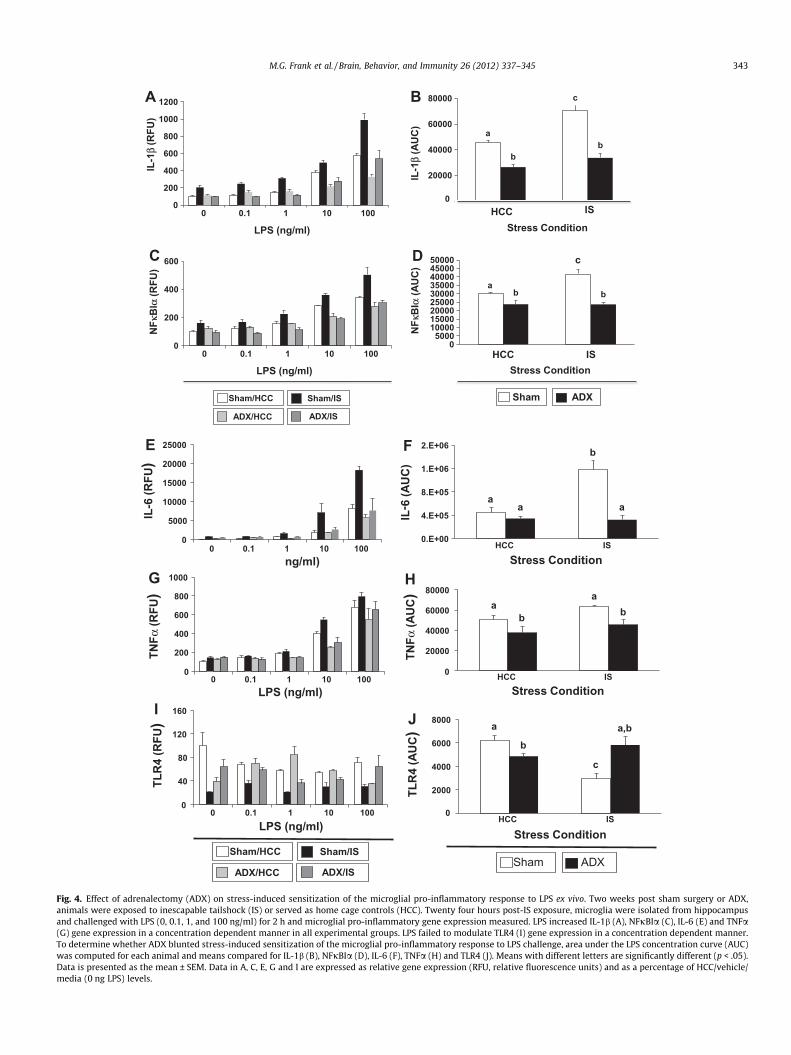
LPS increased IL-1b (Fig. 4A), NFjBIa (Fig. 4C), IL-6 (Fig. 4E) andTNFa (Fig. 4G) gene expression in a concentration dependent man-ner in all experimental groups. LPS failed to modulate TLR4 (Fig. 4I)gene expression in a concentration dependent manner. To deter-mine whether ADX blunted stress-induced sensitization of themicroglial pro-inflammatory response to LPS challenge, area underthe LPS concentration curve (AUC) was computed for each subjectand the interaction between stress and ADX treatment determined.In sham surgery animals, IS significantly potentiated the microglialIL-1b response, which was completely blocked by ADX (Fig. 4B)(F = 6.92, df = 1, 12, p < .05). Independent of stress condition, ADXsignificantly suppressed IL-1b expression compared to sham/HCCanimals. A similar interaction between stress and ADX was ob-served for NFjBIa (Fig. 4D) (F = 9.94, df = 1, 12, p < .01). Again,independent of stress condition, ADX significantly suppressedNFjBIa expression compared to sham/HCC animals. In sham sur-gery animals, IS significantly potentiated the microglial IL-6 re-sponse, which was completely blocked by ADX (Fig. 4F)

0
5
10
15
20
25
30
35
40
HCC IS
Seru
m C
OR
T (μ
g/dl
)
Stress Treatment
sham surgeryADX b
a
a a
Fig. 3. Verification of the effect of adrenalectomy (ADX) on stress-induced serumcorticosterone (CORT) levels. Two weeks post sham surgery or ADX, animals wereexposed to inescapable tailshock (IS) or served as home cage controls (HCC).Twenty four hours post-stress exposure, serum CORT levels were measured inanimals concomitant with isolation of hippocampal microglia. Means with differentletters are significantly different (p < .05). Data is presented as the mean ± SEM.
342 M.G. Frank et al. / Brain, Behavior, and Immunity 26 (2012) 337–345
(F = 15.61, df = 1, 12, p < .01), however, ADX did not significantlysuppress IL-6 expression compared to sham/HCC animals. Theinteraction between stress and ADX was not significant for TNFa(Fig. 4H), although the main effect of ADX on TNFa expressionwas significant indicating that ADX reduced TNFa expression. Insham surgery animals, IS significantly reduced microglial TLR4 lev-els, which was completely blocked by ADX (Fig. 4J) (F = 20.19,df = 1, 12, p < .001). In HCC animals, ADX also resulted in a signifi-cant reduction in TLR4 expression compared to sham surgery.
4. Discussion
The data from the present set of experiments provide converg-ing evidence that GCs mediate stress-induced sensitization ofmicroglia to later pro-inflammatory challenges. Both pharmacolog-ical (RU486) and surgical (ADX) suppression of stress-induced GCsignaling blocked the stress-induced sensitization of the pro-inflammatory response (i.e. IL-1b) of microglia to a pro-inflamma-tory challenge. We have previously demonstrated that microgliaare a neuroimmune substrate for stress-induced potentiation ofCNS pro-inflammatory immune responses (Frank et al., 2007). A re-cent study by Wohleb et al. shows that repeated social defeat alsopotentiates the microglial pro-inflammatory response to LPSex vivo (Wohleb et al., 2011). The present results suggest thatmicroglia are a neuroimmune substrate of stress-induced GC ac-tion, which in turn ‘‘prime’’ microglia to over-respond to pro-inflammatory insults. These results are consistent with severalstudies showing that acute and chronic stressors sensitize theneuroinflammatory response to both peripheral and central immu-nologic challenges (de Pablos et al., 2006; Espinosa-Oliva et al.,2011; Frank et al., 2007; Johnson et al., 2002b, 2003, 2004; Munhozet al., 2006). It is important to note here that stress-induced poten-tiation of pro-inflammatory mediators does not necessarily trans-late into potentiation of behavioral phenotypes such as sicknessresponses because it is the balance of pro- and anti-inflammatorymediators that determines stress-induced behavioral outcomes.We have previously shown that exposure to a severe acute stressor
(IS) as used here potentiates the sickness response to LPS adminis-tered 24 h post-stress (Johnson et al., 2003). Interestingly, admin-istration of a single dose of GCs that mimics the GC response toIS (Fleshner et al., 1993) also potentiates the sickness response toLPS given 24 h post-stress (Hains et al., 2011). In addition, GCsare sufficient to sensitize the neuroinflammatory as well as themicroglial response to pro-inflammatory challenges (Frank et al.,2010; Munhoz et al., 2010).
In prior studies of GC mediation of stress-induced neuroinflam-matory sensitization (de Pablos et al., 2006; Espinosa-Oliva et al.,2011; Munhoz et al., 2006), the pro-inflammatory challenge (i.e.LPS) was administered in vivo, which necessarily convolutes theGC effects of the stressor with the GC effects of LPS. Stress-inducedGCs could modulate LPS-induced GC production (HPA axis;(Beishuizen and Thijs, 2003). Indeed, prior stressors such as ines-capable shock do potentiate the later GC increase produced byLPS (Johnson et al., 2002a). In addition, stress-induced GCs couldmodify later LPS-induced signaling at the receptor level (GR;(Oakley and Cidlowski, 2011), plasma binding proteins (corticoste-roid binding globulin; (Henley and Lightman, 2011), and/or meta-bolic enzymes (11b -hydroxysteroid dehydrogenase Type I and II;(Seckl, 1997). In the present study, because microglia were ex-posed to LPS ex vivo and so LPS could not itself produce a GC re-sponse, the GC effects of stress could be studied independent ofthe GC effects of LPS. Here LPS was used ex vivo to probe the sen-sitization ‘‘status’’ of microglia. LPS is considered a pathogen asso-ciated molecular pattern or motif common to gram-negativebacteria, which is recognized and signals through the pattern rec-ognition receptor TLR4 (Kawai and Akira, 2010). Importantly, TLR4is expressed by microglia (Carpentier et al., 2008). The observationthat stress-induced GCs sensitized the microglia response to LPSthus prompted us to explore whether stress-induced GCs modu-lated the expression of TLR4. Interestingly, exposure to acute stressdownregulated TLR4 gene expression and surgical suppression ofstress-induced GCs abrogated this effect of stress on TLR4 geneexpression. This finding was unexpected given that LPS signalingis mediated by TLR4. However, LPS signaling also requires addi-tional proteins. LPS typically must bind LPS-binding protein, whichis then bound by CD14 and delivered to TLR4 complexed with MD2(Kawai and Akira, 2010). It could be that stress-induced sensitiza-tion of microglia was mediated by stress-induced changes in one ormore of these ancillary LPS signaling proteins. Interestingly, re-peated social defeat stress up-regulated protein expression ofCD14 and TLR4 on microglia (Wohleb et al., 2011). Though specu-lative, one possible explanation for the present results is that thedownregulation of TLR4 mRNA is compensatory for increased pro-tein level. Further studies are warranted to investigate the effect ofstress and GCs on TLR4 protein levels.
Although the present results provide converging lines of evi-dence that GCs play a pivotal role in stress-induced priming ofmicroglial pro-inflammatory responses, ambiguities associatedwith the use of RU486 and ADX should be noted. Though a highlyeffective GR antagonist, RU486 also antagonizes the progesteronereceptor (Sarkar, 2002). Microglia express both GR and MR (Tanakaet al., 1997), however microglia do not express the progesteronereceptor (Sierra et al., 2008) suggesting that the effects of RU486observed here were mediated predominantly through the GR. Ofnote, we found that RU486 potentiated the LPS-induced increasein hippocampal IL-1b in HCC animals. This finding was unexpectedand not observed with NF-jBIa. The effect of RU486 on IL-1b couldbe due to the long half-life of RU486 (20–30 h)(Sarkar, 2002),which may have blocked LPS-induced CORT signaling in the hippo-campus, thereby ameliorating the anti-inflammatory effects ofLPS-induced CORT. The long half-life of RU486 is an additional lia-bility of this manipulation because of the potential effects on LPS-induced CORT responses 24 h post-stress. To obviate the liabilities
0
200
400
600
800
1000
1200
0 0.1 1 10 100
IL-1
β (R
FU)
NFκ
BIα
(RFU
)
IL-1
β(A
UC
)
0
200
400
600
0 0.1 1 10 100
NFκ
BIα
(AU
C)
05000
100001500020000250003000035000400004500050000
HCC IS
A
D
B
C
*
c
a
0
20000
40000
60000
80000
LPS (ng/ml)
LPS (ng/ml)
Stress Condition
Stress ConditionHCC IS
c
bb
Sham/HCC Sham/IS
ADX/HCC ADX/IS
Sham ADX
a
b b
0.E+00
4.E+05
8.E+05
1.E+06
2.E+06
HCC IS0
5000
10000
15000
20000
25000
0 0.1 1 10 100
0
200
400
600
800
1000
0 0.1 1 10 1000
20000
40000
60000
80000
HCC IS
0
40
80
120
160
0 0.1 1 10 100 0
2000
4000
6000
8000
HCC IS
IL-6
(RFU
)
IL- 6
(AU
C)
TNFα
(RFU
)
TNFα
(AU
C)
TLR
4 (R
FU)
TLR
4 (A
UC
)
E F
G H
I J
LPS (ng/ml)
ng/ml)
LPS (ng/ml)
Sham/HCC Sham/IS
ADX/HCC ADX/IS
Stress Condition
Stress Condition
Stress Condition
Sham ADX
aa
a
b
a
aa
b b
a
ba,b
c
Fig. 4. Effect of adrenalectomy (ADX) on stress-induced sensitization of the microglial pro-inflammatory response to LPS ex vivo. Two weeks post sham surgery or ADX,animals were exposed to inescapable tailshock (IS) or served as home cage controls (HCC). Twenty four hours post-IS exposure, microglia were isolated from hippocampusand challenged with LPS (0, 0.1, 1, and 100 ng/ml) for 2 h and microglial pro-inflammatory gene expression measured. LPS increased IL-1b (A), NFjBIa (C), IL-6 (E) and TNFa(G) gene expression in a concentration dependent manner in all experimental groups. LPS failed to modulate TLR4 (I) gene expression in a concentration dependent manner.To determine whether ADX blunted stress-induced sensitization of the microglial pro-inflammatory response to LPS challenge, area under the LPS concentration curve (AUC)was computed for each animal and means compared for IL-1b (B), NFjBIa (D), IL-6 (F), TNFa (H) and TLR4 (J). Means with different letters are significantly different (p < .05).Data is presented as the mean ± SEM. Data in A, C, E, G and I are expressed as relative gene expression (RFU, relative fluorescence units) and as a percentage of HCC/vehicle/media (0 ng LPS) levels.
M.G. Frank et al. / Brain, Behavior, and Immunity 26 (2012) 337–345 343

344 M.G. Frank et al. / Brain, Behavior, and Immunity 26 (2012) 337–345
associated with RU486, ADX was employed as an alternate con-verging strategy to modulate stress-induced GCs. ADX was aseffective as RU486 in abrogating the effect of stress on microgliasensitization. However, in addition to removing the predominantsource of endogenous GCs, ADX also removes a key source ofperipheral mineralocorticoids (aldosterone) and catecholamines(epinephrine). Aldosterone has been found to modulate microglialexpression of inducible nitric oxide synthase (Tanaka et al., 1997),however it is unknown whether aldosterone modulates neuroin-flammatory responses. ADX animals were supplemented with ba-sal replacement CORT in their drinking water, but not withaldosterone. Likewise, ADX also abrogates stress-induced releaseof catecholamines, which have been shown to play a pivotal rolein the effects of acute and chronic stress on central pro-inflamma-tory cytokines.
We have shown that a severe acute stressor as used here in-duces pro-inflammatory cytokines in the CNS (hippocampus,hypothalamus and cortex) within hours of stressor termination(O’Connor et al., 2003) and is blocked by peripheral administrationof b -adrenergic receptor antagonists (Johnson et al., 2005) sug-gesting that stress-induced catecholamines mediate, in part, theimmediate effects of stress on central pro-inflammatory cytokines.Further, peripheral injection of a b –adrenergic receptor agonistwas sufficient to induce central proinflammatory cytokines(Johnson et al., 2005). A subsequent study showed that central,but not peripheral b -adrenergic receptors mediate the effects ofacute stress on central pro-inflammatory cytokines (Johnsonet al., 2008). Similarly, Blandino et al. demonstrated that peripheraladministration of a b-adrenergic receptor antagonist or themicroglia inhibitor minocycline blocked the stress induced up-reg-ulation of hypothalamic IL-1b and CD14 (Blandino et al., 2009).Further, they demonstrated that CORT synthesis inhibition duringstress potentiated the immediate stress effects on central pro-inflammatory cytokines. These results are consistent with a priorstudy showing that adrenalectomy potentiates the central proin-flammatory cytokine response observed immediately after termi-nation of the stressor (Nguyen et al., 1998). While these studiesshow that catecholamines play a pivotal role in the immediate ef-fects of stress on central pro-inflammatory cytokines, a recentstudy by Wohleb et al. showed that peripheral administration ofa b-adrenergic receptor antagonist blocked stress-induced potenti-ation of the microglial pro-inflammatory response to LPS ex vivo(Wohleb et al., 2011). In addition, stress-induced up-regulation ofmicroglial CD14 and TLR4 protein was blocked by prior treatmentin vivo with a b-adrenergic receptor antagonist. Of note, microgliaexpress a- and b-adrenergic receptors (Mori et al., 2002). Taken to-gether, these studies suggest that catecholamines mediate, at leastin part, the pro-inflammatory effects of stress in the brain. How-ever, several studies have shown that catecholamines are sufficientto induce an anti-inflammatory immunophenotype in the CNS(Gleeson et al., 2010; McNamee et al., 2010a,b, 2009). An intriguingpossibility is that catecholamines and GCs may work in concert tointegrate stress-induced inflammatory immune responses. Indeed,a precedent for this possibility has been established in the field oflearning and memory, which has found instances where GCs canamplify the effects of catecholamines on LTP (Joels et al., 2009).
The present results are consistent with a growing body of evi-dence demonstrating the permissive effects of stress and GCs inneuroinflammatory processes (Sorrells and Sapolsky, 2007). Sev-eral key parameters are pivotal in shaping the permissive functionof stress and GCs. Of particular relevance to the present study, thetiming of stress exposure relative to inflammatory challenge ap-pears to play a pivotal role in whether a stressor is anti-inflamma-tory or pro-inflammatory. If a pro-inflammatory stimulus (LPS) isadministered immediately before stress exposure, stress exhibitsan anti-inflammatory effect, including inhibition of LPS-induced
increases in brain IL-1b (Goujon et al., 1995). However, if LPS isadministered after stress exposure, stress potentiates the neuroin-flammatory response to LPS (Johnson et al., 2002b). It isnoteworthy that severe acute stress as used in the present investi-gation induces a rapid (within minutes of stress onset) and pro-found increase in serum GCs (Fleshner et al., 1993) in theabsence of any pro-inflammatory challenge. A considerable litera-ture has attempted to understand the physiological function ofstress-induced GCs, in particular as they relate to host defensemechanisms (Munck and Naray-Fejes-Toth, 1994). It is problem-atic that GCs, which are universally considered anti-inflammatoryand immunosuppressive (Boumpas et al., 1993; De Bosscher et al.,2003), would be elevated during a fight/flight emergency, whenthe chances of injury and infection increase. Notably, repeated so-cial defeat stress increases the ability of splenic macrophages tokill bacteria (Bailey et al., 2007) suggesting that a fight/flight emer-gency may enhance an organism’s innate immune response toimmunological threats or dangers. An organism’s innate immuneresponse to pathogens involves coordinated and bidirectional com-munication between central and peripheral immune response sys-tems (Blalock, 2005). As observed in the CNS, stressors alsosensitize and potentiate the peripheral proinflammatory immuneresponse to proinflammatory stimuli (Bailey et al., 2007, 2009;Johnson et al., 2002b, 2003; Powell et al., 2009; Stark et al.,2001). Further, stress modulates the activation state of peripheralmacrophages. For example, social defeat stress upregulates theexpression of the pattern recognition receptor TLR4 (Bailey et al.,2007), which is an immunophenotype also induced in microgliaafter social defeat stress (Wohleb et al., 2011). Thus, stress-inducedhormones such as GCs and catecholamines may serve to integrateand facilitate the central and peripheral innate immune responseto exogenous danger (i.e. pathogens, injury).
Taken together, the present results support the notion thatexposure to an acute stressor or fight/flight emergency inducesGCs, which function, in part, to ‘‘prime’’ or alert the organism’sCNS innate system to potential ‘‘danger’’ signals. These signalsmay include exogenous pathogens and/or pathogen associatedmolecular patterns or endogenous danger signals such as alarmins,which can be released in response to a wide array of stimuli includ-ing sterile injury. The notion explored here is that stress-inducedGCs can function as an endocrine ‘‘warning signal’’ to the CNS in-nate immune system to induce a preparatory response (microglialsensitization) to subsequent proinflammatory stimuli, therebypotentiating an immune response to threats (i.e. injury and infec-tion), which are more likely to occur during a fight/flightemergency.
Conflict of interest statement
All authors declare that there are no conflicts of interest.
References
Altschul, S.F., Madden, T.L., Schaffer, A.A., Zhang, J., Zhang, Z., Miller, W., Lipman, D.J.,1997. Gapped BLAST and PSI-BLAST: a new generation of protein databasesearch programs. Nucleic Acids Res. 25, 3389–3402.
Anisman, H., 2009. Cascading effects of stressors and inflammatory immune systemactivation: implications for major depressive disorder. J. Psychiatr. Neurosci. 34,4–20.
Bailey, M.T., Engler, H., Powell, N.D., Padgett, D.A., Sheridan, J.F., 2007. Repeatedsocial defeat increases the bactericidal activity of splenic macrophages througha Toll-like receptor-dependent pathway. Am. J. Physiol. Regul. Integr. Comp.Physiol. 293, R1180–R1190.
Bailey, M.T., Kinsey, S.G., Padgett, D.A., Sheridan, J.F., Leblebicioglu, B., 2009. Socialstress enhances IL-1beta and TNF-alpha production by Porphyromonasgingivalis lipopolysaccharide-stimulated CD11b+ cells. Physiol. Behav. 98,351–358.
Beishuizen, A., Thijs, L.G., 2003. Endotoxin and the hypothalamo-pituitary-adrenal(HPA) axis. J. Endotoxin Res. 9, 3–24.

M.G. Frank et al. / Brain, Behavior, and Immunity 26 (2012) 337–345 345
Blalock, J.E., 2005. The immune system as the sixth sense. J. Intern. Med. 257, 126–138.
Blandino Jr., P., Barnum, C.J., Solomon, L.G., Larish, Y., Lankow, B.S., Deak, T., 2009.Gene expression changes in the hypothalamus provide evidence for regionally-selective changes in IL-1 and microglial markers after acute stress. Brain Behav.Immun. 23, 958–968.
Borda, J.T., Alvarez, X., Mohan, M., Hasegawa, A., Bernardino, A., Jean, S., Aye, P.,Lackner, A.A., 2008. CD163, a marker of perivascular macrophages, is up-regulated by microglia in simian immunodeficiency virus encephalitis afterhaptoglobin-hemoglobin complex stimulation and is suggestive of breakdownof the blood-brain barrier. Am. J. Pathol. 172, 725–737.
Boumpas, D.T., Chrousos, G.P., Wilder, R.L., Cupps, T.R., Balow, J.E., 1993.Glucocorticoid therapy for immune-mediated diseases: basic and clinicalcorrelates. Ann. Intern. Med. 119, 1198–1208.
Carpentier, P.A., Duncan, D.S., Miller, S.D., 2008. Glial toll-like receptor signaling incentral nervous system infection and autoimmunity. Brain Behav. Immun. 22,140–147.
De Bosscher, K., Vanden Berghe, W., Haegeman, G., 2003. The interplay between theglucocorticoid receptor and nuclear factor-kappaB or activator protein-1:molecular mechanisms for gene repression. Endocr. Rev. 24, 488–522.
de Pablos, R.M., Villaran, R.F., Arguelles, S., Herrera, A.J., Venero, J.L., Ayala, A., Cano,J., Machado, A., 2006. Stress increases vulnerability to inflammation in the ratprefrontal cortex. J. Neurosci. 26, 5709–5719.
Der-Avakian, A., Will, M.J., Bland, S.T., Deak, T., Nguyen, K.T., Schmid, M.J., Spencer,R.L., Watkins, L.R., Maier, S.F., 2005. Surgical and pharmacological suppressionof glucocorticoids prevents the enhancement of morphine conditioned placepreference by uncontrollable stress in rats. Psychopharmacology (Berl) 179,409–417.
Espinosa-Oliva, A.M., de Pablos, R.M., Villaran, R.F., Arguelles, S., Venero, J.L.,Machado, A., Cano, J., 2011. Stress is critical for LPS-induced activation ofmicroglia and damage in the rat hippocampus. Neurobiol. Aging 32, 85–102.
Fleshner, M., Watkins, L.R., Lockwood, L.L., Grahn, R.E., Gerhardt, G., Meaney, M.J.,Laudenslager, M.L., Maier, S.F., 1993. Blockade of the hypothalamic-pituitary-adrenal response to stress by intraventricular injection of dexamethasone: amethod for studying the stress-induced peripheral effects of glucocorticoids.Psychoneuroendocrinology 18, 251–263.
Frank, M.G., Baratta, M.V., Sprunger, D.B., Watkins, L.R., Maier, S.F., 2007. Microgliaserve as a neuroimmune substrate for stress-induced potentiation of CNS pro-inflammatory cytokine responses. Brain Behav. Immun. 21, 47–59.
Frank, M.G., Miguel, Z.D., Watkins, L.R., Maier, S.F., 2010. Prior exposure toglucocorticoids sensitizes the neuroinflammatory and peripheralinflammatory responses to E. coli lipopolysaccharide.. Brain Behav. Immun.24, 19–30.
Frank, M.G., Wieseler-Frank, J.L., Watkins, L.R., Maier, S.F., 2006. Rapid isolation ofhighly enriched and quiescent microglia from adult rat hippocampus:immunophenotypic and functional characteristics. J. Neurosci. Methods 151,121–130.
Gleeson, L.C., Ryan, K.J., Griffin, E.W., Connor, T.J., Harkin, A., 2010. The beta2-adrenoceptor agonist clenbuterol elicits neuroprotective, anti-inflammatoryand neurotrophic actions in the kainic acid model of excitotoxicity. Brain Behav.Immun. 24, 1354–1361.
Goujon, E., Parnet, P., Laye, S., Combe, C., Kelley, K.W., Dantzer, R., 1995. Stressdownregulates lipopolysaccharide-induced expression of proinflammatorycytokines in the spleen, pituitary, and brain of mice. Brain Behav. Immun. 9,292–303.
Hains, L.E., Loram, L.C., Taylor, F.R., Strand, K.A., Wieseler, J.L., Barrientos, R.M.,Young, J.J., Frank, M.G., Sobesky, J., Martin, T.J., Eisenach, J.C., Maier, S.F.,Johnson, J.D., Fleshner, M., Watkins, L.R., 2011. Prior laparotomy orcorticosterone potentiates lipopolysaccharide-induced fever and sicknessbehaviors. J. Neuroimmunol. 239, 53–60.
Henley, D.E., Lightman, S.L., 2011. New insights into corticosteroid-binding globulinand glucocorticoid delivery. Neuroscience 180, 1–8.
Holfelder, K., Schittenhelm, J., Trautmann, K., Haybaeck, J., Meyermann, R.,Beschorner, R., 2011. De novo expression of the hemoglobin scavengerreceptor CD163 by activated microglia is not associated with hemorrhages inhuman brain lesions. Histol. Histopathol. 26, 1007–1017.
Jacobson, L., Akana, S.F., Cascio, C.S., Shinsako, J., Dallman, M.F., 1988. Circadianvariations in plasma corticosterone permit normal termination ofadrenocorticotropin responses to stress. Endocrinology 122, 1343–1348.
Joels, M., Krugers, H.J., Lucassen, P.J., Karst, H., 2009. Corticosteroid effects oncellular physiology of limbic cells. Brain Res. 1293, 91–100.
Johnson, J.D., Campisi, J., Sharkey, C.M., Kennedy, S.L., Nickerson, M., Greenwood,B.N., Fleshner, M., 2005. Catecholamines mediate stress-induced increases inperipheral and central inflammatory cytokines. Neuroscience 135, 1295–1307.
Johnson, J.D., Cortez, V., Kennedy, S.L., Foley, T.E., Hanson 3rd, H., Fleshner, M., 2008.Role of central beta-adrenergic receptors in regulating proinflammatorycytokine responses to a peripheral bacterial challenge. Brain Behav. Immun.22, 1078–1086.
Johnson, J.D., O’Connor, K.A., Deak, T., Spencer, R.L., Watkins, L.R., Maier, S.F., 2002a.Prior stressor exposure primes the HPA axis. Psychoneuroendocrinology 27,353–365.
Johnson, J.D., O’Connor, K.A., Deak, T., Stark, M., Watkins, L.R., Maier, S.F., 2002b.Prior stressor exposure sensitizes LPS-induced cytokine production. BrainBehav. Immun. 16, 461–476.
Johnson, J.D., O’Connor, K.A., Hansen, M.K., Watkins, L.R., Maier, S.F., 2003. Effects ofprior stress on LPS-induced cytokine and sickness responses. Am. J. Physiol.Regul. Integr. Comp. Physiol. 284, R422–432.
Johnson, J.D., O’Connor, K.A., Watkins, L.R., Maier, S.F., 2004. The role of IL-1beta instress-induced sensitization of proinflammatory cytokine and corticosteroneresponses. Neuroscience 127, 569–577.
Kawai, T., Akira, S., 2010. The role of pattern-recognition receptors in innateimmunity: update on Toll-like receptors. Nat. Immunol. 11, 373–384.
McNamee, E.N., Griffin, E.W., Ryan, K.M., Ryan, K.J., Heffernan, S., Harkin, A., Connor,T.J., 2010a. Noradrenaline acting at beta-adrenoceptors induces expression ofIL-1beta and its negative regulators IL-1ra and IL-1RII, and drives an overallanti-inflammatory phenotype in rat cortex. Neuropharmacology 59, 37–48.
McNamee, E.N., Ryan, K.M., Griffin, E.W., Gonzalez-Reyes, R.E., Ryan, K.J., Harkin, A.,Connor, T.J., 2010b. Noradrenaline acting at central beta-adrenoceptors inducesinterleukin-10 and suppressor of cytokine signaling-3 expression in rat brain:implications for neurodegeneration. Brain Behav. Immun. 24, 660–671.
McNamee, E.N., Ryan, K.M., Kilroy, D., Connor, T.J., 2009. Noradrenaline induces IL-1ra and IL-1 type II receptor expression in primary glial cells and protectsagainst IL-1beta-induced neurotoxicity. Eur. J. Pharmacol. 626, 219–228.
Mori, K., Ozaki, E., Zhang, B., Yang, L., Yokoyama, A., Takeda, I., Maeda, N., Sakanaka,M., Tanaka, J., 2002. Effects of norepinephrine on rat cultured microglial cellsthat express alpha1, alpha2, beta1 and beta2 adrenergic receptors.Neuropharmacology 43, 1026–1034.
Munck, A., Naray-Fejes-Toth, A., 1994. Glucocorticoids and stress: permissive andsuppressive actions. Ann. N Y Acad. Sci. 746, 115–130 (discussion 131–113).
Munhoz, C.D., Lepsch, L.B., Kawamoto, E.M., Malta, M.B., Lima Lde, S., Avellar, M.C.,Sapolsky, R.M., Scavone, C., 2006. Chronic unpredictable stress exacerbateslipopolysaccharide-induced activation of nuclear factor-kappaB in the frontalcortex and hippocampus via glucocorticoid secretion. J. Neurosci. 26, 3813–3820.
Munhoz, C.D., Sorrells, S.F., Caso, J.R., Scavone, C., Sapolsky, R.M., 2010.Glucocorticoids exacerbate lipopolysaccharide-induced signaling in thefrontal cortex and hippocampus in a dose-dependent manner. J. Neurosci. 30,13690–13698.
Nair, A., Bonneau, R.H., 2006. Stress-induced elevation of glucocorticoids increasesmicroglia proliferation through NMDA receptor activation. J. Neuroimmunol.171, 72–85.
Nguyen, K.T., Deak, T., Owens, S.M., Kohno, T., Fleshner, M., Watkins, L.R., Maier, S.F.,1998. Exposure to acute stress induces brain interleukin-1beta protein in therat. J. Neurosci. 18, 2239–2246.
O’Connor, K.A., Johnson, J.D., Hansen, M.K., Wieseler Frank, J.L., Maksimova, E.,Watkins, L.R., Maier, S.F., 2003. Peripheral and central proinflammatorycytokine response to a severe acute stressor. Brain Res. 991, 123–132.
Oakley, R.H., Cidlowski, J.A., 2011. Cellular processing of the glucocorticoid receptorgene and protein: new mechanisms for generating tissue-specific actions ofglucocorticoids. J. Biol. Chem. 286, 3177–3184.
Powell, N.D., Bailey, M.T., Mays, J.W., Stiner-Jones, L.M., Hanke, M.L., Padgett, D.A.,Sheridan, J.F., 2009. Repeated social defeat activates dendritic cells andenhances Toll-like receptor dependent cytokine secretion. Brain Behav.Immun. 23, 225–231.
Sarkar, N.N., 2002. Mifepristone: bioavailability, pharmacokinetics and use-effectiveness. Eur. J. Obstet. Gynecol. Reprod. Biol. 101, 113–120.
Seckl, J.R., 1997. 11beta-Hydroxysteroid dehydrogenase in the brain: a novelregulator of glucocorticoid action? Front. Neuroendocrinol. 18, 49–99.
Sierra, A., Gottfried-Blackmore, A., Milner, T.A., McEwen, B.S., Bulloch, K., 2008.Steroid hormone receptor expression and function in microglia. Glia 56, 659–674.
Sorrells, S.F., Sapolsky, R.M., 2007. An inflammatory review of glucocorticoid actionsin the CNS. Brain Behav. Immun. 21, 259–272.
Stark, J.L., Avitsur, R., Padgett, D.A., Campbell, K.A., Beck, F.M., Sheridan, J.F., 2001.Social stress induces glucocorticoid resistance in macrophages. Am. J. Physiol.Regul. Integr. Comp. Physiol. 280, R1799–R1805.
Tanaka, J., Fujita, H., Matsuda, S., Toku, K., Sakanaka, M., Maeda, N., 1997.Glucocorticoid- and mineralocorticoid receptors in microglial cells: the tworeceptors mediate differential effects of corticosteroids. Glia 20, 23–37.
Tilders, F.J., Schmidt, E.D., 1999. Cross-sensitization between immune and non-immune stressors. A role in the etiology of depression? Adv. Exp. Med. Biol. 461,179–197.
Wohleb, E.S., Hanke, M.L., Corona, A.W., Powell, N.D., Stiner, L.M., Bailey, M.T.,Nelson, R.J., Godbout, J.P., Sheridan, J.F., 2011. Beta-Adrenergic receptorantagonism prevents anxiety-like behavior and microglial reactivity inducedby repeated social defeat. J. Neurosci. 31, 6277–6288.