genetics and epigenetics of aging and longevity
TRANSCRIPT
Review
www.landesbioscience.com Cell Cycle 1063
Cell Cycle 13:7, 1063–1077; April 1, 2014; © 2014 Landes Bioscience
Review
Introduction
During aging, vital bodily functions such as regeneration and reproduction slowly decline. As a result, the organism loses its ability to maintain homeostasis and becomes more susceptible to stress, diseases, and injuries. A loss of essential body functions leads to age-associated pathologies, which ultimately cause death.
Traditionally, there have been many theories of aging, proposing underlying mechanisms of how aging evolved. The major evolutionary theories of aging are the theory of programmed death,1-3 the mutation accumulation theory of aging,4,5 the antagonistic pleiotropic theory of aging,6 and the evolutionary maintenance (see ref. 7 for a review). Weisman initiated the theoretical approach to the evolution of aging, arguing that natural selection inheritably “programs” death to limit individual lifespan and to clear space for new generations. His view was challenged by Haldane, Medawar, and Williams, who proposed that aging is more stochastic then programmed, because the forces of natural selection diminish with adult age, most rapidly after the peak of reproduction. Hamilton published
theoretical work in 1966, deriving a mathematical equation that later became known as “Hamilton’s forces of natural selection” and showing that forces of natural selection indeed decline with age, which was later confirmed experimentally using Drosophila (see ref. 8 and references therein for a review). The mutation accumulation theory of aging postulates that the mechanism of aging evolved through the evolutionary accumulation of germinal mutations with small harmful effects, which do not appear until old age, and thus avoid the negative pressure of natural selection.4,5 The first theory that proposed gerontogenes was the theory of antagonistic pleiotropy.6 Williams postulated positive evolutionary selection of genes that have favorable effects in early life stages but adverse effects in late life (after reaching reproductive success). Indeed, now we know that mutations in many genes important for growth and development (e.g., PI3K, mTOR, see below) can prolong life of model organisms (yeasts, nematodes, flies, and mice). Disposable soma theory, a special case of the theory of antagonistic pleiotropy,9 predicts the existence of genes that control the redistribution of energy resources from body maintenance to growth and reproduction. According to this theory, repair of cellular damage requires energy, competing for energy needs with reproduction. Therefore, in favor to the growth and development conditions of existence, longevity-assurance genes reduce their activity or are temporarily turned off, and aging speed increases. As predicted by this theory, longevity assurance genes exist, as confirmed by experimental overexpression of some antioxidant, DNA-, protein-, and cellular-repair genes, which prolong the lifespan of model animals (fruit flies and mice).
Identification of dozens of genes with mutations that prolong life supports another evolutionary theory, “longevity program” theory.10-14 The longevity program could have arisen in the evolution so the organisms can survive in conditions of short-term extreme environmental stress (overheating, overcooling, overpopulation, reducing caloric intake). Under stress, the program allows the body to exceed its normal lifespan by entering “maintenance mode”. This is associated with such modifications as increased stress resistance, downregulation of the biosynthesis of structural proteins, suspension of growth, and reproduction. Indeed, the survival rate of offspring in circumstances of short-term adverse changes in the environment will be minimal, so it is to evolutionary advantage to reallocate resources to extended longevity of adults, which can start breeding after the
*Correspondence to: Alexey A Moskalev; Email: [email protected]: 02/07/2014; Accepted: 03/04/2014; Published Online: 03/06/2014http://dx.doi.org/10.4161/cc.28433
Genetics and epigenetics of aging and longevityAlexey A Moskalev1,2,3,*, Alexander M Aliper1,2, Zeljka Smit-McBride4, Anton Buzdin1,5,6,7, and Alex Zhavoronkov1,5,8
1Moscow institute of Physics and Technology; Moscow, Russian Federation; 2institute of Biology; Komi Science Center of Russian Academy of Sciences; Syktyvkar, Russian Federation; 3Syktyvkar State University; Syktyvkar, Russian Federation; 4Department of Ophthalmology and vision Science; School of Medicine; University of California
at Davis; Davis, CA USA; 5Federal Clinical Research Center of Pediatric Hematology, Oncology, and immunology; Moscow, Russian Federation; 6Shemyakin-Ovchinnikov institute of Bioorganic Chemistry; Moscow, Russian Federation; 7First Oncology Research and Advisory Center; Moscow, Russian Federation;
8The Biogerontology Research Foundation; London, UK
Keywords: aging, epigenetics, evolution, genetics, longevity
evolutionary theories of aging predict the existence of certain genes that provide selective advantage early in life with adverse effect on lifespan later in life (antagonistic pleiotropy theory) or longevity insurance genes (disposable soma theory). indeed, the study of human and animal genetics is gradually identifying new genes that increase lifespan when overexpressed or mutated: gerontogenes. Furthermore, genetic and epigenetic mechanisms are being identified that have a positive effect on longevity. The gerontogenes are classified as lifespan regulators, mediators, effectors, housekeeping genes, genes involved in mitochondrial function, and genes regulating cellular senescence and apoptosis. in this review we demonstrate that the majority of the genes as well as genetic and epigenetic mechanisms that are involved in regulation of longevity are highly interconnected and related to stress response.
1064 Cell Cycle volume 13 issue 7
improvement of the environmental conditions. For example, C. elegans is showing that genetic program by actively promoting longevity of adults at cold temperatures.15 Artificially induced pro-longevity mutations affect this program, so that individuals go into stress-resistant mode independently of the exogenous conditions. As we shall see, the analysis of large amount of experimental data shows that most of the molecular pathways of longevity are associated with increased stress tolerance.
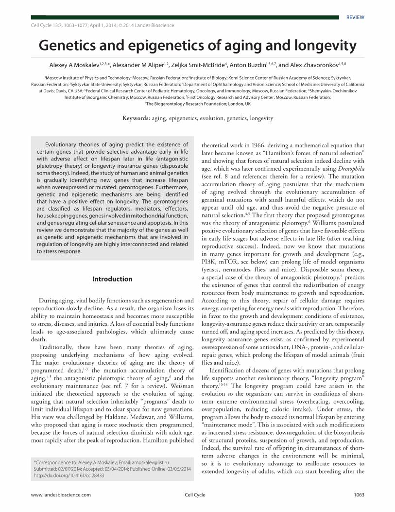
According to accumulation of the errors theory, aging has been viewed as a mechanical exhaustion and accumulation of errors. This model suggests that accidental errors and stress caused by environmental factors result in metabolic abnormalities, increase in free radical production and macromolecular damage at both cellular and tissue levels (Fig. 1).
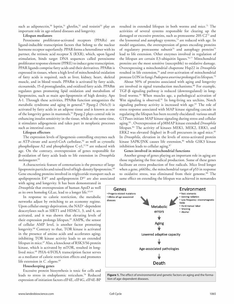
At the same time, it is known that moderate stress could have beneficial effects stimulating innate defense resources of the body, thereby boosting its ability to cope with higher stress levels and slowing down aging.16,17 This is the so-called lifespan hormesis effect.18,19 For instance, in our experiments, we observed the role of DNA repair genes and heat shock protein genes in radiation hormesis in fruit flies.20-22 Moderate stress stimulates expression of genes responsible for stress-resistance promoting prevention or elimination of genetic errors, including the novel and spontaneous ones, thereby delaying the aging process (Fig. 2). On the other hand, prolonged or severe stress exposure exhausts the defense mechanisms, causing drastic accumulation of errors and physiological abnormalities, accelerating the process of aging (Fig. 2).
Aging research has undergone dramatic expansion in recent years, with the discovery of gerontological genes, or gerontogenes, members of conserved biological pathways across species that increase lifespan when overexpressed or mutated. This discovery led to a renewed interest in understanding how aging is regulated and opened up a new field developing pharmacological treatments that can extend healthy lifespan and slow down human aging process, pioneered by Cynthia Kenyon and Linda Partridge.23-27
Genetics of Aging and Longevity
Identification of gerontogenes—the genes controlling aging and longevity—typically involves model organisms to screen for mutant strains whose rate of aging differs significantly from that of a control group.
The two most efficient methods for identifying new genes are: (1) loss of function: lifespan increases when the gene is inactivated; (2) gain of function: lifespan increases in a mutant with an overexpressed candidate gene.
The phenotypic characteristics that are evaluated are increase in longevity, or emergence of functional aberrations associated with aging (e.g., the dynamics in behavioral responses, elevation of cellular levels of lipofuscin, etc.). In order to accelerate these studies, stress factors can be employed, typically a heat shock or oxidative stress, because stress resistance is frequently linked with life extension.28 Some of the genes, like LMNA, whose mutant version leads to decreased longevity, may be used to find clues for ameliorating age-related diseases.29 However, the most valuable
gerontogenes that may ultimately lead to prospective drug candidates for life extension are the genes whose overexpression or polymorphisms lead to increased longevity of the organism.
Using various model organisms, hundreds of genes whose activity was altered in long-lived mutants have been identified. The following signaling pathways are involved in regulating the aging process: insulin/IGF-1, PI3K, TOR, MAPK, AMPK, PKC, NF-κB, TGF-β, Notch and WNT. Under favorable environmental conditions, these signaling cascades control energy balance, cellular plasticity, and the mechanisms supporting homeostasis, growth, and reproduction.30 However, under harsh conditions, the hormonal stimulation of growth is blocked, while stress-resistance proteins are activated. These pathways are evolutionary conserved from invertebrate to mammals.31
Lifespan regulatorsThe most studied pathway in the aging field is the insulin-
like signaling pathway. Upon insulin-like growth factor (IGF-1) binding to its receptor, IGF-1 receptor (IGF-1R), the intracellular phosphoinositol-3-kinase (PI3K) is activated, leading to formation of the downstream intermediate phosphoinositide-3,4,5-triphosphate. The latter binds to 3-phosphoinositid-dependent kinase 1 (PDK-1), which, in turn, phosphorylates and activates the kinases Akt/PKB and SGK-1 that control regular growth processes in the cell. At the same time, the stress-resistance factors, such as FOXO transcriptional factor, are inactivated (see the ref. 28 for review).
It is known, that centenarians are more sensitive to insulin while maintaining low blood levels.32 Insulin-like signaling activity as well as the expression level of insulin-like peptides are reduced in long-lived nematodes, mice, and humans.33-36 Heterozygous mice and humans harboring mutation in a gene encoding receptor for IGF-1 live longer than usual.35,37 Mutations in genes encoding for substrates of insulin receptor 1 and 2 result in the extended lifespan in Drosophila and mouse.38-40 Mutations in genes encoding kinases PI3K, AKT/PKB, and PDK are associated with a prolonged life in animals.41-43 Activity of phosphatases such as PTEN, SHIP1, and SHIP2, counteracting the function of PI3K, also promote longevity.36 Insulin-like signaling inhibits the mechanisms of stress response regulated by FOXO transcription factor. FOXO activity, together with the activity of FOXO-dependent genes, including PEPCK, Hsps, and MnSod, results in life extension.44,45 Another FOXO-dependent gene, GADD45, when overexpressed, leads to a prolonged lifespan and stress resistance in Drosophila and is also associated with a number of age-dependent pathologies in humans.22,46,47
Mutation in a gene encoding kidney hormone Klotho leads to a shortened life in mice, while its overexpression promotes longevity. Klotho suppresses the effect of the insulin/IGF-1 signaling pathway, reinforcing the resistance to oxidative stress at the cellular and organismal levels, thereby promoting longevity.48
A characteristic feature of long-lived Drosophila with the reduced insulin signaling activity is high lipid level.49 Lipid metabolism is downregulated with time, leading to age-dependent diseases such as metabolic syndrome and atherosclerosis. Dyslipidemia is associated with altered activity in a number of genes. Hormones regulating lipid metabolism,
www.landesbioscience.com Cell Cycle 1065
such as adiponectin,50 leptin,51 ghrelin,52 and resistin53 play an important role in age-related diseases and longevity.
Lifespan mediatorsPeroxisome proliferator-activated receptors (PPARs) are
ligand-inducible transcription factors that belong to the nuclear hormone receptor superfamily. PPAR forms a heterodimer with its partner, the retinoic acid receptor X (RXR), which, upon ligand stimulation, binds target DNA sequences called peroxisome proliferator response element (PPRE) to induce gene transcription. PPAR ligands comprise fatty acids and their derivatives. PPARα is expressed in tissues, where a high level of mitochondrial oxidation of fatty acids is required, such as liver, kidney, heart, skeletal muscle, and in blood vessels. PPARα is activated by fatty acids, eicosanoids, 15-d prostaglandin, and oxidized fatty acids. PPARα regulates genes promoting lipid oxidation and metabolism of lipoproteins, such as main apo-lipoprotein of high density, Apo A-1. Through these activities, PPARα function antagonizes the metabolic syndrome and aging in general.54 Pparg-2 (Nr1c3) is activated by fatty acids in an adipose tissue and is known as one of the longevity genes in mammals.55 Pparg-2 plays central role in enhancing insulin sensitivity in the tissue, while at the same time, it stimulates adipogenesis and takes part in neoplastic processes such as intestinal cancer.
Lifespan effectorsThe expression levels of lipogenesis controlling enzymes such
as ATP-citrate and acetyl-CoA carbolase,56 as well as cytosolic phospholipase A2 and phospholipase C-y1,57,58 are reduced with age. On the contrary, overexpression of genes responsible for β-oxidation of fatty acids leads to life extension in Drosophila melanogaster.59
A characteristic feature of centenarians is the presence of large lipoprotein particles and raised level of high-density lipoproteins.60 Gene encoding proteins involved in triglyceride transport such as apolipoprotein E461 and apolipoprotein D62 are also associated with aging and longevity. It has been demonstrated in Drosophila that overexpression of human ApoD as well as its own homolog GLaz, lead to a longer life.63,64
In response to calorie restriction, the metabolic networks adjust by switching to an economy regime. Upon cellular energy deprivation, the NAD+-dependent deacetylases such as SIRT1 and HDAC1, 3, and 4, are activated, and it was shown that elevating levels of their expression prolongs lifespan.65 AMPK, the sensor of cellular AMP level, is another factor promoting longevity.66 Contrary to that, TOR kinase is activated in the presence of amino acids and accelerates aging; inhibiting TOR kinase activity leads to an extended lifespan in mice.67 Also, a knockout of RSK3/S6 protein kinase, which is activated by mTOR, resulted in long-lived mice.68 PHA-4/FOXA transcription factor serves as a mediator of calorie restriction effects and promotes life extension in C. elegans.69
Housekeeping genesExcessive protein biosynthesis is toxic for cells and
leads to stress in endoplasmic reticulum.70 Reduced expression of initiation factors eIF4E, eIF4G, eIF4E-BP
resulted in extended lifespan in both worms and mice.71 The activities of several systems responsible for clearing up the damaged or excessive proteins, such as proteasome 20S C272 and the lysosomal and autophagy systems,73 are reduced with age. In model organisms, the overexpression of genes encoding proteins of regulatory proteasome subunit74 and autophagy proteins75 lead to life extension. Other enzymes involved in regulation of the lifespan are certain E3-ubiquitin ligases.76,77 Mitochondrial proteins are the most sensitive (susceptible) to oxidative damage. Overexpressing a mitochondrial chaperone Hsp22 in Drosophila resulted in life extension,78 and over-activation of mitochondrial protease LON in fungi Podospora anserina prolonged its lifespan.79
About 50% of proteins associated with aging and longevity are involved in signal transduction mechanisms.80 For example, TGF-β signaling pathway is reduced (downregulated) in long-lived worms.34 When muscles age, a pathological activation of Wnt signaling is observed.81 In long-living sea urchins, Notch signaling pathway activity is increased with age.82 The role of stress response associated with MAP kinase signaling cascade in regulating the lifespan has been recently elucidated: various small GTPases initiate MAP kinase signaling during stress and cellular aging.83 . Overexpression of p38MAP kinase extended Drosophila lifespan.84 The activity of kinases MEK1, MEK2, ERK1, and ERK2 was elevated (higher) in B-cell precursors in aged mice.85 In Drosophila, elevation in the levels of stress-activated protein kinase SAPK/JNK causes life extension,86 while GSK3 kinase inhibition leads to cellular aging.87
Genes involved in mitochondrial functionsAnother group of genes playing an important role in aging are
those regulating the free radical production. Some of these genes facilitate an extra production of free radicals. Mice lived longer when a gene, p66Shc, the mitochondrial target of p53 in response to oxidative stress, was eliminated from their genome.88 The same effect on extending the lifespan was achieved in nematodes
Figure 1. The effect of environmental and genetic factors on aging and the forma-tion of age-dependent diseases.
1066 Cell Cycle volume 13 issue 7
carrying a mutation in Clk-1 gene regulating the biosynthesis of a component from electron transport chain in mitochondria and an antioxidant ubiquinone, as well as in mice heterozygous for the same gene.89 Mitochondrial uncoupling proteins UCP-1, -2, and -3 reduce the formation of active oxygen species in mitochondria.90 Oxidative stress sensors VDAC1 and VDAC3 play role in lifespan in different organisms.91
Enhanced activity in a number of proteins involved in antioxidant protection has also been proposed to promote longevity. In a course of cellular response to the oxidative stress through MAP kinase-signaling cascade, SKN-1 transcription factor is activated. SKN-1 activity is elevated in long-lived nematodes, mice, and flies.92 When genes encoding for peroxyredoxin II (Jafrac 1) and peroxyredoxin 5 (dPrx5), which are responsible for controlling peroxide levels in a cell, were overexpressed in Drosophila, the flies lived longer.93,94 Overexpression of Mn-SOD is also beneficial for life extension in flies and mice in number of cases.95 Overexpressing Cu/Zn SOD in neurons extends life in Drosophila.96 Transgenic mice carrying a copy of mitochondrial catalase have shown delayed changes in aging markers in the heart in rodents.97
Genes regulating cellular senescence and apoptosisIn humans, a large number of genes undergo change in
their expression with aging (Fig. 2). Some of these genes are downregulated as growth and development slow down, while other genes become activated in the course of pro-inflammatory and stress responses, which arise due to accumulation of damage and errors at the levels of cells and tissues.
Epigenetics of Aging and Longevity
Epigenetic marks on DNA and chromosomesOne of the main reasons for change in gene expression during
aging is epigenetic regulation, which includes alterations in
the methylated states of regulatory DNA sequences, covalent modifications of histone proteins, and the expression of regulatory non-coding RNAs. Epigenetic theory of aging is a rapidly developing modern concept postulating that non-adaptive epigenetic alterations are fundamental to aging. It is well established that epimutations accumulate with age, leading to activation of genes normally downregulated epigenetically.98,99 Genetically identical twins, as they age, exhibit significant differences in genome methylation pattern, leading to differences in gene expression and, ultimately, lifespan.100,101 Variations in epigenetic markers among different cells within the same tissue of an organism are increased with age.102 A global demethylation of DNA sequence repeats, such as mobile genetic elements, occurs with aging,103 as well as the local hypermethylation of promoters of genes transcribed by RNA polymerase II, such as rRNA.104,105 Senescence is accompanied by the formation of nuclear regions called senescence-associated heterochromatin foci (SAHF). These foci are determined by the recruitment of heterochromatin proteins and Rb protein to E2F-dependent promoters of proliferative genes, leading to the repression of E2F target genes.106 During aging, the activities of methyltransferases DNMT1 and DNMT3a107 as well as deacetylase SIRT1108 are reduced, while the activities of histone demethylases Jmjd3109 and Jarid1b110 are enhanced. These changes result in non-adaptive alterations of epigenetic landscape, thereby changing gene expression and leading to aging.
Non-coding RNANon-coding RNAs include small RNAs, such as microRNAs
and piwi-interacting RNAs, and a wide range of long non-coding RNAs (lnc RNAs).
MicroRNAThe aging process has become a potentially important
target in cancer therapy after realization that cancer cells can be induce to undergo aging-type responses under stress of
chemotherapeutics.111 In a search of appropriate age-related biomarkers, the role of microRNA (miRNA) in induction, regulation, and fine-tuning of the aging process has been discovered.112 miRNAs represent a class of small RNAs that play very important roles in various biological processes in health and in the development of human diseases through specific posttranscriptional downregulation of gene expression. One of the microRNAs, miR-34a, has been designated as an aging marker in several tissues and system. Boon et al. has shown that miR-34a is upregulated in the aging heart, and that miR-34a inhibition reduces cell death and fibrosis following acute myocardial infarction.113 The results of Boon et al. identified miR-34a and its target PNUTS as a key mechanism that regulates cardiac contractile function during aging by inducing DNA damage responses and telomere attrition. Klotho is an anti-aging protein in mice that regulates pathways classically associated with longevity, such as insulin/IGF-1 and Wnt signaling. Protein expression of Klotho decreases in normal aging of mice. In silico analysis has identified miRNA-339 and miRNA-556 to bind to 3′ Figure 2. Stresses of various magnitudes affect aging rate and lifespan through dif-
ferent mechanisms.
www.landesbioscience.com Cell Cycle 1067
untranslated region of Klotho mRNA. In vitro results confirmed that these miRNAs can directly decrease Klotho protein expression, indicating that these miRNAs might be playing a role in age-related downregulation of Klotho mRNA in vivo.114 In addition to intracellular miRNAs, there is a novel category of circulatory miRNAs that can be considered as a completely new intercellular and system level communication. Accumulated evidence suggests that circulatory miRNAs can exert 2 opposite roles, activating as well as inhibiting inflammatory pathways (inflamma-miRs). Several of the circulatory miRNAs seem to be common for the major age-related diseases that share a chronic, low-level proinflammatory status, such as cardiovascular disease, type 2 diabetes mellitus, Alzheimer disease, rheumatoid arthritis, and cancer.115
Long noncoding RNAsThe role of long non-coding RNAs (lncRNAs) in aging has
been suggested in the work of Chang et al., in which he was studying gene expression changes of aged and rejuvenated human skin. He found that skin aging was associated with a significantly altered expression level of 2265 coding and noncoding RNAs,
of which 1293 became “rejuvenated” after broadband light treatment. Rejuvenated genes (RGs) included several known key regulators of organismal longevity and their proximal long noncoding RNAs.116 Abdelmohsen et al. described identification of senescence-associated long non-coding RNAs (SAL-RNAs). He looked at the lncRNAs that are differentially expressed during replicative senescence of human diploid WI-38 fibroblasts by RNA-seq. SAL-RNA1 (XLOC_023166) has been identified as putative age-delaying lncRNA, since its reduction with small inhibitory RNAs (siRNA) induced rapid aging changes of the fibroblasts, such as large cell morphology, positive β-galactosidase activity, and upregulation of p53.117
Pathway Analysis
The longevity genes described in this paper were separated into categories using Gene Ontology (GO), and their interactions were analyzed using GeneGo Metacore.
Most of the longevity genes described are related to stress response. The major regulatory hubs in stress response were
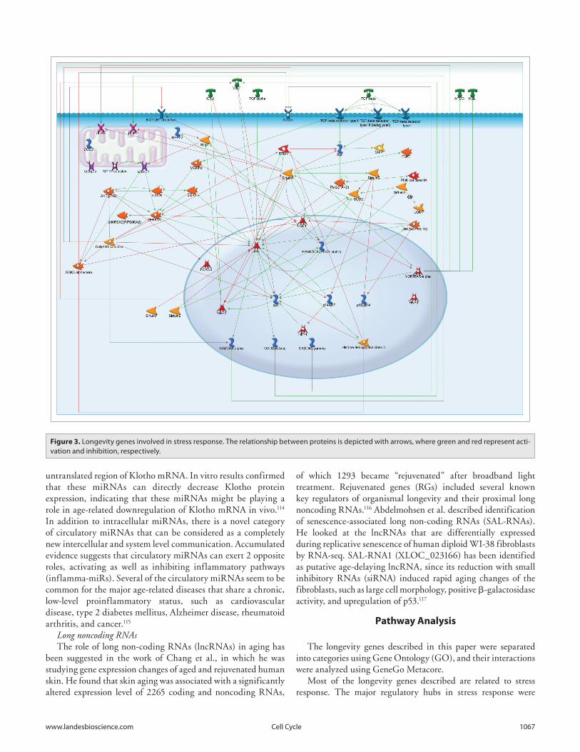
Figure 3. Longevity genes involved in stress response. The relationship between proteins is depicted with arrows, where green and red represent acti-vation and inhibition, respectively.
1068 Cell Cycle volume 13 issue 7
P53, Sirtuin 1, P21, HSF1, and the CoREST and VDR/RXR-α complexes (Fig. 3).
The VDR/RXR-α complex, a complex including over 20 elements, mainly PPAR and RXR, upregulates many proteins in the GADD45 family, P21, APOA1, APOD, WNT 4 UCP2, and UCP3. On the contrary, all of the interactions of the CoREST complex are downregulatory. It downregulates GADD45 α, P53, P21, ERK1/2, PTEN, AKT (PKB), GSK3 α/β complex, Notch pre-cursor, and NOTCH.
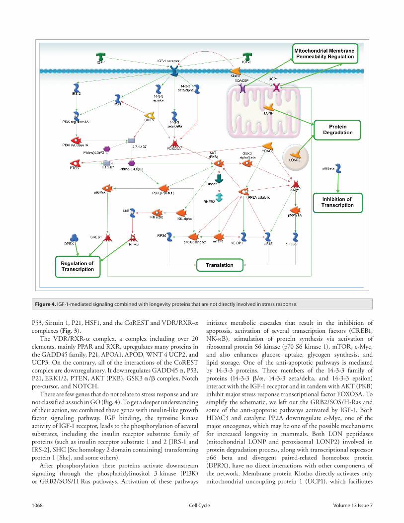
There are few genes that do not relate to stress response and are not classified as such in GO (Fig. 4). To get a deeper understanding of their action, we combined these genes with insulin-like growth factor signaling pathway. IGF binding, the tyrosine kinase activity of IGF-1 receptor, leads to the phosphorylation of several substrates, including the insulin receptor substrate family of proteins (such as insulin receptor substrate 1 and 2 [IRS-1 and IRS-2], SHC [Src homology 2 domain containing] transforming protein 1 [Shc], and some others).
After phosphorylation these proteins activate downstream signaling through the phosphatidylinositol 3-kinase (PI3K) or GRB2/SOS/H-Ras pathways. Activation of these pathways
initiates metabolic cascades that result in the inhibition of apoptosis, activation of several transcription factors (CREB1, NK-κB), stimulation of protein synthesis via activation of ribosomal protein S6 kinase (p70 S6 kinase 1), mTOR, c-Myc, and also enhances glucose uptake, glycogen synthesis, and lipid storage. One of the anti-apoptotic pathways is mediated by 14-3-3 proteins. Three members of the 14-3-3 family of proteins (14-3-3 β/α, 14-3-3 zeta/delta, and 14-3-3 epsilon) interact with the IGF-1 receptor and in tandem with AKT (PKB) inhibit major stress response transcriptional factor FOXO3A. To simplify the schematic, we left out the GRB2/SOS/H-Ras and some of the anti-apoptotic pathways activated by IGF-1. Both HDAC3 and catalytic PP2A downregulate c-Myc, one of the major oncogenes, which may be one of the possible mechanisms for increased longevity in mammals. Both LON peptidases (mitochondrial LONP and peroxisomal LONP2) involved in protein degradation process, along with transcriptional repressor p66 beta and divergent paired-related homeobox protein (DPRX), have no direct interactions with other components of the network. Membrane protein Klotho directly activates only mitochondrial uncoupling protein 1 (UCP1), which facilitates
Figure 4. iGF-1-mediated signaling combined with longevity proteins that are not directly involved in stress response.
www.landesbioscience.com Cell Cycle 1069
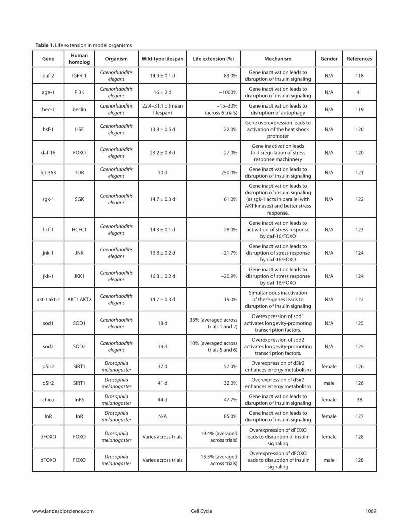
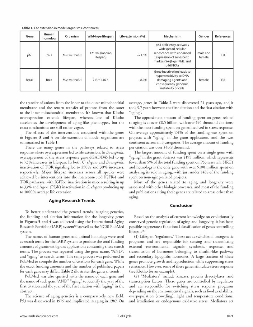
Table 1. Life extension in model organisms (continued)
GeneHuman
homologOrganism Wild-type lifespan Life extension (%) Mechanism Gender References
daf-2 iGFR-1Caenorhabditis
elegans14.9 ± 0.1 d 83.0%
Gene inactivation leads to disruption of insulin signaling
N/A 118
age-1 Pi3KCaenorhabditis
elegans16 ± 2 d ~1000%
Gene inactivation leads to disruption of insulin signaling
N/A 41
bec-1 beclinCaenorhabditis
elegans22.4–31.1 d (mean
lifespan)−15–30%
(across 6 trials)Gene inactivation leads to disruption of autophagy
N/A 119
hsf-1 HSFCaenorhabditis
elegans13.8 ± 0.5 d 22.0%
Gene overexpression leads to activation of the heat shock
promoterN/A 120
daf-16 FOXOCaenorhabditis
elegans23.2 ± 0.8 d −27.0%
Gene inactivation leads to disregulation of stress
response machinneryN/A 120
let-363 TORCaenorhabditis
elegans10 d 250.0%
Gene inactivation leads to disruption of insulin signaling
N/A 121
sgk-1 SGKCaenorhabditis
elegans14.7 ± 0.3 d 61.0%
Gene inactivation leads to disruption of insulin signaling (as sgk-1 acts in parallel with
AKT kinases) and better stress response.
N/A 122
hcf-1 HCFC1Caenorhabditis
elegans14.3 ± 0.1 d 28.0%
Gene inactivation leads to activation of stress response
by daf-16/FOXON/A 123
jnk-1 JNKCaenorhabditis
elegans16.8 ± 0.2 d −21.7%
Gene inactivation leads to disruption of stress response
by daf-16/FOXON/A 124
jkk-1 JKK1Caenorhabditis
elegans16.8 ± 0.2 d −20.9%
Gene inactivation leads to disruption of stress response
by daf-16/FOXON/A 124
akt-1 akt-2 AKT1 AKT2Caenorhabditis
elegans14.7 ± 0.3 d 19.0%
Simultaneous inactivation of these genes leads to
disruption of insulin signalingN/A 122
sod1 SOD1Caenorhabditis
elegans18 d
33% (averaged across trials 1 and 2)
Overexpression of sod1 activates longevity-promoting
transcription factors.N/A 125
sod2 SOD2Caenorhabditis
elegans19 d
10% (averaged across trials 5 and 6)
Overexpression of sod2 activates longevity-promoting
transcription factors.N/A 125
dSir2 SiRT1Drosophila
melanogaster37 d 57.0%
Overexpression of dSir2 enhances energy metabolism
female 126
dSir2 SiRT1Drosophila
melanogaster41 d 32.0%
Overexpression of dSir2 enhances energy metabolism
male 126
chico inRSDrosophila
melanogaster44 d 47.7%
Gene inactivation leads to disruption of insulin signaling
female 38
inR inRDrosophila
melanogasterN/A 85.0%
Gene inactivation leads to disruption of insulin signaling
female 127
dFOXO FOXODrosophila
melanogastervaries across trials
19.4% (averaged across trials)
Overexpression of dFOXO leads to disruption of insulin
signalingfemale 128
dFOXO FOXODrosophila
melanogastervaries across trials
15.5% (averaged across trials)
Overexpression of dFOXO leads to disruption of insulin
signalingmale 128
1070 Cell Cycle volume 13 issue 7
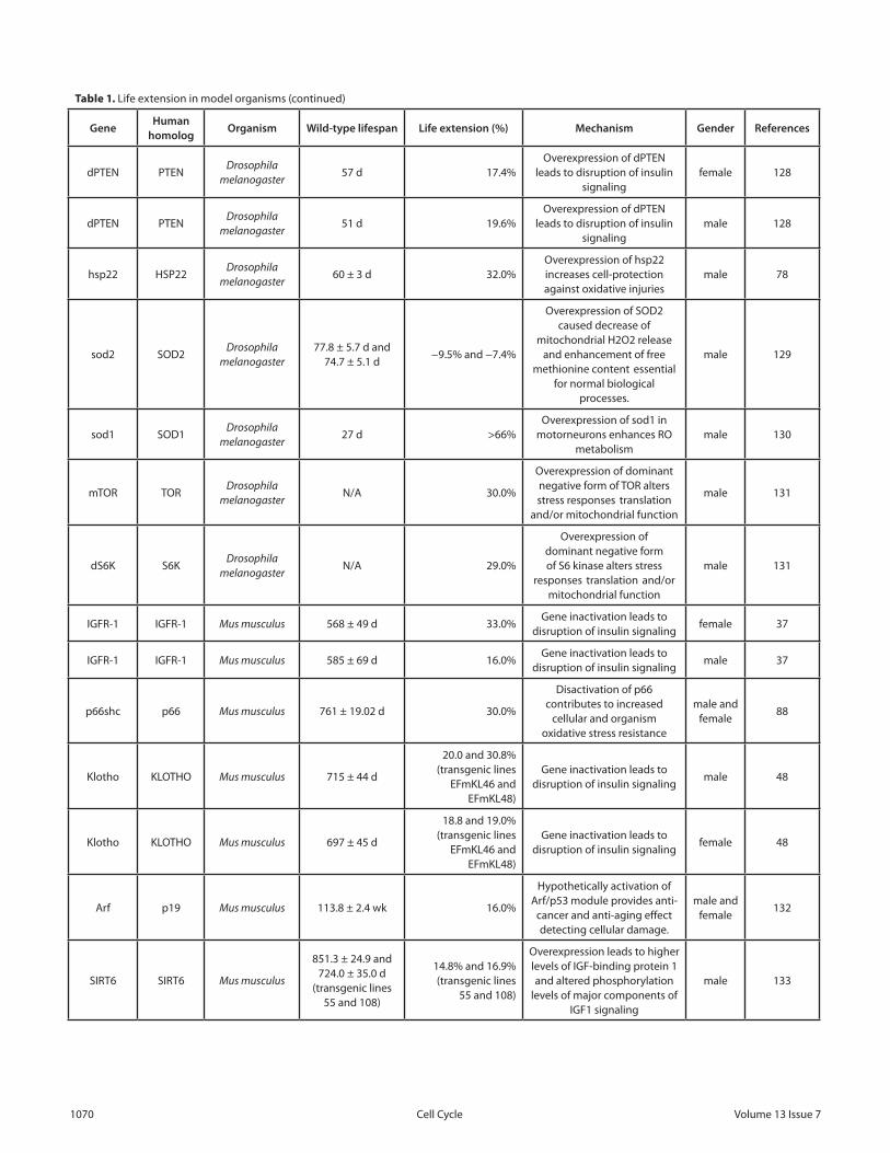
Table 1. Life extension in model organisms (continued)
GeneHuman
homologOrganism Wild-type lifespan Life extension (%) Mechanism Gender References
dPTeN PTeNDrosophila
melanogaster57 d 17.4%
Overexpression of dPTeN leads to disruption of insulin
signalingfemale 128
dPTeN PTeNDrosophila
melanogaster51 d 19.6%
Overexpression of dPTeN leads to disruption of insulin
signalingmale 128
hsp22 HSP22Drosophila
melanogaster60 ± 3 d 32.0%
Overexpression of hsp22 increases cell-protection against oxidative injuries
male 78
sod2 SOD2Drosophila
melanogaster77.8 ± 5.7 d and
74.7 ± 5.1 d−9.5% and −7.4%
Overexpression of SOD2 caused decrease of
mitochondrial H2O2 release and enhancement of free
methionine content essential for normal biological
processes.
male 129
sod1 SOD1Drosophila
melanogaster27 d >66%
Overexpression of sod1 in motorneurons enhances RO
metabolismmale 130
mTOR TORDrosophila
melanogasterN/A 30.0%
Overexpression of dominant negative form of TOR alters stress responses translation
and/or mitochondrial function
male 131
dS6K S6KDrosophila
melanogasterN/A 29.0%
Overexpression of dominant negative form of S6 kinase alters stress
responses translation and/or mitochondrial function
male 131
iGFR-1 iGFR-1 Mus musculus 568 ± 49 d 33.0%Gene inactivation leads to
disruption of insulin signalingfemale 37
iGFR-1 iGFR-1 Mus musculus 585 ± 69 d 16.0%Gene inactivation leads to
disruption of insulin signalingmale 37
p66shc p66 Mus musculus 761 ± 19.02 d 30.0%
Disactivation of p66 contributes to increased
cellular and organism oxidative stress resistance
male and female
88
Klotho KLOTHO Mus musculus 715 ± 44 d
20.0 and 30.8% (transgenic lines
eFmKL46 and eFmKL48)
Gene inactivation leads to disruption of insulin signaling
male 48
Klotho KLOTHO Mus musculus 697 ± 45 d
18.8 and 19.0% (transgenic lines
eFmKL46 and eFmKL48)
Gene inactivation leads to disruption of insulin signaling
female 48
Arf p19 Mus musculus 113.8 ± 2.4 wk 16.0%
Hypothetically activation of Arf/p53 module provides anti-
cancer and anti-aging effect detecting cellular damage.
male and female
132
SiRT6 SiRT6 Mus musculus
851.3 ± 24.9 and 724.0 ± 35.0 d
(transgenic lines 55 and 108)
14.8% and 16.9% (transgenic lines
55 and 108)
Overexpression leads to higher levels of iGF-binding protein 1 and altered phosphorylation
levels of major components of iGF1 signaling
male 133
www.landesbioscience.com Cell Cycle 1071
the transfer of anions from the inner to the outer mitochondrial membrane and the return transfer of protons from the outer to the inner mitochondrial membrane. It’s known that Klotho overexpression extends lifespan, whereas loss of Klotho accelerates the development of aging-like phenotypes, but the exact mechanisms are still rather vague.
The effects of the interventions associated with the genes in Figures 3 and 4 on life extension of model organisms are summarized in Table 1.
There are many genes in the pathways related to stress response where overexpression led to life extension. In Drosophila, overexpression of the stress response gene dGADD45 led to up to 73% increases in lifespan. In both C. elegans and Drosophila, inactivation of TOR signaling led to 250% and 30% increases, respectively. Major lifespan increases across all species were achieved by interventions into the interconnected IGFR-1 and TOR pathways, with IGFR-1 inactivation in mice resulting in up to 33% and Age-1 (PI3K) inactivation in C. elegans producing up to 1000% average life extension.
Aging Research Trends
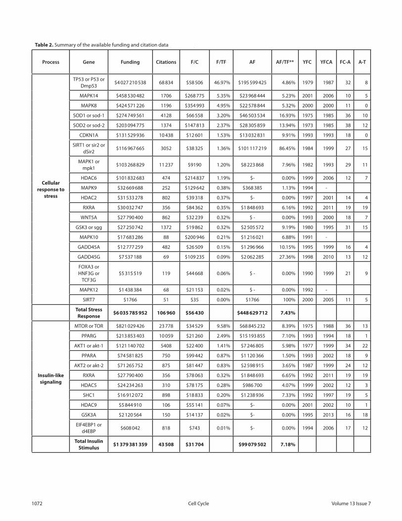
To better understand the general trends in aging genetics, the funding and citation information for the longevity genes in Figures 3 and 4 was collected using the International Aging Research Portfolio (IARP) system136 as well as the NCBI PubMed system.
The names of human genes and animal homologs were used as search terms for the IARP system to produce the total funding amounts of grants with grant applications containing these search terms. The process was repeated using the gene name, “AND”, and “aging” as search terms. The same process was performed in PubMed to compile the number of citations for each gene. While the exact funding amounts and the number of published papers for each gene may differ, Table 2 illustrates the general trends.
PubMed was also queried with the name of each gene and the name of each gene “AND” “aging” to identify the year of the first citation and the year of the first citation with “aging” in the abstract.
The science of aging genetics is a comparatively new field. P53 was discovered in 1979 and implicated in aging in 1987. On
average, genes in Table 2 were discovered 21 years ago, and it took 9.7 years between the first citation and the first citation with “aging”.
The approximate amount of funding spent on genes related to aging is at over $8.5 billion, with over 195 thousand citations, with the most funding spent on genes involved in stress response. On average approximately 7.4% of the funding was spent on projects with “aging” in the grant application, and this was consistent across all 3 categories. The average amount of funding per citation was over $43.9 thousand.
The largest amount of funding spent on a single gene with “aging” in the grant abstract was $195 million, which represents fewer than 5% of the total funding spent on P53 research. SIRT1 and homologs is the only gene with over $100 million spent on analyzing its role in aging, with just under 14% of the funding spent on non-aging-related projects.
Most of the genes related to aging and longevity were associated with other biologic processes, and most of the funding and publications citing these genes are related to areas other than aging.
Conclusion
Based on the analysis of current knowledge on evolutionarily conserved genetic regulation of aging and longevity, it has been possible to generate a functional classification of genes controlling lifespan137
(1) Lifespan “regulators.” These act as switches of ontogenetic programs and are responsible for sensing and transmitting external environmental signals: synthesis, response, and transmission of hormones belonging to insulin-like pathway and secondary lipophilic hormones. A large fraction of these genes promote growth and reproduction while suppressing stress resistance. However, some of these genes stimulate stress response (see Klotho for an example).
(2) “Mediators” include kinases, protein deacetylases, and transcription factors. These genes are controlled by regulators and are responsible for switching stress response programs depending on the environmental signals, such as food availability, overpopulation (crowding), light and temperature conditions, and irradiation or endogenous oxidative stress. Mediators act
Table 1. Life extension in model organisms (continued)
GeneHuman
homologOrganism Wild-type lifespan Life extension (%) Mechanism Gender References
p63 p63 Mus musculus121 wk (median
lifespan)−21.5%
p63 deficiency activates widespread cellular
senescence with enhanced expression of senescent
markers SA-β-gal PML and p16iNK4a
male and female
134
Brca1 Brca Mus musculus 713 ± 146 d −8.0%
Gene inactivation leads to hypersensitivity to DNA damaging agents and
consequently genomic instability of cells
female 135
1072 Cell Cycle volume 13 issue 7
Table 2. Summary of the available funding and citation data (continued)
Process Gene Funding Citations F/C F/TF AF AF/TF** YFC YFCA FC-A A-T
Cellular response to
stress
TP53 or P53 or Dmp53
$4 027 210 538 68 834 $58 506 46.97% $195 599 425 4.86% 1979 1987 32 8
MAPK14 $458 530 482 1706 $268 775 5.35% $23 968 444 5.23% 2001 2006 10 5
MAPK8 $424 571 226 1196 $354 993 4.95% $22 578 844 5.32% 2000 2000 11 0
SOD1 or sod-1 $274 749 561 4128 $66 558 3.20% $46 503 534 16.93% 1975 1985 36 10
SOD2 or sod-2 $203 094 775 1374 $147 813 2.37% $28 305 859 13.94% 1973 1985 38 12
CDKN1A $131 529 936 10 438 $12 601 1.53% $13 032 831 9.91% 1993 1993 18 0
SiRT1 or sir2 or dSir2
$116 967 665 3052 $38 325 1.36% $101 117 219 86.45% 1984 1999 27 15
MAPK1 or mpk1
$103 268 829 11 237 $9190 1.20% $8 223 868 7.96% 1982 1993 29 11
HDAC6 $101 832 683 474 $214 837 1.19% $- 0.00% 1999 2006 12 7
MAPK9 $32 669 688 252 $129 642 0.38% $368 385 1.13% 1994 -
HDAC2 $31 533 278 802 $39 318 0.37% $- 0.00% 1997 2001 14 4
RXRA $30 032 747 356 $84 362 0.35% $1 848 693 6.16% 1992 2011 19 19
wNT5A $27 790 400 862 $32 239 0.32% $ - 0.00% 1993 2000 18 7
GSK3 or sgg $27 250 742 1372 $19 862 0.32% $2 505 572 9.19% 1980 1995 31 15
MAPK10 $17 683 286 88 $200 946 0.21% $1 216 021 6.88% 1991 -
GADD45A $12 777 259 482 $26 509 0.15% $1 296 966 10.15% 1995 1999 16 4
GADD45G $7 537 188 69 $109 235 0.09% $2 062 285 27.36% 1998 2010 13 12
FOXA3 or HNF3G or
TCF3G$5 315 519 119 $44 668 0.06% $ - 0.00% 1990 1999 21 9
MAPK12 $1 438 384 68 $21 153 0.02% $ - 0.00% 1992 -
SiRT7 $1766 51 $35 0.00% $1766 100% 2000 2005 11 5
Total Stress Response
$6 035 785 952 106 960 $56 430 $448 629 712 7.43%
Insulin-like signaling
MTOR or TOR $821 029 426 23 778 $34 529 9.58% $68 845 232 8.39% 1975 1988 36 13
PPARG $213 853 403 10 059 $21 260 2.49% $15 193 855 7.10% 1993 1994 18 1
AKT1 or akt-1 $121 140 702 5408 $22 400 1.41% $7 246 805 5.98% 1977 1999 34 22
PPARA $74 581 825 750 $99 442 0.87% $1 120 366 1.50% 1993 2002 18 9
AKT2 or akt-2 $71 265 752 875 $81 447 0.83% $2 598 915 3.65% 1987 1999 24 12
RXRA $27 790 400 356 $78 063 0.32% $1 848 693 6.65% 1992 2011 19 19
HDAC5 $24 234 263 310 $78 175 0.28% $986 700 4.07% 1999 2002 12 3
SHC1 $16 912 072 898 $18 833 0.20% $1 238 936 7.33% 1992 1997 19 5
HDAC9 $5 844 910 106 $55 141 0.07% $- 0.00% 2001 2002 10 1
GSK3A $2 120 564 150 $14 137 0.02% $- 0.00% 1995 2013 16 18
eiF4eBP1 or d4eBP
$608 042 818 $743 0.01% $- 0.00% 1994 2006 17 12
Total Insulin Stimulus
$1 379 381 359 43 508 $31 704 $99 079 502 7.18%

www.landesbioscience.com Cell Cycle 1073
Table 2. Summary of the available funding and citation data (continued)
Process Gene Funding Citations F/C F/TF AF AF/TF** YFC YFCA FC-A A-T
Regulation of translation
MTOR or TOR $821 029 426 23 778 $34 529 9.58% $68 845 232 8.39% 1975 1988 36 13
AKT1 or akt-1 $121 140 702 5408 $22 400 1.41% $7 246 805 5.98% 1977 1999 34 22
MAPK1 $96 943 128 11 067 $8 760 1.13% $8 223 868 8.48% 1991 1993 20 2
eiF4e $69 771 228 2392 $29 169 0.81% $3 935 384 5.64% 1991 2001 20 10
PTK2B $44 532 289 298 $149 437 0.52% $- 0.00% 1995 2010 16 15
eiF4G1 or eiF4G
$3 998 998 970 $4123 0.05% $911 650 22.80% 1995 2001 16 6
eiF4eBP1 or d4eBP
$608 042 818 $743 0.01% $- 0.00% 1994 2006 17 12
Total Reg. of Translation
$1 158 023 813 44 731 $25 889 $89 162 939 7.70%
Total $8 573 191 124 195 199 $43 920 $636 872 153 7.43%
F/C, funding per citation; F/TF, funding for a specific gene as percentage of total funding; AF, funding for projects with the specific gene name and “aging” in the grant application; YFC, year of first citation; YFCA, year of first citation with “aging” in the abstract; FC-A, the time between first citation of the gene and citation with “aging”; A-T, the time between 2013 and the time of the first citation of the gene with “aging” in the abstract.
either as tissue-specific regulators of effector genes or directly controlling protein activity or lifetime. Mediators also interact among themselves, stimulating or inhibiting one another’s activity.
(3) “Effectors” are stress-resistance genes, including heat shock proteins, antioxidants, protein and DNA damage repair proteins, proteasome components, calpains, autophagy proteins, innate immunity, detoxification of xenobiotics, and metabolic regulators. Overexpression of these genes is usually correlated with extended lifespan. Often, the effectors act in additive manner, becoming activated by distinct “mediators” and extending lifespan under stress conditions. However, a number of “mediators” suppress “effectors” activity.
(4) Housekeeping genes. These act ubiquitously at every stage of life and are responsible for supporting cellular structure, respiration, synthesis of amino acids, lipids, nucleotides, etc. Mutations in these genes are either lethal or result in pathologies. Under stress conditions, some of the housekeeping genes are temporarily repressed by “mediators”, which allows saving energy and resources for “effectors” and extending lifespan.
(5) Genes involved in mitochondrial function. These are components of electron transport chain, Krebs cycle, uncoupling proteins, clk-1 gene in nematodes. These genes regulate energy metabolism, the level of free radicals, and also apoptosis.
(6) Genes regulating cellular senescence and apoptosis (p53, p21, p16, pRB). These genes are responsible for cancer prevention, cell cycle regulation, and elimination of extra or malignant cells during early ontogenesis and maturity. Cellular senescence (replicative or stress-induced) of dividing cells or excessive elimination of postmitotic cells is a pleiotropic side effect of aging.
How do all these new developments in the new science of aging and discovery of genes that drastically alter longevity fit in
with classical evolutionary theories? Which one is standing the test of time and new developments in the field of aging? At this point in time it appears that each of them is holding bits of truth, and each of them is explaining the evolution and mechanism of aging using dualistic principles (adaptive/nonadaptive, molecular/organismic, etc.). There is a newer theory proposed that offers an integrated theory of aging that helps us to better grasp similarity rather than differences among all these processes, the fractal theory of aging.138 The fractal theory is based, first, on the multilevel nature and complexity of aging, as well as self-similarity of those levels. Another important property of a fractal is a combination of stochastic and regular traits. The fractal principle of aging manifests in a combination of random (i.e., aging rates) and regular (i.e., sequence of geriatric changes) traits. Thus, according to this theory, aging can be defined as an age-dependent fractal increase in the number of deviations from homeostasis at the molecular, subcellular, cellular, tissue, and systemic levels. Actually, what would be highly desirable at this point in time is a unified theory of aging that would offer experimentally testable predictions. If we are able to mathematically describe the aging for one (e.g., cellular) level or one biological trait on a small interval of time, this model could be extrapolated to predict the aging at all other levels of organization of life, including individual lifespan. Substituting the model parameters with experimental measurements could lead to finding of biomarkers of aging rate and efficiency of anti-aging interventions.
Our pathway analysis shows that most of the gerontogenes are members of the stress response pathways and confirms the existence of genetics “longevity program”. As a rule, genes, regulators of the longevity program, which suppress mild stress response as well as mutations that make some of those pathways less efficient, provide life-extension benefits. Mild
1074 Cell Cycle volume 13 issue 7
References1. Weismann A. Über die Dauer des Lebens. G. Fisher:
1882.2. Weismann A. Essays Upon Heredity and Kindred
Biological Problems. Clarendon Press: Oxford, 1889.3. Weismann A. Über Leben und Tod. Verlag von
Gustav Fisher, Jena, Germany. Jena, Gustav Fischer: 1892.
4. Medawar PB. Old age and natural death. Modern Quarterly 1946; 1:30-56
5. Medawar PB. An Unsolved Problem of Biology. H.K.Lewis: London, 1952.
6. Williams GC. Pleiotropy, natural selection and the evolution of senescence. Evolution 1957; 11:398-411; http://dx.doi.org/10.2307/2406060
7. Gavrilov LA, Gavrilova NS. Evolutionary theories of aging and longevity. ScientificWorldJournal 2002; 2:339-56; PMID:12806021; http://dx.doi.org/10.1100/tsw.2002.96
8. Rose MR, Burke MK, Shahrestani P, Mueller LD. Evolution of ageing since Darwin. J Genet 2008; 87:363-71; PMID:19147926; http://dx.doi.org/10.1007/s12041-008-0059-6
9. Kirkwood TB. Evolution of ageing. Nature 1977; 270:301-4; PMID:593350; http://dx.doi.org/10.1038/270301a0
10. Guarente L, Kenyon C. Genetic pathways that regulate ageing in model organisms. Nature 2000; 408:255-62; PMID:11089983; http://dx.doi.org/10.1038/35041700
11. Lithgow GJ, White TM, Melov S, Johnson TE. Thermotolerance and extended lifespan conferred by single-gene mutations and induced by thermal stress. Proc Natl Acad Sci U S A 1995; 92:7540-4; PMID:7638227; http://dx.doi.org/10.1073/pnas.92.16.7540
12. Longo VD, Mitteldorf J, Skulachev VP. Programmed and altruistic ageing. Nat Rev Genet 2005; 6:866-72; PMID:16304601; http://dx.doi.org/10.1038/nrg1706
13. Murakami S, Johnson TE. A genetic pathway conferring life extension and resistance to UV stress in Caenorhabditis elegans. Genetics 1996; 143:1207-18; PMID:8807294
14. Partridge L, Gems D, Withers DJ. Sex and death: what is the connection? Cell 2005; 120:461-72; PMID:15734679; http://dx.doi.org/10.1016/j.cell.2005.01.026
15. Xiao R, Zhang B, Dong Y, Gong J, Xu T, Liu J, Xu XZ. A genetic program promotes C. elegans longevity at cold temperatures via a thermosensitive TRP channel. Cell 2013; 152:806-17; PMID:23415228; http://dx.doi.org/10.1016/j.cell.2013.01.020
16. Gaman L, Stoian I, Atanasiu V. Can ageing be slowed?: Hormetic and redox perspectives. J Med Life 2011; 4:346-51; PMID:22514565
17. Gems D, Partridge L. Stress-response hormesis and aging: “that which does not kill us makes us stronger”. Cell Metab 2008; 7:200-3; PMID:18316025; http://dx.doi.org/10.1016/j.cmet.2008.01.001
18. Cornelius C, Perrotta R, Graziano A, Calabrese EJ, Calabrese V. Stress responses, vitagenes and hormesis as critical determinants in aging and longevity: Mitochondria as a “chi”. Immun Ageing 2013; 10:15; PMID:23618527; http://dx.doi.org/10.1186/1742-4933-10-15
19. Rattan SI. Hormesis in aging. Ageing Res Rev 2008; 7:63-78; PMID:17964227; http://dx.doi.org/10.1016/j.arr.2007.03.002
20. Moskalev A. Radiation-induced lifespan alteration of Drosophila lines with genotype differences. Biogerontology 2007; 8:499-504; PMID:17380421; http://dx.doi.org/10.1007/s10522-007-9090-x
21. Moskalev AA, Plyusnina EN, Shaposhnikov MV. Radiation hormesis and radioadaptive response in Drosophila melanogaster f lies with different genetic backgrounds: the role of cellular stress-resistance mechanisms. Biogerontology 2011; 12:253-63; PMID:21234801; http://dx.doi.org/10.1007/s10522-011-9320-0
22. Plyusnina EN, Shaposhnikov MV, Moskalev AA. Increase of Drosophila melanogaster lifespan due to D-GADD45 overexpression in the nervous system. Biogerontology 2011; 12:211-26; PMID:21153055; http://dx.doi.org/10.1007/s10522-010-9311-6
23. López-Otín C, Blasco MA, Partridge L, Serrano M, Kroemer G. The hallmarks of aging. Cell 2013; 153:1194-217; PMID:23746838; http://dx.doi.org/10.1016/j.cell.2013.05.039
24. Partridge L, Thornton J, Bates G. The new science of ageing. Philos Trans R Soc Lond B Biol Sci 2011; 366:6-8; PMID:21115524; http://dx.doi.org/10.1098/rstb.2010.0298
25. Fontana L, Partridge L, Longo VD. Extending healthy lifespan--from yeast to humans. Science 2010; 328:321-6; PMID:20395504; http://dx.doi.org/10.1126/science.1172539
26. Kenyon CJ. The genetics of ageing. Nature 2010; 464:504-12; PMID:20336132; http://dx.doi.org/10.1038/nature08980
27. Partridge L. The new biology of ageing. Philos Trans R Soc Lond B Biol Sci 2010; 365:147-54; PMID:20008392; http://dx.doi.org/10.1098/rstb.2009.0222
28. Moskalev AA. [Prospective trends in genetics of aging and longevity]. Adv Gerontol 2009; 22:92-103; PMID:19827680
29. Zhavoronkov A, Smit-McBride Z, Guinan KJ, Litovchenko M, Moskalev A. Potential therapeutic approaches for modulating expression and accumulation of defective lamin A in laminopathies and age-related diseases. J Mol Med (Berl) 2012; 90:1361-89; PMID:23090008; http://dx.doi.org/10.1007/s00109-012-0962-4
30. Barzilai N, Huffman DM, Muzumdar RH, Bartke A. The critical role of metabolic pathways in aging. Diabetes 2012; 61:1315-22; PMID:22618766; http://dx.doi.org/10.2337/db11-1300
31. Kim SK. Common aging pathways in worms, f lies, mice and humans. J Exp Biol 2007; 210:1607-12; PMID:17449826; http://dx.doi.org/10.1242/jeb.004887
32. Cheng CL, Gao TQ, Wang Z, Li DD. Role of insulin/insulin-like growth factor 1 signaling pathway in longevity. World J Gastroenterol 2005; 11:1891-5; PMID:15800976
33. Bartke A. Insulin and aging. Cell Cycle 2008; 7:3338-43; PMID:18948730; http://dx.doi.org/10.4161/cc.7.21.7012
34. Parrella E, Longo VD. Insulin/IGF-I and related signaling pathways regulate aging in nondividing cells: from yeast to the mammalian brain. ScientificWorldJournal 2010; 10:161-77; PMID:20098959; http://dx.doi.org/10.1100/tsw.2010.8
35. Suh Y, Atzmon G, Cho MO, Hwang D, Liu B, Leahy DJ, Barzilai N, Cohen P. Functionally significant insulin-like growth factor I receptor mutations in centenarians. Proc Natl Acad Sci U S A 2008; 105:3438-42; PMID:18316725; http://dx.doi.org/10.1073/pnas.0705467105
36. Tazearslan C, Ayyadevara S, Bharill P, Shmookler Reis RJ. Positive feedback between transcriptional and kinase suppression in nematodes with extraordinary longevity and stress resistance. PLoS Genet 2009; 5:e1000452; PMID:19360094; http://dx.doi.org/10.1371/journal.pgen.1000452
37. Holzenberger M, Dupont J, Ducos B, Leneuve P, Géloën A, Even PC, Cervera P, Le Bouc Y. IGF-1 receptor regulates lifespan and resistance to oxidative stress in mice. Nature 2003; 421:182-7; PMID:12483226; http://dx.doi.org/10.1038/nature01298
38. Clancy DJ, Gems D, Harshman LG, Oldham S, Stocker H, Hafen E, Leevers SJ, Partridge L. Extension of lifespan by loss of CHICO, a Drosophila insulin receptor substrate protein. Science 2001; 292:104-6; PMID:11292874; http://dx.doi.org/10.1126/science.1057991
39. Selman C, Lingard S, Choudhury AI, Batterham RL, Claret M, Clements M, Ramadani F, Okkenhaug K, Schuster E, Blanc E, et al. Evidence for lifespan extension and delayed age-related biomarkers in insulin receptor substrate 1 null mice. FASEB J 2008; 22:807-18; PMID:17928362; http://dx.doi.org/10.1096/fj.07-9261com
40. Taguchi A, Wartschow LM, White MF. Brain IRS2 signaling coordinates lifespan and nutrient homeostasis. Science 2007; 317:369-72; PMID:17641201; http://dx.doi.org/10.1126/science.1142179
overexpression of effector longevity genes involved in stress response to DNA, protein, or other cellular damages (e.g., Hsps, Sod, GADD45, ATGs) prolongs lifespan. While moderate stress induces “longevity program” by stimulating expression of life-assurance genes and promoting prevention or elimination of errors, chronic or acute stress exposure exhausts the defense mechanisms and therefore accelerates aging. Pro-aging and anti-aging gene-determined processes exist on all levels of the organismal system—from molecules to systems (metabolic, endocrine, immune, inter-cellular communication). Their multi-level organization, the interpenetration of levels, a combination of regular and stochastic elements is what makes the process of aging a fractal process.
Disclosure of Potential Conflicts of Interest
No potential conflicts of interest were disclosed
Acknowledgments
We would like to thank the editorial team and the reviewers for their valuable advice and comments pertaining to the content of this manuscript and Brian Kennedy, Jan Vijg, Yousin Suh, Viktoria Lunyak, Mark Tatar, and Charles Cantor for productive discussions on the genetics and epigenetics of aging and longevity. We would like to thank the UMA Foundation for their help in preparation of this manuscript. The work was supported by Grant of RFBR 14-04-01596 and Grant of President of Russian Federation MD-1090.2014.4.
www.landesbioscience.com Cell Cycle 1075
41. Ayyadevara S, Alla R, Thaden JJ, Shmookler Reis RJ. Remarkable longevity and stress resistance of nematode PI3K-null mutants. Aging Cell 2008; 7:13-22; PMID:17996009; http://dx.doi.org/10.1111/j.1474-9726.2007.00348.x
42. Narasimhan SD, Yen K, Tissenbaum HA. Converging pathways in lifespan regulation. Curr Biol 2009; 19:R657-66; PMID:19674551; http://dx.doi.org/10.1016/j.cub.2009.06.013
43. Puig O, Tjian R. Transcriptional feedback control of insulin receptor by dFOXO/FOXO1. Genes Dev 2005; 19:2435-46; PMID:16230533; http://dx.doi.org/10.1101/gad.1340505
44. Hanson RW, Hakimi P. Born to run; the story of the PEPCK-Cmus mouse. Biochimie 2008; 90:838-42; PMID:18394430; http://dx.doi.org/10.1016/j.biochi.2008.03.009
45. Tower J. Hsps and aging. Trends Endocrinol Metab 2009; 20:216-22; PMID:19394247; http://dx.doi.org/10.1016/j.tem.2008.12.005
46. Moskalev A, Plyusnina E, Shaposhnikov M, Shilova L, Kazachenok A, Zhavoronkov A. The role of D-GADD45 in oxidative, thermal and genotoxic stress resistance. Cell Cycle 2012; 11:4222-41; PMID:23095639; http://dx.doi.org/10.4161/cc.22545
47. Moskalev AA, Smit-McBride Z, Shaposhnikov MV, Plyusnina EN, Zhavoronkov A, Budovsky A, Tacutu R, Fraifeld VE. Gadd45 proteins: relevance to aging, longevity and age-related pathologies. Ageing Res Rev 2012; 11:51-66; PMID:21986581; http://dx.doi.org/10.1016/j.arr.2011.09.003
48. Kurosu H, Yamamoto M, Clark JD, Pastor JV, Nandi A, Gurnani P, McGuinness OP, Chikuda H, Yamaguchi M, Kawaguchi H, et al. Suppression of aging in mice by the hormone Klotho. Science 2005; 309:1829-33; PMID:16123266; http://dx.doi.org/10.1126/science.1112766
49. Broughton SJ, Piper MD, Ikeya T, Bass TM, Jacobson J, Driege Y, Martinez P, Hafen E, Withers DJ, Leevers SJ, et al. Longer lifespan, altered metabolism, and stress resistance in Drosophila from ablation of cells making insulin-like ligands. Proc Natl Acad Sci U S A 2005; 102:3105-10; PMID:15708981; http://dx.doi.org/10.1073/pnas.0405775102
50. Atzmon G, Pollin TI, Crandall J, Tanner K, Schechter CB, Scherer PE, Rincon M, Siegel G, Katz M, Lipton RB, et al. Adiponectin levels and genotype: a potential regulator of lifespan in humans. J Gerontol A Biol Sci Med Sci 2008; 63:447-53; PMID:18511746; http://dx.doi.org/10.1093/gerona/63.5.447
51. Naito M, Fujikura J, Ebihara K, Miyanaga F, Yokoi H, Kusakabe T, Yamamoto Y, Son C, Mukoyama M, Hosoda K, et al. Therapeutic impact of leptin on diabetes, diabetic complications, and longevity in insulin-deficient diabetic mice. Diabetes 2011; 60:2265-73; PMID:21810600; http://dx.doi.org/10.2337/db10-1795
52. Albarran-Zeckler RG, Sun Y, Smith RG. Physiological roles revealed by ghrelin and ghrelin receptor deficient mice. Peptides 2011; 32:2229-35; PMID:21781995; http://dx.doi.org/10.1016/j.peptides.2011.07.003
53. Gharibeh MY, Al Tawallbeh GM, Abboud MM, Radaideh A, Alhader AA, Khabour OF. Correlation of plasma resistin with obesity and insulin resistance in type 2 diabetic patients. Diabetes Metab 2010; 36:443-9; PMID:20739208; http://dx.doi.org/10.1016/j.diabet.2010.05.003
54. Schiff M, Bénit P, Jacobs HT, Vockley J, Rustin P. Therapies in inborn errors of oxidative metabolism. Trends Endocrinol Metab 2012; 23:488-95; PMID:22633959; http://dx.doi.org/10.1016/j.tem.2012.04.006
55. Argmann C, Dobrin R, Heikkinen S, Auburtin A, Pouilly L, Cock TA, Koutnikova H, Zhu J, Schadt EE, Auwerx J. Ppargamma2 is a key driver of longevity in the mouse. PLoS Genet 2009; 5:e1000752; PMID:19997628; http://dx.doi.org/10.1371/journal.pgen.1000752
56. Nogalska A, Pankiewicz A, Goyke E, Swierczynski J. The age-related inverse relationship between ob and lipogenic enzymes genes expression in rat white adipose tissue. Exp Gerontol 2003; 38:415-22; PMID:12670628; http://dx.doi.org/10.1016/S0531-5565(02)00210-3
57. Gentili C, Morelli S, de Boland AR. PTH and phospholipase A2 in the aging process of intestinal cells. J Cell Biochem 2004; 93:312-26; PMID:15368358; http://dx.doi.org/10.1002/jcb.20158
58. Utsuyama M, Wakikawa A, Tamura T, Nariuchi H, Hirokawa K. Impairment of signal transduction in T cells from old mice. Mech Ageing Dev 1997; 93:131-44; PMID:9089578; http://dx.doi.org/10.1016/S0047-6374(96)01837-4
59. Lee SH, Lee SK, Paik D, Min KJ. Overexpression of fatty-acid-β-oxidation-related genes extends the lifespan of Drosophila melanogaster. Oxid Med Cell Longev 2012; 2012:854502; PMID:22997544; http://dx.doi.org/10.1155/2012/854502
60. Atzmon G, Rincon M, Schechter CB, Shuldiner AR, Lipton RB, Bergman A, Barzilai N. Lipoprotein genotype and conserved pathway for exceptional longevity in humans. PLoS Biol 2006; 4:e113; PMID:16602826; http://dx.doi.org/10.1371/journal.pbio.0040113
61. Bonomini F, Filippini F, Hayek T, Aviram M, Keidar S, Rodella LF, Coleman R, Rezzani R. Apolipoprotein E and its role in aging and survival. Exp Gerontol 2010; 45:149-57; PMID:19941948; http://dx.doi.org/10.1016/j.exger.2009.11.006
62. Muffat J, Walker DW, Apolipoprotein D. Apolipoprotein D: an overview of its role in aging and age-related diseases. Cell Cycle 2010; 9:269-73; PMID:20023409; http://dx.doi.org/10.4161/cc.9.2.10433
63. Muffat J, Walker DW, Benzer S, Human Apo D. Human ApoD, an apolipoprotein up-regulated in neurodegenerative diseases, extends lifespan and increases stress resistance in Drosophila. Proc Natl Acad Sci U S A 2008; 105:7088-93; PMID:18458334; http://dx.doi.org/10.1073/pnas.0800896105
64. Walker DW, Muffat J, Rundel C, Benzer S. Overexpression of a Drosophila homolog of apolipoprotein D leads to increased stress resistance and extended lifespan. Curr Biol 2006; 16:674-9; PMID:16581512; http://dx.doi.org/10.1016/j.cub.2006.01.057
65. Virtue S, Vidal-Puig A. It’s not how fat you are, it’s what you do with it that counts. PLoS Biol 2008; 6:e237; PMID:18816166; http://dx.doi.org/10.1371/journal.pbio.0060237
66. Greer EL, Banko MR, Brunet A. AMP-activated protein kinase and FoxO transcription factors in dietary restriction-induced longevity. Ann N Y Acad Sci 2009; 1170:688-92; PMID:19686213; http://dx.doi.org/10.1111/j.1749-6632.2009.04019.x
67. Harrison DE, Strong R, Sharp ZD, Nelson JF, Astle CM, Flurkey K, Nadon NL, Wilkinson JE, Frenkel K, Carter CS, et al. Rapamycin fed late in life extends lifespan in genetically heterogeneous mice. Nature 2009; 460:392-5; PMID:19587680
68. Selman C, Tullet JM, Wieser D, Irvine E, Lingard SJ, Choudhury AI, Claret M, Al-Qassab H, Carmignac D, Ramadani F, et al. Ribosomal protein S6 kinase 1 signaling regulates mammalian lifespan. Science 2009; 326:140-4; PMID:19797661; http://dx.doi.org/10.1126/science.1177221
69. Panowski SH, Wolff S, Aguilaniu H, Durieux J, Dillin A. PHA-4/Foxa mediates diet-restriction-induced longevity of C. elegans. Nature 2007; 447:550-5; PMID:17476212; http://dx.doi.org/10.1038/nature05837
70. Brown MK, Naidoo N. The endoplasmic reticulum stress response in aging and age-related diseases. Front Physiol 2012; 3:263; PMID:22934019; http://dx.doi.org/10.3389/fphys.2012.00263
71. Hipkiss AR. On why decreasing protein synthesis can increase lifespan. Mech Ageing Dev 2007; 128:412-4; PMID:17452047; http://dx.doi.org/10.1016/j.mad.2007.03.002
72. Rodriguez KA, Gaczynska M, Osmulski PA. Molecular mechanisms of proteasome plasticity in aging. Mech Ageing Dev 2010; 131:144-55; PMID:20080121; http://dx.doi.org/10.1016/j.mad.2010.01.002
73. Cuervo AM. Autophagy and aging: keeping that old broom working. Trends Genet 2008; 24:604-12; PMID:18992957; http://dx.doi.org/10.1016/j.tig.2008.10.002
74. Tonoki A, Kuranaga E, Tomioka T, Hamazaki J, Murata S, Tanaka K, Miura M. Genetic evidence linking age-dependent attenuation of the 26S proteasome with the aging process. Mol Cell Biol 2009; 29:1095-106; PMID:19075009; http://dx.doi.org/10.1128/MCB.01227-08
75. Simonsen A, Cumming RC, Brech A, Isakson P, Schubert DR, Finley KD. Promoting basal levels of autophagy in the nervous system enhances longevity and oxidant resistance in adult Drosophila. Autophagy 2008; 4:176-84; PMID:18059160
76. Carrano AC, Liu Z, Dillin A, Hunter T. A conserved ubiquitination pathway determines longevity in response to diet restriction. Nature 2009; 460:396-9; PMID:19553937
77. Li W, Gao B, Lee SM, Bennett K, Fang D. RLE-1, an E3 ubiquitin ligase, regulates C. elegans aging by catalyzing DAF-16 polyubiquitination. Dev Cell 2007; 12:235-46; PMID:17276341; http://dx.doi.org/10.1016/j.devcel.2006.12.002
78. Morrow G, Samson M, Michaud S, Tanguay RM. Overexpression of the small mitochondrial Hsp22 extends Drosophila lifespan and increases resistance to oxidative stress. FASEB J 2004; 18:598-9; PMID:14734639
79. Luce K, Osiewacz HD. Increasing organismal healthspan by enhancing mitochondrial protein quality control. Nat Cell Biol 2009; 11:852-8; PMID:19543272; http://dx.doi.org/10.1038/ncb1893
80. Wolfson M, Budovsky A, Tacutu R, Fraifeld V. The signaling hubs at the crossroad of longevity and age-related disease networks. Int J Biochem Cell Biol 2009; 41:516-20; PMID:18793745; http://dx.doi.org/10.1016/j.biocel.2008.08.026
81. Brack AS, Conboy MJ, Roy S, Lee M, Kuo CJ, Keller C, Rando TA. Increased Wnt signaling during aging alters muscle stem cell fate and increases fibrosis. Science 2007; 317:807-10; PMID:17690295; http://dx.doi.org/10.1126/science.1144090
82. Loram J, Bodnar A. Age-related changes in gene expression in tissues of the sea urchin Strongylocentrotus purpuratus. Mech Ageing Dev 2012; 133:338-47; PMID:22475988; http://dx.doi.org/10.1016/j.mad.2012.03.012
83. DeNicola GM, Tuveson DA. RAS in cellular transformation and senescence. Eur J Cancer 2009; 45(Suppl 1):211-6; PMID:19775620; http://dx.doi.org/10.1016/S0959-8049(09)70036-X
84. Vrailas-Mortimer A, del Rivero T, Mukherjee S, Nag S, Gaitanidis A, Kadas D, Consoulas C, Duttaroy A, Sanyal S. A muscle-specific p38 MAPK/Mef2/MnSOD pathway regulates stress, motor function, and lifespan in Drosophila. Dev Cell 2011; 21:783-95; PMID:22014527; http://dx.doi.org/10.1016/j.devcel.2011.09.002
85. King AM, Van der Put E, Blomberg BB, Riley RL. Accelerated Notch-dependent degradation of E47 proteins in aged B cell precursors is associated with increased ERK MAPK activation. J Immunol 2007; 178:3521-9; PMID:17339447
86. Wang MC, Bohmann D, Jasper H. JNK signaling confers tolerance to oxidative stress and extends lifespan in Drosophila. Dev Cell 2003; 5:811-6; PMID:14602080; http://dx.doi.org/10.1016/S1534-5807(03)00323-X
1076 Cell Cycle volume 13 issue 7
87. Kim YM, Seo YH, Park CB, Yoon SH, Yoon G. Roles of GSK3 in metabolic shift toward abnormal anabolism in cell senescence. Ann N Y Acad Sci 2010; 1201:65-71; PMID:20649541; http://dx.doi.org/10.1111/j.1749-6632.2010.05617.x
88. Migliaccio E, Giorgio M, Mele S, Pelicci G, Reboldi P, Pandolfi PP, Lanfrancone L, Pelicci PG. The p66shc adaptor protein controls oxidative stress response and lifespan in mammals. Nature 1999; 402:309-13; PMID:10580504; http://dx.doi.org/10.1038/46311
89. Lapointe J, Hekimi S. Early mitochondrial dysfunction in long-lived Mclk1+/- mice. J Biol Chem 2008; 283:26217-27; PMID:18635541; http://dx.doi.org/10.1074/jbc.M803287200
90. Caldeira da Silva CC, Cerqueira FM, Barbosa LF, Medeiros MH, Kowaltowski AJ. Mild mitochondrial uncoupling in mice affects energy metabolism, redox balance and longevity. Aging Cell 2008; 7:552-60; PMID:18505478; http://dx.doi.org/10.1111/j.1474-9726.2008.00407.x
91. Messina A, Reina S, Guarino F, De Pinto V. VDAC isoforms in mammals. Biochim Biophys Acta 2012; 1818:1466-76; PMID:22020053; http://dx.doi.org/10.1016/j.bbamem.2011.10.005
92. Tullet JM, Hertweck M, An JH, Baker J, Hwang JY, Liu S, Oliveira RP, Baumeister R, Blackwell TK. Direct inhibition of the longevity-promoting factor SKN-1 by insulin-like signaling in C. elegans. Cell 2008; 132:1025-38; PMID:18358814; http://dx.doi.org/10.1016/j.cell.2008.01.030
93. Lee KS, Iijima-Ando K, Iijima K, Lee WJ, Lee JH, Yu K, Lee DS. JNK/FOXO-mediated neuronal expression of f ly homologue of peroxiredoxin II reduces oxidative stress and extends lifespan. J Biol Chem 2009; 284:29454-61; PMID:19720829; http://dx.doi.org/10.1074/jbc.M109.028027
94. Radyuk SN, Michalak K, Klichko VI, Benes J, Rebrin I, Sohal RS, Orr WC. Peroxiredoxin 5 confers protection against oxidative stress and apoptosis and also promotes longevity in Drosophila. Biochem J 2009; 419:437-45; PMID:19128239; http://dx.doi.org/10.1042/BJ20082003
95. Hu D, Cao P, Thiels E, Chu CT, Wu GY, Oury TD, Klann E. Hippocampal long-term potentiation, memory, and longevity in mice that overexpress mitochondrial superoxide dismutase. Neurobiol Learn Mem 2007; 87:372-84; PMID:17129739; http://dx.doi.org/10.1016/j.nlm.2006.10.003
96. Phillips JP, Parkes TL, Hilliker AJ. Targeted neuronal gene expression and longevity in Drosophila. Exp Gerontol 2000; 35:1157-64; PMID:11113599; http://dx.doi.org/10.1016/S0531-5565(00)00117-0
97. Dai DF, Santana LF, Vermulst M, Tomazela DM, Emond MJ, MacCoss MJ, Gollahon K, Martin GM, Loeb LA, Ladiges WC, et al. Overexpression of catalase targeted to mitochondria attenuates murine cardiac aging. Circulation 2009; 119:2789-97; PMID:19451351; http://dx.doi.org/10.1161/CIRCULATIONAHA.108.822403
98. Bennett-Baker PE, Wilkowski J, Burke DT. Age-associated activation of epigenetically repressed genes in the mouse. Genetics 2003; 165:2055-62; PMID:14704185
99. Salpea P, Russanova VR, Hirai TH, Sourlingas TG, Sekeri-Pataryas KE, Romero R, Epstein J, Howard BH. Postnatal development- and age-related changes in DNA-methylation patterns in the human genome. Nucleic Acids Res 2012; 40:6477-94; PMID:22495928; http://dx.doi.org/10.1093/nar/gks312
100. Bell JT, Tsai PC, Yang TP, Pidsley R, Nisbet J, Glass D, Mangino M, Zhai G, Zhang F, Valdes A, et al.; MuTHER Consortium. Epigenome-wide scans identify differentially methylated regions for age and age-related phenotypes in a healthy ageing population. PLoS Genet 2012; 8:e1002629; PMID:22532803; http://dx.doi.org/10.1371/journal.pgen.1002629
101. Fraga MF, Ballestar E, Paz MF, Ropero S, Setien F, Ballestar ML, Heine-Suñer D, Cigudosa JC, Urioste M, Benitez J, et al. Epigenetic differences arise during the lifetime of monozygotic twins. Proc Natl Acad Sci U S A 2005; 102:10604-9; PMID:16009939; http://dx.doi.org/10.1073/pnas.0500398102
102. Anisimov VN, Bartke A, Barzilai N, Batin MA, Blagosklonny MV, Brown-Borg H, Budovskaya Y, Campisi J, Friguet B, Fraifeld V, et al. The second international conference “genetics of aging and longevity”. Aging (Albany NY) 2012; 4:305-17; PMID:22661237
103. Barbot W, Dupressoir A, Lazar V, Heidmann T. Epigenetic regulation of an IAP retrotransposon in the aging mouse: progressive demethylation and de-silencing of the element by its repetitive induction. Nucleic Acids Res 2002; 30:2365-73; PMID:12034823; http://dx.doi.org/10.1093/nar/30.11.2365
104. Machwe A, Orren DK, Bohr VA. Accelerated methylation of ribosomal RNA genes during the cellular senescence of Werner syndrome fibroblasts. FASEB J 2000; 14:1715-24; PMID:10973920; http://dx.doi.org/10.1096/fj.99-0926com
105. Swisshelm K, Disteche CM, Thorvaldsen J, Nelson A, Salk D. Age-related increase in methylation of ribosomal genes and inactivation of chromosome-specific rRNA gene clusters in mouse. Mutat Res 1990; 237:131-46; PMID:1700292; http://dx.doi.org/10.1016/0921-8734(90)90019-N
106. Narita M, Nũnez S, Heard E, Narita M, Lin AW, Hearn SA, Spector DL, Hannon GJ, Lowe SW. Rb-mediated heterochromatin formation and silencing of E2F target genes during cellular senescence. Cell 2003; 113:703-16; PMID:12809602; http://dx.doi.org/10.1016/S0092-8674(03)00401-X
107. Casillas MA Jr., Lopatina N, Andrews LG, Tollefsbol TO. Transcriptional control of the DNA methyltransferases is altered in aging and neoplastically-transformed human fibroblasts. Mol Cell Biochem 2003; 252:33-43; PMID:14577574; http://dx.doi.org/10.1023/A:1025548623524
108. Marton O, Koltai E, Nyakas C, Bakonyi T, Zenteno-Savin T, Kumagai S, Goto S, Radak Z. Aging and exercise affect the level of protein acetylation and SIRT1 activity in cerebellum of male rats. Biogerontology 2010; 11:679-86; PMID:20467811; http://dx.doi.org/10.1007/s10522-010-9279-2
109. Jung JW, Lee S, Seo MS, Park SB, Kurtz A, Kang SK, Kang KS. Histone deacetylase controls adult stem cell aging by balancing the expression of polycomb genes and jumonji domain containing 3. Cell Mol Life Sci 2010; 67:1165-76; PMID:20049504; http://dx.doi.org/10.1007/s00018-009-0242-9
110. Nijwening JH, Geutjes EJ, Bernards R, Beijersbergen RL. The histone demethylase Jarid1b (Kdm5b) is a novel component of the Rb pathway and associates with E2f-target genes in MEFs during senescence. PLoS One 2011; 6:e25235; PMID:21980403; http://dx.doi.org/10.1371/journal.pone.0025235
111. Cairney CJ, Bilsland AE, Evans TR, Roffey J, Bennett DC, Narita M, Torrance CJ, Keith WN. Cancer cell senescence: a new frontier in drug development. Drug Discov Today 2012; 17:269-76; PMID:22314100; http://dx.doi.org/10.1016/j.drudis.2012.01.019
112. Bilsland AE, Revie J, Keith W. MicroRNA and senescence: the senectome, integration and distributed control. Crit Rev Oncog 2013; 18:373-90; PMID:23614622; http://dx.doi.org/10.1615/CritRevOncog.2013007197
113. Boon RA, Iekushi K, Lechner S, Seeger T, Fischer A, Heydt S, Kaluza D, Tréguer K, Carmona G, Bonauer A, et al. MicroRNA-34a regulates cardiac ageing and function. Nature 2013; 495:107-10; PMID:23426265; http://dx.doi.org/10.1038/nature11919
114. Mehi SJ, Maltare A, Abraham CR, King GD. MicroRNA-339 and microRNA-556 regulate Klotho expression in vitro. Age (Dordr) 2014; 36:141-9; PMID:23818104
115. Olivieri F, Rippo MR, Procopio AD, Fazioli F. Circulating inflamma-miRs in aging and age-related diseases. Front Genet 2013; 4:121; PMID:23805154; http://dx.doi.org/10.3389/fgene.2013.00121
116. Chang AL, Bitter PH Jr., Qu K, Lin M, Rapicavoli NA, Chang HY. Rejuvenation of gene expression pattern of aged human skin by broadband light treatment: a pilot study. J Invest Dermatol 2013; 133:394-402; PMID:22931923; http://dx.doi.org/10.1038/jid.2012.287
117. Abdelmohsen K, Panda A, Kang MJ, Xu J, Selimyan R, Yoon JH, Martindale JL, De S, Wood WH, Becker KG, et al. SAL-RNAs: Senescence-associated long non-coding RNAs. Aging Cell 2013; http://dx.doi.org/10.1111/acel.12115
118. Halaschek-Wiener J, Khattra JS, McKay S, Pouzyrev A, Stott JM, Yang GS, Holt RA, Jones SJ, Marra MA, Brooks-Wilson AR, et al. Analysis of long-lived C. elegans daf-2 mutants using serial analysis of gene expression. Genome Res 2005; 15:603-15; PMID:15837805; http://dx.doi.org/10.1101/gr.3274805
119. Hansen M, Chandra A, Mitic LL, Onken B, Driscoll M, Kenyon C. A role for autophagy in the extension of lifespan by dietary restriction in C. elegans. PLoS Genet 2008; 4:e24; PMID:18282106; http://dx.doi.org/10.1371/journal.pgen.0040024
120. Morley JF, Morimoto RI. Regulation of longevity in Caenorhabditis elegans by heat shock factor and molecular chaperones. Mol Biol Cell 2004; 15:657-64; PMID:14668486; http://dx.doi.org/10.1091/mbc.E03-07-0532
121. Vellai T, Takacs-Vellai K, Zhang Y, Kovacs AL, Orosz L, Müller F. Genetics: influence of TOR kinase on lifespan in C. elegans. Nature 2003; 426:620; PMID:14668850; http://dx.doi.org/10.1038/426620a
122. Hertweck M, Göbel C, Baumeister R. C. elegans SGK-1 is the critical component in the Akt/PKB kinase complex to control stress response and lifespan. Dev Cell 2004; 6:577-88; PMID:15068796; http://dx.doi.org/10.1016/S1534-5807(04)00095-4
123. Rizki G, Iwata TN, Li J, Riedel CG, Picard CL, Jan M, Murphy CT, Lee SS. The evolutionarily conserved longevity determinants HCF-1 and SIR-2.1/SIRT1 collaborate to regulate DAF-16/FOXO. PLoS Genet 2011; 7:e1002235; PMID:21909281; http://dx.doi.org/10.1371/journal.pgen.1002235
124. Oh SW, Mukhopadhyay A, Svrzikapa N, Jiang F, Davis RJ, Tissenbaum HA. JNK regulates lifespan in Caenorhabditis elegans by modulating nuclear translocation of forkhead transcription factor/DAF-16. Proc Natl Acad Sci U S A 2005; 102:4494-9; PMID:15767565; http://dx.doi.org/10.1073/pnas.0500749102
125. Cabreiro F, Ackerman D, Doonan R, Araiz C, Back P, Papp D, Braeckman BP, Gems D. Increased lifespan from overexpression of superoxide dismutase in Caenorhabditis elegans is not caused by decreased oxidative damage. Free Radic Biol Med 2011; 51:1575-82; PMID:21839827; http://dx.doi.org/10.1016/j.freeradbiomed.2011.07.020
126. Rogina B, Helfand SL. Sir2 mediates longevity in the f ly through a pathway related to calorie restriction. Proc Natl Acad Sci U S A 2004; 101:15998-6003; PMID:15520384; http://dx.doi.org/10.1073/pnas.0404184101
127. Tatar M, Kopelman A, Epstein D, Tu MP, Yin CM, Garofalo RS. A mutant Drosophila insulin receptor homolog that extends lifespan and impairs neuroendocrine function. Science 2001; 292:107-10; PMID:11292875; http://dx.doi.org/10.1126/science.1057987
www.landesbioscience.com Cell Cycle 1077
128. Hwangbo DS, Gershman B, Tu MP, Palmer M, Tatar M. Drosophila dFOXO controls lifespan and regulates insulin signalling in brain and fat body. Nature 2004; 429:562-6; PMID:15175753; http://dx.doi.org/10.1038/nature02549
129. Bayne AC, Mockett RJ, Orr WC, Sohal RS. Enhanced catabolism of mitochondrial superoxide/hydrogen peroxide and aging in transgenic Drosophila. Biochem J 2005; 391:277-84; PMID:15954861; http://dx.doi.org/10.1042/BJ20041872
130. Parkes TL, Elia AJ, Dickinson D, Hilliker AJ, Phillips JP, Boulianne GL. Extension of Drosophila lifespan by overexpression of human SOD1 in motorneurons. Nat Genet 1998; 19:171-4; PMID:9620775; http://dx.doi.org/10.1038/534
131. Kapahi P, Zid BM, Harper T, Koslover D, Sapin V, Benzer S. Regulation of lifespan in Drosophila by modulation of genes in the TOR signaling pathway. Curr Biol 2004; 14:885-90; PMID:15186745; http://dx.doi.org/10.1016/j.cub.2004.03.059
132. Matheu A, Maraver A, Klatt P, Flores I, Garcia-Cao I, Borras C, Flores JM, Viña J, Blasco MA, Serrano M. Delayed ageing through damage protection by the Arf/p53 pathway. Nature 2007; 448:375-9; PMID:17637672; http://dx.doi.org/10.1038/nature05949
133. Kanfi Y, Naiman S, Amir G, Peshti V, Zinman G, Nahum L, Bar-Joseph Z, Cohen HY. The sirtuin SIRT6 regulates lifespan in male mice. Nature 2012; 483:218-21; PMID:22367546; http://dx.doi.org/10.1038/nature10815
134. Keyes WM, Wu Y, Vogel H, Guo X, Lowe SW, Mills AA. p63 deficiency activates a program of cellular senescence and leads to accelerated aging. Genes Dev 2005; 19:1986-99; PMID:16107615; http://dx.doi.org/10.1101/gad.342305
135. Jeng YM, Cai-Ng S, Li A, Furuta S, Chew H, Chen PL, Lee EY, Lee WH. Brca1 heterozygous mice have shortened lifespan and are prone to ovarian tumorigenesis with haploinsufficiency upon ionizing irradiation. Oncogene 2007; 26:6160-6; PMID:17420720; http://dx.doi.org/10.1038/sj.onc.1210451
136. Zhavoronkov A, Cantor CR. Methods for structuring scientific knowledge from many areas related to aging research. PLoS One 2011; 6:e22597; PMID:21799912; http://dx.doi.org/10.1371/journal.pone.0022597
137. Moskalev A. Aging and genes. Science: St Petersburg, 2008; p 358.
138. Moskalev AA. Evolutionary ideas on the nature of aging. Adv Gerontol 2011; 1:112-21; http://dx.doi.org/10.1134/S207905701102010X