genetic kidney cancer syndromes
TRANSCRIPT
Thai H. Ho and Eric Jonasch
Genetic Kidney Cancer Syndromes
Harborside Press, 37 Main Street, Cold Spring Harbor, NY 11724 is published by JNCCN – The Journal of the National Comprehensive Cancer Network
Print ISSN: 1540-1405. Online ISSN: 1540-1413.
. All rights reserved. Copyright © 2014 by the National Comprehensive Cancer Network
2014;12:1347-1355J Natl Compr Canc Netw
Online article http://www.jnccn.org/content/12/9/1347.full
Subscriptions
http://www.jnccn.org/site/subscriptions/ is online at Comprehensive Cancer NetworkJNCCN – The Journal of the NationalInformation about subscribing to
Permissionshttp://www.NCCN.org/permissionsmaterial, please go online to
For information about photocopying, republishing, reprinting, or adapting
NCCN.org
. All
right
s re
serv
ed.
Cop
yrig
ht ©
201
4 by
the
Nat
iona
l Com
preh
ensi
ve C
ance
r N
etw
orkfrom
00.
000.
000.
0 o
n S
epte
mbe
r 6,
201
4by
gue
st
jncc
n.or
gD
ownl
oade
d fr
om
. All
right
s re
serv
ed.
Cop
yrig
ht ©
201
4 by
the
Nat
iona
l Com
preh
ensi
ve C
ance
r N
etw
orkfrom
00.
000.
000.
0 o
n S
epte
mbe
r 6,
201
4by
gue
st
jncc
n.or
gD
ownl
oade
d fr
om

Focused Review
© JNCCN—Journal of the National Comprehensive Cancer Network | Volume 12 Number 9 | September 2014
1347
AbstractHereditary forms of renal cell carcinoma (RCC) have yielded clues regarding the molecular pathogenesis of sporadic RCC. The dis-covery of germline mutations in chromatin-modulating enzymes also defined a new hereditary RCC syndrome. Although histologi-cally distinct RCC subtypes exist, emerging themes shared between hereditary and sporadic RCC include dysregulation of the von Hippel-Lindau tumor suppressor protein/hypoxia inducible factor axis, defective ciliogenesis, and aberrant tumor metabolism. This article describes the most common hereditary RCC syndromes and associated extrarenal manifestations. Recent evidence supports developing screening guidelines for early-onset RCC to identify persons with germline mutations in the absence of secondary clini-cal manifestations. (J Natl Compr Canc Netw 2014;12:1347–1355)
tations and incomplete penetrance of RCC, establish-ing screening guidelines for detecting early-onset RCC may help identify persons with germline mutations who have an increased risk of developing RCC.
von Hippel-Lindau Diseasevon Hippel-Lindau (VHL) disease is an autosomal dom-inant disorder caused by germline mutations in the VHL gene, a tumor suppressor found on chromosome 3p25.3 Patients inherit a nonfunctional VHL allele from a par-ent, or have a de novo germline VHL mutation.4 A sto-chastic secondary inactivation of the other allele leads to the development of renal cysts and tumors. The most common sporadic RCC histologic type (75%) is clear cell RCC (ccRCC), which is also associated with loss of VHL function. Mechanisms of biallelic VHL inactiva-tion in sporadic ccRCC include loss of heterozygosity at the VHL locus, somatic VHL mutations, and VHL hypermethylation.5 The VHL gene product, pVHL, acts as an oxygen sensor that regulates degradation of the hypoxia-inducible factors (HIFs) (Figure 1). The HIF family contains 3 subunits: HIF-1α, HIF-2α, and HIF-3α. HIFs bind hypoxia-related elements to transactivate target genes, such as vascular endothelial growth fac-tor (VEGF), involved in cellular adaptation to hypoxia. pVHL acts as the substrate recognition component of the ubiquitin E3 ligase complex.6 Recognition by pVHL requires the hydroxylation of HIF-1α and HIF-2α by an oxygen-dependent prolyl hydroxylase, and in normox-ia, pVHL targets HIF-1α and HIF-2α for proteasomal degradation.7 Conversely, in hypoxic conditions or in pVHL-deficient cells, HIF-1α and HIF-2α bind to HIF-1β, forming a transcription factor leading to expression of hypoxia-responsive genes.8 In the setting of pVHL loss, inhibition of HIF-2α is sufficient to suppress tumor formation.9 pVHL loss is also associated with Aurora
From the aDivision of Hematology and Oncology, Mayo Clinic, Scottsdale, Arizona, and bDepartment of Genitourinary Medical Oncology, The University of Texas MD Anderson Cancer Center, Houston, Texas.Submitted April 8, 2014; accepted for publication August 2, 2014.The authors have disclosed that they have no financial interests, arrangements, affiliations, or commercial interests with the manufacturers of any products discussed in this article or their competitors.Correspondence: Eric Jonasch, MD, Department of Genitourinary Medical Oncology, The University of Texas MD Anderson Cancer Center, 1515 Holcombe Boulevard, Unit 1374, Houston, TX 77030. E-mail: [email protected]
Genetic Kidney Cancer Syndromes
Thai H. Ho, MD, PhD,a and Eric Jonasch, MDb
Hereditary renal cell carcinoma (RCC) has been esti-mated to account for 5% to 8% of all RCC cases, and extrarenal manifestations may present as early as 3 years of age (Table 1).1,2 RCC is a diverse set of cancers that originate from the renal parenchyma. Histologic classi-fications include clear cell, papillary, chromophobe, and translocation; rare subtypes include renal medullary and collecting duct. Hereditary familial RCC syndromes have yielded clues regarding the molecular pathogenesis of sporadic RCC and have served as a framework for the development of targeted therapies. With diverse presen-
. All
right
s re
serv
ed.
Cop
yrig
ht ©
201
4 by
the
Nat
iona
l Com
preh
ensi
ve C
ance
r N
etw
orkfrom
00.
000.
000.
0 o
n S
epte
mbe
r 6,
201
4by
gue
st
jncc
n.or
gD
ownl
oade
d fr
om

Focused Review
Ho and Jonasch
© JNCCN—Journal of the National Comprehensive Cancer Network | Volume 12 Number 9 | September 2014
1348
kinase A overexpression and formation of visceral cysts, a common feature shared with other ciliopa-thies.10
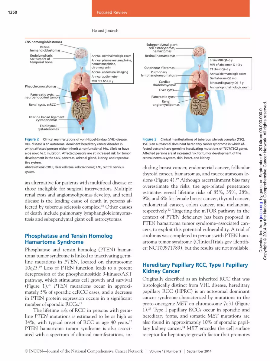
VHL disease is characterized by an increased risk of kidney cysts, ccRCC, pheochromocytomas, and central nervous system hemangioblastomas (Table 1 and Figure 2).4,11 The mean age of RCC onset is 35 years, and RCC is the leading cause of death in
persons affected by VHL disease.12 In a trial evaluat-ing the use of sunitinib in VHL-associated lesions, responses were noted in renal lesions but not heman-gioblastomas.13 Activation of the fibroblast growth factor (FGF) pathway may mediate escape from VEGF inhibition and a trial of dovitinib, an inhibi-tor of VEGF and FGF signaling, is underway in VHL (ClinicalTrials.gov identifier: NCT01266070), but results are not yet available. Other potential thera-peutic interventions include Aurora kinase A inhib-itors to restore primary cilia formation, but whether restoring cilia would reduce tumorigenesis in renal cyst mice models is unknown.
Tuberous Sclerosis ComplexTuberous sclerosis complex is caused by germline mutations in the TSC1/2 genes, located on chromo-somes 9q34 and 16p13, respectively.14 The mTOR complex 1 (mTORC1) signaling cascade is regulat-ed by a heterodimer of TSC1 (hamartin) and TSC2 (tuberin), which function as tumor suppressors (Fig-ure 1). Inactivation of TSC1/2 results in a derepres-sion of mTORC1 inhibition.15 The mTOR path-way responds to both external and internal stimuli to regulate a diverse set of processes, including cell proliferation, nutrient abundance, protein synthesis, and cytoskeletal rearrangements.
Persons who inherit TSC1/2 mutations may de-velop hamartomas, angiomyolipomas, pulmonary lymphangioleiomyomatosis, subependymal giant cell astrocytomas, and RCC (Figure 3).16 Similar to those with VHL disease, persons with TSC1/2 muta-tions have kidney cysts that are associated with cili-ary dysfunction. Angiomyolipomas cause morbidity through spontaneous hemorrhage, invasion of adja-cent renal parenchyma, and chronic kidney disease. The identification of mTORC1 activation caused by TSC1/2 mutations led to clinical trials using siroli-mus, an mTOR inhibitor (ClinicalTrials.gov iden-tifier: NCT00457808), in patients with tuberous sclerosis complex.15 Everolimus was approved by the FDA for angiomyolipoma associated with tuberous sclerosis complex or sporadic lymphangioleiomyo-matosis after a phase III trial showed a response rate of 42%, defined as a reduction in angiomyolipoma volume of 50%.17 In the study, 78% of the patients had bilateral angiomyolipomas and 40% had prior invasive procedures, suggesting that everolimus is
PI3K
mTORC2
HIF-2α HIF-1α
mTORC1
Rheb
TSC1
TSC2
AKT
PTEN
AMPK
FLCN
SDH
FH
PHD
pVHL
E3 Ligase
MET
Proteasome
PcG
BAP1
Cell nucleus
Figure 1 Genes mutated in hereditary kidney cancer syndromes. Mutations (blue) in MET, PTEN, TSC1/TSC2, FLCN, VHL, SDH, FH, and BAP1 are linked to sporadic RCC and hereditary kidney cancer syndromes. In hereditary RCC syndromes, the age of onset is approximately 27 years younger than observed in the general population with sporadic RCC. TSC1/TSC2, VHL, and FLCN mutations also share features with ciliopathies, with cyst formation in target organs. FH or SDH mutations cause accumulation of tricarboxylic acid cycle intermediates that inhibit PHDs and also stabilize HIF-1α and HIF-2α. Similarly, VHL mutations disrupt pVHL binding to HIF-1α and HIF-2α, and HIF-1α and HIF-2α escape ubiquitin mediated proteolysis. Abbreviations: AMPK, adenosine monophosphate–dependent protein kinase; HIF, hypoxia-inducible factor; mTORC1, mammalian target of rapamycin complex 1; mTORC2, mTOR complex 2; PcG, polycomb group; PHD, prolyl hydroxylase; PI3K, phosphoinositide 3-kinase; RCC, renal cell carcinoma. Adapted from Linehan WM, Bratslavsky G, Pinto PA, et al. Molecular diagnosis and therapy of kidney cancer. Ann Rev Med 2010;61:329–343.
. All
right
s re
serv
ed.
Cop
yrig
ht ©
201
4 by
the
Nat
iona
l Com
preh
ensi
ve C
ance
r N
etw
orkfrom
00.
000.
000.
0 o
n S
epte
mbe
r 6,
201
4by
gue
st
jncc
n.or
gD
ownl
oade
d fr
om

Focused Review
Genetic Kidney Cancer Syndromes
© JNCCN—Journal of the National Comprehensive Cancer Network | Volume 12 Number 9 | September 2014
1349
Tab
le 1
Her
edit
ary
Ren
al C
ance
r C
arci
no
ma
Syn
dro
mes
Synd
rom
ePa
tter
n of
In
heri
tanc
eG
ene
Gen
e Pr
oduc
tRC
C H
isto
logi
c Ch
arac
teri
stic
Ass
ocia
ted
Clin
ical
Syn
drom
esSu
gges
ted
Surv
eilla
nce
Poss
ible
RCC
The
rapi
es a
nd
Dru
g Tr
ials
VH
LA
DV
HL
pVH
Lcc
RCC
Hem
angi
obla
stom
asPh
eoch
rom
ocyt
omas
Endo
lym
phat
ic s
ac t
umor
sPa
raga
nglio
mas
Cyst
aden
omas
of
epid
idym
isV
isce
ral c
ysts
At
age
≥15
y:A
nnua
l oph
thal
mol
ogic
ex
amin
atio
nA
nnua
l pla
sma
met
anep
hrin
e,
norm
etan
ephr
ine,
chr
omog
rani
n
test
ing
Ann
ual a
bdom
inal
imag
ing
Ann
ual a
udio
met
ryM
RI o
f CN
S q2
y11
Suni
tini
b13
Pazo
pani
b(C
linic
alTr
ials
.gov
iden
tifi
er:
NCT
0143
6227
)D
ovit
inib
(Clin
ical
Tria
ls.g
ov
iden
tifi
er: N
CT01
2660
70)
Van
deta
nib
(Clin
ical
Tria
ls.
gov
iden
tifi
er:
NCT
0056
6995
)
Tube
rous
scl
eros
is
com
plex
AD
TSC1
/TS
C2H
amar
tin/
tube
rin
Ang
iom
yolip
oma,
RC
CA
ngio
myo
lipom
asBr
ain
MRI
q1–
3y (a
ge ≥
25 y
)Te
msi
rolim
us, e
vero
limus
14,1
7
Sube
pend
ymal
gia
nt c
ell
astr
ocyt
omas
Ham
arto
mas
Cuta
neou
s fi
brom
asA
utis
m s
pect
rum
dis
orde
rPu
lmon
ary
lym
phan
giol
eiom
yom
atos
is
MRI
abd
omen
q1–
3yCT
che
st (w
ith
lung
cys
ts) q
2–3y
Ann
ual d
erm
atol
ogic
exa
min
atio
nD
enta
l exa
m q
6mo
Echo
card
iogr
aphy
q1–
3y b
ased
on
sy
mpt
oms
Ann
ual o
phth
alm
olog
ic
exam
inat
ion14
PTEN
ham
arto
ma
synd
rom
eA
DPT
ENPT
ENcc
RCC
Brea
st c
ance
r19
Folli
cula
r th
yroi
d ca
rcin
oma
Endo
met
rial
can
cer
Muc
ocut
aneo
us p
apul
esA
utis
m s
pect
rum
dis
orde
rM
acro
ceph
aly
Lher
mit
te-D
uclo
s di
seas
e
Adu
lts:
ann
ual t
hyro
id u
ltra
soun
d
and
derm
atol
ogic
exa
min
atio
n,
colo
nosc
opy
(age
≥35
y),
abdo
min
al
imag
ing
q2y
(age
≥40
y)
Wom
en (a
ge ≥
30 y
): an
nual
bre
ast
scre
enin
g, g
ynec
olog
ic u
ltra
soun
dCo
nsid
er s
cree
ning
5–1
0 y
befo
re
youn
gest
age
of
diag
nosi
s in
fam
ily22
Tem
siro
limus
, eve
rolim
us
Her
edit
ary
papi
llary
RC
C, t
ype
I pap
illar
y ki
dney
can
cer
AD
MET
c-M
ETPa
pilla
ry t
ype
IN
o re
port
ed e
xtra
rena
l m
anif
esta
tion
sA
nnua
l abd
omin
al im
agin
g26
Cabo
zant
inib
(Clin
ical
Tria
ls.
gov
iden
tifi
er:
NCT
0186
5747
)
INC2
80 (C
linic
alTr
ials
.gov
id
enti
fier
: NCT
0201
9693
) Fo
reti
nib27
Her
edit
ary
leio
myo
mat
osis
and
RC
C, t
ype
II pa
pilla
ry
kidn
ey c
ance
r
AD
FHFH
Papi
llary
typ
e II
Cuta
neou
s le
iom
yom
as
Ute
rine
leio
myo
mas
Re
nal c
ysts
Der
mat
olog
ic e
xam
inat
ions
q1–
2y
Ann
ual M
RI t
o ev
alua
te f
or r
enal
le
sion
sA
nnua
l gyn
ecol
ogic
ult
raso
und29
,34
Beva
cizu
mab
/erl
otin
ib
(Clin
ical
Tria
ls.g
ov id
enti
fier
: N
CT01
1305
19)
SDH
-ass
ocia
ted
kidn
ey
canc
erA
DSD
HSD
H
subu
nits
B/
C/D
ccRC
C,
chro
mop
hobe
, on
cocy
tom
a
Para
gang
liom
a Ph
eoch
rom
ocyt
oma
Gas
troi
ntes
tina
l str
omal
tum
ors
Abd
omin
al im
agin
g35
MRI
of
the
para
gang
lial s
yste
m
Birt
-Hog
g-D
ubé
AD
FLCN
Folli
culin
Mix
ed o
ncoc
ytic
/ ch
rom
opho
beFi
brof
ollic
ulom
as
Pulm
onar
y cy
sts
Tric
hodi
scom
as
Acr
ocho
rdon
s
Age
≥20
y:
Ann
ual a
bdom
inal
imag
ing
or
q36m
o in
per
sons
wit
hout
ren
al
lesi
ons
(MRI
pre
fera
ble)
Ba
selin
e ch
est
imag
ing38
,41
BAP1
pre
disp
osit
ion
to
fam
ilial
ccR
CCPr
edis
posi
tion
ge
neBA
P1BR
CA1-
asso
ciat
ed
prot
ein-
1
ccRC
CU
veal
mel
anom
a M
elan
oma
Mes
othe
liom
a
Not
est
ablis
hed,
con
side
r an
nual
ab
dom
inal
imag
ing
Abb
revi
atio
ns: A
D, a
utos
omal
dom
inan
t; B
AP1
, BRC
A1-
asso
ciat
ed p
rote
in-1
; ccR
CC, c
lear
cel
l ren
al c
ell c
arci
nom
a; C
NS,
cen
tral
ner
vous
sys
tem
; FH
, fum
arat
e hy
drat
ase;
PTE
N, p
hosp
hata
se a
nd t
ensi
n ho
mol
og; p
VH
L, v
on H
ippe
l-Lin
dau
tu
mor
sup
pres
sor
prot
ein;
RCC
, ren
al c
ell c
arci
nom
a; S
DH
, suc
cina
te d
ehyd
roge
nase
; TSC
, tub
erou
s sc
lero
sis
com
plex
; VH
L, v
on H
ippe
l-Lin
dau.
. All
right
s re
serv
ed.
Cop
yrig
ht ©
201
4 by
the
Nat
iona
l Com
preh
ensi
ve C
ance
r N
etw
orkfrom
00.
000.
000.
0 o
n S
epte
mbe
r 6,
201
4by
gue
st
jncc
n.or
gD
ownl
oade
d fr
om
Focused Review
Ho and Jonasch
© JNCCN—Journal of the National Comprehensive Cancer Network | Volume 12 Number 9 | September 2014
1350
an alternative for patients with multifocal disease or those ineligible for surgical intervention. Multiple renal cysts and angiomyolipomas develop, and renal disease is the leading cause of death in persons af-fected by tuberous sclerosis complex.18 Other causes of death include pulmonary lymphangioleiomyoma-tosis and subependymal giant cell astrocytomas.
Phosphatase and Tensin Homolog Hamartoma SyndromePhosphatase and tensin homolog (PTEN) hamar-toma tumor syndrome is linked to inactivating germ-line mutations in PTEN, located on chromosome 10q23.19 Loss of PTEN function leads to a potent derepression of the phosphoinositide 3-kinase/AKT pathway, which stimulates cell growth and survival (Figure 1).20 PTEN mutations occur in approxi-mately 5% of sporadic ccRCC cases, and a decrease in PTEN protein expression occurs in a significant number of sporadic RCCs.21
The lifetime risk of RCC in persons with germ-line PTEN mutations is estimated to be as high as 34%, with typical onset of RCC at age 40 years.22 PTEN hamartoma tumor syndrome is also associ-ated with a spectrum of clinical manifestations, in-
cluding breast cancer, endometrial cancer, follicular thyroid cancer, hamartomas, and mucocutaneous le-sions (Figure 4).19 Although ascertainment bias may overestimate the risks, the age-related penetrance estimates reveal lifetime risks of 85%, 35%, 28%, 9%, and 6% for female breast cancer, thyroid cancer, endometrial cancer, colon cancer, and melanoma, respectively.22 Targeting the mTOR pathway in the context of PTEN deficiency has been proposed in PTEN hamartoma tumor syndrome–associated can-cers, to exploit this potential vulnerability. A trial of sirolimus was completed in persons with PTEN ham-artoma tumor syndrome (ClinicalTrials.gov identifi-er: NCT00971789), but the results are not available.
Hereditary Papillary RCC, Type I Papillary Kidney CancerOriginally described as an inherited RCC that was histologically distinct from VHL disease, hereditary papillary RCC (HPRC) is an autosomal dominant cancer syndrome characterized by mutations in the proto-oncogene MET on chromosome 7q31 (Figure 1).23 Type I papillary RCCs occur in sporadic and hereditary forms, and somatic MET mutations are also found in approximately 10% of sporadic papil-lary kidney cancer.24 MET encodes the cell surface receptor for hepatocyte growth factor that promotes
CNS hemangioblastomas
Retinal hemangioblastomas
Endolymphatic sac tumors of temporal bone
Pheochromocytomas
Pancreatic cysts, neuroendocrine tumors
Renal cysts, ccRCC
Uterine broad ligament cystadenomas
Epididymal cystadenomas
Annual ophthalmologic exam
Annual plasma metanephrine, normetanephrine, chromogranin
Annual abdominal imaging
Annual audiometry
MRI of CNS Q2 y
Figure 2 Clinical manifestations of von Hippel-Lindau (VHL) disease. VHL disease is an autosomal dominant hereditary cancer disorder in which affected persons either inherit a nonfunctional VHL allele or have a de novo VHL mutation. Affected persons are at increased risk for tumor development in the CNS, pancreas, adrenal gland, kidney, and reproduc-tive system. Abbreviations: ccRCC, clear cell renal cell carcinoma; CNS, central nervous system.
Figure 3 Clinical manifestations of tuberous sclerosis complex (TSC). TSC is an autosomal dominant hereditary cancer syndrome in which af-fected persons have germline inactivating mutations of TSC1/TSC2 genes. Affected persons are at increased risk for tumor development of the central nervous system, skin, heart, and kidney.
Subependymal giant cell astrocytomas,
hamartomasRetinal hamartomas
Cutaneous fibromasPulmonary
lymphangiomyomatosis
Cardiac rhabdomyomas
Liver cysts
Pancreatic cysts
Renal angiomyolipomas
Brain MRI Q1–3 y
MRI of abdomen Q1–3 y
CT chest Q2–3 y
Annual dermatologic exam
Dental exam Q6 mo
Echocardiography Q1–3 y
Annual ophthalmologic exam
. All
right
s re
serv
ed.
Cop
yrig
ht ©
201
4 by
the
Nat
iona
l Com
preh
ensi
ve C
ance
r N
etw
orkfrom
00.
000.
000.
0 o
n S
epte
mbe
r 6,
201
4by
gue
st
jncc
n.or
gD
ownl
oade
d fr
om

Focused Review
Genetic Kidney Cancer Syndromes
© JNCCN—Journal of the National Comprehensive Cancer Network | Volume 12 Number 9 | September 2014
1351
migration, invasion, proliferation, and angiogenesis. Unlike sporadic ccRCC and VHL disease, these RCC tumors lack 3p alterations but exhibit trisomy 7, leading to multiple copies of MET and tumorigen-esis initiated by the combination of a MET mutation and duplication of the mutated allele.25
Persons affected by this cancer syndrome can have bilateral involvement with more than 1000 tumors per kidney, suggesting the presence of mi-croscopic precursors that may not be apparent on routine imaging.26 Extrarenal manifestations have not been reported. The presence of MET pathway activation or germline MET mutations was recently associated with responses to foretinib, a multikinase inhibitor targeting MET/VEGF receptor 2.27 A risk-adapted approach of observing patients with small tumors (<3 cm), independent of location or number of tumors, was developed to guide surveillance based on a 10-year follow-up study of parenchymal-sparing surgery in hereditary RCC.28 Papillary RCC is hy-poechoic and can be missed by ultrasound.29 Active surveillance with CT/MRI is recommended until a kidney tumor reaches 3 cm to mitigate the risk of metastatic disease and preserve renal function.
Hereditary Leiomyomatosis and RCC, Type II Papillary Kidney Cancer Hereditary leiomyomatosis and RCC (HLRCC) is characterized by a germline mutation in FH (fuma-rate hydratase), found on chromosome 1q42.30 FH is a Krebs cycle enzyme that converts fumarate to malate, and FH inactivation causes a metabolic shift to aerobic glycolysis in FH-deficient kidney cancer.31 The buildup of fumarate inhibits prolyl hydroxylase (PHD), interferes with the recognition of HIF-1α and HIF-2α by pVHL, and leads to an increase in VEGF-mediated angiogenesis (Figure 1).32
Affected persons can present with cutaneous leiomyomas, uterine leiomyomas, and type II papil-lary RCC (Figure 5).29,30 Systemic therapy specific for patients with HLRCC has yet to be fully developed. Expression profiling of an Fh1-deficient mouse model and HLRCC cell lines shows a common pathway of dysregulated glycolytic genes, suggesting that FH de-ficiency may sensitize cells to glycolytic inhibitors.31 For example, the glycolytic inhibitor 2-deoxy-D- glucose was used to treat a patient with HLRCC af-ter experiencing disease progression while receiving an mTOR inhibitor, and although the patient expe-rienced hypoglycemia consistent with inhibition of glycolysis, no antitumor effect occurred.33 The me-dian age of onset for the development of papillary RCC is 37 years, and individuals in approximately 15% of families with HLRCC develop disease.1,29 Although a 3-cm threshold is used to guide manage-ment of RCC in other hereditary syndromes, im-mediate surgical management of any identified HL-RCC-associated renal tumors is suggested because of aggressive growth and a propensity for early metasta-ses.34 Papillary RCC is hypoechoic, can be missed by ultrasound, and is best screened with CT/MRI im-aging.29 Uterine leiomyomas also develop earlier in women affected by cutaneous leiomyomas compared with the general population.34 Because of symptom-atic leiomyomas, women affected by HLRCC have higher rates of hysterectomy and undergo hysterec-tomy at a younger age, indicating a need for family planning counseling.
Succinate Dehydrogenase–Associated Kidney CancerSuccinate dehydrogenase (SDH)–associated kidney cancer is linked to a germline mutation in genes that
Figure 4 Clinical manifestations of phosphatase and tensin homolog hamartoma syndrome (PHTS). PHTS is an autosomal dominant hereditary cancer syndrome in which affected persons have germline inactivating mutations of PTEN. Affected persons are at increased risk for tumor development of the central nervous system, breast, colon, thyroid, skin, endometrium, and kidney. Abbreviations: GI, gastrointestinal; RCC, renal cell carcinoma.
Adults: annual thyroid ultrasound and dermatologic examination, colonscopy (age 35 y), abdominal imaging Q2 y (age ≥40 y)
Women (age ≥30 y): annual breast screening, gynecological ultrasound
Consider screening 5–10 y before youngest age of diagnosis in family
Lhermitte-Duclos,macrocephaly
Mucocutaneous lesions
Follicular thyroidcarcinoma
Breast cancer
RCC
Colon cancer,GI hamartomas
Endometrial cancer
. All
right
s re
serv
ed.
Cop
yrig
ht ©
201
4 by
the
Nat
iona
l Com
preh
ensi
ve C
ance
r N
etw
orkfrom
00.
000.
000.
0 o
n S
epte
mbe
r 6,
201
4by
gue
st
jncc
n.or
gD
ownl
oade
d fr
om

Focused Review
Ho and Jonasch
© JNCCN—Journal of the National Comprehensive Cancer Network | Volume 12 Number 9 | September 2014
1352
encode for subunits of the tricarboxylic acid cycle enzyme, SDH.35 SDH genes include SDHA (chro-mosome 5p15), SDHB (chromosome 1p36), SDHC (chromosome 1q23), and SDHD (chromosome 11q23). Mutations in SDH cause accumulation of succinate and, similar to fumarate in HLRCC, also inhibit prolyl hydroxylation of HIF-1α and HIF-2α (Figure 1).36
RCC may be the only clinical presentation in persons with germline SDHB, SDHC, or SDHD mutations.2 Extrarenal manifestations include pheo-chromocytomas, paragangliomas, carotid body tu-mors, and gastrointestinal stromal tumors. Because of the rarity of these tumors, no established screen-ing guidelines have been developed. Mutations in Krebs cycle enzymes shift the cells toward increased glucose uptake, aerobic glycolysis, and fatty acid synthesis.37 The increased dependence on glycolytic pathways suggests that inhibitors of glucose uptake, glycolysis, and fatty acid synthesis could exploit the vulnerability of these tumors. Among the SDH sub-unit mutations, SDHB and SDHD carriers are more likely to develop RCC, with a median age of onset of 30 years.1 Symptoms related to pheochromocytomas and paragangliomas may present as early as 3 years of age in germline carriers, supporting a role for germ-line testing of SDHB and SDHD at initial presenta-
tion.2 Subsequent surveillance for renal masses and extrarenal manifestations is likely warranted in this population.35
Birt-Hogg-Dubé SyndromeBirt-Hogg-Dubé (BHD) is an autosomal dominant syndrome caused by mutations of the FLCN gene, located on chromosome 17p11.38 The FLCN gene encodes for folliculin, a downstream target of both adenosine monophosphate–dependent protein ki-nase and mTORC1 signaling; folliculin also local-izes to cilia (Figure 1).39 Loss of folliculin function leads to mTORC1 activation and dysregulation of ciliogenesis.
Clinically, BHD is characterized by fibrofollicu-lomas, pulmonary cysts, and early-onset RCC, com-monly with mixed oncocytoma and chromophobe histologic characteristics.38 Similar to the VHL and tuberous sclerosis complex disease syndromes, the BHD-associated pulmonary cysts share characteris-tics consistent with a ciliopathy. The risk of devel-oping RCC at age 70 years has been estimated to be 16% in patients with BHD, with characteristic hybrid histologic characteristics of chromophobe RCC and oncocytomas.40 Similar to VHL disease and HPRC, active surveillance is recommended for the management of renal lesions less than 3 cm.28,41 When adjusted for age, persons with BHD have a 50-fold increased risk of pneumothorax.42 Pulmonary consultation is recommended for those at risk of am-bient atmospheric pressure changes, and smoking cessation may reduce morbidity associated with pul-monary cysts. mTOR inhibitors have prolonged sur-vival of folliculin-deficient mice and have been pro-posed to treat the clinical manifestations of BHD.43 However, a trial of topical rapamycin to treat BHD-associated fibrofolliculomas did not reduce the size or number of cutaneous lesions.44
BRCA1-Associated Protein-1 Predisposition to Familial ccRCCSystematic sequencing of sporadic ccRCC identified loss-of-function mutations in histone-modulating enzymes, such as the BRCA1-associated protein-1 (BAP1) (5%–15%) gene on chromosome 3p21.1.21,45 BAP1 functions as a nuclear deubiquitinase that in-teracts with polycomb group (PcG) proteins at open
Dermatologic examinations Q 1–2 y
Annual MRI to evaluate for renal lesionsAnnual gynecological ultrasound
Cutaneousleiomyomas
Papillary RCC,type II
Uterineleiomyomas
Figure 5 Clinical manifestations of hereditary leiomyomatosis and renal cell carcinoma (HLRCC). HLRCC is an autosomal dominant hereditary cancer syndrome in which affected persons have germline inactivating mutations of FH. Affected persons are at increased risk for tumor development of skin, uterus, and kidney.Abbreviation: RCC, renal cell carcinoma.
. All
right
s re
serv
ed.
Cop
yrig
ht ©
201
4 by
the
Nat
iona
l Com
preh
ensi
ve C
ance
r N
etw
orkfrom
00.
000.
000.
0 o
n S
epte
mbe
r 6,
201
4by
gue
st
jncc
n.or
gD
ownl
oade
d fr
om

Focused Review
Genetic Kidney Cancer Syndromes
© JNCCN—Journal of the National Comprehensive Cancer Network | Volume 12 Number 9 | September 2014
1353
chromatin and promotes double-strand break repair (Figure 1).46
After the discovery of BAP1 mutations in spo-radic ccRCC, germline mutations were linked with familial ccRCC syndromes.47 Germline BAP1 muta-tions are also associated with uveal melanomas and mesotheliomas. However, the BAP1 syndrome is rare, making it difficult to assess its prevalence in the general population and estimate tumor risks to guide surveillance. The use of general guidelines for early-onset kidney screening may be considered based on age of initial presentation.1
Overlapping Mechanisms Between Hereditary and Sporadic ccRCC The overlap of gene mutations in VHL, TSC1, MTOR, PTEN, and BAP1 in both hereditary and sporadic ccRCC has increased the understanding of tumorigenesis. The 4 most frequently mutated genes (VHL, ≈52%; polybromo-1, ≈33%; SETD2, ≈12%; and BAP1, ≈10%) are located on chromosome 3p, a region that is deleted in more than 90% of sporadic ccRCCs, supporting a mechanism of biallelic inacti-vation of these tumor suppressors.21 In parallel with the clinical benefit seen with mTOR inhibitors in the tuberous sclerosis complex syndrome, a subset of patients with sporadic ccRCC and TSC1 inactiva-tion or MTOR mutations may experience a durable response to mTOR inhibitors.48 PTEN (≈4%) and
PIK3CA (≈3%) mutations occur in approximately 4% of sporadic ccRCC cases, supporting a phenotyp-ic convergence on the PI3K/mTOR axis (Figure 1). An integrated analysis of molecular pathways iden-tified a common metabolic phenotype of increased glycolysis between sporadic ccRCC and HLRCC.21 Krebs cycle mutations in HLRCC and SDH-as-sociated kidney cancer stabilize HIF and shift me-tabolism to increased reliance on aerobic glycolysis, characterized as “the Warburg effect,” by mobilizing nutrients (nucleotides, amino acids, lipids) for cell proliferation.32 Similarly, in sporadic ccRCC, bial-lelic inactivation of VHL leads to HIF stabilization, and tumors with metabolic shifts toward aerobic gly-colysis are associated with a worse survival.21
Establishing a Role for Early-Onset Kidney Cancer ScreeningThe median age of patients with sporadic RCC was 64 years in an analysis of the SEER 17 registry program, which was considerably older than the median age of 37 years in a cohort of patients with hereditary kidney cancer. At the NCI Urologic Oncology Branch, pa-tients with VHL, BHD, HLRCC, HPRC, and SDHB syndromes had median ages of onset ranging from 35 to 50 years. Because the median age of presentation for hereditary RCC is 27 years younger than that for RCC observed in the general population, patients with RCC who are 46 years old or younger should consider ge-
A B C D
Clear CellVHL
SDHCBAP1TAC1TSC2
Papillary,Type IMET
Papillary,Type II
FH
Chromophobe FLCN TSC1 TSC2 PTEN
Figure 6 Germline testing of selected genes based on renal cell carcinoma (RCC) histology: (A) clear cell, (B) papillary type I, (C) papillary type II, and (D) chromophobe. Persons with RCC aged 46 years or younger should be considered for genetic counseling and germline mutations testing, even in the absence of secondary clinical manifestations. The RCC histologic characteristics may guide which germline mutations to test. (Hematoxylin and eosin stain; original magnification x10 and x40 [inset]) Courtesy of Dr. Melissa L. Stanton, Mayo Clinic Arizona.
. All
right
s re
serv
ed.
Cop
yrig
ht ©
201
4 by
the
Nat
iona
l Com
preh
ensi
ve C
ance
r N
etw
orkfrom
00.
000.
000.
0 o
n S
epte
mbe
r 6,
201
4by
gue
st
jncc
n.or
gD
ownl
oade
d fr
om
Focused Review
Ho and Jonasch
© JNCCN—Journal of the National Comprehensive Cancer Network | Volume 12 Number 9 | September 2014
1354
netic counseling and germline mutation testing even in the absence of secondary clinical manifestations.1 The histology of early-onset RCC may guide which germ-line mutations to test (Figure 6).49
ConclusionsPatients with hereditary RCC syndromes provide the clinician with unique diagnostic, surveillance, and therapeutic challenges. Many of the described syndromes demonstrate highly distinct phenotypes, allowing clinical findings to guide genetic testing. Once a hereditary RCC syndrome is identified, care-ful adherence to surveillance strategies, appropri-ate management of the renal and extrarenal disease manifestations, and attention to at-risk family mem-bers will lead to improvement in patient outcome.
The hereditary drivers of several nonsyndromic RCC cohorts have long eluded investigators. Ad-vances in genomewide sequencing technologies have led to the identification of additional mutations in RCC and to the discovery of the most recently de-scribed BAP1 familial RCC syndrome.47
Despite the complexity of networks involved in hereditary and sporadic RCC, these alterations share a common dysregulation of the HIF-VEGF axis and aberrant tumor metabolism.21 The identification of genes linked to RCC syndromes has led to the devel-opment of clinical trials selecting for patients affect-ed by hereditary syndromes (Table 1).21 Additional drug targets may exist outside the mTOR and VEGF pathways, such as proteins that influence metabolic sensitivity or chromatin remodeling proteins. Ulti-mately, the identification of new druggable targets for sequential or combination therapies may provide a more robust form of therapy for patients who have sporadic and hereditary RCC.
The observed early onset of hereditary RCC in-dicates that primary preventative strategies are more likely to increase life expectancy in affected persons than administering targeted therapies in the meta-static setting. Persons affected with RCC at age 46 years or younger should be considered for genetic counseling and germline mutational testing, even in the absence of secondary clinical manifestations.1 As life expectancy increases, additional clinical mani-festations in hereditary RCC syndromes will un-doubtedly become apparent, and clinicians and fam-ilies must maintain vigilant and report new findings.
References1. Shuch B, Vourganti S, Ricketts CJ, et al. Defining early-onset kidney
cancer: implications for germline and somatic mutation testing and clinical management. J Clin Oncol 2013.
2. Ricketts CJ, Forman JR, Rattenberry E, et al. Tumor risks and genotype-phenotype-proteotype analysis in 358 patients with germline mutations in SDHB and SDHD. Hum Mutat 2010;31:41–51.
3. Latif F, Tory K, Gnarra J, et al. Identification of the von Hippel-Lindau disease tumor suppressor gene. Science 1993;260:1317–1320.
4. Prowse AH, Webster AR, Richards FM, et al. Somatic inactivation of the VHL gene in Von Hippel-Lindau disease tumors. Am J Hum Genet 1997;60:765–771.
5. Latif F, Duh FM, Gnarra J, et al. von Hippel-Lindau syndrome: cloning and identification of the plasma membrane Ca(++)-transporting ATPase isoform 2 gene that resides in the von Hippel-Lindau gene region. Cancer Res 1993;53:861–867.
6. Cockman ME, Masson N, Mole DR, et al. Hypoxia inducible factor-alpha binding and ubiquitylation by the von Hippel-Lindau tumor suppressor protein. J Biol Chem 2000;275:25733–25741.
7. Maxwell PH, Wiesener MS, Chang GW, et al. The tumour suppressor protein VHL targets hypoxia-inducible factors for oxygen-dependent proteolysis. Nature 1999;399:271–275.
8. Semenza GL. Targeting HIF-1 for cancer therapy. Nat Rev Cancer 2003;3:721–732.
9. Kondo K, Kim WY, Lechpammer M, Kaelin WG Jr. Inhibition of HIF2alpha is sufficient to suppress pVHL-defective tumor growth. PLoS Biol 2003;1:E83.
10. Xu J, Li H, Wang B, et al. VHL inactivation induces HEF1 and Aurora kinase A. J Am Soc Nephrol 2010;21:2041–2046.
11. Binderup ML, Bisgaard ML, Harbud V, et al. Von Hippel-Lindau Disease (vHL). National Clinical Guideline for Diagnosis and Surveillance in Denmark. 3rd ed. Dan Med J 2013;60:B4763.
12. Maher ER, Neumann HP, Richard S. von Hippel-Lindau disease: a clinical and scientific review. Eur J Hum Genet 2011;19:617–623.
13. Jonasch E, McCutcheon IE, Waguespack SG, et al. Pilot trial of sunitinib therapy in patients with von Hippel-Lindau disease. Ann Oncol 2011;22:2661–2666.
14. Krueger DA, Northrup H. Tuberous sclerosis complex surveillance and management: recommendations of the 2012 International Tuberous Sclerosis Complex Consensus Conference. Pediatr Neurol 2013;49:255–265.
15. Laplante M, Sabatini DM. mTOR signaling in growth control and disease. Cell 2012;149:274–293.
16. Crino PB, Nathanson KL, Henske EP. The tuberous sclerosis complex. N Engl J Med 2006;355:1345–1356.
17. Bissler JJ, Kingswood JC, Radzikowska E, et al. Everolimus for angiomyolipoma associated with tuberous sclerosis complex or sporadic lymphangioleiomyomatosis (EXIST-2): a multicentre, randomised, double-blind, placebo-controlled trial. Lancet 2013;381:817–824.
18. Shepherd CW, Gomez MR, Lie JT, Crowson CS. Causes of death in patients with tuberous sclerosis. Mayo Clin Proc 1991;66:792–796.
19. Pilarski R, Burt R, Kohlman W, et al. Cowden syndrome and the PTEN hamartoma tumor syndrome: systematic review and revised diagnostic criteria. J Natl Cancer Inst 2013;105:1607–1616.
20. Sun H, Lesche R, Li DM, et al. PTEN modulates cell cycle progression and cell survival by regulating phosphatidylinositol 3,4,5,-trisphosphate and Akt/protein kinase B signaling pathway. Proc Natl Acad Sci U S A 1999;96:6199–6204.
21. Comprehensive molecular characterization of clear cell renal cell carcinoma. Nature 2013;499:43–49.
22. Tan MH, Mester JL, Ngeow J, et al. Lifetime cancer risks in individuals with germline PTEN mutations. Clin Cancer Res 2012;18:400–407.
23. Schmidt L, Duh FM, Chen F, et al. Germline and somatic mutations in the tyrosine kinase domain of the MET proto-oncogene in papillary renal carcinomas. Nat Genet 1997;16:68–73.
24. Linehan WM, Srinivasan R, Schmidt LS. The genetic basis of kidney cancer: a metabolic disease. Nat Rev Urol 2010;7:277–285.
. All
right
s re
serv
ed.
Cop
yrig
ht ©
201
4 by
the
Nat
iona
l Com
preh
ensi
ve C
ance
r N
etw
orkfrom
00.
000.
000.
0 o
n S
epte
mbe
r 6,
201
4by
gue
st
jncc
n.or
gD
ownl
oade
d fr
om
Focused Review
Genetic Kidney Cancer Syndromes
© JNCCN—Journal of the National Comprehensive Cancer Network | Volume 12 Number 9 | September 2014
1355
25. Zhuang Z, Park WS, Pack S, et al. Trisomy 7-harbouring non-random duplication of the mutant MET allele in hereditary papillary renal carcinomas. Nat Genet 1998;20:66–69.
26. Ornstein DK, Lubensky IA, Venzon D, et al. Prevalence of microscopic tumors in normal appearing renal parenchyma of patients with hereditary papillary renal cancer. J Urol 2000;163:431–433.
27. Choueiri TK, Vaishampayan U, Rosenberg JE, et al. Phase II and biomarker study of the dual MET/VEGFR2 inhibitor foretinib in patients with papillary renal cell carcinoma. J Clin Oncol 2013;31:181–186.
28. Herring JC, Enquist EG, Chernoff A, et al. Parenchymal sparing surgery in patients with hereditary renal cell carcinoma: 10-year experience. J Urol 2001;165:777–781.
29. Toro JR, Nickerson ML, Wei MH, et al. Mutations in the fumarate hydratase gene cause hereditary leiomyomatosis and renal cell cancer in families in North America. Am J Hum Genet 2003;73:95–106.
30. Tomlinson IP, Alam NA, Rowan AJ, et al. Germline mutations in FH predispose to dominantly inherited uterine fibroids, skin leiomyomata and papillary renal cell cancer. Nat Genet 2002;30:406–410.
31. Ashrafian H, O’Flaherty L, Adam J, et al. Expression profiling in progressive stages of fumarate-hydratase deficiency: the contribution of metabolic changes to tumorigenesis. Cancer Res 2010;70:9153–9165.
32. Pollard PJ, Spencer-Dene B, Shukla D, et al. Targeted inactivation of fh1 causes proliferative renal cyst development and activation of the hypoxia pathway. Cancer Cell 2007;11:311–319.
33. Yamasaki T, Tran TA, Oz OK, et al. Exploring a glycolytic inhibitor for the treatment of an FH-deficient type-2 papillary RCC. Nat Rev Urol 2011;8:165–171.
34. Smit DL, Mensenkamp AR, Badeloe S, et al. Hereditary leiomyomatosis and renal cell cancer in families referred for fumarate hydratase germline mutation analysis. Clin Genet 2011;79:49–59.
35. Ricketts CJ, Shuch B, Vocke CD, et al. Succinate dehydrogenase kidney cancer: an aggressive example of the Warburg effect in cancer. J Urol 2012;188:2063–2071.
36. Selak MA, Armour SM, MacKenzie ED, et al. Succinate links TCA cycle dysfunction to oncogenesis by inhibiting HIF-alpha prolyl hydroxylase. Cancer Cell 2005;7:77–85.
37. Pollard PJ, Briere JJ, Alam NA, et al. Accumulation of Krebs cycle intermediates and over-expression of HIF1alpha in tumours which result from germline FH and SDH mutations. Hum Mol Genet 2005;14:2231–2239.
38. Menko FH, van Steensel MA, Giraud S, et al. Birt-Hogg-Dube syndrome: diagnosis and management. Lancet Oncol 2009;10:1199–1206.
39. Luijten MN, Basten SG, Claessens T, et al. Birt-Hogg-Dube syndrome is a novel ciliopathy. Hum Mol Genet 2013;22:4383–4397.
40. Houweling AC, Gijezen LM, Jonker MA, et al. Renal cancer and pneumothorax risk in Birt-Hogg-Dube syndrome; an analysis of 115 FLCN mutation carriers from 35 BHD families. Br J Cancer 2011;105:1912–1919.
41. Stamatakis L, Metwalli AR, Middelton LA, et al. Diagnosis and management of BHD-associated kidney cancer. Fam Cancer 2013;12:397–402.
42. Zbar B, Alvord WG, Glenn G, et al. Risk of renal and colonic neoplasms and spontaneous pneumothorax in the Birt-Hogg-Dube syndrome. Cancer Epidemiol Biomarkers Prev 2002;11:393–400.
43. Baba M, Furihata M, Hong SB, et al. Kidney-targeted Birt-Hogg-Dube gene inactivation in a mouse model: Erk1/2 and Akt-mTOR activation, cell hyperproliferation, and polycystic kidneys. J Natl Cancer Inst 2008;100:140–154.
44. Gijezen LM, Vernooij M, Martens H, et al. Topical rapamycin as a treatment for fibrofolliculomas in Birt-Hogg-Dube syndrome: a double-blind placebo-controlled randomized split-face trial. PLoS One 2014;9:e99071.
45. Pena-Llopis S, Vega-Rubin-de-Celis S, Liao A, et al. BAP1 loss defines a new class of renal cell carcinoma. Nat Genet 2012;44:751–759.
46. Yu H, Pak H, Hammond-Martel I, et al. Tumor suppressor and deubiquitinase BAP1 promotes DNA double-strand break repair. Proc Natl Acad Sci U S A 2014;111:285–290.
47. Popova T, Hebert L, Jacquemin V, et al. Germline BAP1 mutations predispose to renal cell carcinomas. Am J Hum Genet 2013;92:974–980.
48. Voss MH, Hakimi AA, Pham CG, et al. Tumor genetic analyses of patients with metastatic renal cell carcinoma and extended benefit from mTOR inhibitor therapy. Clin Cancer Res 2014;20:1955–1964.
49. Linehan WM. Evaluation and screening for hereditary renal cell cancers. Can Urol Assoc J 2013;7:324–325.
. All
right
s re
serv
ed.
Cop
yrig
ht ©
201
4 by
the
Nat
iona
l Com
preh
ensi
ve C
ance
r N
etw
orkfrom
00.
000.
000.
0 o
n S
epte
mbe
r 6,
201
4by
gue
st
jncc
n.or
gD
ownl
oade
d fr
om