genetic analysis of thedrosophilaβ3-tubulin gene demonstrates that the microtubule cytoskeleton in...
TRANSCRIPT
DEVELOPMENTAL BIOLOGY 177, 117–135 (1996)ARTICLE NO. 0150
Genetic Analysis of the Drosophila b3-TubulinGene Demonstrates That the MicrotubuleCytoskeleton in the Cells of the VisceralMesoderm Is Required for Morphogenesisof the Midgut Endoderm
Robert W. Dettman,1 F. Rudolf Turner, and Elizabeth C. Raff2
Department of Biology and Institute for Molecular and Cellular Biology,Indiana University, Bloomington, Indiana 47405
We have investigated the cellular basis for lethality of mutant alleles of the Drosophila melanogaster b3-tubulin gene,bTub60D. Lethal b3 mutations can be grouped into two classes: the most severe mutations (Class I alleles) cause deathduring the first larval instar, while weaker alleles (Class II) cause death in later larval stages or in early pupal development.Since b3 is not expressed during larval development, lethality of the Class I mutations must reflect essential functions ofb3 in embryogenesis. b3-tubulin is zygotically expressed during midembryogenesis in the developing mesoderm, and themajor site of b3 accumulation is in the developing muscles during myogenesis. We show that the embryonic pattern ofb3 expression, including accumulation in the developing musculature, is conserved in other Drosophila species. However,we found that loss of b3 function does not cause discernible defects in either the ultrastructure or function of the larvalmuscle. Thus b3-tubulin is dispensable in its highest site of accumulation. Rather, the essential site of function of b3 inembryos is in cells of the visceral mesoderm. Lethality of Class I alleles is caused by defects in midgut morphogenesis andfailure of gut function. Although the folding pattern is irregular and the gut is smaller than normal, a complete folded gutforms in mutant larvae, and the visceral muscle functions normally to move food through the gut. However, mutant larvaecannot absorb nutrients across the gut wall. Thus loss of b3 function in the mesoderm results in defects in the underlyingendodermally derived layer of the gut. Our data provide an assay for cellular interactions between mesoderm and endodermaltissues and reveal a role for the microtubule cytoskeleton of the visceral mesodermal cells in differentiation of the endoder-mal cell layer of the larval gut. q 1996 Academic Press, Inc.
INTRODUCTION plays diverse roles both in prepositioning of developmentalcues in the oocyte, and in the mitotic divisions and cyto-plasmic reorganizations in the embryo (Theurkauf et al.,Microtubules are an important component of the eukary-1993, 1994). All of these early developmental functions areotic cytoskeleton. The major structural components of mi-supported by microtubules assembled from the b1-tubulincrotubules, the a- and b-tubulins, are encoded in multipleisoform (b1), which is both maternally supplied and zygoti-gene families, each member of which has a unique temporalcally expressed (Raff et al., 1982; Bialojan et al., 1984; Gaschand spatial pattern of expression. Drosophila has four b-et al., 1988; Buttgereit et al., 1991). Expression of a secondtubulin genes that encode four distinct isoforms. In earlyisoform, b3-tubulin (b3), does not begin until midembryo-Drosophila development, the microtubule cytoskeletongenesis, in cells that have ceased to divide and will undergoextensive cell shape changes (Raff et al., 1982; Bialojan etal., 1984; Leiss et al., 1988; Gasch et al., 1988; Kimble et1 Current address: Department of Pediatrics, University of Cali-al., 1989). Dissection of the role of b3 would thus providefornia, San Francisco, San Francisco, CA 94143.insight into the role of the microtubule cytoskeleton in2 To whom correspondence should be addressed at Departmentcells undergoing morphogenetic changes in the final stagesof Biology, Jordan Hall, Indiana University, Bloomington, IN 47405.
Fax: 812-855-6705. E-mail: [email protected]. of differentiation.
117
0012-1606/96 $18.00Copyright q 1996 by Academic Press, Inc.All rights of reproduction in any form reserved.
AID DB 8230 / 6X0f$$$$21 06-14-96 18:18:34 dba AP: Dev Bio

118 Dettman, Turner, and Raff
The b3 gene has a complex pattern of expression. In em- sin et al., 1985; Reedy and Beall, 1993a,b). Moreover, weexamined expression of the homologous isoform in otherbryos, b3 is expressed at high levels in the developing mus-
culature, where it is the single zygotically expressed b-tu- Drosophila species and determined that embryonic b3 ex-pression in myogenesis has been conserved in evolution.bulin (Gasch et al., 1988; Leiss et al., 1988; Kimble et al.,
1989), but the protein can no longer be detected in first or We therefore speculated that the b3 isoform might havespecialized functional properties required for myogenesis.second instar larvae (Dettman, 1995; Dettman and Raff, in
preparation). After 16 hr postfertilization, b3 message can- However, to our surprise, we found that b3 is dispensablefor development and function of the larval muscle in Dro-not be detected until the third instar larval molt (Bialojan
et al., 1984; Andres et al., 1993), at which time the protein sophila melanogaster. We have considered the possiblemeaning of evolutionarily conserved muscle expression ofbegins to accumulate in the male gonads (Gonczy et al.,
1992) and in imaginal discs (Kimble et al., 1989). During b3 in the light of this observation. We argue that b3 expres-sion serves to increase the tubulin pool levels in myogenicpupal development, b3 is expressed in several tissues, in-
cluding each of the developing muscles; expression persists cells, and that this confers a selective advantage for mainte-nance of muscle expression, but is not essential for thein the adult only in the somatic cells of the gonads (Kimble
et al., 1989). development of an individual larva. To define the functionsof b3 that are essential in development, we examined otherAlthough there may be some exceptions, in the embryo
b3 is expressed primarily in cells of mesodermal origin aspects of the mutant phenotypes. We found that lethalityassociated with b3 mutations results from abnormal devel-(Leiss et al., 1988; Kimble et al., 1989). When expression of
b3 begins after 8 hr of development, it is expressed in the opment and function of the gut. Importantly, a major contri-bution to the lethality lies in the failure of the endodermalcephalic, visceral, and somatic mesoderm. In addition to
the pharyngeal, gut, and body wall musculature, b3 is ex- layer of the intestine to function correctly. This suggeststhat correct cytoarchitecture of the visceral mesoderm cellspressed in nonmuscle cells derived from the mesoderm, in-
cluding the macrophages (Leiss et al., 1988), the transverse is important for the differentiation of the underlying endo-derm.nerve exit glia (Gorczyca et al., 1994), and the cap cells of
chordotonal organs and the lymph and ring glands (Dettmanand Raff, in preparation). The three distinct mesoderm lay-ers may become determined early to perform distinct func- MATERIAL AND METHODStions later. Cells of the cephalic mesoderm form the pharyn-geal and foregut musculature and incorporate into several Fliesother organs in the head segments, such as support cells
Drosophila stocks were maintained at 257C on standard corn-of sensory organs. The visceral mesoderm forms the gutmeal/molasses/agar food. Visible markers, deficiency chromo-muscles and also mediates events that allow the midgut tosomes, and balancer chromosomes used in this study are describedfold (Reuter and Scott, 1990). Cells of the somatic meso-in Lindsley and Zimm (1992) or FlyBase (1994). The deficiencyderm give rise to body wall muscles and support cells ofchromosome Df(2R)Px 2, bw sp2 (designated Px2 throughout the
chordotonal organs. text) deletes region 60C5,6–60D9,10 of chromosome 2, includingThe bTub60D gene, which encodes b3, is essential. Mu- bTub60D, the gene that encodes the b3-tubulin isoform (Kimble
tant alleles of bTub60D (B3t) cause lethality, male and fe- et al., 1990). Second chromosome balancers used in this study weremale sterility, flightlessness, and defects in bristle struc- CyO (In(2LR)O,Cy dplvI pr cn2) and CyO-P[y/], the CyO balancerture—phenotypes consistent with known sites of b3 ex- carrying a copy of the wild-type yellow gene in the P[y/] transpo-
son. All other stocks mentioned in the text were either generatedpression (Kimble et al., 1990). To define the roles ofby mutagenesis or obtained from the stock centers in Bloomingtonmicrotubules in late events in embryogenesis, we investi-and Bowling Green or from other laboratories.gated the cellular basis for lethality of B3t alleles. The most
severe mutations, designated Class I alleles, cause deathearly in larval development, while less severe Class II alleles
Isolation of B3t Allelescause death much later in larval development, or duringpupal development. Mutations in the bTub60D gene were recovered by F2 screens
using standard methods of mutagenesis; mutant alleles are desig-Our initial prediction was that the early-dying Class Inated as B3tn. The B3t lethal phenotype maps to the 60D regionmutants would reflect loss of b3 function in the somaticby recombination and is rescued with a P-element containing wild-musculature, the embryonic tissue with the highest levelstype bTub60D genomic DNA (Kimble et al., 1990). In addition toof b3 and in which b3 is the only zygotically expressed b-the five EMS-induced alleles described in our original study (B3t1–5;tubulin isoform (Bialojan et al., 1984; Gasch et al., 1988;Kimble et al., 1990), we generated eight new alleles in this study,Kimble et al., 1989). That there are conserved general func-six of which were induced by EMS (B3t 6–11), one by gamma irradia-
tions in both vertebrates and invertebrates for microtubules tion (B3t sk) and one by P-element mutagenesis (B3t p126). Screensas essential cytoskeletal elements in myogenesis is sup- were designed to isolate mutagenized chromosomes that failed toported by numerous ultrastructural and drug studies complement either the Px2 deficiency or B3t2, the most severe(Bischoff and Holtzer, 1968; Persitianis and Gregory, 1971; lethal allele. Viable allele combinations were scored for fertility,
bristle morphology, and ability to fly; flight tests were performedCrossley, 1972a,b; Warren, 1974; Toyama et al., 1982; Tas-
Copyright q 1996 by Academic Press, Inc. All rights of reproduction in any form reserved.
AID DB 8230 / 6X0f$$$$21 06-14-96 18:18:34 dba AP: Dev Bio
119b3-Tubulin Function in Drosophila
on flies that had not been anesthetized for at least 24 hr. Sterility,flightlessness, and defective bristle phenotypes remained tightlylinked to B3t lethality for every allele isolated. B3t P126 was gener-ated by mobilization of an insertion of the P[lacZ ] element in lineA1-2-26 isolated by Bier et al. (1989). The original P[lacZ] insertionis in the 5* region of the bTub60D gene and has no mutant pheno-type but expresses lacZ in the same pattern as b3 (Dettman andRaff, in preparation). Southern analysis of the B3t P126 chromosomerevealed that the new position of the P-element insertion is withinthe first intron of bTub60D, approximately 1 kb from the 5* splicesite; DNA rearrangements were not detected in any of the otheralleles (Dettman, 1995). B3t P126/B3t 2 animals die as pharate adults.All viable combinations with B3t P126 are male sterile; in combina-tions of B3t P126 with severe B3t alleles, including Px2, survivingadults are male and female sterile and also exhibit bristle defects(Dettman, 1995). The B3t alleles are all recessive mutations thatexhibit different degrees of loss of function; none of the alleles isa protein null. The mutations can be ordered from most to leastsevere as 2 ú 3 ú 10 ú 9 Å 6 ú 8 Å 7 ú sk ú 11 ú 5 ú 1 ú 4 úP126 (Dettman, 1995).
Larval Lethal Phase and Feeding Experiments
Lethal phase and phenotype analysis was facilitated by use of FIG. 1. Interallelic complementation matrix for B3t alleles atthe CyO-P[y/] balancer to allow identification of the genotype of 257C showing allele combinations that are lethal, semilethal, andfirst instar larvae. When B3t alleles are balanced by CyO-P[y/] in viable. Viable adult animals carrying combinations of B3t mutanta yellow mutant background, larvae carrying the balancer chromo- alleles exhibit other phenotypes. Phenotypes of alleles B3t1 –B3t5
some have wild-type darkened mouth hooks (y/), whereas larvae are described in detail in Kimble et al. (1990); phenotypes of newhomozygous for the B3t allele have yellow mouth hooks (y0). Eggs alleles isolated in this study are described under Materials andwere collected on agar plates treated with penicillin/streptomycin Methods and Results. Allele combinations that are viable or par-to prevent bacterial growth in cultures of sickly mutant larvae tially viable at 257C are semi-lethal or lethal at 297C, and some(Sigma, 20 mg/ml). When culturing larvae, to prevent desiccation phenotypes are also more severe at 297C than at 257C, indicating(to which B3t mutant larvae are particularly vulnerable), the under- that developmental processes involving b3 are intrinsically temper-side of the tops of the plates were sprayed with a fine mist of ature-dependent.water, and plates were sealed with parafilm and placed in humiditychambers inside incubators. The instar at which larvae died wasdetermined by squashing dead larvae and observing the teeth onthe mouth hooks (Bodenstein, 1965). Experiments to determine
Electron Microscopyfood intake and excretion were done by feeding larvae a yeast pastecontaining blue food dye and 1.4 nCi/mmol [35S]methionine ([35S]-
Samples were prepared and analyzed by transmission electronmet) as a tracer. Washed, individual larvae were placed on DE81microscopy as described by Fackenthal et al. (1993).filters (Whatman, 2.4 cm diameter) and counted by liquid scintilla-
tion in 4 ml of Biosafe II (Research Products International). Drugfeeding experiments for 20-OH ecdysone (Sigma) were done by dis-solving the given dilution of ecdysone in a specified amount (dryweight) of autoclaved yeast. Larvae were transferred to fresh plates RESULTSwith a fresh dose of drug daily.
Lethal Mutations in the b3-Tubulin Gene CauseImmunocytochemistry Death during Larval Development
Preparation and purification of antibodies and immunolocaliza- Figure 1 shows an inter se complementation matrix fortion experiments were done as described in Kimble et al. (1989). the b3 mutations. All of the B3t alleles result in recessiveAntibodies used in this study were anti-b3, an affinity purified lethality, with the exception of B3t4 and B3t P126, semilethalpolyclonal antiserum specific to Drosophila b3-tubulin (Kimble et alleles for which homozygous viable adults are sterile. Aal., 1989); anti-b1, a monoclonal antiserum specific to Drosophila
number of interallelic B3t mutant combinations are alsob1-tubulin prepared by M. T. Fuller (Fuller and Raff, unpublishedviable or partly viable; animals carrying these allele combi-data) that recognizes only the b1 isoform on both Western blotsnations exhibit defects in fertility, bristle morphology, andand in Drosophila tissues (unpublished data and Fig. 2); and anti-flight, revealing essential b3 functions during pupal devel-Mhc, a polyclonal antiserum specific to myosin heavy chain (Young
et al., 1991). opment.
Copyright q 1996 by Academic Press, Inc. All rights of reproduction in any form reserved.
AID DB 8230 / 6X0f$$$$21 06-14-96 18:18:34 dba AP: Dev Bio

120 Dettman, Turner, and Raff
TABLE 1 manually eclosed B3t 1 homozygotes are weak, short-lived,Analysis of Lethal B3t Alleles sterile, flightless, and have severe bristle defects (Kimble et
al., 1990), similar to phenotypes exhibited by animals withB3t mutant Lethal No. days No. animals other viable allele combinations (Fig. 1).
allele larval instar as larva observed
Class I alleles b3 Is the Major Isoform in Embryonic Muscle ofDrosophila melanogaster, and Muscle ExpressionB3t2 L1 2–3 100
B3t6 L1 5 30 of the b3 Homolog Is Conserved in D. simulansB3t7 L1 5 45 and D. virilisB3t8 L1 3 55
Previous studies showed that b3 is the predominant b-B3t9 L1 5 64tubulin isoform in developing muscle cells (Gasch et al.,B3t10 L1 3 541988; Kimble et al., 1989; Buttgereit et al., 1991). In lateB3t11 L1 3 38stage embryos, zygotic expression of the b1 gene supportsaccumulation of high levels of b1 in the developing nervousClass II allelessystem and in the segmental muscle attachment cells, the
B3t1 L3/P ú5 30 apodemes, but the b1 gene is not zygotically expressed inB3t5 L3/P ú5 30
other tissues (Gasch et al., 1988; Buttgereit et al., 1991).B3tsk L3/P ú5 30However, abundant levels of both b1 mRNA and protein
Note. The larval lethal phases at 257C are shown for Class I and are preloaded into the egg during oogenesis, and significantClass II B3t lethal alleles. ‘‘Lethal larval instar’’ is the final instar levels of maternally supplied tubulins persist until the endthat mutant larvae reach before death: L1, first instar, L3/P, some of embryogenesis (Raff et al., 1982; Raff, 1984; Gasch et al.,die as third instar larvae and some die as pupae. ‘‘No. days as larva’’ 1988; Buttgereit et al., 1991; Theurkauf, 1992; Dettman,is the average number of days that a larva of the given genotype Hoyle, and Raff unpublished data). Thus myogenic cellssurvives in the lethal instar. (Compare to the normal develop- descend from cells wherein b1 is maternally loaded. Wemental time course at 257C: hatching, 1 day post egg lay; time in
used a monoclonal antibody specific to the b1 isoform tolarval instars: L1, 1 day; L2, 1 day; L3, 3 days; pupariation, 5 dayssee if b1 is present along with b3 in differentiated muscleposthatching; eclosion to adult, 10 days). The alleles B3t3, B3t4,cells. As shown in Fig. 2, the lateral body wall muscles ofand B3tP126 shown in Fig. 1 were not included in this study, becauseeach segment exhibit high levels of staining with the b3-B3t3 carries a tightly linked secondary embryonic lethal mutation,
and B3t4 and B3tP126 are semiviable male sterile alleles (see Kimble specific antibody (Fig. 2A) but not with the b1-specific anti-et al., 1990, and Materials and Methods). body (Fig. 2B). We observed specific high-level b1 staining
in the apodemes, consistent with the zygotic expression inthese cells demonstrated by Buttgereit et al. (1991). We didnot detect b1 staining in embryonic muscle above the gener-alized background caused by the presence of b1 throughoutLethal B3t alleles fall into two classes (Table 1). Class I
mutants hatch and live for several days as first instar larvae the embryo (Figs. 2B and 2C). These data confirm the previ-ous observation that b3 is the predominant b-tubulin iso-but never grow and fail to molt into the second instar. The
time elapsed before death varies, depending on the strength form in developing muscle and demonstrate that the devel-oping body wall muscles do not contain high levels of b1.of the particular allele and the conditions under which the
larvae are reared. Given optimal culture conditions, mutant However, the generalized b1 staining in late embryos mayobscure low-level b1 staining in muscle; thus our resultslarvae homozygous for more severe Class I alleles (B3t 2,
B3t8, B3t 10, and B3t 11) die in the second or third day after do not preclude the possibility that some maternally derivedb1 persists in the muscle cells. The data we present belowhatching; animals homozygous for weaker Class I alleles
(B3t6, B3t 7, and B3t 9) do not die until the third to fifth day show that maternally derived b1 is present in the initialstages of myogenesis.after hatching, but they also rarely molt into the second
instar. The most severe Class I allele is B3t2. Most homozy- The accumulation of high levels of b3 in the developingmusculature was suggestive that b3 has an important rolegous or hemizygous B3t2 larvae die in the second day after
hatching, with B3t 2 hemizygotes appearing more sickly in this tissue. As shown in Fig. 3, a second important obser-vation consistent with this hypothesis is our finding thatthan B3t 2 homozygotes before death.
Class II alleles comprise a less severe class of mutations the localization pattern of the anti-b3 antibody is conservedin other species of Drosophila. We expected that Drosophilathat exhibit multiphasic lethality, dying late in the third
instar or during pupal development. The stage of develop- b3 might have homologs in other species, in analogy withthe conserved b-tubulin isotype classes in vertebrates, andment reached by Class II alleles also depends on the strength
of the allele and the culture conditions of larvae. Three of as has been demonstrated for the testis-specific b2 isoformsin D. melanogaster and D. hydeii (Michiels et al., 1987).the five weakest lethal alleles defined by inter se comple-
mentation (B3t 1, B3t5, and B3t sk) make up the Class II mu- We stained embryos from a closely related species (D. sim-ulans) and a more distantly related species (D. virilis) withtants. The weakest Class II allele is B3t1; rare viable or
Copyright q 1996 by Academic Press, Inc. All rights of reproduction in any form reserved.
AID DB 8230 / 6X0f$$$$21 06-14-96 18:18:34 dba AP: Dev Bio
121b3-Tubulin Function in Drosophila
anti-b3. The staining pattern shows that each species has formation (Fig. 4A), as has been observed in other species. Inlater stages, microtubules are intermingled among growinga b3 homolog expressed in a pattern similar to that of the
D. melanogaster b3 isoform (Fig. 3A). In both species, anti- sarcomere elements (Figs. 4B and 4C). Microtubules remainassociated with growing myofibrils, but the number of mi-b3 stained body wall muscles (Figs. 3B–3F), pharyngeal
muscles (Figs. 3B and 3D), gut muscles (Fig. 3D, not shown crotubules with each myofibril decreases as the islands ofthick and thin filaments increase in diameter (Fig. 4C). Wefor D. simulans), and the dorsal vessel (Fig. 3B, not shown
for D. virilis). Myoblasts were also stained in both D. virilis examined sections of muscle from first, second, and thirdinstar larvae; few microtubules persist after sarcomeresand D. simulans embryos (data not shown), as we have
observed for D. melanogaster (Dettman and Raff, unpub- form. We only rarely observed microtubules in muscle cellsof first instar larvae and never observed microtubules inlished observations). We also observed staining in macro-
phages (Figs. 3C and 3F) and cells in the anterior-most por- association with myofibrils in second and third instar larvae(not shown). Thus, muscle-associated microtubules aretion of the embryo (Figs. 3B and 3D) in D. simulans and D.
virilis embryos, similar to that in D. melanogaster (Dett- transient and are disassembled after completion of myogen-esis, consistent with the transient accumulation of b3. Theman and Raff, in preparation), demonstrating that b3 ex-
pression is also conserved in nonmuscle tissues. temporal course and placement of microtubules in Dro-sophila melanogaster are consistent with a role for microtu-bules in the orientation or organization of sarcomere forma-
Microtubule Arrays Precede Myofibril Formation tion, as has been postulated for myogenesis in other species,and Are Associated with Growing Myofibrils in and as also appears to be the case during development ofEmbryonic Body Wall and Gut Muscle the flight muscles during Drosophila pupal development
(Reedy and Beall, 1993a,b). During development of the flightAssembly of arrays of microtubules has been shown toaccompany sarcomere formation in a number of vertebrate muscles, however, the microtubule cytoskeleton is very
highly organized, unlike the seemingly unpatterned micro-and invertebrate species (Persitianis and Gregory, 1971;Crossley, 1972a,b; Warren, 1974; Toyama et al., 1982; Tas- tubule arrays that we observed during embryonic myogen-
esis. Correspondingly, the sarcomeres in the Drosophila lar-sin et al., 1985; Reedy and Beall, 1993a,b). As shown in Fig.4, ultrastructural examination of developing larval muscle val muscles are also much less tightly organized than those
in the adult flight muscles; the larval musculature exhibitsin wild-type Drosophila embryos revealed that in myogeniccells, microtubules form in arrays parallel to the eventual considerable variability in size and spacing of thick and thin
filaments and Z lines (Fig. 4D).muscle fibers. Microtubule assembly precedes sarcomere
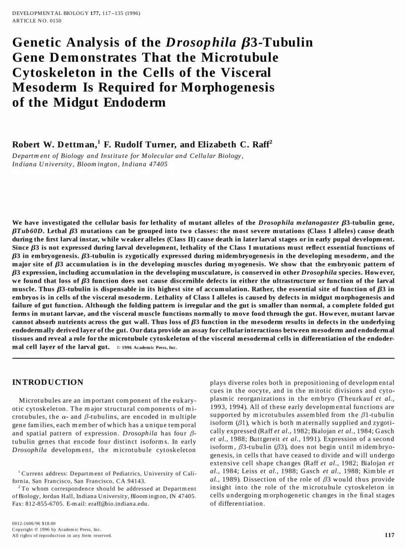
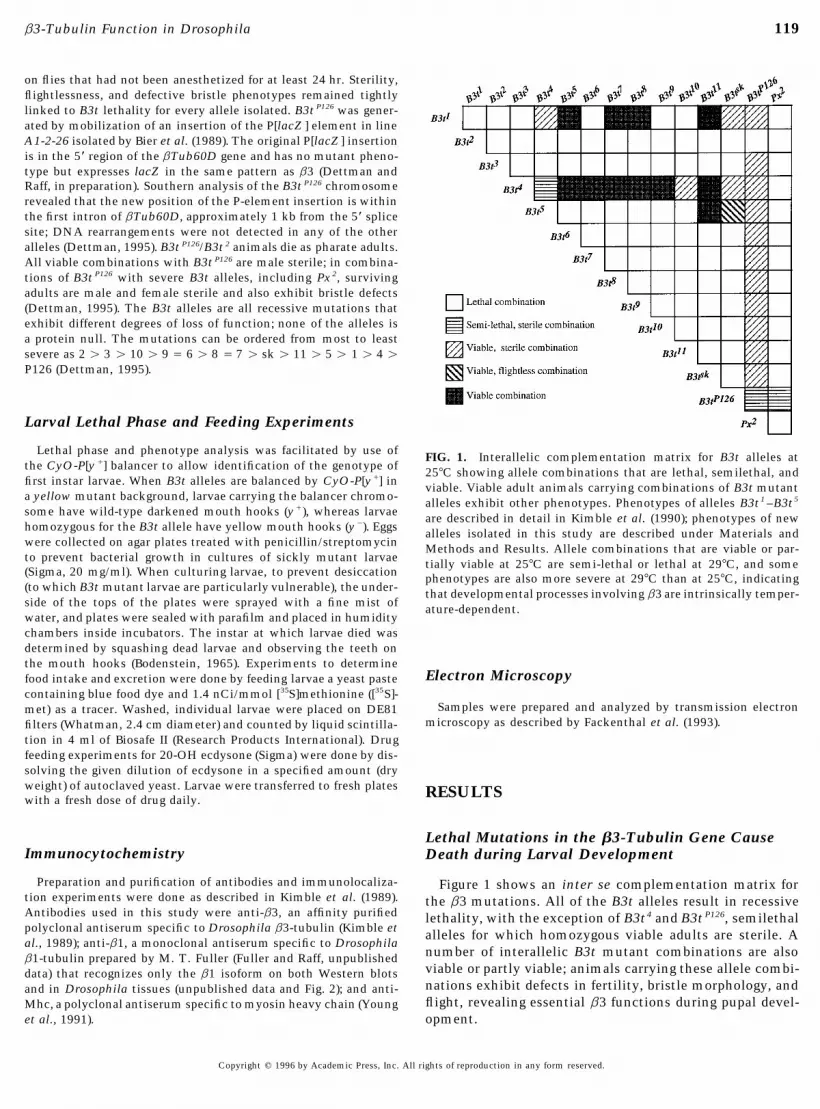
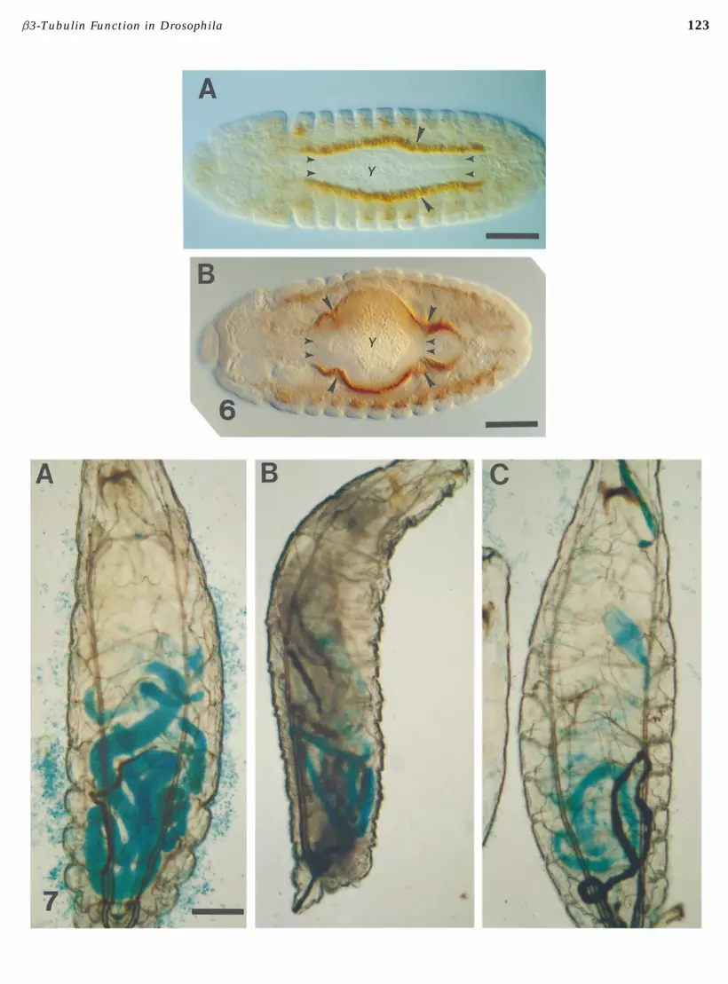
FIG. 2. b1-tubulin does not accumulate in the body wall musculature. (A) Stage 16 wild-type embryo stained with anti-b3. b3accumulates in the dorso-lateral body wall muscles (small arrowheads) but is not detectable in the apodemes (large arrowhead). (Band C) Stage 16 wild-type embryo stained with anti-b1. Higher magnification (B) shows that b1 is accumulated in the apodemes andin clusters of neuronal cells adjacent to the body wall muscles (large arrowheads), but is not accumulated above background levelsin the dorso-lateral body wall muscles. Generalized staining of b1 is also visible in the gut and other tissues. Lower magnification(C) shows the overall localization pattern of b1 in the late embryo. b1 is present at high levels in the tissues in which it is zygoticallyexpressed (nervous system and apodemes), but maternally derived b1 is also present throughout the embryo. Bar in A represents 0.03mm, for A and B. Bar in C is 0.1 mm.FIG. 3. b3 homologs accumulate in the embryonic mesoderm of D. virilis and D. simulans in a pattern similar to that in D. melanogaster.(A) Stage 16 wild-type D. melanogaster embryo stained with anti-b3. The predominant site of accumulation is in the somatic musculature;b3-staining can also be seen in the pharyngeal and gut muscles (arrowheads). (B and C) D. simulans embryos stained with anti-b3.Substantial accumulation of the b3 homolog in the somatic muscles can be seen in both embryos. Arrowheads in B indicate anti-b3staining in the pharyngeal muscles (left) and the dorsal vessel (right). Arrowheads in C indicate anti-b3 staining in macrophages. (D–F )D. virilis embryos stained with anti-b3. Accumulation of the b3 homolog in the body wall muscles can be seen in all embryos; the focalplane in E shows the pattern of the somatic musculature. The arrowhead in D indicates staining in the pharyngeal muscles; arrowheadsin F indicate staining in the macrophages. Embryos are oriented with the anterior end to the left. Bar in A represents 0.08 mm. Bar in Fis 0.06 mm, for B –F.FIG. 6. b3 expression in the visceral mesoderm and the apposition of the visceral mesoderm and endoderm during formation of themidgut. Embryos are stained with anti-b3, showing the accumulation of b3 in the visceral mesoderm (large arrowheads) that surroundsthe columnar endodermal cells of the gut (small arrowheads). The yolk mass (Y) is visible in the lumen of the gut. (A) Early stage 14embryo, prior to constriction of the midgut. The visceral mesoderm cells are also columnar at this stage. (B) Later stage 14 embryo, whenconstriction of the midgut is proceeding, and the visceral mesodermal cells have flattened. Large arrowheads indicate accumulation ofb3 at the site of midgut constriction. Bars are 0.08 mm.FIG. 7. B3t 2 larvae have morphologically abnormal midguts. Larvae tested were fed yeast paste mixed with blue food dye to visualizegut morphology. All larvae are first instar larvae, oriented with their anterior ends to the top of the page. (A) Wild-type larva, showingnormal midgut morphology. (B and C) Examples of smaller midguts typical of Class I mutant larvae: (B) B3t 2 homozygote. (C) B3t2/Px 2
larva. Bar in A represents 0.08 mm, for all panels.
Copyright q 1996 by Academic Press, Inc. All rights of reproduction in any form reserved.
AID DB 8230 / 6X0f$$$$21 06-14-96 18:18:34 dba AP: Dev Bio
122
06-14-96 18:18:34 dba AP: Dev Bio
123b3-Tubulin Function in Drosophila
06-14-96 18:18:34 dba AP: Dev Bio

124 Dettman, Turner, and Raff
Myogenesis and Sarcomere Integrity Are Normal in were present in all of the developing muscles we examined(Figs. 4E and 4F). We also examined the ultrastructure ofHomozygous Px2 Embryos and in B3t Mutant
Larvae and Adults body wall, gut, and pharyngeal muscle in first instar larvaehemizygous for B3t 2, the most severe recessive lethal allele.
Since the Px 2 chromosome deletes the bTub60D gene, As predicted from the homozygous Px2 phenotype, the mus-we anticipated that in homozygous Px 2 embryos we could cles in B3t 2/Px 2 larvae were indistinguishable from thoseexamine muscle formation in the absence of microtubules of wild-type controls (Figs. 4G and 4H). We did not observeand that the defects in morphogenesis or function of the any qualitative difference between wild-type controls andmuscles would reveal the specific function played in myo- b3 mutants.genesis by the b3-based microtubule cytoskeleton. We The results described above were such a surprise to usknew that loss of b3 function does not prevent formation of that we constructed another prd, Px2 stock and repeatedmuscle, since in prelethal terminal stage Px2 homozygotes, our analysis in order to eliminate the possibility that thesomatic muscles are present and moreover can undergo co- original stock had broken down and that in the first experi-ordinated rhythmic contractions along the body segments, ments the prd embryos we had examined were in fact notalbeit with much less vigor than in prehatching wild-type homozygous for Px 2. The results were the same in the sec-animals (Kimble et al., 1990; this study). We therefore ex- ond experiment: Muscle in homozygous prd, Px 2 embryosamined the organization of the muscle fibers in homozy- was normal. The only difference between wild-type animalsgous Px 2 embryos and in B3t mutants by immunolocaliza- and animals deficient for b3 that we could discern in eithertion and scanning electron microscopy (not shown). We experiment was that there were fewer microtubules in thefixed embryos laid by heterozygous Px2 or B3t n parents and muscle cells of Px 2 homozygotes than in wild-type controls.stained them with anti-Mhc, an antibody directed at muscle This observation was consistent with our expectation thatmyosin (see Materials and Methods). We examined muscle the size of the tubulin pool in myogenic cells would bemyosin staining in populations of stained embryos, one considerably diminished when b3 is absent.quarter of which will be the homozygotes for the mutant Since microtubules are still present in myogenic cells inallele being tested. We observed no defects in the muscle the absence of b3 expression, there must be another sourcefiber pattern in any of the embryos in any of the genotypes of b-tubulin. b1-tubulin is the only likely candidate, sinceexamined. We used SEM to examine the organization of b1 and b3 are the only isoforms expressed at the appropriatethe body wall muscle in homozygous Px 2 embryos at the time in development (Raff et al., 1982; Gasch et al., 1988).terminal stage of development (prehatching) and in B3t mu- Of the remaining two b-tubulin isoforms, b2 is exclusivelytant larvae. For each of the genotypes examined, muscles expressed in the male germ line (Kemphues et al., 1979,of the correct size and morphology were organized correctly 1982; Hoyle et al., 1995) and b4 is first expressed only dur-in the typical fiber pattern for each hemisegment. Thus ing late embryogenesis and is not detectable in muscle byoverall muscle development is not disrupted by loss of b3 immunolocalization (H. B. Diaz and E. C. Raff, unpublishedfunction. observations). Once the muscles have formed, we did not
We then examined myofibrillar development at the ultra- detect high levels of b1 in muscle in wild-type embryosstructural level in Px 2 homozygotes to determine if there (Fig. 2B). We considered the possibility that residual b1-were any defects in the cytoarchitecture of individual sar- tubulin from earlier stages in development might be prefer-comeres formed in the absence of b3. To identify Px 2 homo- entially stabilized when b3 is not expressed. To determinezygotes at the time when myogenesis occurs during midem- if b1-tubulin is present above wild-type levels in muscle inbryogenesis, the prd, Px2 stock was utilized as described in Px 2 homozygotes, we collected stage 16 and 17 embryosKimble et al. (1990). The prd segmentation defect allows from Px 2/CyO parents and stained them with anti-b1.identification of homozygous prd, Px2 embryos, but the prd There was not a population of embryos with preferentialmutation does not affect the structure of muscle fibers accumulation of b1 (data not shown). We therefore con-within a given segment. Our results were unexpected. We cluded that b1 accumulation and/or stability is not en-analyzed a large number of sections of somatic muscle rep- hanced in Px2 homozygotes. Thus, in the absence of b3resenting multiple homozygous prd, Px2 embryos. Muscles expression, assembly of microtubules must be supported byin prd, Px 2 embryos were indistinguishable from muscles the maternally derived b1 that normally persists from ear-in wild-type embryos (Figs. 4E and 4F). We could identify lier stages in development.no intrinsic abnormalities in the sarcomeres of the prd, Px 2 Formation of muscle fibers begins during early stage 13embryos either in longitudinal or cross sections. Neither and is complete by stage 16 (Campos-Ortega andthe length of the sarcomeres nor the organization in cross Hartenstein, 1985; and see Fig. 4). We observed that micro-section were appreciably different in the prd, Px 2 embryos tubule assembly in the myogenic cells begins in stage 12compared to wild-type controls, based on counts of thick (Figs. 4A and 4E). For homozygous Px 2 embryos, therefore,and thin filaments in cross sections and measurements of the question is the amount of maternally derivedb1 protein,the length of the sarcomeres in longitudinal sections (data and hence the size of the available tubulin pool, that isnot shown). Not only was the structure of sarcomeres in present when microtubule assembly commences in the ini-
tial stages of myogenesis. Available data show that the ma-homozygous prd, Px 2 embryos normal, but microtubules
Copyright q 1996 by Academic Press, Inc. All rights of reproduction in any form reserved.
AID DB 8230 / 6X0f$$$$21 06-14-96 18:18:34 dba AP: Dev Bio
125b3-Tubulin Function in Drosophila
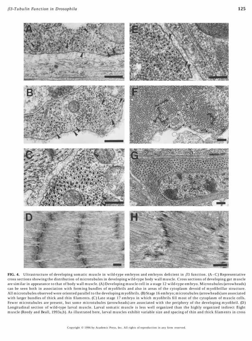
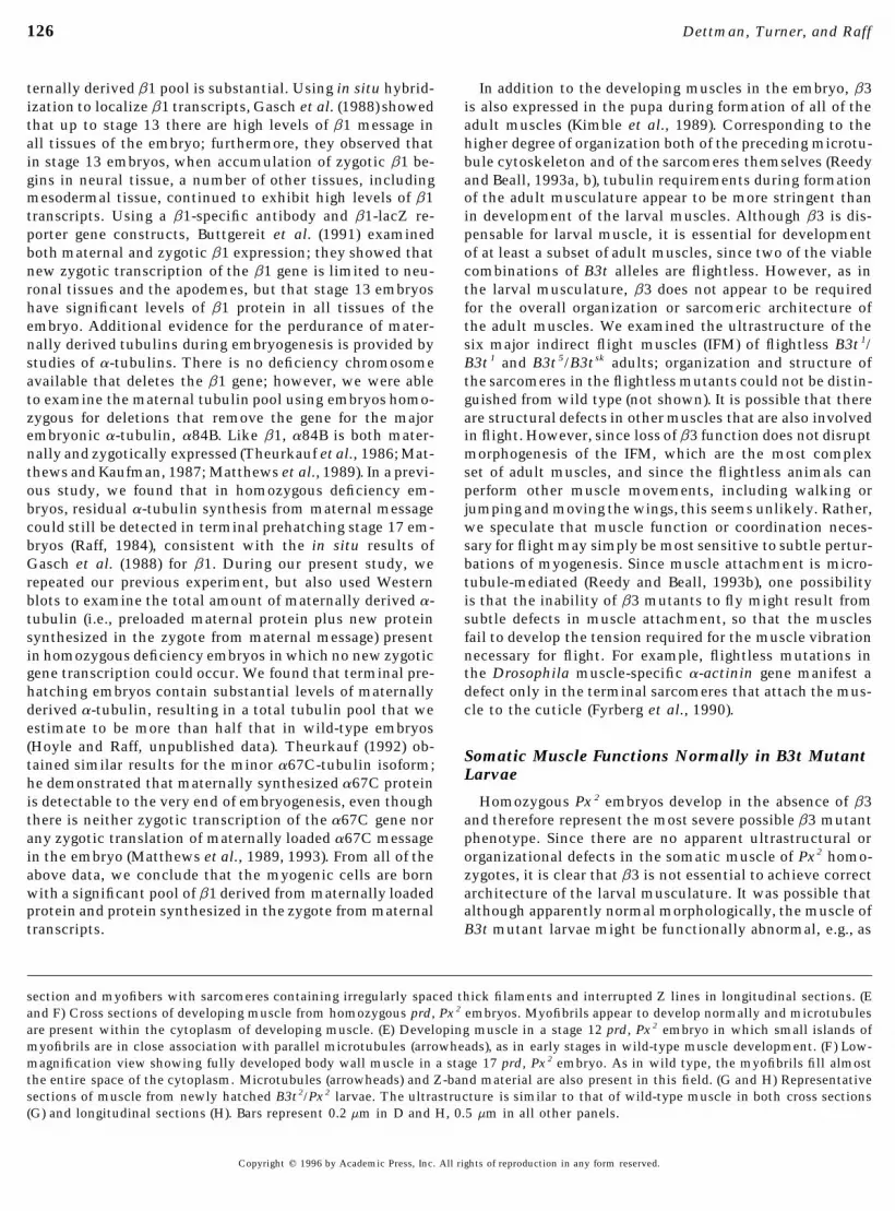
FIG. 4. Ultrastructure of developing somatic muscle in wild-type embryos and embryos deficient in b3 function. (A–C) Representativecross sections showing the distribution of microtubules in developing wild-type body wall muscle. Cross sections of developing gut muscleare similar in appearance to that of body wall muscle. (A) Developing muscle cell in a stage 12 wild-type embryo. Microtubules (arrowheads)can be seen both in association with forming bundles of myofibrils and also in areas of the cytoplasm devoid of myofibrillar structure.All microtubules observed were oriented parallel to the developing myofibrils. (B) Stage 16 embryo; microtubules (arrowheads) are associatedwith larger bundles of thick and thin filaments. (C) Late stage 17 embryo in which myofibrils fill most of the cytoplasm of muscle cells.Fewer microtubules are present, but some microtubules (arrowheads) are associated with the periphery of the developing myofibril. (D)Longitudinal section of wild-type larval muscle. Larval somatic muscle is less well organized than the highly organized indirect flightmuscle (Reedy and Beall, 1993a,b). As illustrated here, larval muscles exhibit variable size and spacing of thin and thick filaments in cross
Copyright q 1996 by Academic Press, Inc. All rights of reproduction in any form reserved.
AID DB 8230 / 6X0f$$8230 06-14-96 18:18:34 dba AP: Dev Bio
126 Dettman, Turner, and Raff
ternally derived b1 pool is substantial. Using in situ hybrid- In addition to the developing muscles in the embryo, b3is also expressed in the pupa during formation of all of theization to localize b1 transcripts, Gasch et al. (1988) showed
that up to stage 13 there are high levels of b1 message in adult muscles (Kimble et al., 1989). Corresponding to thehigher degree of organization both of the preceding microtu-all tissues of the embryo; furthermore, they observed that
in stage 13 embryos, when accumulation of zygotic b1 be- bule cytoskeleton and of the sarcomeres themselves (Reedyand Beall, 1993a, b), tubulin requirements during formationgins in neural tissue, a number of other tissues, including
mesodermal tissue, continued to exhibit high levels of b1 of the adult musculature appear to be more stringent thanin development of the larval muscles. Although b3 is dis-transcripts. Using a b1-specific antibody and b1-lacZ re-
porter gene constructs, Buttgereit et al. (1991) examined pensable for larval muscle, it is essential for developmentof at least a subset of adult muscles, since two of the viableboth maternal and zygotic b1 expression; they showed that
new zygotic transcription of the b1 gene is limited to neu- combinations of B3t alleles are flightless. However, as inthe larval musculature, b3 does not appear to be requiredronal tissues and the apodemes, but that stage 13 embryos
have significant levels of b1 protein in all tissues of the for the overall organization or sarcomeric architecture ofthe adult muscles. We examined the ultrastructure of theembryo. Additional evidence for the perdurance of mater-
nally derived tubulins during embryogenesis is provided by six major indirect flight muscles (IFM) of flightless B3t 1/B3t 1 and B3t 5/B3tsk adults; organization and structure ofstudies of a-tubulins. There is no deficiency chromosome
available that deletes the b1 gene; however, we were able the sarcomeres in the flightless mutants could not be distin-guished from wild type (not shown). It is possible that thereto examine the maternal tubulin pool using embryos homo-
zygous for deletions that remove the gene for the major are structural defects in other muscles that are also involvedin flight. However, since loss of b3 function does not disruptembryonic a-tubulin, a84B. Like b1, a84B is both mater-
nally and zygotically expressed (Theurkauf et al., 1986; Mat- morphogenesis of the IFM, which are the most complexset of adult muscles, and since the flightless animals canthews and Kaufman, 1987; Matthews et al., 1989). In a previ-
ous study, we found that in homozygous deficiency em- perform other muscle movements, including walking orjumping and moving the wings, this seems unlikely. Rather,bryos, residual a-tubulin synthesis from maternal message
could still be detected in terminal prehatching stage 17 em- we speculate that muscle function or coordination neces-sary for flight may simply be most sensitive to subtle pertur-bryos (Raff, 1984), consistent with the in situ results of
Gasch et al. (1988) for b1. During our present study, we bations of myogenesis. Since muscle attachment is micro-tubule-mediated (Reedy and Beall, 1993b), one possibilityrepeated our previous experiment, but also used Western
blots to examine the total amount of maternally derived a- is that the inability of b3 mutants to fly might result fromsubtle defects in muscle attachment, so that the musclestubulin (i.e., preloaded maternal protein plus new protein
synthesized in the zygote from maternal message) present fail to develop the tension required for the muscle vibrationnecessary for flight. For example, flightless mutations inin homozygous deficiency embryos in which no new zygotic
gene transcription could occur. We found that terminal pre- the Drosophila muscle-specific a-actinin gene manifest adefect only in the terminal sarcomeres that attach the mus-hatching embryos contain substantial levels of maternally
derived a-tubulin, resulting in a total tubulin pool that we cle to the cuticle (Fyrberg et al., 1990).estimate to be more than half that in wild-type embryos(Hoyle and Raff, unpublished data). Theurkauf (1992) ob-
Somatic Muscle Functions Normally in B3t Mutanttained similar results for the minor a67C-tubulin isoform;Larvaehe demonstrated that maternally synthesized a67C protein
is detectable to the very end of embryogenesis, even though Homozygous Px 2 embryos develop in the absence of b3and therefore represent the most severe possible b3 mutantthere is neither zygotic transcription of the a67C gene nor
any zygotic translation of maternally loaded a67C message phenotype. Since there are no apparent ultrastructural ororganizational defects in the somatic muscle of Px2 homo-in the embryo (Matthews et al., 1989, 1993). From all of the
above data, we conclude that the myogenic cells are born zygotes, it is clear that b3 is not essential to achieve correctarchitecture of the larval musculature. It was possible thatwith a significant pool of b1 derived from maternally loaded
protein and protein synthesized in the zygote from maternal although apparently normal morphologically, the muscle ofB3t mutant larvae might be functionally abnormal, e.g., astranscripts.
section and myofibers with sarcomeres containing irregularly spaced thick filaments and interrupted Z lines in longitudinal sections. (Eand F) Cross sections of developing muscle from homozygous prd, Px2 embryos. Myofibrils appear to develop normally and microtubulesare present within the cytoplasm of developing muscle. (E) Developing muscle in a stage 12 prd, Px2 embryo in which small islands ofmyofibrils are in close association with parallel microtubules (arrowheads), as in early stages in wild-type muscle development. (F) Low-magnification view showing fully developed body wall muscle in a stage 17 prd, Px2 embryo. As in wild type, the myofibrils fill almostthe entire space of the cytoplasm. Microtubules (arrowheads) and Z-band material are also present in this field. (G and H) Representativesections of muscle from newly hatched B3t2/Px2 larvae. The ultrastructure is similar to that of wild-type muscle in both cross sections(G) and longitudinal sections (H). Bars represent 0.2 mm in D and H, 0.5 mm in all other panels.
Copyright q 1996 by Academic Press, Inc. All rights of reproduction in any form reserved.
AID DB 8230 / 6X0f$$$$21 06-14-96 18:18:34 dba AP: Dev Bio
127b3-Tubulin Function in Drosophila
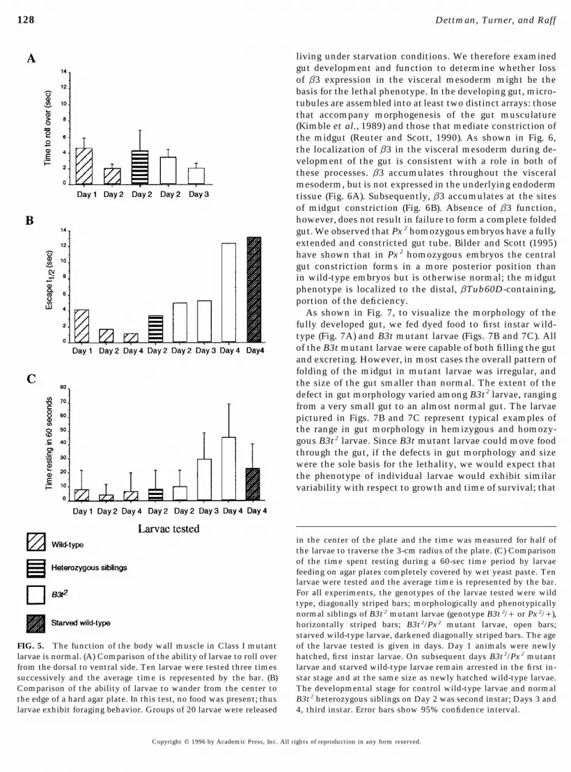
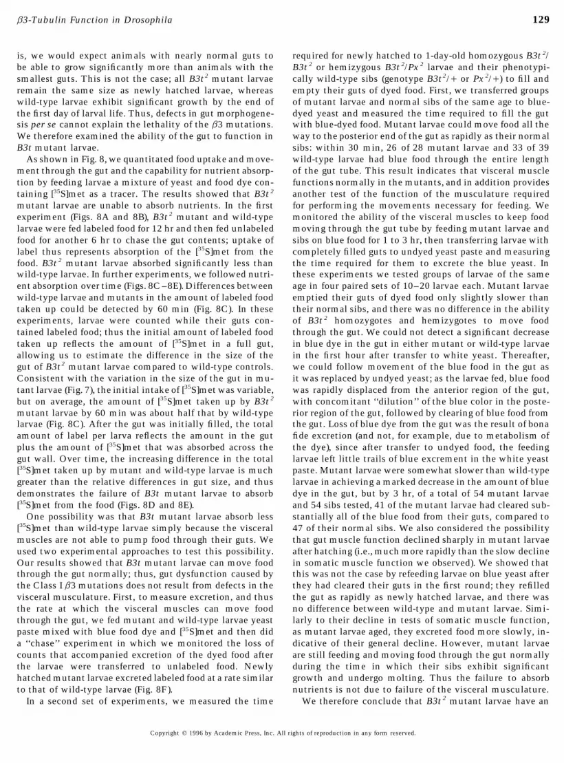
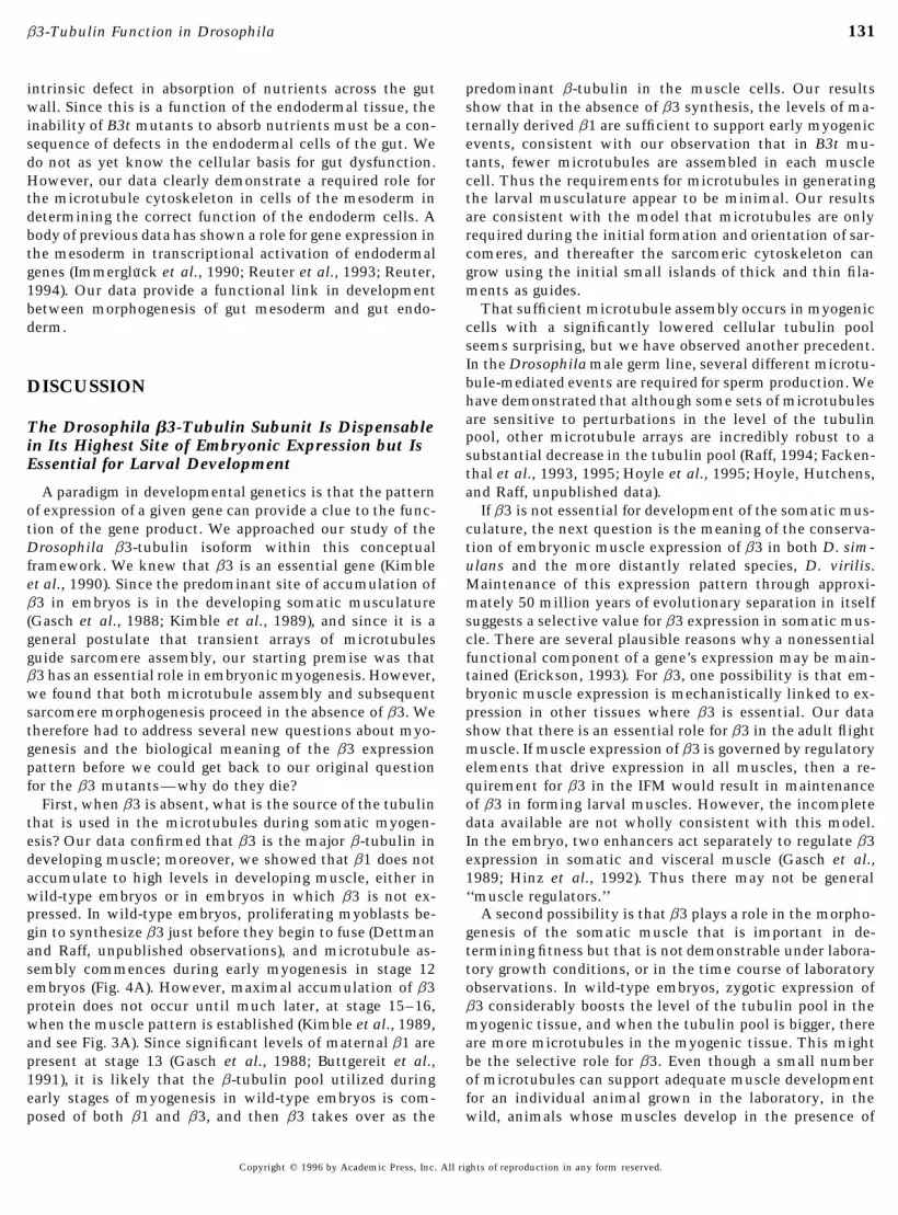
appears to be the case for the flight muscle in flightless B3t very little during feeding. However, aging mutant larvae restfor increasing amounts of time.mutant adults. We therefore tested somatic muscle func-
The data in Fig. 5 show that somatic muscle functiontion in mutant larvae hemizygous or homozygous for theand coordination are not intrinsically defective in larvaemost severe allele, B3t2. Hemizygous B3t2 larvae were vid-hemizygous for B3t 2. Since B3t2 is not a protein null allele,eotaped in motion to observe locomotion and contractionsthese experiments may not reflect the condition of larvalof the segmental body wall muscles. Newly hatched B3t 2
muscles that have developed in the complete absence of b3hemizygotes were able to contract their body wall musclesfunction. However, the phenotype resulting from loss ofnormally and move at the same rate as wild-type controlb3 function cannot be more severe than the phenotype oflarvae. Additional tests to examine other aspects of muscleanimals homozygous for the Px 2 deficiency, which deletesmovements in larvae developed from embryos defective in12 other essential genes in addition to the b3 gene (Kimbleb3 function were modeled on assays for the larval pheno-et al., 1990). Px 2 results in lethality during stage 17, thetypes of mutations in the foraging and a-actinin genes offinal stage of embryonic development. Thus, the only possi-Drosophila (deBelle et al., 1989; Roulier et al., 1992). Mu-ble lethal phase for the hypothetical B3t null allele is be-tant larvae were examined in behavioral assays to measuretween late stage 17 and after hatching to the L1 stage. Boththe time taken to complete various muscular movementsthe somatic and visceral muscles are morphologically nor-that require coordination of different muscle contractions.mal in Px 2 homozygotes. Moreover, somatic muscle in Px 2
Our expectation was that if the function of some or all ofhomozygotes is functional at least to some extent, sincethe body muscles were defective, then the mutant animalsprelethal embryos undergo muscle movements, includingwould not be able to perform them, or would take longerrhythmic contractions along the body wall. Thus our datato perform these movements than wild-type larvae. Asunambiguously show that b3 is not required either for em-shown in Fig. 5, all tests showed that the somatic musclesbryonic myogenesis or for the basic features of larval musclefunction normally in B3t2 mutant animals.contraction and coordination. Since we do not knowWe first measured the time required for an individualwhether B3t 2 is a complete loss of function allele, we can-larva to roll onto its ventral side after it was manually rollednot eliminate the possibility that the quality of muscleonto its dorsal side (Fig. 5A). The time taken to roll overfunction might be diminished by a true b3 null mutation.varied from animal to animal even in wild-type larvae, butIndeed, the B3t adult flightless phenotypes demonstratethere was no difference in the average time for mutant andthat perturbing b3 function can affect muscle functioncontrol larvae of similar sizes to roll over, nor did we ob-without causing gross defects in sarcomere architecture.serve any difference in the manner in which they rolledHowever, since somatic muscle function is normal in B3t 2
over. As wild-type larvae grew, they took longer to roll overmutant animals, the lethal phenotype reveals that b3 has(not shown), corresponding to the large size difference be-an essential role in some other developmental process. Intween first and third instar larvae.order to understand why B3t mutants die, we had to defineIn the second test, ‘‘the maggot circus,’’ we measured thethe other role(s) of b3 in embryogenesis.half-time elapsed for groups of mutant larvae to move 3 cm
The key feature of the Class I lethal phenotype is the failurefrom the center of a hard agar plate to the edge, providing
to grow coupled with terminal arrest at the first larval instar.a test of whether mutants could control the coordination Starvation has been shown to cause larval arrest in other spe-of their muscle contractions in forward motion (Fig. 5B). In cies of insects (Wigglesworth, 1972). We therefore examinedthe first two days of larval life, mutants were able to reach the effects of starvation on wild-type larvae, as an approachthe outer edge of the plate at a rate equal to that of control to understanding the failure of Class I mutants to grow orlarvae. As mutant larvae aged they became increasingly molt. We found that starving wild-type larvae remain arrestedsluggish and moved much more slowly than larger wild- in the first instar, similar to B3t Class I mutant larvae. Starv-type larvae of the same age; however, we interpreted this ing wild-type larvae continued to actively forage throughoutto be the result of a general prelethal state rather than paral- the period of the experiment and did not exhibit the rapidysis or a specific loss in muscle coordination. decline in vitality shown by B3t mutant larvae. As long as
The third test was to measure the proportion of time indi- starving larvae remained hydrated, most lived for many days;vidual mutant larvae ‘‘rest’’ (i.e., cease making food scraping starving larvae up to 10 days old were able to recover andmovements) while feeding on wet yeast on a hard agar plate complete development after they were placed back onto food.(Fig. 5C). Class I mutant larvae fail to grow any larger than Thus starving wild-type larvae are more robust than B3t mu-the size of newly hatched larvae, even though they live for tant larvae. However, in our behavioral tests for muscle func-up to 3 days after hatching. We speculated that failure to grow tion, starving wild-type larvae behaved similarly to B3t mu-might reflect inability to perform the muscle movements nec- tant larvae (Figs. 5B and 5C).essary for scraping food during feeding. However, similar to
Midgut Morphology and Gut Function Arethe other tests, although observations of feeding behavior re-Defective in b3 Mutant Larvae, but Visceralvealed a general decline over time in the vitality of B3t mutantMuscle Functions Normallylarvae, we did not observe any specific defect in the ability to
perform feeding movements or in the amount of time spent The partial phenocopy of the Class I phenotype by starva-tion of wild-type larvae suggested that mutant larvae arefeeding. Wild-type larvae and recently hatched B3t2 larvae rest
Copyright q 1996 by Academic Press, Inc. All rights of reproduction in any form reserved.
AID DB 8230 / 6X0f$$$$21 06-14-96 18:18:34 dba AP: Dev Bio
128 Dettman, Turner, and Raff
living under starvation conditions. We therefore examinedgut development and function to determine whether lossof b3 expression in the visceral mesoderm might be thebasis for the lethal phenotype. In the developing gut, micro-tubules are assembled into at least two distinct arrays: thosethat accompany morphogenesis of the gut musculature(Kimble et al., 1989) and those that mediate constriction ofthe midgut (Reuter and Scott, 1990). As shown in Fig. 6,the localization of b3 in the visceral mesoderm during de-velopment of the gut is consistent with a role in both ofthese processes. b3 accumulates throughout the visceralmesoderm, but is not expressed in the underlying endodermtissue (Fig. 6A). Subsequently, b3 accumulates at the sitesof midgut constriction (Fig. 6B). Absence of b3 function,however, does not result in failure to form a complete foldedgut. We observed that Px 2 homozygous embryos have a fullyextended and constricted gut tube. Bilder and Scott (1995)have shown that in Px 2 homozygous embryos the centralgut constriction forms in a more posterior position thanin wild-type embryos but is otherwise normal; the midgutphenotype is localized to the distal, bTub60D-containing,portion of the deficiency.
As shown in Fig. 7, to visualize the morphology of thefully developed gut, we fed dyed food to first instar wild-type (Fig. 7A) and B3t mutant larvae (Figs. 7B and 7C). Allof the B3t mutant larvae were capable of both filling the gutand excreting. However, in most cases the overall pattern offolding of the midgut in mutant larvae was irregular, andthe size of the gut smaller than normal. The extent of thedefect in gut morphology varied among B3t2 larvae, rangingfrom a very small gut to an almost normal gut. The larvaepictured in Figs. 7B and 7C represent typical examples ofthe range in gut morphology in hemizygous and homozy-gous B3t2 larvae. Since B3t mutant larvae could move foodthrough the gut, if the defects in gut morphology and sizewere the sole basis for the lethality, we would expect thatthe phenotype of individual larvae would exhibit similarvariability with respect to growth and time of survival; that
in the center of the plate and the time was measured for half ofthe larvae to traverse the 3-cm radius of the plate. (C) Comparisonof the time spent resting during a 60-sec time period by larvaefeeding on agar plates completely covered by wet yeast paste. Tenlarvae were tested and the average time is represented by the bar.For all experiments, the genotypes of the larvae tested were wildtype, diagonally striped bars; morphologically and phenotypicallynormal siblings of B3t2 mutant larvae (genotype B3t 2// or Px2//),horizontally striped bars; B3t2/Px2 mutant larvae, open bars;starved wild-type larvae, darkened diagonally striped bars. The ageof the larvae tested is given in days. Day 1 animals were newlyFIG. 5. The function of the body wall muscle in Class I mutanthatched, first instar larvae. On subsequent days B3t2/Px2 mutantlarvae is normal. (A) Comparison of the ability of larvae to roll overlarvae and starved wild-type larvae remain arrested in the first in-from the dorsal to ventral side. Ten larvae were tested three timesstar stage and at the same size as newly hatched wild-type larvae.successively and the average time is represented by the bar. (B)The developmental stage for control wild-type larvae and normalComparison of the ability of larvae to wander from the center toB3t2 heterozygous siblings on Day 2 was second instar; Days 3 andthe edge of a hard agar plate. In this test, no food was present; thus4, third instar. Error bars show 95% confidence interval.larvae exhibit foraging behavior. Groups of 20 larvae were released
Copyright q 1996 by Academic Press, Inc. All rights of reproduction in any form reserved.
AID DB 8230 / 6X0f$$$$21 06-14-96 18:18:34 dba AP: Dev Bio
129b3-Tubulin Function in Drosophila
is, we would expect animals with nearly normal guts to required for newly hatched to 1-day-old homozygous B3t 2/B3t 2 or hemizygous B3t 2/Px 2 larvae and their phenotypi-be able to grow significantly more than animals with the
smallest guts. This is not the case; all B3t2 mutant larvae cally wild-type sibs (genotype B3t2// or Px 2//) to fill andempty their guts of dyed food. First, we transferred groupsremain the same size as newly hatched larvae, whereas
wild-type larvae exhibit significant growth by the end of of mutant larvae and normal sibs of the same age to blue-dyed yeast and measured the time required to fill the gutthe first day of larval life. Thus, defects in gut morphogene-
sis per se cannot explain the lethality of the b3 mutations. with blue-dyed food. Mutant larvae could move food all theway to the posterior end of the gut as rapidly as their normalWe therefore examined the ability of the gut to function in
B3t mutant larvae. sibs: within 30 min, 26 of 28 mutant larvae and 33 of 39wild-type larvae had blue food through the entire lengthAs shown in Fig. 8, we quantitated food uptake and move-
ment through the gut and the capability for nutrient absorp- of the gut tube. This result indicates that visceral musclefunctions normally in the mutants, and in addition providestion by feeding larvae a mixture of yeast and food dye con-
taining [35S]met as a tracer. The results showed that B3t 2 another test of the function of the musculature requiredfor performing the movements necessary for feeding. Wemutant larvae are unable to absorb nutrients. In the first
experiment (Figs. 8A and 8B), B3t 2 mutant and wild-type monitored the ability of the visceral muscles to keep foodmoving through the gut tube by feeding mutant larvae andlarvae were fed labeled food for 12 hr and then fed unlabeled
food for another 6 hr to chase the gut contents; uptake of sibs on blue food for 1 to 3 hr, then transferring larvae withcompletely filled guts to undyed yeast paste and measuringlabel thus represents absorption of the [35S]met from the
food. B3t 2 mutant larvae absorbed significantly less than the time required for them to excrete the blue yeast. Inthese experiments we tested groups of larvae of the samewild-type larvae. In further experiments, we followed nutri-
ent absorption over time (Figs. 8C–8E). Differences between age in four paired sets of 10–20 larvae each. Mutant larvaeemptied their guts of dyed food only slightly slower thanwild-type larvae and mutants in the amount of labeled food
taken up could be detected by 60 min (Fig. 8C). In these their normal sibs, and there was no difference in the abilityof B3t 2 homozygotes and hemizygotes to move foodexperiments, larvae were counted while their guts con-
tained labeled food; thus the initial amount of labeled food through the gut. We could not detect a significant decreasein blue dye in the gut in either mutant or wild-type larvaetaken up reflects the amount of [35S]met in a full gut,
allowing us to estimate the difference in the size of the in the first hour after transfer to white yeast. Thereafter,we could follow movement of the blue food in the gut asgut of B3t2 mutant larvae compared to wild-type controls.
Consistent with the variation in the size of the gut in mu- it was replaced by undyed yeast; as the larvae fed, blue foodwas rapidly displaced from the anterior region of the gut,tant larvae (Fig. 7), the initial intake of [35S]met was variable,
but on average, the amount of [35S]met taken up by B3t 2 with concomitant ‘‘dilution’’ of the blue color in the poste-rior region of the gut, followed by clearing of blue food frommutant larvae by 60 min was about half that by wild-type
larvae (Fig. 8C). After the gut was initially filled, the total the gut. Loss of blue dye from the gut was the result of bonafide excretion (and not, for example, due to metabolism ofamount of label per larva reflects the amount in the gut
plus the amount of [35S]met that was absorbed across the the dye), since after transfer to undyed food, the feedinglarvae left little trails of blue excrement in the white yeastgut wall. Over time, the increasing difference in the total
[35S]met taken up by mutant and wild-type larvae is much paste. Mutant larvae were somewhat slower than wild-typelarvae in achieving a marked decrease in the amount of bluegreater than the relative differences in gut size, and thus
demonstrates the failure of B3t mutant larvae to absorb dye in the gut, but by 3 hr, of a total of 54 mutant larvaeand 54 sibs tested, 41 of the mutant larvae had cleared sub-[35S]met from the food (Figs. 8D and 8E).
One possibility was that B3t mutant larvae absorb less stantially all of the blue food from their guts, compared to47 of their normal sibs. We also considered the possibility[35S]met than wild-type larvae simply because the visceral
muscles are not able to pump food through their guts. We that gut muscle function declined sharply in mutant larvaeafter hatching (i.e., much more rapidly than the slow declineused two experimental approaches to test this possibility.
Our results showed that B3t mutant larvae can move food in somatic muscle function we observed). We showed thatthis was not the case by refeeding larvae on blue yeast afterthrough the gut normally; thus, gut dysfunction caused by
the Class I b3 mutations does not result from defects in the they had cleared their guts in the first round; they refilledthe gut as rapidly as newly hatched larvae, and there wasvisceral musculature. First, to measure excretion, and thus
the rate at which the visceral muscles can move food no difference between wild-type and mutant larvae. Simi-larly to their decline in tests of somatic muscle function,through the gut, we fed mutant and wild-type larvae yeast
paste mixed with blue food dye and [35S]met and then did as mutant larvae aged, they excreted food more slowly, in-dicative of their general decline. However, mutant larvaea ‘‘chase’’ experiment in which we monitored the loss of
counts that accompanied excretion of the dyed food after are still feeding and moving food through the gut normallyduring the time in which their sibs exhibit significantthe larvae were transferred to unlabeled food. Newly
hatched mutant larvae excreted labeled food at a rate similar growth and undergo molting. Thus the failure to absorbnutrients is not due to failure of the visceral musculature.to that of wild-type larvae (Fig. 8F).
In a second set of experiments, we measured the time We therefore conclude that B3t 2 mutant larvae have an
Copyright q 1996 by Academic Press, Inc. All rights of reproduction in any form reserved.
AID DB 8230 / 6X0f$$$$21 06-14-96 18:18:34 dba AP: Dev Bio

130 Dettman, Turner, and Raff
FIG. 8. B3t 2 larvae can move food through the gut, but absorption of nutrients is defective. (A and B) Results from two separateexperiments in which the capability for absorption of nutrients from the gut was monitored by uptake of [35S]met from the food. Larvaewere fed yeast paste containing [35S]met for 12 hr, washed, and allowed to feed for 6 hr on nonradioactive yeast paste (chase), and [35S]metuptake was determined. Symbols are wild-type larvae, diagonally striped bars; homozygous B3t2 larvae, open bars. Day 1 animals werenewly hatched, first instar larvae. On subsequent days homozygous B3t2 mutant larvae remain arrested in the first instar stage and remainthe same size as newly hatched wild-type larvae. Day 3 wild-type larvae are in the third instar. Twenty larvae were counted for each timepoint, except for Day 3 B3t 2 larvae, in which five were counted. (A) includes a control experiment showing background counts obtainedfor wild-type larvae fed on yeast with no [35S]met added. Error bars show 95% confidence intervals. (C–E) Time course experimentsquantifying total uptake of [35S]met in newly hatched wild-type larvae (solid squares) compared to newly hatched homozygous B3t2 mutantlarvae (open squares) or 2-day-old B3t2 mutant larvae (open circles). Fifty to 100 larvae were placed on food containing [35S]met and 5 (C)or 10 (D and E) larvae were removed at each time point, washed, and counted. (F) Excretion by wild-type larvae (solid squares) and B3t2
homozygous larvae (open squares). Larvae were fed [35S]met-containing food for 4 hr, and then placed on unlabeled food. Five larvae wereremoved at each time point, washed, and counted. The upturn in the last time point for wild-type larvae reflects the reconsumption oflabeled excretion products. Error bars (C–F) indicate the range on the graphs for one standard deviation unit.
AID DB 8230 / 6X0f$$823006-14-96 18:18:34 dba AP: Dev Bio
131b3-Tubulin Function in Drosophila
intrinsic defect in absorption of nutrients across the gut predominant b-tubulin in the muscle cells. Our resultsshow that in the absence of b3 synthesis, the levels of ma-wall. Since this is a function of the endodermal tissue, the
inability of B3t mutants to absorb nutrients must be a con- ternally derived b1 are sufficient to support early myogenicevents, consistent with our observation that in B3t mu-sequence of defects in the endodermal cells of the gut. We
do not as yet know the cellular basis for gut dysfunction. tants, fewer microtubules are assembled in each musclecell. Thus the requirements for microtubules in generatingHowever, our data clearly demonstrate a required role for
the microtubule cytoskeleton in cells of the mesoderm in the larval musculature appear to be minimal. Our resultsare consistent with the model that microtubules are onlydetermining the correct function of the endoderm cells. A
body of previous data has shown a role for gene expression in required during the initial formation and orientation of sar-comeres, and thereafter the sarcomeric cytoskeleton canthe mesoderm in transcriptional activation of endodermal
genes (Immergluck et al., 1990; Reuter et al., 1993; Reuter, grow using the initial small islands of thick and thin fila-ments as guides.1994). Our data provide a functional link in development
between morphogenesis of gut mesoderm and gut endo- That sufficient microtubule assembly occurs in myogeniccells with a significantly lowered cellular tubulin poolderm.seems surprising, but we have observed another precedent.In the Drosophila male germ line, several different microtu-bule-mediated events are required for sperm production. WeDISCUSSIONhave demonstrated that although some sets of microtubulesare sensitive to perturbations in the level of the tubulinThe Drosophila b3-Tubulin Subunit Is Dispensablepool, other microtubule arrays are incredibly robust to ain Its Highest Site of Embryonic Expression but Issubstantial decrease in the tubulin pool (Raff, 1994; Facken-Essential for Larval Developmentthal et al., 1993, 1995; Hoyle et al., 1995; Hoyle, Hutchens,and Raff, unpublished data).A paradigm in developmental genetics is that the pattern
of expression of a given gene can provide a clue to the func- If b3 is not essential for development of the somatic mus-culature, the next question is the meaning of the conserva-tion of the gene product. We approached our study of the
Drosophila b3-tubulin isoform within this conceptual tion of embryonic muscle expression of b3 in both D. sim-ulans and the more distantly related species, D. virilis.framework. We knew that b3 is an essential gene (Kimble
et al., 1990). Since the predominant site of accumulation of Maintenance of this expression pattern through approxi-mately 50 million years of evolutionary separation in itselfb3 in embryos is in the developing somatic musculature
(Gasch et al., 1988; Kimble et al., 1989), and since it is a suggests a selective value for b3 expression in somatic mus-cle. There are several plausible reasons why a nonessentialgeneral postulate that transient arrays of microtubules
guide sarcomere assembly, our starting premise was that functional component of a gene’s expression may be main-tained (Erickson, 1993). For b3, one possibility is that em-b3 has an essential role in embryonic myogenesis. However,
we found that both microtubule assembly and subsequent bryonic muscle expression is mechanistically linked to ex-pression in other tissues where b3 is essential. Our datasarcomere morphogenesis proceed in the absence of b3. We
therefore had to address several new questions about myo- show that there is an essential role for b3 in the adult flightmuscle. If muscle expression of b3 is governed by regulatorygenesis and the biological meaning of the b3 expression
pattern before we could get back to our original question elements that drive expression in all muscles, then a re-quirement for b3 in the IFM would result in maintenancefor the b3 mutants—why do they die?
First, when b3 is absent, what is the source of the tubulin of b3 in forming larval muscles. However, the incompletedata available are not wholly consistent with this model.that is used in the microtubules during somatic myogen-
esis? Our data confirmed that b3 is the major b-tubulin in In the embryo, two enhancers act separately to regulate b3expression in somatic and visceral muscle (Gasch et al.,developing muscle; moreover, we showed that b1 does not
accumulate to high levels in developing muscle, either in 1989; Hinz et al., 1992). Thus there may not be general‘‘muscle regulators.’’wild-type embryos or in embryos in which b3 is not ex-
pressed. In wild-type embryos, proliferating myoblasts be- A second possibility is that b3 plays a role in the morpho-genesis of the somatic muscle that is important in de-gin to synthesize b3 just before they begin to fuse (Dettman
and Raff, unpublished observations), and microtubule as- termining fitness but that is not demonstrable under labora-tory growth conditions, or in the time course of laboratorysembly commences during early myogenesis in stage 12
embryos (Fig. 4A). However, maximal accumulation of b3 observations. In wild-type embryos, zygotic expression ofb3 considerably boosts the level of the tubulin pool in theprotein does not occur until much later, at stage 15–16,
when the muscle pattern is established (Kimble et al., 1989, myogenic tissue, and when the tubulin pool is bigger, thereare more microtubules in the myogenic tissue. This mightand see Fig. 3A). Since significant levels of maternal b1 are
present at stage 13 (Gasch et al., 1988; Buttgereit et al., be the selective role for b3. Even though a small numberof microtubules can support adequate muscle development1991), it is likely that the b-tubulin pool utilized during
early stages of myogenesis in wild-type embryos is com- for an individual animal grown in the laboratory, in thewild, animals whose muscles develop in the presence ofposed of both b1 and b3, and then b3 takes over as the
Copyright q 1996 by Academic Press, Inc. All rights of reproduction in any form reserved.
AID DB 8230 / 6X0f$$$$21 06-14-96 18:18:34 dba AP: Dev Bio
132 Dettman, Turner, and Raff
more microtubules may be at an advantage over animals dermal cells during the initial stages of gut folding is shownin Fig. 6B. Throughout these processes, endodermal cellswhose muscles develop with fewer microtubules. Expres-
sion of the b3 gene may not bring an essential specialized must differentiate, since the final endodermal layer of thelarval midgut contains multiple cell types (Dimitriadis andfunctional property to myogenesis, but may serve to regu-
late the size of the tubulin pool. We think that this explana- Kastritsis, 1984). Several events in midgut morphogenesiscould require cytoskeletal mediated cell–cell interactions.tion may be the most likely for the evolutionary conserva-
tion of the embryonic b3 expression pattern. Reuter and colleagues (1993) have shown that cell migrationand the establishment of the midgut epithelium requiresthe visceral mesoderm. They propose that the mesodermal
Disruption of b3-Tubulin Function Reveals That layer provides developmental cues required for cell migra-the Cytoskeleton of Visceral Mesodermal Cells Is tion. These signals could be provided either by secretedEssential for Morphogenesis of the Endoderm molecules or transmembrane cell adhesion proteins in the
overlying mesodermal cells. In addition, they postulatedOur results show that the essential requirement for b3expression is in the visceral mesoderm and that b3 has mul- that since proteins such as Notch and Delta are expressed
in both the mesoderm and the endoderm, they could betiple roles in morphogenesis of the larval gut. Loss of b3function results in defects in midgut morphogenesis, and essential components of a mechanism that mediates the
close association and transient cell shape changes of endo-perhaps in some variability in the robustness of visceralmuscle function. However, while these factors may contrib- dermal and mesodermal cells. The placement of such sig-
nals and the ability of cells to change shape may depend onute to the severity of the mutant phenotype, the proximalcause of lethality in the Class I B3t mutants is defective the proper function of the cytoskeleton in mesodermal cells.
Utilization of the b3 isoform to satisfy tubulin require-function of the endodermal tissue of the gut. Previous stud-ies have demonstrated an inductive relationship between ments in cells where the microtubule cytoskeleton is in-
volved in mediating cell–cell interactions is not unique togene expression in the visceral mesoderm and the endoderm(Immergluck et al., 1990; Reuter et al., 1993; Reuter, 1994). the interactions between mesoderm and endoderm. Analy-
sis of the male sterility phenotype of viable B3t mutantOur data reveal that disruption of cell structure in one layercan affect developmental events in the other layer. adults has revealed a similar requirement for b3 in de-
termining somatic cell–germ cell interactions. In the testis,There are two direct roles for b3 in visceral mesodermcells, but for which b3 is at least partly dispensable. First, b3 is expressed in the somatic cells that enclose each cyst
of developing germ cells (Kimble et al., 1989). We havewe have shown that transient cytoskeletal microtubulesaccompany sarcomere formation in the visceral muscles; shown that loss of b3 function in the somatic cells disrupts
crucial developmental events in the germ cells (Raff andhowever, myogenesis is little if at all affected by disruptionof b3 function. Second, microtubules are involved in con- Dettman, unpublished observations). We hypothesize that
just as there are cell signaling and attachment molecules,striction and folding of the midgut. Reuter and Scott (1990)observed dense microtubule arrays in the three sites of con- such as Notch and Delta, that mediate many different cell–
cell interactions, there may also be generalized cytoskeletalstriction of visceral mesoderm closest to the yolk mass,beginning at stage 14. Since b3 is expressed in these cells components, of which b3 is one, that function in such con-
texts in numerous cell types.and since the gut is often abnormally shaped in b3 mutants,it is likely that b3 is required in gut constriction furrowmicrotubules. Gut constriction microtubules are assembled
The Phenotypes of Class I and Class II b3later than the early myogenic microtubules; their functionMutations Suggest Additional Developmental Rolesmay be optimal in the presence of b3 because maternal b1-for b3 in Sensory Perception and Ecdysistubulin is more depleted by this time. However, since a
complete and folded (albeit abnormally) gut forms in Px2 Other factors in addition to starvation resulting from gutdysfunction must contribute to the B3t mutant phenotype,homozygotes, assembly of the gut constriction microtu-
bules is not dependent on the presence of b3. since B3t mutant larvae are less vigorous and die muchsooner than starving wild-type larvae. Class I B3t mutantThe gut defects in Class I b3 mutations reveal an unex-
pected and essential role for the b3-containing microtubule larvae exhibit behavioral abnormalities that suggest a rolefor b3 in sensory perception. Recently hatched mutant lar-cytoskeleton in visceral mesoderm cells in directing mor-
phogenesis of the midgut endoderm. To form the midgut, vae feed actively, but they invariably begin to exhibit wan-dering behavior even when they are still in the presence ofendodermal cells migrate from two sites to form a single
luminal epithelial cell layer that is surrounded by the meso- a source of food. This behavior does not appear to be similarto the wandering behavior of wild-type third instar larvaedermally derived muscle cell layer (Reuter et al., 1993).
After cell migration is completed, cells in both layers ex- prior to pupariation; we did not observe any other behav-ioral or physical characteristics of the onset of pupariation,pand into contiguous layers of columnar cells that interact
to fold the midgut, as shown in Fig. 6A. After folding of the such as tanning or hardening of the cuticle, in the wanderingClass I larvae. Rather, even when food is present, the behav-midgut, endodermal and mesodermal cells flatten sur-
rounding the yolk. The change in configuration of the meso- ior of the mutants appears similar to the foraging behavior
Copyright q 1996 by Academic Press, Inc. All rights of reproduction in any form reserved.
AID DB 8230 / 6X0f$$$$21 06-14-96 18:18:34 dba AP: Dev Bio

133b3-Tubulin Function in Drosophila
exhibited by starving wild-type larvae, suggesting that Class addition of ecdysone to the food also had no effect on ClassI mutant larvae. Although the Class I lethality results fromI B3t mutant larvae cannot sense the presence of food. This
might be the result of defects in the ability to detect food developmental defects that are ‘‘upstream’’ of the defectsin ecdysis revealed by the Class II phenotypes, loss of b3via chemosensory organs or to sense that the gut is filled
via stretch receptors. We have identified two sites of zygotic function in the responsible cells may be a contributing fac-tor in the Class I lethality.b3 expression in which defective microtubule function
might account for the abnormal behavior of Class I mutants.We have observed accumulation of b3 in the support cellsof the chordotonal organs, the mechanosensory organs in
What Is the General Role for b3 in Development?the body wall, and also in sets of previously undefined cellsin the anterior-most portion of the embryo, which we postu-
The complex developmental pattern of expression of b3,late may be sensory organs (Dettman and Raff, in prepara-coupled with our finding that b3 is not essential in a num-tion). A precedent for such a role for microtubules can beber of cell types in which it is expressed, raises the questionfound in the touch receptor cells in C. elegans, in whichof the specific functions that b3 performs in the microtu-the expression of the mec7 b-tubulin gene is required forbule cytoskeleton. Differential expression of multiple tu-assembly of the array of microtubules that serve as rigidbulin genes may provide an organism with a repertoire ofcytoskeletal elements whose deformation mediates mecha-tubulin isoforms, each of which has perhaps subtly distinctnochemical signal transduction (Savage et al., 1989). How-assembly properties, selected in evolution for optimizationever, in Drosophila, loss of b3 does not completely abolishof microtubule function in different cellular contexts. Alter-touch sensitivity, since even prelethal homozygous Px2 em-natively, different isoforms might be functionally equiva-bryos respond to touch by undergoing muscle contraction,lent but differential expression might serve to allow controlnor did we observe any structural abnormalities in theover the time, place, and amount of tubulin synthesis inchordotonal organs in B3t mutants (Dettman and Raff, indifferent cell types. In fungi, even structurally diverged iso-preparation).forms have been shown to be functionally interchangeableThe Class II B3t lethal phenotype reveals a requirementin vivo (Schatz et al., 1986; May, 1989), whereas develop-for b3 in ecdysis. At death, Class II larvae often possessmental studies in Drosophila have provided evidence formultiple mouth parts, suggesting difficulty in shedding thefunctional specialization of tubulin isoforms (Hoyle andcuticle after a molt, and small spots of tanned cuticle, show-Raff, 1990; Hoyle et al., 1995; Matthews et al., 1993). Weing incomplete entry into the pupal stage. The steroid hor-cannot eliminate the possibility that b3 has properties thatmone ecdysone, produced by the ring gland, is an essentialmay be specialized for some subset of its sites of expression;signal that induces larval molts and pupariation in insectsin fact we suspect that this is likely. However, we would(Wigglesworth, 1972). There are two possible clues to a rela-argue that our observations on the utilization of b3 in devel-tionship between developmental processes involving ecdy-opment are consistent with the possibility that the generalsone and b3. We have found that b3 is expressed in the ringrole for b3 expression may be to provide a way for cells togland (Raff and Dettman, in preparation), and there is alsocontrol tubulin pool size. First, in previous experiments, weevidence that b3 is an ecdysone-responsive gene (Bruhat etshowed that b3 is considerably restricted in its functionalal., 1990; Andres et al., 1993). Thus b3 might function incapacity relative to the b2-tubulin isoform utilized in themicrotubule-mediated events required for generating or re-male germ line (Hoyle and Raff, 1990; Hoyle et al., 1995).sponding to ecdysone. We examined the effects of includingSecond, in its normal sites of expression, b3 is expressed in1–5 mg/ml ecdysone in the diet of wild-type or mutantterminally differentiated cells that require microtubules tolarvae, amounts shown by Garen et al. (1977) to bypassmediate cell shape changes and tissue interactions. b3 ex-larval arrest in ecdysone mutants. Ecdysone had a deleteri-pression may serve to boost the tubulin pool in such cellsous effect on the development of wild-type larvae: 80% ofand would be essential only in cells in which accumulationanimals died as larvae, exhibiting pleiotropic defects, in-of other b-tubulins is below some critical threshold. Thiscluding improper shedding of the cuticle during moltinghypothesis is a testable one: If the role of b3 in other cellsand cuticular tanning, similar to the lethal phenotypes ofis primarily to augment the tubulin pool, then just as myo-Class II mutant larvae. Addition of ecdysone to the food ofgenic cells are capable of utilizing the pre-existing b1 pool,homozygous Class II larvae had no effect on most larvae,expression of another b-tubulin isoform should be able tobut some larvae died earlier than the normal lethal period.fulfill b3’s roles in cells where b3 is normally essential.The Class II phenotype thus more closely resembles theThis is precisely what has been observed in fungi, where aneffect of excess rather than insufficient levels of hormone.‘‘essential’’ tubulin isoform may be replaced by another,We tested whether exogenous ecdysone could overcomestructurally diverged isoform so long as the tubulin pool isthe developmental arrest caused by starvation by rearingmaintained above some minimal level (Schatz et al., 1986;starving, arrested wild-type larvae on a glycerol solutionMay, 1989). We are currently testing the ability of the b1containing ecdysone. None of these larvae molted. Thusisoform to rescue mutant phenotypes that result from lossexogenous ecdysone is not sufficient to induce molting in
the absence of growth. As expected, given these results, of b3 function.
Copyright q 1996 by Academic Press, Inc. All rights of reproduction in any form reserved.
AID DB 8230 / 6X0f$$$$21 06-14-96 18:18:34 dba AP: Dev Bio

134 Dettman, Turner, and Raff
Dettman, R. W. (1995). A genetic analysis of a developmentallyACKNOWLEDGMENTSregulated b-tubulin in Drosophila melanogaster. Ph.D. thesis,Indiana University.
We thank H. Hoyle, J. Hutchens, S. Karsan, and G. Montgomery Dimitriadis, V. K., and Kastritsis, C. D. (1984). Ultrastructural anal-for valuable assistance in this work, M. Reedy for assistance in ysis of the midgut of Drosophila auraria larvae. Morphologicalanalyzing the structure of the indirect flight muscle in flightless observations and their physiological implications. Can. J. Zool.b3 mutant adults, and D. Kiehart for the anti-Mhc antiserum. We 62, 659–669.thank R. A. Raff for critical reading of the manuscript. Our study
Erickson, H. P. (1993). Gene knockouts of c-src, transformingwas supported by a research grant to E.C.R. from the U.S. Public
growth factor b1, and enascin suggest superfluous, nonfunctionalHealth Service (RO1 HD-16739). R.W.D. was supported in part asexpression of proteins. J. Cell Biol. 120, 1079–1081.a predoctoral trainee under a Department of Health and Human
Fackenthal, J. D., Turner, F. R., and Raff, E. C. (1993). Tissue-spe-Services National Research Service Training Grant awarded to thecific microtubule functions in Drosophila spermatogenesis re-Indiana University Department of Biology. Assistance to S. Karsanquire the b2-tubulin isotype-specific carboxy terminus. Dev.and G. Montgomery was also provided by a Howard Hughes Under-Biol. 158, 213–227.graduate Research award to the Department of Biology at Indiana
Fackenthal, J. D., Hutchens, J. A., Turner, F. R., and Raff, E. C.University.(1995). Structural analysis of mutations in the Drosophila b2-tubulin isoform reveal regions in the b-tubulin molecule requiredfor general and for tissue-specific functions. Genetics 139, 267 –
REFERENCES 286.FlyBase. (1994). The Drosophila Genetic Database. Nucleic Acids
Res. 22, 3456–3458. [Available from the flybase.bio.indiana.eduAndres, J. A., Fletcher, J. C., Karim, F. D., and Thummel, C. S.network server and Gopher site](1993). Molecular analysis of the initiation of insect metamor-
Fyrberg, E., Kelly, M., Ball, E., Fyrberg, C., and Reedy, M. C. (1990).phosis: A comparative study of Drosophila ecdysteroid-regulatedMolecular genetics of Drosophila alpha-actinin: Mutant allelestranscription. Development 160, 388 –404.disrupt Z disc integrity and muscle insertions. J. Cell Biol. 110,Bialojan, S., Falkenburg, D., and Renkawitz-Pohl, R. (1984). Charac-1999–2011.terization and developmental expression of b-tubulin genes in
Drosophila melanogaster. EMBO J. 3, 2543–2548. Garen, A., Kauvar, L., and Lepesant, J.-A. (1977). Roles of ecdysoneBier, E., Vassin, H., Shepherd, S., Lee, K., McCall, K., Barbel, S., in Drosophila development. Proc. Natl. Acad. Sci. USA 74, 5099–
Ackerman, L., Carretto, R., Uemura, T., and Grell, E. (1989). 5103.Searching for pattern and mutation in the Drosophila genome Gasch, A., Hinz, U., Leiss, D., and Renkawitz-Pohl, R. (1988). Thewith a P-lacZ vector. Genes Dev. 3, 1273–1287. expression of b1 and b3 tubulin genes of Drosophila melanogas-
Bilder, D., and Scott, M. P. (1995). Genomic regions required for ter is spatially regulated during embryogenesis. Mol. Gen. Genet.the morphogenesis of the Drosophila embryonic midgut. Genet- 211, 8–16.ics 141, 1087–1100. Gasch, A., Hinz, U., and Renkawitz-Pohl, R. (1989). Intron and
Bischoff, R., and Holtzer, H. (1968). The effect of mitotic inhibitors upstream sequences regulate expression of the Drosophila beta-on myogenesis in vitro. J. Cell Biol. 36, 111–127. 3-tubulin gene in the visceral and somatic musculature, respec-
Bodenstein, D. (1965). The postembryonic development of Dro- tively. Proc. Natl. Acad. Sci. USA 86, 3215–3218.sophila., In ‘‘Biology of Drosophila ’’ (M. Demerec, Ed.), pp. 275– Gonczy, P., Viswanathan, S., and DiNardo, S. (1992). Probing sper-367. Haefner, New York/London. matogenesis in Drosophila with P-element enhancer detectors.
Bruhat, A., Tourmente, S., Chapel., S., Sobrier, M. L., Couderc, J. L., Development 114, 89 –98.and Dastugue, B. (1990). Regulatory elements in the first intron Gorczyca, M. G., Phillis, R. W., and Budnik, V. (1994). The role ofcontribute to transcriptional regulation of the b3 tubulin gene tinman, a mesodermal fate gene, in axon pathfinding during theby 20-hydroxy ecdysone in Drosophila Kc cells. Nucleic Acids development of the transverse nerve in Drosophila. DevelopmentRes. 18, 2861–2867. 120, 2143–2152.
Buttgereit, D., Leiss, D., Michiels, F., and Renkawitz-Pohl, R.Hinz, U., Wolk, A., and Renkawitz-Pohl, R. (1992). Ultrabithorax
(1991). During Drosophila embryogenesis the beta-1 tubulin geneis a regulator of b3- tubulin expression in the Drosophila visceralis specifically expressed in the nervous system and the apodemes.mesoderm. Development 116, 543–554.Mech. Dev. 33, 107–118.
Hoyle, H. D., and Raff, E. C. (1990). Two Drosophila beta-tubulinCampos-Ortega, J. A., and Hartenstein, V. (1985). ‘‘The Embryonicisoforms are not functionally equivalent. J. Cell Biol. 111, 1009–Development of Drosophila melanogaster.’’ Springer-Verlag,1026.New York.
Hoyle, H. D., Hutchens, J. A., Turner, F. R., and Raff, E. C. (1995).Crossley, A. C. (1972a). Ultrastructural changes during transitionRegulation of b-tubulin function and expression in Drosophilaof larval to adult intersegmental muscle at metamorphosis inspermatogenesis. Dev. Genet. 16, 148–170.the blowfly Calliphora erythrocephala. I. Dedifferentiation and
Immergluck, K., Lawrence, P. A., and Bienz, M. (1990). Inductionmyoblast fusion. J. Embryol. Exp. Morphol. 27, 43–74.across germ layers in Drosophila mediated by a genetic cascade.Crossley, A. C. (1972b). Ultrastructural changes during transitionCell 62, 261–268.of larval to adult intersegmental muscle at metamorphosis in
Kemphues, K. J., Raff, R. A., Kaufman, T. C., and Raff, E. C. (1979).the blowfly Calliphora erythrocephala. II. The formation of adultMutation in a structural gene for a b-tubulin specific to testis inmuscle. J. Embryol. Exp. Morphol. 27, 75 –101.Drosophila melanogaster. Proc. Natl. Acad. Sci. USA 76, 3991–de Belle, J. S., Hilliker, A. J., and Sokolowski, M. B. (1989). Genetic3995.localization of foraging (for): A major gene for larval behavior in
Drosophila melanogaster. Genetics 123, 157–163. Kemphues, K. J., Kaufman, T. C., Raff, R. A., and Raff, E. C. (1982).
Copyright q 1996 by Academic Press, Inc. All rights of reproduction in any form reserved.
AID DB 8230 / 6X0f$$$$21 06-14-96 18:18:34 dba AP: Dev Bio

135b3-Tubulin Function in Drosophila
The testis-specific b-tubulin subunit in Drosophila melanogaster flight muscle in Drosophila. II. Formation of the myotendonjunction. Dev. Biol. 160, 466–479.has multiple functions in spermatogenesis. Cell 31, 655–670.
Reuter, R., and Scott, M. P. (1990). Expression and function of theKimble, M., Incardona, J. P., and Raff, E. C. (1989). A variant b-homeotic genes Antennapadia and Sex combs reduced in thetubulin isoform of Drosophila melanogaster (b3) is expressedembryonic midgut of Drosophila. Development 109, 289 –303.primarily in tissues of mesodermal origin in embryos and pupae,
Reuter, R., Grunewald, B., and Leptin, M. (1993). A role for theand is utilized in populations of transient microtubules. Dev.mesoderm in endodermal migration and morphogenesis in Dro-Biol. 131, 415–429.sophila. Development 119, 1135–1145.Kimble, M., Dettman, R. W., and Raff, E. C. (1990). The b3-tubulin
Reuter, R. (1994). The gene serpent has homeotic properties andgene of Drosophila melanogaster is essential for viability andspecifies endoderm versus ectoderm within the Drosophila gut.fertility. Genetics 126, 991–1005.Development 120, 1123–1135.Leiss, D., Hinz, U., Gasch, A., Mertz, R., and Renkawitz-Pohl, R.
Roulier, E. M., Fyrberg, C., and Fyrberg, E. A. (1992). Perturbations(1988). b3-tubulin expression characterizes the differential meso-of Drosophila a-actinin cause muscle paralysis, weakness and
dermal germ layer during Drosophila embryogenesis. Develop-atrophy but do not confer obvious nonmuscle phenotypes. J. Cell
ment 104, 525–532. Biol. 116, 911–922.Lindsley, D. L., and Zimm, G. G. (1992). ‘‘The Genome of Drosoph- Savage, C., Hamelin, M., Culotti, J. G., Coulson, A., Albertson,
ila melanogaster.’’ Academic Press, San Diego. D. G., and Chalfie, M. (1989). mec-7 is a b-tubulin gene requiredMatthews, K. A., and Kaufman, T. C. (1987). Developmental conse- for the production of 15-protofilament microtubules in Caeno-
quences of mutations in the 84B alpha-tubulin gene of Drosoph- rhabditis elegans. Genes Dev. 3, 870–881.ila melanogaster. Dev. Biol. 119, 100–114. Schatz, P. J., Solomon, F., and Botstein, D. (1986). Genetically es-
Matthews, K. A., Miller, D. F., and Kaufman, T. C. (1989). Develop- sential and nonessential a-tubulin genes specify functionally in-mental distribution of RNA and protein products of the Drosoph- terchangeable proteins. Mol. Cell. Biol. 6, 3722–3733.ila alpha-tubulin gene family. Dev. Biol. 132, 45–61. Tassin, A. M., Maro, B., and Bornens, M. (1985). Fate of microtubule
organizing centers during myogenesis in vitro. J. Cell Biol. 100,Matthews, K. A., Rees, D., and Kaufman, T. C. (1993). A function-35–46.ally specialized alpha-tubulin is required for oocyte meiosis and
Theurkauf, W. E., Alberts, B. M., Jan, Y. N., and Jongens, T. A.cleavage mitoses in Drosophila. Development 117, 977–991.(1993). A central role for microtubules in the differentiation ofMay, G. S. (1989). The highly divergent b-tubulins of AspergillusDrosophila oocytes. Development 118, 1169–1180.nidulans are functionally interchangeable. J. Cell Biol. 109,
Theurkauf, W. E. (1994). Premature microtubule-dependent cyto-2267–2274.plasmic streaming in cappuccino and spire mutant oocytes. Sci-Michiels, F., Falkenburg, D., Muller, A., Hinz, U., Otto, U., Bell-ence 265, 2093–2096.man, R., Glatzer, K. H., Brand, R., Bialojan, S., and Renkawitz-
Theurkauf, W. E. (1992). Behavior of structurally divergent alpha-Pohl, R. (1987). Testis-specific b2 tubulins are identical in D.tubulin isotypes during Drosophila embryogenesis: Evidence formelanogaster and D. hydeii but differ from the ubiquitous b1post-translational regulation of isotype abundance. Dev. Biol.
tubulin. Chromosoma 95, 387–395. 154, 205–217.Persitianis, G. C., and Gregory, D. W. (1971). Early stages of flight Theurkauf, W. E., Baum, H., Bo, J., and Wensink, P. C. (1986).
muscle development in the blowfly Lucilia cuprina: A light and Tissue-specific and constitutive alpha-tubulin genes of Drosoph-electron microscopic study. J. Insect Physiol. 17, 1005–1022. ila melanogaster code for structurally distinct proteins. Proc.
Raff, E. C., Fuller, M. T., Kaufman, T. C., Kemphues, K., Rudolph, Natl. Acad. Sci. USA 83, 8477–8481.J., and Raff, R. A. (1982). Regulation of tubulin gene expression Toyama, Y., Forry-Schaudies, S., Hoffman, B., and Holtzer, H.during embryogenesis in Drosophila melanogaster. Cell 28, 33– (1982). Effects of taxol and colcemid on myofibrillogenesis. Proc.40. Natl. Acad. Sci. USA 79, 6556–6560.
Raff, E. C. (1984). The genetics of microtubule systems. J. Cell Biol. Warren, R. H. (1974). Microtubular organization in elongating myo-99, 1–10. genic cells. J. Cell Biol. 63, 550–566.
Wigglesworth, V. B. (1972). ‘‘The Principles of Insect Physiology.’’Raff, E. C. (1994). The role of multiple tubulin isoforms in cellularChapman–Hall, London.microtubule function. In ‘‘Microtubules’’ (J. S. Hyams, and
Young, P. E., Pesacreta, T. C., and Kiehart, D. P. (1991). DynamicC. W. Lloyd, Eds.), pp. 85–111. Wiley, New York.changes in the distribution of cytoplasmic myosin during Dro-Reedy, M. C., and Beall, C. (1993a). Ultrastructure of developingsophila embryogenesis. Development 111, 1–14.flight muscle in Drosophila. I. Assembly of myofibrils. Dev. Biol.
160, 443–465. Received for publication December 4, 1995Accepted April 4, 1996Reedy, M. C., and Beall, C. (1993b). Ultrastructure of developing
Copyright q 1996 by Academic Press, Inc. All rights of reproduction in any form reserved.
AID DB 8230 / 6X0f$$$$21 06-14-96 18:18:34 dba AP: Dev Bio