fungal foliar diseases of rauwolfiaserpentina in wild, its seasonaloccurrence, seed transmission...
TRANSCRIPT

This article was downloaded by: [Kuvempu Univ]On: 08 April 2013, At: 23:26Publisher: Taylor & FrancisInforma Ltd Registered in England and Wales Registered Number: 1072954 Registeredoffice: Mortimer House, 37-41 Mortimer Street, London W1T 3JH, UK
Archives Of Phytopathology And PlantProtectionPublication details, including instructions for authors andsubscription information:http://www.tandfonline.com/loi/gapp20
Fungal foliar diseases of Rauwolfiaserpentina in wild, its seasonaloccurrence, seed transmission anddisease managementThyagarthi Ramappa Parashurama a & Manchanahally ByrappaShivanna aa Department of Studies in Applied Botany, Kuvempu University,Shimoga, IndiaVersion of record first published: 08 Apr 2013.
To cite this article: Thyagarthi Ramappa Parashurama & Manchanahally Byrappa Shivanna(2013): Fungal foliar diseases of Rauwolfia serpentina in wild, its seasonal occurrence, seedtransmission and disease management, Archives Of Phytopathology And Plant Protection,DOI:10.1080/03235408.2013.772769
To link to this article: http://dx.doi.org/10.1080/03235408.2013.772769
PLEASE SCROLL DOWN FOR ARTICLE
Full terms and conditions of use: http://www.tandfonline.com/page/terms-and-conditions
This article may be used for research, teaching, and private study purposes. Anysubstantial or systematic reproduction, redistribution, reselling, loan, sub-licensing,systematic supply, or distribution in any form to anyone is expressly forbidden.
The publisher does not give any warranty express or implied or make any representationthat the contents will be complete or accurate or up to date. The accuracy of anyinstructions, formulae, and drug doses should be independently verified with primarysources. The publisher shall not be liable for any loss, actions, claims, proceedings,demand, or costs or damages whatsoever or howsoever caused arising directly orindirectly in connection with or arising out of the use of this material.

Fungal foliar diseases of Rauwolfia serpentina in wild, its seasonaloccurrence, seed transmission and disease management
Thyagarthi Ramappa Parashurama and Manchanahally Byrappa Shivanna*
Department of Studies in Applied Botany, Kuvempu University, Shimoga, India
(Received 16 January 2013; final version received 29 January 2013)
Rauwolfia serpentina is an important medicinal herb. Studies were conducted todetermine causal organisms of foliar diseases in R. serpentina in Bhadra WildlifeSanctuary during 2006–2009. The foliar disease incidence and its distribution anddisease severity in nine state forest regions of the sanctuary were determined.The seedborne nature and transmission of the causal organism was alsodetermined. The management of seedborne inoculum was done by seed dressingwith fungicides. The effect of foliage infection on secondary metabolite contentwas also determined. Results of the present study indicated that Cercosporarauwolfiae is major leaf spot disease causing pathogen. The disease is homoge-neously distributed through the study area. The foliar disease severity was highin Kagemanegiri forest during October–November. The minor leaf spot diseasecaused by Alternaria alternata occurred occasionally. Alternaria alternata is seed-borne and seed transmitted and could be managed by seed treatment with Captraor Hyzeb. The secondary metabolites like alkaloids and steroids decreased withincrease in foliar infection by C. rauwolfiae, while phenol and flavonoid contentsincreased. The study suggested that R. serpentina is affected in wild byC. rauwolfiae and A. alternata. The latter pathogen is seedborne and seed trans-mitted and controlled by seed treatment.
Keywords: Rauwolfia serpentina; Cercospora rauwolfiae; disease severity; seed-borne; Captra; secondary metabolites
Introduction
Rauwolfia serpentina Benth.,the Indian snake root, is an important Ayurvedic medicinalplant used for centuries for different medicinal properties in India. R. serpentina is agood source of alkaloids and dry root contains about 30 indole alkaloids (0.7–2.4%).Among various alkaloids, reserpine is the active constituent and is well known for itsanti-hypertensive action. It depletes the stores of catecholamines at nerve endings. Otherimportant alkaloids are ajmalicine, rescinnamine, yohimbine and serpentine (Chopraet al. 1980). Pure isolated alkaloids as well as their synthetic derivatives are used asbasic medicinal agents for their analgesic, anti-spasmodic and bactericidal effects (Stary1998; Okwu and Okwu 2004). In Indian systems of medicine, Indian snakeroot is alsoused for snake and reptile bites (Kokate et al. 2003), feverish intestinal diseases, liverailments, rheumatism, dropsy (oedema), general debilities, mental illness, epilepsy, ner-vousness (Ojha and Mishra 1985), insomnia and insanity. In view of its sharply rising
*Corresponding author. Email: [email protected]
Archives of Phytopathology and Plant Protection, 2013http://dx.doi.org/10.1080/03235408.2013.772769
� 2013 Taylor & Francis
Dow
nloa
ded
by [
Kuv
empu
Uni
v] a
t 23:
26 0
8 A
pril
2013

pharmaceutical value, the large-scale cultivation has been taken up in recent years inorder to maintain sustainable supply to the pharmaceutical industries. However, it isthreatened with extinction (Mao et al. 2009; Singh et al. 2010) due to over-exploitationby local people, government agencies and pharmaceutical companies (Mamgain et al.1998), and its loss in natural habitat (Sukumaran and Raj 2008) and limited cultivationowing to the poor seed viability and seed germination rate (Dutta et al. 1962).
A survey of forests in the Western ghats of India indicated that R. serpentina occursin Bhadra Wildlife sanctuary (BWLS) and is affected by foliar fungal diseases in seed-lings and mature plants during different seasons of the year in varying severity (Shivan-na et al. 2007). Rauwolfia serpentina is affected by a number of foliar diseases causedby Alternaria alternata, Cercospora rauwolfiae and Rhizoctonia solani which causeconsiderable damage to plants (Mohanty and Addy 1957; Ganguly and Pandotra 1962).These studies are limited to the description of diseases; however, a detailed study offoliar diseases throughout the year, the pathogen transmission and disease control islacking. In this study, an attempt was made to determine disease incidence and severityin the entire sanctuary through different growing stages and seasons, seedborne natureand transmission of pathogen(s) and pathogen management by seed treatment. Theeffect of foliar disease on certain secondary metabolite content was also determined.
Materials and methods
Selection of study area
Bhadra Wildlife sanctuary (13°34′ to 13°46′ N lat. and 75°29′ to 75°45′ E long), locatedin the southern central part of the Western Ghats region of Karnataka, is comprised of12 state forest regions with an area of 492.46 km2. Nine forest regions- Hebbegiri,Gangegiri, Madla, Lakkavalli, Aldara, Kakanahosudi, Madhuguni, Muthodi andKagemanegiri that run across Bhadravathi and Tarikere taluks of Shimoga and Chika-magalur districts, respectively, were selected as study regions. The study area supportsdry-deciduous, moist-deciduous and semi-evergreen forests and receives an annualrainfall of 1600–2000mm. In each study region, three study sites were identifiedrandomly; each of the study sites consisted of three quadrates (10� 10m2) representingthree replicates. Plant samples were collected from the study area and identified basedon their morphology (Gamble 1935; Yoganarasimhan et al. 1982) and in comparisonwith herbarial specimens in the department. The species identity was kindly confirmedby Prof. Emeritus G. R. Shivamurthy, University of Mysore, Mysore.
Isolation and characterisation of causal organisms and leaf bioassay
The diseased plants were studied for symptoms during different growing stages andseasons. The diseased plant parts of R. serpentina were collected in sterilisedpolypropylene covers, surface disinfected (0.2%, NaOCl, 2min), segmented into piecesand incubated on moistened blotter discs in Petri dishes under light–darkness cycle of12/12 h at 23 ± 2 °C for five days. The fungal species associated with incubated diseasedplant materials were identified based on their morphological characteristics by referringto identification manuals (Booth 1977; Barnett et al. 1998; Sivanesan 1983;Subramanian 1983) and by visiting Index Fungorum (www.indexfungorum.org). Thefungal species were cultured on PDA and their identity was confirmed by comparingwith the characteristics of fungal colonies on incubated diseased parts.
2 T. R. Parashurama and M.B. Shivanna
Dow
nloa
ded
by [
Kuv
empu
Uni
v] a
t 23:
26 0
8 A
pril
2013

The pathogenicity of fungal species associated with disease symptom was deter-mined by detached leaf bioassay technique (Shivanna and Mallikarjunaswamy 2009).For this purpose, surface disinfected apparently healthy leaves were prick-inoculatedwith sterile water or spore suspension (4� 106 or 6� 106 sporesml�1) of the fungalspecies cultured on PDA and incubated on moistened blotter discs as described previ-ously.
Seasonal occurrence and distribution of foliar disease in the sanctuary
The data of disease incidence (DI%) and severity (DS%) were recorded every monthfor a period of 36months during 2006–2009. The DI and DS were determined asdescribed (Shivanna and Mallikarjunaswamy 2009).
The distribution of foliar disease incidence in the study area during 2006–2009 wasanalysed by Taylor’s power law (Taylor 1984) using the data of disease incidence. Tay-lor’s power law relates the observed variance (Vobs) to the population mean (m) forcount data with no upper limit by
Vobs ¼ Amb ð1Þ
where A and b are parameters, binomial distribution [log (Vr)] equals the mean andestimated by regression after logarithmic transformation of Equation 1 to
logðVobsÞ ¼ logðAÞ þ blogðVrÞ ð2Þ
The parameter b is considered an index of aggregation, providing a measure of thedegree of “disease incidence dependence of aggregation” (Taylor and Taylor 1977). Forcount data, vobs =m (i.e. a= b = 1) indicates a random pattern of disease incidence asrepresented by a binomial distribution. Parameter estimates of a and b> 1 indicate heter-ogeneity as an aggregation at the scale of the sampling unit which is dependent on dis-ease incidence. Least squares regression was used to estimate the intercept and slopeparameters. Significance in relationships between log (vobs) and log (Vr) was determinedwith F-test. The coefficient of determination (R2) and the mean square error were usedto determine the goodness of fit of the model. Differences between the observed andpredicted variance were plotted against predicted values to evaluate the appropriatenessof the model. For comparative purpose and because of the importance of obtainingaccurate estimate of A and b, two other approaches were evaluated for estimatingparameters, namely geometric mean and resistant line regression. For geometric meanregression, the slope is always greater than the least squares slope (Madden et al. 1995;Gent et al. 2008). Least squares regression was used to estimate the intercept and slopeparameters of Taylor’s power law using MINITAB version 15 (Achar & Shivanna2013).
Determination of seed mycoflora
Seeds of R. serpentina were collected from the study sites in nine forest regions andseeds of the same range were mixed to obtain three samples representing Lakkavalli,Hebbe and Muthodi ranges. Four hundred seeds of R. serpentina from each range wereincubated (12 h alternating cycles of fluorescent light/darkness at 22 ± 2 °C for 7 days)by standard blotter method (Anonymous 2003). The fungal species occurring on
Archives of Phytopathology and Plant Protection 3
Dow
nloa
ded
by [
Kuv
empu
Uni
v] a
t 23:
26 0
8 A
pril
2013

incubated seeds were identified based on their morphological characteristics, asdescribed previously. Data of the fungal incidence in seeds were analysed by ANOVA.
Seed localisation and transmission of fungal pathogen
Seeds were subjected to component plating (Maden et al. 1975) to determine the locali-sation of fungal pathogen. Accordingly, seeds were dissected into seed coat, cotyledonand embryonic axis, which were surface disinfected and incubated by blotter method atroom temperature. Seed transmission of the causal organism was determined by thesand method (Shivanna and Shetty 1991; Vishunavat and Kolte 2005). Seed samples(100) collected from study region were sown in poly pots (5� 7) containing autoclavedpotting soil (farmyard manure: red soil: sand; 2:1:1) and irrigated with sterile distilledwater and cultured for a period of six weeks in the greenhouse. The data on pre- andpost-emergence seedling mortalities and disease occurrence were collected. Seedlingswith disease symptoms were incubated by the blotter method for confirmation of thepathogen involvement. Dead and ungerminated seeds were dug out and similarly stud-ied for the pathogen presence. The data of seedling mortality and disease incidence andseverity were subjected to the ANOVA at 5% level of probability (Steel and Torrie1980).
Management of seedborne fungal flora
All the three seed samples were mixed and subjected to fungicide treatment by dustingwith Bavistin (Carbendazim 50% WP), Mancozeb 75% WP (Hyzeb M-45), Antracol(Propineb 70% WP) or Captra (Captan 50% WP) @ 2% (mg g�1). The untreated andtreated seeds were incubated by blotter method as described previously. The plates werearranged in RCBD with four replications. The data of fungal colonies occurring onincubated seeds as well as seed germination were collected and subjected to ANOVA.
Assay of phytochemical compounds in infected foliages
Leaves of R. serpentina were collected from the study sites during the maximumdisease severity and graded into (i) sample containing only apparently healthy leaves(no visible symptom when collected or zero), (ii) sample containing infected leavesthat are partially diseased (650% DS) and (iii) sample containing only infectedleaves or totally diseased (100% DS), based on the blotter test. The dried leaf sam-ples were powdered (0.5-mm size particles using a coffee grinding hand mill) anddetermined quantitatively for secondary metabolites like phenols (Folin Denis reagentmethod (Folin and Denis 1939), flavonoids (Swain and Hillis 1959), sterols (Sanchezet al. 1972) and alkaloids (Ikan 1969). The data of secondary metabolites were ana-lysed by ANOVA.
Results
Disease symptomatology, pathogen characterisation and pathogenicity confirmation
The population of R. serpentina is found distributed in nine state forest regions –Hebbegiri, Gangegiri, Madla, Lakkavalli, Kakanahosudi, Aldara, Madhuguni, Muthodiand Kagemanegiri state forest regions. The foliar disease symptom appeared as
4 T. R. Parashurama and M.B. Shivanna
Dow
nloa
ded
by [
Kuv
empu
Uni
v] a
t 23:
26 0
8 A
pril
2013
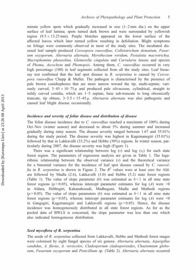
minute yellow spots which gradually increased in size (1–2mm dia.) on the uppersurface of leaf lamina; spots turned dark brown and were surrounded by yellowishregion (9.5� 13.25mm). Purple blotches appeared on the lower surface of theaffected leaves which later turned yellow resulting in defoliation. Blight symptomson foliage were commonly observed in most of the study sites. The incubated dis-eased leaf sample produced Cercospora rauwolfiae, Colletotrichum dematium, Fusar-ium oxysporum, Alternaria alternata, Myrothecium roridum, Pestalotia macrotrichia,Macrophomina phaseolina, Glomerella cingulata and Curvularia lunata and speciesof Phoma, Ascochyta and Phomopsis. Among them, C. rauwolfiae occurred in veryhigh percentage (100) in leaf segments collected from all the study sites. Leaf bioas-say test confirmed that the leaf spot disease in R. serpentina is caused by Cercos-pora rauwolfiae Chupp & Muller. The pathogen is characterised by the presence ofpale brown conidiophores that are more narrow toward the tip, multi-septate, vari-ously curved, 3–45� 10–75 μ and produced pale olivaceous, cylindrical, straight tomildy curved conidia, which are 1–5 septate, base sub-truncate to long obconicallytruncate, tip obtuse, 3–5.5� 15–45 μ. Alternaria alternata was also pathogenic andcaused leaf blight disease occasionally.
Incidence and severity of foliar disease and distribution of disease
The foliar disease incidence due to C. rauwolfiae reached a maximum of 100% duringOct–Nov (winter season) and decreased to about 5% during summer and increasedgradually during rainy season. The disease severity ranged between 1.67 and 35.01%during the study period. The disease severity was highest in Kagemanegiri (35.01%)followed by that in Lakkavalli (33.2%) and Hebbe (30%) regions. In winter season, par-ticularly during 2007, the disease severity was high (Figure 1).
There was a significant relationship between log (v) and log (vr) for each stateforest region. The parameters of regression analysis are given in Table 1. The loga-rithmic relationship between the observed variance (v) and the theoretical variancefor a binomial variance for the incidence of leaf spot disease caused by C. rauwol-fia in R. serpentina is shown in Figure 2. The R2 values were at least zero for Ald-ara followed by Madla (2.6), Lakkavalli (3.0) and Hebbe (5.2) state forest regions(Table 1). The value of slope parameter (b) was estimated as b< 1 in all nine stateforest regions (p> 0.05), whereas intercept parameter estimates for log (A) were >0in Aldara, Hebbegiri, Kakanahosudi, Madhuguni, Madla and Muthodi regions(p> 0.05). The value of slope parameters (b) was estimated as b < 1 in all nine stateforest regions (p> 0.05), whereas intercept parameter estimates for log (A) were <0in Gangegiri, Kagemanegiri and Lakkavalli regions (p> 0.05). Hence, the diseaseincidence was homogeneously distributed in all state forest regions. As for as thepooled data of BWLS is concerned, the slope parameter was less than one whichalso indicated homogeneous distribution.
Seed mycoflora of R. serpentina
The seeds of R. serpentina collected from Lakkavalli, Hebbe and Muthodi forest rangeswere colonised by eight fungal species of six genera- Alternaria alternata, Aspergilluscandidus, A. flavus, A. versicolor, Cladosporium cladosporioides, Chaetomium globo-sum, Fusarium oxysporum and Penicillium sp. (Table 2). Alternaria alternata occurred
Archives of Phytopathology and Plant Protection 5
Dow
nloa
ded
by [
Kuv
empu
Uni
v] a
t 23:
26 0
8 A
pril
2013

0
20
40
60
100
0
20
40
60
8 0
100
0
20
40
60
100
0
20
40
60
8 0
100
Max.RH
Min.RH
0
10
20
30
0
10
20
30 RainFall Temp.Max Temp.Min
A LD K K H
MD G
MTD
K G M
GNG
MD L
LK V
A S O N D J F M A M J J0
10
20
30
40
0
10
20
30
40HEB
Dis
ease
inci
denc
e (%
)
Dis
ease
inci
denc
e (%
)
Dis
ease
sev
erity
(%)
Dis
ease
sev
erity
(%)
Rai
n Fa
ll (c
m)
Rel
ativ
e H
umid
ity (%
)
Rel
ativ
e H
umid
ity (%
)Te
mpe
ratu
re (C
0 )
80
80
MonthsFigure 1. Cercospora rauwolfiae leaf spot disease incidence and severity in Rauwolfiaserpentina in Bhadra Wild life sanctuary, Karnataka.Note: HEB: Hebbegiri; GNG: Gangegiri; MDL: Madla; LKV: Lakkavalli; KKH: Kakanahosudi;MDG: Madhuguni; MTD: Muthodi; KGM: Kagemanegiri; ALD: Aldhar; Data presented is anaverage of data collected during August 2006 to July 2009.
Table 1. Mean disease incidence, slope and intercept parameter estimates of the binary powerlaw for the incidence of leaf spot disease of Rauwolfia serpentina in the sanctuary.
Sl. no State forest regions P1 SE2 (P) A3 SE4 (A) b5 SE6 (b) 7R2
1 Aldhar 0.231 0.046 0.071 0.052 �0.003 0.089 0.02 Gangegiri 0.234 0.047 �0.015 0.071 �0.185 0.115 7.03 Hebbe 0.330 0.061 0.179 0.067 �0.130 0.095 5.24 Kagemanegiri 0.166 0.038 �0.041 0.077 0.200 0.120 7.55 Kakanahosudi 0.207 0.048 0.071 0.069 0.214 0.122 8.36 Madhuguni 0.374 0.065 0.132 0.052 �0.369 0.064 49.27 Lakkavalli 0.236 0.050 �0.024 0.075 0.101 0.099 3.08 Madla 0.306 0.058 0.162 0.062 �0.090 0.094 2.69 Muthodi 0.297 0.053 0.068 0.045 �0.262 0.057 38.0BWLS Pooled 0.280 0.050 �0.408 0.199 0.349 0.145 14.6
Note: 1Average mean disease incidence (Aug 2006 to July 2009): 2,4,6Standard error: 3Intercept parameter ofthe binary power law: 5Slope parameter of the binary power law: 7Co-efficient of determination, higher thevalue the better fit of the data to the analysis.
6 T. R. Parashurama and M.B. Shivanna
Dow
nloa
ded
by [
Kuv
empu
Uni
v] a
t 23:
26 0
8 A
pril
2013

in 10.96% of seeds. The average percentage of seed germination was 43%, with highgermination (47%) in Muthodi and low germination (38%) in Lakkavalli regions.
Localisation and seed transmission of A. alternata and its management
By component plating technique, A. alternata was found to localise mainly in theembryonic axis (18.5%), followed by that in cotyledons (8.38%) and seed coat (5.13%).
Figure 2. Relationship between log of observed variance and the binomial variance for theincidence of foliar disease caused by Cercospora rauwolfiae in Rauwolfia serpentina.
Archives of Phytopathology and Plant Protection 7
Dow
nloa
ded
by [
Kuv
empu
Uni
v] a
t 23:
26 0
8 A
pril
2013
In the pot experiment, the naturally infected seeds (with 44.66% germination) expressed55.33% pre- and 13.67% post-emergence mortalities, however, 11.33% of seedlingsexhibited leaf spot disease symptom on cotyledonary leaves and leaves at 21 days.
All the seed dressing fungicides tested in the study inhibited the expression of fun-gal colony growth on seeds. Captra or Hyzeb was highly effective in reducing the seed-borne occurrence of A. alternata. Bavistin improved seed germination ability from 45%in untreated to 65% in treated seeds (Table 3).
Assay of phytochemical compounds in infected foliages
The alkaloids of R. serpentina decreased significantly (p< 0.05) by 45.99% in par-tially infected and 84.53% in completely diseased samples as compared to thehealthy one (28.20mg g�1). However, the total phenol content increased by 90.03and 92.33% over the control (35.09 μgml�1) in the partially and completely infectedleaf samples, respectively. Similarly, the flavonoids increased in both partially(34.80%) and completely infected samples (24.94%) as compared to the healthysample (208.64 μgml�1). On the other hand, the steroid content decreased in bothinfection categories (45.76 and 36.08%) when compared to the healthy sample(1752.32 μgml�1) (Table 4).
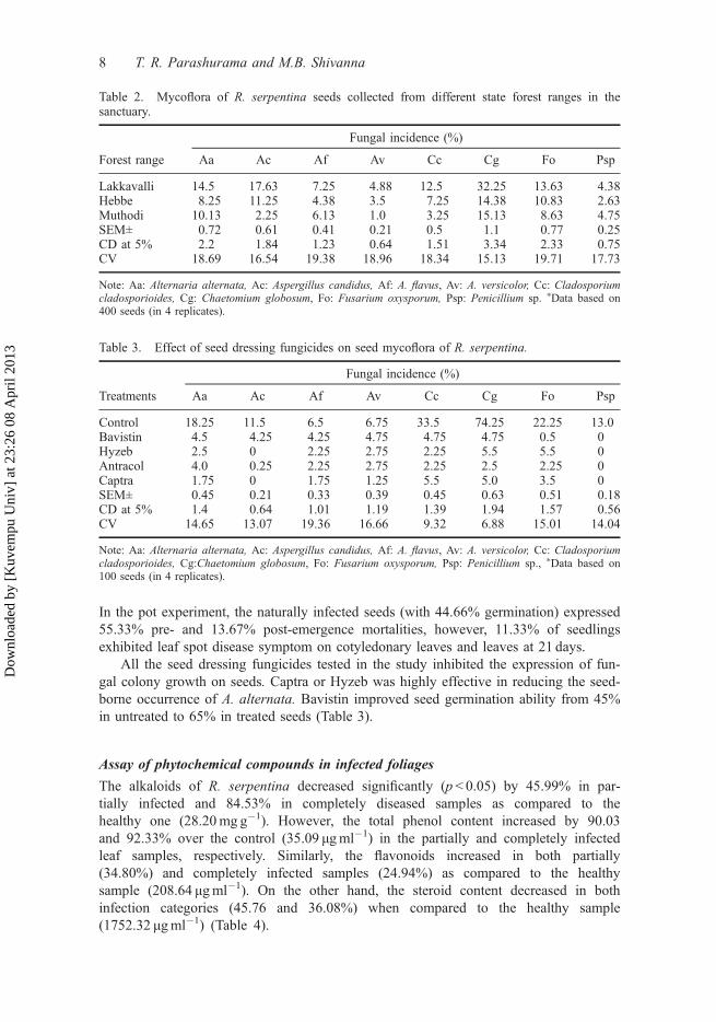
Table 3. Effect of seed dressing fungicides on seed mycoflora of R. serpentina.
Treatments
Fungal incidence (%)
Aa Ac Af Av Cc Cg Fo Psp
Control 18.25 11.5 6.5 6.75 33.5 74.25 22.25 13.0Bavistin 4.5 4.25 4.25 4.75 4.75 4.75 0.5 0Hyzeb 2.5 0 2.25 2.75 2.25 5.5 5.5 0Antracol 4.0 0.25 2.25 2.75 2.25 2.5 2.25 0Captra 1.75 0 1.75 1.25 5.5 5.0 3.5 0SEM± 0.45 0.21 0.33 0.39 0.45 0.63 0.51 0.18CD at 5% 1.4 0.64 1.01 1.19 1.39 1.94 1.57 0.56CV 14.65 13.07 19.36 16.66 9.32 6.88 15.01 14.04
Note: Aa: Alternaria alternata, Ac: Aspergillus candidus, Af: A. flavus, Av: A. versicolor, Cc: Cladosporiumcladosporioides, Cg:Chaetomium globosum, Fo: Fusarium oxysporum, Psp: Penicillium sp., ⁄Data based on100 seeds (in 4 replicates).
Table 2. Mycoflora of R. serpentina seeds collected from different state forest ranges in thesanctuary.
Forest range
Fungal incidence (%)
Aa Ac Af Av Cc Cg Fo Psp
Lakkavalli 14.5 17.63 7.25 4.88 12.5 32.25 13.63 4.38Hebbe 8.25 11.25 4.38 3.5 7.25 14.38 10.83 2.63Muthodi 10.13 2.25 6.13 1.0 3.25 15.13 8.63 4.75SEM± 0.72 0.61 0.41 0.21 0.5 1.1 0.77 0.25CD at 5% 2.2 1.84 1.23 0.64 1.51 3.34 2.33 0.75CV 18.69 16.54 19.38 18.96 18.34 15.13 19.71 17.73
Note: Aa: Alternaria alternata, Ac: Aspergillus candidus, Af: A. flavus, Av: A. versicolor, Cc: Cladosporiumcladosporioides, Cg: Chaetomium globosum, Fo: Fusarium oxysporum, Psp: Penicillium sp. ⁄Data based on400 seeds (in 4 replicates).
8 T. R. Parashurama and M.B. Shivanna
Dow
nloa
ded
by [
Kuv
empu
Uni
v] a
t 23:
26 0
8 A
pril
2013

Discussion
Survey of the study area for fungal diseases in R. serpentina indicated that leaf spot dis-ease caused by C. rauwolfiae occurred in Hebbegiri, Gangegiri, Madla, Lakkavalli,Kakanahosudi, Aldhara, Madhuguni, Muthodi and Kagemanegiri forest regions of thesanctuary. The incubated diseased leaf segments of R. serpentina were associated withC. rauwolfiae in high percentage and produced circular leaf spot symptoms in seedlingsand mature plants. Pathogenicity test confirmed the association of C. rauwolfiae withfoliar disease in R. serpentina. The above pathogen has also been found to be associ-ated with foliar disease of Rauwolfia plants (Mohanty and Addy 1957; Ganguly andPandotra 1962). The survey of study regions also indicated that the C. rauwolfiae foliardisease incidence which was initiated during the rainy season reached high during thewinter and decreased during summer. Maximum foliar DS was found in Kagemanegiriforest region. The study suggested that the same pathogen is capable of causing diseasein Rauwolfia plants under cultivation (Pandrotra and Husain 1966), as well as in thewild. In the study area, rainfall, minimum and maximum temperature and maximumhumidity positively correlated (p < 0.05) with disease development. Ijaz et al. (2011)also obtained similar correlation on Cercospora leaf spot disease in peanut. An averagetemperature of 24–28 °C and average RH of 75–85% and rainfall (102mm) were foundto be also associated with the foliar disease development.
Power law analysis based on data of leaf spot disease incidence caused by C. rau-wolfiae in R. serpentina indicated homogeneous distribution of disease in all the indi-vidual forest regions of the sanctuary, as well as in the entire sanctuary. The findingscorroborated with the previous study in Centella asiatica (Parashurama & Shivanna2013). Achar and Shivanna (2013) showed that Colletotrichum leaf spot disease inLakkavalli region of the above sanctuary is homogeneously distributed, while that inKemmannugundi region is heterogeneously distributed. They opined that such variationin spatial distribution depended on the type of forest and prevailing weather conditions.Siddiqui and Shaukat (2002), on the other hand, found the heterogeneous distributionof root disease in egg plants.
The seedborne incidence of fungal species in R. serpentina was high (p < 0.05) inseed sample collected from Lakkavalli range forest as compared to those collected from
Table 4. The secondary metabolite contents in foliages of Rauwolfia serpentina infected withC. rauwolfiae.
Disease category (% DS)
Secondary metabolites
Alkaloids(mg g�1)
Flavonoids(μg ml�1)
Phenolics(μg ml�1)
Steroids(μg ml�1)
Apparently healthy (control) 28.2 208.64 35.09 1752.32Partial diseased (650%) 15.23
(45.99)⁄⁄320.0
(53.37)⁄352.07
(903.19)⁄950.44 (45.76)⁄⁄
Completely diseased(100%)
4.36 (84.53)⁄⁄ 278.0(24.94)⁄
457.59 (1204)⁄ 1120.03(36.08)⁄⁄
SEm± 0.04 0.5 0.72 1.02CD (p = 0.05) 0.14 1.54 2.22 3.13CV 0.74 0.49 0.68 0.21
Note: Data of secondary metabolites was subjected to ANOVA (p6 0.005, 0.001); ⁄Values in parenthesis indi-cate the per cent increase in secondary metabolite content in infected over healthy category; ⁄⁄Values in paren-thesis indicate the per cent decrease of secondary metabolites in infected over healthy category.
Archives of Phytopathology and Plant Protection 9
Dow
nloa
ded
by [
Kuv
empu
Uni
v] a
t 23:
26 0
8 A
pril
2013

Hebbe and Muthodi range forests which could be due to weather conditions favouringseed infection (temperature of 25 °C and RH of 85%). The foliar pathogen C. rauwol-fiae was neither detected on the seed surface nor in its components and hence it is notseedborne. However, it is a major pathogen of R. serpentina in the study area. Thereduced seed germination could be due to hard seed coat and seed dormancy (Trivediand Kumari 2011). Alternaria alternata is a minor pathogen in the study area and it isseedborne (Patil and Pandule 2000; Chavan and Mukadam 2001). The fact that theembryonic axis and cotyledons were colonised by A. alternata might suggest the occur-rence of seedling disease and contribution to inoculum spread. Observations of manyauthors (Maude and Humpherson-Jones 1980) on the internal infection of seeds withAlternaria in many plant species are concurrent with the present observation. The inter-nal seed infection might suggest the pathogen transmission from the mother plant toembryo through the connective tissue. Further, the seed infection by A. alternata andthe resulting seedling disease suggested its seed to seedling transmission. This pathogenis also shown to be seed transmitted in other plants (Rathod 2012). On the other hand,none of the seedlings showed symptoms of disease due to C. rauwolfiae. This pathogenalthough not seedborne and seed transmitted caused severe disease in R. serpentinawhich could be attributed to foliar infection by airborne inoculum of C. rauwolfiae. Thepathogen is capable of surviving in plant debris in soil (Yang 2004) and produced copi-ous spores during the onset of rainy season. The spores thus caused infection in foliagesand disease spread in post-rainy season. Seed treatment with Captra significantlyreduced seedborne occurrence of A. alternata and other fungal species. This lead to theimprovement in seed germination and improved seedling stand. Similarly, Captan hasbeen shown to most effectively inhibit growth of Alternaria fungus in radish (Kumaret al. 2006). Bavistin, a systemic fungicide, is also reported to manage seedborne infec-tion by F. oxysporum and A. alternata (Van Nghiep and Gaur 2005).
Among the secondary metabolites tested, the alkaloid and steroid contentsdecreased in both partially infected and completely diseased foliar samples of R. serpen-tina. This corroborated with the observations made for alkaloids in the above plant spe-cies (Rosazza 1978). In contrast to the above, Shivanna and Mallikarjunaswamy (2009)reported increase in alkaloids, as the disease progressed, in Terminalia species. The ste-roid content was also shown to decrease in infected leaf samples (Prasad and Sah1991). However, the total phenols and flavonoids increased in both the partially andcompletely infected leaf samples. Total phenol content was also shown to increase inblight infected tea plants rather than in the healthy plants (Chakraborty et al. 2002).Vidyasagar et al. (2003) showed significant increase in flavonol content in infected overthe healthy leaves. This suggested that phenolic compounds might accumulate in dis-eased parts, to protect plant systems in response to infection by plant pathogens (Mazidet al. 2011). Flavonols have also been shown to protect plants from damages that mightbe caused by UV-light and pathogens (Takahashi and Ohnishi 2004). Flavonoids arepotent water-soluble antioxidants and free radical scavengers, which prevent oxidativecell damage, and have strong anticancer activity (Salah et al. 1995; Del-Rio et al.1997). As antioxidants, flavonoids from R. serpentina provide anti-inflammatory activity(Okwu, 2004) and hence used in the treatment of diseases.
In conclusion, results of present study indicated that the leaf spot disease in R. ser-pentina is caused by C. rauwolfiae which caused considerable damage to foliagesthrough different seasons in different forest regions of the sanctuary. Cercospora rau-wolfiae is a major leaf spot pathogen but is not seedborne and seed transmitted. How-ever, A. alternata is a minor pathogen which is seedborne and transmitted to seedlings.
10 T. R. Parashurama and M.B. Shivanna
Dow
nloa
ded
by [
Kuv
empu
Uni
v] a
t 23:
26 0
8 A
pril
2013

The seedborne inoculum of A. alternata could be effectively managed by seed dressingwith Captra or Hyzeb. Foliar infection of R. serpentina with C. rauwolfiae resulted inthe decrease in alkaloid and steroid contents and increase in phenolics and flavonoids.The decrease in alkaloid content, and possibly its quality, could be a cause of concernsince the alkaloids in R. serpentina are therapeutically important compounds used intreating various diseases and disorders in humans. The study helped in the generationof baseline data which could be used while compiling the package of practices forintensive cultivation of R. serpentina and in pharmaceutical industries.
AcknowledgementsThe financial assistance of the Department of Science and Technology, New Delhi is gratefullyacknowledged.
ReferencesAchar KGS, Shivanna MB. 2013. Foliar disease of Clitorea ternatea due to Colletotrichum dema-
tium and its effect on secondary metabolte production, doi:10.1080/03235408.2012.755854.Anonymous. 2003. International rules for seed testing Annexe to chapter 7: Seed Health Testing
Methods, Int. Seed Testing Association (ISTA), Bassersdorf, Switzerland, 7-002: 1-6.Barnett HL, Hunter BB. 1998. Illustrated genera of imperfect fungi, 4th ed. St Paul (MN): APS
Press.Booth C. 1977. Fusarium laboratory guide to the identification of the major species. Surrey:
Commonwealth Mycological Institute.Chakraborty BN, Datta S, Chakraborty U. 2002. Biochemical responses of tea plants induced by
foliar infection with Exobasidium vexans. Indian Phytopathol. 55:8–13.Chavan AM, Mukadam DS. 2001. Role of fungal pigments in seed discolouration. Paper pre-
sented at: National seminar on Recent Advances in Mycology; Plant Pathology and Biotech-nology; April 6–7, Pune, p. 38.
Chopra RN, Chopra IC, Verma BS. 1980. Publications and Information Directorate, Council ofScientific & Industrial Research, New Delhi. p. 86.
Del-Rio A, Obdululio BG, Casfillo J, Marin FG, Ortuno A. 1997. Uses and Properties of citrusflavonoids. J Agric Food Chem. 45:4505–4515.
Dutta PK, Choudhury SB, Rao PR. 1962. Germination and chemical composition of Rauwolfiaserpentina seeds. Ind J Pharm. 24:61–63.
Folin D, Denis W. 1939. A calorimetric estimation phenols (Phenol derivatives) in urine. J BiolChem. 22:305–308.
Gamble JS. 1935. Flora of Presidency of Madras, vol. 1–3. [Reprint] Dehra Dun, India: BishenSingh Mahendra Pal Singh Publications, p. 2017.
Ganguly D, Pandotra VR. 1962. Some of the commonly occurring diseases of important medici-nal and aromatic plants in Jammu and Kashmir. Indian Phytopathol. 15:50–54.
Gent DH, Turechek WW, Mahaffee WF. 2008. Spatial and temporal stability of the estimatedparameters of the binary power law. Phytopathology. 98:1107–1117.
Ijaz M, Haque MI, Rauf CA, Fayyaz-UL-Hassan AR, Mughal SM. 2011. Correlation betweenhumid thermal ratio and epidemics of Cercospora leaf spot of peanut in Rothwar. Pak J Bot.43:2011–2016.
Ikan R. 1969. Natural products: a laboratory guide. London: Academic Press.Kokate CK, Purohit AP, Gokhale SB. 2003. Phamacognosy, 24th ed. Pune: Nirali-Prakashan.Kumar P, Prajapati CR, Singh DV. 2006. Efficacy of some fungitoxicants against Alternaria
brassicae causing Alternaria blight of radish. Ind J Pl Pathol. 24:29–31.Madden LV, Hughes G, Ellis MA. 1995. Spatial heterogeneity of the incidence of grape downy
mildew. Phytopathology. 85:269–275.Maden SS, Mathur DSB, Neerggard P. 1975. Detection and location of seedborne inoculum of
Ascochyta rabiei and its transmission in chick pea (Cicer arietinum). Seed Sci Technol.3:667–681.
Archives of Phytopathology and Plant Protection 11
Dow
nloa
ded
by [
Kuv
empu
Uni
v] a
t 23:
26 0
8 A
pril
2013

Mamgain SK, Goel AK, Sharma SC. 1998. Conservation of assessment of some important threa-tened medicinal plants of India. J Non-Timber Fore Prod. 5:1–9.
Mao AA, Hynniewta TM, Sanjappa M. 2009. Plant wealth of northeast India with reference toEthnobotany. Ind J Trad Knowl. 8:96–103.
Maude RB, Humpherson-Jones FM. 1980. Ann Appl Biol. 95:311–319.Mazid M, Khan TA, Mohammad F. 2011. Role of secondary metabolites in defense mechanisms
of plants. Biol Med. 3:232–249.Mohanthy NN, Addy SK. 1957. Cercospora leaf spot of Rauwolfia serpentina. Benth. Curr Sci.
26:289–290.Ojha J, Mishra U. 1985. Dhanvantari Nighantuh, with Hindi translation and commentary, Ist ed.
Varanasi: Department of Dravyaguna, Institute of Medical Sciences BHU.Okwu DE, Okwu ME. 2004. Chemical composition of Spondias mombin Linn. plant parts. J Sus-
tain Agric Environ. 6:140–147.Pandotra VR, Husain A. 1966. Fungi on medicinal and aromatic plants in the north Himalayas.
IV. Mycopath Mycol Appl. 29:155–159.Parashurama TR, Parinitha M, Mallikarjunaswamy GE, Shivanna MB. 2013. Cercospora leaf spot
disease in Centella asiatica and its effect on pharmaceutical components. Inter J Bio &Pharma Res. 4:1–8.
Patil PJ, Pandule DN. 2000. Effects of grain mould fungi on seed germination and seedlings vig-our index of sorghum seeds. Var. (SH.9) in Western Maharashtra. Seed Res. 28:190–192.
Prasad MM, Sah RP. 1991. Level of sterol in two medicinal plants under colonization by fungi.Indian Phytopath. 44:409–411.
Rathod S. 2012. Seed borne Alternaria species: A review. Curr Bot. 3:21–23.Rosazza TP. 1978. Microbial transformations of natural antitumour agents. Lloydia. 41:279–311.Salah N, Miller NJ, Pagangeg G, Tijburg L, Bolwellg P, Rice E, Evans C. 1995. Polyphenolic
flavonols as scavenger of aqueous phase radicals as chain breaking antioxidant. Arch Bio-chem Broph. 2:339–346.
Sanchez GL, Medina AJC, Solo RR. 1972. Spectrophotometric determination of disogenin inDioscorea composite following thin layer chromatography. Analyst. 97:973.
Shivanna MB, Mallikarjunaswamy GE. 2009. Fungal diseases and their effect on phytochemicalconstituents of medicinally important Terminalia species in Bhadra wild life Sanctuary, Kar-nataka. Indian Phytopathol. 62:37–43.
Shivanna MB, Shetty HS. 1991. Occurrence of fungal diseases and its relationship with growthstages in cluster bean during different seasons. Int J Trop Plant Dis. 9:53–64.
Shivanna MB, Parinitha M, Parashurama TR. 2007. Fungal diseases of Rauwolfia serpentina inBhadra wildlife sanctuary and their effect on phytochemical composition, National Confer-ence on Medicinal and Aromatic plants. Gulbarga: Department of Post-Graduate Studies andResearch in Botany Gulbarga University.
Siddiqui IA, Shaukat SS. 2002. Spatial pattern analysis of root rot-root knot disease complex inan infested egg plant field. Nematol Medit. 30:131–135.
Singh PK, Kumar V, Tiwari RK, Sharma A, Rao CV, Singh RH. 2010. Medico-ethnobotany of‘Chatara’ block of District Sonebhadra, Uttar Pradesh. India Adv Biol Res. 4:65–80.
Sivanesan A. 1983. The Bitunicate Ascomycetes and their Anamorphs. Hirschberg: Strands andCramer Gmbh.
Stary F. 1998. The natural guide to medicinal herbs sand plants. London: Tiger Books Interna-tional.
Steel RGD, Torrie JH. 1980. Principles and procedures of statistics, 2nd ed. New York (NY):McGraw Hill Book.
Subramanian CV. 1983. Hyphomycetes: Taxonomy and biology, Vol. I and II. London: AcademicPress.
Sukumaran S, Raj ADS. 2008. Rare and endemic plants in the sacred groves of Kanyakumari dis-trict in Tamilnadu. Ind J For. 31:611–616.
Swain T, Hillis WE. 1959. The phenolic constituents of Brunus domestica L. The qualitative anal-ysis of phenol constituents. S J Sci Food Agric. 10:63–68.
Takahashi A, Ohnishi T. 2004. The significance of the study about the biological effects of solarultraviolet radiation using the exposed facility on the internal space station. Biol Sci Space.18:255–260.
12 T. R. Parashurama and M.B. Shivanna
Dow
nloa
ded
by [
Kuv
empu
Uni
v] a
t 23:
26 0
8 A
pril
2013

Taylor LR. 1984. Assessing and interpreting the spatial distributions of insect populations. AnnuRev Entomol. 29:321–357.
Taylor LR, Taylor R. 1977. Aggregation, migration and population mechanics. Nature. 265:415–421.
Trivedi MP, Kumari R. 2011. Ethano-botanical and germinational aspects of Rauwolfia serpentina(L.) Benth. ex Kurz. Our Nat. 9:176–178.
Van Nghiep H, Gaur A. 2005. Efficacy of seed treatment in improving seed quality in rice.Omonnice. 13:42–51.
Vidyasagar GM, Kotresha D, Shivakumar D. 2003. Biochemical changes during powdery mildewdisease development in mulberry (Morus alba L.). Bull Ind Acad Servi. 7:104–107.
Yang XB. 2004. Soybean Cercospora diseases show up. Integrated crop management, Iowa stateUniver. July 26. IC-492:17.
Vishunavat K, Kolte SJ. 2005. Essentials of phytopathological techniques. New Delhi: Kalyani.Yoganarasimhan SN, Subramanyam K, Razi BA. 1982. Flora of Chikmagalur District, Karnataka,
India. Dehra Dun: International Book Distributors.
Archives of Phytopathology and Plant Protection 13
Dow
nloa
ded
by [
Kuv
empu
Uni
v] a
t 23:
26 0
8 A
pril
2013