functional electrical stimulation and spinal cord injury
TRANSCRIPT
Functional ElectricalStimulation and Spinal
Cord InjuryChester H. Ho, MDa,*, Ronald J. Triolo, PhDb,c,d,e,Anastasia L. Elias, PhDf, Kevin L. Kilgore, PhDd,e,g,h,Anthony F. DiMarco, MDe,h, Kath Bogie, DPhilb,c,d,g,Albert H. Vette, PhDi,j, Musa L. Audu, PhDb,d, Rudi Kobetic, MSb,Sarah R. Chang, BSb,d, K. Ming Chan, MDk,Sean Dukelow, MD, PhDa, Dennis J. Bourbeau, PhDg,h,Steven W. Brose, DOg,h,l, Kenneth J. Gustafson, PhDd,g,h,Zelma H.T. Kiss, MD, PhDm, Vivian K. Mushahwar, PhDk
KEYWORDS
� Electric stimulation � Electrodes � Spinal cord injuries � Rehabilitation� Muscle spasticity � Pressure ulcer � Neurogenic urinary bladder � Paralysis
a Division of Physical Medicine & Rehabilitation, Department of Clinical Neurosciences,Foothills Medical Centre, Room 1195, 1403-29th Street NW, Calgary, Alberta T2N 2T9, Can-ada; b Louis Stokes Cleveland VA Medical Center, Advanced Platform Technology Center, 151AW/APT, 10701 East Boulevard, Cleveland, OH 44106, USA; c Department of Orthopaedics,Case Western Reserve University, MetroHealth Medical Center, 2500 MetroHealth Drive, Cleve-land, OH 44109, USA; d Department of Biomedical Engineering, Case Western Reserve Univer-sity, 10900 Euclid Avenue, Cleveland, OH 44106, USA; e MetroHealth Medical Center, 2500MetroHealth Drive, Cleveland, OH 44109, USA; f Chemical and Materials Engineering,W7-002 ECERF, University of Alberta, Edmonton, Alberta T6G 2V4, Canada; g Louis StokesCleveland VA Medical Center, 10701 East Boulevard, Cleveland, OH 44106, USA; h ClevelandFES Center, 11000 Cedar Avenue, Suite 230, Cleveland, OH 44106-3056, USA; i Department ofMechanical Engineering, University of Alberta, 4-9 Mechanical Engineering Building, Edmon-ton, Alberta T6G 2G8, Canada; j Glenrose Rehabilitation Hospital, Alberta Health Services,10230 - 111 Avenue, Edmonton, Alberta T5G 0B7, Canada; k Division of Physical Medicineand Rehabilitation, Centre for Neuroscience, University of Alberta, 5005 Katz Group Centre,11361-87 Avenue, Edmonton, Alberta T6G 2E1, Canada; l Ohio University Heritage College ofOsteopathic Medicine, Grosvenor Hall, Athens, OH 45701, USA; m Department of ClinicalNeurosciences, Foothills Medical Centre, Room 1195, 1403-29th Street NW, Calgary, AlbertaT2N 2T9, Canada* Corresponding author.E-mail address: [email protected]
Phys Med Rehabil Clin N Am 25 (2014) 631–654http://dx.doi.org/10.1016/j.pmr.2014.05.001 pmr.theclinics.com1047-9651/14/$ – see front matter � 2014 Elsevier Inc. All rights reserved.
KEY POINTS
� Functional electrical stimulation (FES) of the peripheral and central nervous system maybe used for rehabilitation and management of complications after spinal cord injury (SCI).
� FES may improve the functional status and quality of life of many persons with spinal cordinjuries.
� Many of the FES strategies are already commercially available, whereas others are beingtested in human and laboratory studies.
� FES should be routinely considered as part of the rehabilitation and medical managementof eligible persons with spinal cord injuries.
Ho et al632
An injury to the spinal cord can disrupt communications between the brain and body,leading to a loss of control over otherwise intact neuromuscular systems. By takingadvantage of these intact neuromuscular systems, several neuroprostheses havebeen developed to restore functions through functional electrical stimulation (FES)of the central and peripheral nervous system. Neuroprostheses using FES to controlthe paralyzed muscles may prevent many secondary medical complications andimprove functional independence by providing a means to exercise and negotiatephysical barriers. Improvements in multiple body systems and functions have beenreported through the use of FES, and they are discussed in this article. These devicesrange in complexity and include components such as power supplies (which may becompletely external to the body or implanted and recharged with radio frequencywaves), a control circuit (ie, the brains of the device), lead wires, connectors, externalbraces, and sensors. This article describes the basic properties of the electrodes, thecurrent FES system being developed in research and in clinical practice, and the futureof these devices.
THE BASIC PROPERTIES OF ELECTRODES FOR NERVE STIMULATION
In neuroprostheses, electrodes are the interface between the external circuitry andthe tissue, delivering a charge that stimulates the nerves connected to the musclesof interest. This charge perturbs the resting potential of the neuron (typically around�65 mV); if this value is raised beyond a threshold, membrane depolarization oc-curs. This depolarization results in an influx of Na1 ions, initiating an action potentialthat can travel spatially down the length of an axon. A coordinated group of actionpotentials can lead to a muscle contraction.1 By targeting nerves rather than themuscle fibers themselves (which can also be stimulated electrically), substantiallysmaller charge densities may be used, consuming less power and avoiding tissuedamage.2
Provided that the neuromuscular system is intact, stimulation may be achieved at avariety of locations (from the origin of the neuron in the spinal cord to the peripheralnerve and to the skin above the muscle) using various types of electrodes. Thesimplest configuration uses large (of the order of square centimeters) electrodesplaced on the surface of the skin. The electrodes are easily replaced; however,achieving accurate and precise positioning is challenging, and charge is distributedover a large area. A more invasive approach is to implant needlelike electrodes percu-taneously into the muscle of interest. This method is considered a precursor to fullyimplanted systems, although subcutaneous electrodes themselves can remain func-tional for years.3 When electrodes are fully implanted in close proximity to the nerve,

FES and Spinal Cord Injury 633
even more precise targeting can be achieved using even smaller current densities,which are less likely to damage the tissue.Electrodes have been designed to wrap around individual nerves, with a range of
geometries, including spiral,4 helical,5 and rectangular.6 To selectively address smallergroups of axons within a nerve and to reach areas that are not readily accessible fromthe surface, intrafascicular electrodes may be inserted into the nerve itself.7 Pools ofneurons may also be stimulated directly in the spinal cord in intraspinal microstimula-tion (ISMS).8 Although implanted devices offer superior targeting, the obvious draw-back is the invasiveness of the insertion process and the potential risk of infection,although this has not been reported as a significant issue.9
In FES, the electrode typically acts as a conductor, delivering electrical charge froma power supply to the tissue. Charge transfer occurs when voltage applied betweenthe active electrode and a second electrode (called the reference electrode) generatesan electric field, which in turn forces electrical charge to flow. In systems in which mul-tiple stimulation channels are used, a single reference electrode may be used. When avoltage is applied, the energy can drive several unwanted chemical reactions. Toavoid generating H2 gas from water, the voltage generated between the electrodesmust not exceed the amount required to electrolyze water (�0.6 V to �0.8 V depend-ing on electrode type10). The amount of charge that can be delivered within these limitsdepends on the impedance of the material, which should be low to maximize the cur-rent delivered. To balance the charge injected to stimulate the neurons and preventthe electrochemical decomposition of tissue, a secondary pulse of opposite polarityshould be included in the stimulation profile (ie, a biphasic pulse should be applied).The electrodes themselves must be selected to be resistant to corrosion under phys-iologic conditions, even under an applied voltage. Common electrode materials forimplanted devices include corrosion-resistant stainless steel and noble metals suchas PtIr or Pt (which have highly stable atomic configurations and therefore are resistantto chemical processes such as corrosion or oxidation). Other metals (including silver,iron, and copper) are known to elicit dramatic inflammatory response in vivo andshould be avoided.11
The time-dependent failure of neural interfaces in vivo is an impediment to long-termuse, particularly for recording electrodes and stimulating electrodes, which injectsmall currents into small target areas. The principal cause of failure of these devicesis the encapsulation, which occurs as a part of the foreign body response, insulatingthe electrodes from their surroundings.12 To avoid scar formation initiated by mechan-ical mismatch between stiff electrodes and soft tissues, there is an increasing interestin fabricating electrodes and arrays from soft (low modulus) materials such as siliconeelastomer.13 Beyond this, several strategies have been undertaken to modify the sur-face properties of electrodes to improve the interactions that take place with sur-rounding tissue and reduce glial scar formation.14 When developing new electrodes,arrays, and coatings, in vitro testing may be used initially to screen the cellularresponse, but they must be tested in vivo following the standard ISO 10993.
UPPER EXTREMITY FUNCTIONAL RESTORATION WITH FES
For persons with cervical-level SCI, restoration of hand function is their top priority.15
Neuroprostheses using FES provide the most promising method for significant gainin hand and arm function for this population. Muscle contractions can be orchestratedto produce coordinated grasp opening and closing; thumb opening, closing, andpositioning; wrist extension and flexion; forearm pronation; and elbow extensionfor persons with C5-C6–level SCI. Neuroprostheses can be coupled with tendon

Ho et al634
transfers to maximize function.16 The objectives of these neuroprostheses are toreduce the need to rely on assistance from others; the need for adaptive equipment,braces, or other orthotic devices; and the time it takes to perform tasks. Neuropros-theses make use of the patient’s own paralyzed musculature to provide the powerfor grasp and the patient’s voluntary musculature to control the grasp. Typically,persons with SCI use the neuroprosthesis for eating, personal hygiene, writing, andoffice tasks.Neuroprostheses have been clinically implemented and investigated using systems
based on surface electrodes, percutaneous electrodes, and implanted devices. Sur-face and percutaneous systems have potential application in muscle conditioningand in short-term research or clinical applications.17 Implanted systems are generallyused for long-term functional enhancement.All existing upper extremity neuroprosthetic systems consist of (1) a stimulator that
activates the muscles of the forearm and hand and (2) an input transducer and controlunit. The control signal for grasp is derived from an action that the user has retainedvoluntary control over, which can include joint movement, muscle activity, respiration,or voice control.18 A coordinated stimulation pattern is developed so that the musclesare activated in a sequence that produces a functional grasp pattern as the user typi-cally has control over grasp opening and closing but does not have direct control overthe activation of each muscle.Surface stimulation of the forearm and hand can be used to exercise and to produce
functional movements. Nathan19 developed a splint that incorporates surface elec-trodes for grasp. This system is commercially available (NESS H200, Bioness, Valen-cia, CA, USA) and is primarily intended for therapeutic applications after stroke or SCI,such as building muscle strength, preventing joint contractures, and improving tissueviability. Popovic and colleagues20 have developed a surface stimulation systemcalled the ETHZ-ParaCare neuroprosthesis. This system is capable of 4 channels ofstimulation and can be interfaced with a variety of control inputs. Early functional re-sults indicate that subjects can use the system to perform a variety of activities of dailyliving (ADL) in the home.21
Implanted FES systems have been used for long-term functional enhancement forpersons with cervical SCI. The largest clinical trial of an upper extremity neuropros-thesis was the Freehand trial, initiated by the Cleveland Functional Electrical Stimula-tion Center in 1992.22 The Freehand neuroprosthesis used an implanted 8-channelreceiver-stimulator, and control of grasp opening and closing was achieved throughgraded elevation of the user’s contralateral shoulder. Using the neuroprosthesis,100% of the participants (n 5 28) improved in independence in at least 1 task, and78% were less dependent in at least 3 tasks. More than 90% were satisfied withthe neuroprosthesis.23 The Freehand system was transferred to industry (NeuroCon-trol Corp, Elyria, OH, USA) and was implemented successfully in more than 200 pa-tients with SCI using neuroprostheses.24 Despite the clinical success, the companyexited the SCI market in 2001 and no longer markets the Freehand System.A second-generation implanted neuroprosthesis has been developed, improving on
the features of the Freehand System.25 This system, called the Implanted StimulatorTelemeter Twelve-channel System (IST-12), has 12 stimulation channels and 2 chan-nels of myoelectric signal recording acquisition.26 To date, 12 subjects with SCI havebeen implanted with the IST-12 system, including 3 subjects with systems forrestoring movement in both hands. Subjects successfully use the processedmyoelec-tric signal from a wrist extensor for proportional control of grasp opening and closing.Every subject has demonstrated improvement in at least 2 activities and as many as11 activities. Most commonly, improvement was demonstrated in eating with a fork

FES and Spinal Cord Injury 635
and writing with a pen. Other tasks in which subjects showed improvement includedoffice tasks, using a cell phone, getting money out of a wallet, and embroidery,25 asillustrated in Fig. 1.
Availability
At present, commercially available FES systems for grasp function in cervical SCI arelimited to surface stimulation systems. Specifically, the NESS H200 is available by pre-scription at multiple sites throughout the world (www.bioness.com). Other systems,such as the Compex system, are primarily targeted for exercise training rather thanfunction benefit. Efforts are underway to increase the availability of implanted neuro-prostheses to persons with SCI (http://casemed.case.edu/ifr/).
Future Directions
Future directions for FES hand systems include the development of fully implantedsystems that eliminate the need to don and doff components27 and the expandeduse of myoelectric control algorithms to control multiple functions at the sametime.28 The use of signals derived directly from the brain (brain-computer interface),either externally or through implanted electrodes, is expected to result in more naturalhand system control.29 In addition, systems are being developed to provide whole-arm function for those with C4 or higher SCI.30
LOWER EXTREMITY FUNCTIONAL RESTORATION WITH FES
In persons with SCI, the inability to stand or step significantly limits the performance ofmany ADL such as washing dishes at a counter or reaching items on high shelves.For persons with thoracic-level complete SCI, stimulated contractions of the lowerextremity muscles can enable standing and stepping, increase personal mobility,and improve general health and quality of life.31 In persons with incomplete injuries,walking performance can be improved.32
Eight channels of continuous stimulation to the knee, hip, and trunk extensors canpower the sit-to-stand transition and support the body vertically against collapse(Fig. 2).33 Stimulation to the hip ab/adductors and ankle plantar/dorsiflexors hasbeen included in experimental systems for sensor-based control of standing balancein the coronal and sagittal planes.34 Existing neuroprostheses for lower extremity func-tion use maximal levels of constant stimulation at the hips and knees.35 Recipients of aneuroprosthesis with epimysial and intramuscular electrodes that continuously acti-vated the vasti, gluteals, hamstrings, and lumbar erector spinae exhibited mean andmedian standing times of 10 and 3 minutes, respectively.33 This time is sufficient forfacilitating transfers to high surfaces, performing swing-to gait for short distances inwheelchair-inaccessible environments, and participating in other social, work, andpersonal activities. Some implant recipients in a phase II clinical trial of the systemwere able to stand for more than 20 minutes, and all were able to release 1 handfrom a walker or assistive device to reach objects overhead (Fig. 3). On average,90% of body weight was placed on the legs, reducing requirements on the arms toonly light touch to maintain balance. System performance and patterns of usagewere maintained after discharge for at least 1 year of follow-up. Although therewere no discernible interactions between injury level, degree of preserved sensation,or time postinjury and system performance, outcomes seem to be inversely propor-tional to height and weight, implying that bodymass indexmay be an important clinicalfactor for determining expectations.35 Long-term use of neuroprostheses for standingwas safe and effective and had no adverse physiologic effects.

Fig. 1. Functional activities performed using the IST-12 myoelectrically controlled neuroprosthesis. From left to right: eating with a fork, holding a pento write, holding a cup, needle embroidery, holding a tennis racquet.
Hoetal
636

Fig. 2. Implant recipient (C7AISC) standingwithFES to theknee,hip, and trunkextensors, andhip/trunkab/adductors.Multicontact cuff electrodeson the femoralnerves selectively activatethe uniarticular heads of the quadriceps (vastus lateralis, intermedius, and medialis).
Fig. 3. Eight-channel implant recipient (T9 AIS A) releases one hand for overhead reachingactivities while standing with the neuroprosthesis.
FES and Spinal Cord Injury 637

Ho et al638
Stepping of up to 100m has also been achieved after paralysis with simple preprog-rammed patterns of open-loop stimulation delivered from the surface or via 8- and16-channel implanted pulse generators.36 Once initiated by the user, steppingmotionscan cycle continuously while the appropriate adjustments are made with the upperbody until the pattern is stopped. Alternatively, the stimulation for sequential stepscan be triggered from successive depressions of ring- or walker-mounted switchesor automatically from body-mounted sensors, such as inclinometers, accelerometers,gyroscopes, or foot or heel switches.37 The largest potential impact of stimulation maybe for people with motor incomplete injuries (Fig. 4) who require activation of a smallnumber of muscles during the gait cycle to become household or community ambula-tors.38 In such cases, gait training with stimulation can have a therapeutic effect interms of improved voluntary strength, walking speed, stride length, and cadenceeven after completion of aggressive conventional therapies.39 Interactive use of stim-ulation to assist gait resulted consistently in an additional 20% improvement in walkingspeed and 6-minute distance, as well as a more than 3-fold increase in maximumwalking distance, illustrating a significant neuroprosthetic effect. Walking with stimula-tion was also more dynamic as evidenced by decreased time spent in the double sup-port phases of gait. The electromyographic activity of muscles under volitional controlhas also been exploited as a command source to control stimulation in persons withincomplete injuries, which has the potential to coordinate stimulated contractionswith voluntary motor function, and in so doing reinforce voluntary movement patternsand provide a mechanism to continuously modulate walking speed and cadence.40
Surface FES to the lower extremity muscles with intact innervation has allowedcycling movement that simulates exercise training, leading to increase in oxygen con-sumption during exercise,41 muscle mass and strength, and quality of life in personswith chronic SCI.42
Fig. 4. Subject with incomplete SCI (C5 AIS D) walking with an 8-channel implanted receiverstimulator for activation of hip flexors and ankle dorsiflexors.

FES and Spinal Cord Injury 639
Availability
Although implanted standing and walking systems clearly provide significant func-tional and clinical benefits, such systems are only available on a research basis.Limited lower extremity function is possible with commercially available surface stim-ulators with reduced channel counts.43
FES cycling devices are available through Restorative Therapies, Inc. (www.restorative-therapies.com) and Therapeutic Alliances, Inc. (www.musclepower.com)in the United States.
Future Directions
Standing performance with implanted neuroprostheses can be improved significantlyby using nerve-based electrodes, which more fully recruit the target muscles. Contin-uous stimulation of the femoral nerve with a multicontact cuff electrode below thebranches to the rectus femoris and sartorius was shown to extend standing timeand accelerate progress through reconditioning rehabilitation and balance trainingwith the system.44 The potential to delay the effects of fatigue by alternating activationof independent motor unit pools within a muscle via multicontact nerve cuffs or mul-tiple independent nerve- or muscle-based electrodes is also being investigated.45 Atpresent, neuroprostheses are generally unresponsive to environmental disturbances,necessitating use of the arms for balance on an assistive device. Additional research isalso focusing on automatically modulating stimulation in response to perturbations toreduce reliance on the upper extremities, allow users to alter their postures in advanceof anticipated disturbances, and minimize the risk of falls while standing or usingadvanced biomechanical modeling techniques to optimize stimulus patterns duringwalking or while assuming various task-dependent standing postures.46 Anotherpromising development involves the combination of FES with exoskeletal bracingthat can lock, unlock, or couple the joints as necessary to avoid fatigue and smoothlyshape limb trajectories or that can inject small amounts of assistive power when thestimulated responses are too weak or fatigued to complete a motion.47 With suchan approach, users would be able to walk under their own power and therefore accruethe physiologic benefits of exercising the paralyzed muscles in addition to those ofstanding, weight bearing, and mobilization.
TRUNK CONTROL AND POSTURE WITH FES
After SCI, trunk muscles can oftentimes not provide the necessary forces toadequately control trunk posture because of a lack of innervation48 and/or muscle at-rophy,49 significantly limiting their performance during ADL50 and even leading to sec-ondary health complications such as reduced respiratory capacity.51 To compensatefor insufficient muscle control during sitting, persons with SCI usually tilt their pelvisfurther backward to increase stability in the anterior direction.52 When reaching,they oftentimes use one arm thrown over the back of their chair to provide the externalforces necessary to keep the trunk from bending forward uncontrollably. Compensa-tional sitting arrangements can, however, lead to kyphosis52 and pressure ulcers (PUs)that arise from asymmetric trunk orientation and infrequent weight redistribution. It istherefore not surprising that persons with SCI have prioritized the recovery of trunkcontrol over the recovery of walking function and other essential functional abilities.15
Bracing devices such as corsets are perhaps the most common items for stabilizingthe trunk after SCI. To improve reaching and wheelchair propulsion, some personswith SCI use chest straps.53 In the general case of reaching from a wheelchair duringADL, chest straps or other restraints are highly undesirable as they hinder free and

Ho et al640
spontaneous movement, decrease available trunk range of motion, and draw undueattention to themselves. Other studies have shown that the large forces exerted onthe abdomen by a fabric corset might cause abnormal increases in the intra-abdominal pressure, potentially leading to disturbance of the viscera.54
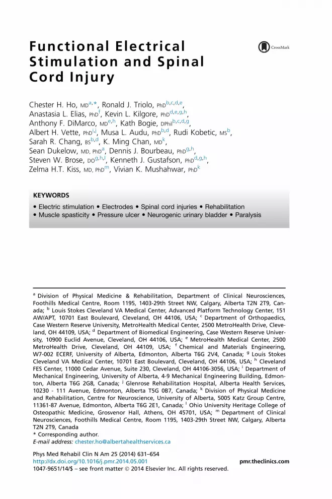
Stiffening the paralyzed trunk and hip extensors with continuous FES has a multi-tude of benefits: it can correct kyphotic seated postures, normalize lateral vertebralalignment, improve ventilation and respiratory volumes, and alter interface pres-sures.55 It can also expand bimanual workspace,48 statically stabilize the torso(Fig. 5), increase the forces that can be exerted on objects with the upper extremities,return users to erect sitting from a fully forward-flexed posture, and improve manualwheelchair propulsion efficiency at comfortable speeds.56 Independent bed turningand wheelchair transfers can also be facilitated by more rigidly coupling the pelvisto the shoulders when the paralyzed core trunk muscles are continuously activatedwith stimulation to stiffen the torso.57 In addition, activating the quadratus lumborumwith surface or implanted electrodes has been shown to enhancemediolateral stabilityand assist with attaining side leaning postures, whereas coactivation with the abdom-inal muscles can further stiffen the trunk while seated or assist in attaining forwardleaning postures. Some of the required muscles to achieve these clinical outcomescan be accessed via surface stimulation; however, strong and isolated contractionsare robustly and repeatably achieved by exciting the T12-L2 spinal nerves associatedwith the lumbar erector spinae and other muscles (Fig. 6) using intramuscular elec-trodes and surgically implanted pulse generators.58 The strategy of continuouslyactivating the core trunk and hip muscles only substitutes one statically stable posturefor another. Upper extremity effort is still required to stabilize the body during transi-tions between nonstimulated and stimulated postures and to maintain balance orrestore erect sitting when exposed to internal or external perturbations.
Fig. 5. Effect of FES on seated posture. By stimulating the trunk and hip muscles, consistentsignificant changes in posterior pelvic tilt and shoulder height were recorded.
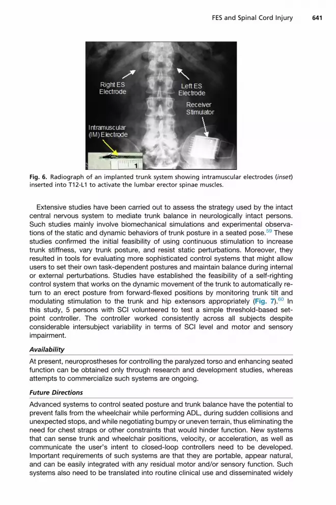
Fig. 6. Radiograph of an implanted trunk system showing intramuscular electrodes (inset)inserted into T12-L1 to activate the lumbar erector spinae muscles.
FES and Spinal Cord Injury 641
Extensive studies have been carried out to assess the strategy used by the intactcentral nervous system to mediate trunk balance in neurologically intact persons.Such studies mainly involve biomechanical simulations and experimental observa-tions of the static and dynamic behaviors of trunk posture in a seated pose.59 Thesestudies confirmed the initial feasibility of using continuous stimulation to increasetrunk stiffness, vary trunk posture, and resist static perturbations. Moreover, theyresulted in tools for evaluating more sophisticated control systems that might allowusers to set their own task-dependent postures and maintain balance during internalor external perturbations. Studies have established the feasibility of a self-rightingcontrol system that works on the dynamic movement of the trunk to automatically re-turn to an erect posture from forward-flexed positions by monitoring trunk tilt andmodulating stimulation to the trunk and hip extensors appropriately (Fig. 7).60 Inthis study, 5 persons with SCI volunteered to test a simple threshold-based set-point controller. The controller worked consistently across all subjects despiteconsiderable intersubject variability in terms of SCI level and motor and sensoryimpairment.
Availability
At present, neuroprostheses for controlling the paralyzed torso and enhancing seatedfunction can be obtained only through research and development studies, whereasattempts to commercialize such systems are ongoing.
Future Directions
Advanced systems to control seated posture and trunk balance have the potential toprevent falls from the wheelchair while performing ADL, during sudden collisions andunexpected stops, and while negotiating bumpy or uneven terrain, thus eliminating theneed for chest straps or other constraints that would hinder function. New systemsthat can sense trunk and wheelchair positions, velocity, or acceleration, as well ascommunicate the user’s intent to closed-loop controllers need to be developed.Important requirements of such systems are that they are portable, appear natural,and can be easily integrated with any residual motor and/or sensory function. Suchsystems also need to be translated into routine clinical use and disseminated widely
Fig. 7. Simple threshold-based control of seated balance based on trunk tilt in a subject withC8 tetraplegia. Without stimulation (top) of the hip and trunk extensors, the subject cannotreturn to erect sitting from a fully forward-flexed position without use of the arms. With thecontroller active (bottom), forward trunk tilt is arrested before a forward fall, and uprightposture is automatically restored.
Ho et al642
in home and community environments. Future directions also include the timing of thestimulation to coincide with different phases of the manual wheelchair propulsion cy-cle to improve efficiency during ramp ascent or varying speeds, utilization duringrowing exercise, and early introduction of trunk control systems soon after injury toprevent the development of spinal deformities and help vary posture to augment pres-sure relief maneuvers.
FES TECHNIQUES TO RESTORE RESPIRATORY MUSCLE FUNCTION
The use of FES to improve respiratory muscle function is discussed in depth in thearticle by Dalal and DiMarco elsewhere in this issue.
PREVENTION OF PRESSURE ULCERS THROUGH FUNCTIONAL ELECTRICALSTIMULATION
PUs are a common complication after SCI. They cause psychological distress, have adetrimental effect on quality of life, and place a significant burden on health care sys-tems, with costs estimated at $6 to $15 billion per year in the United States.61 Prevent-ing PUs from developing in the first place reduces patient suffering, improves patientoutcomes and quality of life, and reduces the large health care costs associated withtreating them. Indeed, it has been estimated that prevention of PUs is approximately2.5 times more economical than treating them.62
PUs can develop in one of 2 ways. They can originate at the surface of the skin andprogress inward if unattended. Skin inspections are often effective in detecting theseulcers at an early stage of development. If unattended, these ulcers can progressively

FES and Spinal Cord Injury 643
affect deeper tissue layers ending at the bone. PUs can also originate at deep muscle-bone interfaces and progress outward. These ulcers have only recently been acknowl-edged clinically and are now referred to as deep tissue injury (DTI). Sustained pressureleads to unrelieved mechanical deformation, tissue ischemia, and ischemia-reperfusion injury. Muscle is more susceptible to breakdown because of mechanicaldeformation and ischemia-reperfusion injury than skin; thus, damage originates withinmuscle tissue around bony prominences much sooner than in the skin. Skin inspec-tions are ineffective in detecting DTI at their earliest stages of development, and thereare no clinically viable methods for the early detection of DTI. Therefore, these ulcersoften develop unbeknownst to the affected individual or their caregiver. Once DTIsexhibit obvious skin signs, for example, purple discoloration, extensive damage inthe underlying soft tissue had already occurred. Prevention strategies such as pres-sure redistributing surfaces (mattresses and seating cushions) and periodical weightshifts have not decreased the incidence of PU; in fact, with the improved awarenessof DTI, the prevalence of PU is on the rise.63 Other approaches to prevent PU arenecessary. FES through surface stimulation and implanted electrodes are two novelways to prevent PU, each having their own specific advantages and disadvantages.Both systems require intact innervation to the gluteal muscles.
Intermittent Electrical Stimulation for the Prevention of DTI
Intermittent electrical stimulation64 (IES) was developed for the prevention of DTI. Thismethod applies brief electrical stimulation through surface electrodes to musclesaround bony prominences that are loaded during sitting or lying down (eg, the gluteusmaximus muscles) every few minutes causing them to contract. These periodical con-tractions mimic the subconscious postural adjustments conducted by able-bodiedpersons in response to discomfort while sitting or lying down. Ten seconds of IEScausing fusedmuscle contractions in the gluteusmuscles every 10minuteswhile sittingredistribute surface pressure away from the ischial tuberosities and increase tissueoxygenation in studyparticipants independently of glutealmusclemass.65,66Moreover,IES-induced contractions significantly redistribute internal pressure away from thebony prominences67 and reduce tissue deformation in the muscles between the ischialtuberosity and skin evenwhen loading levels ashigh as75%ofbodyweight in adult pigswith SCI were applied.67 IES is effective in significantly reducing or completely elimi-nating the formation of DTI in adult rats and pigs,68 thus establishing a strong scientificsupport for the utility of IES as a means for preventing DTI in clinical settings.
Implanted Neuromuscular Stimulation for Tissue Health and Pressure UlcerPrevention
Another approach of FES for PU prevention is through stimulation of the inferiorgluteal nerve, which innervates the gluteus maximus muscle and lies deep to thebuttock surface and close to the sciatic nerve. Surface electrode placement for pref-erential recruitment of the inferior gluteal nerve can be difficult for users to achieve.Moreover, repeatable electrode placement in the upper buttock region may be hardto accomplish for either independent users or their carers. Implanted neuromuscularelectrical stimulation (NMES) systems for long-term therapeutic use have dual advan-tages. The stimulating tip of the electrode can be located close to the motor point ofthe nerve of interest; this reduces the charge required to elicit a contractile responseand ensures that the response is repeatable and predicable. The user does not haveto replace the stimulating electrode every day, so the system is both reliable andsimple to use.

Ho et al644
The gluteal stimulation v1 (GSTIM I) system using implanted electrodes with percu-taneous leads provides both concurrent bilateral and alternating gluteal stimulation todeliver muscle conditioning and regular weight shifting to the user. GSTIM I has beenshown to have a positive impact on multiple aspects of tissue health. Subjects whoreceived GSTIM I have shown statistically significant changes between baseline andpostintervention ischial region interface pressure (Fig. 8). Maximum gluteal musclethickness significantly increased and was maintained with regular use of glutealNMES.69 Tissue oxygen levels also improved with regular use of dynamic stimulationbut decreased on withdrawal.In addition to the long-term changes in muscle characteristics, weight shifting
induced by gluteal NMES dynamically alters conditions at the seating support inter-face facilitated by stimulated muscular contractions. This dynamic effect increasesover time as the paralyzed muscles become stronger with regular use of implantedgluteal NMES. Chronic application of gluteal stimulation is thus uniquely able to affectthe intrinsic properties of paralyzed muscle through contractile responses to repeatedstimulation, increasing muscle thickness and blood flow together with reducingregional interface pressures.70,71 Use of GSTIM I also increased sitting toleranceand minimized the impact of minor incidents such as skin tears due to poor transfers,which were reported to be resolved in days rather than in weeks.Therapeutic implanted NMES provides a unique intrinsic approach to reducing the
risk of PU development for persons with SCI. Daily use of NMES is indicated to
Fig. 8. Multistage longitudinal analysis and self-registration analysis maps showing areas ofsignificant change in seated interface pressures over time (output adjusted for simultaneoustesting at multiple locations).
FES and Spinal Cord Injury 645
maintain hypertrophy of paralyzed muscles. Long-term use of gluteal NMES usingimplanted systems may provide an adjunctive method to ensure a regular pressure-relief regimen in high-risk persons, which can reduce the risk of PU developmentand allow users to participate more fully in ADL.
Availability
Both the IES system and the fully implanted NMES for the prevention of PUs are underresearch protocol use only.
Future Directions
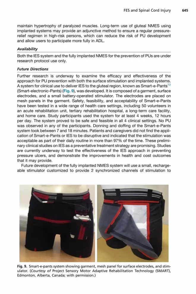
Further research is underway to examine the efficacy and effectiveness of theapproach for PU prevention with both the surface stimulation and implanted systems.A system for clinical use to deliver IES to the gluteal region, known as Smart-e-Pants72
(Smart-electronic-Pants) (Fig. 9), was developed. It is composed of a garment, surfaceelectrodes, and a small battery-operated stimulator. The electrodes are placed onmesh panels in the garment. Safety, feasibility, and acceptability of Smart-e-Pantshave been tested in a wide range of health care settings, including 50 volunteers inan acute rehabilitation unit, tertiary rehabilitation hospital, a long-term care facility,and home care. Study participants used the system for at least 4 weeks, 12 hoursper day. The system proved to be safe and feasible in all 4 clinical settings. No PUwas observed in any of the participants. Donning and doffing of the Smart-e-Pantssystem took between 7 and 18 minutes. Patients and caregivers did not find the appli-cation of Smart-e-Pants or IES to be disruptive and indicated that the stimulation wasacceptable as part of their daily routine in more than 97% of the time. These prelimi-nary clinical studies on IES as a preventative treatment strategy are promising. Studiesare currently underway to test the effectiveness of the IES approach in preventingpressure ulcers, and demonstrate the improvements in health and cost outcomesthat it may provide.Future development of the fully implanted NMES system will use a small, recharge-
able stimulator customized to provide 2 synchronized channels of stimulation to
Fig. 9. Smart-e-pants system showing garment, mesh panel for surface electrodes, and stim-ulator. (Courtesy of Project Sensory Motor Adaptive Rehabilitation Technology (SMART),Edmonton, Alberta, Canada; with permission.)
Ho et al646
automatically produce the regular weight-shift maneuvers recommended for periodicpressure relief when seated in the wheelchair.
FES FOR RESTORING BLADDER CONTROL
The lower urinary tract (LUT) functions in the storage and emptying of urine. After SCIwith an upper motor neuron injury to the sacral nerve roots, volitional control of thesefunctions is frequently lost and the LUT becomes hyperreflexive. Incontinence canoccur when the detrusor produces large, uninhibited reflex contractions at low vol-umes of stored urine. Simultaneously with detrusor contractions, the external urethralsphincter (EUS) may reflexively contract as pressure builds in the urethra during void-ing, producing detrusor-sphincter dyssynergia (DSD). This uncoordinated reflex andthe subsequent high bladder pressures can result in inefficient voiding, incontinence,and ureteric reflux, causing renal injury. In addition, DSD can also cause autonomicdysreflexia (AD), which can be life-threatening if not resolved. Finally, loss of bladdercontrol has a severe impact on quality of life and self-image. Persons with SCI listbladder function restoration among the highest priority for restoration, more thanstanding and ambulation.15
Persons with SCI frequently report ineffectiveness with existing bladder manage-ment, medication side effects, challenges associated with bladder catheterizationstrategies, and complications associated with surgical solutions. Similar to many othercomplications of SCI discussed earlier, there remains a critically unmet need to restorebladder function lost to SCI and the use of FES may offer an effective solution.FES offers a means to restore LUT function by activating the bladder and inhibiting
the urethral sphincter to produce voiding or by inhibiting the bladder to provide urinarycontinence and reduce triggers for AD and restore LUT function.73 The Brindleyapproach was the first widely clinically available FES system for bladder function.74
This approach produces bladder contractions by stimulating bladder motor efferentsin the sacral roots. To avoid cocontraction of the EUS and detrusor preventing fluidflow, stimulation is delivered in repeated bursts. After each burst, the striated EUSmus-cle relaxes, but the smooth-muscle bladder relaxes more slowly, maintaining bladderpressure and creating a pressure gradient that causes poststimulus urine flow. Thissystem has been implanted in thousands of persons with SCI and is both medicallyeffective and cost-effective.75 However, this approach requires transection of the dor-sal spinal roots (dorsal rhizotomy) to eliminate unwanted bladder and urethral reflexesdue to sensory feedback. This rhizotomy also eliminates desirable reflexes that affectsexual and bowel functions and removes the opportunity for future clinical therapies,markedly reducing acceptance of this approach by persons with SCI.76
Stimulation of peripheral sensory pathways can access and influence the spinalneural circuits that control pelvic reflexes and function. Afferent-mediated neural pros-theses take advantage of natural nervous system processes and are potentially lessinvasive than spinal-root-based approaches such as the Brindley system. Thisapproach has the potential to provide more natural function than motor-driven ap-proaches, although it is more dependent on stimulation patterns and other inputs tothe spinal circuits. One such approach uses genital nerve stimulation to achieve directspinal level bladder inhibition. This approach has primarily been applied for immediateuse, but it has also shown to improve urinary continence and bladder capacity in per-sons with SCI during short-duration use.77,78 If longer-term use is effective, then thisapproach may provide both a noninvasive and implanted option. Bladder inhibitionvia implanted electrodes on the pudendal nerve79 and sacral roots80 can also providebladder inhibition in persons with SCI.

FES and Spinal Cord Injury 647
Availability
There are several neural prostheses in development to restore pelvic functions forpersons with SCI to activate or inhibit the bladder and urethral sphincter and providea rhizotomy-free Brindley system. They are not commercially available yet.
Future Directions
Some approaches have been shown to be effective in animal models and may bepromising for human studies. Bladder activation and voiding via pudendal urethralafferent stimulation has been demonstrated in animal models, and human studies sug-gest that bladder excitation can be achieved.81 This approach may provide aperipheral-based alternative to sacral-root-based bladder activation.Urethral sphincter inhibition and bladder voiding can be obtained with patterned
afferent stimulation of sacral dermatomes.82 This approach has achieved clinical dailyvoiding of awake animals with chronic SCI. It may potentially provide a less-invasivealternative in humans. Finally, high-frequency (kilohertz) stimulation can provide tem-porary, reversible, and complete conduction block of the pudendal nerve, and thusstop reflexive EUS contractions, allowing bladder voiding equivalent to nerve transec-tion.83 Bilateral pudendal nerve block can provide clinical daily voiding of awakeanimals with chronic SCI. If this approach is effective in humans, it could be combinedwith pudendal bladder inhibition to restore bladder function with a single implant.
INTRASPINAL MICROSTIMULATION FOR GAIT RESTORATION
Apart from the aforementioned systems that are either commercially available orcloser to clinical availability, one novel experimental approach is worth noting. A sig-nificant limitation of the surface stimulation system to restore walking is that many ofthe key muscles required for walking lie deep in the leg and are not accessible withsurface electrodes. Even with the percutaneous implantation system, many channelsare required to stimulate these different muscles. Mushahwar and colleagues 84–87
have pioneered the use of implanted electrodes in the spinal cord to overcome theseproblems. This approach, known as intraspinal microstimulation (ISMS), has shownpromising results in pre-clinical animal studies. Intraspinal microstimulation entailsthe implantation of a few fine, hair-like wires in the ventral horns of the small lumbosa-cral enlargement of the spinal cord (w5 cm long in humans). This region contains thecell bodies of the motoneurons innervating all the muscles of the lower extremity, aswell as large proportions of the neural networks involved in locomotion. Tappinginto this region allows access to the standing and stepping control centre in the spinalcord. Stimulation through a single ISMS microwire can produce selective movementsaround one joint.85,86,89 A single ISMS microwire can also activate synergistic musclegroups that produce coordinated multi-joint movements such as downward full limbextension, upward flexion, forward reaching, and backward propulsion,87 which elim-inates the need for routing electrodes widely through the body to each member of amuscle group producing these movements. The levels of stimulation with ISMS(<0.1 mA) are orders of magnitude less than those required for stimulation throughthe skin or percutaneous electrodes. Moreover, the levels required for generatingfunctional limb movements generate no signs of discomfort or pain in consciousexperimental animals implanted with ISMSmicrowires. By activating the underlying lo-comotor-related networks in the lumbosacral enlargement, ISMS activates the moto-neurons trans-synaptically. This in turn recruits the motor units in a near normalphysiological order which produces graded recruitment of force and large improve-ments in fatigue-resistance relative to FES systems targeting the muscles or

Ho et al648
peripheral nerves.84,90 In chronically implanted animals, ISMS microwires remainedstable throughout the duration of implantation and produced negligible damage inthe spinal cord.88,91 In adult cats, ISMS was effective in producing long durations ofstanding that, on average, were 5 times longer than durations produced by peripheralFES.92 Moreover, ISMS produced long durations of in-place stepping in animals withchronic SCI and atrophied muscles.93 In deeply anesthetized animals, ISMS wascapable of producing long distances (>1 km) of weight-bearing and propulsiveover-ground walking94; these distances were 10 times longer than walking distancesproduced by peripheral FES.95 The produced walking was adaptable on-the fly usingintelligent control strategies that adjusted the stimulation pattern based on miniatureforce and position sensors mounted on the legs.95,96 The intelligent control strategiesallowed for automatic adaptations to perturbations as well as muscle fatigue. Interest-ingly, both standing and walking with ISMS were produced with as few as 4 micro-wires in each side of the spinal cord.Given the promising results obtained by ISMS, a proof-of-principle human study is
currently planned, and a number of considerations are under discussion. Theseinclude patient selection, instrumentation, level of spinal cord injury, and fusion of spi-nal vertebra around the implant region. The most common sites of traumatic SCI arethe cervical and thoracolumbar junction, while mid-thoracic injuries constitute 35% ofall injuries. The appropriate volunteers for the study would be drawn from the mid-thoracic pool. While younger patients are generally better candidates for any experi-mental therapy, a temporary implant or an intra-operative mapping procedure todetermine the ability of ISMS to produce functional leg movements may precludethese persons from undergoing permanent implantation in the future. Multiple pen-etrations of the spinal cord may result in gliosis, and opening the dura mater may pro-duce a scar in the pia mater and arachnoid layers, making surgical re-exploration moredifficult. Other surgical considerations include the ease of implanting very fine wires<100 mm in diameter into the spinal cord. Specialized instrumentation has beendesigned to inject stem cells successfully into the anterior horn of lumbar spinal seg-ments97,98 and could be adapted to insert electrodes as well. As with stem cell injec-tion, anticipated complications include cerebrospinal fluid leakage. Minimally invasiveinsertion methods may be considered and fusion may be undertaken as part of theprocedure to secure wires ema.
SUMMARY
Functional electrical stimulation has been well-studied and can be used in manyways to improve the well-being and functionality of persons with SCI. The scopeof applications for FES in SCI continues to grow. Many options are commerciallyavailable both for institutional and home use, while others are still in research phase.When appropriate, SCI clinicians are encouraged to consider the use of FES aspart of their standard regimen for rehabilitation or medical management of personswith SCI.
ACKNOWLEDGMENTS
The authors would like to thank the following institutions for their financial support:Alberta Innovates - Health Solutions, Canada Foundation for Innovation, Canadian In-stitutes of Health Research, Congressionally Directed and Peer-Reviewed Medical,Research Programs of the US Department of Defense (CDMRP/PRMRP), InternationalSpinal Research Trust, National Institutes of Health (NIBIB and NINDS), Natural Sci-ences and Engineering Research Council of Canada, Project SMART, Rick Hansen

FES and Spinal Cord Injury 649
Man in Motion Fund, Spinal Cord Injury Treatment Centre Society, US Department ofDefense Spinal Cord Injury Research Program, and US Department of Veterans AffairsRehabilitation Research & Development Service.
REFERENCES
1. Popovic MR, Keller T, Papas IP, et al. Surface-stimulation technology for graspingand walking neuroprostheses. IEEE Eng Med Biol Mag 2001;20(1):82–93.
2. Ragnarsson KT. Functional electrical stimulation after spinal cord injury: currentuse, therapeutic effects and future directions. Spinal Cord 2007;46(4):255–74.
3. Agarwal S, Kobetic R, Nandurkar S, et al. Functional electrical stimulation forwalking in paraplegia: 17-year follow-up of 2 cases. J Spinal Cord Med 2003;26(1):86–91.
4. Naples GG, Mortimer JT, Scheiner A, et al. A spiral nerve cuff electrode for pe-ripheral nerve stimulation. IEEE Trans Biomed Eng 1988;35:905–16.
5. Agnew WF, McCreery DB, Yuen TG, et al. Histologic and physiologic evaluationof electrically stimulated peripheral nerve: considerations for the selection of pa-rameters. Ann Biomed Eng 1989;17(1):39–60.
6. Tyler DJ, Durand DM. Functionally selective peripheral nerve stimulation with aflat interface nerve electrode. IEEE Trans Neural Syst Rehabil Eng 2002;10:294–303.
7. Tyler DJ, Durand DM. A slowly penetrating interfascicular nerve electrode for se-lective activation of peripheral nerves. IEEE Trans Rehabil Eng 1997;5(1):51–61.
8. Bamford JA, Mushahwar VK. Intraspinal microstimulation for the recovery offunction following spinal cord injury. Prog Brain Res 2011;194:227–39.
9. Agarwal S, Triolo RJ, Kobetic R, et al. Long-term user perceptions of an im-planted neuroprosthesis for exercise, standing, and transfers after spinal cordinjury. J Rehabil Res Dev 2003;40(3):241–52.
10. Cogan SF. Neural stimulation and recording electrodes. Annu Rev Biomed Eng2008;10(1):275–309.
11. Geddes LA, Roeder R. Criteria for the selection of materials for implanted elec-trodes. Ann Biomed Eng 2003;31(7):879–90.
12. Prasad A, Sanchez JC. Quantifying long-term microelectrode array functionalityusing chronic in vivo impedance testing. J Neural Eng 2012;9(2):026028.
13. Khaled I, Cheng C, Elmallah S, et al. A flexible base electrode array for intraspi-nal microstimulation. IEEE Trans Biomed Eng 2013;60:2904–13.
14. Grill WM, Norman SE, Bellamkonda RV. Implanted neural interfaces: bio-challenges and engineered solutions. Annu Rev Biomed Eng 2009;11:1–24.
15. Anderson K. Targeting recovery: priorities of the spinal cord injured population.J Neurotrauma 2004;21(10):1371–83.
16. Keith MW, Kilgore KL, Peckham PH, et al. Tendon transfers and functional elec-trical stimulation for reconstruction of hand function in spinal cord injury. J HandSurg Am 1996;21:89–99.
17. Chae J, Kilgore KL, Triolo RJ, et al. Functional neuromuscular stimulation in spi-nal cord injury. Phys Med Rehabil Clin N Am 2000;11(1):209–26.
18. Scott TR, Haugland M. Command and control interfaces for advanced neuro-prosthetic applications. Neuromodulation 2001;4(4):165–75.
19. Nathan RH. Functional electrical stimulation of the upper limb: charting the fore-arm surface. Med Biol Eng Comput 1979;17(6):729–36.
20. Popovic M, Popovic D, Keller T. Neuroprostheses for grasping. Neurol Res2002;24:443–52.

Ho et al650
21. Popovic R, Thrasher T, Adams M, et al. Functional electrical therapy: retraininggrasping in spinal cord injury. Spinal Cord 2006;44(3):143–51.
22. Peckham PH, Keith MW, Kilgore KL, et al. Efficacy of an implanted neuropros-thesis for restoring hand grasp in tetraplegia: a multicenter study. Arch PhysMed Rehabil 2001;82:1380–8.
23. Wuolle KS, Van Doren CL, Bryden AM, et al. Satisfaction and usage of a handneuroprosthesis. Arch Phys Med Rehabil 1999;80:206–13.
24. Taylor P, Esnouf J, Hobby J. The functional impact of the Freehand system ontetraplegic hand function, clinical results. Spinal Cord 2002;40:560–6.
25. Kilgore KL, Hoyen HA, Bryden AM, et al. An implanted upper extremity neuro-prosthesis utilizing myoelectric control. J Hand Surg Am 2008;33:539–50.
26. Hart RL, Bhadra N, Montague FW, et al. Design and testing of an advancedimplantable neuroprosthesis with myoelectric control. IEEE Trans Neural SystRehabil Eng 2011;19(1):45–53.
27. Peckham PH, Kilgore KL. Challenges and opportunities in restoring functionafter paralysis. IEEE Trans Biomed Eng 2013;60(3):602–9.
28. Moss CW, Kilgore KL, Peckham PH. A novel command signal for motor neuro-prosthetic control. Neurorehabil Neural Repair 2011;25(9):847–54.
29. Donoghue JP, Nurmikko A, Black M, et al. Assistive technology and robotic con-trol using motor cortex ensemble-based neural interface systems in humanswith tetraplegia. J Physiol 2007;579(Pt 3):603–11.
30. Williams MR, Kirsch RF. Evaluation of head orientation and neck muscle EMGsignals as command inputs to a human-computer interface for individualswith high tetraplegia. IEEE Trans Neural Syst Rehabil Eng 2008;16(5):485–96.
31. Rohde L, Bonder B, Triolo R. An exploratory study of perceived quality of life withimplanted standing neuroprostheses. J Rehabil Res Dev 2012;49(2):265–78.
32. Creasey GH, Ho CH, Triolo RJ, et al. Clinical applications of electrical stimulationafter spinal cord injury. J Spinal Cord Med 2004;27:365–75.
33. Triolo RJ, Bailey SN, Miller ME, et al. Longitudinal performance of a surgicallyimplanted neuroprosthesis for lower extremity exercise, standing, and transfersafter spinal cord injury. Arch Phys Med Rehabil 2012;93(5):896–904. http://dx.doi.org/10.1016/J.APMR.2012.01.001.
34. Nataraj R, Audu M, Triolo R. Comparing joint kinematics and center of mass ac-celeration for feedback control of standing by functional neuromuscular stimula-tion. J Neuroeng Rehabil 2012;9:25. http://dx.doi.org/10.1186/1743-0003-9-25.
35. Mushahwar VK, Jacobs PL, Normann RA, et al. New functional electrical stimu-lation approaches to standing and walking. J Neural Eng 2007;4:S181–97.
36. Kobetic R, Triolo RJ, Uhlir J, et al. Implanted functional electrical stimulation sys-tem for mobility in paraplegia: a follow-up case report. IEEE Trans Rehabil Eng1999;7(4):390–8.
37. Cikajlo I, Matjacic Z, Bajd T, et al. Sensory supported FES control in gaittraining of incomplete spinal cord injury persons. Artif Organs 2005;29(6):459–61.
38. Hardin E, Kobetic R, Murray L, et al. Walking after incomplete spinal cord injurywith an implanted FES system. J Rehabil Res Dev 2007;44(3):333–46.
39. Bailey SN, Hardin E, Kobetic R, et al. Neuroprosthetic and neurotherapeutic ef-fects of implanted electrical stimulation for ambulation after incomplete spinalcord injury. J Rehabil Res Dev 2010;47(1):7–16.
40. Dutta A, Kobetic R, Triolo R. Gait initiation with electromyographically triggeredelectrical stimulation in people with partial paralysis. J Biomech Eng 2009;131(8):081002. http://dx.doi.org/10.1115/1.3086356.

FES and Spinal Cord Injury 651
41. Hettinga DM, Andrews BJ. Oxygen consumption during functional electricalstimulation-assisted exercise in persons with spinal cord injury: implicationsfor fitness and health. Sports Med 2008;38(10):825–38.
42. Sadowsky CL, Hammond ER, Strohl AB, et al. Lower extremity functional elec-trical stimulation cycling promotes physical and functional recovery in chronicspinal cord injury. J Spinal Cord Med 2013;36(6):623–31. http://dx.doi.org/10.1179/2045772313Y.0000000101.
43. Dutta A, Kobetic R, Triolo R. Ambulation after incomplete spinal cord injury withEMG-triggered functional electrical stimulation. IEEE Trans Biomed Eng 2008;55(2):791–4.
44. Fisher L, Miller M, Nogan S, et al. Standing after spinal cord injury with four con-tact nerve-cuff electrodes for quadriceps stimulation. IEEE Trans Neural SystRehabil Eng 2008;16(5):473–8. http://dx.doi.org/10.1109/TNSRE.2008.2003390.
45. Fisher L, Tyler D, Triolo R. Optimization of selective stimulation parameters formulti-contact electrodes. J Neuroeng Rehabil 2013;10:25. http://dx.doi.org/10.1186/1743-0003-10-25.
46. Audu M, Nataraj R, Gartman S, et al. Posture shifting after spinal cord injuryusing functional neuromuscular stimulation – a computer simulation study.J Biomech 2011;44:1639–45. http://dx.doi.org/10.1016/j.jbiomech.2010.12.020.
47. Kobetic R, To C, Schnellenberger J, et al. Development of a hybrid orthosis forstanding, walking and stair climbing after spinal cord injury. J Rehabil Res Dev2009;46(3):447–62.
48. Kukke SN, Triolo RJ. The effects of trunk stimulation on bimanual seated work-space. IEEE Trans Neural Syst Rehabil Eng 2004;12:177–85.
49. Spungen AM, Adkins RH, Steward CA, et al. Factors influencing body compo-sition in persons with spinal cord injury: a cross-sectional study. J Appl Physiol(1985) 2003;95:2398–407.
50. Potten YJ, Seelen HA, Drukker J, et al. Postural muscle responses in the spi-nal cord injured persons during forward reaching. Ergonomics 1999;42:1200–15.
51. Hart N, Laffont I, de La Sota A, et al. Respiratory effects of combined truncal andabdominal support in patients with spinal cord injury. Arch Phys Med Rehabil2005;86:1447–51.
52. Hobson DA, Tooms RE. Seated lumbar/pelvic alignment. A comparison betweenspinal cord-injured and noninjured groups. Spine 1992;17:293–8.
53. Curtis KA, Kindlin CM, Reich KM, et al. Functional reach in wheelchair users: theeffects of trunk and lower extremity stabilization. Arch Phys Med Rehabil 1995;76:360–7.
54. Ueyoshi A, Shima Y. Studies on spinal braces. With special reference to theeffects of increased abdominal pressure. Int Orthop 1985;9:255–8.
55. Wu GA, Lombardo LM, Triolo RJ, et al. The effects of combined trunk and glutealneuromuscular electrical stimulation on posture and tissue health in spinal cordinjury. PM R 2013;5:688–96.
56. Triolo RJ, Bailey SN, Miller ME, et al. Effects of stimulating hip and trunk muscleson seated stability, posture, and reach after spinal cord injury. Arch Phys MedRehabil 2013;94:1766–75.
57. Triolo RJ, Bailey SN, Lombardo LM, et al. Effects of intramuscular trunk stimula-tion on manual wheelchair propulsion mechanics in 6 subjects with spinal cordinjury. Arch Phys Med Rehabil 2013;94:1997–2005.
58. Davis JA, Triolo RJ, Uhlir JP, et al. Surgical technique for installing an 8-channelneuroprosthesis for standing. Clin Orthop Relat Res 2001;4:237–52.

Ho et al652
59. Vette AH, Yoshida T, Thrasher TA, et al. A comprehensive three-dimensional dy-namic model of the human head and trunk for estimating lumbar and cervicaljoint torques and forces from upper body kinematics. Med Eng Phys 2012;34(5):640–9.
60. Murphy JO,AuduML,LombardoLM,et al. Feasibility of aclosed-loopcontroller forrighting seated posture after spinal cord injury. J Rehabil Res Dev 2014, in press.
61. Markova A, Mostow EN. US skin disease assessment: ulcer and wound care.Dermatol Clin 2012;30(1):107–11, ix.
62. Lyder CH, Wang Y, Metersky M, et al. Hospital-acquired pressure ulcers: resultsfrom the national Medicare Patient Safety Monitoring System study. J Am GeriatrSoc 2012;60:1603–8.
63. VanGilder C, MacFarlane GD, Harrison P, et al. The demographics of suspecteddeep tissue injury in the United States: an analysis of the International PressureUlcer Prevalence Survey 2006-2009. Adv Skin Wound Care 2010;23:254–61.
64. Mushahwar VK, Solis L. Mitigation of pressure ulcers using electrical stimulation.US patent application 12/362,725, Jan 2009.
65. Solis LR, Gyawali S, Seres P, et al. Effects of intermittent electrical stimulation onsuperficial pressure, tissue oxygenation, and discomfort levels for the preven-tion of deep tissue injury. Ann Biomed Eng 2011;39:649–63.
66. Gyawali S, Solis L, Chong SL, et al. Intermittent electrical stimulation redistrib-utes pressure and promotes tissue oxygenation in loaded muscles of individualswith spinal cord injury. J Appl Physiol (1985) 2011;110:246–55.
67. Solis LR, Liggins A, Uwiera RR, et al. Distribution of internal pressure aroundbony prominences: implications to deep tissue injury and effectiveness of inter-mittent electrical stimulation. Ann Biomed Eng 2012;40(8):1740–59. http://dx.doi.org/10.1007/s10439-012-0529-0.
68. Solis LR, Twist E, Seres P, et al. Prevention of deep tissue injury through musclecontractions induced by intermittent electrical stimulation after spinal cord injuryin pigs. J Appl Physiol (1985) 2013;114(2):286–96. http://dx.doi.org/10.1152/japplphysiol.00257.2012.
69. Bogie KM, Wang X, Triolo RJ. Long term prevention of pressure ulcers in highrisk individuals: a single case study of the use of gluteal neuromuscular electri-cal stimulation. Arch Phys Med Rehabil 2006;87(4):585–91.
70. Bogie KM, Triolo RJ. The effects of regular use of neuromuscular electrical stim-ulation on tissue health. J Rehabil Res Dev 2003;40(6):469–75.
71. Bogie K, Ho CH, Chae J, et al. Dynamic therapeutic neuromuscular electricalstimulation for pressure relief. Am J Phys Med Rehabil 2004;83(3):240.
72. Mushahwar VK, Isaacson G, Ahmetovic A, et al. Apparatus and method for elec-trically stimulating pressure-loaded muscles. WIPO application WO 2013/113099 A1, January 2013.
73. Gaunt RA, Prochazka A. Control of urinary bladder function with devices: suc-cesses and failures. Prog Brain Res 2006;152:163–94.
74. Brindley GS. An implant to empty the bladder or close the urethra. J Neurol Neu-rosurg Psychiatry 1977;40:358–69.
75. Creasey GH, Dahlberg JE. Economic consequences of an implanted neuro-prosthesis for bladder and bowel management. Arch Phys Med Rehabil 2001;82(11):1520–5.
76. Sanders PM, Ijzerman MJ, Roach MJ, et al. Patient preferences for next genera-tion neural prostheses to restore bladder function. Spinal Cord 2011;49(1):113–9.
77. FaragFF,MartensFM,RijkhoffNJ, et al. Dorsal genital nerve stimulation in patientswith detrusor overactivity: a systematic review. Curr Urol Rep 2012;13(5):385–8.

FES and Spinal Cord Injury 653
78. Lee YH, Kim SH, Kim JM, et al. The effect of semiconditional dorsal penile nerveelectrical stimulation on capacity and compliance of the bladder with deformityin spinal cord injury patients: a pilot study. Spinal Cord 2012;50(4):289–93.
79. Possover M, Schurch B, Henle KP. New strategies of pelvic nerves stimulationfor recovery of pelvic visceral functions and locomotion in paraplegics. Neuro-urol Urodyn 2010;29(8):1433–8.
80. Kirkham AP, Knight SL, Craggs MD, et al. Neuromodulation through sacral nerveroots 2 to 4 with a Finetech-Brindley sacral posterior and anterior root stimulator.Spinal Cord 2002;40(6):272–81.
81. Yoo PB, Horvath EE, Amundsen CL, et al. Multiple pudendal sensory pathwaysreflexly modulate bladder and urethral activity in patients with spinal cord injury.J Urol 2011;185(2):737–43.
82. McCoin JL, Bhadra N, Gustafson KJ. Electrical stimulation of sacral derma-tomes can suppress aberrant urethral reflexes in felines with chronic spinalcord injury. Neurourol Urodyn 2013;32(1):92–7.
83. Boger AS, Bhadra N, Gustafson KJ. High frequency sacral root nerve blockallows bladder voiding. Neurourol Urodyn 2012;31(5):677–82.
84. Mushahwar VK, Horch KW. Proposed Specifications for a Lumbar Spinal CordElectrode Array for Control of Lower Extremities in Paraplegia. IEEE Trans Reha-bil Eng 1997;5:237–43.
85. Mushahwar VK, Horch KW. Selective Activation of Muscles in the Feline Hin-dlimb through Electrical Microstimulation of the Ventral Lumbo-Sacral SpinalCord. IEEE Trans Rehabil Eng 2000;8(1):11–21.
86. Mushahwar VK, Horch KW. Muscle Recruitment through Electrical Stimulation ofthe Lumbo-sacral Spinal Cord. IEEE Trans Rehabil Eng 2000;8(1):22–9.
87. Mushahwar VK, Collins DK, Prochazka A. Spinal Cord Microstimulation Gener-ates Functional Movements in Chronically Implanted Cats. Exp Neurol 2000;163(2):422–9.
88. Prochazka A, Mushahwar VK, McCreery D. Neural Prostheses: Pros and Cons. JPhysiol (London) 2001;533:99–109.
89. Mushahwar VK, Horch KW. Selective Activation and Graded Recruitment ofFunctional Muscle Groups through Spinal Cord Stimulation. Ann N Y Acad Sci1998;860:531–5.
90. Bamford J, Putman CT, Mushahwar VK. Intraspinal Microstimulation Preferen-tially Recruits Fatigue-Resistant Muscle Fibres and Generates Gradual Forcein Rat. J Physiol (London) 2005;569.3:873–84.
91. Bamford JA, Todd KG, Mushahwar VK. The Effects of Intraspinal Microstimula-tion on Spinal Cord Tissue in the Rat. Biomaterials 2010;31:5552–63.
92. Lau B, Guevremont L, Mushahwar VK. Open- and Closed-loop Control Strate-gies for Restoring Standing using Intramuscular and Intraspinal Stimulation.IEEE Trans Neural Syst Rehabil Eng 2007;15:273–85.
93. Saigal R, Renzi CG, Mushahwar VK. Intraspinal Microstimulation GeneratesFunctional Limb Movements after Spinal Cord Injury. IEEE Trans Neural SystRehabil Eng 2004;12:430–40.
94. Holinski BJ, Mazurek KA, Everaert DG, et al. Restoring stepping after spinalcord injury using intraspinal microstimulation and novel control strategies.Conf Proc IEEE Eng Med Biol Soc 2011;2011:5798–801. http://dx.doi.org/10.1109/IEMBS.2011.6091435.
95. Mazurek K, Holinski BJ, Everaert DG, et al. Feedforward and Feedback Controlfor Over-ground Locomotion in Anesthetized Cats. J Neural Eng 2012;9(2):026003.
Ho et al654
96. Guevremont L, Norton JA, Mushahwar VK. A Physiologically-based Controllerfor Generating Overground Locomotion using Functional Electrical Stimulation.J Neurophysiol 2007;97:2499–510.
97. Federici T, Hurtig CV, Burks KL, et al. Surgical technique for spinal cord deliveryof therapies: demonstration of procedure in gottingen minipigs. J Vis Exp2012;(70):e4371.
98. Riley J, Federici T, Polak M, et al. Intraspinal stem cell transplantation in amyo-trophic lateral sclerosis: a phase I safety trial, technical note, and lumbar safetyoutcomes. Neurosurgery 2012;71(2):405–16. http://dx.doi.org/10.1227/NEU.0b013e31825ca05f [discussion: 416].