foxp2 regulates gene networks implicated in neurite outgrowth in the developing brain
TRANSCRIPT
Foxp2 Regulates Gene Networks Implicated in NeuriteOutgrowth in the Developing BrainSonja C. Vernes1¤a, Peter L. Oliver2, Elizabeth Spiteri3¤b, Helen E. Lockstone1, Rathi Puliyadi1, Jennifer M.
Taylor1, Joses Ho1, Cedric Mombereau4, Ariel Brewer4, Ernesto Lowy1, Jerome Nicod1, Matthias
Groszer1,4, Dilair Baban1, Natasha Sahgal1, Jean-Baptiste Cazier1, Jiannis Ragoussis1, Kay E. Davies2,
Daniel H. Geschwind3,5, Simon E. Fisher1,6*
1 Wellcome Trust Centre for Human Genetics, University of Oxford, Oxford, United Kingdom, 2 Medical Research Council Functional Genetics Unit, University of Oxford,
Oxford, United Kingdom, 3 Program in Neurogenetics, Department of Neurology, University of California Los Angeles, Los Angeles, California, United States of America,
4 INSERM Institute du Fer a Moulin, University Pierre and Marie Curie, UMR-S 839, Paris, France, 5 Semel Institute and Department of Human Genetics, David Geffen School
of Medicine, University of California Los Angeles, Los Angeles, California, United States of America, 6 Language and Genetics Department, Max Planck Institute for
Psycholinguistics, Nijmegen, The Netherlands
Abstract
Forkhead-box protein P2 is a transcription factor that has been associated with intriguing aspects of cognitive function inhumans, non-human mammals, and song-learning birds. Heterozygous mutations of the human FOXP2 gene cause amonogenic speech and language disorder. Reduced functional dosage of the mouse version (Foxp2) causes deficientcortico-striatal synaptic plasticity and impairs motor-skill learning. Moreover, the songbird orthologue appears criticallyimportant for vocal learning. Across diverse vertebrate species, this well-conserved transcription factor is highly expressed inthe developing and adult central nervous system. Very little is known about the mechanisms regulated by Foxp2 duringbrain development. We used an integrated functional genomics strategy to robustly define Foxp2-dependent pathways,both direct and indirect targets, in the embryonic brain. Specifically, we performed genome-wide in vivo ChIP–chip screensfor Foxp2-binding and thereby identified a set of 264 high-confidence neural targets under strict, empirically derivedsignificance thresholds. The findings, coupled to expression profiling and in situ hybridization of brain tissue from wild-typeand mutant mouse embryos, strongly highlighted gene networks linked to neurite development. We followed up ourgenomics data with functional experiments, showing that Foxp2 impacts on neurite outgrowth in primary neurons and inneuronal cell models. Our data indicate that Foxp2 modulates neuronal network formation, by directly and indirectlyregulating mRNAs involved in the development and plasticity of neuronal connections.
Citation: Vernes SC, Oliver PL, Spiteri E, Lockstone HE, Puliyadi R, et al. (2011) Foxp2 Regulates Gene Networks Implicated in Neurite Outgrowth in theDeveloping Brain. PLoS Genet 7(7): e1002145. doi:10.1371/journal.pgen.1002145
Editor: Joshua M. Akey, University of Washington, United States of America
Received October 22, 2010; Accepted May 7, 2011; Published July 7, 2011
Copyright: � 2011 Vernes et al. This is an open-access article distributed under the terms of the Creative Commons Attribution License, which permitsunrestricted use, distribution, and reproduction in any medium, provided the original author and source are credited.
Funding: This work was supported by the Wellcome Trust (grant numbers 075491 and 080971), the Royal Society (fellowship to SEF), Autism Speaks, the SimonsFoundation Autism Research Initiative (SFARI, grant number 137593 to SEF), and the Max Planck Society. SCV was supported by the Christopher Welch BiologicalSciences Scholarship and the Wellcome Trust V.I.P programme. JN was supported by a Marie Curie Intra-European Fellowship. HEL, NS, JBC, DB and JR weresupported by the Wellcome Trust Core Grant Award (grant number 075491/Z/04). MG and CM are supported by the Ecole des Neurosciences de Paris Ile-de-France(ENP) and the Inserm AVENIR programme. This research was supported in part by National Institutes of Health (NIH) grant 5R21MH075028. ES was supported by NIHgrant T32GM008243. The funders had no role in study design, data collection and analysis, decision to publish, or preparation of the manuscript.
Competing Interests: The authors have declared that no competing interests exist.
* E-mail: [email protected]
¤a Current address: Research Institute of Molecular Pathology, Vienna, Austria¤b Current address: Cedars Sinai Medical Center, Department of Pathology and Laboratory Medicine, Los Angeles, California, United States of America
Introduction
Forkhead-box protein P2 is a highly conserved vertebrate
protein, belonging to an important group of transcription factors
[1]. By modulating the expression of downstream target genes,
forkhead-box proteins influence a diverse array of processes,
including cell-cycle regulation, signal transduction, differentiation,
patterning and metabolism [2]. They thereby play crucial roles
during embryogenesis, in postnatal development and in the
mature organism, and many have been linked to disease states
[3]. The P subgroup is a divergent branch of forkhead-box
proteins that share a distinctive DNA-binding domain located near
the C-terminal end of the protein, as well as zinc-finger/leucine-
zipper motifs that mediate dimerization, and a glutamine-rich
region towards the N-terminus [4,5].
Functional evidence from multiple species implicates Forkhead-
box protein P2 in particularly intriguing aspects of brain
development and function [1]. (Here we adopt the standard
accepted nomenclature to refer to the protein in different species:
FOXP2 in humans, Foxp2 in mice, FoxP2 in other chordates, with
the corresponding gene names in italics [6].) In humans, damage to
one copy of the FOXP2 gene causes a rare neurodevelopmental
disorder, characterised by difficulties mastering sequences of mouth
movements during speech, as well as impaired language processing
[4,7,8]. Heterozygous disruptions of the mouse orthologue (Foxp2)
yield dramatic reductions in synaptic plasticity of cortico-striatal
PLoS Genetics | www.plosgenetics.org 1 July 2011 | Volume 7 | Issue 7 | e1002145
brain circuits, associated with deficits in learning of rapid motor
skills [9]. Mouse pups with homozygous Foxp2 mutations show more
severe neural effects – gross motor impairments, delayed postnatal
maturation of the cerebellum and dramatic reductions in emission
of ultrasonic vocalisations – against a background of reduced
weight-gain and postnatal lethality [9-11]. In addition, the avian
orthologue (FoxP2) is required for normal vocal learning in
songbirds [12,13]. Selective knockdown of the gene in a key striatal
nucleus in juvenile zebrafinches leads to incomplete and inaccurate
imitation of tutor songs [14].
Studies of both human FOXP2 and mouse Foxp2 identified
similarly strong CNS (central nervous system) expression during
embryogenesis, which is confined to neurons (absent from glial cells)
and enriched in various brain structures, including deep layers of the
developing cortical plate, and parts of the striatum, thalamus and
cerebellum [15,16]. These embryonic expression patterns appear
highly concordant in the different species, and show remarkable
overlaps with sites of pathology identified by neuroimaging of
human children and adults carrying FOXP2 mutations [16,17].
Neural expression of the gene continues postnatally and into
adulthood [4,15], and is also observed in certain non-neural tissues,
most notably the distal alveolar lung epithelium, and the outflow
tract and atrium of the cardiovascular system [18].
The above observations of well-conserved and specific CNS
expression patterns [15,16] suggest that Foxp2 is likely to have
important functions in neurodevelopment. Nevertheless, as data
continue to accumulate regarding its impacts on the postnatal
brain [9,11,14], the specific roles of Foxp2 in the developing CNS
remain largely elusive. One route for gaining insights into the
biological processes controlled by a transcription factor is to define
the regulatory networks that are directly downstream of it [1]. An
efficient strategy for identifying direct targets exploits chromatin
immunoprecipitation (ChIP) methods to screen the tissue of
interest [19]. Two previous investigations have coupled ChIP with
hybridisation to promoter microarrays (ChIP-chip) in order to
uncover binding sites of FOXP2 in human foetal brain tissue [20]
and in human neurons grown in culture [21]. Both screens were
of limited scope – the microarrays in these studies comprised
fragments from the 59 ends of ,5,000 loci [20,21], representing a
small percentage of the known gene promoters in the genome.
Neither study combined ChIP data with large-scale expression
analyses. A more recent report used mRNA expression profiling in
human neuronal models transfected with different versions of
FOXP2 to explore regulatory differences between the human and
chimpanzee orthologues, but did not include any ChIP screening
for direct targets [22].
In the present study, we performed a systematic large-scale in vivo
ChIP-chip screen of the embryonic mouse brain, coupling Foxp2-
ChIP to high-density arrays with oligonucleotides tiled across
.17,000 promoters. We robustly established the empirical signifi-
cance of our ChIP results in wild-type brains by determining the null
distribution of signals generated by matched control tissue from
littermates that expressed no Foxp2 protein. Under strict empirical
thresholds that minimised false positive signals, we isolated a set of
264 high-confidence in vivo targets. Gene ontology (GO) analyses of
the ChIP-chip data, as well as genome-wide expression profiling and
in situ hybridisations of wild-type and mutant mice, converged on
neurite outgrowth as one of the most prominent biological themes
associated with Foxp2 function in the embryonic CNS. We went on
to directly demonstrate, using neuronal cell models and primary
neurons from the embryonic mouse brain, that Foxp2 alters
expression of neurite-outgrowth targets and thereby influences
neurite process length and branch number.
Results
Genome-wide identification of in vivo Foxp2 targets inembryonic mouse brain
In vivo Foxp2-ChIP screening was carried out using brains
harvested from embryonic mice. Experiments were performed
with mice that were wild-type for Foxp2, as well as homozygous
littermates that do not express any Foxp2 protein (Foxp2-S321X
mutants; see Materials and Methods) [9]. The different types of
sample were screened in parallel, undergoing identical experi-
mental manipulations and data processing. In this context, the
homozygous mutant mouse tissue acts as an ideal negative control
[21]. Since such samples completely lack Foxp2 protein (see Figure
S1 and [9]), fragments that are pulled down by Foxp2-ChIP in
these cases give an unbiased empirical indication of background
noise and false positive rates yielded by the procedure. Whole
mouse brains from wild-type or mutant mice were harvested at
embryonic day 16 (E16), corresponding to a timepoint at which
particularly high levels of Foxp2 expression are observed in the
developing CNS [16]. Chromatin isolated by Foxp2-ChIP was
labelled and hybridised to DNA microarrays covering the
promoter regions of ,17,000 mouse transcripts (Agilent Technol-
ogies), using total input DNA as a reference sample. Each
promoter on these arrays is represented by an average of twenty-
five 60-mer probes spanning ,5.5 kb upstream and ,2.5 kb
downstream of the transcription start site, allowing peak regions of
binding to be precisely defined (Figure 1). Moreover, the presence
of multiple probes for each promoter scattered throughout the
array gives independent enrichment values within the same
promoter, which aids discrimination of real biological targets
from false positive events. Specifically, since the shearing process
during ChIP produces overlapping fragments of chromatin, true
targets should show evidence of enrichment for multiple probes
across the promoter region, while promoters with only a single
enriched probe are most likely to be false positive results.
In order to identify enriched promoters, Foxp2-ChIP data were
analysed as per Materials and Methods. Briefly, array data from
independent biological replicates (three independent ChIP experi-
ments hybridised to one each of three array sets) were normalised for
each genotype (wild-type or mutant control) separately. Normalised
array data (excluding probes with a negative average enrichment
Author Summary
Foxp2 codes for an intriguing regulatory protein thatprovides a window into unusual aspects of brain functionin multiple species. For example, the gene is implicated inspeech and language disorders in humans, song learningin songbirds, and learning of rapid movement sequencesin mice. Foxp2 acts by tuning the expression levels ofother genes (its downstream targets). In this study we usedgenome-wide techniques to comprehensively identify themajor targets of Foxp2 in the embryonic brain, in order tounderstand its roles in fundamental biological pathwaysduring neurodevelopment, which we followed up throughfunctional analyses of neurons. Most notably, we foundthat Foxp2 directly and indirectly regulates networks ofgenes that alter the length and branching of neuronalprojections, an important route for modulating the wiringof neural connections in the developing brain. Overall, ourfindings shed light on how Foxp2 directs particularfeatures of nervous system development, helping us tobuild bridges between genes and complex aspects ofbrain function.
Foxp2 Regulates Neurite Outgrowth Pathways
PLoS Genetics | www.plosgenetics.org 2 July 2011 | Volume 7 | Issue 7 | e1002145
across replicate experiments) were subjected to a sliding window
analysis, using a similar method to that employed in genome-wide
ChIP-chip studies of other forkhead transcription factors [23]. Each
probe was assigned a value (window-adjusted score) based on the
median fold enrichment of itself and its neighbouring probe on either
side (within 500 bp upstream and 500 bp downstream), and then
probes were ranked based on this window score.
By analysing the distribution of window scores observed in the
mutant null control experiments we were able to derive an
empirical threshold for significance, which could then be applied
to the wild-type data. We found that window scores greater than
or equal to 0.974 (corresponding to ,2-fold enrichment) excluded
99% of the data-points in the mutant null control experiments.
When we applied this threshold to data from wild-type
experiments, we identified a set of 1,217 promoter regions that
were consistently enriched by Foxp2-ChIP over 3 replicates in
wild-type mouse brains (Table S1). On inspection of the locations
of the enriched probes throughout the mouse genome, no
positional bias was observed (Figure S2). Since some of the
enriched regions lay close to the transcriptional start site (TSS) of
more than one gene, the 1,217 promoter regions corresponded to
1,253 genes. Of note, using the same analysis parameters, only 147
genes were enriched in the mutant null controls, suggesting a low
false discovery rate. Nevertheless, in order to minimize false-
positive findings, we excluded any enriched genes from the wild-
type dataset that also had window scores exceeding the 99%
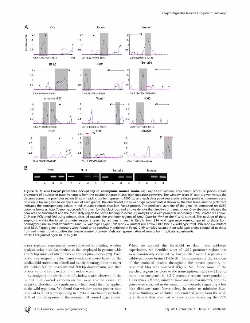
Figure 1. In vivo Foxp2 promoter occupancy in embryonic mouse brain. (A) Foxp2-ChIP window enrichment scores of probes acrosspromoters of a subset of putative targets from the neurite outgrowth and axon guidance pathways. The window score (Y axis) is given versus thedistance across the promoter region (X axis) - each cross bar represents 1000 bp and each data point represents a single probe (chromosome andposition in bp are given below the X axis of each graph). The enrichment in the wild-type experiments is shown by the blue trace, and the pink traceindicates the corresponding values in null mutant controls that lack Foxp2 protein. The predicted start site of the gene (as annotated on UCSCgenome browser: http://genome.ucsc.edu/) is given by the black box and arrows denote the direction of transcription. Grey shading indicates thepeak area of enrichment and the most likely region for Foxp2 binding to occur. (B) Analysis of in vivo promoter occupancy. DNA isolated via Foxp2-ChIP was PCR amplified using primers directed towards the promoter regions of Nrp2, Sema3a, Nrn1 or the b-actin control. The position of theseamplicons within the target promoter region is given by red bars in part A. Results from E16 wild type mice were compared to those fromhomozygous null mutant littermates. Lane 1 = wild-type Foxp2-ChIP, lane 2 = mutant null Foxp2-ChIP, lane 3 = wild-type total DNA, lane 4 = mutanttotal DNA. Target gene promoters were found to be specifically enriched in Foxp2-ChIP samples isolated from wild-type brains compared to thosefrom null mutant brains, unlike the b-actin control promoter. Gels are representative of results from triplicate experiments.doi:10.1371/journal.pgen.1002145.g001
Foxp2 Regulates Neurite Outgrowth Pathways
PLoS Genetics | www.plosgenetics.org 3 July 2011 | Volume 7 | Issue 7 | e1002145
threshold in the mutant control dataset. This filtering process
yielded a slightly smaller set of 1,164 putative targets (Table S2).
When we applied stricter thresholds to the wild-type data,
selecting only those promoters in which at least one probe gave a
window score of $ 1.5, we identified a shortlist of 259 promoter
regions. Since a small number of peak regions lay directly between
the TSSs of two different genes, these 259 promoters corresponded
to a slightly higher total of 266 genes. Crucially, the same analyses
of the entire mutant null control dataset identified only a single
gene in the genome with a window score of $ 1.5 (the Pigt gene),
indicating an extremely low rate of false positives under these
stricter selection criteria. We excluded two genes from the strict
wild-type shortlist (Pigt and Zfp496) since they contained probes
that exceeded the 99% threshold (i.e. window score of .0.974) in
mutant null controls (Figure S3). The outcome of these analyses
was a final curated shortlist of 264 high-confidence in vivo targets
(Table S3).
Given that DNA is sheared randomly during the ChIP process,
we would expect a true Foxp2 binding event to be represented by
a peak of enrichment at a target promoter. This peak would result
from the sheared DNA forming a series of overlapping fragments,
with the region closest to the binding site showing the highest
degree of enrichment (i.e. highest number of fragments pulled
down during immunoprecipitation) and with progressively less
enrichment observed as the distance to the binding site increases
on either side. Figure 1A gives typical examples of the enrichment
peaks observed for putative targets from our Foxp2-ChIP dataset.
Examination of corresponding data from mutant control exper-
iments emphasises the relative lack of enrichment in nulls that lack
Foxp2 protein, indicating that the enrichment in wild-type samples
results from highly specific Foxp2-mediated interactions. Further-
more, we followed up a subset of candidates with qPCR,
consistently confirming their enrichment (Figure 1B).
Enriched regions represented in the shortlist of high-confidence
targets were assessed in silico for any over-represented sequence
motifs (see Text S1). This analysis did not enforce a priori
conditions of motif sequence, other than a length restriction of 8
bases. This meant that rather than limiting our search to
occurrences of known patterns in the promoters, we obtained an
unbiased list of motifs that were characteristic of the Foxp2-ChIP
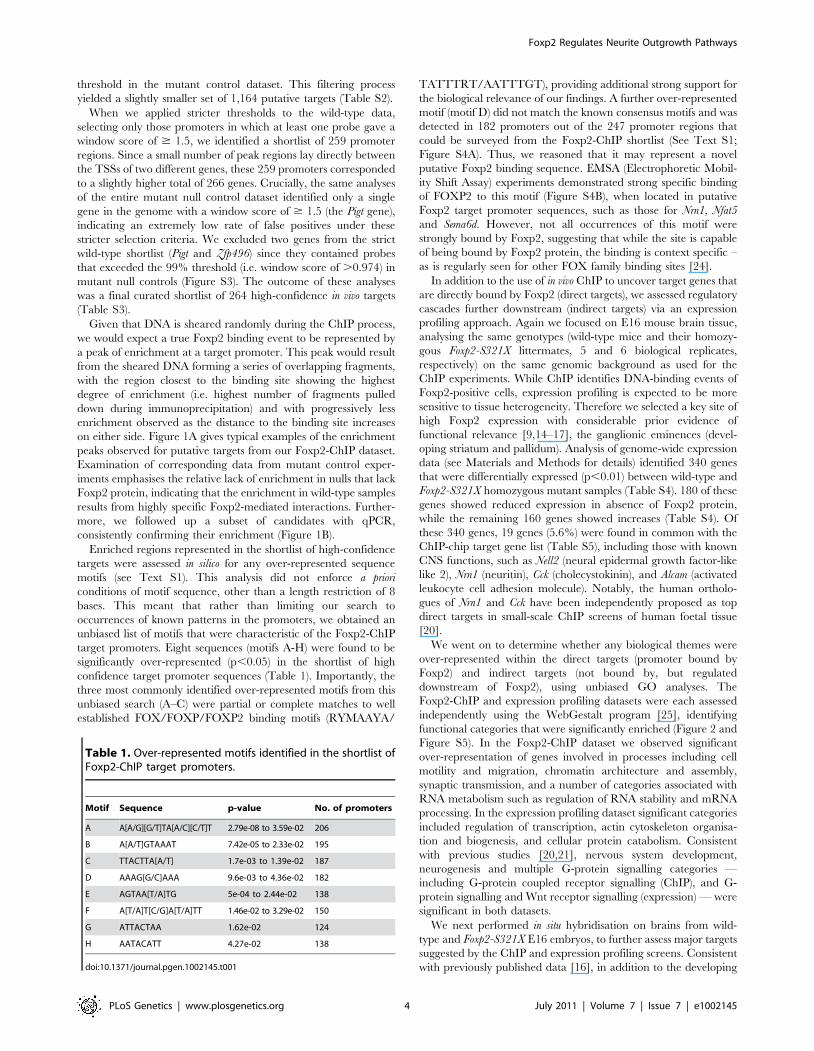
target promoters. Eight sequences (motifs A-H) were found to be
significantly over-represented (p,0.05) in the shortlist of high
confidence target promoter sequences (Table 1). Importantly, the
three most commonly identified over-represented motifs from this
unbiased search (A–C) were partial or complete matches to well
established FOX/FOXP/FOXP2 binding motifs (RYMAAYA/
TATTTRT/AATTTGT), providing additional strong support for
the biological relevance of our findings. A further over-represented
motif (motif D) did not match the known consensus motifs and was
detected in 182 promoters out of the 247 promoter regions that
could be surveyed from the Foxp2-ChIP shortlist (See Text S1;
Figure S4A). Thus, we reasoned that it may represent a novel
putative Foxp2 binding sequence. EMSA (Electrophoretic Mobil-
ity Shift Assay) experiments demonstrated strong specific binding
of FOXP2 to this motif (Figure S4B), when located in putative
Foxp2 target promoter sequences, such as those for Nrn1, Nfat5
and Sema6d. However, not all occurrences of this motif were
strongly bound by Foxp2, suggesting that while the site is capable
of being bound by Foxp2 protein, the binding is context specific –
as is regularly seen for other FOX family binding sites [24].
In addition to the use of in vivo ChIP to uncover target genes that
are directly bound by Foxp2 (direct targets), we assessed regulatory
cascades further downstream (indirect targets) via an expression
profiling approach. Again we focused on E16 mouse brain tissue,
analysing the same genotypes (wild-type mice and their homozy-
gous Foxp2-S321X littermates, 5 and 6 biological replicates,
respectively) on the same genomic background as used for the
ChIP experiments. While ChIP identifies DNA-binding events of
Foxp2-positive cells, expression profiling is expected to be more
sensitive to tissue heterogeneity. Therefore we selected a key site of
high Foxp2 expression with considerable prior evidence of
functional relevance [9,14–17], the ganglionic eminences (devel-
oping striatum and pallidum). Analysis of genome-wide expression
data (see Materials and Methods for details) identified 340 genes
that were differentially expressed (p,0.01) between wild-type and
Foxp2-S321X homozygous mutant samples (Table S4). 180 of these
genes showed reduced expression in absence of Foxp2 protein,
while the remaining 160 genes showed increases (Table S4). Of
these 340 genes, 19 genes (5.6%) were found in common with the
ChIP-chip target gene list (Table S5), including those with known
CNS functions, such as Nell2 (neural epidermal growth factor-like
like 2), Nrn1 (neuritin), Cck (cholecystokinin), and Alcam (activated
leukocyte cell adhesion molecule). Notably, the human ortholo-
gues of Nrn1 and Cck have been independently proposed as top
direct targets in small-scale ChIP screens of human foetal tissue
[20].
We went on to determine whether any biological themes were
over-represented within the direct targets (promoter bound by
Foxp2) and indirect targets (not bound by, but regulated
downstream of Foxp2), using unbiased GO analyses. The
Foxp2-ChIP and expression profiling datasets were each assessed
independently using the WebGestalt program [25], identifying
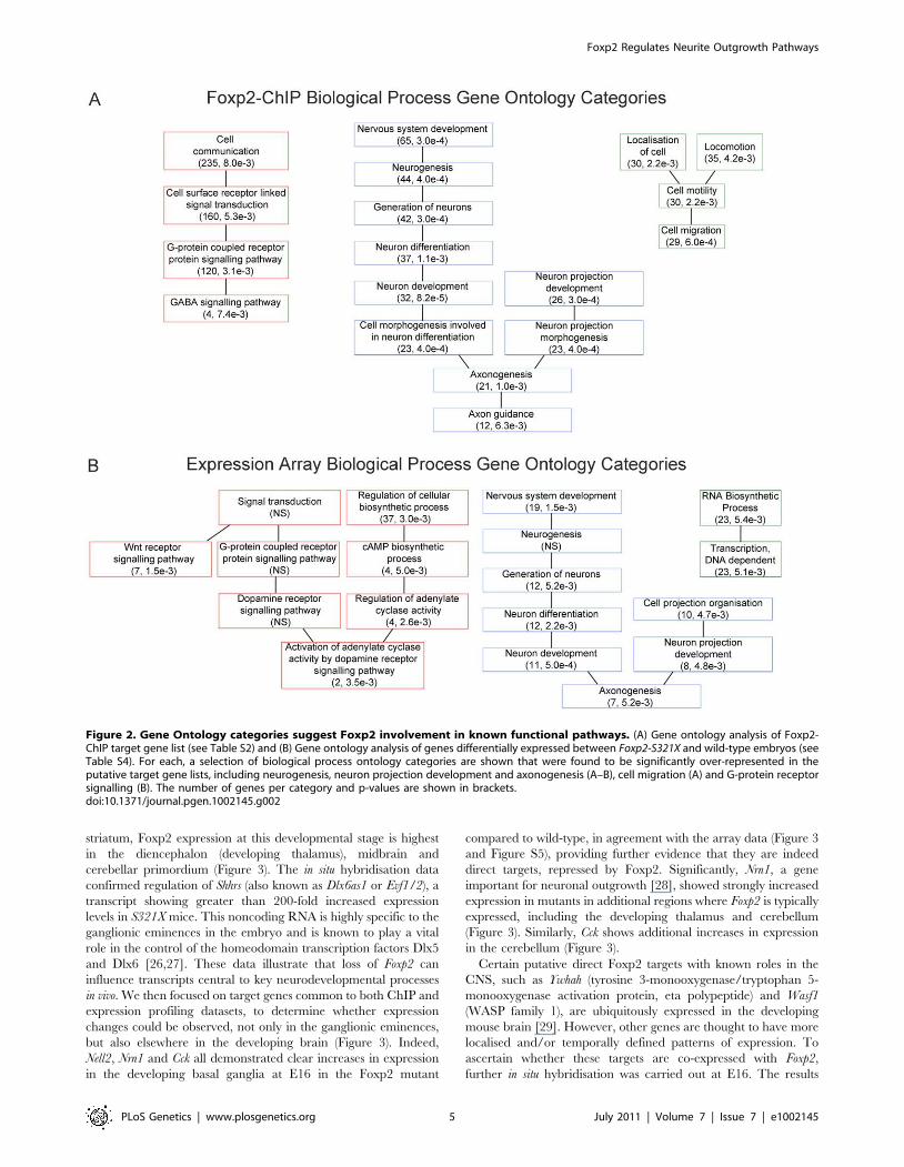
functional categories that were significantly enriched (Figure 2 and
Figure S5). In the Foxp2-ChIP dataset we observed significant
over-representation of genes involved in processes including cell
motility and migration, chromatin architecture and assembly,
synaptic transmission, and a number of categories associated with
RNA metabolism such as regulation of RNA stability and mRNA
processing. In the expression profiling dataset significant categories
included regulation of transcription, actin cytoskeleton organisa-
tion and biogenesis, and cellular protein catabolism. Consistent
with previous studies [20,21], nervous system development,
neurogenesis and multiple G-protein signalling categories —
including G-protein coupled receptor signalling (ChIP), and G-
protein signalling and Wnt receptor signalling (expression) — were
significant in both datasets.
We next performed in situ hybridisation on brains from wild-
type and Foxp2-S321X E16 embryos, to further assess major targets
suggested by the ChIP and expression profiling screens. Consistent
with previously published data [16], in addition to the developing
Table 1. Over-represented motifs identified in the shortlist ofFoxp2-ChIP target promoters.
Motif Sequence p-value No. of promoters
A A[A/G][G/T]TA[A/C][C/T]T 2.79e-08 to 3.59e-02 206
B A[A/T]GTAAAT 7.42e-05 to 2.33e-02 195
C TTACTTA[A/T] 1.7e-03 to 1.39e-02 187
D AAAG[G/C]AAA 9.6e-03 to 4.36e-02 182
E AGTAA[T/A]TG 5e-04 to 2.44e-02 138
F A[T/A]T[C/G]A[T/A]TT 1.46e-02 to 3.29e-02 150
G ATTACTAA 1.62e-02 124
H AATACATT 4.27e-02 138
doi:10.1371/journal.pgen.1002145.t001
Foxp2 Regulates Neurite Outgrowth Pathways
PLoS Genetics | www.plosgenetics.org 4 July 2011 | Volume 7 | Issue 7 | e1002145
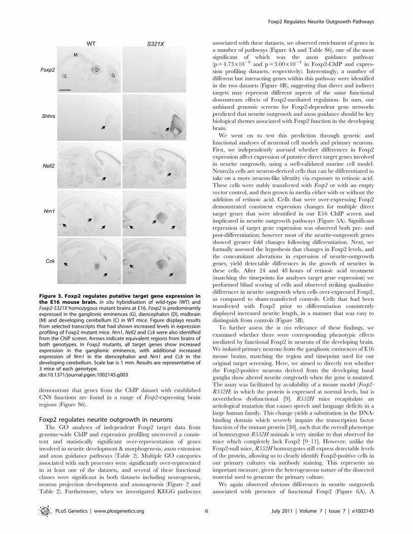
striatum, Foxp2 expression at this developmental stage is highest
in the diencephalon (developing thalamus), midbrain and
cerebellar primordium (Figure 3). The in situ hybridisation data
confirmed regulation of Shhrs (also known as Dlx6as1 or Evf1/2), a
transcript showing greater than 200-fold increased expression
levels in S321X mice. This noncoding RNA is highly specific to the
ganglionic eminences in the embryo and is known to play a vital
role in the control of the homeodomain transcription factors Dlx5
and Dlx6 [26,27]. These data illustrate that loss of Foxp2 can
influence transcripts central to key neurodevelopmental processes
in vivo. We then focused on target genes common to both ChIP and
expression profiling datasets, to determine whether expression
changes could be observed, not only in the ganglionic eminences,
but also elsewhere in the developing brain (Figure 3). Indeed,
Nell2, Nrn1 and Cck all demonstrated clear increases in expression
in the developing basal ganglia at E16 in the Foxp2 mutant
compared to wild-type, in agreement with the array data (Figure 3
and Figure S5), providing further evidence that they are indeed
direct targets, repressed by Foxp2. Significantly, Nrn1, a gene
important for neuronal outgrowth [28], showed strongly increased
expression in mutants in additional regions where Foxp2 is typically
expressed, including the developing thalamus and cerebellum
(Figure 3). Similarly, Cck shows additional increases in expression
in the cerebellum (Figure 3).
Certain putative direct Foxp2 targets with known roles in the
CNS, such as Ywhah (tyrosine 3-monooxygenase/tryptophan 5-
monooxygenase activation protein, eta polypeptide) and Wasf1
(WASP family 1), are ubiquitously expressed in the developing
mouse brain [29]. However, other genes are thought to have more
localised and/or temporally defined patterns of expression. To
ascertain whether these targets are co-expressed with Foxp2,
further in situ hybridisation was carried out at E16. The results
Figure 2. Gene Ontology categories suggest Foxp2 involvement in known functional pathways. (A) Gene ontology analysis of Foxp2-ChIP target gene list (see Table S2) and (B) Gene ontology analysis of genes differentially expressed between Foxp2-S321X and wild-type embryos (seeTable S4). For each, a selection of biological process ontology categories are shown that were found to be significantly over-represented in theputative target gene lists, including neurogenesis, neuron projection development and axonogenesis (A–B), cell migration (A) and G-protein receptorsignalling (B). The number of genes per category and p-values are shown in brackets.doi:10.1371/journal.pgen.1002145.g002
Foxp2 Regulates Neurite Outgrowth Pathways
PLoS Genetics | www.plosgenetics.org 5 July 2011 | Volume 7 | Issue 7 | e1002145
demonstrate that genes from the ChIP dataset with established
CNS functions are found in a range of Foxp2-expressing brain
regions (Figure S6).
Foxp2 regulates neurite outgrowth in neuronsThe GO analyses of independent Foxp2 target data from
genome-wide ChIP and expression profiling uncovered a consis-
tent and statistically significant over-representation of genes
involved in neurite development & morphogenesis, axon extension
and axon guidance pathways (Table 2). Multiple GO categories
associated with such processes were significantly over-represented
in at least one of the datasets, and several of these functional
classes were significant in both datasets including neurogenesis,
neuron projection development and axonogenesis (Figure 2 and
Table 2). Furthermore, when we investigated KEGG pathways
associated with these datasets, we observed enrichment of genes in
a number of pathways (Figure 4A and Table S6), one of the most
significant of which was the axon guidance pathway
(p = 4.7361028 and p = 3.0061024 in Foxp2-ChIP and expres-
sion profiling datasets, respectively). Interestingly, a number of
different but interacting genes within this pathway were identified
in the two datasets (Figure 4B), suggesting that direct and indirect
targets may represent different aspects of the same functional
downstream effects of Foxp2-mediated regulation. In sum, our
unbiased genomic screens for Foxp2-dependent gene networks
predicted that neurite outgrowth and axon guidance should be key
biological themes associated with Foxp2 function in the developing
brain.
We went on to test this prediction through genetic and
functional analyses of neuronal cell models and primary neurons.
First, we independently assessed whether differences in Foxp2
expression affect expression of putative direct target genes involved
in neurite outgrowth, using a well-validated murine cell model.
Neuro2a cells are neuron-derived cells that can be differentiated to
take on a more neuron-like identity via exposure to retinoic acid.
These cells were stably transfected with Foxp2 or with an empty
vector control, and then grown in media either with or without the
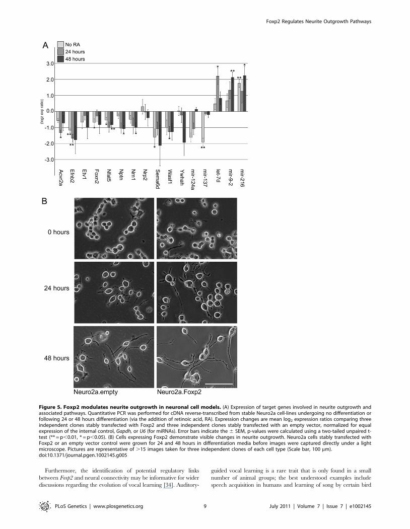
addition of retinoic acid. Cells that were over-expressing Foxp2
demonstrated consistent expression changes for multiple direct
target genes that were identified in our E16 ChIP screen and
implicated in neurite outgrowth pathways (Figure 5A). Significant
repression of target gene expression was observed both pre- and
post-differentiation; however most of the neurite-outgrowth genes
showed greater fold changes following differentiation. Next, we
formally assessed the hypothesis that changes in Foxp2 levels, and
the concomitant alterations in expression of neurite-outgrowth
genes, yield detectable differences in the growth of neurites in
these cells. After 24 and 48 hours of retinoic acid treatment
(matching the timepoints for analyses target gene expression) we
performed blind scoring of cells and observed striking qualitative
differences in neurite outgrowth when cells over-expressed Foxp2,
as compared to sham-transfected controls. Cells that had been
transfected with Foxp2 prior to differentiation consistently
displayed increased neurite length, in a manner that was easy to
distinguish from controls (Figure 5B).
To further assess the in vivo relevance of these findings, we
examined whether there were corresponding phenotypic effects
mediated by functional Foxp2 in neurons of the developing brain.
We isolated primary neurons from the ganglionic eminences of E16
mouse brains, matching the region and timepoint used for our
original target screening. Here, we aimed to directly test whether
the Foxp2-positive neurons derived from the developing basal
ganglia show altered neurite outgrowth when the gene is mutated.
The assay was facilitated by availability of a mouse model (Foxp2-
R552H) in which the protein is expressed at normal levels, but is
nevertheless dysfunctional [9]. R552H mice recapitulate an
aetiological mutation that causes speech and language deficits in a
large human family. This change yields a substitution in the DNA-
binding domain which severely impairs the transcription factor
function of the mutant protein [30], such that the overall phenotype
of homozygous R552H animals is very similar to that observed for
mice which completely lack Foxp2 [9–11]. However, unlike the
Foxp2-null mice, R552H homozygotes still express detectable levels
of the protein, allowing us to clearly identify Foxp2-positive cells in
our primary cultures via antibody staining. This represents an
important measure, given the heterogeneous nature of the dissected
material used to generate the primary culture.
We again observed obvious differences in neurite outgrowth
associated with presence of functional Foxp2 (Figure 6A). A
Figure 3. Foxp2 regulates putative target gene expression inthe E16 mouse brain. In situ hybridisation of wild-type (WT) andFoxp2-S321X homozygous mutant brains at E16. Foxp2 is predominantlyexpressed in the ganglionic eminences (G), diencephalon (D), midbrain(M) and developing cerebellum (C) in WT mice. Figure displays resultsfrom selected transcripts that had shown increased levels in expressionprofiling of Foxp2 mutant mice. Nrn1, Nell2 and Cck were also identifiedfrom the ChIP screen. Arrows indicate equivalent regions from brains ofboth genotypes. In Foxp2 mutants, all target genes show increasedexpression in the ganglionic eminence, with additional increasedexpression of Nrn1 in the diencephalon and Nrn1 and Cck in thedeveloping cerebellum. Scale bar is 1 mm. Results are representative of3 mice of each genotype.doi:10.1371/journal.pgen.1002145.g003
Foxp2 Regulates Neurite Outgrowth Pathways
PLoS Genetics | www.plosgenetics.org 6 July 2011 | Volume 7 | Issue 7 | e1002145

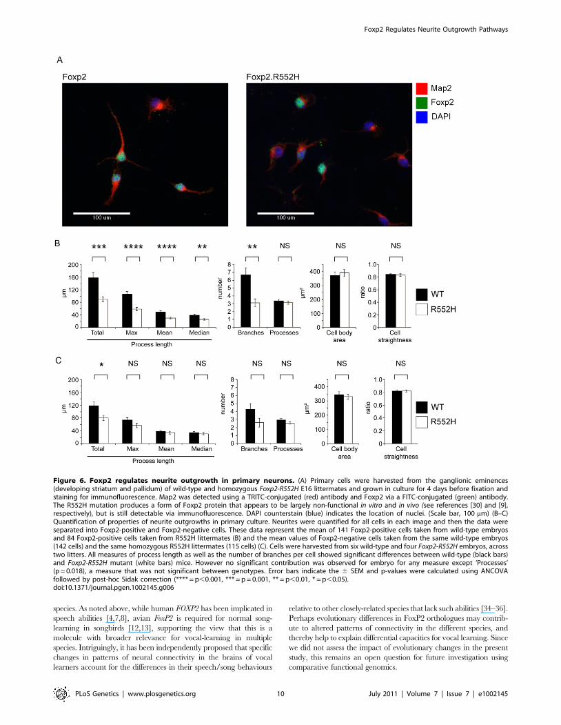
blinded analysis revealed statistically significant changes in
quantitative measures of neurite outgrowth for Foxp2-expressing
neurons from wild-type embryos as compared to those from
homozygous Foxp2-R552H littermates (Figure 6B). In particular,
the latter showed significant reductions in total outgrowth
(p = 0.001); mean (p,0.001), median (p = 0.008) and maximum
process length (p,0.001); and average number of branches
(p = 0.003). Thus, loss of Foxp2 function in striatal neurons that
normally express this transcription factor yields significant
reductions in multiple indices of neurite outgrowth. When
Foxp2-negative cells from the wild-type cultures were compared
to equivalent cells from mutants (Figure 6C), it was only the total
outgrowth that met significance (p = 0.013).
These findings are strongly in agreement with differences in levels
of Foxp2 expression, neurite outgrowth and correlated physiological
properties between the two major subpopulations of striatal medium
spiny neurons (MSNs) in vivo. While both striatonigral (Drd1a) and
striatopallidal (Drd2) MSNs continue to increase their dendritic area
well into adulthood, Drd1a MSNs develop significantly more
dendrites [31]. This dichotomy in dendritic growth contributes to
key physiological differences between both MSN populations,
although the underlying mechanisms remain unknown [31].
Furthermore, studies of cultured striatal neurons demonstrate that
Drd1a MSNs have larger dendritic trees than Drd2 MSNs, invoking
intrinsic mechanisms [31]. To study whether these intrinsic
differences in dendritic growth correlate with Foxp2 expression
levels, we investigated mice expressing enhanced green fluorescent
protein (EGFP) either mainly in Drd1a or Drd2 MSNs [32]. We
found that Foxp2 shows consistently high expression in striatonigral
Drd1a MSNs and very low expression in Drd2 MSNs throughout
the striatum (Figure S7), further supporting roles for Foxp2 in neurite
outgrowth.
Discussion
Although early studies of Foxp2 orthologues in multiple species
suggested that it may play crucial roles in neurodevelopment [15,16],
the exact nature of such roles has not been established. Indeed, much
of the existing knowledge regarding neuronal functions of this
transcription factor instead concerns its impacts on the postnatal CNS
[9,14]. In the present study we employed a high-throughput
functional genomic strategy to shed new light on the in vivo activities
of Foxp2-dependent pathways in the developing CNS.
Of note, among the biological themes that we identified, our
comprehensive ChIP-chip and expression profiling in midgestation
brain tissue independently and consistently highlighted gene
networks underlying neurite development and morphogenesis,
axon extension and axon guidance. These findings drove us to
specifically assess the impact of the Foxp2 gene on neurite
outgrowth phenotypes in genetically manipulated neuronal cell
models and primary neurons from embryos of mutant mice. Our
functional experiments confirmed regulation of the highlighted
gene networks and indicated that wild-type Foxp2 thus enhances
multiple facets of neurite development in vivo, including outgrowth
process length and branch number. The data suggest that the
mode of action may be predominantly cell autonomous, since the
functional effects were mainly restricted to the subset of Foxp2-
expressing cells within a mixed population of neurons. This
possibility of cell-autonomous effects is an interesting hypothesis
that could be clarified in further studies.
Our neurite outgrowth findings are in line with new evidence
regarding the functional impact of evolutionary differences
between FOXP2 orthologues [33]. For example, it is known that
this transcription factor underwent two amino-acid substitutions
on the human lineage after splitting from the chimpanzee lineage,
leading to speculation that such changes may have been important
for evolution of spoken language. In a recent study, researchers
inserted the relevant substitutions into the endogenous Foxp2 gene
of mice, and observed that striatal neurons had significantly longer
dendrites and increased synaptic plasticity [33]. By contrast, we
have shown that mice with loss of function of Foxp2 have
statistically significant reductions in neurite outgrowth (Figure 6 in
the present paper) and decreased synaptic plasticity [9].
Table 2. GO categories significantly over-represented in Foxp2-ChIP (Table S2) and expression profiling (Table S4) datasets.
Gene Ontology Category Foxp2-ChIP Expression arrays
Nervous system development 3.00e-04 1.50e-03
Generation of neurons 3.00e-04 5.20e-03
Neurogenesis 4.00e-04 -
Regulation of neurogenesis 4.20e-04 -
Negative regulation of neurogenesis 2.00e-03 -
Neuron development 8.21e-05 5.00e-04
Neuron differentiation 1.10e-03 2.20e-03
Cellular morphogenesis involved in differentiation - 1.20e-03
Cell projection 3.66e-02 9.10e-03
Cell projection organization - 4.70e-03
Negative regulation of cell projection organization 4.00e-04 -
Cell projection morphogenesis 1.80e-03 -
Neuron projection development 3.00e-04 4.80e-03
Neuron projection morphogenesis 4.00e-04 -
Regulation of neuron projection development 2.00e-03 -
Axonogenesis 1.00e-03 5.20e-03
Axon guidance 6.30e-03 -
doi:10.1371/journal.pgen.1002145.t002
Foxp2 Regulates Neurite Outgrowth Pathways
PLoS Genetics | www.plosgenetics.org 7 July 2011 | Volume 7 | Issue 7 | e1002145
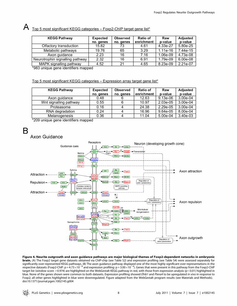
Figure 4. Neurite outgrowth and axon guidance pathways are major biological themes of Foxp2-dependent networks in embryonicbrain. (A) The Foxp2 target gene datasets obtained via ChIP-chip (see Table S2) and expression profiling (see Table S4) were assessed separately forsignificantly over-represented KEGG pathways. (B) The axon guidance pathway displayed one of the most highly significant over-representations in therespective datasets (Foxp2-ChIP: p = 4.7361028 and expression profiling: p = 3.0061024). Genes that were present in this pathway from the Foxp2-ChIPtarget list (window score .0.974) are highlighted on the WebGestalt KEGG pathway in red, with those from expression analysis (p,0.01) highlighted inblue. None of the genes shown were common to both datasets. Expression profiling showed Efnb1 and Plxna4 to be upregulated in vivo in response toFoxp2; all other genes highlighted in blue were downregulated. Figure adapted from the WebGestalt program results (see Materials and Methods).doi:10.1371/journal.pgen.1002145.g004
Foxp2 Regulates Neurite Outgrowth Pathways
PLoS Genetics | www.plosgenetics.org 8 July 2011 | Volume 7 | Issue 7 | e1002145
Furthermore, the identification of potential regulatory links
between Foxp2 and neural connectivity may be informative for wider
discussions regarding the evolution of vocal learning [34]. Auditory-
guided vocal learning is a rare trait that is only found in a small
number of animal groups; the best understood examples include
speech acquisition in humans and learning of song by certain bird
Figure 5. Foxp2 modulates neurite outgrowth in neuronal cell models. (A) Expression of target genes involved in neurite outgrowth andassociated pathways. Quantitative PCR was performed for cDNA reverse-transcribed from stable Neuro2a cell-lines undergoing no differentiation orfollowing 24 or 48 hours differentiation (via the addition of retinoic acid, RA). Expression changes are mean log2 expression ratios comparing threeindependent clones stably transfected with Foxp2 and three independent clones stably transfected with an empty vector, normalized for equalexpression of the internal control, Gapdh, or U6 (for miRNAs). Error bars indicate the 6 SEM, p-values were calculated using a two-tailed unpaired t-test (** = p,0.01, * = p,0.05). (B) Cells expressing Foxp2 demonstrate visible changes in neurite outgrowth. Neuro2a cells stably transfected withFoxp2 or an empty vector control were grown for 24 and 48 hours in differentiation media before images were captured directly under a lightmicroscope. Pictures are representative of .15 images taken for three independent clones of each cell type (Scale bar, 100 mm).doi:10.1371/journal.pgen.1002145.g005
Foxp2 Regulates Neurite Outgrowth Pathways
PLoS Genetics | www.plosgenetics.org 9 July 2011 | Volume 7 | Issue 7 | e1002145
species. As noted above, while human FOXP2 has been implicated in
speech abilities [4,7,8], avian FoxP2 is required for normal song-
learning in songbirds [12,13], supporting the view that this is a
molecule with broader relevance for vocal-learning in multiple
species. Intriguingly, it has been independently proposed that specific
changes in patterns of neural connectivity in the brains of vocal
learners account for the differences in their speech/song behaviours
relative to other closely-related species that lack such abilities [34–36].
Perhaps evolutionary differences in FoxP2 orthologues may contrib-
ute to altered patterns of connectivity in the different species, and
thereby help to explain differential capacities for vocal learning. Since
we did not assess the impact of evolutionary changes in the present
study, this remains an open question for future investigation using
comparative functional genomics.
Figure 6. Foxp2 regulates neurite outgrowth in primary neurons. (A) Primary cells were harvested from the ganglionic eminences(developing striatum and pallidum) of wild-type and homozygous Foxp2-R552H E16 littermates and grown in culture for 4 days before fixation andstaining for immunofluorescence. Map2 was detected using a TRITC-conjugated (red) antibody and Foxp2 via a FITC-conjugated (green) antibody.The R552H mutation produces a form of Foxp2 protein that appears to be largely non-functional in vitro and in vivo (see references [30] and [9],respectively), but is still detectable via immunofluorescence. DAPI counterstain (blue) indicates the location of nuclei. (Scale bar, 100 mm) (B–C)Quantification of properties of neurite outgrowths in primary culture. Neurites were quantified for all cells in each image and then the data wereseparated into Foxp2-positive and Foxp2-negative cells. These data represent the mean of 141 Foxp2-positive cells taken from wild-type embryosand 84 Foxp2-positive cells taken from R552H littermates (B) and the mean values of Foxp2-negative cells taken from the same wild-type embryos(142 cells) and the same homozygous R552H littermates (115 cells) (C). Cells were harvested from six wild-type and four Foxp2-R552H embryos, acrosstwo litters. All measures of process length as well as the number of branches per cell showed significant differences between wild-type (black bars)and Foxp2-R552H mutant (white bars) mice. However no significant contribution was observed for embryo for any measure except ‘Processes’(p = 0.018), a measure that was not significant between genotypes. Error bars indicate the 6 SEM and p-values were calculated using ANCOVAfollowed by post-hoc Sidak correction (**** = p,0.001, *** = p = 0.001, ** = p,0.01, * = p,0.05).doi:10.1371/journal.pgen.1002145.g006
Foxp2 Regulates Neurite Outgrowth Pathways
PLoS Genetics | www.plosgenetics.org 10 July 2011 | Volume 7 | Issue 7 | e1002145
To our knowledge, the current report represents the first large-
scale in vivo characterisation of direct and indirect Foxp2 targets in
the embryonic brain. It is of interest to consider how the present
findings relate to published screens that used more limited ChIP
surveys [19–21], or that employed expression profiling [22,33].
The extent of direct overlap with previous datasets is complicated
by three confounding factors. First, there are differences in scope
of screening; the prior ChIP-chip investigations only queried a
small subset of known promoters [20,21]. Second, there are
differences in species under investigation. Previous target screens
largely focused on human and/or chimpanzee FOXP2, and the
differences between the two [19–22,33], while here we chose to
comprehensively define the pathways regulated by murine Foxp2.
Mouse models offer considerable advantages for functional
genomics, and careful integration of murine data with those from
other species will enhance our understanding of evolutionary roles
of this gene. Finally, the majority of earlier studies screened
neuron-like cells grown in culture [19–21], and no investigation of
this transcription factor has reported integrated use of genome-
wide ChIP and expression profiling to screen the same tissue.
Nevertheless, many important consistencies are observed
between the different datasets, particularly in the biological themes
and processes that they implicate. For example neurite outgrowth
pathways and synaptic plasticity are over-represented in all FoxP2
ChIP-chip datasets across different species and neuronal cell-type,
in vitro and in vivo [20,21]. These processes are closely related
during the development of neuronal networks. Genes controlling
neurite outgrowth or axon guidance during early development
have crucial roles in maturation and stabilisation of synaptic
connectivity at later stages and eventually in activity-dependent
synaptic plasticity in the mature brain throughout life (such as
neurotrophins, semaphorins and ephrins) [37,38]. Hence, the
strong impact of Foxp2 on neurite outgrowth during one
particular stage at E16 might even reflect major Foxp2 functions
that are relevant throughout the development and maintenance of
neuronal networks. A case in point is provided by our data
demonstrating that Nrn1 is a highly robust downstream target. The
Nrn1 gene encodes neuritin, which is already expressed at
embryonic stages of development and was initially identified as a
downstream effector of neuronal activity and neurotrophin-
induced neurite outgrowth [28]. Nrn1 not only showed one of
the strongest enrichment signals in our in vivo ChIP experiments,
but was independently detected as a target in our systematic
expression profiling experiments of equivalent tissue and by in situ
hybridisation – the corresponding human homologue was also one
of the top direct targets reported in a small-scale ChIP screen of
human foetal brain tissue [20].
A number of additional genes, which overlap with earlier
studies, merit further comment. The Cck gene, which showed
convergent evidence in our embryonic ChIP experiments,
expression profiling screens and in situ hybridisation analyses,
was reported as a direct target in both prior published human
ChIP-chip studies [20,21]. Lmo4 (Lim domain only 4) was found to
be indirectly downregulated by Foxp2 in our analyses of
embryonic brain tissue (Table S4) and the human orthologue
LMO4 was similarly repressed by FOXP2 in previous expression
profiling studies of human neuron-like cells by Konopka and
colleagues [22]. Interestingly, in that earlier study using cellular
models, this indirect target was repressed both by human and
chimpanzee versions of FOXP2 [22]. LMO4 encodes a transcrip-
tion factor that plays important roles in cortical patterning, and is
one of the few genes known to show asymmetric expression in the
embryonic human brain [39]. Efnb2 (Ephrin-b2), a well-validated
direct target (Figure 1, Figure 4, Figure 5) was identified in the
Konopka et al. study as one of a small number of genes that may
be differentially regulated by human and chimpanzee FOXP2
orthologues [22]. This gene is of particular interest since it is
implicated in neurite outgrowth and axon guidance (and also
synaptic plasticity) in the basal ganglia and related brain structures
[40]. In addition, Nell2, a validated ChIP and expression array
target (Figure 3), has also been linked to neurite outgrowth [41],
and has recently been shown to promote neuronal survival by
trans-activation by estrogen [42].
Given the substantially enhanced scope of ChIP screening in
the present study, we were able to identify many interesting novel
targets that could not be isolated in the earlier work. For
example, our high-confidence shortlist of direct targets includes
Pak3 – a downstream effector of the Rho family of GTPases
which plays critical roles in pathways restraining neurite growth
[43]; Nptn (neuroplastin) – encoding a synaptic glycoprotein
involved both in development/maintenance of synaptic connec-
tions [44] and in long-term plasticity [45]; Wasf1 – a gene that
regulates activity-induced changes in dendritic spine morpho-
genesis [46] and is involved in actin remodelling during axon
growth [47]; the neuronal semaphorins Sema4f [48] and Sema6d
[49]; as well as Ywhah (also known as 14-3-3), which encodes an
adapter protein implicated in presynaptic plasticity [50] (Figure 1,
Figure 4, Figure 5; Table S3). Although the screening tissue was
embryonic brain, many of the relevant genes have functions that
go beyond this to also influence neural plasticity at later stages.
Overall, this dataset will be important for directing follow-up
studies of Foxp2-dependent pathways and assessing their
involvement in traits such as acquisition of motor-skills [9], vocal
learning [14], and spoken language [1]. While it is likely to be an
indirect target of Foxp2 regulation, it is noteworthy that Evf1/2
(Shhrs) showed such highly increased expression in Foxp2-S321X
mice. It has been shown that the Evf2 RNA molecule co-operates
with the Dlx2 protein to activate the Dlx5/6 enhancer element
[27]. Thus it is interesting that both the DLX1/2 and DLX5/6
loci have been implicated in autism via independent studies,
including a common polymorphism in the DLX5/6 enhancer
itself [51–53].
Of 340 genes showing differential expression (p,0.01) between
mutant and wild-type ganglionic eminences, only 19 (,5%)
corresponded to putative direct targets of Foxp2 from the ChIP-
chip screens. Thus, most of the expression differences observed in
the transcriptional profiling experiments are unlikely to represent
direct modulation due to Foxp2 binding, but could instead
represent cascade effects further downstream (i.e. loss of Foxp2
directly alters expression of a relatively small subset of genes,
which in turn indirectly affect many others). Discrepancies
between the ChIP-chip and expression profiling datasets may also
result from our experimental design: the former could potentially
detect binding events of Foxp2-expressing neurons anywhere in
the brain, while the latter was targeted specifically at the
ganglionic eminences, a region showing particularly high Foxp2
levels. Foxp2 target genes that are not expressed in this structure
could therefore be observed in the ChIP study, but would not be
detected in the expression analysis. An example of such a target is
Sema3a. The promoter of this gene was bound by Foxp2 in our
ChIP study (Figure 1), but its expression only overlaps with Foxp2
expression in the cerebellum (Figure S6). Nevertheless, it is not
unusual in studies of transcription factor function to observe
substantial differences between promoter occupancy maps and
transcriptional profiling data. It is well established that transcrip-
tion factors can be poised ready at particular genomic sites,
awaiting important co-factors, before modulating expression of the
relevant targets [2,54,55].
Foxp2 Regulates Neurite Outgrowth Pathways
PLoS Genetics | www.plosgenetics.org 11 July 2011 | Volume 7 | Issue 7 | e1002145
The present investigation queried the vast majority of known
promoters in the genome, but we acknowledge that the screening
strategy is unable to uncover potential regulatory sequences that
lie outside classical promoter regions. In earlier work, based on
low-throughput shotgun sequencing of human FOXP2-ChIP
fragments, we identified a FOXP2-bound element in the first
intron of CNTNAP2 (contactin-associated-protein-like-2) a gene
implicated in language impairments and autism [19]. Although the
mouse genome contains an orthologous region to the human
FOXP2-bound regulatory element of CNTNAP2, this was not
represented on the arrays used in this study, and hence it escaped
detection. When we carried out ChIP-PCR experiments using the
same mouse embryonic brain tissue as used for ChIP-chip we
demonstrated clear Foxp2 occupancy of the orthologous region in
mouse Cntnap2. Specific enrichment was observed in the wild-type
brains; while no enrichment was found in equivalent tissue from
the mutant null controls (see Figure S8 and Table S7). Studies are
now underway using ‘ChIP-seq’ techniques (coupling ChIP to
next-generation-sequencing) to allow a fully unbiased view of
FOXP2/Foxp2 binding throughout the genome.
Among the validated direct targets of Foxp2 identified in our
study there were a number of microRNA (miRNA) molecules,
including mir-124a and mir-137. miRNAs are an extensive class of
short (,18–23 nucleotide) noncoding molecules which provide
extra layers of dynamic control in networks of gene expression
[56]. miRNAs are abundant in the brain and implicated in critical
aspects of nervous system development and function, ranging from
early neurogenesis and proliferation [57], through neural differ-
entiation and dendrite morphogenesis [58], to adaptive mecha-
nisms in mature neurons, including learning and memory [59].
They play pivotal roles in processes such as neurite outgrowth,
axonal pathfinding and synaptic plasticity, mechanisms for which
localised rapid control of protein synthesis is paramount [58,59].
In conclusion, the use of in vivo genomic screening strategies in
the developing embryonic brain has proved to be a powerful
approach for understanding the biology of Foxp2, one of the most
intriguing transcription factors of the CNS. This starting point led
us to functional characterisation of new mechanisms of Foxp2
action, in particular the modulation of networks involved in
neurite outgrowth, axonogenesis and other core aspects of neural
development. Future studies will define how these regulatory
networks differ between distinct species, what role miRNAs play in
Foxp2-related pathways and phenotypes and will investigate
whether it is possible to rescue the established neurobiological
effects associated with loss of Foxp2 function, through manipula-
tion of key targets. Ultimately, such work promises to fully uncover
the functional pathways that connect Foxp2 with plasticity of the
developing CNS.
Materials and Methods
In vivo chromatin immunoprecipitationIn vivo Foxp2-ChIP in embryonic mouse brain tissue was
performed according to the protocol previously described by
Vernes and colleagues [21]. Each of the three replicates included
whole brain tissue (from the telencephalon to the brain stem at the
level of the foramen magnum) at E16 (embryonic day 16), a
developmental timepoint of high Foxp2 expression [16], pooled
from 5–6 mice of matching genotype. Experiments were carried
out either with wild-type embryos, or homozygous Foxp2-S321X
mutants as negative controls. S321X mutants carry an early
nonsense mutation that disrupts Foxp2; the resulting combination
of nonsense-mediated RNA decay and protein instability leads to a
complete lack of detectable Foxp2 protein in the brain [9]. The
wild-type embryos and mutant controls used in these experiments
were all matched littermates, backcrossed for at least ten
generations into a C57BL/6J strain, maximizing the homogeneity
of the genomic background. Although homozygous mutants
display developmental delays and reduced cerebellar growth after
birth, they show no gross anomalies in brain anatomy or
development during embryogenesis [9]. All animal work was
carried out conforming to the regulatory standards of the UK
Home Office, under Project Licence 30/2016.
E16 mouse brains were extracted, snap frozen in liquid nitrogen
and stored at 280uC until use. Each whole brain was weighed,
then chopped finely with a razor on ice. Brains were pooled to
achieve a total weight of between 0.3 and 0.5 g of tissue (between
5–6 brains per replicate) and resuspended in 5 ml PBS. A 1/10
volume (500 ml) of cross-linking buffer was added prior to 15
minutes incubation with agitation at room temperature. Formal-
dehyde was quenched via the addition of 125 mM glycine. Cross-
linked tissue was washed in PBS before brief mechanical
homogenisation. Pellets were then incubated in two in vivo ChIP
lysis buffers at room temperature for ten minutes each: Buffer 1
(50 mM HEPES-KOH pH = 7.5, 140 mM NaCl, 1 mM EDTA,
10% glycerol, 0.5% NP-40, 0.25% Triton X-100, protease
inhibitors); Buffer 2 (200 mM NaCl, 1 mM EDTA, 0.5 mM
EGTA, 10 mM Tris pH = 8, protease inhibitors). After collection
via centrifugation, nuclei were resuspended in 5 ml sonication
buffer (10 mM Tris-HCl pH = 8, 1 mM EDTA, 0.5 mM EGTA,
protease inhibitors). Samples underwent 15 rounds of 20-second
sonication pulses at 30% power, with 60 seconds on ice between
each round (Branson Digital Sonifier - S450D). Agarose gel
electrophoresis was used to confirm that fragment size was 300–
1000 bp. Cells were centrifuged at 10,000 g and 4uC for 10
minutes to remove cell debris. 10 mg of polyclonal rabbit anti-
Foxp2 antibody (C-terminal antibody, Geschwind Laboratory,
UCLA) [20] pre-coupled to 100 ml Dynal M-280 rat anti-rabbit
IgG magnetic protein-A beads was added and incubated at 4uC,
rotating overnight. Beads were washed five times in RIPA buffer
and once in TE buffer. Chromatin was eluted from beads in TE
buffer with 1% SDS at 65uC for 10 minutes with agitation. The
chromatin was then incubated at 65uC overnight to reverse cross-
links. Purified chromatin was amplified via Ligation Mediated
PCR (LMPCR) according to published protocols [60]. Size and
purity of DNA was assessed via spectrophotometry and gel
electrophoresis.
Hybridisation of Foxp2-ChIP products to promotermicroarrays
2 mg of amplified immunoprecipitated chromatin, or total input
DNA was fluorescently labelled with Cy5 and Cy3 respectively
using random primers provided in the BioPrime DNA labelling
system (Invitrogen). The labelling reaction was allowed to proceed
for 16 hours at 37uC, before purification by sodium acetate
precipitation. Hybridisation to mouse promoter arrays (Agilent
Technologies, #G4490A) was carried out by the UCLA
microarray core facility, according to the manufacturer’s instruc-
tions. Arrays consisted of 60-mer oligonucleotides spanning ,8 kb
(5.5 kb upstream and 2.5 kb downstream of TSS) at each of
,17,000 mouse promoter regions. Probes were spaced on
average, between 100–300 bp apart, with approximately 25
probes for each promoter region. Three littermate matched sets
of pooled wild-type or mutant control chromatin samples were
applied to microarrays, each using its respective input DNA
sample as the internal reference on the array. Thus, the three wild-
type and three mutant control datasets represent signals obtained
from a total of 34 individual mouse embryos.
Foxp2 Regulates Neurite Outgrowth Pathways
PLoS Genetics | www.plosgenetics.org 12 July 2011 | Volume 7 | Issue 7 | e1002145
Analysis of promoter microarray dataArray images were scanned using the Axon GenePix 4000B.
Data were retrieved and initial quality control carried out using
the Axon GenePix 4000B software package. All promoter
coordinates and probes were mapped with reference to the NCBI
m36 mouse assembly. Microarray data analysis was carried out
using the mArray package for R [61]. LOESS normalisation and
background correction was performed within each array. Data
were normalised between arrays using quantile normalisation, and
mean values were calculated from three biological replicates (wild-
type or mutant control experiments) for each probe - called ‘probe
scores’, such that a score of 1 corresponds to 2-fold enrichment in
ChIP versus total input DNA. All negative probe scores were
assigned a value of zero. A ‘window-adjusted score’ for each probe
was then calculated as the median value of each probe score and
its nearest neighbour on either side. Neighbouring probes were
only considered if they fell within 500 bp upstream or 500 bp
downstream of the central probe. This window size was based on
the average size of the labelled DNA fragments, estimated to be
approx 1000 bp. Thus, a true binding event would likely be
indicated by positive scores of multiple neighbouring probes within
a 1000 bp window. In cases where there were less than three
probes located within this 1000 bp window showing a signal
greater than background then the window-adjusted score was set
to zero. This process helps to guard against artificial skewing of
enrichment values at edges of promoter regions.
The use of mutant null controls enabled us to robustly assess the
empirical significance of wild-type ChIP results. The data from the
mutant control experiments were used to estimate a null
distribution of window scores; that is, the non-specific signals
produced by the Foxp2-ChIP protocol even when there is no
Foxp2 protein available for pulldown. (Note that a subset of
binding events in mutant null controls could potentially be due to
crossreactivity of Foxp2 antibodies with closely related proteins,
such as Foxp1 or Foxp4, that may bind to the same promoter.
However, prior work with the antibody used here suggests that
levels of crossreactivity are extremely low [20].) From this null
distribution of window scores we calculated the threshold which
excluded 99% of all datapoints in controls. This threshold could
then be applied to the wild-type array data.
Semi-quantitative PCRChromatin isolated during Foxp2-ChIP in mutant and wild-
type mouse brains was amplified using a semi-quantitative PCR
technique, as described previously [21], using primers directed
towards the peak regions of enrichment (Table 3). The b-actin
housekeeping gene promoter was used as a negative control.
Expression profilingThe ganglionic eminences, sites of particularly high embryonic
Foxp2 expression [16], were dissected from E16 brains of six wild-
type mice and six homozygous Foxp2-S321X mutant littermates.
For each embryo, the left- and right-hemisphere ganglionic
eminences were pooled in TRIzol reagent and RNA was extracted
using the QIAGEN RNeasy kit, according to the manufacturer’s
instructions. RNA yield was measured using a NanoDrop ND-
1000 spectrophotometer (NanoDrop Technologies, Wilmington,
DE), and its quality was assessed using RNA6000 Nano Assays on
an Agilent Bioanalyzer 2100 (Agilent Technologies, Santa Clara,
CA).
Gene expression profiling was performed using whole-genome
mouse BeadChip arrays from Illumina (San Diego, California,
USA), which include 45,281 probes representing 31,492 mouse
transcripts. In brief, 500 ng of total RNA was reverse transcribed
to synthesize first- and second-strand complementary DNA
(cDNA). Following purification on spin columns, in vitro transcrip-
tion was used to synthesize biotin-labelled complementary RNA
(cRNA). 1500 ng of biotin-labelled cRNA was hybridized to
Mouse WG-6 V2 Expression BeadChip arrays (Illumina Inc., San
Diego, CA) at 55uC for 18 h. The hybridized arrays were washed
and labelled with streptavidin-Cy3 according to the manufactur-
er’s protocols before being scanned with the Illumina Bead Array
Scanner. Raw data were exported from the Illumina BeadStudio
software (v3.4.0) for further processing and analysis using the R
statistical software [62] and BioConductor packages [63]. Signal
data and detection scores were extracted for each of the 12
samples. Signal data were background corrected by subtracting
the average signal from the negative control probes on each array,
prior to being transformed and normalised using the ‘VSN’
package [64]. Quality control analyses, including hierarchical
clustering and principal component analysis (PCA), identified one
outlier sample (from the wild-type group). This sample had very
low signal compared to other samples while hybridisation and
labelling metrics were normal, suggesting a sample problem rather
than a technical issue. It was sufficiently outlying to remove from
further analysis and the remaining 11 samples were re-normalised.
The dataset was then filtered to remove probes that were not
detected (detection score ,0.95 in all samples), resulting in a final
dataset of 24,479 probes.
Statistical analysis was performed using the ‘Linear Models for
Microarray Analysis’ (Limma) package [65]. Differential expres-
sion between mutant and wild-type animals was assessed using a
linear model that included effects for genotype and litter. Raw p-
values were corrected for multiple testing using the false discovery
rate (FDR) controlling procedure of Benjamini and Hochberg
[66]. Fourteen probes were significant at a FDR of 5%. The larger
set of 340 probes significant at p,0.01 was used for further
biological investigation. We performed permutation tests on the
genotype labels (11 choose 5), taking litter effects into account, and
found that $340 genes were differentially expressed at p = 0.01 in
only 9 out of the possible 108 permutations (,8%). Gene
annotation was added to the final probe list using the relevant
annotation file (MouseWG-6_V2_0_R0_11278593_A.txt) from
the Illumina website (http://www.illumina.com).
Cell culture and reagentsNeuro2a (murine neuroblastoma) cells were cultured in ‘Growth
media’: Modified Eagles Medium (MEM) (Sigma) supplemented
with 10% Foetal Calf Serum (Sigma), 2 mM L-glutamine (Sigma)
and 2 mM Penicillin/Streptomycin (Sigma). Cells were grown at
37uC in the presence of 5% CO2. Stable Neuro2a cell-lines
overexpressing Foxp2 protein or non-expressing controls, were
generated via transfection with pcDNA3.1/Foxp2 (mouse isoform I
- untagged) or the empty vector, using Genejuice (Novagen)
according to the manufacturers’ instructions. Cells were cultured
Table 3. Semi-quantitative PCR primers.
GENE FORWARD PRIMER (59R39) REVERSE PRIMER (59R39)
Nrp2 CAGCAACCAGTGATGCTTGT AGCAAGGAGAGTTGGAGCAA
Sema3a GGGCACTGAGTAGCTTCCAC GAATGCAAGAAAAGTTGTCCTC
Nrn1 TCCCCCAAACAAATTCTCAA AATCCTTGCAGCATTTCAGG
b-actin AGGGTACCACCGGAAAAGTC CCCCAAAGGCTGAGAAGTTA
doi:10.1371/journal.pgen.1002145.t003
Foxp2 Regulates Neurite Outgrowth Pathways
PLoS Genetics | www.plosgenetics.org 13 July 2011 | Volume 7 | Issue 7 | e1002145
in complete medium supplemented with 500 mg/ml G418
(Calbiochem) as a selective agent. Resistant single colonies were
isolated 20 days after transfection, then cultured and expanded
independently in the presence of G418 (500 mg/ml). Expression of
recombinant Foxp2 was confirmed using qRT-PCR and Western
blotting with two polyclonal antibodies recognizing different
epitopes of the protein (goat N-terminal antibody, Santa Cruz
Biotechnology [30]; rabbit C-terminal antibody, Geschwind
Laboratory, UCLA [20]). Three Foxp2-transfected clones with a
high and consistent level of expression and three empty vector
clones were chosen for use in further experiments. Neuro2a cells
were differentiated via the addition of Modified Eagles Medium
supplemented with 2% Foetal Calf Serum (Sigma), 2 mM L-
glutamine (Sigma), 2 mM Penicillin/Streptomycin (Sigma) and
20 mM all-trans retinoic acid (‘Differentiation media’).
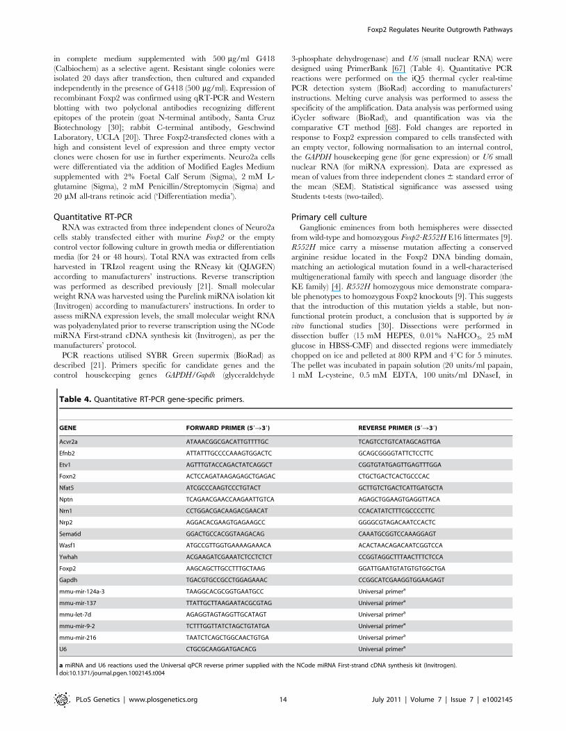
Quantitative RT-PCRRNA was extracted from three independent clones of Neuro2a
cells stably transfected either with murine Foxp2 or the empty
control vector following culture in growth media or differentiation
media (for 24 or 48 hours). Total RNA was extracted from cells
harvested in TRIzol reagent using the RNeasy kit (QIAGEN)
according to manufacturers’ instructions. Reverse transcription
was performed as described previously [21]. Small molecular
weight RNA was harvested using the Purelink miRNA isolation kit
(Invitrogen) according to manufacturers’ instructions. In order to
assess miRNA expression levels, the small molecular weight RNA
was polyadenylated prior to reverse transcription using the NCode
miRNA First-strand cDNA synthesis kit (Invitrogen), as per the
manufacturers’ protocol.
PCR reactions utilised SYBR Green supermix (BioRad) as
described [21]. Primers specific for candidate genes and the
control housekeeping genes GAPDH/Gapdh (glyceraldehyde
3-phosphate dehydrogenase) and U6 (small nuclear RNA) were
designed using PrimerBank [67] (Table 4). Quantitative PCR
reactions were performed on the iQ5 thermal cycler real-time
PCR detection system (BioRad) according to manufacturers’
instructions. Melting curve analysis was performed to assess the
specificity of the amplification. Data analysis was performed using
iCycler software (BioRad), and quantification was via the
comparative CT method [68]. Fold changes are reported in
response to Foxp2 expression compared to cells transfected with
an empty vector, following normalisation to an internal control,
the GAPDH housekeeping gene (for gene expression) or U6 small
nuclear RNA (for miRNA expression). Data are expressed as
mean of values from three independent clones 6 standard error of
the mean (SEM). Statistical significance was assessed using
Students t-tests (two-tailed).
Primary cell cultureGanglionic eminences from both hemispheres were dissected
from wild-type and homozygous Foxp2-R552H E16 littermates [9].
R552H mice carry a missense mutation affecting a conserved
arginine residue located in the Foxp2 DNA binding domain,
matching an aetiological mutation found in a well-characterised
multigenerational family with speech and language disorder (the
KE family) [4]. R552H homozygous mice demonstrate compara-
ble phenotypes to homozygous Foxp2 knockouts [9]. This suggests
that the introduction of this mutation yields a stable, but non-
functional protein product, a conclusion that is supported by in
vitro functional studies [30]. Dissections were performed in
dissection buffer (15 mM HEPES, 0.01% NaHCO3, 25 mM
glucose in HBSS-CMF) and dissected regions were immediately
chopped on ice and pelleted at 800 RPM and 4uC for 5 minutes.
The pellet was incubated in papain solution (20 units/ml papain,
1 mM L-cysteine, 0.5 mM EDTA, 100 units/ml DNaseI, in
Table 4. Quantitative RT-PCR gene-specific primers.
GENE FORWARD PRIMER (59R39) REVERSE PRIMER (59R39)
Acvr2a ATAAACGGCGACATTGTTTTGC TCAGTCCTGTCATAGCAGTTGA
Efnb2 ATTATTTGCCCCAAAGTGGACTC GCAGCGGGGTATTCTCCTTC
Etv1 AGTTTGTACCAGACTATCAGGCT CGGTGTATGAGTTGAGTTTGGA
Foxn2 ACTCCAGATAAGAGAGCTGAGAC CTGCTGACTCACTGCCCAC
Nfat5 ATCGCCCAAGTCCCTGTACT GCTTGTCTGACTCATTGATGCTA
Nptn TCAGAACGAACCAAGAATTGTCA AGAGCTGGAAGTGAGGTTACA
Nrn1 CCTGGACGACAAGACGAACAT CCACATATCTTTCGCCCCTTC
Nrp2 AGGACACGAAGTGAGAAGCC GGGGCGTAGACAATCCACTC
Sema6d GGACTGCCACGGTAAGACAG CAAATGCGGTCCAAAGGAGT
Wasf1 ATGCCGTTGGTGAAAAGAAACA ACACTAACAGACAATCGGTCCA
Ywhah ACGAAGATCGAAATCTCCTCTCT CCGGTAGGCTTTAACTTTCTCCA
Foxp2 AAGCAGCTTGCCTTTGCTAAG GGATTGAATGTATGTGTGGCTGA
Gapdh TGACGTGCCGCCTGGAGAAAC CCGGCATCGAAGGTGGAAGAGT
mmu-mir-124a-3 TAAGGCACGCGGTGAATGCC Universal primera
mmu-mir-137 TTATTGCTTAAGAATACGCGTAG Universal primera
mmu-let-7d AGAGGTAGTAGGTTGCATAGT Universal primera
mmu-mir-9-2 TCTTTGGTTATCTAGCTGTATGA Universal primera
mmu-mir-216 TAATCTCAGCTGGCAACTGTGA Universal primera
U6 CTGCGCAAGGATGACACG Universal primera
a miRNA and U6 reactions used the Universal qPCR reverse primer supplied with the NCode miRNA First-strand cDNA synthesis kit (Invitrogen).doi:10.1371/journal.pgen.1002145.t004
Foxp2 Regulates Neurite Outgrowth Pathways
PLoS Genetics | www.plosgenetics.org 14 July 2011 | Volume 7 | Issue 7 | e1002145
dissection buffer) on ice for 5 minutes then at 37uC for 10 minutes,
agitating regularly. The enzymatic reaction was halted by addition
of Ovo-BSA solution (10 mg/ml ovomucoid, 10 mg/ml BSA in
dissection buffer). Cells were pelleted at 1000 RPM for 5 minutes
at 4uC and the pellet was washed then re-suspended in complete
medium (neurobasal media (Sigma) supplemented by 2 mM
Glutamax (Sigma). 2 mM Penicillin/Streptomycin (Sigma) and
1X B27 supplement). Suspension was triturated using plastic and
glass pipettes to dissociate any remaining cell clumps before
passing the cell suspension through a 70 mm cell strainer. Single
cell suspensions were seeded onto laminin and poly-D-lysine
coated coverslips (BD Biosciences) at a density of 6.36104 cells per
well into 24 well plates and grown at 37uC in the presence of 5%
CO2 in complete medium.
After 4 days in culture, cells were fixed using 4% Paraformal-
dehyde solution for 15 minutes at room temperature and
permeablised in wash solution (0.1% Triton X-100 in TBS).
Antibodies were diluted in Blocking Solution (1% Fish Gelatine,
0.1% Triton X-100, 5% BSA in PBS). Cells were co-stained at
4uC overnight, using two primary antibodies; an anti-MAP2 rabbit
polyclonal antibody (Chemicon) and an anti-Foxp2 mouse
monoclonal antibody recognising an epitope near the C-terminal
end of the protein (Gift from Prof. A. Banham). Detection was
then facilitated via four rounds of antibody incubation, which
allowed magnification of the Foxp2 signal. Cells were incubated
with anti-rabbit TRITC (Alexa Fluor 568, Molecular Probes) plus
anti-mouse biotinylated (BA9200, Vector Labs) secondary anti-
bodies, followed by incubation with anti-rabbit TRITC plus anti-
biotin FITC (Alexa Fluor 488, Molecular Probes) antibodies, each
for 1 hour, shaking under limited light exposure. This secondary/
tertiary antibody incubation was then repeated under the same
conditions. Nuclei were visualised using mounting media contain-
ing a DAPI counterstain (VectaShield). Cells were viewed on a
Nikon Eclipse TE2000U fluorescence inverted microscope.
Images were captured using a Hamamatsu black and white
C4742-95 Orca hi-sensitivity CCD camera with IPLab imaging
software (Scanalytics Inc), and analysed using the neurite
outgrowth function of Metamorph Version 7.6 (Molecular
Devices). Statistical analyses were carried out using ANCOVA
(analysis of covariance) for genotype and embryo, followed by
post-hoc Sidak correction. Data are expressed as the mean 6
standard error of the mean (SEM).
In situ hybridisationIn situ hybridization was carried out on 10 mM frozen sections of
E16.5 embryo heads as previously described [69]. An approxi-
mately 500 bp fragment of each target transcript was subcloned
into pCR4-TOPO (Invitrogen) for dioxygenin-labelled riboprobe
synthesis. Primer sequences for the riboprobes are available on
request. Equivalent parasagittal sections were hybridized in
parallel from three wild-type and three homozygous Foxp2-
S321X mutant embryos and all slides were developed for 16 hours,
or 6 hours in the case of Shhrs. In all cases a sense-strand negative
control riboprobe gave no specific signal (data not shown).
Supporting Information
Figure S1 Western blot analysis of endogenous Foxp2 protein.
(TIF)
Figure S2 Chromosome distribution of enriched probes.
(TIF)
Figure S3 Window scores for all probes that exceeded the strict
threshold (enrichment score of .1.5) in wild-type Foxp2-chip.
(TIF)
Figure S4 EMSA analysis of a novel Foxp2 DNA-binding motif.
(TIF)
Figure S5 Differential expression of Foxp2 target genes in E16
developing mouse ganglionic eminences.
(TIF)
Figure S6 Co-expression of Foxp2 and putative target genes in
the E16 mouse brain.
(TIF)
Figure S7 Expression of Foxp2 in medium spiny neurons
(MSNs).
(TIF)
Figure S8 In vivo binding of mouse Foxp2 to intron 1 of Cntnap2
in embryonic brain tissue.
(TIF)
Table S1 Foxp2-ChIP (window score $0.97) complete dataset.
(XLS)
Table S2 Foxp2-ChIP putative target promoters (curated long
list n = 1164, window score $0.97).
(XLS)
Table S3 Foxp2-ChIP putative target promoters (curated short
list n = 264, at least 1 window score $1.50).
(XLS)
Table S4 Foxp2-dependent gene expression changes in the E16
mouse brain (p,0.01).
(XLS)
Table S5 Overlapping target genes from Foxp2-ChIP and
expression analysis studies.
(XLS)
Table S6 Significant over-represented KEGG pathways identi-
fied in the Foxp2-ChIP and gene expression datasets.
(XLS)
Table S7 Cntnap2 semi-quantitative PCR primers.
(XLS)
Text S1 Supporting Materials and Methods.
(DOC)
Acknowledgments
We are grateful to Alice Page and Radhika Chadha for assistance with
target gene validation assays. We thank Denis Herve and Jean-Antoine
Girault, Inserm UMR-S839, for their support.
Author Contributions
Conceived and designed the experiments: SCV SEF. Performed the
experiments: SCV PLO ES RP JH CM AB JN MG DB. Analyzed the
data: SCV HEL JMT EL NS J-BC SEF. Contributed reagents/materials/
analysis tools: JR KED DHG SEF. Wrote the paper: SCV SEF.
References
1. Fisher SE, Scharff C (2009) FOXP2 as a molecular window into speech and
language. Trends Genet 25: 166–177.
2. Carlsson P, Mahlapuu M (2002) Forkhead transcription factors: key players in
development and metabolism. Dev Biol 250: 1–23.
Foxp2 Regulates Neurite Outgrowth Pathways
PLoS Genetics | www.plosgenetics.org 15 July 2011 | Volume 7 | Issue 7 | e1002145
3. Hannenhalli S, Kaestner KH (2009) The evolution of Fox genes and their role in
development and disease. Nat Rev Genet 10: 233–240.
4. Lai CS, Fisher SE, Hurst JA, Vargha-Khadem F, Monaco AP (2001) A
forkhead-domain gene is mutated in a severe speech and language disorder.
Nature 413: 519–523.
5. Wang B, Lin D, Li C, Tucker P (2003) Multiple domains define the expression
and regulatory properties of Foxp1 forkhead transcriptional repressors. J Biol
Chem 278: 24259–24268.
6. Kaestner KH, Knochel W, Martinez DE (2000) Unified nomenclature for the
winged helix/forkhead transcription factors. Genes Dev 14: 142–146.
7. Feuk L, Kalervo A, Lipsanen-Nyman M, Skaug J, Nakabayashi K, et al. (2006)
Absence of a paternally inherited FOXP2 gene in developmental verbal
dyspraxia. Am J Hum Genet 79: 965–972.
8. MacDermot KD, Bonora E, Sykes N, Coupe AM, Lai CS, et al. (2005)
Identification of FOXP2 truncation as a novel cause of developmental speech
and language deficits. Am J Hum Genet 76: 1074–1080.
9. Groszer M, Keays DA, Deacon RM, de Bono JP, Prasad-Mulcare S, et al. (2008)
Impaired synaptic plasticity and motor learning in mice with a point mutation
implicated in human speech deficits. Curr Biol 18: 354–362.
10. French CA, Groszer M, Preece C, Coupe AM, Rajewsky K, et al. (2007)
Generation of mice with a conditional Foxp2 null allele. Genesis 45: 440–446.
11. Shu W, Cho JY, Jiang Y, Zhang M, Weisz D, et al. (2005) Altered ultrasonic
vocalization in mice with a disruption in the Foxp2 gene. Proc Natl Acad
Sci U S A 102: 9643–9648.
12. Haesler S, Wada K, Nshdejan A, Morrisey EE, Lints T, et al. (2004) FoxP2
expression in avian vocal learners and non-learners. J Neurosci 24: 3164–3175.
13. Teramitsu I, Kudo LC, London SE, Geschwind DH, White SA (2004) Parallel
FoxP1 and FoxP2 expression in songbird and human brain predicts functional
interaction. J Neurosci 24: 3152–3163.
14. Haesler S, Rochefort C, Georgi B, Licznerski P, Osten P, et al. (2007)
Incomplete and inaccurate vocal imitation after knockdown of FoxP2 in
songbird basal ganglia nucleus Area X. PLoS Biol 5: e321. doi:10.1371/
journal.pbio.0050321.
15. Ferland RJ, Cherry TJ, Preware PO, Morrisey EE, Walsh CA (2003)
Characterization of Foxp2 and Foxp1 mRNA and protein in the developing
and mature brain. J Comp Neurol 460: 266–279.
16. Lai CS, Gerrelli D, Monaco AP, Fisher SE, Copp AJ (2003) FOXP2 expression
during brain development coincides with adult sites of pathology in a severe
speech and language disorder. Brain 126: 2455–2462.
17. Watkins KE, Vargha-Khadem F, Ashburner J, Passingham RE, Connelly A,
et al. (2002) MRI analysis of an inherited speech and language disorder:
structural brain abnormalities. Brain 125: 465–478.
18. Shu W, Yang H, Zhang L, Lu MM, Morrisey EE (2001) Characterization of a
new subfamily of winged-helix/forkhead (Fox) genes that are expressed in the
lung and act as transcriptional repressors. J Biol Chem 276: 27488–27497.
19. Vernes SC, Newbury DF, Abrahams BS, Winchester L, Nicod J, et al. (2008) A
functional genetic link between distinct developmental language disorders.
N Engl J Med 359: 2337–2345.
20. Spiteri E, Konopka G, Coppola G, Bomar J, Oldham M, et al. (2007)
Identification of the transcriptional targets of FOXP2, a gene linked to speech
and language, in developing human brain. Am J Hum Genet 81: 1144–1157.
21. Vernes SC, Spiteri E, Nicod J, Groszer M, Taylor JM, et al. (2007) High-
throughput analysis of promoter occupancy reveals direct neural targets of
FOXP2, a gene mutated in speech and language disorders. Am J Hum Genet
81: 1232–1250.
22. Konopka G, Bomar JM, Winden K, Coppola G, Jonsson ZO, et al. (2009)
Human-specific transcriptional regulation of CNS development genes by
FOXP2. Nature 462: 213–217.
23. Marson A, Kretschmer K, Frampton GM, Jacobsen ES, Polansky JK, et al.
(2007) Foxp3 occupancy and regulation of key target genes during T-cell
stimulation. Nature 445: 931–935.
24. Overdier DG, Porcella A, Costa RH (1994) The DNA-binding specificity of the
hepatocyte nuclear factor 3/forkhead domain is influenced by amino-acid
residues adjacent to the recognition helix. Mol Cell Biol 14: 2755–2766.
25. Zhang B, Kirov S, Snoddy J (2005) WebGestalt: an integrated system for
exploring gene sets in various biological contexts. Nucleic Acids Res 33:
W741–748.
26. Faedo A, Quinn JC, Stoney P, Long JE, Dye C, et al. (2004) Identification and
characterization of a novel transcript down-regulated in Dlx1/Dlx2 and up-
regulated in Pax6 mutant telencephalon. Dev Dyn 231: 614–620.
27. Feng J, Bi C, Clark BS, Mady R, Shah P, et al. (2006) The Evf-2 noncoding
RNA is transcribed from the Dlx-5/6 ultraconserved region and functions as a
Dlx-2 transcriptional coactivator. Genes Dev 20: 1470–1484.
28. Naeve GS, Ramakrishnan M, Kramer R, Hevroni D, Citri Y, et al. (1997)
Neuritin: a gene induced by neural activity and neurotrophins that promotes
neuritogenesis. Proc Natl Acad Sci U S A 94: 2648–2653.
29. Visel A, Thaller C, Eichele G (2004) GenePaint.org: an atlas of gene expression
patterns in the mouse embryo. Nucleic Acids Res 32: D552–556.
30. Vernes SC, Nicod J, Elahi FM, Coventry JA, Kenny N, et al. (2006) Functional
genetic analysis of mutations implicated in a human speech and language
disorder. Hum Mol Genet 15: 3154–3167.
31. Gertler TS, Chan CS, Surmeier DJ (2008) Dichotomous anatomical properties
of adult striatal medium spiny neurons. J Neurosci 28: 10814–10824.
32. Valjent E, Bertran-Gonzalez J, Herve D, Fisone G, Girault JA (2009) Looking
BAC at striatal signaling: cell-specific analysis in new transgenic mice. Trends
Neurosci 32: 538–547.
33. Enard W, Gehre S, Hammerschmidt K, Holter SM, Blass T, et al. (2009) A
humanized version of Foxp2 affects cortico-basal ganglia circuits in mice. Cell
137: 961–971.
34. Jarvis ED (2004) Learned birdsong and the neurobiology of human language.
Ann N Y Acad Sci 1016: 749–777.
35. Fischer J, Hammerschmidt K (2010) Ultrasonic vocalizations in mouse models
for speech and socio-cognitive disorders: insights into the evolution of vocal
communication. Genes Brain Behav 10: 17–27.
36. Jurgens U (2002) Neural pathways underlying vocal control. Neurosci Biobehav
Rev 26: 235–258.
37. Chao MV (2003) Neurotrophins and their receptors: a convergence point for
many signalling pathways. Nat Rev Neurosci 4: 299–309.
38. Shen K, Cowan CW Guidance molecules in synapse formation and plasticity.
Cold Spring Harb Perspect Biol 2: a001842.
39. Sun T, Patoine C, Abu-Khalil A, Visvader J, Sum E, et al. (2005) Early
asymmetry of gene transcription in embryonic human left and right cerebral
cortex. Science 308: 1794–1798.
40. Yue Y, Widmer DA, Halladay AK, Cerretti DP, Wagner GC, et al. (1999)
Specification of distinct dopaminergic neural pathways: roles of the Eph family
receptor EphB1 and ligand ephrin-B2. J Neurosci 19: 2090–2101.
41. Nelson BR, Claes K, Todd V, Chaverra M, Lefcort F (2004) NELL2 promotes
motor and sensory neuron differentiation and stimulates mitogenesis in DRG in
vivo. Dev Biol 270: 322–335.
42. Choi EJ, Kim DH, Kim JG, Kim DY, Kim JD, et al. (2010) Estrogen-dependent
transcription of the NEL-like 2 (NELL2) gene and its role in protection from cell
death. J Biol Chem 285: 25074–25084.
43. Cobos I, Borello U, Rubenstein JL (2007) Dlx transcription factors promote
migration through repression of axon and dendrite growth. Neuron 54:
873–888.
44. Marzban H, Khanzada U, Shabir S, Hawkes R, Langnaese K, et al. (2003)
Expression of the immunoglobulin superfamily neuroplastin adhesion molecules
in adult and developing mouse cerebellum and their localisation to parasagittal
stripes. J Comp Neurol 462: 286–301.
45. Smalla KH, Matthies H, Langnase K, Shabir S, Bockers TM, et al. (2000) The
synaptic glycoprotein neuroplastin is involved in long-term potentiation at
hippocampal CA1 synapses. Proc Natl Acad Sci U S A 97: 4327–4332.
46. Sung JY, Engmann O, Teylan MA, Nairn AC, Greengard P, et al. (2008)
WAVE1 controls neuronal activity-induced mitochondrial distribution in
dendritic spines. Proc Natl Acad Sci U S A 105: 3112–3116.
47. Tahirovic S, Hellal F, Neukirchen D, Hindges R, Garvalov BK, et al. Rac1
regulates neuronal polarization through the WAVE complex. J Neurosci 30:
6930–6943.
48. Schultze W, Eulenburg V, Lessmann V, Herrmann L, Dittmar T, et al. (2001)
Semaphorin4F interacts with the synapse-associated protein SAP90/PSD-95.
J Neurochem 78: 482–489.
49. Taniguchi M, Shimizu T (2004) Characterization of a novel member of murine
semaphorin family. Biochem Biophys Res Commun 314: 242–248.
50. Simsek-Duran F, Linden DJ, Lonart G (2004) Adapter protein 14-3-3 is required
for a presynaptic form of LTP in the cerebellum. Nat Neurosci 7: 1296–1298.
51. Liu X, Novosedlik N, Wang A, Hudson ML, Cohen IL, et al. (2009) The
DLX1and DLX2 genes and susceptibility to autism spectrum disorders.
Eur J Hum Genet 17: 228–235.
52. Nakashima N, Yamagata T, Mori M, Kuwajima M, Suwa K, et al. (2010)
Expression analysis and mutation detection of DLX5 and DLX6 in autism.
Brain Dev 32: 98–104.
53. Poitras L, Yu M, Lesage-Pelletier C, Macdonald RB, Gagne JP, et al. (2010) An
SNP in an ultraconserved regulatory element affects Dlx5/Dlx6 regulation in the
forebrain. Development 137: 3089–3097.
54. Carroll JS, Brown M (2006) Estrogen receptor target gene: an evolving concept.
Mol Endocrinol 20: 1707–1714.
55. Cirillo LA, Zaret KS (2007) Specific interactions of the wing domains of FOXA1
transcription factor with DNA. J Mol Biol 366: 720–724.
56. Chen K, Rajewsky N (2007) The evolution of gene regulation by transcription
factors and microRNAs. Nat Rev Genet 8: 93–103.
57. Fineberg SK, Kosik KS, Davidson BL (2009) MicroRNAs potentiate neural
development. Neuron 64: 303–309.
58. Saba R, Schratt GM (2010) MicroRNAs in neuronal development, function and
dysfunction. Brain Res 1338: 3–13.
59. Ashraf SI, Kunes S (2006) A trace of silence: memory and microRNA at the
synapse. Curr Opin Neurobiol 16: 535–539.
60. Oberley MJ, Farnham PJ (2003) Probing chromatin immunoprecipitates with
CpG-island microarrays to identify genomic sites occupied by DNA-binding
proteins. Methods Enzymol 371: 577–596.
61. Wang J, Nygaard V, Smith-Sorensen B, Hovig E, Myklebost O (2002) MArray:
analysing single, replicated or reversed microarray experiments. Bioinformatics
18: 1139–1140.
62. Team" RDC (2007) R: A language and environment for statistical computing, R
Foundation for Statistical Computing, Vienna Austria. (http://wwwR-projectorg).
Accessed Feb, 2009.
Foxp2 Regulates Neurite Outgrowth Pathways
PLoS Genetics | www.plosgenetics.org 16 July 2011 | Volume 7 | Issue 7 | e1002145

63. Gentleman RC, Carey VJ, Bates DM, Bolstad B, Dettling M, et al. (2004)
Bioconductor: open software development for computational biology and
bioinformatics. Genome Biol 5: R80.
64. Huber W, von Heydebreck A, Sultmann H, Poustka A, Vingron M (2002)
Variance stabilization applied to microarray data calibration and to the
quantification of differential expression. Bioinformatics 18 Suppl 1: S96–104.
65. Smyth GK, Michaud J, Scott HS (2005) Use of within-array replicate spots for
assessing differential expression in microarray experiments. Bioinformatics 21:
2067–2075.
66. Benjamini Y, Hochberg, Y (1995) Controlling the false discovery rate: a practical
and powerful approach to multiple testing. Journal of the Royal StatisticalSociety, Series B (Methodological) 57: 289–300.
67. Wang X, Seed B (2003) A PCR primer bank for quantitative gene expression
analysis. Nucleic Acids Res 31: e154.68. Livak KJ, Schmittgen TD (2001) Analysis of relative gene expression data using
real-time quantitative PCR and the 2(-Delta Delta C(T)) Method. Methods 25:402–408.
69. Isaacs AM, Oliver PL, Jones EL, Jeans A, Potter A, et al. (2003) A mutation in
Af4 is predicted to cause cerebellar ataxia and cataracts in the robotic mouse.J Neurosci 23: 1631–1637.
Foxp2 Regulates Neurite Outgrowth Pathways
PLoS Genetics | www.plosgenetics.org 17 July 2011 | Volume 7 | Issue 7 | e1002145