foxn1gfp/w reporter hescs enable identification of integrin-β4, hla-dr, and epcam as markers of...
TRANSCRIPT

Stem Cell Reports
ResourceFOXN1GFP/w Reporter hESCs Enable Identification of Integrin-b4, HLA-DR,and EpCAM as Markers of Human PSC-Derived FOXN1+ Thymic EpithelialProgenitors
Chew-Li Soh,1 Antonietta Giudice,1 Robert A. Jenny,1 David A. Elliott,2 Tanya Hatzistavrou,1
Suzanne J. Micallef,1 Korosh Kianizad,3 Natalie Seach,1 Juan Carlos Zuniga-Pflucker,3 Ann P. Chidgey,1
Alan Trounson,4 Susan K. Nilsson,5,6 David N. Haylock,5,6 Richard L. Boyd,1 Andrew G. Elefanty,1,2
and Edouard G. Stanley1,2,7,*1Department of Anatomy and Developmental Biology, Monash University, Clayton, VIC 3800, Australia2Murdoch Childrens Research Institute, The Royal Children’s Hospital, Parkville, VIC 3052, Australia3Sunnybrook Research Institute, Department of Immunology, University of Toronto, Toronto, ON M4N 3M5, Canada4California Institute for Regenerative Medicine, San Francisco, CA 94107, USA5Materials Science and Engineering, Commonwealth Scientific and Industrial Research Organisation, Clayton, VIC 3168, Australia6Australian Regenerative Medicine Institute, Monash University, Clayton, VIC 3800, Australia7Department of Paediatrics, University of Melbourne, Parkville, VIC 3050, Australia
*Correspondence: [email protected]
http://dx.doi.org/10.1016/j.stemcr.2014.04.009
This is an open access article under the CC BY license (http://creativecommons.org/licenses/by/3.0/).
SUMMARY
Thymic epithelial cells (TECs) play a critical role in T cell maturation and tolerance induction. The generation of TECs from in vitro
differentiation of human pluripotent stem cells (PSCs) provides a platform on which to study the mechanisms of this interaction and
has implications for immune reconstitution. To facilitate analysis of PSC-derived TECs, we generated hESC reporter lines in which
sequences encoding GFP were targeted to FOXN1, a gene required for TEC development. Using this FOXN1GFP/w line as a readout, we
developed a reproducible protocol for generating FOXN1-GFP+ thymic endoderm cells. Transcriptional profiling and flow cytometry
identified integrin-b4 (ITGB4, CD104) and HLA-DR as markers that could be used in combination with EpCAM to selectively purify
FOXN1+ TEC progenitors from differentiating cultures of unmanipulated PSCs. Human FOXN1+ TEC progenitors generated from
PSCs facilitate the study of thymus biology and are a valuable resource for future applications in regenerative medicine.
INTRODUCTION
T cells undergo most of their development in the thymus,
the primary lymphoid organ that regulates their differ-
entiation and maturation from blood-borne bone-
marrow-derived precursors and appropriate selection for
the induction of self-tolerance (Anderson et al., 2007).
Thymic function is critically dependent on thymic epithe-
lial cells (TECs), the most abundant cellular constituent of
the stromal microenvironment. TECs are classified as two
morphologically and functionally distinct subsets based
on their localization to the thymic cortex (cTECs) or
medulla (mTECs). TEC development and identity require
the forkhead-box transcription factor Foxn1, which, in
the mouse, demarcates the prospective thymic primor-
dium within the third pharyngeal pouch at embryonic
day 11.5 (E11.5). Loss of Foxn1 results in a ‘‘nude’’ pheno-
type in mice and rats (Nehls et al., 1994, 1996) and in
humans (Pignata et al., 1996), characterized by congenital
hairlessness and defective TEC differentiation, the latter of
which results in the absence of functional Tcells and severe
immunodeficiency.
T cell insufficiency is associated with other congenital
thymic hypoplasias, such as DiGeorge syndrome (Jerome
Ste
and Papaioannou, 2001), age-related thymus atrophy, or
cytoablative therapy-induced thymic involution. In the
last case, patients undergoing high-dose chemotherapy
often experience chronic immunosuppression, predispos-
ing them to a host of opportunistic infections. In all of
these instances, replenishment of the thymic epithelial
compartment might provide an avenue to augment
thymus function and boost T cell output.
The derivation of tissues by in vitro differentiation of
pluripotent stem cells (PSCs) has been advanced as a
platform for use in the emerging fields of cell therapy and
regenerative medicine. PSCs possess the capacity to give
rise to the three embryonic germ layers, including the
definitive endoderm, the precursor of thymic epithelium.
PSC differentiation protocols that promote definitive
endoderm formation (D’Amour et al., 2005; Kubo et al.,
2004; Tada et al., 2005; Yasunaga et al., 2005) and the
subsequent generation of posterior foregut derivatives,
including pancreatic cells (D’Amour et al., 2006; Kroon
et al., 2008) and hepatocytes (Cai et al., 2007; Gouon-Evans
et al., 2006), are well established. Similarly, strategies
designed to generate anterior foregut derivatives, such as
the lung, thyroid, and thymus, have also been reported
(Green et al., 2011; Lai and Jin, 2009; Longmire et al.,
m Cell Reports j Vol. 2 j 925–937 j June 3, 2014 j ª2014 The Authors 925
D21KGF
D14/21KGF
D7/14/21KGF
D
HindIII
+ Cre Recombinase3.9kb9.6kba
5.4kb
3.7kb
GFP Probe
GFP
GFP
neoR
neoR
GFP
SwaISwaI HindIII
HindIII
HindIII
HindIII
HindIII
a b b
c c
c c
e2 e3
e3
e3
e3
4.2kb
FOXN1w
TargetingVector
FOXN1GFPneoR
FOXN1GFP
A
B
C
0 0.31 0.63 1.25 2.5 5 10 20 40 80 160 3200
5
10
20
40
80
160320
0.8%
8.4%
0 5 10 15 20 25 30
BMP4 (ng ml-1)
Act
ivin
A (n
g m
l-1)
%GFP+
Spin Aggregation
Day 7 Platedown
EB Culture
Analysis
Day 0-4: Activin ADay 5-6: No Factors
Day 7-13: No FactorsDay 14 Onwards: KGF
Day 28 Onwards
GFP- GFP+
E
0
8
16
24
32
NoKGF
KGF Addition Timecourse
%G
FP
9.6kb
3.9kb5.4kb3.7kb 4.2kb
22.9%
GFP
FOXN1GFPNeoR/w
FOXN1GFP/w
FOXN1w/w
FOXN1w/w
FOXN1GFPNeoR/w
FOXN1GFPNeoR/w
FOXN1GFP/w
FOXN1w/w
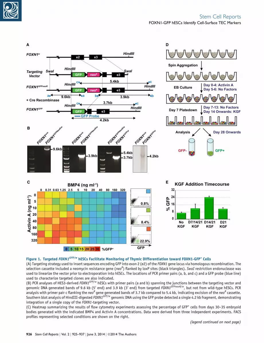
Figure 1. Targeted FOXN1GFP/w hESCs Facilitate Monitoring of Thymic Differentiation toward FOXN1-GFP+ Cells(A) Targeting strategy used to insert sequences encoding GFP into exon 2 (e2) of the FOXN1 gene locus via homologous recombination. Theselection cassette included a neomycin resistance gene (neoR) flanked by loxP sites (black triangles). SwaI restriction endonuclease wasused to linearize the vector prior to electroporation into hESCs. The locations of PCR primer pairs (a, b, and c) and a GFP probe (blue line)used to characterize targeted clones are also indicated.(B) PCR analyses of HES3-derived FOXN1GFP/w hESCs with primer pairs (a and b) spanning the junctions between the targeting vector andgenomic DNA generated bands of 9.6 kb (50 end) and 3.9 kb (30 end) from targeted FOXN1GFPneoR/w, but not from wild-type hESCs. PCRanalysis with primer pair c flanking the neoR gene generated bands of 3.7 kb compared to 5.4 kb, indicating excision of the neoR cassette.Southern blot analysis of HindIII-digested FOXN1GFP/w genomic DNA using the GFP probe detected a single 4.2 kb fragment, demonstratingintegration of a single copy of the FOXN1-targeting vector.(C) Heatmap summarizing the results of flow cytometry experiments assessing the percentage of GFP+ cells from days 30–35 embryoidbodies generated with the indicated BMP4 and Activin A concentrations. Data were derived from three independent experiments. FACSprofiles representing selected conditions are shown on the right.
(legend continued on next page)
926 Stem Cell Reports j Vol. 2 j 925–937 j June 3, 2014 j ª2014 The Authors
Stem Cell ReportsFOXN1-GFP hESCs Identify Cell-Surface TEC Markers

Stem Cell ReportsFOXN1-GFP hESCs Identify Cell-Surface TEC Markers
2012). Recently, two groups reported methods for the
generation of thymic endoderm from human pluripotent
stem cells (Parent et al., 2013; Sun et al., 2013). Impor-
tantly, these studies showed that differentiated mixed cul-
tures containing thymic progenitors could mature in vivo
to form grafts capable of supporting T cell development
in nude (Foxn1�/�) or other immunocompromised mice.
However, because of the lack of appropriate surface
markers, it was not possible to dissect the contributions
or requirements of the various cell types present in the
differentiation cultures at the time of transplantation.
This deficit could be partly remedied by the availability of
FOXN1 reporter lines or surface markers that allow further
fractionation of cultures containing FOXN1+ cells.
We generated FOXN1GFP/w human embryonic stem cell
(hESC) reporter lines that were used to develop a robust
serum-free protocol for the generation of FOXN1+ thymic
endodermal progenitors. We found that high levels of
Activin A and KGF efficiently induced the differentiation
of FOXN1+ cells and that these cells expressed genes
involved in endoderm and thymus development. Tran-
scriptional profiling of purified FOXN1-GFP+ cells allowed
the identification of several combinations of cell-surface
markers that could selectively isolate FOXN1+ TEC pro-
genitor populations derived from unmodified PSC lines.
Collectively, these reagents and findings represent a valu-
able resource for the further investigation of thymic devel-
opment from pluripotent stem cells.
RESULTS
FOXN1GFP/w hESCs Facilitate Analysis of Thymic
Differentiation In Vitro
To facilitate analysis of thymic differentiation of PSCs, we
used homologous recombination to target a GFP reporter
gene to the endogenous FOXN1 locus in MEL1 (Millipore)
and HES3 (Richards et al., 2002) hESCs (Figures 1A and 1B;
Figure S1A available online). Targeted FOXN1GFP/w hESCs
retained expression of pluripotent stem cell markers,
formed multilineage teratomas when transplanted into
immunodeficient NOD/SCID mice, and maintained
normal karyotypes (Figures S1B–S1D). In order to identify
conditions that favored the formation of FOXN1+
endodermal cells, we differentiated FOXN1GFP/w hESCs in
serum-free media as spin embryoid bodies (EBs) (Ng et al.,
2008) and cross-titrated bone morphogenetic protein-4
(BMP4) and Activin A, growth factors that activate
(D) Spin embryoid body protocol for generating FOXN1-GFP+ cells.(E) Summary of flow cytometric data indicating the effects of KGF adddays 7, 14, and 21, days 14 and 21, or day 21. Differentiations were peat days 5 and 6. Error bars show SEM for three independent experime
Ste
signaling pathways required for the generation and
patterning of mesendoderm and have been previously
shown to promote PSC differentiation in vitro (D’Amour
et al., 2005; Davis et al., 2008; Kubo et al., 2004; Tada
et al., 2005; Yasunaga et al., 2005). This heatmap analysis
employed an iterative process (Elliott et al., 2011) and
identified combinations of BMP4 and Activin A that
promoted the appearance of FOXN1-GFP+ cells, which
also costained with the endodermal marker EpCAM (Fig-
ure 1C; Figures S2A–S2C). Based on these results and find-
ings from experiments using different batches of Activin
A (data not shown), for all subsequent experiments, we
differentiated hESCs for 5 days in serum-free medium
supplemented with 150 ng ml�1 Activin A alone (Fig-
ure 1D). These high Activin conditions were similar to
those used in previously published differentiation pro-
tocols where definitive endoderm derivatives were the
desired outcome (D’Amour et al., 2006; Green et al.,
2011). Flow cytometric analysis of differentiating
MIXL1GFP/w hESCs, which can be used to monitor mesen-
doderm induction (Davis et al., 2008), indicated that at
differentiation day 4, over 99% of cells were MIXL1-GFP+,
arguing against the possibility that FOXN1-GFP+ cells rep-
resented ectodermally derived keratinocytes (Figure S3A).
Time course quantitative real-time PCR analysis of differen-
tiation cultures demonstrated that from day 12, cells pro-
gressively upregulated expression of the pharyngeal pouch
markers HOXA3 and PAX9, as well as the thymic markers
INVOLUCRIN, FGFR2 (the receptor for KGF), KERATIN-10,
and FOXN1 itself. Expression of the general endodermal
markers FOXA2 and SOX17 declined from a peak at day
12, consistent with expansion of a specialized anterior
endoderm population that does not express these markers
at high levels (Figure S3B).
Keratinocyte growth factor (KGF or FGF7), a factor previ-
ously implicated in thymic development, was required to
expand the FOXN1-GFP+ population once it emerged in
culture, with the timing of the KGF supplementation
affecting the percentage of GFP+ cells obtained (Figures
1D and 1E). KGF is expressed in the thymus by the sur-
roundingmesenchyme and by different thymocyte subsets
(Rossi et al., 2007) and exerts a paracrine effect by binding
to the FgfR2IIIb receptor, which, within the thymus, is
expressed exclusively by TECs (Erickson et al., 2002). More-
over, exogenous administration of KGF has been shown to
increase thymic cellularity by enhancing the proliferation
and function of TECs (Erickson et al., 2002; Min et al.,
2007; Rossi et al., 2007). After the addition of KGF, GFP+
ition. Following EB culture with Activin A, KGF was either added atrformed in APEL or BPEL medium with or without 50 ng ml�1 nogginnts.
m Cell Reports j Vol. 2 j 925–937 j June 3, 2014 j ª2014 The Authors 927
Stem Cell ReportsFOXN1-GFP hESCs Identify Cell-Surface TEC Markers
cells could be visualized using fluorescence microscopy
from approximately day 25 of differentiation. Flow cyto-
metric analysis of cells differentiated as outlined in Fig-
ure 1D indicated that the first FOXN1-GFP+ cells emerged
from a PDGFRa� and EpCAM+ population as early as differ-
entiation day 15 and increased in frequency thereafter
(up to 38% GFP+ at day 35) (Figure 2A). MEL1-derived
FOXN1GFP/w hESCs differentiated with similar kinetics to
theHES3-derived line, suggesting our protocol is applicable
to other PSC lines (Figure 2A). Quantitative real-time PCR
analysis of cells purified on the basis of GFP and EpCAM
expression showed that FOXN1 transcripts were confined
to the GFP+ population (Figure 2B), confirming the fidelity
of theGFP reporter gene. This conclusion was supported by
intracellular flow cytometric analysis of FOXN1 protein
expression in purified GFP+ and GFP� cells (Figure S3C).
Prolonged culture of aggregates of fluorescence-activated
cell sorting (FACS)-purified GFP+EpCAM+ cells indicated
that GFP expression was retained for up to 3 weeks
(Figure 2B).
Gene Expression Analysis of hESC-Derived FOXN1+
TEC Progenitors
We next performed microarray analysis to survey genes
expressed in the GFP+ fraction and to search for markers
thatmight identify FOXN1+ cells in differentiating cultures
derived from unmanipulated human PSCs. We first
compared the global gene expression within the GFP+
andGFP� EpCAM+ epithelial subpopulations. This analysis
identified 115 genes that were upregulated in the GFP+
EpCAM+ fraction compared to the GFP�EpCAM+ fraction
(Figure 3A; see Tables S1 and S2 for the full list of genes
differentially upregulated by 3-fold), and many of these
have been previously associatedwith thymus development
and/or FOXN1 expression. These included the canonical
WNT-signaling molecules WNT4 and WNT3 and the
WNT regulator KRM2, the NOTCH ligands DLL1, thought
to be required for crosstalk in driving T-lymphopoiesis,
and JAG2 (Candi et al., 2007), a downstream effector of
the DNP63 isoform of the TP63 transcription factor,
required for the proliferation of TECs and other stratified
epithelia, human TEC type I cytokeratins KRT14, KRT15,
and KRT16, and the chemokine CXCL14 whose transcript
is expressed in fetal TECs and may aid in the recruitment
of T cell precursors (Liu et al., 2005). Conversely, 213
transcripts were preferentially upregulated in the GFP�
EpCAM+ population (Figure 3A). These included HAND1
and ISL1, genes expressed during cardiac morphogenesis
and in neural crest derivatives, although ISL1 is also ex-
pressed in pancreatic endoderm, other definitive endo-
dermal markers, such as APOA1, SOX9, and TGFB1, and
human TEC type II cytokeratins KRT6, KRT7, and KRT8.
Interestingly, the chemokineCXCL12was also upregulated
928 Stem Cell Reports j Vol. 2 j 925–937 j June 3, 2014 j ª2014 The Author
within the GFP�EpCAM+ population. CXCL12 is expressed
within TECs and bone marrow niches, where it acts as a
chemoattractant for T cell precursors, monocytes, and
hematopoietic stem cells (HSCs) (Greenbaum et al., 2013;
Liu et al., 2005). Overall, the GFP�EpCAM+ population
expressed genes associated with gene ontology (GO) cate-
gories including ‘‘blood circulation’’ (p value < 0.002), ‘‘cir-
culatory system process’’ (p value < 0.002), ‘‘cell migration’’
(p value < 0.003), and ‘‘cell motility’’ (p value < 0.006) (Fig-
ure S4A). In contrast, GFP+EpCAM+ cells were enriched for
genes associated with FOXN1 expression, thymus develop-
ment, or epithelial-related GO categories (Figure S4A).
Although it is not possible to infer from gene expression
data which genes are direct targets of FOXN1, the ability
to generate and purify a large number of FOXN1+ cells pro-
vides an opportunity to examine this issue using tech-
niques such as chromatin immunoprecipitation (ChIP).
Last, we also compared gene expression of the GFP+
EpCAM+ and GFP-EpCAM- populations (Figure S4B).
Included among the transcripts preferentially expressed
withinGFP+EpCAM+ cells were genes with a role in thymus
development (KRM2, JAG2, WNT4, BCL11B, EFNA1, and
EYA2), epithelial morphogenesis (KRT5, TP63, EpCAM,
KRT14, ECAD, ITGB4, and EGFR), and endoderm specifica-
tion (ALB and AFP).
Keratin-5 and Keratin-8 have been detected within the
human thymus as markers of the medulla and cortex,
respectively (Laster et al., 1986; Shezen et al., 1995). Our
microarray analysis demonstrated high coexpression of
Keratin-5 and its heterodimerization partner Keratin-14
within FOXN1+ (GFP+EpCAM+) cells (Figure 3B). The
former result was confirmed by quantitative real-time
PCR (Figure S4C). Interestingly, although also detected in
GFP+EpCAM+ cells, Keratin-8 is preferentially expressed
by the GFP�EpCAM+ subpopulation (Figure 3A; Fig-
ure S4C). These results might suggest that the FOXN1+ cells
generated in our protocol more closely reflect a medullary
phenotype. Finally, FOXN1+ cells were devoid of genes
that mark the parathyroid gland, including GCM2, PTH,
and CaSR (Figures S4D and S4E), consistent with the obser-
vation that these two developmentally related tissues can
be delineated on the basis of FOXN1 and GCM2 expression
(Gordon et al., 2001). Similarly, other genes associatedwith
the demarcation of thymic versus parathyroid primordia
(BMP4, BMPR1A, BMPR2, and NOGGIN) were not restricted
to specific subpopulations (Figure S4E).
Examination of microarray data indicated that a number
of transcription factors, signalingmolecules, and structural
proteins implicated in TEC differentiation were preferen-
tially expressed in GFP+EpCAM+ cells (Figure 3B). We
were particularly interested in identifying transmembrane
proteins for which well-characterized cell-surface anti-
bodies were readily available. Such antibodies could then
s

A
B
FOXN1
Figure 2. Activin A and KGF Induce GFP Expression in Differentiating FOXN1GFP/w hESCs(A) Flow cytometric analysis of differentiating HES3-derived FOXN1GFP/w hESCs shows FOXN1-GFP+ cells emerge at day 15 and subsequentlyincrease thereafter. FOXN1-GFP+ cells coexpress the thymic epithelial marker EpCAM, but not the mesodermal marker PDGFRa. Flowcytometric analysis of a second independent MEL1-derived FOXN1GFP/w hESC line demonstrated that the time course of GFP and EpCAMinduction was comparable between HES3- and MEL1-derived lines.(B) Quantitative real-time PCR analysis showed that FOXN1 expression was confined to the GFP+EPCAM+ fraction, with expression absentfrom the GFP�EpCAM+ and GFP�EpCAM� subpopulations. Aggregated GFP+EpCAM+ cells retained GFP expression for up to 3 weeks.
Stem Cell Reports j Vol. 2 j 925–937 j June 3, 2014 j ª2014 The Authors 929
Stem Cell ReportsFOXN1-GFP hESCs Identify Cell-Surface TEC Markers

BADay 30 Microarray: GFP+EpCAM+ vs GFP-EpCAM+
115 probe sets > 3.0-foldupregulatedin GFP+EpCAM+
CXCL14 26.5COL17A1 16.0KRM2 11.8KRT14 11.2KRT15 10.4WNT4 8.8DLL1 7.5TP63 6.5JAG2 6.4KRT16 6.2WNT3 4.2
HAND1 10.2KRT6 6.5APOA1 5.8SOX9 4.9CXCL12 4.6KRT7 4.5TGFB1 4.4KRT8 4.2ISL1 4.1BMP4 3.5LIN28 3.3
213 probe sets > 3.0-foldupregulated in GFP-EpCAM+
020004000 KRT5
0600
1200 KRT14
0500
1000 BCL11B
012002400 TP63
012002400 WNT3
0800
1600 WNT4
0400800 WNT10A
020004000 KRM2
010002000 DLL1
015003000 JAG2
030006000 NOTCH1
015003000 EFNA1
0800
1600 SEMA3A
0250500 ITGA6
0200400 ITGB4
0300600 VIPR1
GFP+ EpCAM+ GFP- EpCAM+ GFP- EpCAM-102
103
104
102 103 104
Rel
ativ
e G
ene
Expr
essi
on
Figure 3. Microarray Analysis of FOXN1-GFP+ Thymic Epithelial Progenitor Cells(A) Comparison of gene expression in day 30 GFP+EpCAM+ and GFP�EpCAM+ cells. Selected differentially regulated key genes are listedalong with the fold change in gene expression between the two populations.(B) Genes associated with endoderm commitment and thymus development are preferentially expressed in GFP+EpCAM+ cells compared toGFP�EpCAM+ and GFP�EpCAM� cells. The cell-surface marker ITGB4 was among the transcripts upregulated.
Stem Cell ReportsFOXN1-GFP hESCs Identify Cell-Surface TEC Markers
be employed to isolate FOXN1+ cells from differentiating
cultures of genetically unmodified PSC lines. One such
gene upregulated in GFP+EpCAM+ cells was integrin-b4
(ITGB4 or CD104) (Figure 3B). The ITGB4 subunit
commonly associates with the integrin-a6 (ITGA6) subunit
to form an integrin a6b4 heterodimer that functions as a
receptor for laminins within the extracellularmatrix (Gian-
cotti, 1996). ITGB4 is expressed in normal human TECs,
where it is thought to activate intracellular signaling path-
ways that regulate production of the thymic cytokine IL-6,
which in turn affects TEC differentiation and thymocyte
homing (Mainiero et al., 2003). Moreover, ITGB4 is also
expressed by cultured human TEC lines (Beaudette-Zlata-
nova et al., 2011). Time course analysis showed low expres-
sion of ITGB4 in early differentiating embryoid bodies,
which preceded the appearance of FOXN1-GFP+ cells,
and was associated with a population that concomitantly
upregulated EpCAM expression (Figure 4A). Indeed as
differentiation proceeded, by day 15 the majority of
EpCAM+ cells coexpressed ITGB4, consistent with the
expression of these markers on a broad range of epithelial
tissues (Figure 4A). EpCAM+, FOXN1-GFP+ cells appearing
from differentiation day 20 onward expressed high levels
of ITGB4 (ITGB4hi). Furthermore, as the number of
FOXN1-GFP+ cells increased in response to KGF treatment,
GFP expression became more clearly associated with the
ITGB4hiEpCAM+ subpopulation. Quantitative real-time
PCR analysis of FACS-purified day 34 cells derived from
the differentiation of FOXN1GFP/w hESCs confirmed
that FOXN1 gene expression was largely confined to the
ITGB4hiEpCAM+ population and that expression levels
930 Stem Cell Reports j Vol. 2 j 925–937 j June 3, 2014 j ª2014 The Author
were comparable to those seen in cells sorted on the
basis of GFP expression alone (Figure 4B). Negligible
FOXN1 expression was observed in sorted populations
that expressed intermediate or low levels of ITGB4 or
were EpCAM�.To explore the utility of these cell-surface makers for
monitoring developing TECs, we examined other human
pluripotent stem cell lines, including MEL1-derived
FOXN1GFP/w hESCs, genetically unmodified H9 hESCs
(Thomson et al., 1998) and DF19-9-7T human iPSCs (Yu
et al., 2009) (Figure 4B). All human PSC lines generated
ITGB4hiEpCAM+ cells with parallel kinetics, although the
proportion of this population varied between different
cell lines. Cell populations that expressed ITGB4 and
EpCAMbut were not ITGB4hi (designated ITGB4+EpCAM+)
varied in composition between different PSC lines. Never-
theless, we found FOXN1 expression to be solely confined
to those EpCAM+ cells that were ITGB4hi (Figures 4B and
4C). Overall, flow cytometric analysis of HES3 and MEL1
FOXN1GFP/w cultures indicated that FOXN1-GFP+ cells
comprised over 80% of the ITGB4hiEpCAM+ fraction (Fig-
ure S4F). Conversely, 95% of the GFP+ population was
found to be ITGB4hiEpCAM+, testifying to the close associ-
ation between these markers and FOXN1 expression
(Figure S4G).
In addition to thosemarkers identified by transcriptional
profiling, we examined the expression of the TEC-associ-
ated receptor, HLA-DR: a human major histocompatibility
complex class II (MHCII) cell-surface receptor encoded by
the human leukocyte antigen (HLA) complex (Walsh
et al., 2003). Within the thymus, the primary function of
s
d10 d15 d20 d25 d30 d40d5
αITG
B4
αEpCAM
0.0 58.80.2 40.9
1.4 29.923.5 45.3
1.6 13.679.7 5.1
7.3 20.669.0 3.1
6.6 24.462.7 6.3
11.5 19.862.7 5.9
19.2 18.056.9 5.9
d60
32.1 14.248.5 5.2
A
B
Rel
ativ
e FOXN1
Expr
essi
on
HES3 FOXN1GFP/w(hESCs)
10.2
7.8
13.4
7.4
17.4
0
60
120
180
240
I-E- IhiE+ I+E+ GFP- GFP+
0
25
50
75
100
0
60
120
180
240
4.3 14.975.7 5.1MEL1
FOXN1GFP/w(hESCs)
H9(hESCs)
DF19-9-7T(iPSCs)
13.578.9 6.31.3
5.9 11.975.6 6.7
I-E- IhiE+ I+E+ GFP- GFP+
I-E- IhiE+ I+E+
αITG
B4
αEpCAMGFP
αEpC
AM
44.8
0
60
120
180
240
FL2
8.0 35.945.3 10.8
I-E- IhiE+ I+E+
I-E- IhiE+ I+E+0
60
120
180
240
50
100
I-E- IhiE+ I+E+
C
Rel
ativ
eFOXN1
Expr
essi
on ***
P<0.05P<0.001
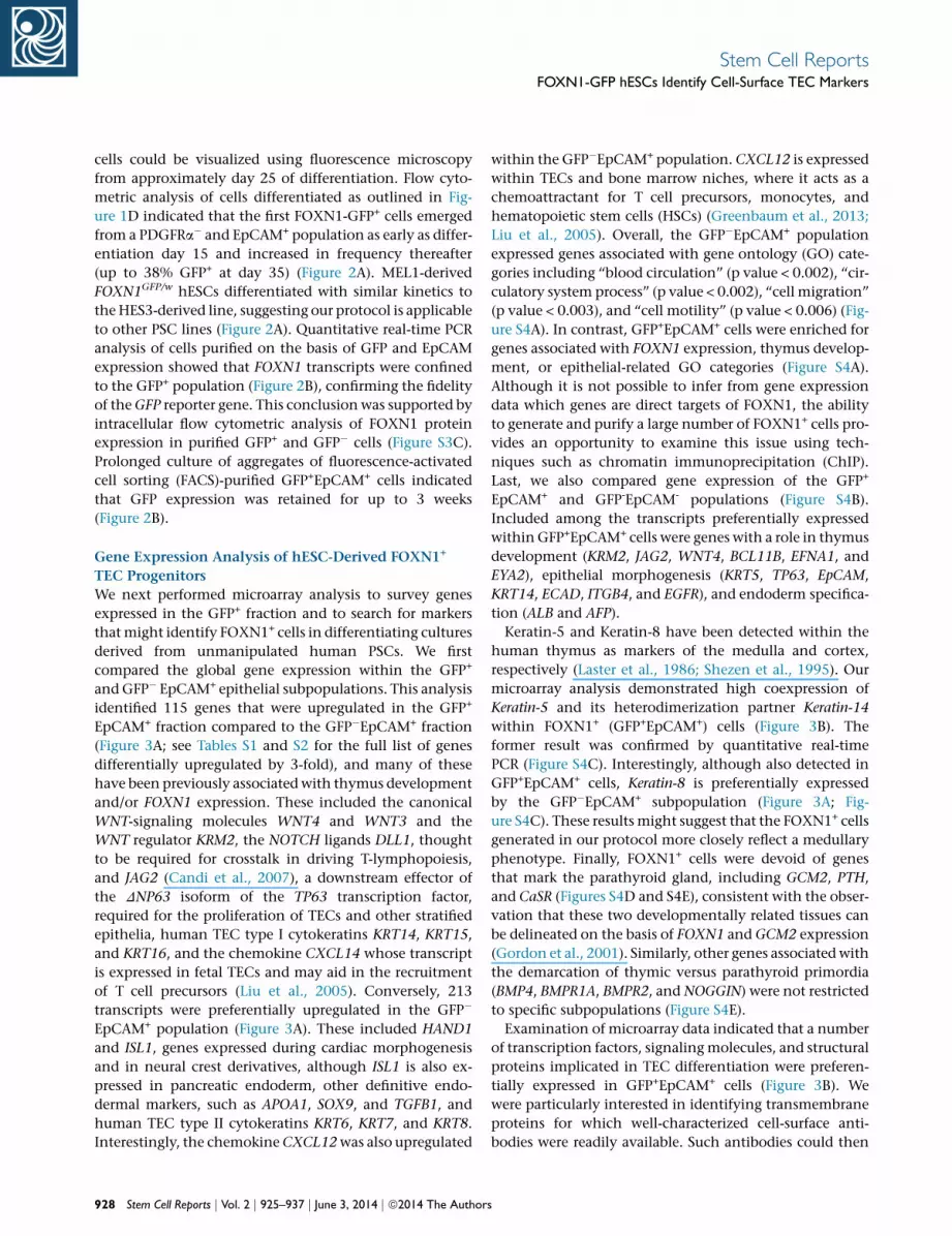
Figure 4. Integrin-b4 and EpCAM Identify FOXN1+ Thymic Epithelial Progenitor Cells(A) Time course analysis of differentiating HES3-derived FOXN1GFP/w cells demonstrated that FOXN1-GFP+ cells could be identified on thebasis of integrin-b4 (ITGB4) and EpCAM expression. GFP expression was localized to EpCAM+ cells that expressed high levels of ITGB4.EpCAM+ cells are shown gated in light blue, whereas GFP+ cells are shown gated in green. Numbers indicate the percentage of cells in thecorresponding quadrants.(B) Quantitative real-time PCR analysis confirmed that FOXN1 transcripts were restricted to the ITGB4hiEpCAM+ population differentiatedfrom HES3-derived FOXN1GFP/w cells and were present at levels similar to those observed in cells isolated on the basis of FOXN1-GFPexpression alone. FOXN1 expression in differentiating MEL1-derived FOXN1GFP/w cells and H9 hESCs or DF19-9-7T human iPSCs was alsoconfined to the ITGB4hiEpCAM+ population. Colored boxes represent the gating strategy used to isolate subpopulations for flow cytometricsorting. I, ITGB4; E, EpCAM. �, +, and hi indicate negative, positive, and high levels of expression, respectively.(C) Histograms demonstrating the summary of raw (left panel) and normalized (right panel) data from individual quantitative real-time PCRexperiments shown in Figure 4B (n = 4). Data are shown as the mean ± SEM. p values were calculated using a two-tailed Student’s t test.
Stem Cell ReportsFOXN1-GFP hESCs Identify Cell-Surface TEC Markers
HLA-DR, similar to otherMHCII genes, is to present peptide
antigens during positive and negative Tcell selection in the
thymic cortex and medulla (Yang et al., 2006). TECs are
Ste
unique in that they constitutively express MHCII without
the need for interferon-g (IFN-g) stimulation (Drukker
et al., 2002; Yang et al., 2006), consistent with the fact
m Cell Reports j Vol. 2 j 925–937 j June 3, 2014 j ª2014 The Authors 931

d10 d15 d20 d25 d30 d40d5αH
LAD
R
αEpCAM
0.0 3.30.1 96.6
0.0 1.710.5 87.8
1.0 3.473.2 22.5
1.3 8.471.1 19.3
1.1 9.265.6 24.2
1.3 9.172.3 17.3
1.8 11.569.3 17.5
16.5
10.1
6.7
0.1 4.384.7 10.9
0
90
180
270
360
H-E- H+E+ H-E+ GFP- GFP+
0
120
240
360
480
Rel
ativ
eFOXN1
Expr
essi
on
4.4 6.577.1 12.1
A
BHES3FOXN1GFP/w(hESCs)
MEL1 FOXN1GFP/w(hESCs)
H9(hESCs)
αHLA
DR
αEpCAMGFP
αEpC
AM FL2
11.8
8.0
0.4 8.171.6 19.9
0
25
50
75
100
H-E- H+E+ H-E+ GFP- GFP+
H-E- H+E+ H-E+
H-E- H+E+ H-E+0
100
200
300
400
50
100
H-E- H+E+ H-E+
C
Rel
ativ
eFOXN1
Expr
essi
on
* P<0.05
Figure 5. HLA-DR and EpCAM Identify PSC-Derived FOXN1+ Thymic Epithelial Progenitor Cells(A) Time course analysis of differentiating HES3-derived FOXN1GFP/w cells demonstrated that FOXN1-GFP+ cells could be identified on thebasis of coexpression of HLA-DR and EpCAM. EpCAM+ cells are shown gated in light blue, whereas GFP+ cells are shown gated in green.Numbers indicate the percentage of cells in the corresponding quadrants.(B) Quantitative real-time PCR analysis confirmed that FOXN1 transcripts were substantially enriched in the HLA-DR+EpCAM+ populationdifferentiated from HES3-derived FOXN1GFP/w cells and were present at levels similar to those observed in FOXN1-GFP+ cells. FOXN1expression in differentiating MEL1-derived FOXN1GFP/w cells and H9 hESCs was also enriched in the HLA-DR+EpCAM+ population. Coloredboxes represent the gating strategy used to isolate subpopulations for flow cytometric sorting. H, HLA-DR; E, EpCAM.(C) Histograms demonstrating the summary of raw (left panel) and normalized (right panel) data from individual quantitative real-timePCR experiments shown in Figure 5B (n = 3). Data are shown as the mean ± SEM. p values were calculated using a two-tailed Student’st test.
Stem Cell ReportsFOXN1-GFP hESCs Identify Cell-Surface TEC Markers
that IFN-g-deficient mice undergo normal thymic develop-
ment (Dalton et al., 1993). To explore whether HLA-DR
could also identify FOXN1-GFP+ cells, we performed flow
cytometry to monitor the expression of HLA-DR and
EpCAM over a 40 day differentiation time course (Fig-
ure 5A). Similar to previous results (Drukker et al., 2002),
HLA-DR was not expressed on the surface of undifferenti-
ated hESCs (data not shown) or during the early stages of
embryoid body formation. Expression was first observed
from approximately day 15 and increased thereafter. By
day 40, we detected a distinct population of HLA-DR+
EpCAM+ cells that contained the majority of FOXN1-
932 Stem Cell Reports j Vol. 2 j 925–937 j June 3, 2014 j ª2014 The Author
GFP+ cells (Figure 5A). Furthermore, all HLA-DR+ cells
were also EpCAM+, suggesting that HLA-DR is restricted
to only thymic epithelial cells in our culture system.
Consistent with past studies, HLA-DR expression on
FOXN1-GFP+ TEC progenitors was not upregulated in
response to IFN-g stimulation. Conversely, as expected,
IFN-g treatment induced expression of HLA-DR in the
non-GFP population (Figure S4H). Finally, quantitative
real-time PCR analysis was performed on day 30 differenti-
ated cells purified on the basis of HLA-DR and EpCAM
expression (Figure 5B). This analysis showed that FOXN1
transcripts were greatly enriched in the HLA-DR+EpCAM+
s

Stem Cell ReportsFOXN1-GFP hESCs Identify Cell-Surface TEC Markers
fraction, with expression levels comparable to those seen in
cells sorted on the basis of GFP alone. The veracity of
this result was confirmed in similar analyses of HLA-DR+
EpCAM+ cells isolated from differentiating cultures of
MEL1 FOXN1GFP/w and H9 hESC lines (Figures 5B and
5C). Similar to results obtained for cells isolated on the
basis of ITGB4 and EpCAM expression, flow cytometric
analysis indicated FOXN1-GFP+ cells comprised over 70%
of cells within the HLA-DR+EpCAM+ fraction (Figure S4I).
The clear association of FOXN1 expression with HLA-DR
further substantiates the thymic identity of cells generated
using our protocol.
The upregulation of HLA-DR in conjunction with gene
expression analyses indicating the presence of NOTCH
signaling receptors and ligands prompted us to test
whether FOXN1-GFP+ cells generated with our differ-
entiation protocol might be competent to promote the
development of T lineage cells from human CD34+
hematopoietic progenitors. To test this hypothesis, we
cocultivated purified FOXN1-GFP+ with human umbilical
cord blood-derived CD34+CD7+ T lineage progenitors, a
cell type previously shown to differentiate into CD4+ or
CD8+ T cells (Awong et al., 2009; La Motte-Mohs et al.,
2005). Over a 4 week coculture period, we analyzed the
hematopoietic component for expression of CD1a, CD5,
CD4, CD8, and CD3, markers of immature T lineage
progenitors, differentiating proT-cells and mature T cells.
Although we observed the induction of CD1a, CD5, CD4,
and CD8 expression, the differentiation step was not
specifically dependent on the presence of FOXN1-GFP+
cells (Figures S5A–S5C and S5E). Moreover, we did not
observe convincing expression of the T-lineage-specific
marker CD3 (Figure S5D). Rather, a significant proportion
of CD34+ cells differentiated into cells with a myeloid
phenotype (CD45+CD14+) (Figure S5E). Hence, although
our humanPSC-derived cells possess a TEC-like phenotype,
we believe that they represent a stage that is too immature
to drive T cell differentiation. Further investigations into
the growth factors necessary for the commitment of
FOXN1+ TEC progenitors into mature TECs in our culture
system are therefore required.
DISCUSSION
We devised a simple method for generating and isolating
FOXN1+ thymic epithelial progenitor cells differentiated
from human pluripotent stem cells. We inserted a GFP
reporter gene into the locus encoding the key thymic tran-
scription factor FOXN1, to facilitatemonitoring of the early
steps involved in human thymus specification. This
reporter gene faithfully identified cells expressing FOXN1
and enabled isolation of FOXN1+ thymic progenitors
Ste
from heterogeneous differentiation cultures. Although
targeted lines represent an extremely useful research tool,
genetically manipulated lines are undesirable for potential
future clinical use. Moreover, the technical requirements
for genetic modification make the application of this
approach impractical for the ever-expanding number of
available pluripotent stem cell lines. With these points in
mind, we interrogated the expression profile of FACS-puri-
fied FOXN1-GFP+ cells through microarray analysis with
the aim of identifying genes that could serve as a surrogate
marker of FOXN1 expression. From this screen, we identi-
fied ITGB4 as a cell-surface marker that, in conjunction
with EpCAM, could be used to isolate FOXN1+ cells from
genetically unmodified hESCs and from human iPSCs.
Similarly, our analysis showed that HLA-DR and EpCAM
were also specific markers of FOXN1+ TEC progenitors.
Taken together, our study provides a model system to
investigate the signaling pathways required for TEC
commitment and differentiation.
Gene expression analysis revealed that cells differenti-
ated for up to 30 days expressed markers implicated in
endoderm specification and TEC development but lacked
the expression of important late-stage TEC markers asso-
ciated with functional maturation. In particular, relative
to human pediatric thymic stroma (data not shown),
in vitro-derived FOXN1+ TEC progenitors showed low
expression of autoimmune regulator (AIRE), a gene critical
for intrathymic expression of tissue-restricted antigens,
which in turn is required to induce tolerance to peripheral
antigens (Anderson et al., 2007). In addition, CD80, a
costimulatory molecule expressed on functionally mature
mTECs (Derbinski et al., 2005), was also not enriched on
FOXN1+ cells. These data indicated that the thymic endo-
derm produced using our culture conditions most likely
represented an early stage of ontogeny in which overt
thymic functional characteristics were yet to be acquired.
The latter stages of thymic epithelial development are
dependent on the presence of hematopoietic cells that, in
the mouse, infiltrate the thymic primordium at embryonic
day 12, soon after the onset of FOXN1 expression. Sub-
sequent to this, the thymic endoderm and lymphoid
progenitors engage in signaling crosstalk that ultimately
yields functionally mature TECs and educated T cells. The
Notch-Delta system is a central signaling mechanism
implicated in directing this crosstalk (Mohtashami et al.,
2010). In the thymus, Notch1 expressed on hematopoietic
progenitors interacts with Dll1 or Dll4 on TECs to either
maintain or induce T-lineage commitment and differentia-
tion. Because of this, we tested whether coculture of
FOXN1+ thymic endoderm with hematopoietic progenitor
cells could provide an environment whereby both cell
types could undergo further maturation. However, under
the conditions used, we saw no evidence of ongoing
m Cell Reports j Vol. 2 j 925–937 j June 3, 2014 j ª2014 The Authors 933

Stem Cell ReportsFOXN1-GFP hESCs Identify Cell-Surface TEC Markers
Tcell differentiation.Moreover, analysis of these cocultures
suggested that the viability of thymic endoderm cells
was compromised, suggesting that further work will be
required to define conditions that are permissive for
the continued growth and development of both hemato-
poietic and endodermal cells types.
Recently, Parent et al. (2013) and Sun et al. (2013)
reported methods for the generation of thymic endoderm
from pluripotent stem cells. Following induction of defini-
tive endoderm by Activin A treatment, both groups could
promote the appearance of anterior foregut and subse-
quently pharyngeal endoderm by carefully choreographed
manipulation of WNT, retinoic acid, and BMP4 signaling,
mirroring the inductive and repressive actions of these
pathways during early thymic ontogeny. Both studies
showed that mixed differentiation cultures containing
thymic progenitors could instruct T cell development in
xenotransplantation models. In our differentiation proto-
col, the activities of the WNT, retinoic acid, and BMP4
pathways were not deliberately manipulated with exoge-
nous growth factors or inhibitors, potentially explaining
the inability of our FOXN1+ cells to upregulate markers of
late-stage TEC differentiation or to support T cell develop-
ment. Having said this, we did not formally test whether
heterogeneous non-FACS-purified populations containing
FOXN1+ cells could differentiate further following trans-
plantation or indeed support T cell development in vitro.
Consequently, the potential functionality and differentia-
tion status of the cells generated using our simple method
is difficult to directly compare with cells described by
Parent et al. (2013) and Sun et al. (2013), where mixed
cultures were used for in vivo studies.
The cell lines and surface markers reported in this study
provide a facile platform on which to begin the process
of identifying key events and critical cell populations
required to derive fully functional TECs from PSCs. These
FOXN1GFP/w reporter lines represent a unique research
tool to analyze human thymus development, whereas the
cell-surface marker combinations recognized by antibodies
against ITGB4, HLA-DR, and EpCAM can be used to purify
TEC progenitors from cultures of differentiating un-
manipulated PSCs. Although the exogenous growth factors
required for ongoing differentiation of FOXN1+ progenitor
cells into functionally mature TECs remain to be estab-
lished, our study contributes critical tools for efforts to
generate this clinically important cell type.
EXPERIMENTAL PROCEDURES
Construction of the FOXN1-Targeting VectorThe FOXN1-targeting vector was assembled using standard cloning
techniques and Gene Bridges Red/ET Recombination technology
(see the Supplemental Experimental Procedures). The final
934 Stem Cell Reports j Vol. 2 j 925–937 j June 3, 2014 j ª2014 The Author
18.2 kb FOXN1-targeting vector comprised a 9.6 kb 50 homology
arm, a GFP-coding sequence, a loxP-flanked neomycin resistance
gene driven by the mouse phosphoglycerate kinase (PGK) gene
promoter, a 3.9 kb 30 homology arm, and a pBR322-based plasmid
backbone. The targeting vector was linearized by SwaI digestion
prior to electroporation.
Generation, Identification, and Characterization of
Targeted FOXN1GFP/w hESCsThe FOXN1-targeting vector was electroporated into hESCs as
previously described (Costa et al., 2007) and as detailed in the
Supplemental Experimental Procedures. The loxP-flanked neoR
cassette was removed by Cre recombinase-mediated excision as
previously described (Davis et al., 2008). For one of the two
independently derived targeted hESC lines (HES3 FOXN1GFP/w
and MEL1 FOXN1GFP/w), Southern blot analysis with a labeled
GFP probe (Figure 1A) was used to confirm the presence of a
single-integration event (Figure 1B). Karyotype analysis was
performed by the Cytogenetics Department at Southern Cross
Pathology, Monash Medical Centre. Teratoma formation and
analysis were performed as previously described (Costa et al.,
2005). Animal experiments were conducted under the approval
of the Monash University Animal Ethics Committee (number
SOBSA/MIS/2009/07).
hESC and iPSC Culture and DifferentiationhESC lines (HES3, MEL1, and H9 [Thomson et al., 1998]) and the
induced pluripotent stem cell (iPSC) line DF19-9-7T (Yu et al.,
2009) were maintained as previously described (Costa et al.,
2008). hESCs and iPSCs were differentiated as spin embryoid
bodies in serum-free media as previously described (Ng et al.,
2008). Briefly, 1 day prior to differentiation, cells were passaged
onto a new tissue culture flask seeded with low density (1 3 104
cells/cm2) mouse embryonic fibroblasts. At day 0, cells were
harvested and deposited into each well (3 3 103 cells/well) of a
96-well round-bottom nonadherent plate (Nunc) and briefly
centrifuged to promote cell aggregation.
Heatmap experiments were performed using APEL medium (Ng
et al., 2008), supplemented with 0–320 ng ml�1 Activin A (R&D
Systems) and 0–320 ng ml�1 bone morphogenetic protein-4
(BMP4; R&D Systems) in 96-well round-bottom low-attachment
plates (Costar) (Elliott et al., 2011). Cytokines were removed on
day 5; at day 7, embryoid bodies were transferred to 96-well flat-
bottom adherent tissue culture-treated plates (BD Falcon) contain-
ing APEL medium lacking PVA (AEL medium) (Ng et al., 2008).
After 30–35 days of differentiation, embryoid bodies were dissoci-
ated with TrypLE Select (Invitrogen), stained with the relevant
antibodies, and analyzed with FACSDIVA software (BD Biosci-
ences) on either the BD LSRII or BD LSRFortessa cell analyzers
(BD Biosciences), fitted with a BD high-throughput sampler
(HTS) module.
For thymic endoderm differentiation assays, cells were initially
cultured in APEL or BPEL medium (Ng et al., 2008), supplemented
with 150 ng ml�1 human Activin A. On day 5, Activin A-contain-
ing APEL or BPEL medium was replaced with APEL or BPEL me-
dium alone. On day 7, EBs were transferred to 96-well flat-bottom
adherent plates (BD Falcon) in AEL or BEL medium (media lacking
s
Stem Cell ReportsFOXN1-GFP hESCs Identify Cell-Surface TEC Markers
PVA) (Ng et al., 2008). AEL or BEL medium supplemented with
40 ngml�1 human keratinocyte growth factor (KGF; R&DSystems)
was used to replenish the cultures on days 14, 21, 28, and 35.
Embryoid bodies were harvested for analysis by flow cytometry
at the times indicated. For experiments using MIXL1GFP/w hESCs
(Davis et al., 2008), cells were differentiated in BPEL medium sup-
plemented either with 150 ng ml�1 Activin A, 20 ng ml�1 human
BMP4, and 100 ng ml�1 Activin A, or with 100 ng ml�1 FGF2
(Peprotech). In all instances, hESC and iPSC cultures and differen-
tiations were maintained at 37�C, in a 5% CO2/air environment.
Flow Cytometric Analysis and SortingFor analysis and sorting of live cells, hESCs and embryoid bodies
were dissociated to a single-cell suspension using TrypLE Select
(Invitrogen), filtered through a 35 mMcell-strainer cap (BD Falcon),
and labeled with the appropriate antibodies (see the Supplemental
Experimental Procedures) as previously described (Davis et al.,
2008). Flow cytometry was performed using a FACSCalibur,
FACSDiva, or Influx Cell Sorter (all from BD Biosciences).
Flow cytometric gates were set using unmodified hESCs or
FOXN1GFP/w-targeted cells labeled with an appropriate isotype
control antibody. Live cells were identified on the basis of forward
scatter, side scatter, and propidium iodide (PI) exclusion.
Intracellular flow cytometry using mouse anti-human OCT4
(clone C-10; Santa Cruz Biotechnology) and mouse anti-human
FOXN1 (clone E-3; Santa Cruz Biotechnology) antibodies was per-
formed essentially as described by Davis et al. (2008) and as
detailed in the Supplemental Experimental Procedures.
Culture of Sorted Cell AggregatesCells purified by flow cytometry were aggregated by centrifugation
for 5 min at 1,500 rpm, using the spin EB protocol (5–10 3 103
cells/well) in BPEL medium supplemented with 5 mM Y27632
ROCK inhibitor, as previously described (Goulburn et al., 2011).
Aggregated cell clusters were cultured on gelatin-coated wells of a
96-well flat-bottom adherent plate in BEL medium supplemented
with 40 ng ml�1 KGF.
Quantitative Real-Time PCR and Microarray Gene
Expression AnalysisTotal RNA was prepared using a High Pure RNA Isolation Kit
(Roche), in accordance with the manufacturer’s instructions.
Quantitative real-time PCR was performed essentially as described
by Pick et al. (2007), with probes detailed in the Supplemental
Experimental Procedures. For microarray analysis, RNA samples
were amplified, labeled, and hybridized to the Human WG-6
(v. 3.0) BeadChip and the Human HT-12 (v. 3.0) BeadChip (Illu-
mina) at the Australian Genome Research Facility (The Walter
and Eliza Hall Institute of Medical Research). Data were analyzed
usingGenomeStudio Gene ExpressionModule (v. 1.5.4) (Illumina)
using average normalization across all samples, with additional
analysis performed using GeneSpring GX software (Agilent Tech-
nologies), as previously described (Goulburn et al., 2011).
IFN-g Stimulation AssayHuman IFN-g (R&D Systems) at 10 ng ml�1 was added to day 30
thymic differentiation cultures in BEL medium. After 72 hr of
Ste
IFN-g stimulation, differentiated cells were harvested and analyzed
for HLA-DR expression by flow cytometry.
CD34+CD7+ proT-Cell and FOXN1-GFP+ TEC
Progenitor Coculture AssaysUmbilical cord blood (UCB) mononuclear cells were obtained
and processed as previously described (La Motte-Mohs et al.,
2005). They were pre-enriched into lineage-negative (Lin�)fractions, sorted into CD34+ hematopoietic stem cells, and
cultured on OP9-DL1 cells for 9–10 days, as previously described
(Awong et al., 2009). CD45+CD34+CD7+ proT-cells were FACS
purified and either cultured alone (1 3 103 cells/well) or with
FOXN1-GFP+ (3.5 3 103 cells/well) or GFP� (3.5 3 103 cells/well)
cells in 96-well round-bottom low-attachment plates (Costar),
containing APEL medium supplemented with 20% FBS, rhFLT3L
(5 ng ml�1, Peprotech), rhIL7 (5 ng ml�1, Peprotech), and rhSCF
(30 ngml�1, Peprotech). A half-media changewas performed every
3–4 days. Cultures were analyzed by flow cytometry using the indi-
cated antibodies and as detailed in the Supplemental Experimental
Procedures.
ACCESSION NUMBERS
TheGEO accessionnumber for themicroarray data reported in this
paper is GSE56373.
SUPPLEMENTAL INFORMATION
Supplemental Information includes Supplemental Experimental
Procedures, five figures, and two tables and can be found
with this article online at http://dx.doi.org/10.1016/j.stemcr.
2014.04.009.
ACKNOWLEDGMENTS
The authors thank Robyn Mayberry and the staff of StemCore
Victoria for the provision of hESCs and iPSCs and Andrew Fryga
and the staff of FlowCore (Monash University) for flow cytometric
sorting. They also thank Jade Homann and the staff of Monash
University Animal Research Laboratories for assistancewithmouse
experiments and Dr. Mahmood Mohtashami for technical advice
on proT-cell cocultures. This work was supported by grants from
the Australian Stem Cell Centre, Stem Cells Australia, the Juvenile
Diabetes Research Foundation, the National Health and Medical
Research Council of Australia (NHMRC), and the Victoria-
California Stem Cell Alliance. K.K. is a recipient of an Alexander
Graham Bell Doctoral Award from the Natural Sciences and Engi-
neering Research Council of Canada. J.C.Z.-P. is supported by a
Canada Research Chair in Developmental Immunology. S.K.N.
and D.N.H. are supported by Australian Research Council Future
Fellowships. A.G.E. and E.G.S. are Senior Research Fellows of the
NHMRC.
Received: November 21, 2013
Revised: April 15, 2014
Accepted: April 16, 2014
Published: May 22, 2014
m Cell Reports j Vol. 2 j 925–937 j June 3, 2014 j ª2014 The Authors 935

Stem Cell ReportsFOXN1-GFP hESCs Identify Cell-Surface TEC Markers
REFERENCES
Anderson, G., Lane, P.J., and Jenkinson, E.J. (2007). Generating
intrathymic microenvironments to establish T-cell tolerance.
Nat. Rev. Immunol. 7, 954–963.
Awong, G., Herer, E., Surh, C.D., Dick, J.E., La Motte-Mohs, R.N.,
and Zuniga-Pflucker, J.C. (2009). Characterization in vitro and
engraftment potential in vivo of human progenitor T cells gener-
ated from hematopoietic stem cells. Blood 114, 972–982.
Beaudette-Zlatanova, B.C., Knight, K.L., Zhang, S., Stiff, P.J.,
Zuniga-Pflucker, J.C., and Le, P.T. (2011). A human thymic epithe-
lial cell culture system for the promotion of lymphopoiesis from
hematopoietic stem cells. Exp. Hematol. 39, 570–579.
Cai, J., Zhao, Y., Liu, Y., Ye, F., Song, Z., Qin, H., Meng, S., Chen, Y.,
Zhou, R., Song, X., et al. (2007). Directed differentiation of human
embryonic stem cells into functional hepatic cells. Hepatology 45,
1229–1239.
Candi, E., Rufini, A., Terrinoni, A., Giamboi-Miraglia, A., Lena,
A.M.,Mantovani, R., Knight, R., andMelino, G. (2007). DeltaNp63
regulates thymic development through enhanced expression of
FgfR2 and Jag2. Proc. Natl. Acad. Sci. USA 104, 11999–12004.
Costa, M., Dottori, M., Ng, E., Hawes, S.M., Sourris, K., Jamshidi, P.,
Pera, M.F., Elefanty, A.G., and Stanley, E.G. (2005). The hESC line
Envy expresses high levels of GFP in all differentiated progeny.
Nat. Methods 2, 259–260.
Costa, M., Dottori, M., Sourris, K., Jamshidi, P., Hatzistavrou, T.,
Davis, R., Azzola, L., Jackson, S., Lim, S.M., Pera, M., et al. (2007).
Amethod for geneticmodification of human embryonic stem cells
using electroporation. Nat. Protoc. 2, 792–796.
Costa, M., Sourris, K., Hatzistavrou, T., Elefanty, A.G., and Stanley,
E.G. (2008). Expansion of human embryonic stem cells in vitro.
Curr. Protoc. Stem Cell Biol. Chapter 1, 1C.1.1–1C.1.7.
D’Amour, K.A., Agulnick, A.D., Eliazer, S., Kelly, O.G., Kroon, E.,
and Baetge, E.E. (2005). Efficient differentiation of human embry-
onic stem cells to definitive endoderm. Nat. Biotechnol. 23, 1534–
1541.
D’Amour, K.A., Bang, A.G., Eliazer, S., Kelly, O.G., Agulnick, A.D.,
Smart, N.G., Moorman, M.A., Kroon, E., Carpenter, M.K., and
Baetge, E.E. (2006). Production of pancreatic hormone-expressing
endocrine cells from human embryonic stem cells. Nat.
Biotechnol. 24, 1392–1401.
Dalton, D.K., Pitts-Meek, S., Keshav, S., Figari, I.S., Bradley, A., and
Stewart, T.A. (1993). Multiple defects of immune cell function in
mice with disrupted interferon-gamma genes. Science 259, 1739–
1742.
Davis, R.P., Ng, E.S., Costa,M.,Mossman, A.K., Sourris, K., Elefanty,
A.G., and Stanley, E.G. (2008). Targeting a GFP reporter gene to the
MIXL1 locus of human embryonic stem cells identifies human
primitive streak-like cells and enables isolation of primitive
hematopoietic precursors. Blood 111, 1876–1884.
Derbinski, J., Gabler, J., Brors, B., Tierling, S., Jonnakuty, S., Hergen-
hahn, M., Peltonen, L., Walter, J., and Kyewski, B. (2005). Promis-
cuous gene expression in thymic epithelial cells is regulated at
multiple levels. J. Exp. Med. 202, 33–45.
936 Stem Cell Reports j Vol. 2 j 925–937 j June 3, 2014 j ª2014 The Author
Drukker,M., Katz, G., Urbach, A., Schuldiner, M., Markel, G., Itsko-
vitz-Eldor, J., Reubinoff, B., Mandelboim, O., and Benvenisty, N.
(2002). Characterization of the expression of MHC proteins in
human embryonic stem cells. Proc. Natl. Acad. Sci. USA 99,
9864–9869.
Elliott, D.A., Braam, S.R., Koutsis, K., Ng, E.S., Jenny, R., Lagerqvist,
E.L., Biben, C., Hatzistavrou, T., Hirst, C.E., Yu, Q.C., et al. (2011).
NKX2-5(eGFP/w) hESCs for isolation of human cardiac progeni-
tors and cardiomyocytes. Nat. Methods 8, 1037–1040.
Erickson, M., Morkowski, S., Lehar, S., Gillard, G., Beers, C.,
Dooley, J., Rubin, J.S., Rudensky, A., and Farr, A.G. (2002). Regula-
tion of thymic epithelium by keratinocyte growth factor. Blood
100, 3269–3278.
Giancotti, F.G. (1996). Signal transduction by the alpha 6 beta 4
integrin: charting the path between laminin binding and nuclear
events. J. Cell Sci. 109, 1165–1172.
Gordon, J., Bennett, A.R., Blackburn, C.C., and Manley, N.R.
(2001). Gcm2 and Foxn1 mark early parathyroid- and thymus-
specific domains in the developing third pharyngeal pouch.
Mech. Dev. 103, 141–143.
Goulburn, A.L., Alden, D., Davis, R.P., Micallef, S.J., Ng, E.S., Yu,
Q.C., Lim, S.M., Soh, C.L., Elliott, D.A., Hatzistavrou, T., et al.
(2011). A targeted NKX2.1 human embryonic stem cell reporter
line enables identification of human basal forebrain derivatives.
Stem Cells 29, 462–473.
Gouon-Evans, V., Boussemart, L., Gadue, P., Nierhoff, D., Koehler,
C.I., Kubo, A., Shafritz, D.A., and Keller, G. (2006). BMP-4 is
required for hepatic specification of mouse embryonic stem cell-
derived definitive endoderm. Nat. Biotechnol. 24, 1402–1411.
Green, M.D., Chen, A., Nostro, M.C., d’Souza, S.L., Schaniel, C.,
Lemischka, I.R., Gouon-Evans, V., Keller, G., and Snoeck, H.W.
(2011). Generation of anterior foregut endoderm from human
embryonic and induced pluripotent stem cells. Nat. Biotechnol.
29, 267–272.
Greenbaum, A., Hsu, Y.M., Day, R.B., Schuettpelz, L.G., Christo-
pher, M.J., Borgerding, J.N., Nagasawa, T., and Link, D.C. (2013).
CXCL12 in early mesenchymal progenitors is required for haema-
topoietic stem-cell maintenance. Nature 495, 227–230.
Jerome, L.A., and Papaioannou, V.E. (2001). DiGeorge syndrome
phenotype in mice mutant for the T-box gene, Tbx1. Nat. Genet.
27, 286–291.
Kroon, E., Martinson, L.A., Kadoya, K., Bang, A.G., Kelly, O.G.,
Eliazer, S., Young, H., Richardson, M., Smart, N.G., Cunningham,
J., et al. (2008). Pancreatic endoderm derived from human embry-
onic stem cells generates glucose-responsive insulin-secreting cells
in vivo. Nat. Biotechnol. 26, 443–452.
Kubo, A., Shinozaki, K., Shannon, J.M., Kouskoff, V., Kennedy, M.,
Woo, S., Fehling, H.J., and Keller, G. (2004). Development of defin-
itive endoderm from embryonic stem cells in culture. Develop-
ment 131, 1651–1662.
La Motte-Mohs, R.N., Herer, E., and Zuniga-Pflucker, J.C. (2005).
Induction of T-cell development from human cord blood hemato-
poietic stem cells by Delta-like 1 in vitro. Blood 105, 1431–1439.
s

Stem Cell ReportsFOXN1-GFP hESCs Identify Cell-Surface TEC Markers
Lai, L., and Jin, J. (2009). Generation of thymic epithelial cell
progenitors by mouse embryonic stem cells. Stem Cells 27,
3012–3020.
Laster, A.J., Itoh, T., Palker, T.J., and Haynes, B.F. (1986). The
human thymic microenvironment: thymic epithelium contains
specific keratins associated with early and late stages of epidermal
keratinocyte maturation. Differentiation 31, 67–77.
Liu, C., Ueno, T., Kuse, S., Saito, F., Nitta, T., Piali, L., Nakano, H.,
Kakiuchi, T., Lipp, M., Hollander, G.A., and Takahama, Y. (2005).
The role of CCL21 in recruitment of T-precursor cells to fetal
thymi. Blood 105, 31–39.
Longmire, T.A., Ikonomou, L., Hawkins, F., Christodoulou, C.,
Cao, Y., Jean, J.C., Kwok, L.W., Mou, H., Rajagopal, J., Shen, S.S.,
et al. (2012). Efficient derivation of purified lung and thyroid
progenitors from embryonic stem cells. Cell Stem Cell 10,
398–411.
Mainiero, F., Colombara,M., Antonini, V., Strippoli, R.,Merola,M.,
Poffe, O., Tridente, G., and Ramarli, D. (2003). p38 MAPK is a crit-
ical regulator of the constitutive and the beta4 integrin-regulated
expression of IL-6 in human normal thymic epithelial cells. Eur.
J. Immunol. 33, 3038–3048.
Min, D., Panoskaltsis-Mortari, A., Kuro-O, M., Hollander, G.A.,
Blazar, B.R., and Weinberg, K.I. (2007). Sustained thymopoiesis
and improvement in functional immunity induced by exogenous
KGF administration in murine models of aging. Blood 109, 2529–
2537.
Mohtashami, M., Shah, D.K., Nakase, H., Kianizad, K., Petrie, H.T.,
and Zuniga-Pflucker, J.C. (2010). Direct comparison of Dll1- and
Dll4-mediated Notch activation levels shows differential lympho-
myeloid lineage commitment outcomes. J. Immunol. 185,
867–876.
Nehls, M., Pfeifer, D., Schorpp, M., Hedrich, H., and Boehm, T.
(1994). Newmember of the winged-helix protein family disrupted
in mouse and rat nude mutations. Nature 372, 103–107.
Nehls,M., Kyewski, B., Messerle,M.,Waldschutz, R., Schuddekopf,
K., Smith, A.J., and Boehm, T. (1996). Two genetically separable
steps in the differentiation of thymic epithelium. Science 272,
886–889.
Ng, E.S., Davis, R., Stanley, E.G., and Elefanty, A.G. (2008). A proto-
col describing the use of a recombinant protein-based, animal
product-free medium (APEL) for human embryonic stem cell
differentiation as spin embryoid bodies. Nat. Protoc. 3, 768–776.
Parent, A.V., Russ, H.A., Khan, I.S., LaFlam, T.N., Metzger, T.C.,
Anderson, M.S., and Hebrok, M. (2013). Generation of functional
thymic epithelium from human embryonic stem cells that
supports host T cell development. Cell Stem Cell 13, 219–229.
Pick, M., Azzola, L., Mossman, A., Stanley, E.G., and Elefanty, A.G.
(2007). Differentiation of human embryonic stem cells in serum-
freemedium reveals distinct roles for bonemorphogenetic protein
Ste
4, vascular endothelial growth factor, stem cell factor, and fibro-
blast growth factor 2 in hematopoiesis. Stem Cells 25, 2206–2214.
Pignata, C., Fiore,M., Guzzetta, V., Castaldo, A., Sebastio, G., Porta,
F., andGuarino, A. (1996). Congenital Alopecia and nail dystrophy
associated with severe functional T-cell immunodeficiency in two
sibs. Am. J. Med. Genet. 65, 167–170.
Richards, M., Fong, C.Y., Chan, W.K., Wong, P.C., and Bongso, A.
(2002). Human feeders support prolonged undifferentiated growth
of human inner cell masses and embryonic stem cells. Nat.
Biotechnol. 20, 933–936.
Rossi, S.W., Jeker, L.T., Ueno, T., Kuse, S., Keller, M.P., Zuklys, S.,
Gudkov, A.V., Takahama, Y., Krenger, W., Blazar, B.R., and
Hollander, G.A. (2007). Keratinocyte growth factor (KGF)
enhances postnatal T-cell development via enhancements in
proliferation and function of thymic epithelial cells. Blood 109,
3803–3811.
Shezen, E., Okon, E., Ben-Hur, H., and Abramsky, O. (1995). Cyto-
keratin expression in human thymus: immunohistochemical
mapping. Cell Tissue Res. 279, 221–231.
Sun, X., Xu, J., Lu, H., Liu, W., Miao, Z., Sui, X., Liu, H., Su, L., Du,
W., He, Q., et al. (2013). Directed differentiation of human embry-
onic stem cells into thymic epithelial progenitor-like cells reconsti-
tutes the thymic microenvironment in vivo. Cell Stem Cell 13,
230–236.
Tada, S., Era, T., Furusawa, C., Sakurai, H., Nishikawa, S., Kinoshita,
M., Nakao, K., Chiba, T., and Nishikawa, S. (2005). Characteriza-
tion of mesendoderm: a diverging point of the definitive endo-
derm and mesoderm in embryonic stem cell differentiation
culture. Development 132, 4363–4374.
Thomson, J.A., Itskovitz-Eldor, J., Shapiro, S.S., Waknitz, M.A.,
Swiergiel, J.J., Marshall, V.S., and Jones, J.M. (1998). Embryonic
stem cell lines derived from human blastocysts. Science 282,
1145–1147.
Walsh, E.C., Mather, K.A., Schaffner, S.F., Farwell, L., Daly, M.J.,
Patterson, N., Cullen, M., Carrington, M., Bugawan, T.L., Erlich,
H., et al. (2003). An integrated haplotypemap of the humanmajor
histocompatibility complex. Am. J. Hum. Genet. 73, 580–590.
Yang, S.J., Ahn, S., Park, C.S., Holmes, K.L., Westrup, J., Chang,
C.H., and Kim, M.G. (2006). The quantitative assessment of
MHC II on thymic epithelium: implications in cortical thymocyte
development. Int. Immunol. 18, 729–739.
Yasunaga, M., Tada, S., Torikai-Nishikawa, S., Nakano, Y., Okada,
M., Jakt, L.M., Nishikawa, S., Chiba, T., Era, T., and Nishikawa, S.
(2005). Induction and monitoring of definitive and visceral endo-
derm differentiation of mouse ES cells. Nat. Biotechnol. 23, 1542–
1550.
Yu, J., Hu, K., Smuga-Otto, K., Tian, S., Stewart, R., Slukvin, I.I., and
Thomson, J.A. (2009). Human induced pluripotent stem cells free
of vector and transgene sequences. Science 324, 797–801.
m Cell Reports j Vol. 2 j 925–937 j June 3, 2014 j ª2014 The Authors 937