fourier transform infrared (ftir) spectroscopic study of acid soluble collagen and gelatin from...
TRANSCRIPT
Food
Food Chemistry 86 (2004) 325–332
www.elsevier.com/locate/foodchem
Chemistry
Fourier transform infrared (FTIR) spectroscopic studyof acid soluble collagen and gelatin from skins and bones
of young and adult Nile perch (Lates niloticus)
J.H. Muyonga a,b, C.G.B. Cole c, K.G. Duodu b,*
a Department of Food Science and Technology, Makerere University, P.O. Box 7062, Kampala, Ugandab Department of Food Science, University of Pretoria, Pretoria 0002, South Africa
c Davis Gelatine (South Africa), P.O. Box 5019 West Krugersdorp, 1742, South Africa
Received 25 April 2003; received in revised form 8 September 2003; accepted 8 September 2003
Abstract
Fourier transform infrared (FTIR) spectroscopy was conducted on type A gelatins derived from skins and bones of young and
adult Nile perch (Lates niloticus) by a sequential extraction process. Spectra for gelatins were compared to each other and to that of
acid soluble collagen from young Nile perch skins, in order to elucidate changes in protein secondary structure during collagen to
gelatin transformation. The first gelatin extracts showed diminished amide III bands while the last gelatin extracts showed distinct
amide III bands and their amide I bands consisted of a higher percent area of a component around 1690 cm�1. The differences
suggested that the collagen to gelatin transition leads to loss of molecular order. The later gelatin extracts exhibited higher molecular
order than earlier gelatin extracts, probably because the former contained surviving crosslinks or/and because renaturation of the
low molecular weight gelatin fractions (later gelatin extracts) led to formation of more protein–protein linkages.
� 2003 Published by Elsevier Ltd.
Keywords: Nile perch; Gelatin; Collagen; FTIR; Protein structure
1. Introduction
Fourier transform infrared (FTIR) spectroscopy has
been used to study changes in the secondary structure of
collagen and gelatin. It has been used to study collagen
crosslinking (Paschalis et al., 2001), denaturation (Friess
& Lee, 1996), thermal self assembly (Jakobsen, Brown,
Hutson, Fink, & Veis, 1983; George & Veis, 1991) as
well as gelatin melting (Prystupa & Donald, 1996). The
spectral changes which are indicative of changes incollagen secondary structure have been shown to include
changes in the amide A (Milch, 1964), amide I (1636–
1661 cm�1), amide II (1549–1558 cm�1) (Renugopala-
krishnan et al., 1989) and the amide III (1200–1300
cm�1) regions (Friess & Lee, 1996).
* Corresponding author. Tel.: +27-12-420-3202/420-4299; fax: +27-
12-4202839.
E-mail address: [email protected] (K.G. Duodu).
0308-8146/$ - see front matter � 2003 Published by Elsevier Ltd.
doi:10.1016/j.foodchem.2003.09.038
Fibrillogenesis (self assembly) of collagen has been
found to be associated with broadening and a slight shiftto lower wave number of the amide A peak (Milch,
1964), increase in intensity and slight shift to lower wave
number of amide III peak (Jakobsen et al., 1983), band
broadening and shift of amide I peak to lower wave
number (Jakobsen et al., 1983; George & Veis, 1991;
Prystupa & Donald, 1996) and shift of amide II peak to
lower wave number (Jakobsen et al., 1983; George &
Veis, 1991). Shift of amide I, II and III peaks to lowerwave numbers, increase in intensity of amide III and
broadening of amide I are therefore associated with in-
creased intermolecular interactions (by hydrogen
bonding) in collagen.
Denaturation of collagen, on the other hand, has
been found to lead to reduction in the intensity of amide
A, I, II and III peaks (Friess & Lee (1996), narrowing
of amide I band (Prystupa & Donald, 1996), increasein amide I component found around 1630 cm�1 and
326 J.H. Muyonga et al. / Food Chemistry 86 (2004) 325–332
reduction in the intensity of amide I component, found
around 1660 cm�1 (George & Veis, 1991; Payne & Veis,
1988; Renugopalakrishnan et al., 1989).
Prystupa & Donald (1996) studied gelatin melting
and found it to be associated with reduction in the 1678cm �1 peak and 1660/1690 cm�1 peak intensity ratio and
increase in amide I components occurring around 1613,
1629 and 1645 cm�1. These authors assigned the bands
occurring at 1645–1657 cm�1to random coils and the
1660 cm�1band to triple helix, with contribution from a-helix and b-turns. The amide I component, at 1690
cm�1, has been attributed to helices of aggregated col-
lagen-like peptides (Doyle, Bendit, & Blout, 1975; Pry-stupa & Donald, 1996). According to Doyle et al. (1975)
this peak vanishes with hydration of collagen or gelatin.
As animals age, the extent of crosslinking of their
collagen increases and the type of crosslinks change
(Sims & Bailey, 1992; Bailey, Paul, & Knott, 1998; Sims,
Avery, & Bailey, 2000; Hickman et al., 2000). According
to Bailey et al. (1998) collagen from skins of immature
animals mainly contain the intermediate crosslinks de-hydroxylysinonorleucine (deHLNL), whereas collagen
from bones of immature animals contain hydroxylysin-
oketonorleucine (HLKNL). These intermediate divalent
crosslinks are, respectively, converted to the more stable
trivalent histidinohydroxylysinonorleucine (HHL) and
pyridolines (PYR) during maturation. It has been shown
that differences in the quantities of the two types of
crosslinks are manifested in the amide I region of theFTIR spectra of collagenous tissue (Paschalis et al.,
2001). There is a positive correlation between the ratio
of the components (1660/1690 cm�1) and the relative
abundance of PYR and HHL crosslinks.
Age-related increase in stability of collagen, through
increase in the amount and stability of crosslinks, affects
the stability of collagen to denaturation processes, e.g.
heat. Collagens with more extensive crosslinks, e.g.those from mature bovine hide, require a more severe
process to break the crosslinks and allow collagen de-
naturation and solubilisation into gelatin (Reich, Wal-
ther, & Stather, 1962). During such severe processes,
more peptide bonds are broken but some intermolecular
crosslinks survive. The triple helices of collagen from
young animals are mainly held together by hydrogen
bonds and Van der Waals forces. In such collagens, heattreatment mainly leads to breaking of hydrogen bonds
and the triple helical structure is more likely to decom-
pose, mainly to intact alpha chains. It is not clear,
however, whether the secondary structures of gelatins
derived from young and old animals differ. Nile perch
(Lates niloticus) is a warm water fish species, with po-
tential for giving gelatin with gelling properties more
similar to mammalian gelatins than cold water fishspecies.
In this study, the FTIR spectra of gelatins derived
from young and adult Nile perch skins and bones were
determined and compared to those of acid-soluble col-
lagen from the same species, in an effort to elucidate
changes in secondary structure that occur during the
conversion of collagen to gelatin. The first and last
gelatin extracts from skins and bones of young and adultNile perch were studied.
2. Materials and methods
2.1. Preparation of acid soluble collagen
Acid-soluble collagen was prepared from skins ofyoung Nile perch (skin thickness < 0:4 mm), as de-
scribed by G�omez-Guill�en &Montero (2001). Briefly the
method involved washing of the skins with chilled (�5
�C) water for a period of 10 min. During this time, the
skins were pressed intermittently by hand. The skins
were then washed with 0.8 M NaCl for 3 periods, of 10
min each, followed by rinsing in running water, after
each wash, with NaCl. Collagen was then extracted us-ing 0.5 M acetic acid solution (1:20 w/v). The extraction
was conducted for 16 h, during which the skins were
stirred intermittently. The viscous collagenous material
was separated from the insoluble components by sieving
through cheesecloth and collagen was precipitated
using 0.9 M NaCl, washed with distilled water and
freeze-dried.
An attempt was made to extract collagen from bonesusing 0.5 M acetic acid, but no collagen could be pre-
cipitated from the acetic acid liquor, after 5 days holding
at room temperature.
2.2. Preparation of gelatins
The gelatins used in this study were derived from
Nile perch skins and bones by the acid process. Gel-atin was extracted from young (skin thickness < 0:4mm and skeleton length < 40 cm) and adult (skin
thickness > 1:5 mm and skeleton length > 95 cm) fish.
Briefly, extraction of skin gelatin involved acidulation
with concentrated sulphuric acid to a pH of 2.5–3.0
and maintaining this pH range throughout the swell-
ing period (16 h) by adding more acid solution until
the skins were adequately swollen. The skins werethen transferred to beakers, covered with warm (�60
�C) water and gelatin extracted in water baths at 50,
60 and 70 �C, in a sequential process. In the case of
young fish skins, extraction was conducted at only 50
and 60 �C because, after the 60 �C extraction, the
residue left was very small and would give very small
amounts of gelatin at 70 �C. The gelatin extracts (light
liquors) were filtered through compressed cotton wool.The light liquor concentrations were determined by
evaporating duplicate 10 ml portions to a stable
weight (48 h at 105 �C) and the concentration was
Assignment
Reference
B(70)
78
NH
stretch,coupledwithHB
SaiandBabu(2001)
CH
2asymmetricalStretch
AbeandKrimm
(1972)
CH
2symmetricalStretch
AbeandKrimm
(1972)
52
C@O
stretch/H
Bcoupled
withCOO-
Jackson,Choo,Watson,
Halliday,andMantsch
(1995)
40
NH
bendcoupledwithCN
stretch
Jacksonet
al.(1995)
50
CH
2bend
Jacksonet
al.(1995)
CH
2waggingofproline
Jacksonet
al.(1995)
36
NH
bend
Jacksonet
al.(1995)
27
CAO
stretch
Jacksonet
al.(1995)
76
Skeletalstretch
AbeandKrimm
(1972)
4Skeletalstretch
AbeandKrimm
(1972)
1 –Gelatinextracted
from
adultfish
skins,YB–Gelatinextracted
from
youngfish
bones,
der,–Nocommonnameforthespectralregion,DM
–Dim
inished
peak,HB–Hydrogen
J.H. Muyonga et al. / Food Chemistry 86 (2004) 325–332 327
used in calculation of % gelatin extractability as
follows:
Amount of gelatin extracted at a given temp
sum of gelatin extracted at all temp� 100%
¼ %gelatin extractability at a given temp:
The light liquors were then passed through a column
of activated carbon (GRC 22, BHT water treatment,
Chloorkop, South Africa) at a rate of �5 bed volumesper hour. The pH of all the light liquors was adjusted to
�5.0 using 5% ammonia solution and the gelatin extract
was dried in a cross-flow air drier at 42 �C, until brittle.The brittle sheets were broken into small pieces and
milled using a domestic coffee grinder to pass through a
1 mm mesh sieve.
Bones used for gelatin extraction were cleaned, by
scraping with a knife, to reduce the flesh contamination.They were then decreased by tumbling in warm (35 �C)water and demineralised using 3% HCl at room tem-
perature (20–25 �C) for a period of 9–12 days, with the
liquor changed after every three days, until the bones
did not have any hard cores. The demineralised bones
were then treated in the same way as the acidulated
skins. The extractability and Bloom of the gelatins are
presented in Table 1.
perch
skin
andbonegelatins
0)
AS(70)
YB(50)
AB(50)
YB(70)
A
3404
3421
3456
3310
34
2923
2924
DM
Sh
Sh
2853
DM
2355
DM
1653
1647
1644
1656
16
1541
1558
DM
1544
15
1451
DM
1457
1451
14
1335
1402
1335
1240
1243
12
1082
1122
1107
11
1006
1082
10
670
870
866
87
670
701
67
,YS–Gelatinextracted
from
youngfish
skins,AS
re(�C)forthegelatin,Sh–Peakappearingasshoul
2.3. Fourier transform infrared spectroscopy
FTIR spectra were obtained from discs containing 2
mg sample in approximately 100 mg potassium bromide
(KBr). All spectra were obtained using a Bruker infrared
spectrophotometer (Bruker Instruments, Billerica, MA)
from 4000 to 500 cm�1at data acquisition rate of 2 cm�1
per point. Background was subtracted using the Opus
software (Bruker Instruments, Billerica, MA). Triplicate
samples of collagen and gelatins were analysed and
spectra for the triplicate runs averaged. Fourier self
deconvolution was conducted on the average spectra for
the amide I band, using a resolution enhancement factor
Table 1
Source, extractability and Bloom of gelatins used
Source Extraction
temperature (�C)Extractability
(%)
Bloom
(g)
Fish skin gelatins
Adult fish 50 70.0 240
Young fish 50 86.5 217
Adult fish 70 10.6 134
Young fish 60 12.9 0
Fish bone gelatins
Adult fish 50 33.0 84
Young fish 50 33.3 156
Adult fish 70 9.6 155
Young fish 70 22.6 0
Table
2
FTIR
spectrapeakpositionandassignments
forNile
Region
Peakwavenumber
cm�1
ASC
YS(50)
AS(50)
YS(6
AmideA
3434
3623
3648
3411
–2924
2923
2924
DM
–2853
2853
2853
–2355
2355
2356
AmideI
1650
1648
1650
1654
AmideII
1542
DM
1541
1542
–1457
1458
1457
1452
–DM
DM
AmideIII
1235
1234
DM
DM
–1026
1011
–871
863
867
–670
670
660
669
– ASC–YoungNileperch
skinsacid-solublecollagen
AB–Gelatinextracted
from
adultfish
bones.
Numbersin
bracketsrepresentextractiontemperatu
bonding.
328 J.H. Muyonga et al. / Food Chemistry 86 (2004) 325–332
of 1.8 and full height band width of 13 cm�1. The self
deconvolution provided information on the number and
location of components. Curve fitting was then per-
formed using peakfit software (SPSS Inc., Chicago, IL,
USA).
3. Results and discussion
3.1. Frequencies
The frequencies at which major peaks occurred for
acid soluble collagen and the different gelatins and col-lagens are summarised in Table 2.
3.2. Spectra for skin gelatins
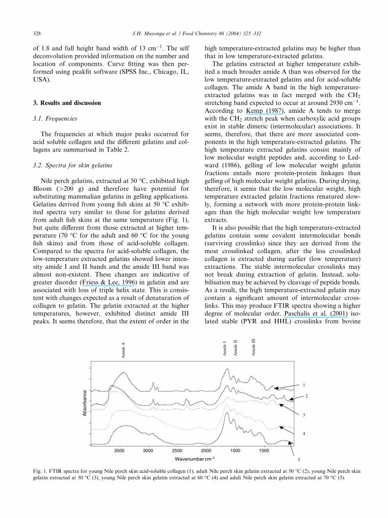
Nile perch gelatins, extracted at 50 �C, exhibited high
Bloom (>200 g) and therefore have potential for
substituting mammalian gelatins in gelling applications.
Gelatins derived from young fish skins at 50 �C exhib-ited spectra very similar to those for gelatins derived
from adult fish skins at the same temperature (Fig. 1),
but quite different from those extracted at higher tem-
perature (70 �C for the adult and 60 �C for the young
fish skins) and from those of acid-soluble collagen.
Compared to the spectra for acid-soluble collagen, the
low-temperature extracted gelatins showed lower inten-
sity amide I and II bands and the amide III band wasalmost non-existent. These changes are indicative of
greater disorder (Friess & Lee, 1996) in gelatin and are
associated with loss of triple helix state. This is consis-
tent with changes expected as a result of denaturation of
collagen to gelatin. The gelatin extracted at the higher
temperatures, however, exhibited distinct amide III
peaks. It seems therefore, that the extent of order in the
225003000 3500
Wavenumber
Abs
orba
nce
Am
ide
A
Fig. 1. FTIR spectra for young Nile perch skin acid-soluble collagen (1), adu
gelatin extracted at 50 �C (3), young Nile perch skin gelatin extracted at 60
high temperature-extracted gelatins may be higher than
that in low temperature-extracted gelatins.
The gelatins extracted at higher temperature exhib-
ited a much broader amide A than was observed for the
low temperature-extracted gelatins and for acid-solublecollagen. The amide A band in the high temperature-
extracted gelatins was in fact merged with the CH2
stretching band expected to occur at around 2930 cm�1.
According to Kemp (1987), amide A tends to merge
with the CH2 stretch peak when carboxylic acid groups
exist in stable dimeric (intermolecular) associations. It
seems, therefore, that there are more associated com-
ponents in the high temperature-extracted gelatins. Thehigh temperature extracted gelatins consist mainly of
low molecular weight peptides and, according to Led-
ward (1986), gelling of low molecular weight gelatin
fractions entails more protein-protein linkages than
gelling of high molecular weight gelatins. During drying,
therefore, it seems that the low molecular weight, high
temperature extracted gelatin fractions renatured slow-
ly, forming a network with more protein-protein link-ages than the high molecular weight low temperature
extracts.
It is also possible that the high temperature-extracted
gelatins contain some covalent intermolecular bonds
(surviving crosslinks) since they are derived from the
most crosslinked collagen, after the less crosslinked
collagen is extracted during earlier (low temperature)
extractions. The stable intermolecular crosslinks maynot break during extraction of gelatin. Instead, solu-
bilisation may be achieved by cleavage of peptide bonds.
As a result, the high temperature-extracted gelatin may
contain a significant amount of intermolecular cross-
links. This may produce FTIR spectra showing a higher
degree of molecular order. Paschalis et al. (2001) iso-
lated stable (PYR and HHL) crosslinks from bovine
1000 1500000
cm-1
1
2
3
4
5
Am
ide
I
Am
ide
II
Am
ide
III
lt Nile perch skin gelatin extracted at 50 �C (2), young Nile perch skin
�C (4) and adult Nile perch skin gelatin extracted at 70 �C (5).
10001500200025003000 3500
Absorbance
5
21
3
4
Wavenumber cm-1
Am
ide
A
Am
ide
I
Am
ide
III
Am
ide
II
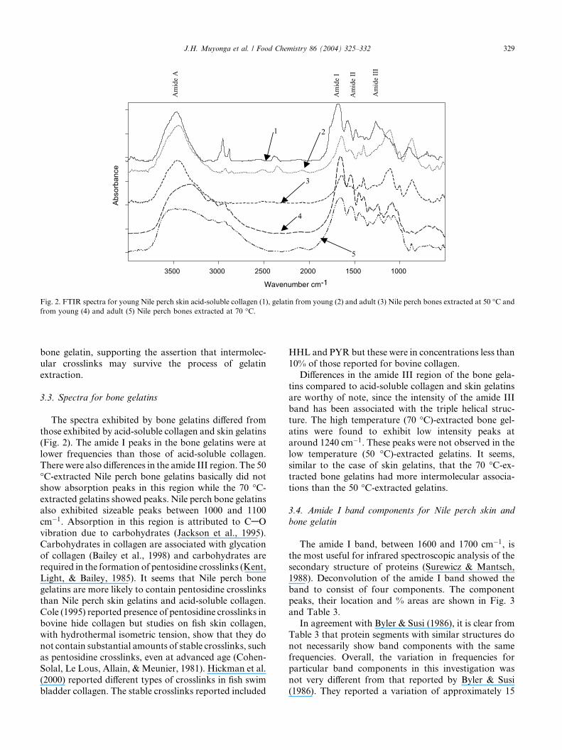
Fig. 2. FTIR spectra for young Nile perch skin acid-soluble collagen (1), gelatin from young (2) and adult (3) Nile perch bones extracted at 50 �C and
from young (4) and adult (5) Nile perch bones extracted at 70 �C.
J.H. Muyonga et al. / Food Chemistry 86 (2004) 325–332 329
bone gelatin, supporting the assertion that intermolec-
ular crosslinks may survive the process of gelatin
extraction.
3.3. Spectra for bone gelatins
The spectra exhibited by bone gelatins differed from
those exhibited by acid-soluble collagen and skin gelatins
(Fig. 2). The amide I peaks in the bone gelatins were at
lower frequencies than those of acid-soluble collagen.
There were also differences in the amide III region. The 50
�C-extracted Nile perch bone gelatins basically did not
show absorption peaks in this region while the 70 �C-extracted gelatins showed peaks. Nile perch bone gelatinsalso exhibited sizeable peaks between 1000 and 1100
cm�1. Absorption in this region is attributed to CAO
vibration due to carbohydrates (Jackson et al., 1995).
Carbohydrates in collagen are associated with glycation
of collagen (Bailey et al., 1998) and carbohydrates are
required in the formation of pentosidine crosslinks (Kent,
Light, & Bailey, 1985). It seems that Nile perch bone
gelatins are more likely to contain pentosidine crosslinksthan Nile perch skin gelatins and acid-soluble collagen.
Cole (1995) reported presence of pentosidine crosslinks in
bovine hide collagen but studies on fish skin collagen,
with hydrothermal isometric tension, show that they do
not contain substantial amounts of stable crosslinks, such
as pentosidine crosslinks, even at advanced age (Cohen-
Solal, Le Lous, Allain, &Meunier, 1981). Hickman et al.
(2000) reported different types of crosslinks in fish swimbladder collagen. The stable crosslinks reported included
HHL and PYR but these were in concentrations less than
10% of those reported for bovine collagen.
Differences in the amide III region of the bone gela-
tins compared to acid-soluble collagen and skin gelatins
are worthy of note, since the intensity of the amide III
band has been associated with the triple helical struc-ture. The high temperature (70 �C)-extracted bone gel-
atins were found to exhibit low intensity peaks at
around 1240 cm�1. These peaks were not observed in the
low temperature (50 �C)-extracted gelatins. It seems,
similar to the case of skin gelatins, that the 70 �C-ex-tracted bone gelatins had more intermolecular associa-
tions than the 50 �C-extracted gelatins.
3.4. Amide I band components for Nile perch skin and
bone gelatin
The amide I band, between 1600 and 1700 cm�1, is
the most useful for infrared spectroscopic analysis of the
secondary structure of proteins (Surewicz & Mantsch,
1988). Deconvolution of the amide I band showed the
band to consist of four components. The componentpeaks, their location and % areas are shown in Fig. 3
and Table 3.
In agreement with Byler & Susi (1986), it is clear from
Table 3 that protein segments with similar structures do
not necessarily show band components with the same
frequencies. Overall, the variation in frequencies for
particular band components in this investigation was
not very different from that reported by Byler & Susi(1986). They reported a variation of approximately 15
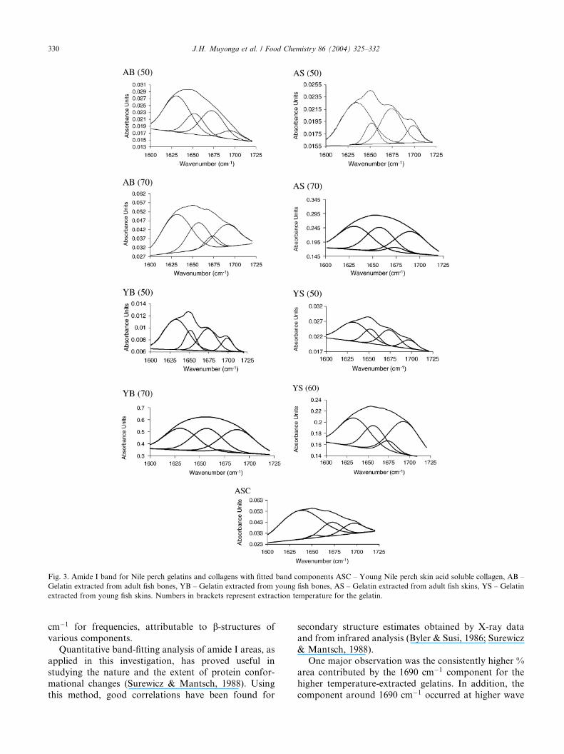
Fig. 3. Amide I band for Nile perch gelatins and collagens with fitted band components ASC – Young Nile perch skin acid soluble collagen, AB –
Gelatin extracted from adult fish bones, YB – Gelatin extracted from young fish bones, AS – Gelatin extracted from adult fish skins, YS – Gelatin
extracted from young fish skins. Numbers in brackets represent extraction temperature for the gelatin.
330 J.H. Muyonga et al. / Food Chemistry 86 (2004) 325–332
cm�1 for frequencies, attributable to b-structures of
various components.
Quantitative band-fitting analysis of amide I areas, as
applied in this investigation, has proved useful in
studying the nature and the extent of protein confor-
mational changes (Surewicz & Mantsch, 1988). Using
this method, good correlations have been found for
secondary structure estimates obtained by X-ray data
and from infrared analysis (Byler & Susi, 1986; Surewicz
& Mantsch, 1988).
One major observation was the consistently higher %
area contributed by the 1690 cm�1 component for the
higher temperature-extracted gelatins. In addition, the
component around 1690 cm�1 occurred at higher wave
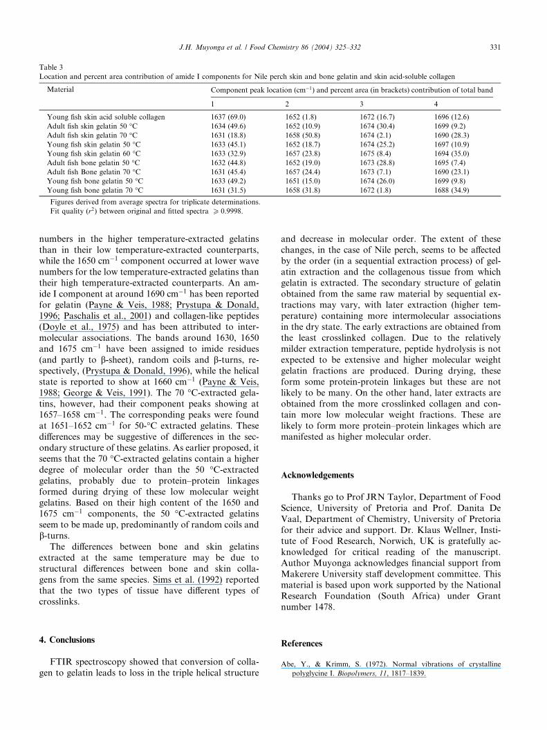
Table 3
Location and percent area contribution of amide I components for Nile perch skin and bone gelatin and skin acid-soluble collagen
Material Component peak location (cm�1) and percent area (in brackets) contribution of total band
1 2 3 4
Young fish skin acid soluble collagen 1637 (69.0) 1652 (1.8) 1672 (16.7) 1696 (12.6)
Adult fish skin gelatin 50 �C 1634 (49.6) 1652 (10.9) 1674 (30.4) 1699 (9.2)
Adult fish skin gelatin 70 �C 1631 (18.8) 1658 (50.8) 1674 (2.1) 1690 (28.3)
Young fish skin gelatin 50 �C 1633 (45.1) 1652 (18.7) 1674 (25.2) 1697 (10.9)
Young fish skin gelatin 60 �C 1633 (32.9) 1657 (23.8) 1675 (8.4) 1694 (35.0)
Adult fish bone gelatin 50 �C 1632 (44.8) 1652 (19.0) 1673 (28.8) 1695 (7.4)
Adult fish Bone gelatin 70 �C 1631 (45.4) 1657 (24.4) 1673 (7.1) 1690 (23.1)
Young fish bone gelatin 50 �C 1633 (49.2) 1651 (15.0) 1674 (26.0) 1699 (9.8)
Young fish bone gelatin 70 �C 1631 (31.5) 1658 (31.8) 1672 (1.8) 1688 (34.9)
Figures derived from average spectra for triplicate determinations.
Fit quality (r2) between original and fitted spectra P 0.9998.
J.H. Muyonga et al. / Food Chemistry 86 (2004) 325–332 331
numbers in the higher temperature-extracted gelatins
than in their low temperature-extracted counterparts,
while the 1650 cm�1 component occurred at lower wave
numbers for the low temperature-extracted gelatins than
their high temperature-extracted counterparts. An am-
ide I component at around 1690 cm�1 has been reported
for gelatin (Payne & Veis, 1988; Prystupa & Donald,
1996; Paschalis et al., 2001) and collagen-like peptides(Doyle et al., 1975) and has been attributed to inter-
molecular associations. The bands around 1630, 1650
and 1675 cm�1 have been assigned to imide residues
(and partly to b-sheet), random coils and b-turns, re-spectively, (Prystupa & Donald, 1996), while the helical
state is reported to show at 1660 cm�1 (Payne & Veis,
1988; George & Veis, 1991). The 70 �C-extracted gela-
tins, however, had their component peaks showing at1657–1658 cm�1. The corresponding peaks were found
at 1651–1652 cm�1 for 50-�C extracted gelatins. These
differences may be suggestive of differences in the sec-
ondary structure of these gelatins. As earlier proposed, it
seems that the 70 �C-extracted gelatins contain a higher
degree of molecular order than the 50 �C-extractedgelatins, probably due to protein–protein linkages
formed during drying of these low molecular weightgelatins. Based on their high content of the 1650 and
1675 cm�1 components, the 50 �C-extracted gelatins
seem to be made up, predominantly of random coils and
b-turns.The differences between bone and skin gelatins
extracted at the same temperature may be due to
structural differences between bone and skin colla-
gens from the same species. Sims et al. (1992) reportedthat the two types of tissue have different types of
crosslinks.
4. Conclusions
FTIR spectroscopy showed that conversion of colla-
gen to gelatin leads to loss in the triple helical structure
and decrease in molecular order. The extent of these
changes, in the case of Nile perch, seems to be affected
by the order (in a sequential extraction process) of gel-
atin extraction and the collagenous tissue from which
gelatin is extracted. The secondary structure of gelatin
obtained from the same raw material by sequential ex-
tractions may vary, with later extraction (higher tem-
perature) containing more intermolecular associationsin the dry state. The early extractions are obtained from
the least crosslinked collagen. Due to the relatively
milder extraction temperature, peptide hydrolysis is not
expected to be extensive and higher molecular weight
gelatin fractions are produced. During drying, these
form some protein-protein linkages but these are not
likely to be many. On the other hand, later extracts are
obtained from the more crosslinked collagen and con-tain more low molecular weight fractions. These are
likely to form more protein–protein linkages which are
manifested as higher molecular order.
Acknowledgements
Thanks go to Prof JRN Taylor, Department of Food
Science, University of Pretoria and Prof. Danita De
Vaal, Department of Chemistry, University of Pretoria
for their advice and support. Dr. Klaus Wellner, Insti-
tute of Food Research, Norwich, UK is gratefully ac-
knowledged for critical reading of the manuscript.
Author Muyonga acknowledges financial support from
Makerere University staff development committee. Thismaterial is based upon work supported by the National
Research Foundation (South Africa) under Grant
number 1478.
References
Abe, Y., & Krimm, S. (1972). Normal vibrations of crystalline
polyglycine I. Biopolymers, 11, 1817–1839.
332 J.H. Muyonga et al. / Food Chemistry 86 (2004) 325–332
Bailey, A. J., Paul, R. G., & Knott, L. (1998). Mechanisms of
maturation and aging of collagen. Mechanism of Aging and
Development, 106, 1–56.
Byler, D. M., & Susi, H. (1986). Examination of the secondary
structure of proteins by deconvolved FTIR spectra. Biopolymers,
25, 469–487.
Cole, C. G. B. (1995). Occurrence, measurement and origins of gelatin
colour as determined by flourescence and electrophoresis. Ph.D.
Thesis, University of Pretoria. Pretoria, Republic of South Africa.
Cohen-Solal, L., Le Lous, M., Allain, J., & Meunier, F. (1981).
Absence of maturation of collagen crosslinks in fish skin. Febs
Letters, 123, 282–284.
Doyle, B. B., Bendit, E. G., & Blout, E. R. (1975). Infrared
spectroscopy of collagen and collagen-like polypeptides. Biopoly-
mers, 14, 937–957.
Friess, W., & Lee, G. (1996). Basic thermoanalytical studies of
insoluble collagen matrices. Biomaterials, 17, 2289–2294.
George, A., & Veis, A. (1991). FTIRS in H2O demonstrates that
collagen monomers undergo a conformational transition prior to
thermal self-assembly in vitro. Biochemistry, 30, 2372–2377.
G�omez-Guill�en, M. C., & Montero, P. (2001). Extraction of gelatin
from megrim (Lepidorhombus boscii) skins with several organic
acids. Journal of Food Science, 66, 213–216.
Hickman, D., Sims, T. J., Miles, C. A., Bailey, A. J., de Mari, M., &
Koopmans, M. (2000). Isinglass/collagen: Denaturation and func-
tionality. Journal of Biotechnology, 79, 245–257.
Jackson, M., Choo, L., Watson, P. H., Halliday, W. C., & Mantsch, H.
H. (1995). Beware of connective tissue proteins: Assignment and
implications of collagen absorptions in infrared spectra of human
tissues. Biochima et Biophysica Acta, 1270, 1–6.
Jakobsen, R. L., Brown, L. L., Hutson, T. B., Fink, D. J., & Veis, A.
(1983). Intermolecular interactions in collagen self-assembly as
revealed by fourier transform infrared spectroscopy. Science, 220,
1288–1290.
Kemp, W. (1987). Organic Spectroscopy (second ed.). Hampshire:
Macmillan Education Ltd..
Kent, M. J. C., Light, N. D., & Bailey, A. J. (1985). Evidence for
glucose-mediated covalent cross-linking of collagen after glycosyl-
ation in vitro. Biochemical Journal, 225, 745–752.
Ledward, D. A. (1986). Gelation of Gelatin. In J. R. Mitchell & D. A.
Ledward (Eds.), Functional Properties of Food Macromolecules (pp.
171–201). New York: Elsevier.
Milch, R. A. (1964). Infra-red spectra of deuterated gelatin sols.
Nature, 202, 84–85.
Paschalis, E. P., Verdelis, K., Doty, S. S., Boskey, A. L., Mendelesohn,
R., & Yamauchi, M. (2001). Spectroscopic characterisation of
collagen cross-links in bone. Journal of Bone and Mineral Research,
16, 1821–1828.
Payne, K. J., & Veis, A. (1988). Fourier transform IR spectroscopy
of collagen and gelatin solutions: Deconvolution of the
Amide I band for conformational studies. Biopolymers, 27, 1749–
1760.
Prystupa, D. A., & Donald, A. M. (1996). Infrared study of gelatin
conformations in gel and sol states. Polymer Gels and Networks, 4,
87–110.
Reich, G., Walther, S., & Stather, F. (1962). The influence of the age of
cattle and pigskin on the yield and the quality of the gelatines
obtained after the acid conditioning process. In Investigation of
Collagen and Gelatine IV (vol. 18, pp. 24–30). Freiberg/SA:
Deutsche Lederinstitut.
Renugopalakrishnan, V., Chandarakasan, G., Moore, S., Hutson, T.
B., Berney, C. V., & Ravejendra, S. B. (1989). Bound water in
collagen. Evidence from fourier transform infrared and Fourier
transform infrared photoacoustic spectroscopic study. Macromol-
ecules, 22, 4124–4124.
Sai, P. K., & Babu, M. (2001). Studies on Rana tigerina skin collagen.
Comparative Biochemistry and Physiology, 128(B), 81–90.
Sims, J. T., & Bailey, A. J. (1992). Quantitative analysis of collagen
and elastin crosslinks using a single-column system. Journal of
Chromatography, 582, 49–55.
Sims, J. T., Avery, N. C., & Bailey, A. J. (2000). Quantitative
determination of collagen crosslinks. InC. Streuli & M.
Grant (Eds.), Extracellular Matrix Protocols: vol. 139.
Methods in Molecular Biology (pp. 11–26). Totowa: Humana Press
Inc.
Surewicz, W. K., & Mantsch, H. H. (1988). New insight into protein
secondary structure from resolution enhanced infrared spectra.
Biochimica et Biophysica Acta, 952, 115–130.