forsterite amorphisation by ion irradiation: monitoring by infrared spectroscopy
TRANSCRIPT

Astronomy & Astrophysics manuscript no. paper˙revised˙babbage July 23, 2003(DOI: will be inserted by hand later)
Forsterite amorphisation by ion irradiation:
monitoring by infrared spectroscopy
J.R. Brucato1 G. Strazzulla2 G. Baratta2 and L. Colangeli1
1 INAF Osservatorio Astronomico di Capodimonte, via Moiariello 16, 80131 Napoli, Italy
e-mail: [email protected], [email protected]
2 INAF Osservatorio Astrofisico di Catania, via S.Sofia 78, 95123 Catania, Italy
e-mail: [email protected], [email protected]
Abstract.
We present experimental results on crystal–amorphous transition of forsterite
(Mg2SiO4) silicate under ion irradiation. The aim of this work is to study the structural
evolution of one of the most abundant crystalline silicates observed in space driven by ion
irradiation. To this aim, forsterite films have been sythesised in laboratory and irradiated
with low energy (30–60 keV) ion beams. Structural changes during irradiation with H+,
He+, C+, and Ar++ have been observed and monitored by infrared spectroscopy. The frac-
tion of crystalline forsterite converted into amorphous is a function of the energy deposited
by nuclear collision by ions in the target.
Laboratory results indicate that ion irradiation is a mechanism potentially active in
space for the amorphisation of silicates. Physical properties obtained in this work can be
used to model the evolution of silicate grains during their life cycle from evolved stars,
through different interstellar environments and up to be incorporated in Solar System ob-
jects.
Key words. methods: laboratory – techniques: spectroscopy – ISM: dust, evolution – cos-
mic rays
1. Introduction
During late stages of evolution, stars loose mass in a short time compared to the life they spend in
the main sequence. In the gaseous expanding circumstellar envelopes dust condensation occurs.
In particular, the envelopes of giant stars belonging to the asymptotic branch (AGB) (Gehrz 1989)
and of supernovae (SN) (Jones 1997) are considered the principal sites of cosmic dust formation.
Once formed, grains are injected in the interstellar medium (ISM) driven by the stellar winds.
Send offprint requests to: J.R. Brucato
2 J.R. Brucato G. Strazzulla G. Baratta and L. Colangeli: Ion amorphisation of forsterite
Recent detailed observations of AGB M-type stars in the medium and far infrared spectral re-
gions obtained with the ISO satellite revealed series of bands testifying that both amorphous and
crystalline silicate phases are present in the envelopes (Kemper et al. 2001; Molster et al. 2002a,
2002b, 2002c). The results showed the presence of two principal crystalline silicate components,
forsterite (Mg2SiO4) and enstatite (MgSiO3). Relevant correlations between silicate grain proper-
ties and outflow or disk source geometries were found. In particular, disk–like stars show strong
crystalline silicate bands, while outflow geometry is characterised by weak crystalline silicate
bands. Amorphous silicates were detected at temperatures higher than those of crystalline ones.
Molster et al. (2002c) showed that enstatite and forsterite masses correlate eachother and that
enstatite is more abundant than forsterite by a factor 3-4. Fitting analysis by laboratory spectra of
the Spectral Energy Distributions (SED) indicates that, for example, in the source OH-127.8+0.0,
80% of silicates is amorphous olivine ((Mg,Fe)2SiO4), 3% is forsterite, 3% enstatite, 4% metal-
lic iron, and 10% crystalline water ice (Kemper et al. 2002). A value of 3% for forsterite and
enstatite was also calculated for many AGB stars (Sylvester et al. 1999) and considered typical
for these stars (Kemper et al. 2001).
The distribution of crystalline and amorphous silicate grains according to AGB mass-loss
rates was derived (Sogawa & Kozasa 1999; Kemper et al. 2001; Suh 2002). In particular, Kemper
et al. (2001) showed that the presence of crystalline silicates does not depend on the mass-loss
rate. Moreover, they demonstrated that the lack of spectroscopic signatures of crystalline silicates
in the ISO spectra does not imply the absence of crystalline dust in the envelope. This result was
obtained considering that, if two separate populations of amorphous and crystalline grains are
used in the radiative transfer model of circumstellar envelopes, up to 40 % of crystals can be
included in the envelope dust without observing any spectroscopic evidence.
Further analyses of AGB stars spectra were performed by Suh (2002) by using a different dust
model. An average single grain population for mixed amorphous and crystalline silicate grains
was chosen. It was obtained that about 10 to 20% of crystalline silicates are produced in high
mass-loss rate AGB stars, but no crystalline silicates were found in low mass-loss rate stars. The
discrepancy of these results depends on the formation mechanism chosen to be active in AGB
envelopes. In the first case, two grain populations are formed with different temperatures, while
in the second case a single amorphous component is formed that subsequently crystallises totally
or partially by annealing, reaching a single final temperature.
The model of Sogawa and Kozasa (1999) already confirmed the absence of crystalline sili-
cates in heterogeneous grains condensed in low mass–loss stars (≤ 3 · 10−5M�yr−1). However,
condensation models of minerals in stellar winds of M-stars indicate that a multicomponent mix-
ture is mainly formed, dominated by olivine and iron grains (Gail and Sedlmayr 1999). The tem-
perature evolution of the condensed grains in the outflow favours the birth of crystalline grains
when their radius is ≤ 100 nm. For larger grains the temperature is too low to crystallise the
materials, giving rise to a further population of crystalline core grains with an external coating of
amorphous material.

J.R. Brucato G. Strazzulla G. Baratta and L. Colangeli: Ion amorphisation of forsterite 3
Thus, if we consider that AGB stars with circumstellar envelopes are expected to be ubiqui-
tous in our Galaxy (Habing 1996) and that they account for 50% of the total stellar mass-loss
of the Galaxy, the crystalline silicate component formed in these stars should be observed in the
Interstellar medium (ISM). On the contrary, analysis of ISO spectra showed that an upper limit
of only few percent of crystals is present in dense and diffuse ISM (Li and Draine 2001; Demyk
et al. 1999).
Among various mechanisms that could explain the absence or the non observability of crys-
talline silicates in ISM (e.g. selective destruction, low production rate or interstellar dust dilution)
in this work we experimentally investigate the amorphisation process by interaction with 30–60
keV ions.
Previous laboratory irradiation experiments of forsterite with energetic (1.5 MeV) protons
(Day 1977) and of clinoenstatite with 400 keV and 1 MeV helium ions (Jager et at. 2003), made
at fluences similar to those of cosmic rays, did not show any infrared spectroscopic evidence
of structural modification of crystals. Complete amorphisation for low energy (4 and 10 keV)
helium ions irradiation at similar fluences was observed (Demyk et al. 2001; Carrez et al. 2002).
The crystal–amorphous transition process can be affected significantly by various physical
parameters of the impinging ions, as e.g. the mass, charge and the kinetic energy. Thus, further
laboratory studies are needed to investigate the amorphisation process of silicates.
In this work, experimental results on structural modifications suffered by crystalline forsterite
under ion irradiation are presented. Thin films of forsterite are synthesised in order to monitor in
situ, by infrared transmission spectroscopy, the crystalline to amorphous transition during irra-
diation. Processing effects are investigated, the correlation with the energy deposition is studied
and the cross–section of nuclear collision which is directly related to amorphisation is derived.
In Section 2 we describe the experimental apparatus and procedures. Results are presented in
Section 3 and discussed in Section 4. Astrophysical implications of experimental results and
concluding remarks are reported in Section 5.
2. Experimental
Among various laboratory techniques which give information on the chemical and physical prop-
erties of silicates of various nature, the infrared spectroscopy is widely used. The results obtained
by this technique can be used directly for comparisons with space observations, once a dust
model is applied. Up to now a series of infrared spectra are available in literature for different
classes of crystalline silicates where peak positions and intensities were linked to properties of
the samples, as morphology, chemical composition and crystallographic structure (e.g. Koike et
al. 1993, Mennella et al. 1998, Jager et al. 1998, Koike et al. 2000, Fabian et al. 2001, Suto et al.
2002, Chihara et al. 2002, Brucato et al. 2002, Colangeli et al. 2003). In the present work the ex-
perimental procedure used is based on the possibility to acquire transmission infrared spectra of
crystalline silicate samples and to observe their evolution during irradiation process. To this aim,

4 J.R. Brucato G. Strazzulla G. Baratta and L. Colangeli: Ion amorphisation of forsterite
thin films of crystalline forsterite were prepared in laboratory for in situ infrared transmission
spectroscopy and ion bombardment processing.
2.1. Thin silicate film synthesis
Silicate films were prepared at the Cosmic Physics Laboratory of INAF–Osservatorio
Astronomico di Capodimonte by using a Nd–YAG solid state pulsed laser. Its fundamental wave-
length output is at 1064 nm with a mean energy of 650 mJ. The power laser output is 108 W cm−2
per laser pulse. A set of two crystals was used to get II and IV harmonics at 532 and 266 nm,
respectively. The energy output was 80 mJ at 266 nm, 120 mJ at 532 nm and 190 mJ at 1064
nm. By an optical set–up, the 266 nm wavelength was selected and directed onto the target. By
using a focusing lens, the power density was mantained at 108 W cm−2 by compensating for the
decreased mean energy. Targets were prepared by using mixtures of periclase (MgO) and quartz
(SiO2) at the stochiometric ratio (2:1) of forsterite Mg2SiO4. The mixture was pressed at 10 tons
obtaining pellets 13 mm in diameter and few millimetres thick. The target pellet was mounted
inside a vaporisation chamber which was designed to be filled by different gases. The presence
of a quenching reactive gas affects the chemical composition of the laser ablated sample, while
its pressure drives the morphology of the condensed materials. The lower the pressure inside the
chamber the smaller is the size of the condensed grains. In previous laser ablation experiments
the vaporisation chamber was filled with oxygen at 10 mbar (Brucato et al. 1999, 2002). This was
done to maintain the chemical composition of the condensed sample similar to that of the tar-
get. Moreover, the presence of oxygen favours the super–saturation of the hot quenched vapour.
This is responsible of the formation of grains with sizes following a log normal distribution with
average size of few tens of nanometers. In order to produce films thin enough in order to obtain
observable transmission IR spectra before and during ion irradiation, the chamber pressure was
maintained at 10−5 mbar. With these experimental conditions the mean free path of the atoms of
the plasma plume produced by the laser ablation can be considered infinitely larger than the dis-
tance target–sample (3 cm). Thus, the super saturation condition, necessary to condense grains,
is not reached along the path of the atoms to the substrate. Films of amorphous silicates are pro-
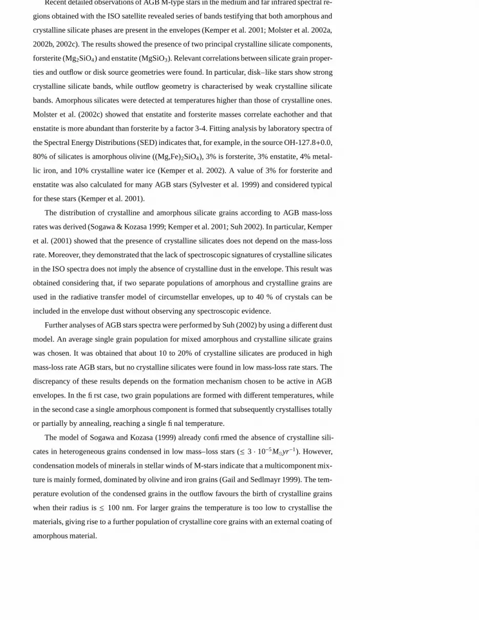
duced after 15 min deposition on silicon wafer substrates. The infrared spectrum of condensed
sample, shown in Figure 1, presents two smooth and broad bands at 10.6 and around 20 µm
typical of amorphous silicate.
In order to prepare samples with crystalline structure, the amorphous silicate films were an-
neled at 900 ◦C for 1 hour at the pressure of 10−6 mbar. The annealing temperature is reached
at a rate of 23 C min−1 and controlled electronically. The transition from the amorphous to crys-
talline phase is evidenced by the appearance in the infrared spectum of a series of sharp bands
(Figure 1), typical of crystalline materials.
J.R. Brucato G. Strazzulla G. Baratta and L. Colangeli: Ion amorphisation of forsterite 5
10 200
0.05
0.1
0.15
Fig. 1. Infrared optical depth spectra of laser synthesised amorphous film (thin line) and of crystalline
forsterite after thermal annealing (thick line).
2.2. Ion irradiation
Ion irradiation was performed by a Danphysik (1080–30) ion implantation system of INAF–
Astrophysical Observatory laboratory of Catania. The gas is ionised in a source and a reduced
pressure favours the production of a plasma. Ions are accelerated by 30 kV potential and mean ki-
netic energies of 30 keV and 60 keV are obtained for once and double ionised ions, respectively.
Once extracted, the ions travel a mass separator where defined m/q are selected by a magnetic
field. An electrostatic scanning system is used to deflect the ion trajectories. This allows to ir-
radiate uniformly the sample with low current density (of the order of 1 µA cm−2), avoiding
undesirable annealing of the sample. The target assembly is designed for in situ spectroscopy.
For further details on the experimental apparatus see Strazzulla et al. (2001). In the present ex-
periment, samples were irradiated with 30 keV H+, He+, C+ and with 60 keV Ar++ with ion
fluences up to 1017 cm−2. Light (hydrogen and helium) and heavy (carbon and argon) ions were
chosen to study how different classes of ions affect silicate structure.
The thin films were characterised by Fourier transform infrared spectroscopy (Bruker
Equinox 55) in the mid-infrared with resolution of 4 cm−1. Spectra were acquired after laser
deposition and thermal annealing and in situ during ion irradiation of samples, in order to moni-
tor the chemical and physical evolution of the silicate films.
3. Results
3.1. Chemical composition and thickness
In order to check composition and thickness, the IR spectrum of synthesised films was compared
with that of crystalline olivine. The optical constants of Mg1.9Fe0.1SiO4 olivine derived by Fabian

6 J.R. Brucato G. Strazzulla G. Baratta and L. Colangeli: Ion amorphisation of forsterite
10 15 20 250
0.05
0.1
0.15
Fig. 2. Infrared optical depth spectra of films of synthesised forsterite normalised to the continuum (thick
line) and of crystalline olivine (Mg1.9Fe0.1SiO4) calculated by the optical constant of Fabian et al. (2000)
(thin line).
et al. (2000) were used to evaluate the optical depth, τ, of the film. This is defined according to the
equation τ = 4πdk/λ, where d is the thickness of the film and k is the immaginary part of complex
refractive index. The average assorbance τ = 13 [τx + τy + τz] along the (x,y,z) crystallographic
axes for a film of thickness d = 380 Åis reported in Figure 2 and compared with the absorbance
of a thin film sample synthesised in this work. Unfortunately, the sample used by Fabian et al. is
not the pure forsterite (Mg2SiO4) end member of olivines, a small fraction of iron being present.
The presence of iron in olivine induces a shift in the peak positions towards longer wavelengths
that according to Jager et al (1998) is correlated to the mass percentage of FeO according to the
relation: [FeO]/∆ν = −1.8 ± 0.1.
Comparing the spectra in Figure 2 it is evident the presence of all the peaks of forsterite in
the synthetic sample spectrum, even if a mismatch of peak positions and intensities is observed.
Differences in peak intensities are probably due to a preferential axis of growing of the crystals
during the thermal annealing. Peak positions are reported in Table 1, together with the peak
positions expected for pure forsterite thin film after correction of wavelength shifts. The peak at
9.48 µm is due to the presence of a fraction of quartz which does not participate to the olivine
formation.
In order to give a further estimate of the film thickness, analysis of scanning electron micro-
graphs of the samples was performed. An average film thickness of 500 ± 200 Å was obtained.
This value is compatible with that obtained by the calculation of the optical depth and comparable
with the penetration ranges of the ions (Table 2).
J.R. Brucato G. Strazzulla G. Baratta and L. Colangeli: Ion amorphisation of forsterite 7
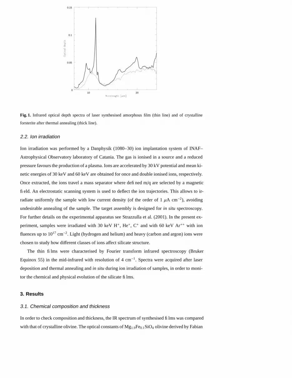
Table 1. Peak positions for synthetic forsterite film, olivine Mg1.9Fe0.1SiO4 film as obtained by optical
constant from Fabian et al. (2000), and after wavelength shift correction for pure forsterite according to
J ager et al. (1998).
Forsterite Olivine Forsterite
corrected
9.48 – –
10.19 10.25 10.28
10.46 10.48 10.51
11.50 11.42 11.46
11.94 11.92 11.96
16.31 16.56 16.64
– 19.02 19.12
19.91 19.91 20.02
21.35 21.11 21.24
– 22.18 22.32
24.5 24.21 24.38
3.2. Ion amorphisation
Infrared spectra of forsterite irradiated with 30 keV He+ at different fluences are shown in
Figure 3. A progressive decrease of the intensities of the crystalline peaks is observed increas-
ing the ion fluence and the two large and smooth bands at around 10 and 20 µm, typical of the
amorphous silicates, appear in the spectra. A similar trend is observed in Figure 4, where spec-
tra of forsterite irradiated with 30 keV H+ and C+ are shown. Low intensity residue crystalline
peaks are yet observed in Figure 3 for the sample irradiated at the highest fluence. This fluence is
that for which an extended ion irradiation does not produce further observable variations in peak
intensities. This means that the samples, at the end of the process, are not completely amorphi-
sised. A possible explanation is that the thickness of the film is larger than the ion penetration
ranges reported in Table 2. Moreover, ejecta of micron sizes coming from the laser target were
observed in scanning electron micrographs, deposited with the film (Figure 5). The inclination of
45 degree of the target surface with respect to the ion beam of the implanter device could prevent
a complete ion processing of a fraction of the film which is shielded by the ejecta. Moreover,
the ejecta could be forsterite grains with sizes larger than the ion penetration range, that are not
completely amorphised by the irradiation.
4. Discussion
The spectra in Figures 3 and 4 show that, for about the same ion fluence, the irradiation of
forsterite with different ions at the same kinetic energy, produces different amounts of damage of
the crystalline structure. This is evidenced by different intensities of sharp (crystalline) and broad

8 J.R. Brucato G. Strazzulla G. Baratta and L. Colangeli: Ion amorphisation of forsterite
Table 2. Nuclear, S n, and electronic, S e, stopping powers and penetration ranges, R, in forsterite target
computed by TRIM Montecarlo simulation program for ions with kinetic energy, E.
Ion E [keV] S n [eV/Å] S e [eV/Å] R [Å]
H+ 30 0.08 16 2300
1500 0.003 5.0 1.9 105
He+ 4 2.5 3.6 324
10 1.7 7.4 800
30 0.9 16 2000
C+ 30 12.5 27 630
Ar++ 60 86 43 431
10 20
Fig. 3. Evolution of infrared optical depth spectra of forsterite (a) before and after irradiation with (b) 8.0
1014, (c) 4.9 1015, (d) 1.0 1016, (e) 2.4 1016, and (f) 1.04 1017 He+ cm−2 with kinetic energy of 30 keV.
(amorphous) bands. In order to quantify the effects on the crystal–amorphous phase transition
of forsterite the infrared spectra, S , are fitted by a linear combination of crystalline, Sc, and
amorphous, S a, spectra of forsterite (Figure 1):
S = Fc · S c + Fa · S a (1)
where Fc and Fa are the fractions of crystalline and amorphous components, respectively.
It is also important to evaluate the energy deposition rate on forsterite versus mass, charge,
and kinetic energy of the ions. The mean energies deposited by the impinging ions, through
nuclear (elastic), S n, and electronic (anelastic), S e, collisions with the target atoms along the ions
path were computed by the TRIM Montecarlo simulation program and are reported in Table 2. To
evidence if the amorphisation process depends on the energy deposition process in the target, the
fractions of crystalline forsterite, Fc, remaining after the irradiation versus nuclear, Dn = Φ · S n,

J.R. Brucato G. Strazzulla G. Baratta and L. Colangeli: Ion amorphisation of forsterite 9
10 20
Fig. 4. Evolution of infrared optical depth spectra of forsterite before (thick line), after irradiation of 6.8
1015 H+ cm−2 (thin line), and of 1.4 1015 C+ cm−2 (dashed line) with kinetic energies of 30 keV. The crystal
damage induced by proton irradiation is negligible if compared to that due to carbon ions, even if the kinetic
energies are the same and the fluence of proton irradiation is about 5 times larger then that of carbon ions.
Fig. 5. Scanning electron micrograph of forsterite film. Ejecta coming from the laser target are observed
with the silicate film.
and electronic, De = Φ · S e, irradiation doses are considered, where Φ is the ion fluence. The
results are plotted in Figure 6.
Similar experiments were performed by irradiating forsterite grains with 1.5 MeV of H+ (Day
1977) and with 4 and 10 keV of He+ (Demyk et al. 2001). The results can be compared with
our data in terms of Fc vs. Dn. No change in the infrared spectra of H+ irradiated forsterite was
observed by Day (1977) (Fc = 1), while a complete amorphisation was observed by transmission
electron microscopy by Demyk et al. (2001) after irradiation with He+ (Fc = 0). Starting from
the fluences reported by the authors, 7 · 1017 1.5 MeV H+/cm2 (Day 1977) and 5 · 1016 4 keV
He+/cm2 and 1018 10 keV He+/cm2 (Demyk et al. 2001), Dn are calculated by using the S n of

10 J.R. Brucato G. Strazzulla G. Baratta and L. Colangeli: Ion amorphisation of forsterite
Table 2. The data well correlate with those obtained in this work, if Fc is reported in funcion of
Dn, while further missed correlation among the data is evident if Fc is reported in funcion of De.
To quantify the correlation of Fc versus Dn, the function:
Fc = Fc0 + A · exp(−κ · Dn) (2)
is fitted to the data points, where Fc0 is the asymptotic non irradiated crystalline fraction, A is the
fraction of the film which has been amorphised by the ion irradiation and κ is the cross–section
expressed in cm3 eV−1. This quantity represents the volume of crystalline forsterite amorphised
per unit energy deposited by elastic collisions. The data points describe the trend of destruction
of the crystalline structure and follow well the decreasing exponential law (Figure 6). The best
fit is obtained for Fc0 = 0.21 ± 0.03, A = 0.74 ± 0.04, and κ = 2.2 ± 0.4 · 10−24 cm3 eV−1
with a coefficient of determination R2 = 0.97. From Figure 6 (bottom panel) it is evident that
Fc and De do not correlate. The dependence of the forsterite amorphisation on nuclear (elastic)
dose demonstrates that it is a consequence of the effect of displacements of the target atoms by
nucleus–nucleus collisions due to the impinging ions.
The results obtained in this work confirm that low energy ion irradiation is an efficient process
for the amorphisation of silicate grains strongly dependent on Dn.
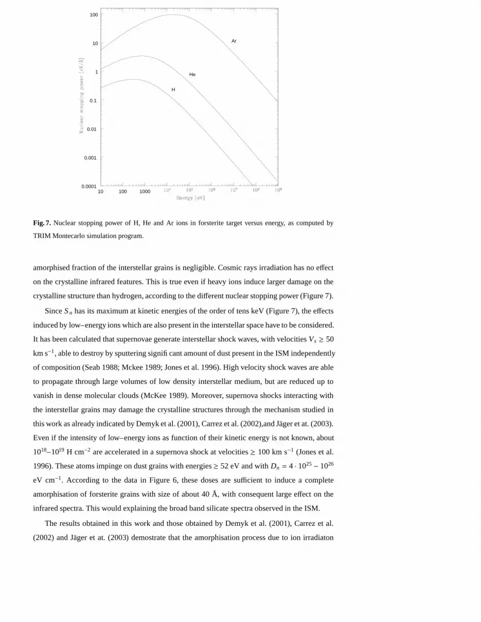
For forsterite, the S n of the light H, He, and heavy Ar ions were computed by TRIM
Montecarlo simulation for kinetic energies in the range 10 eV to 1 GeV (Figure 7). The stop-
ping power increases by increasing the incident ion energy, reaches a maximum and, at higher
energies, decreases. The maximum stopping is obtained at higher energies as the mass of the ion
increases(S n(H)=0.5 eV/Å at 0.28 keV; S n(He)=3.4 eV/Å at 0.7 keV; S n(Ar)=96.2 eV/Å at 20
keV). However, over the whole energy range, the stopping is larger of about 2 orders of magni-
tude for argon with respect to hydrogen. This means that about 1 % of heavier elements produces
the same structural effects than hydrogen on forsterite.
5. Astrophysical application
To study if ion irradiation in space is efficient to amorphise forsterite it is necessary to know
the flux in space of the ions crossing the grains according to their energies. The intensity of
cosmic rays is relatively well known at energies exceeding a few GeV, but becomes increasingly
uncertain at lower energies. The intensity measured on Earth understimates the lower–energy
particles which are sweeped back out into the interstellar space due to the presence of the solar
magnetic field.
Measures of cosmic rays intensities for nuclei of various charges were made by different
authors and extrapolated down to energies as low as few tens of MeV per nucleon. The minimum
cosmic ray flux in interstellar space was evaluated by Spitzer and Tomasko (1968) and converted
to proton densities by using the relative abundances of particles given by Webber (1967). The
J.R. Brucato G. Strazzulla G. Baratta and L. Colangeli: Ion amorphisation of forsterite 11
D n [x10 23 eV cm -3 ]
0 20 40 60 80 100 120 140
F c
0.0
0.2
0.4
0.6
0.8
1.0 He + 30 keV H + 30 keV C + 30 keV Ar ++ 60 keV Fit He + 4 keV (a) H + 1.5 MeV (b)
D e [x10 24 eV cm -3 ]
0 100 200 300 400
F c
0.0
0.2
0.4
0.6
0.8
1.0
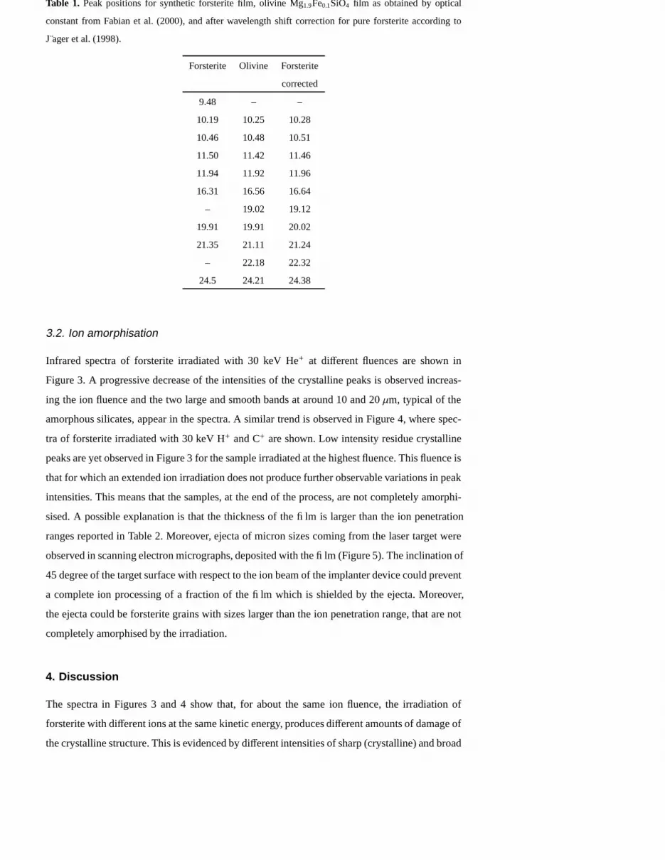
Fig. 6. In the top panel the evolution of forsterite crystalline fraction, Fc , versus nuclear (elastic) doses, Dn
is reported. The thin line represents the fit by the decreasing exponential law Fc = 0.21 + 0.74 · exp(−2.2 ·
10−24· Dn) to the evolution of the crystalline fraction of forsterite, Fc , versus nuclear dose, Dn, for different
ion irradiations. The evolution of forsterite crystalline fraction, Fc, versus electronic (anelastic) doses, De,
for different ion irradiations in the bottom panel is reported. Values deduced from the experiments of (a)
Demyk et al. (2001) and (b) Day (1977) are also reported.
analytical form of the lower limit of cosmic ray intensity adopted by Spitzer and Tomasko (1968)
is:
J(E) =0.90
(0.85 + EG)2.6
1(1 + 0.01/EG)
particles cm−2 sec−1 sterad−1 (3)
where EG is the kinetic energy in unit of 109 eV. This equation is applicable in the range of
kinetic energies for nucleon of 10 MeV–10 GeV. Assuming that the time, t, spent by a grain
of radius a = 0.1µm in the interstellar medium is 107− 108 yr, using the cross–section for the
amorphisation by ion irradiation of forsterite, derived in this work, the fraction of forsterite grains
that is amorphised by high–energy ions can be written as Fc = 1 − exp(−4π2a2tkJ(E)S n). For
proton with energies larger then 10 MeV, S n is less then 6 · 10−4 eV Å−1(Figure 7); therefore, the
12 J.R. Brucato G. Strazzulla G. Baratta and L. Colangeli: Ion amorphisation of forsterite
10 100 10000.0001
0.001
0.01
0.1
1
10
100
Ar
He
H
Fig. 7. Nuclear stopping power of H, He and Ar ions in forsterite target versus energy, as computed by
TRIM Montecarlo simulation program.
amorphised fraction of the interstellar grains is negligible. Cosmic rays irradiation has no effect
on the crystalline infrared features. This is true even if heavy ions induce larger damage on the
crystalline structure than hydrogen, according to the different nuclear stopping power (Figure 7).
Since S n has its maximum at kinetic energies of the order of tens keV (Figure 7), the effects
induced by low–energy ions which are also present in the interstellar space have to be considered.
It has been calculated that supernovae generate interstellar shock waves, with velocities Vs ≥ 50
km s−1, able to destroy by sputtering significant amount of dust present in the ISM independently
of composition (Seab 1988; Mckee 1989; Jones et al. 1996). High velocity shock waves are able
to propagate through large volumes of low density interstellar medium, but are reduced up to
vanish in dense molecular clouds (McKee 1989). Moreover, supernova shocks interacting with
the interstellar grains may damage the crystalline structures through the mechanism studied in
this work as already indicated by Demyk et al. (2001), Carrez et al. (2002),and Jager et at. (2003).
Even if the intensity of low–energy ions as function of their kinetic energy is not known, about
1018–1019 H cm−2 are accelerated in a supernova shock at velocities ≥ 100 km s−1 (Jones et al.
1996). These atoms impinge on dust grains with energies ≥ 52 eV and with Dn = 4 · 1025− 1026
eV cm−1. According to the data in Figure 6, these doses are sufficient to induce a complete
amorphisation of forsterite grains with size of about 40 Å, with consequent large effect on the
infrared spectra. This would explaining the broad band silicate spectra observed in the ISM.
The results obtained in this work and those obtained by Demyk et al. (2001), Carrez et al.
(2002) and Jager et at. (2003) demostrate that the amorphisation process due to ion irradiaton
J.R. Brucato G. Strazzulla G. Baratta and L. Colangeli: Ion amorphisation of forsterite 13
may be an efficient process in space and it is able to explain the presence of amorphous silicates
in the ISM. In this work it has been shown that the amorphisation process of forsterite dependens
on the nuclear elastic collisions between the impinging ions and the target atoms. In contrast,
no correlations has been obtained between the irradiation effects on the silicate structure and
the anelastic electronic collitions. This result shows that the process of damaging in the case
of forsterite acts differently than on water ice, for which a dependence on the total (elastic +
anelastic) dose was observed (Leto & Baratta 2003). Large effects on the silicate infrared spectral
features are produced by irradiation of low–energy ions that could be responsible of the absence
of crystalline peaks in the ISO spectra of silicate dust in the ISM.
Acknowledgements. We are grateful to F. Spinella for the technical support given during the ion irradiation
experiments. We would like to thank S. Inarta for his collaboration for the SEM analysis. This research has
been supported by the MIUR.
References
Brucato, J. R., Colangeli, L., Mennella, V., Palumbo, P., Bussoletti, E. 1999, A&A, 348, 1012
Brucato, J. R., Mennella, V., Colangeli, L., Rotundi, A., Palumbo, P. 2002, PSS, 50, 829
Carrez, P., Demyk, K., Cordier, P., et al. 2002, Meteor. & Planet. Sci., 37, 1599
Chihara, H., Koike, C., Tsuchiyama, A., Tachibana, S., Sakamoto, D. 2002, A&A, 391, 267
Colangeli, L., Henning, Th., Brucato, J.R., et al. 2003, A&ARev., 11, 97
Day, K. L. 1977, MNRAS, 178, 49P
Demyk, K., Jones, A. P., Dartois, E., Cox, P., d’Hendecourt, L. 1999, A&A, 394, 267
Demyk, K., Carrez, Ph., Leroux, H., et al. 2001, A&A, 368, L38
Fabian, D., J ager, C., Henning, Th., Dorschner, J., Mutschke, H. 2000, A&A, 364, 282
Fabian, D., Posch, Th., Mutschke, H., Kerschbaum, F., Dorschner, J. 2001, A& A, 373, 1125
Gail, H.P. & Sedlmayr, E. 1999, A&A, 347, 594
Gehrz, R. 1989, Sources of Stardust in the Galaxy. In Interstellar Dust, ed. L. J. Allamandola & A. G. G.
M. Tielens, Kluwer Academic Publishers, Dordrecht, IAU Symp. 135, 445
Habing, H. J. 1996, A&A Rev., 7, 97
J ager, C., Molster, F. J., Dorschner, J., et al. 1998, A&A, 339, 904
J ager, C., Fabian, D., Schrempel, F., Dorschner, J., Henning, Th., Wesch, W. 2003, A&A, 401, 57
Jones, A. P., Tielens, A. G. G. M., Hollenbach, D. J. 1996, ApJ, 469, 740
Jones, A.P. 1997, The Lifecycle of Interstellar Dust. In From Stardust to Planetesimals. ASP Conference
Series, ed. Y. J. Pendleton & A. G. G. M. Tielens, Vol. 122, 97
Kemper, F., Waters, L. B. F. M., de Koter, A., Tielens, A. G. G. M. 2001, A&A, 369, 132
Kemper, F., de Koter, A., Waters, L. B. F. M., Bouwman, J., Tielens, A. G. G. M. 2002, A& A, 384, 585
Koike, C., Shibai, H., Tuchiyama, A. 1993, MNRAS, 264, 654
Koike, C., Tsuchiyama, A., Shibai, H., et al. 2000, A&A, 363, 1115
Leto, G. & Baratta G. A. 2003, A&A, 397, 7
Li, A. & Draine, B. T. 2001, ApJ, 550, L213
McKee, C. F. 1989, ApJ, 345, 782
Mennella, V., Brucato, J. R., Colangeli, L., et al. 1998, ApJ, 496, 1058

14 J.R. Brucato G. Strazzulla G. Baratta and L. Colangeli: Ion amorphisation of forsterite
Molster, F. J., Waters, L. B. F. M., Tielens, A. G. G. M., Barlow, M. J. 2002a, A&A, 382, 184
Molster, F. J., Waters, L. B. F. M., Tielens, A. G. G. M 2002b, A&A, 382, 222
Molster, F. J., Waters, L. B. F. M., Tielens, A. G. G. M., Koike, C., Chihara, H. 2002c, A&A, 382, 241
Seab, C. G. 1988, Grain destruction and growth. In: Dust in the universe, Cambridge University Press, 303
Sogawa, H. & Kozasa, T. 1999, ApJ, 516, L33
Spitzer, L. Jr., & Tomasko, M. G. 1968, ApJ, 152, 972
Suh, K. W. 2002, MNRAS, 332, 513
Suto, H., Koike, C., Sogawa, H., et al. 2002, A&A, 389, 568
Sylvester, R. J., Kemper, F., Barlow, M.J., et al. 1999, A& A, 252, 587
Strazzulla, G., Baratta, G. A., Palumbo, M. E. 2001, AcSpe, 57, 825
Webber, W. R. 1967, AJ, 72, 836