foetal age determination and development in elephants
TRANSCRIPT
Proc. R. Soc. B (2007) 274, 323–331
doi:10.1098/rspb.2006.3738
Foetal age determination and developmentin elephants
Thomas Hildebrandt1,*, Barbara Drews1, Ann P. Gaeth2, Frank Goeritz1,
Robert Hermes1, Dennis Schmitt3, Charlie Gray4, Peter Rich4,
Wolf Juergen Streich1, Roger V. Short2,5 and Marilyn B. Renfree2
1Institute for Zoo and Wildlife Research, Alfred-Kowalke-Straße 17 10315 Berlin, Germany2Department of Zoology, and 5Faculty of Medicine, University of Melbourne, Melbourne, Victoria 3010, Australia
3Department of Agriculture, Missouri State University, Springfield, MO 65897, USA4African Lion Safari, Cambridge, Ont, Canada N1R 5S2
Published online 31 October 2006
*Autho
ReceivedAccepted
Elephants have the longest pregnancy of all mammals, with an average gestation of around 660 days, so
their embryonic and foetal development have always been of special interest. Hitherto, it has only been
possible to estimate foetal ages from theoretical calculations based on foetal mass. The recent
development of sophisticated ultrasound procedures for elephants has now made it possible to monitor
the growth and development of foetuses of known gestational age conceived in captivity from natural
matings or artificial insemination. We have studied the early stages of pregnancy in 10 captive Asian and 9
African elephants by transrectal ultrasound. Measurements of foetal crown–rump lengths have provided
the first accurate growth curves, which differ significantly from the previous theoretical estimates based on
the cube root of foetal mass. We have used these to age 22 African elephant foetuses collected during
culling operations. Pregnancy can be first recognized ultrasonographically by day 50, the presumptive yolk
sac by about day 75 and the zonary placenta by about day 85. The trunk is first recognizable by days
85–90 and is distinct by day 104, while the first heartbeats are evident from around day 80. By combining
ultrasonography and morphology, we have been able to produce the first reliable criteria for estimating
gestational age and ontological development of Asian and African elephant foetuses during the first
third of gestation.
Keywords: ultrasonography; foetal growth; foetal development; Proboscidea; gestation
1. INTRODUCTIONThere are three recognized elephant species: the African
savannah elephant (Loxodonta africana); the African forest
elephant (Loxodonta cyclotis); and the Asian elephant
(Elephas maximus; Roca et al. 2001). Their gestation
length is the by far longest of all mammals (623–729 days
in the Asian and 640–673 days in the African elephant;
Sukumar 2003; Meyer et al. 2004). The Asian elephant is
regarded as an endangered species in the wild (Sukumar
2003), and captive breeding may be its only hope of long-
term survival. The lack of success with captive breeding of
either species has made it extremely difficult to study foetal
growth and development, hence research has been
dependent on African elephant foetuses of unknown
gestational age collected during culling operations which
took place from 1967 to 1995.
Gaeth et al. (1999) serially sectioned one elephant
embryo and six foetuses obtained from culling operations
in the Kruger National Park, South Africa, between 1993
and 1995. Craig’s (1984) formula was used to estimate the
age of the specimens. According to this, the embryo was at
58 days of gestation and the foetuses ranged from 97 to
166 days. This is the first account of elephant organogen-
esis, and it revealed the unique nature of the mesonephric
kidney with its nephrostomes and the reasons why the
r for correspondence ([email protected]).
14 August 200612 September 2006
323
testes do not descend into a scrotum. Based on these and
earlier findings, Gaeth et al. suggested that elephants
probably have an aquatic ancestry. This is further
supported by a subsequent study of foetal lung develop-
ment, pleural fusion and the evolution of the trunk,
which enables the elephant to snorkel (West 2001, West
et al. 2003). The earliest descriptions of placentation in the
African elephant highlighted its zonary placenta
(Amoroso & Perry 1964; Perry 1974; Allen et al. 2003),
and in an important series of papers, Allen and colleagues
have defined the unusual aspects of its placental
physiology and endocrinology (reviewed in Allen 2006).
However, in all of these studies, foetal age could only be
given as an estimate.
The only methods presently available for estimating
foetal age of the elephant are the formulae of Huggett &
Widdas (1951) and Craig (1984) based on a theoretical
linear relationship between the cube root of foetal mass
and the foetal age. The formula was tZu1/3/aCt0, where
t is the foetal age in days; u is the foetal mass in kg; a is the
specific growth velocity; and t0 is the minimal age to be
determined from the equation. Huggett & Widdas
arbitrarily assumed t0 to be one-tenth of the gestation
length. Perry (1953) determined that the ‘specific foetal
growth velocity’ for both African and Asian elephants was
0.08, given that the average weight at birth is 120 kg
and the gestation length is 22 months. Therefore, the
This journal is q 2006 The Royal Society

Table 1. Pregnant elephants monitored by transrectalultrasound.
ID
total no. of
examinations species
date of
conception
gestational
age (d )
SEa1 1 Emb 13 Jan 2004 NBc 69
SE2 1 Em 18 Jul 1994 NB 189
SE3 1 Em 19 Aug 2003 NB 216
SE4 1 Em 19 Mar 2004 AId 133
SE5 1 Lae 30 Jan 2004 NB 122
SE6 1 La 13 Aug 2001 AI 129
SE7 1 La 23 May 2001 AI 150
SE8 1 La 16 Jun 2001 AI 68
SE9 1 La 23 Aug 2001 AI 132
SE10 2 La 3 Dec 2000 AI 88–172
SE11 2 Em 22 Feb 2000 AI 97–158
SE12 3 Em 15 Apr 2995 NB 79–189
SE13 3 Em 18 Jan 1998 NB 76–130
SE14 3 La 16 Apr 2000 NB 69–110
SE15 3 Em 18 Dec 2002 NB 64–163
SE5 4 La 23 May 1999 AI 58–126
SE16 6 La 23 Jul 1999 AI 60–240
324 T. Hildebrandt et al. Elephant foetal development
elephant equation according to Huggett &Widdas was tZ120.5u1/3C66. Craig (1984) modified this equation to
give greater values for a and t0 in view of his observations
on foetuses collected during culling operations in
Zimbabwe from 1980 to 1982. Craig concluded that the
assumed specific growth velocity was too low, and he
revised the formula to tZ105u1/3C138, assuming that
prior to day 138 the foetus had a different but unknown
growth velocity. Therefore, he suggested that the Hugget &
Widdas equation be modified to tZ105u1/3K(u1/3C
0.199)K3C140, to account for a much slower nonlinear
growth during early gestation, despite not having any actual
data on foetuses younger than 138 days of gestation.
The introduction of transrectal ultrasonography
coupled with successful captive breeding programmes
has opened up a whole new way of studying the
reproductive biology of elephants (Hildebrandt et al.
1998). We have been able to produce accurate growth
curves for Asian and African elephant foetuses, and follow
their development from implantation until 240 days of
gestation.
SE17 14 Em 4 Apr 2004 NB 80–234SE18 35 Em 10 Jul 2004 NB 85–226
SE19 56 Em 27 Oct 2004 NB 47–201
a SE, scanned elephant. b Em, Elephas maximus. c NB, naturalbreeding. d AI, artificial insemination. e La, Loxodonta africana.
2. MATERIAL AND METHODS
(a) Fixed elephant specimens
Six formalin-fixed foetuses from the Kruger National Park
Museum (water bath, WB1–6) measured in 1997, 15 African
elephant foetuses (elephant foetus, EF1–15) collected during
culling operations in the Kruger National Park in 1974, 1993
and 1995 and an additional foetus (EF16) collected in 1967
from a cull in the Luangwa Valley, Zambia were used in the
study. The specimens were all photographed, weighed and
relevant morphological measurements such as crown–rump
length (CRL, distance from the vertex of the skull to the base
of the tail) were taken. Specimens WB1–6 were then
sonographically assessed in a water bath (3.5–10 MHz,
Hitachi EUB 495) and the ultrasound scans were recorded
on S-VHS tape and analysed.
(b) Pregnant elephants
In the period from 1995 to 2005, 9 captive African elephants
(La) and 10 captive Asian elephants (Em) were examined
(scanned elephants, SE1–19; table 1). In the course of
artificial insemination (AI) programmes, the hormone
profiles together with frequent ultrasound examinations
allowed for the exact timing of ovulation. Rupturing of the
leading Graafian follicle was expected 12 h after the second
luteinizing hormone (LH) peak. Growth of the Graafian
follicle as well as its disappearance were detected sonogra-
phically. AI was performed in most cases two to three times
when the Graafian follicle was still visible and after it had
disappeared. Therefore, the date of ovulation was defined by
the absence of a previously enlarged Graafian follicle at the
time of AI (nZ7) or by the detection of a second LH peak
measured in daily blood serum samples (Kapustin et al. 1996;
Brown et al. 2004) at the time of mating. Multiple AIs were
performed owing to poor semen quality. (For most of the
performed AIs, the semen was collected at a different
institution from where the recipient elephant cow was
housed. This made long transport necessary, which had a
negative impact on the semen quality.)
Proc. R. Soc. B (2007)
(c) Ultrasonography
During the first third of pregnancy, sonographic examinations
were performed transrectally using the technique of Hildeb-
randt et al. (2000). Some elephants were in the standing
position and others in lateral recumbency, as previously
described (Hildebrandt et al. 2000).
Later in gestation, transcutaneous ultrasound was
applied (Hildebrandt et al. 2006). In most cases, portable
B-mode real-time ultrasound systems (Sonosite, Inc. USA;
EUB 459, Hitachi Medical Corporation, Japan) with a
transducer frequency range from 10.0 to 2.0 MHz were
employed. Three elephants were assessed with stationary
three-dimensional ultrasound systems (Voluson 530 and
Voluson 730, GE Medical Systems, Austria) equipped with
7.0–4.0 MHz transducers. All ultrasound examinations
were videotaped on analogue S-VHS or digital tapes for
retrospective analysis. Three-dimensional scans were stored
on magneto-optical discs.
(d) Retrospective analysis of the ultrasound data
and measurements
All ultrasound recordings (WB1–6 and SE1–19) were
analysed retrospectively. For the measurement of different
biometric parameters, such as CRL, biparietal diameter
(BPD), thoracic diameter (THD) and femur length (FL),
three-dimensional sonograms had to be created from the two-
dimensional recordings (ADOBE PREMIERE PRO 1.5, Adobe
Systems Inc., USA) before foetal structures could be
measured with a special software program (ANALYSIS v. 3.1,
Olympus, Germany).
By working with the three-dimensional technique,
biometric measurements could be derived directly from the
scans (4DView, GEMedical Systems, Austria). In the earliest
embryos, where there was no bone ossification, the length of
the embryo on its longest axis was defined as the CRL. In the
later stages, the CRL was measured as the distance from the

Table 2. Data collected from fixed African elephant (Loxodonta Africana) specimens.
ID
indirect, sonographicCRL measurement(mm)
direct CRLmeasurement(mm)
head length(mm) mass (g)
calculated agefrom formula(3.4) (days)
calculated agefrom formula(3.7) (days)
EFa1 5.4 1.5 0.04 60 60EF2 18.5 8.8 0.8 82 81EF3 19.0 9.9 1.3 82 85EF4 17.6 9.7 1.5 81 86EF5 27.5 12.7 2.6 92 92EF6 36.4 17.5 4.0 100 97EF7 39.5 15.7 6.6 103 103WBb1 30.2 33.0 11.4 7.2 97 103EF8 46.4 22.8 9.2 109 107EF9 61.4 24.9 17.0 120 115EF10 59.5 25.0 18.5 119 117WB2 64.5 66.8 20.8 24.4 123 121WB3 73.3 78.1 30.6 47.8 131 132EF11 112.4 48.7 107.0 150 147EF12 113.4 49.0 113.7 150 148EF13 116.1 50.6 114.6 152 148WB4 120.0 116.0 43.1 133.7 152 151EF14 123.2 52.3 168.6 155 156EF15 124.9 55.0 205.1 156 160WB5 140.0 146.2 50.3 288.1 166 168EF16 150.0 65.8 299.8 167 169WB6 180.0 185.5 59.0 554.3 182 184
a EF, elephant foetus.b WB, water bath.
Elephant foetal development T. Hildebrandt et al. 325
vertex of the skull to the base of the tail, as in the fixed
specimens. The CRL was preferably measured from a lateral
scan of the foetus. If that could not be achieved, the CRL was
obtained from a horizontal section. Sometimes it was not
possible to image the complete CRL of the foetus in one
frame, so the lengths of the head and the torso were
combined.
The BPD of the foetal head was measured from the outer
margin of one parietal bone to the other. The FL was defined
as the maximal length of the diaphysis. The THD was the
maximal diameter of the rib cage in the horizontal section.
(e) Statistics
Curvilinear regression analyses were used to establish
relationships between the age of the foetuses and their
morphological parameters. Analysis of covariance was used
to compare the regression lines for Asian and African
elephants. The curve fits were performed with TABLECURVE
2D v. 5 (AISN Software, Inc. Mapleton, OR, USA). All other
statistical calculations were performed using the SPSS v. 12.0
(SPSS Inc., Chicago, IL, USA) statistical software package.
3. RESULTS(a) Validation of ultrasound measurements
Data collected from foetal elephant specimens (WB1–6
and EF1–16) of different developmental stages are listed
in table 2. In order to test the reliability of the ultrasound
measurements, the CRLs of six foetuses (WB1–6) were
measured directly and indirectly by ultrasound in a water
bath. The results of the two methods did not differ
significantly (regression through the origin: CRLZ1.026!CRLultrasound, R2Z0.999, p!0.001; 95% CI of
slope: 0.999–1.1060).
Proc. R. Soc. B (2007)
(b) New mathematical models for foetal growth
using ultrasound data
Since the dates of ovulation of SE1–19 were known, each
ultrasound measurement could be assigned a known
gestational age. The CRL was an accessible parameter
from 62 to 202 days of gestation and ranged from 5.0 to
205.3 mm. It became more difficult to measure with
increasing growth of the foetus and its varying flexure.
BPD could be measured transrectally from 83 to 226 days
of gestation, and it ranged from 7.7 to 91.3 mm. THD
ranged from 10.5 to 88.0 mm between days 99 and 240.
FL was a parameter that could only be determined later in
gestation. It ranged from 6.4 to 58.0 mm and could be
reliably measured from 119 to 234 days of gestation.
Regression models were fitted to the data in order to
construct mean curves for age and CRL, BPD, THD and
FL (figure 1a). The data were examined to see if the foetal
growth rates differed between Asian and African elephants.
No significant differences in the slopes or the intercepts
were observed between Asian and African elephants (age
with CRL1/2: p(slopes)Z0.091, p(intercepts)Z0.947;
age with BPD: p(slopes)Z0.405, p(intercepts)Z0.751;
age with THD: p(slopes)Z0.522, p(intercepts)Z0.647; age with FL: p(slopes)Z0.890, p(intercepts)Z0.979). Therefore, all subsequent calculations were
performed using the combined data for both species.
The regression analyses yielded the following equations:
ageZ 78:98C1:95!BPD; R2 Z0:95;
Fð1; 105ÞZ 2195:5; p!0:001;ð3:1Þ
ageZ 85:32C1:69!THD; R2 Z 0:98;
Fð1; 76ÞZ 3640:7; p!0:001;ð3:2Þ
ageZ 113:55C2:02!FL; R2 Z0:97;
Fð1; 54ÞZ 1467:2; p!0:001:ð3:3Þ

250(a) (b)
200
150
mm
CR
L (
mm
)
100
50
0 50 100 150foetal age (days) foetal age (days)
200 250 500 100 150 200 250
Figure 1. (a) Foetal growth curves based on ultrasound measurements. The ultrasound data (SE1–19) show the relationshipbetween true gestation age and crown–rump length (CRL). Crown–rump length (open square), biparietal diameter (open uptriangle), thoracic diameter (open circle) and femur length (open down triangle) are plotted against foetal age. (b) Evaluation ofexisting models for the determination of foetal age. The foetal specimens (WB1–6 and EF1–16) have been aged using ourformula (3.4) (solid line), the formulae by Huggett/Widdas (filled down triangle) and Craig for under 138 days (open uptriangle) and Craig for over 140 days (filled up triangle), respectively.
326 T. Hildebrandt et al. Elephant foetal development
The relationship between CRL and age is nonlinear, and it
can be adequately fitted by a square root function
ageZ 35:14C10:80!CRL1=2; R2 Z 0:98;
Fð1; 114ÞZ 5936:67; p!0:001:ð3:4Þ
The 95% prediction band around the regression curve
(3.4) (not shown) is expected to enclose 95%of future data
points, thus indicating the precision of future estimations.
The prediction band boundaries are in a vertical distance
between G9.2 and G9.7 days (standard deviation (s.d.))
from the regression curve for CRLR5 mm or %213 mm.
Existing formulae for foetal age determination describe
the relationship between foetal mass and foetal age. Since
foetal mass cannot be determined for living foetuses, we
used the CRL–age data assessed by ultrasound for
comparative purposes. For this, data from foetal elephant
specimens (WB1–6 and EF1–16) comprising both foetal
CRL and foetal mass measurements were used. Foetal age
for these specimens was calculated on the basis of foetal
mass according to Huggett/Widdas (using the foetal
growth velocity as determined by Perry 1953) and Craig.
We then supplemented the CRL–age plot of these esti-
mations by the regression line of formula (3.4), which is
derived fromour ultrasound data (figure 1b). TheHuggett/
Widdas formula (using the foetal growth velocity as
determined by Perry 1953) gave ages that were approxi-
mately 20 days younger than the known age (figure 1b). In
contrast, both Craig formulae overestimated foetal age by
25–65 days, with the exception of the age calculated for
the youngest embryo using the formula developed for
younger foetuses (figure 1b). Therefore, we derived a new
formula to describe the age–mass relationship, based on
our ultrasound measurements. Using the data from all the
foetal elephant specimens (WB1-6 and EF1-16), a linear
regression of the cube root of foetal mass u (g) against
foetal CRL (mm) revealed
CRLZ 23:10!u1=3K3:28; R2 Z 0:99;
Fð1; 20ÞZ3177:7; p!0:001:ð3:5Þ
To substitute the CRL in equation (3.4) by that of
equation (3.5) would result in a complicated mass–age
formula with four different parameters. Therefore, we
Proc. R. Soc. B (2007)
estimated the age of the foetal elephant specimens from
the CRL measurements (WB1–6 and EF1–16) using
equation (3.4) in order to find a simpler age–mass
relationship (figure 1b). The formulae used by Huggett/
Widdas and Craig assume a linear relationship between
the cube root of foetal mass u and the foetal age. However,
the relationship is nonlinear. A square root function
provides an adequate fit, resulting in the equations
ageZ 28:434C54:20!ððmassÞ1=3Þ1=2;
R2 Z 0:99; Fð1;20ÞZ 3177:7; p!0:001;ð3:6Þ
or
ageZ 28:434C54:20!ðmassÞ1=6: ð3:7Þ
Thus, our formulae are accurate up to at least 202G9.7 days (s.d.).
(c) Foetal growth from observations of fixed
foetal elephant specimens
As would be expected, the CRL, head, tail and trunk
lengths increased with increasing body mass. In some
cases where foetuses were of similar mass, the heavier
foetus sometimes had shorter crown–rump, head, tail and
trunk lengths. For example, foetuses EF3 and 4 were of
similar mass—1.32 and 1.46 g, respectively—yet EF3 was
longer in CRL (19 mm compared to 17.6 mm), head
length (9.9 mm compared to 9.7 mm) and in tail length
(2 mm compared to 1.3 mm). The average length of the
hind limbs was always shorter than the average length of
the forelimbs, as is observed in adult elephants. Head
length was 28% of the CRL of EF1. This increased with
age to 55% of EF4 and then decreased with age to plateau
at around 42–44%.
(d) Developmental milestones related to foetal age
Sonographic imaging of foetuses of known age, coupled
with the description of the gross morphology of foetal
specimens with the calculated age according to equation
(3.4), have allowed the definition of ontogenetic mile-
stones for the elephant during the first 167 days of
gestation (table 3). On day 46, there was evidence of
constrained, oval-shaped fluid accumulation within the
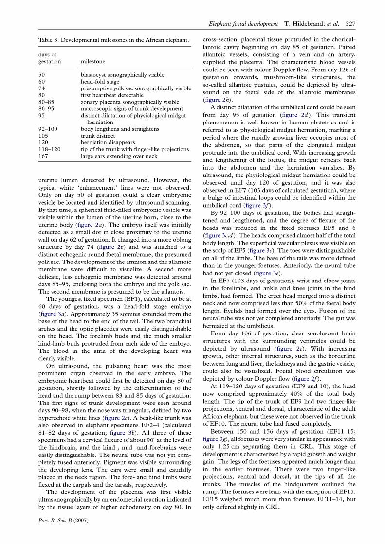
Table 3. Developmental milestones in the African elephant.
days ofgestation milestone
50 blastocyst sonographically visible60 head-fold stage74 presumptive yolk sac sonographically visible80 first heartbeat detectable80–85 zonary placenta sonographically visible86–95 macroscopic signs of trunk development95 distinct dilatation of physiological midgut
herniation92–100 body lengthens and straightens105 trunk distinct120 herniation disappears118–120 tip of the trunk with finger-like projections167 large ears extending over neck
Elephant foetal development T. Hildebrandt et al. 327
uterine lumen detected by ultrasound. However, the
typical white ‘enhancement’ lines were not observed.
Only on day 50 of gestation could a clear embryonic
vesicle be located and identified by ultrasound scanning.
By that time, a spherical fluid-filled embryonic vesicle was
visible within the lumen of the uterine horn, close to the
uterine body (figure 2a). The embryo itself was initially
detected as a small dot in close proximity to the uterine
wall on day 62 of gestation. It changed into a more oblong
structure by day 74 (figure 2b) and was attached to a
distinct echogenic round foetal membrane, the presumed
yolk sac. The development of the amnion and the allantoic
membrane were difficult to visualize. A second more
delicate, less echogenic membrane was detected around
days 85–95, enclosing both the embryo and the yolk sac.
The second membrane is presumed to be the allantois.
The youngest fixed specimen (EF1), calculated to be at
60 days of gestation, was a head-fold stage embryo
(figure 3a). Approximately 35 somites extended from the
base of the head to the end of the tail. The two branchial
arches and the optic placodes were easily distinguishable
on the head. The forelimb buds and the much smaller
hind-limb buds protruded from each side of the embryo.
The blood in the atria of the developing heart was
clearly visible.
On ultrasound, the pulsating heart was the most
prominent organ observed in the early embryo. The
embryonic heartbeat could first be detected on day 80 of
gestation, shortly followed by the differentiation of the
head and the rump between 83 and 85 days of gestation.
The first signs of trunk development were seen around
days 90–98, when the nose was triangular, defined by two
hyperechoic white lines (figure 2c). A beak-like trunk was
also observed in elephant specimens EF2–4 (calculated
81–82 days of gestation; figure 3b). All three of these
specimens had a cervical flexure of about 908 at the level of
the hindbrain, and the hind-, mid- and forebrains were
easily distinguishable. The neural tube was not yet com-
pletely fused anteriorly. Pigment was visible surrounding
the developing lens. The ears were small and caudally
placed in the neck region. The fore- and hind limbs were
flexed at the carpals and the tarsals, respectively.
The development of the placenta was first visible
ultrasonographically by an endometrial reaction indicated
by the tissue layers of higher echodensity on day 80. In
Proc. R. Soc. B (2007)
cross-section, placental tissue protruded in the chorioal-
lantoic cavity beginning on day 85 of gestation. Paired
allantoic vessels, consisting of a vein and an artery,
supplied the placenta. The characteristic blood vessels
could be seen with colour Doppler flow. From day 126 of
gestation onwards, mushroom-like structures, the
so-called allantoic pustules, could be depicted by ultra-
sound on the foetal side of the allantoic membranes
(figure 2h).
A distinct dilatation of the umbilical cord could be seen
from day 95 of gestation (figure 2d ). This transient
phenomenon is well known in human obstetrics and is
referred to as physiological midgut herniation, marking a
period where the rapidly growing liver occupies most of
the abdomen, so that parts of the elongated midgut
protrude into the umbilical cord. With increasing growth
and lengthening of the foetus, the midgut retreats back
into the abdomen and the herniation vanishes. By
ultrasound, the physiological midgut herniation could be
observed until day 120 of gestation, and it was also
observed in EF7 (103 days of calculated gestation), where
a bulge of intestinal loops could be identified within the
umbilical cord (figure 3f ).
By 92–100 days of gestation, the bodies had straigh-
tened and lengthened, and the degree of flexure of the
heads was reduced in the fixed foetuses EF5 and 6
(figure 3c,d ). The heads comprised almost half of the total
body length. The superficial vascular plexus was visible on
the scalp of EF5 (figure 3c). The toes were distinguishable
on all of the limbs. The base of the tails was more defined
than in the younger foetuses. Anteriorly, the neural tube
had not yet closed (figure 3e).
In EF7 (103 days of gestation), wrist and elbow joints
in the forelimbs, and ankle and knee joints in the hind
limbs, had formed. The erect head merged into a distinct
neck and now comprised less than 50% of the foetal body
length. Eyelids had formed over the eyes. Fusion of the
neural tube was not yet completed anteriorly. The gut was
herniated at the umbilicus.
From day 106 of gestation, clear sonoluscent brain
structures with the surrounding ventricles could be
depicted by ultrasound (figure 2e). With increasing
growth, other internal structures, such as the borderline
between lung and liver, the kidneys and the gastric vesicle,
could also be visualized. Foetal blood circulation was
depicted by colour Doppler flow (figure 2f ).
At 119–120 days of gestation (EF9 and 10), the head
now comprised approximately 40% of the total body
length. The tip of the trunk of EF9 had two finger-like
projections, ventral and dorsal, characteristic of the adult
African elephant, but these were not observed in the trunk
of EF10. The neural tube had fused completely.
Between 150 and 156 days of gestation (EF11–15;
figure 3g), all foetuses were very similar in appearance with
only 1.25 cm separating them in CRL. This stage of
development is characterized by a rapid growth and weight
gain. The legs of the foetuses appeared much longer than
in the earlier foetuses. There were two finger-like
projections, ventral and dorsal, at the tips of all the
trunks. The muscles of the hindquarters outlined the
rump. The foetuses were lean, with the exception of EF15.
EF15 weighed much more than foetuses EF11–14, but
only differed slightly in CRL.

EvEm
Ys
Ea
Tr Mh
Ta
TrPc
Uv
Av
He
He
AP
FlEy
Pl
Tr
(a)
(c)
(e)
(g) (h)
( f )
(d )
(b)
Figure 2. Sonographic milestones. (a) Embryonic vesicle (Ev) at day 50. (b) An early embryo at day 74 of gestation. (c) Three-dimensional reconstruction of an embryo at 97 days of gestation with its yolk sac (Ys). The eyes (Ey) and the beak-like trunk (Tr) areclearly depicted. The placenta (Pl) is well developed. (d ) Three-dimensional reconstruction of an early foetus of 102 days of gestationshowing thephysiologicalmidgutherniation(Mh).Therightear (Ea), the typical trunk(Tr)andone fore limb(Fl)areeasy torecognize.(e)At 108days of gestation, the echogenic plexus choroideus (Pc) alreadyfillsmost of the lateral ventricle.The trunk (Tr) is positionedbetween the fore limbs. ( f )Characterization of foetal circulation at day 126 of gestation.The foetal heart (He), arteria vertebralis (Av)andumbilical vessels (Uv) are outlinedby colourDoppler flow. (g)Lateral positionof the foetus at 133days of gestation. (h) In the laterstages of pregnancy, here at day 303 of gestation, the allantoic pustules (AP) protrude into the allantoic cavity. Scale bars, 1 cm.
328 T. Hildebrandt et al. Elephant foetal development
The second largest of the fixed foetuses, EF16
(calculated 167 days of gestation), had large ears
extending back over the neck (figure 3h). On both sides
of the head, rounded masses of tissue beneath the skin
Proc. R. Soc. B (2007)
were easily identified between the eye and the ear. These
were thought to be the temporal glands. The trunk had
furrows at its base and the tip of the trunk had dorsal and
ventral finger-like processes. The chin was still identifiable

a
ts ba
h
oe
ta
fl
(a)
(c) (d )
(b)
(e) ( f )
(g) (h)
Figure 3. African savannah elephant embryo and foetuses. (a) EF1 (60 days of gestation). a, amnion; ba, branchial arches; fl, forelimb; h, heart; oe, optic evagination; s, somites; ta, tail; t, trunk. Scale bar, 0.25 cm. (b) EF4 (81 days of gestation). (c) EF5(92 days of gestation). (d ) EF6 (97 days of gestation). (e) Dorsal aspect of EF6 (100 days of gestation) showing the extent ofneural tube development. ( f ) EF7 (103 days of gestation). ( g) EF11 (150 days of gestation). (h) EF16 (167 days of gestation).Scale bars, 1 cm. Panel (a) from Gaeth et al. (1999).
Elephant foetal development T. Hildebrandt et al. 329
from the lower lip, a feature not seen in adult elephants.
The small invaginations of the mammary primordia were
between the front legs. The soles of the feet were rounded,
and there were three nails and one forming on each of the
forefeet and three nails on each of the hind feet. There
were no body or tail hairs.
Proc. R. Soc. B (2007)
After 240 days of gestation, foetuses could no longer be
visualized by transrectal ultrasound. However, trans-
cutaneous ultrasound was possible in the middle of the
second third of pregnancy. Foetal hair, heartbeat, rib
cage and skull could be seen, as well as the free allantoic
membranes and the allantoic pustules (figure 2h).

330 T. Hildebrandt et al. Elephant foetal development
4. DISCUSSIONThe present study describes the first longitudinal moni-
toring of known-age Asian and African elephant foetuses
by ultrasound. This is also the first accurate description of
the ontogenetic development of a series of elephant
foetuses in the first third of pregnancy. Foetal growth
was monitored by relating ultrasound measurements such
as CRL, BPD, THD and FL to a known foetal age. The
fact that these biometric parameters were always closely
correlated with foetal age confirms the accuracy of the
timing of conception. The date of ovulation was
determined by the disappearance of the previously
observed Graafian follicle in AI programmes or by the
detection of a second LH surge in daily blood samples
associated with mating. Owing to individual variability in
gestation lengths (G46 days in the Asian and G16 days in
the African elephant; Meyer et al. 2004), the time of
ovulation cannot be accurately inferred from the date of
birth. Observations of mating in captivity without
reference to hormonal profiles are also an unreliable
method of timing gestation (Eisenberg et al. 1971). All our
observations are critically dependent on knowing the
precise timing of the day of ovulation.
New formulae for foetal age determination have been
developed based on ultrasound measurements of CRL,
BPD, THD and FL. Developmental milestones as seen by
ultrasound were correlated with development directly
observed in fixed specimens. Pregnancy could be
confirmed by ultrasound after about 50 days of gestation,
when the blastocyst began to distend the uterine lumen.
The embryo itself was not detected until day 62 of
gestation. Between days 74 and 85, foetal membranes,
presumed to be the yolk sac and the allantois, respec-
tively, became sonographically visible. One embryo in
Amoroso & Perry’s (1964) study had a discernable yolk
sac, similar to those seen in our ultrasound images. Using
our formulae, we can now say that the 2 g (4 cm CRL)
embryo of Amoroso & Perry was approximately 103 days
old, not one to twomonths they estimated, but the yolk sac
was already regressing. Amoroso & Perry (1964) report
that in their 10 g (10 cm CRL) foetus, the yolk sac was no
longer visible and the extra-embryonic coelom was
obliterated by the fusion of the allantois with the chorion.
Comparing this 10 cm CRL foetus with the specimens of
our study, we believe that there was a typographical error
and that the 10 cm CRL foetus actually weighed 100 g.
According to our formula (3.4), which is based on the
CRL, the age of a 10 cm CRL foetus would be 143 days
and according to our formula (3.7), which is based on
mass, the 10 g foetus would be 108 days old and a 100 g
foetus would be 145 days old. Furthermore, with
ultrasound, the yolk sac is still visible at 108 days, but
they report that the yolk sac had disappeared, thus their
10 g foetus must actually weigh 100 g.
With advancing gestation, the head and rump of the
embryo became more pronounced (83 days of gestation),
shortly followed by the development of fore- and hind
limbs, ears and the characteristic elephant trunk (days
86–90of gestation),whose early appearance suggests that it
is a phylogenetically ancient trait of the elephant. Between
days 95 and 120, a dilatation of the umbilical cord close to
the abdominal wall was observed by ultrasound. This
physiological midgut herniation is similar to that seen in
developing human foetuses (Warren et al. 1989). It was also
Proc. R. Soc. B (2007)
detected in two of the fixed elephant specimens
(EF7, 103 days of gestation, and WB2, 121 days of
gestation). On days 82–100 of gestation, a lengthening
of the body and the fragmentation of the brain in fore-,
mid- and hindbrains were apparent in EF2–6. In EF7, the
eyelids had formed and by 119–120 days (EF9 and 10), the
body flexure had further reduced. The neural tube
had fused completely. If the key criteria from human
embryology are used, it appears that embryogenesis in the
elephant is completed by 100–120 days of gestation.
Subsequent development was characterized by rapid foetal
growth and further differentiation of the organs. Not
surprisingly, fine morphological details were noted earlier
in the fixed specimens of equivalent ages than in the
foetuses assessed by ultrasound.
There was a significant difference between our direct
measurements of foetal age and the previous mathematical
estimates. The Huggett & Widdas (1951) formula under-
estimated foetal age, whereas both of Craig’s (1984)
formulae overestimated foetal age. Although the slope of
all three graphs is very similar, the starting point differs.
The Huggett/Widdas and Craig formulae are dependent
on a hypothetical intercept with the time axis, which does
not reflect actual foetal growth. With our newly developed
formulae (3.1)–(3.7), gestational age can be accurately
determined for at least 202G9.7 days. We do not know the
rate at which foetal growth takes place later in gestation.
However, it is known from other mammalian species that
growth curves based on the CRL are S-shaped (Evans &
Sack 1973). Therefore, we assume that CRL increases at a
much slower rate, while foetal mass increases more rapidly
in the last third of pregnancy.
From the observations in EF3 and 4, we know that
foetuses with similar masses can differ in relation to
crown–rump, head, tail and trunk lengths, although some
of these differences may be due to fixation artefacts. Mass
is not an ideal parameter for determining foetal age, as
mass cannot be determined in living foetuses. Therefore,
the ultrasound approach of measuring CRL makes
elephant foetal aging possible. Thus, the new formulae
(3.1)–(3.7) combined with ultrasonography provide the
first reliable criteria for recording gestational age and
ontogeny of the elephant foetus.
We thank Ian Whyte, Kruger National Park, for his assistancewith the collection of the elephant foetuses from culledanimals. We also thank the staff at the different elephantkeeping institutions for their great support. The DAAD(Deutscher Akademischer Austauschdienst) provided finan-cial support for the ultrasound assessments of three pregnantelephants at the African Lion Safari, Canada.
REFERENCESAllen, W. R. 2006 Ovulation, pregnancy, placentation and
husbandry in the African elephant (Loxodonta africana).Phil. Trans. R. Soc. B 361, 821–834. (doi:10.1098/rstb.2006.1831)
Allen, W. R., Mathias, S., Wooding, F. B. & van Aarde, R. J.2003 Placentation in the African elephant (Loxodontaafricana): II Morphological changes in the uterus andplacenta throughout gestation. Placenta 24, 598–617.(doi:10.1016/S0143-4004(03)00102-4)
Amoroso, E. C. & Perry, J. S. 1964 The foetal membranesand placenta of the African elephant (Loxodonta africana).Phil. Trans. R. Soc. B 248, 1–34.

Elephant foetal development T. Hildebrandt et al. 331
Brown, J. L., Walker, S. & Moeller, T. 2004 Comparativeendocrinology of cycling and non-cycling Asian (Elephasmaximus) and African (Loxodonta africana) elephants. Gen.Comp. Endocrinol. 136, 360–370. (doi:10.1016/j.ygcen.2004.01.013)
Craig,G.C.1984Foetalmass anddateof conception inAfricanelephants: a revised formula. S. Afr. J. Sci. 80, 512–516.
Eisenberg, J. F., McKay, G. M. & Jainudeen, M. R. 1971Reproductive behaviour of the Asiatic elephant (Elephasmaximus maximus L.). Behaviour 38, 193–225.
Evans, H. E. & Sack, W. O. 1973 Prenatal development ofdomestic and laboratory mammals: growth curves, externalfeatures and selected references. Anat. Histol. Embryol. 2,11–45.
Gaeth, A. P., Short, R. & Renfree, M. B. 1999 The developingrenal, reproductive, and respiratory system of the Africanelephant suggest an aquatic ancestry. Proc. Natl Acad. Sci.USA 96, 5555–5558. (doi:10.1073/pnas.96.10.5555)
Hildebrandt, T. B., Goeritz, F., Stetter, M. D., Hermes, R. &Hofman, R. R. 1998 Application of sonography invertebrates. Zoology 101, 200–209.
Hildebrandt, T. B., Goeritz, F., Pratt, N. C., Brown, J. L.,Montali, R. J., Schmitt, D. L., Fritsch, G. & Hermes, R.2000 Ultrasonography of the urogenital tract in elephants(Loxodonta africana and Elephas maximus): an importanttool for assessing female reproductive function. Zool. Biol.19, 321–332. (doi:10.1002/1098-2361(2000)19:5!321::AID-ZOO4O3.0.CO;2-K)
Hildebrandt, T. B. 2006 Reproductive and diagnostic ultra-sonography. In Biology, medicine, and surgery of elephants(ed. M. Fowler & S. Mikota), pp. 357–376, 1st edn.Ames, IA: Blackwell Publishing.
Huggett, A. S. & Widdas, W. 1951 The relationship betweenmammalian foetal weight and conception age. J. Physiol.114, 306–317.
Proc. R. Soc. B (2007)
Kapustin, N., Critzer, J., Olson, D. & Malven, P. V. 1996
Nonluteal estrous cycles of 3-week duration are initiated
by anovulatory luteinizing hormone peaks in African
elephants. Biol. Reprod. 55, 1147–1154. (doi:10.1095/
biolreprod55.5.1147)
Meyer, J. M., Walker, S., Freeman, E. W., Steinetz, B. G. &
Brown, J. L. 2004 Species and fetal gender effects
on endocrinology of pregnancy in elephants. Gen. Comp.
Endocrinol. 138, 263–270. (doi:10.1016/j.ygcen.2004.
06.010)
Perry, J. S. 1953 The reproduction of the African elephant,
Loxodonta africana. Phil. Trans. R. Soc. B 237, 11–149.
Perry, J. S. 1974 Implantation, foetal membranes and early
placentation of the African elephant, Loxodonta africana.
Phil. Trans. R. Soc. B 269, 109–135.
Roca, A. L., Georgiadis, N., Pecon-Slattery, J. & O’Brien,
S. J. 2001 Genetic evidence for two species of elephant in
Africa. Science 293, 1473–1477. (doi:10.1126/science.
1059936)
Sukumar, R. 2003 The living elephants: evolutionary ecology,
behaviour and conservation, p. 478. Oxford, UK: Oxford
University Press.
Warren, W. B., Timor-Tritsch, I., Peisner, D. B., Raju, S. &
Rosen, M. G. 1989 Dating the early pregnancy by
sequential appearance of embryonic structures. Am.
J. Obstet. Gynecol. 161, 747–753.
West, J. B. 2001 Snorkel breathing in the elephant explains
the unique anatomy of its pleura. Respir. Physiol. 126, 1–8.
(doi:10.1016/S0034-5687(01)00203-1)
West, J. B., Zhenxing, F., Gaeth, A. P. & Short, R. V. 2003
Fetal lung development in the elephant reflects the
adaptations required for snorkelling in adult life. Respir.
Physiol. Neurobiol. 138, 325–333. (doi:10.1016/S1569-
9048(03)00199-X)