fas ligand-dependent inflammatory regulation in acute myocarditis induced by trypanosoma cruzi...
TRANSCRIPT
Immunopathology and Infectious Disease
Fas Ligand-Dependent Inflammatory Regulation inAcute Myocarditis Induced by Trypanosoma cruziInfection
Gabriel Melo de Oliveira, Rafaela Lopes Diniz,Wanderson Batista, Marcelo Meuser Batista,Cristiane Bani Correa,Tania Cremonini de Araujo-Jorge,and Andrea Henriques-PonsFrom the Fundacao Oswaldo Cruz, Instituto Oswaldo Cruz,
Departamento de Ultra-estrutura e Biologia Celular,
Laboratorio de Biologia Celular, Rio de Janeiro, Brazil
Fas/Fas ligand (Fas-L) engagement, a potent inducerof apoptosis, is also important for cellular activation,regulation of effector and chemotactic activity, andsecretion of chemokines and cytokines. We evaluatedthe relevance of Fas/Fas-L in the regulation of myo-carditis induced by Trypanosoma cruzi infection andobserved that in Fas-L�/� mice (gld/gld), cardiac in-filtration was significantly reduced, accordinglyshowing less cardiomyocyte destruction. Fluores-cence-activated cell sorting analysis of cardiac inflam-matory cells showed higher numbers of CD8� T cellsin BALB/c compared with gld/gld mice but similarlevels of lymphocyte function-associated antigen-1,intercellular adhesion molecule, CD2, and CD69 ex-pression; MAC-1� myeloid cells and mast cells wereincreased in BALB/c mice, whereas gld/gld mice ex-hibited an enrichment of CD4�/low T cells. Intracellu-lar labeling of cytokines revealed no clear cardiacskewing of Th1 or Th2 responses, but we found ahigher number of interleukin-10� cells in gld/gldmice and a deficient expression of vascular cell adhe-sion molecule-1 on cardiac endothelial cells in gld/gldmice. Finally, we found a population of CD3� butCD4/CD8 double negative cardiac T cells in bothgroups of infected mice, but down-regulation of someadhesion molecules and surface receptors was onlyobserved in gld/gld mice, indicating a targeted T-cellpopulation mostly affected by the lack of Fas-L engage-ment. These results point to a role for myocarditis reg-ulation by Fas/Fas-L beyond its possible direct rele-vance in cellular death. (Am J Pathol 2007, 171:79–86;DOI: 10.2353/ajpath.2007.060643)
Lymphocyte death triggered by antibody-inducedcrosslinked surface Fas molecules was described almost20 years ago.1 Since then, many groups have demon-strated the enormous importance of this molecular interac-tion, as well as downstream adaptor molecules, in embry-onic tissue remodeling,2 tumor surveillance,3 autoimmunity,and control of acquired immune responses.4,5 All thesefindings strengthened the notion that Fas/Fas-L (Fas ligand)engagement is necessary for regulated and physiologicalapoptosis in a number of systems. Although Fas signalshave become inextricably associated to cell death, it is nowclear that Fas-triggering induces cellular and immunologi-cal responses far beyond its relevance in apoptosis.6 Fascan induce cell activation, proliferation, differentiation, se-cretion of cytokines and chemokines, recruitment of inflam-matory cells, cell survival, and more.7
Fas is a member of the tumor necrosis factor receptorsuperfamily of cytokine receptors that triggers intracellu-lar signals through the death domain formed after en-gagement of two adjacent trimeric Fas-L.8 Intracellulareffects of Fas engagement are mediated by a cascade ofadaptor and effector molecules.7 FADD (Fas-associateddeath domain) couples Fas to oligomers of caspase 8, aswell as caspase 10, for autocatalytic activation and celldeath. However, FADD is associated to not only apopto-sis. Activation-induced T-cell proliferation, thymic matu-ration in an age-dependent manner, and B-cell develop-ment are defective in the absence of FADD.9 Apparently,this balance of proliferation/activation � apoptosis isbased in a single serine amino acid in the position 191,because this punctual mutation abrogates its role in pro-liferation without impairing its apoptotic function.10 Otherintermediate molecules in the Fas-based pathways havealso been depicted as nonexclusive apoptotic players,6
such as Daxx and RIP. These molecules trigger signal
Supported by Fundacao Oswaldo Cruz and the Brazilian agency CNPq.
Accepted for publication March 20, 2007.
Address reprint requests to Andrea Henriques-Pons, Fundacao Os-waldo Cruz/Instituto Oswaldo Cruz, Departamento de Ultra-estrutura eBiologia Celular (DUBC), Laboratorio de Biologia Celular, Rio de Janeiro(RJ); Brasil, Av. Brasil 4365, Manguinhos, 21045-900. E-mail: [email protected].
The American Journal of Pathology, Vol. 171, No. 1, July 2007
Copyright © American Society for Investigative Pathology
DOI: 10.2353/ajpath.2007.060643
79
transduction pathways based in nuclear factor-�B, extra-cellular signal-regulated kinase, or mitogen-activatedprotein kinase kinase for proliferation, transcription induc-tion or repression, differentiation, and survival.7
In the case of inflammatory regulation, recent dataindicate that acute inflammatory response is either pre-vented or attenuated after blockade of Fas/Fas-L. Al-though there are some controversial data regarding theprimary importance of soluble Fas-L (sFas-L) versusmembrane Fas-L (mFas-L),11,12 it is clear that Fas-basedintracellular signaling is important for secretion of inter-leukin (IL)-1�, IL-6, IL-17, IL-18, macrophage inflamma-tory protein MIP-2, MIP-1�, MIP-1�, IL-8, and chemotaxisof neutrophils.13 For example, in vitro experimentsshowed that sFas-L required a CD8� memory T cell [alsoexpressing natural killer (NK) cell markers] to recruit in-flammatory cells. These cells were not conventional NK/Tcells but were necessary for CC and CXC chemokinesecretion and Fas-L-dependent neutrophil recruitment.11
On the other hand, in vivo approaches have indicated thatmFas-L and not sFas-L is responsible for its inflammatoryactivity.12
Mice bearing a disrupted gene for Fas (lpr) or Fas-L(gld) have attenuated neutrophilic infiltration and fibro-sis,14 with lessened aggressive idiopathic pulmonary fi-brosis, silicosis, and acute lung injury.15 Besides this,local administration of adenovirus carrying the Fas-Lgene into an inflamed joint protects or reduces the se-verity of systemic collagen-induced arthritis, where auto-reactive T cells are recruited by Fas-Lhigh cells and de-stroyed.16 In the case of severe myocarditis induced bycoxsackievirus B3, C3H/HE mice treated with anti-Fas-L,C3H/HE-lpr/lpr, or C3H/HE-gld/gld mice showed reducedinflammatory infiltration.17–19
Based on these results, we evaluated the importanceof Fas/Fas-L engagement in the regulation of myocarditisinduced by Trypanosoma cruzi infection. The disease ischaracterized by an initial acute phase with trypomasti-gote forms in the blood and acute myocarditis,20,21 fol-lowed by a silent, indeterminate period with subpatentlevels of parasitemia. Approximately 30% of infected hu-man subjects progress to a severe, diffuse, and fatalchronic myocardiopathy.22 It is mostly accepted thatmononuclear cells, mainly T lymphocytes, are responsi-ble for cardiomyocyte destruction and heart failure in anantigen-dependent manner. However, autoimmunity to-ward cardiac components23 and microvascular lesionshave also been purposed to be involved in the physio-pathology of chagasic heart disease.24
In the last few years, many attempts have been madeto characterize the phenotype of cardiac inflammatorycells and molecular mechanisms involved in cellular re-cruitment and cytotoxic events in T. cruzi infection. Thepredominance of CD8� T cells in cardiac inflammatoryfoci has been reported by different groups,25 especiallyin the chronic phase, but only recently has the phenotypeof these cells been assessed by flow cytometry. CardiacCD8� T cells have been discerned as L-selectinlow
(CD62-Llow), leukocyte function-associated antigen 1high
(LFA-1high), and very late antigen 4high (VLA-4high).26
These cells do express activated/memory cell surface
markers (CD44�/CD11a�) in the chronic phase but havesharply attenuated effector functions, as ascertained bycytotoxic assays and production of interferon-� (IFN-�).27
This may be, at least in part, one of the mechanisms bywhich T. cruzi persists in the heart tissue, causing along-lasting myocarditis.
A predominant Th1 response is of central importanceto controlling parasitism and mounting a protective im-mune response in T. cruzi infection.28 In the early acutephase, activated macrophages secrete inflammatory cy-tokines, especially tumor necrosis factor-� and IL-12,which in turn stimulate the secretion of IFN-� by NKcells,29 although CD4� and CD8��� T cells are alsoimportant sources of IFN-�.30 Control of acute inflamma-tion in nonlymphoid tissues and avoidance of secondarydamage are believed to be based on a subsequent waveof anti-inflammatory cytokines, such as transforminggrowth factor-� and IL-10, but also IL-4.31
The particular aspect relating to cytotoxic cells andmolecular pathways directed against cardiomyocytes isone of the most obscure points of the infection. Perforin-deficient mice infected with T. cruzi exhibited cardiaccellular inflammatory infiltration and cardiomyocyte de-struction much more intensely than infected counter-parts.32 These data indicate that perforin has an impor-tant role in the regulation of inflammatory response butnot in the direct destruction of cardiac cells.
Our present results using Fas-L-deficient mice indicatefor the first time that this molecule, just as perforin, playsa role in the regulation of cardiac inflammatory responseinduced by T. cruzi infection. We observed reduced myo-carditis in gld/gld mice, as well as an enrichment of car-diac IL-10-producing cells and weak expression of vas-cular cell adhesion molecule (VCAM)-1 on endothelialcells. We observed fewer cardiac CD8� T cells, MAC-1�
myeloid cells, and mast cells in gld/gld mice and a tar-geted T-cell population CD3�/CD4�/CD8� that was phe-notypically down-regulated by the absence of Fas-L. Therefined balance between pro- and anti-inflammatory me-diators, now including Fas/Fas-L, is possibly the key tothe control of the infection and maintenance of asymp-tomatic status.
Materials and Methods
Mice
Male, specific pathogen-free Fas-L�/� (gld/gld) mice (6to 7 weeks old) and their counterparts isogenic BALB/c,perforin�/� (C57Bl/6 background), and C57Bl/6 micewere purchased from the Breeding Laboratory AnimalCenter at Fundacao Oswaldo Cruz. Mice were housed for7 to 10 days in the Laboratory of Cellular Biology, Divisionof Animal Experimentation, under environmental factorsand sanitation conforming to the guide for the Care andUse of Laboratory Animals. This project was approved byFiocruz Committee of Ethics in Research (0099-01), ac-cording to resolution 196/96 of the National Health Coun-cil of Brazilian Ministry of Health.
80 Oliveira et alAJP July 2007, Vol. 171, No. 1

Parasites and Infection
T. cruzi Y strain was maintained in vivo by passage innonsyngeneic Swiss Webster mice. Infected blood wascollected, and trypomastigote forms were isolated aspreviously described.33 Thereafter, parasites were di-luted in saline and counted in a hemacytometer to adjustthe inocula to 1 � 103 parasites per 200 �l for intraperi-toneal injection in both groups of mice. Noninfected micereceived 200 �l of saline.
Parasitemia, Mortality, and Blood Samples
Individual parasitemia was counted in 5 �l of blood col-lected from tail snips on indicated days postinfection(dpi), and mortality was scored daily. For plasma analy-sis, 100 �l of blood samples were also collected from tailsnips in heparinized capillaries and centrifuged. For cre-atine kinase-MB activity (CK-MB, the cardiac isoform ofCK), plasma from noninfected and infected BALB/c andgld/gld mice was collected on 0, 8, and 15 dpi andanalyzed using commercially available kits (Merck KGaA,Darmstadt, Germany) as described elsewhere.34 Thisquantitative assay is used as a marker of cardiomyocytesdamage, and it is expressed as a rate of NADPH increase(�E/min) in seven sequential readings in a spectropho-tometer (Molecular Devices, Sunnyvale, CA) at 340 nm.
Histopathological Analysis
Mice were euthanized with CO2 on 0, 8, and 15 dpi,tissue fragments were fixed in Millonig-Rosman solution(10% formaldehyde in phosphate-buffered saline), andparaffin-embedded samples were further processed andstained with hematoxylin and eosin in 3-�m-thick slices.The number of inflammatory foci (composed by �10inflammatory cells), inflammatory cells per foci, T. cruziparasite nests, and number of parasites per nest weredetermined by scanning the whole-tissue slice in a totalof around 300 individual microscopic fields for each pa-rameter and time point (one field � 0.196 mm2). ForVCAM-1- or MAC-1-based immunohistochemistry, weused primary monoclonal antibodies (mAbs) (SouthernLaboratories, Birmingham, AL) and secondary goat anti-rat conjugated to horseradish peroxidase (Sigma) in1-hour incubation and revealed with 3-amino-9-ethylcar-bazole (AEC; Sigma) as described elsewhere.26 Directquantification of mast cells was performed by toluidineblue staining in cardiac slices 16-�m thick to confirmfluorescence-activated cell sorting analysis (see below).
Inflammatory Cell Harvesting
Hearts from four to five infected BALB/c and gld/gld micewere collected on 15 dpi and cut into fragments of 1 to 2mm in ice-cold phosphate-buffered saline (Sigma, St.Louis, MO). Fragments were transferred to a solution ofcollagenase type IV 0.2% (lot 51K8610, 2.6 units/mgsolid; Sigma) and submitted to four to five cycles ofenzymatic digestion of 20 minutes at 37°C. Isolated cells
were centrifuged and immediately transferred to ice-coldDulbecco’s modified Eagle’s medium (Sigma) plus 10%of fetal calf serum (Sigma) and maintained on ice. Cellu-lar debris were removed by fetal calf serum centrifugationat 25 � g for 5 minutes, and before phenotypic labeling,the cells were incubated for 20 minutes at 4°C with 10%solution of inactivated normal sheep serum to block Fcreceptors.
Flow Cytometry
For phenotypic analysis by flow cytometry, we performedfour-color labeling of the samples for 30 minutes at 4°Cwith anti-CD3 (Southern Laboratories), anti-CD8, andanti-CD4 (Southern) monoclonal antibodies and then withanti-adhesion molecules intercellular adhesion molecule(ICAM)-1 or LFA-1 (Southern Laboratories), co-receptorCD2, or early activation T-cell marker CD69. For analysisin all T-cell subpopulations (CD3�/CD4�; CD3�/CD8�
and CD3�/CD4�/CD8�), we first gated CD3� cells,opened the CD4�CD8 dot plot, and after gating all sub-populations, we performed the phenotypic determination.We also used anti-CD45R (B220) anti-�� T cells, andanti-MAC-1. All monoclonal antibodies were purchasedfrom BD Pharmingen (San Jose, CA), except when indi-cated. SSCinterm�MAC-1� were considered MAC-1�-myeloid cells (macrophages, neutrophils, and dendriticcells), and mast cells were SSChigh�MAC-1�. For cyto-kine detection, we labeled the cells with anti-CD3, fixedthe samples with formaldehyde 1% (Sigma), and perme-abilized plasma membranes with saponin 0.2% (Sigma).We then used anti-IL-2, -IL-4, -IL-10, or -IFN-�, washed,and acquired in a FACSCalibur flow cytometer (BectonDickinson, San Jose, CA). Data analysis was performedusing CellQuest software version 3.2 (Becton Dickinson).
Statistical Analysis
The Mann-Whitney nonparametric test was used to com-pare two sets of data. P values were as indicated in figurelegends. Mortality rate was evaluated according to thelog-rank test with P � 0.0002.
Results
Parasitemia was consistently similar in both BALB/c andgld/gld mice in the early acute phase after infection with Ystrain of T. cruzi (Figure 1A). This is in accordance withour previous data where we also observed equivalentlevels of parasitemia in the first 2 weeks of infection.28
We observed 20% mortality at 19 dpi in BALB/c mice and80% at 20 dpi (Figure 1B). gld/gld mice, in turn, showed75% mortality at 15 dpi and 100% at 18 dpi (Figure 1B).Although gld/gld mice died before 20 dpi, approximately20% of BALB/c mice reached the chronic phase (data notshown). We then thought that possibly, contrary to otherinflammatory models that are ameliorated in the absenceof Fas engagement, the cardiac inflammatory infiltrationinduced by T. cruzi infection was more intense in gld/gld
Fas-L Police in T. cruzi Myocarditis 81AJP July 2007, Vol. 171, No. 1
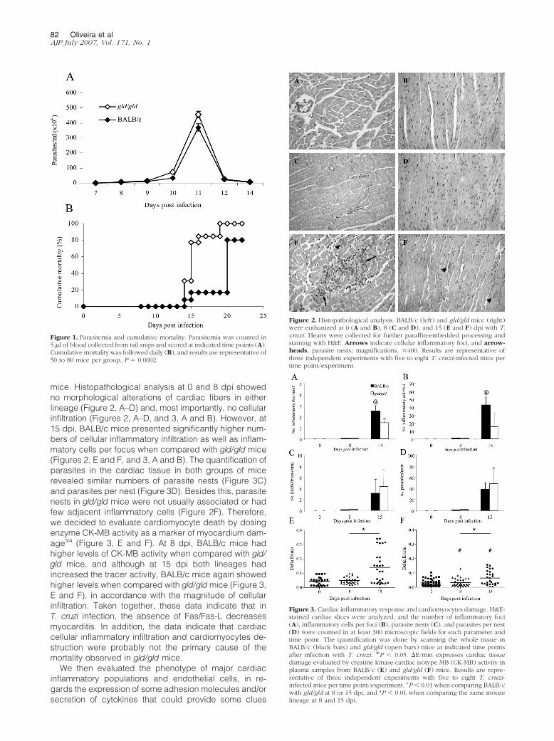
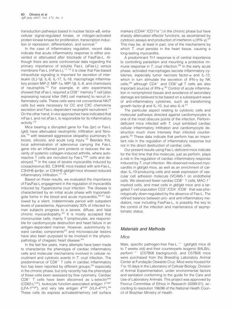
mice. Histopathological analysis at 0 and 8 dpi showedno morphological alterations of cardiac fibers in eitherlineage (Figure 2, A–D) and, most importantly, no cellularinfiltration (Figures 2, A–D, and 3, A and B). However, at15 dpi, BALB/c mice presented significantly higher num-bers of cellular inflammatory infiltration as well as inflam-matory cells per focus when compared with gld/gld mice(Figures 2, E and F, and 3, A and B). The quantification ofparasites in the cardiac tissue in both groups of micerevealed similar numbers of parasite nests (Figure 3C)and parasites per nest (Figure 3D). Besides this, parasitenests in gld/gld mice were not usually associated or hadfew adjacent inflammatory cells (Figure 2F). Therefore,we decided to evaluate cardiomyocyte death by dosingenzyme CK-MB activity as a marker of myocardium dam-age34 (Figure 3, E and F). At 8 dpi, BALB/c mice hadhigher levels of CK-MB activity when compared with gld/gld mice, and although at 15 dpi both lineages hadincreased the tracer activity, BALB/c mice again showedhigher levels when compared with gld/gld mice (Figure 3,E and F), in accordance with the magnitude of cellularinfiltration. Taken together, these data indicate that inT. cruzi infection, the absence of Fas/Fas-L decreasesmyocarditis. In addition, the data indicate that cardiaccellular inflammatory infiltration and cardiomyocytes de-struction were probably not the primary cause of themortality observed in gld/gld mice.
We then evaluated the phenotype of major cardiacinflammatory populations and endothelial cells, in re-gards the expression of some adhesion molecules and/orsecretion of cytokines that could provide some clues
Figure 2. Histopathological analysis. BALB/c (left) and gld/gld mice (right)were euthanized at 0 (A and B), 8 (C and D), and 15 (E and F) dpi with T.cruzi. Hearts were collected for further paraffin-embedded processing andstaining with H&E. Arrows indicate cellular inflammatory foci, and arrow-heads, parasite nests; magnifications, �400. Results are representative ofthree independent experiments with five to eight T. cruzi-infected mice pertime point/experiment.
Figure 3. Cardiac inflammatory response and cardiomyocytes damage. H&E-stained cardiac slices were analyzed, and the number of inflammatory foci(A), inflammatory cells per foci (B), parasite nests (C), and parasites per nest(D) were counted in at least 300 microscopic fields for each parameter andtime point. The quantification was done by scanning the whole tissue inBALB/c (black bars) and gld/gld (open bars) mice at indicated time pointsafter infection with T. cruzi. @P � 0.05. �E/min expresses cardiac tissuedamage evaluated by creatine kinase cardiac isotype MB (CK-MB) activity inplasma samples from BALB/c (E) and gld/gld (F) mice. Results are repre-sentative of three independent experiments with five to eight T. cruzi-infected mice per time point/experiment. #P � 0.01 when comparing BALB/cwith gld/gld at 8 or 15 dpi, and *P � 0.01 when comparing the same mouselineage at 8 and 15 dpi.
Figure 1. Parasitemia and cumulative mortality. Parasitemia was counted in5 �l of blood collected from tail snips and scored at indicated time points (A).Cumulative mortality was followed daily (B), and results are representative of50 to 80 mice per group, P � 0.0002.
82 Oliveira et alAJP July 2007, Vol. 171, No. 1
about the inferior gravity of cardiac inflammatory reactionin gld/gld mice. We first isolated cardiac inflammatorycells from T. cruzi-infected perforin�/� and perforin�/�
mice and observed an enrichment of CD8� (Figure 4A)over CD4� T cells in perforin�/� (Figure 4B), confirmingour previous results based on immunohistochemistry.32
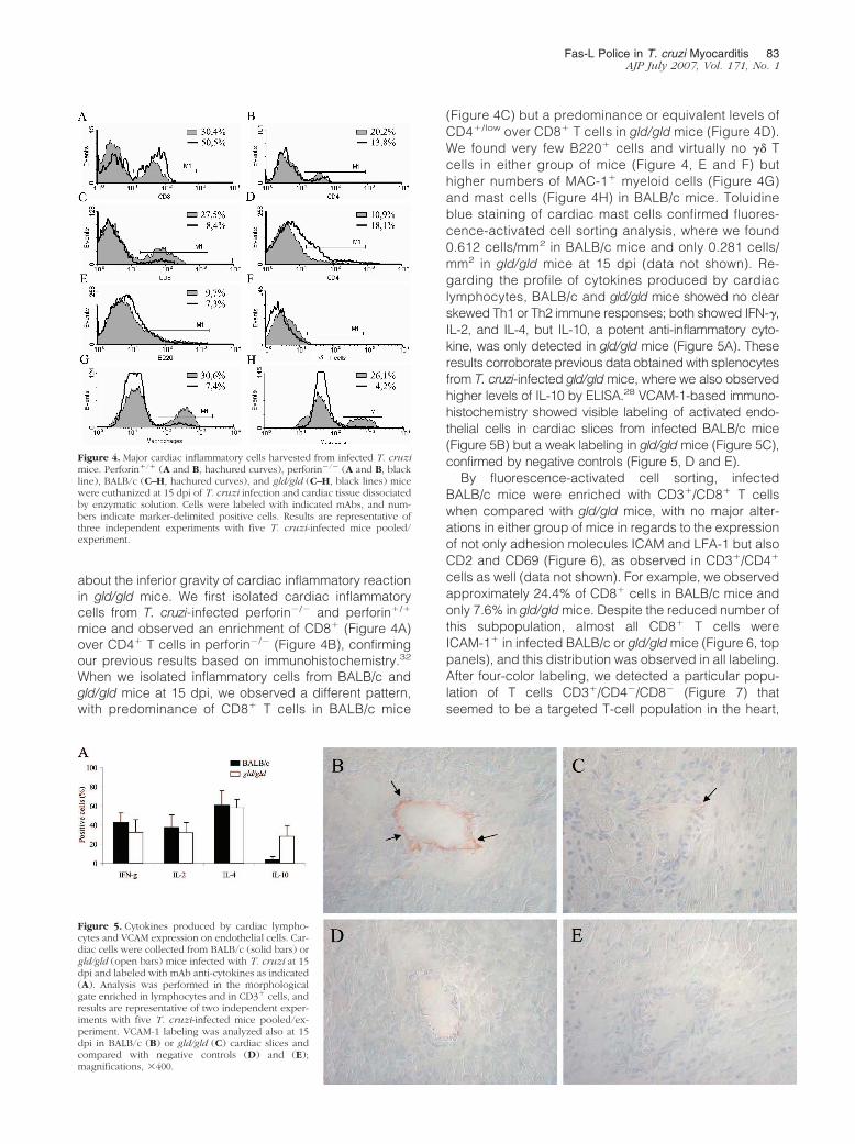
When we isolated inflammatory cells from BALB/c andgld/gld mice at 15 dpi, we observed a different pattern,with predominance of CD8� T cells in BALB/c mice
(Figure 4C) but a predominance or equivalent levels ofCD4�/low over CD8� T cells in gld/gld mice (Figure 4D).We found very few B220� cells and virtually no �� Tcells in either group of mice (Figure 4, E and F) buthigher numbers of MAC-1� myeloid cells (Figure 4G)and mast cells (Figure 4H) in BALB/c mice. Toluidineblue staining of cardiac mast cells confirmed fluores-cence-activated cell sorting analysis, where we found0.612 cells/mm2 in BALB/c mice and only 0.281 cells/mm2 in gld/gld mice at 15 dpi (data not shown). Re-garding the profile of cytokines produced by cardiaclymphocytes, BALB/c and gld/gld mice showed no clearskewed Th1 or Th2 immune responses; both showed IFN-�,IL-2, and IL-4, but IL-10, a potent anti-inflammatory cyto-kine, was only detected in gld/gld mice (Figure 5A). Theseresults corroborate previous data obtained with splenocytesfrom T. cruzi-infected gld/gld mice, where we also observedhigher levels of IL-10 by ELISA.28 VCAM-1-based immuno-histochemistry showed visible labeling of activated endo-thelial cells in cardiac slices from infected BALB/c mice(Figure 5B) but a weak labeling in gld/gld mice (Figure 5C),confirmed by negative controls (Figure 5, D and E).
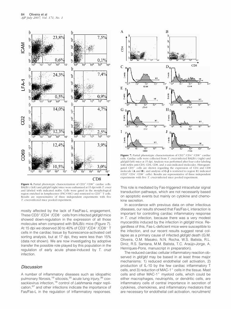
By fluorescence-activated cell sorting, infectedBALB/c mice were enriched with CD3�/CD8� T cellswhen compared with gld/gld mice, with no major alter-ations in either group of mice in regards to the expressionof not only adhesion molecules ICAM and LFA-1 but alsoCD2 and CD69 (Figure 6), as observed in CD3�/CD4�
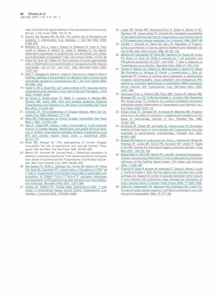
cells as well (data not shown). For example, we observedapproximately 24.4% of CD8� cells in BALB/c mice andonly 7.6% in gld/gld mice. Despite the reduced number ofthis subpopulation, almost all CD8� T cells wereICAM-1� in infected BALB/c or gld/gld mice (Figure 6, toppanels), and this distribution was observed in all labeling.After four-color labeling, we detected a particular popu-lation of T cells CD3�/CD4�/CD8� (Figure 7) thatseemed to be a targeted T-cell population in the heart,
Figure 4. Major cardiac inflammatory cells harvested from infected T. cruzimice. Perforin�/� (A and B, hachured curves), perforin�/� (A and B, blackline), BALB/c (C–H, hachured curves), and gld/gld (C–H, black lines) micewere euthanized at 15 dpi of T. cruzi infection and cardiac tissue dissociatedby enzymatic solution. Cells were labeled with indicated mAbs, and num-bers indicate marker-delimited positive cells. Results are representative ofthree independent experiments with five T. cruzi-infected mice pooled/experiment.
Figure 5. Cytokines produced by cardiac lympho-cytes and VCAM expression on endothelial cells. Car-diac cells were collected from BALB/c (solid bars) orgld/gld (open bars) mice infected with T. cruzi at 15dpi and labeled with mAb anti-cytokines as indicated(A). Analysis was performed in the morphologicalgate enriched in lymphocytes and in CD3� cells, andresults are representative of two independent exper-iments with five T. cruzi-infected mice pooled/ex-periment. VCAM-1 labeling was analyzed also at 15dpi in BALB/c (B) or gld/gld (C) cardiac slices andcompared with negative controls (D) and (E);magnifications, �400.
Fas-L Police in T. cruzi Myocarditis 83AJP July 2007, Vol. 171, No. 1
mostly affected by the lack of Fas/Fas-L engagement.These CD3�/CD4�/CD8� cells from infected gld/gld miceshowed down-regulation in the expression of all thosemolecules when compared with BALB/c mice (Figure 7).At 15 dpi we observed 30 to 40% of CD3�/CD4�/CD8� Tcells in the cardiac tissue by fluorescence-activated cellsorting analysis, but at 17 dpi, they were less than 15%(data not shown). We are now investigating by adoptivetransfer the possible role played by this population in theregulation of early acute phase-induced by T. cruziinfection.
Discussion
A number of inflammatory diseases such as idiopathicpulmonary fibrosis,19 silicosis,35 acute lung injury,36 cox-sackievirus infection,18 control of Leishmania major repli-cation,37 and other infections indicate the importance ofFas/Fas-L in the regulation of inflammatory responses.
This role is mediated by Fas-triggered intracellular signaltransduction pathways, which are not necessarily basedon apoptotic events but mainly on cytokine and chemo-kine secretion.
In accordance with previous data on other infectiousdiseases, our results showed that Fas/Fas-L interaction isimportant for controlling cardiac inflammatory responsein T. cruzi infection, because there was a very modestmyocarditis induced by the infection in gld/gld mice. Re-gardless of this, Fas-L-deficient mice were susceptible tothe infection, and our recent results suggest renal col-lapse as a primary cause of infected gld/gld death (G.M.Oliveira, O.M. Masako, N.N. Rocha, W.S. Batista, R.L.Diniz, R.S. Santana, M.M. Batista, T.C. Araujo-Jorge, A.Henriques-Pons, manuscript in preparation).
The reduced cardiac cellular inflammatory reaction ob-served in gld/gld may be based in at least three majormechanisms: 1) reduced endothelial cell activation, 2)production of IL-10 by the few cardiac inflammatory Tcells, and 3) reduction of MAC-1� cells in the tissue. Mastcells and other MAC-1� myeloid cells, which could beeither macrophages, neutrophils, or dendritic cells, areinflammatory cells of central importance in secretion ofcytokines, chemokines, and inflammatory mediators thatare necessary for endothelial cell activation, recruitment/
Figure 6. Partial phenotypic characterization of CD3�/CD8� cardiac cells.BALB/c (left) and gld/gld (right) mice were euthanized at 15 dpi with T. cruziand labeled with indicated mAbs. Cells were gated in the morphologicalregion enriched in lymphocytes (FSC�SSC) and restricted to CD3� T cells.Results are representative of three independent experiments with fiveT. cruzi-infected mice pooled/experiment.
Figure 7. Partial phenotypic characterization of CD3�/CD4�/CD8� cardiaccells. Cardiac cells were collected from T. cruzi-infected BALB/c (right) andgld/gld (left) mice at 15 dpi. Analysis was performed after four-color labelingwith mAbs anti-CD3, CD4, CD8, and x axis-indicated molecules. Histogram-gated CD3� cells are shown regarding the expression of CD4 and CD8molecule (A and B), and analysis of C–J is restricted to region R1 indicated(CD3�/CD4�/CD8� cells). Results are representative of three independentexperiments with five T. cruzi-infected mice pooled/experiment.
84 Oliveira et alAJP July 2007, Vol. 171, No. 1

activation of inflammatory cells, and destruction of intra-cellular parasites. We do not know why these cells arereduced in the cardiac tissue of infected gld/gld mice. Itcould be possible that Fas/Fas-L interaction is necessaryfor sustained expression of MAC-1 or even mast cell andother myeloid cell survival during infection. The increasein IL-10 observed in inflamed hearts, and also in thespleen,28 of gld/gld mice may be one of the major causesof reduced cellular cardiac inflammation, down-regulat-ing proinflammatory cells, as well as inhibiting endothelialcell activation and recruitment of inflammatory blood cellsin gld/gld.
We observed in both BALB/c and gld/gld mice a CD3�/CD4�/CD8� double negative (DN) T-cell population rep-resenting 30 to 40% of the CD3� T cells harvested fromcardiac tissue. Apparently, this population is present inthe heart only in the early acute phase, because at 17 dpithey were barely detected. This could explain why theyhad not yet been observed, because other groups thatisolate cardiac T cells from T. cruzi-infected mice do it atlater time points. We are now investigating the immuno-logical relevance of these cells, in regards to secretion ofcytokines and adoptive transfer. We believe that Fas/Fas-L interaction is important for a given effector and/orregulatory function, because DN cells from infected gld/gld down-regulate some important co-receptors and ad-hesion molecules for regular T cells.
It has long been known that gld/gld mice, and lpr/lprmice, have B220�/CD3� CD4 and CD8 DN cells.38 Weobserved this population in spleens of gld/gld but not inthe heart, because less than 10% were B220� cardiaccells. It may be possible that these cardiac DN cells werepreviously the B220�/CD3� population that down-regu-lated the B220� marker. However, not only infectedBALB/c mice but also Swiss Webster, C3H, and C57Bl/6have the DN population at 15 dpi (R.L.D. and A.H.-P.,manuscript in preparation). These data suggest that DNcells compose a particular population of T cells, notexpressing NK cell markers, possibly with no previouslydescribed functions in myocarditis induced by T. cruziinfection.
CD3�/CD8� and CD3�/CD4� cardiac T cells col-lected from BALB/c and gld/gld mice showed no relevantalterations in the expression of ICAM-1, LFA-1, CD2, andCD69, although with clearly less CD8� T cells in gld/gldmice. It seems that, in both groups of mice, these cellsare compatible with the phenotype of effector/activated Tcells. In addition, the enrichment of CD4�/low T cells ingld/gld mice observed in most experiments could becaused by the lack of normally observed activation-in-duced cell death of CD4� cells by Fas-L, as observed inspleens.28 This activation-induced cell death deficiency,altering that subpopulation of cardiac T cells, could becentral to controlling pathogenic cells, as observed incoxsackievirus B3 infection. In this model of myocarditis,gld/gld mice also have modest cardiac inflammatoryresponses based on selective destruction of T cells.Wild-type mice have a predominant Th1-driven immuneresponse with high levels of IFN-�, whereas Fas-L- orFas-deficient mice have a dominant CD4�/IL-4� (Th2)T-cell population. Adoptive transfer of �� T cells bearing
Fas-L to gld/gld mice and cytotoxic assays have indi-cated that cardiac subpopulation V�4� cells selectivelykill virus-specific Th2 CD4� T cells in a Fas-basedpathway, enriching the inflamed heart in pathogenicTh1 cells.17
The delicate balance between a protective inflamma-tory response, controlling parasite burden and infection,and a harmful infiltration causing secondary damage tothe tissue is certainly based on a number of cellularpopulations, soluble inflammatory mediators, and mem-brane receptors. Taken together, our results point to arelevant role of Fas/Fas-L interaction in the control ofmyocarditis induced by T. cruzi infection and add moreevidence of molecular triggering pathways that are im-portant in the control of this important pathology.
Acknowledgment
We thank Mr. Marcos Meuser Batista for excellent tech-nical assistance.
References
1. Trauth BC, Klas C, Peters AM, Matzku S, Moller P, Falk W, DebatinKM, Krammer PH: Monoclonal antibody-mediated tumor regressionby induction of apoptosis. Science 1989, 245:301–305
2. Yeh WC, Pompa JL, McCurrach ME, Shu HB, Elia AJ, Shahinian A, NgM, Wakeham A, Khoo W, Mitchell K, El-Deiry WS, Lowe SW, GoeddelDV, Mak TW: FADD: essential for embryo development and signalingfrom some, but not all, inducers of apoptosis. Science 1998,279:1954–1958
3. Abrams SI: Positive and negative consequences of Fas/Fas ligandinteractions in the antitumor response. Front Biosci 2005, 10:809–821
4. Li-Weber M, Krammer PH: Function and regulation of the CD95(APO-1/Fas) ligand in the immune system. Semin Immunol 2003,15:145–157
5. Choi C, Benveniste EN: Fas ligand/Fas system in the brain: regulatorof immune and apoptotic responses. Brain Res Brain Res Rev 2004,44:65–81
6. Wajant H: The Fas signaling pathway: more than a paradigm. Science2002, 296:1635–1636
7. Lambert C, Landau AM, Desbarats J: Fas-beyond death: a regener-ative role for Fas in the nervous system. Apoptosis 2003, 8:551–562
8. Holler N, Tardivel A, Kovacsovics-Bankowski M, Hertig S, Gaide O,Martinon F, Tinel A, Deperthes D, Calderara S, Schulthess T, Engel J,Schneider P, Tschopp J: Two adjacent trimeric Fas ligands are re-quired for Fas signaling and formation of a death-inducing signalingcomplex. Mol Cell Biol 2003, 23:1428–1440
9. Zhang J, Cado D, Chen A, Kabra NH, Winoto A: Fas-mediated apo-ptosis and activation-induced T-cell proliferation are defective in micelacking FADD/Mort1. Nature 1998, 392:296–300
10. Hua ZC, Sohn SJ, Kang C, Cado D, Winoto A: A function of Fas-associated death domain protein in cell cycle progression localizedto a single amino acid at its C-terminal region. Immunity 2003,18:513–521
11. Giroux M, Denis F: CD1d-unrestricted human NKT cells release che-mokines upon Fas engagement. Blood 2005, 105:703–710
12. Shudo K, Kinoshita K, Imamura R, Fan H, Hasumoto K, Tanaka M,Nagata S, Suda T: The membrane-bound but not the soluble form ofhuman Fas ligand is responsible for its inflammatory activity. EurJ Immunol 2001, 31:2504–2511
13. Umemura M, Kawabe T, Shudo K, Kidoya H, Fukui M, Asano M,Iwakura Y, Matsuzaki G, Imamura R, Suda T: Involvement of IL-17 inFas ligand-induced inflammation. Int Immunol 2004, 16:1099–1108
14. Kuwano K, Hagimoto N, Kawasaki M, Yatomi T, Nakamura N, NagataS, Suda T, Kunitake R, Maeyama T, Miyazaki H, Hara N: Essential
Fas-L Police in T. cruzi Myocarditis 85AJP July 2007, Vol. 171, No. 1
roles of the Fas-Fas ligand pathway in the development of pulmonaryfibrosis. J Clin Invest 1999, 104:13–19
15. Dosreis GA, Borges VM, Zin WA: The central role of Fas-ligand cellsignaling in inflammatory lung diseases. J Cell Mol Med 2004,8:285–293
16. Modiano JF, Sun J, Lang J, Vacano G, Patterson D, Chan D, Fran-zusoff A, Gianani R, Meech SJ, Duke R, Bellgrau D: Fas ligand-dependent suppression of autoimmunity via recruitment and subse-quent termination of activated T cells. Clin Immunol 2004, 112:54–65
17. Huber SA, Born W, O’Brien R: Dual functions of murine gammadeltacells in inflammation and autoimmunity in coxsackievirus B3-inducedmyocarditis: role of V�1� and V�4� cells. Microbes Infect 2005,7:537–543
18. Seko Y, Kayagaki N, Seino K, Yagita H, Okumura K, Nagai R: Role ofFas/FasL pathway in the activation of infiltrating cells in murine acutemyocarditis caused by Coxsackievirus B3. J Am Coll Cardiol 2002,39:1399–1403
19. Huber S, Shi C, Budd RC: �� T cells promote a Th1 response duringcoxsackievirus B3 infection in vivo: role of Fas and Fas ligand. J Virol2002, 76:6487–6494
20. de Souza E, Araujo-Jorge TC, Bailly C, Lansiaux A, Batista MM,Oliveira GM, Soeiro MN: Host and parasite apoptosis followingTrypanosoma cruzi infection in in vitro and in vivo models Cell TissueRes 2003, 314:223–235
21. Andrade ZA: Immunopathology of Chagas disease. Mem Inst Os-waldo Cruz 1999, 94(Suppl 1):71–80
22. Rossi MA: Pathogenesis of chronic Chagas’ myocarditis. Sao PauloMed J 1995, 113:750–756
23. Iwai LK, Juliano MA, Juliano L, Kalil J, Cunha-Neto E: T-cell molecularmimicry in Chagas disease: identification and partial structural anal-ysis of multiple cross-reactive epitopes between Trypanosoma cruziB13 and cardiac myosin heavy chain. J Autoimmun 2005,24:111–117
24. Rossi MA, Mengel JO: [The pathogenesis of chronic Chagas’myocarditis: the role of autoimmune and vascular factors]. Portu-guese. Rev Inst Med Trop Sao Paulo 1992, 34:593–599
25. Marino AP, Azevedo MI, Lannes-Vieira J: Differential expression ofadhesion molecules shaping the T-cell subset prevalence during theearly phase of autoimmune and Trypanosoma cruzi-elicited myocar-ditis. Mem Inst Oswaldo Cruz 2003, 98:945–952
26. dos Santos PV, Roffe E, Santiago HC, Torres RA, Marino AP, PaivaCN, Silva AA, Gazzinelli RT, Lannes-Vieira J: Prevalence of CD8���
T cells in Trypanosoma cruzi-elicited myocarditis is associated withacquisition of CD62LLowLFA-1HighVLA-4High activation phenotypeand expression of IFN-gamma-inducible adhesion and chemoattrac-tant molecules. Microbes Infect 2001, 3:971–984
27. Leavey JK, Tarleton RL: Cutting edge: dysfunctional CD8� T cellsreside in nonlymphoid tissues during chronic Trypanosoma cruziinfection. J Immunol 2003, 170:2264–2268
28. Lopes MF, Nunes MP, Henriques-Pons A, Giese N, Morse III HC,Davidson WF, Araujo-Jorge TC, Dosreis GA: Increased susceptibilityof Fas ligand-deficient gld mice to Trypanosoma cruzi infection due toa Th2-biased host immune response. Eur J Immunol 1999, 29:81–89
29. Cardillo F, Voltarelli JC, Reed SG, Silva JS: Regulation of Trypano-soma cruzi infection in mice by gamma interferon and interleukin 10:role of NK cells. Infect Immun 1996, 64:128–134
30. Martins GA, Campanelli AP, Silva RB, Tadokoro CE, Russo M, CunhaFQ, Rizzo LV, Silva JS: CD28 is required for T cell activation andIFN-gamma production by CD4� and CD8� T cells in response toTrypanosoma cruzi infection. Microbes Infect 2004, 6:1133–1144
31. Talvani A, Ribeiro CS, Aliberti JC, Michailowsky V, Santos PV, MurtaSM, Romanha AJ, Almeida IC, Farber J, Lannes-Vieira J, Silva JS,Gazzinelli RT: Kinetics of cytokine gene expression in experimentalchagasic cardiomyopathy: tissue parasitism and endogenous IFN-gamma as important determinants of chemokine mRNA expressionduring infection with Trypanosoma cruzi. Microbes Infect 2000,2:851–866
32. Henriques-Pons A, Oliveira GM, Paiva MM, Correa AF, Batista MM,Bisaggio RC, Liu CC, Cotta-De-Almeida V, Coutinho CM, PersechiniPM, Araujo-Jorge TC: Evidence for a perforin-mediated mechanismcontrolling cardiac inflammation in Trypanosoma cruzi infection. Int JExp Pathol 2002, 83:67–79
33. Araujo-Jorge TC, Sampaio EP, de Souza W, Meirelles MN: Trypano-soma cruzi: the effect of variations in experimental conditions on thelevels of macrophage infection in vitro. Parasitol Res 1989,75:257–263
34. de Souza AP, Olivieri BP, de Castro SL, Araujo-Jorge TC: Enzymaticmarkers of heart lesion in mice infected with Trypanosoma cruzi andsubmitted to benznidazole chemotherapy. Parasitol Res 2000,86:800–808
35. Borges VM, Falcao H, Leite-Junior JH, Alvim L, Teixeira GP, Russo M,Nobrega AF, Lopes MF, Rocco PM, Davidson WF, Linden R, YagitaH, Zin WA, Dosreis GA: Fas ligand triggers pulmonary silicosis. J ExpMed 2001, 194:155–164
36. Matute-Bello G, Winn RK, Martin TR, Liles WC: Sustained lipopolysac-charide-induced lung inflammation in mice is attenuated by functionaldeficiency of the Fas/Fas ligand system. Clin Diagn Lab Immunol2004, 11:358–361
37. Chakour R, Guler R, Bugnon M, Allenbach C, Garcia I, Mauel J, LouisJ, Tacchini-Cottier F: Both the Fas ligand and inducible nitric oxidesynthase are needed for control of parasite replication within lesionsin mice infected with Leishmania major whereas the contribution oftumor necrosis factor is minimal. Infect Immun 2003, 71:5287–5295
38. Sobel ES, Kakkanaiah VN, Rapoport RG, Eisenberg RA, Cohen PL:The abnormal lpr double-negative T cell fails to proliferate in vivo. ClinImmunol Immunopathol 1995, 74:177–184
86 Oliveira et alAJP July 2007, Vol. 171, No. 1