factors affecting luteal oxytocin synthesis - oregon state
TRANSCRIPT
AN ABSTRACT OF THE THESIS OF
Elizabeth M. Paslay for the degree of Master of Science in Animal Sciencepresented on July 17, 2002.Title: Factors Affecting Luteal Oxytocin Synthesis and/or Secretion by theOvine and Bovine Corpus Luteum.
Abstract approvedFredrick Stormshak
Experiments were conducted to determine whether
endogenous progesterone regulates synthesis and/or secretion of luteal
oxytocin (OT). In experiment 1, mature ewes (n=5 per group) were
assigned randomly to control or mifepristone (RU 486) treatment groups.
Ewes were injected twice daily s.c. with vehicle or 10 mg RU 486 from days
5-7 of the estrous cycle (estrus = day 0). On day 8, following an i.v.
prostaglandin F2a (250 ig cloprostenol) challenge, venous samples were
collected at frequent intervals to determine plasma OT concentrations.
Plasma OT in RU 486-treated animals did not differ significantly from those
of the control animals (P> 0.05). In Experiment 2, ewes were injected s.c.
daily with vehicle or 175 mg RU 486 from days 2-5 of the estrous cycle
followed by a prostaglandin F2a (250 tg cloprostenol) challenge on day 6.
Four of five RU 486-treated ewes exhibited "split-estrus" (estrous behavior
Redacted for Privacy
through 36 hours and again 84 to 108 hours after the onset of initial estrus).
There was no significant difference in mean plasma OT or progesterone
levels between treatment groups (P > 0.05). Mean mature corpus luteum
(CL) weights of control and RU 486-treated ewes on day 6 did not differ
(394.8 ± 28.8 vs. 319.5 ± 48.3 mg; P > 0.05). Mifepristone-treated ewes
contained mature CL, new CL (2 of 4 ewes), and/or preovulatory follicles (>
10 mm, 2 of 4 ewes). Average interestrous interval for RU 486-treated
ewes was 9 days longer than that of control animals (26.2 ± 2.9 vs. 17 -'- 0.5
days; P <0.025).
A subsequent study was conducted to determine the effects of
gonadotropin-releasing hormone (Gn RH)-stimulated release of luteinizing
hormone (LH) on luteal OT and progesterone production in beef heifers.
Ten heifers with normal estrous cycles were assigned randomly in equal
numbers to a control and treatment group. On day 2 of the estrous cycle
(estrusday 0) heifers were injected with either physiological saline or 100
pg GnRH every 4 hours for 56 hours. Samples were collected 0 mm pre-
and 180 mm post-GnRH challenge for progesterone analysis. Sixty hours
after the initial injection of GnRH or saline, heifers were challenged with an
i.v. injection of 500 pg prostaglandin F2a (cloprostenol) and blood was
collected at frequent intervals for OT analysis. Luteal OT synthesis was
suppressed (P <0.01) in heifers receiving repeated injections of GnRH
compared to saline-treated control animals. Progesterone secretion was
significantly greater in saline-treated animals compared to GnRH-treated
animals pre- and post-challenge (1.0 ± 0.06 vs. 0.93 ± 0.11 ng/ml and 1.16
0.05 vs. 0.96 ± 0.13 ng/ml, respectively; P < 0.05).
©Copyright by Elizabeth M. PaslayJuly 17, 2002
All Rights Reserved

FACTORS AFFECTING LUTEAL OXYTOCIN SYNTHESIS AND/ORSECRETION BY THE OVINE AND BOVINE CORPUS LUTEUM
byElizabeth M. Paslay
A THESIS
submitted to
Oregon State University
in partial fulfillment ofthe requirements for
the degree of
Master of Science
Presented July 17, 2002
Commencement June 2003
Master of Science thesis of Elizabeth M. Paslay presented on July 17, 2002
APPROVED:
Major Professor, representing Animal Science
Head qVhe Department of Animal Sciences
Dean of the GrdUaté School
I understand that my thesis will become part of the permanent collection of
Oregon State University libraries. My signature below authorizes release of
my thesis to any reader upon request.
Redacted for Privacy
Redacted for Privacy
Redacted for Privacy
Redacted for Privacy

ACKNOWLEDGMENTS
There is no place that I can really find to begin. When I embarked
upon this journey, after my undergraduate studies, I can honestly say it was
because I did not know where I wanted to go in life at that time. A door
opened for me when I met a man that suggested I contact Dr. Stormshak
and continue my education. Little did I know that these two men would
become a very important part in my life, one as a mentor and the other as
my friend and husband.
The appropriate place to begin would be with the man who has
shaped my mind and career. "Stormy", as we have come to know him, has
given me a great opportunity. Through his wisdom, words, and his militant
regime in the Jaboratory and studies, he has built a strong foundation for me
to base my future from. The road wasn't always easy, the many questions
never seemed to have a straight answer, but he seemed to know when to
push my limits and when to guide me when I felt I would break. He and
"The Boys", his faithful companions, have given me two years that changed
the course of my life for the better. Thanks for your guidance, patience,
friendship and most enjoyable disagreements.
Now, I did mention a husband. John was a great blessing in my life.
His love, patience, and support carried me through some very rough times
(biochemistry, among others). I would like to thank you for all of the little

things you did to brighten my days and help me attain my goals. You never
hesitated to put me first, thank you. You also brought a little light into my
life. Cora, thank you for the hugs you shared when I needed them, the
laughs you brought me at the right times, and letting me experience all of
the little things that go along with being a mom. Of course, I cannot forget
my faithful companion, Rudy. Thanks for always being there to warm my
feet late at night when I was studying and for always being happy to see
me, no matter what my day was like.
I would like to conclude by thanking Katie Dunlap, as well as the
other contributing members of our lab, for the laughs, the muscle, and the
brain power you have contributed to my research. Without your teamwork,
the road could have ended up being pretty rough! Good luck to you all in
your future endeavors.
Lastly, thank you Mom and Dad. Your love, support, and EXTREME
patience are what my success as a student, researcher, daughter, wife, and
mother is built on. I couldn't have chosen two better people to influence
and shape my life than you. I love you all!!
CONTRIBUTION OF AUTHORS
Chapter 2, John Jaeger assisted with the collection of the data.
Chapter 3, John Jaeger assisted with the collection and analysis of the
data.

TABLE OF CONTENTS
Page
GENERAL INTRODUCTION 1
OVERVIEW OF THE OVINE ESTROUS CYCLE 1
OVERVIEW OF OXYTOCIN 3
Actions of Hypothalamic Oxytocin 5
Luteal Oxytocin 6
LUTEAL OXYTOCIN SYNTHESIS 7
LUTEAL OXYTOCIN GENE EXPRESSION 9
PROGESTERONE, PROGESTERONE ANTAGONISTS,
AND PROGESTERONE INHIBITORS 13
LUTEAL OXYTOCIN SECRETION 19
UTERINE OXYTOCIN RECEPTOR 25
EFFECTS OF OXYTOCIN ON THE UTERUS 27
EFFECT OF PITUITARY HORMONES ON LUTEAL
OXYTOCIN 29
STATEMENT OF THE PROBLEM 36
MIFEPRJSTONE (RU 486)-INDUCED CHANGES IN OVARIAN
FUNCTION OF EWES
ABSTRACT 38
INTRODUCTION 39
TABLE OF CONTENTS (CONTINUED)
Page
MATERIALS AND METHODS 42
Animals 42Experiment 1 Luteal Oxytocin Synthesis 43Experiment 1 Ovarian Morphology 44Experiment 2 Luteal Oxytocin Synthesis 44Experiment 2 Ovarian Morphology 45Oxytocin Radioimmunoassay 46Progesterone Radioimmunoassay 46Statistical Analysis 47
RESULTS 48
Experiment 1 - Luteal Oxytocin Synthesis 48Experiment 1 Ovarian Morphology 50Experiment 2 Luteal Oxytocin Synthesis 50Experiment 2 Ovarian Morphology 53
DISCUSSION 54
SUPPRESSION OF LUTEAL OXYTOCIN ANDPROGESTERONE SECRETION BY EXOGENOUSGONADOTROPIN-RELEASING HORMONEIN HEIFERS 58
ABSTRACT 58
INTRODUCTION 59
MATERIALS AND METHODS 61
Oxytocin Radioimmunoassay 62Progesterone Radioimmunoassay 63Statistical Analysis 64
RESULTS 64

TABLE OF CONTENTS (CONTINUED)
DISCUSSION
GENERAL CONCLUSIONS
BIBLIOGRAPHY
Page
66
69
71

LIST OF FIGURES
Figure Page
Mean (± SE) plasma concentrations ofoxytocin following administration of prostaglandin F2aon day 8 (0 mm) of the estrous cycle in ewes treatedwith corn oil (n=5) or 10 mg of RU 486 (n=5) twice dailyon days 5-7. 49
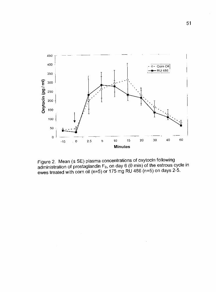
2. Mean (± SE) plasma concentrations of oxytocinfollowing administration of prostaglandin F2 on day 6(0 mm) of the estrous cycle in ewes treated with corn oil(n=5) or 175 mg RU 486 (n=5) on days 2-5. 51
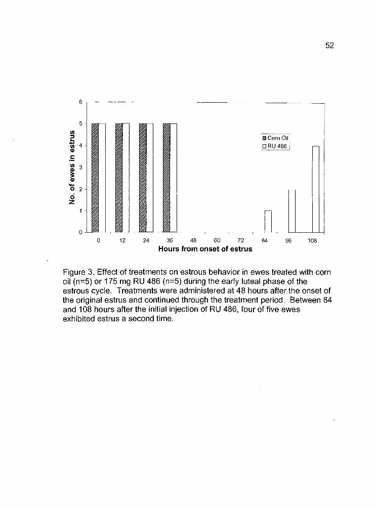
3. Effect of treatments on estrous behavior in ewes treated withcorn oil (n=5) or 175 mg RU 486 (n=5) during the early lutealphase of the estrous cycle. Treatments were administeredat 48 hours after the onset of the original estrus and continuedthrough the treatment period. 52
4. Mean (± SE) plasma concentrations of oxytocinfollowing prostaglandin F2 challenge (0 mm) onday 4 of the estrous cycle in heifers treated with saline(n=5) or 100 pg GnRH (n=5) every 4 hours ondays 2-4 of the estrous cycle. 65

LIST OF TABLES
TablePage
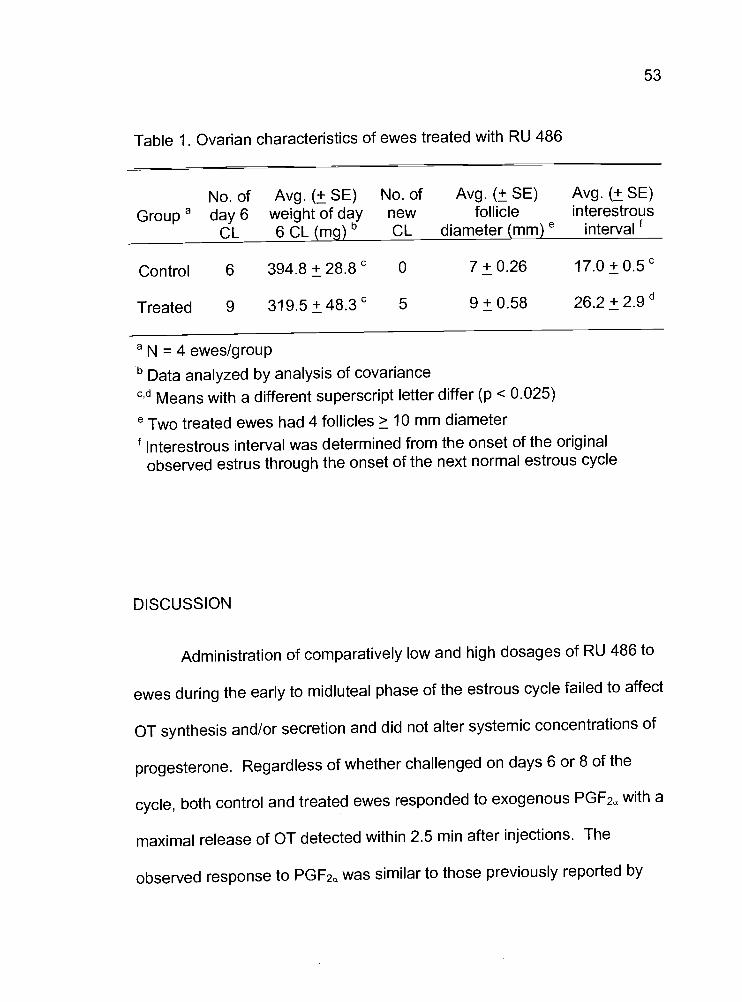
1. Ovarian characteristics of ewes treated with RU 486 53
2. Mean (± SE) serum concentrations of progesterone in control
and treated heifers 66
FACTORS AFFECTING LUTEAL OXYTOCIN SYNTHESIS AND/ORSECRETION BY THE OVINE AND BOVINE CORPUS LUTEUM
GENERAL INTRODUCTION
OVERVIEW OF THE OVINE ESTROUS CYCLE
The domestic ruminant estrous cycle consists of four distinct stages
that encompass the follicular and luteal phases of the cycle. These four
stages are proestrus, estrus, metestrus, and diestrus. The follicular phase,
which includes proestrus and estrus, is characterized by declining or low
levels of progesterone. Proestrus begins as a result of luteolysis and ends
at the onset of estrus. The period of proestrus can last several days,
depending on the animal species, and is noted for its hormonal transition
from high to low levels of progesterone, and subsequently increasing
estrogen levels. Gonadotropins, specifically follicle stimulating hormone
(FSH) and luteinizing hormone (LH), are primarily responsible for this
hormonal shift. Follicle stimulating hormone and LH recruit follicles to be
ovulated, and it is these follicles that secrete large quantities of estrogen
that induce the onset of estrus.
Estrus is the most notable phase of the cycle due to behaviors that
are displayed during this period. These behaviors occur concomitantly with
2
the rising levels of estrogen and result in male receptiveness and ovulation.
In the ewe, estrus can be detected for 18-48 hours with ovulation occurring
24-30 hours after the onset of estrus (Senger, 1999). Estrus exhibited by
the cow lasts about 15-18 hours and ovulation occurs about 10-12 hours
after the end of estrus (Senger, 1999).
After ovulation, the follicular cells undergo morphological changes
and begin to luteinize after exposure to a surge of LH. The period after
ovulation and before the formation of a fully functional corpus luteum (CL) is
denoted as metestrus, and is characterized by low levels of estrogen and
increasing levels of progesterone.
The CL becomes the dominant "gland" in the ovary during diestrus,
secreting massive quantities of progesterone. Progesterone prepares the
uterus for embryonic attachment, inhibits ovulation, and male receptivity.
This phase is the longest in duration, extending 14-18 days in length
depending on the species.
While species like the sow and cow cycle continually throughout the
year, some species are affected by season. This seasonal effect on
reproduction is especially apparent in the ewe and the mare and can be
largely attributed to photoperiod (Hafez, 1952; Kooistra and Ginther, 1975).
These photosensitive species use the number of hours of daylight as
a cue to time their reproductive period. The ewe is seasonally polyestrus

and is anestrus during the summer months, beginning to display estrous
behavior as hours of daylight become reduced in the fall, and hence, is
referred to as a "short-day" breeder. The ewe has a uniform distribution of
estrous cycles during the annual period of reproduction beginning in late
summer, as the hours of daylight shorten (Hafez, 1952). In contrast, the
mare is a so-called "long-day" breeder because estrous behavior begins as
hours of daylight increase and ceases during the winter months of anestrus.
OVERVIEW OF OXYTOCIN
Oxytocin (OT) is classified as a peptide hormone, consisting of nine
amino acids. This nonapeptide is structurally related to vasopressin (VP)
and is believed to be derived from the same ancestral gene, although they
are secreted by separate neurons in the hypothalamus. Due to the
structural similarities, 01 and VP can have the same sites of biological
actions due to regional homologies (Norman and Litwack, 1997).
Oxytocin is a product of magnocelluar neurons located in the
hypothalamus, and is transported down long axons to be stored in the
posterior pituitary until stimulated for release. Ribosomal assembly of
oxytocin occurs in the neurons of the paraventricular nuclei, and the

ru
precursor molecules of this hormone are transferred to the Golgi complex to
be packaged into neurosecretory granules that move along the axon.
During progression towards the posterior pituitary, post-translational
processing of a single gene transcript results in production of VP, OT, and
neurophysin (NP) proteins. Oxytocin and VP form a complex with NP; OT
complexes with the NP I form, while VP associates with the nearly identical
peptide, NP II (Sampson, 2000).
During axonal transport to the posterior pituitary, the signal peptide
of the preprotein OTis cleaved by proteases to the NP protein sequence.
The prohormone is then further cleaved at the carboxy terminus, resulting in
the OT-NP I complex. Neurophysin is believed to stabilize the complex in
its granular form during transport to the posterior pituitary for exocytosis, as
well as increasing the hormone's half-life in the blood stream (Norman and
Litwack, 1997).
Actions of Hypothalamic Oxytocin
Oxytocin has two primary modes of action; milk ejection and uterine
contractions during parturition. During the physical stimulus of suckling
young, OT is released in response to a rapid spinal arc to the
paraventricular nucleus. The stimulatory response is evoked by way of the
chemical messenger acetylcholine (Sampson, 2000). However, OT release
is inhibited by norepinephrine in response to fear or stress (Sampson,
2000).
During parturition the OT cascade responds to different stimuli.
Nearing the termination of gestation, progesterone levels begin to decline
drastically. Concomitantly, estrogen levels rise and upregulate the
production of OT receptors (OTR) in the myometrium, increasing the
sensitivity of the uterus to OT-induced uterine contractions (Zhang et al.,
1992). Due to the increase in OTR, low levels of OT released from the
posterior pituitary are effective in stimulating powerful uterine contractions
that occur during parturition (Dyer, 1988). However, OT can also stimulate
uterine contractions in the nonpregnant animal (Gilbert et al., 1992).

Luteal Oxytocin
The traditional concept is that oxytocin is a neurohormone produced
by the hypothalamus, and stored and secreted by the posterior pituitary.
However, previous studies have indicated that OT is synthesized in and
secreted by the CL of ruminants during the luteal phase of the estrous cycle
(Flint et al., 1990).
Wathes and Swann (1982) did not know how correct they were when
they hypothesized OT was also an ovarian hormone. Peptides extracted
from luteal tissue and assayed for OT indicated that the peptide was indeed
OT. In the luteal tissue that was assayed, it was determined that ovarian
OT represents about 15% of the total hypothalamo-neurohypophyseal store
of hormone in nonpregnant ewes during the luteal phase and roughly 0.2%
during pregnancy (Wathes and Swann, 1982). Such a high concentration
of hormone within the luteal tissue suggested that it was synthesized there.
Further studies validated this hypothesis when intact and
ovariectomized ewes were injected intramuscularly with a synthetic
prostaglandin F2 (PGF2a), cloprostenol, and ovarian venous blood samples
were collected. Within 10 minutes of administration of PGF2 to intact
ewes, there was a transient rise in plasma OT that lasted up to 40 mm (Flint
and Sheldrick, 1982). However, this transient increase in OT was absent in
7
ovariectomized ewes. Administration of PGF2a was unable to stimulate
secretion of OT by the posterior pituitary in the ovariectomized ewes,
confirming that the CL was the source of the peptide hormone during the
luteal phase of the estrous cycle (Flint and Sheldrick, 1982; Schams et al.,
1982).
LUTEAL OXYTOCIN SYNTHESIS
In the hypothalamic-neurohypophyseal system, OT is synthesized as
a precursor molecule along with NP I (Richter, 1983). Ovarian OT
collected from bovine luteal extracts and utero-ovarian plasma has been
shown to contain immunoreactive NP I (Wathes et al., 1983; Swann et al.,
1984). Previous research has determined that luteal OT, and its associated
NP, is released in a 1:1 equimolar equivalence upon exocytosis, just as in
the posterior pituitary (Schams et at, 1985b). This simultaneous release
indicates a potentially similar synthesis mechanism in the CL as in the
brain. Neurophysin I and 01 are always secreted concomitantly in major
quantities from the CL and in minor quantities from follicles; the
concentration depending on the number of CL or follicles present, not on
the rate of progesterone secretion (Schams et al., 1985a).
Oxytocin and its associated NP have been localized in secretory
granules of large luteal cells (Fields and Fields, 1986). When granulosa
cells, theca cells, and endothelial cells were analyzed by electron
microscopy, secretory granules of OT-NP were identified in the granulosa
cells only (Theodosis et al., 1986). Immunocytochemistry failed to detect
OT in tissues such as the corpus albicans or liver cells (Jones and Flint,
1988). Luteal OT granules are larger in size than those of the posterior
pituitary, containing a two-part core structure to distinguish them from the
neurosecretory granules (Theodosis et al., 1986).
Oxytocin and its associated NP have been localized in secretory
granules of granulosa cells (Wathes and Swann, 1982; Swann et al., 1984;
Theodosis et al., 1986; Fields and Fields, 1986). These granules are
formed in the Golgi apparatus and migrate to the membrane for exocytosis,
consistent with secretion of other peptide hormones from endocrine tissues
(Fields and Fields, 1986). It has been suggested that NP may serve to
stabilize OT molecules, preventing intragranular degradation as suggested
for OT granules of the posterior pituitary (Shuckovski et al., 1991). Little is
known about the specific biological actions of the luteal NP. Shuckovski et
al. (1991) also found evidence that biologically active luteal OTis
associated, in non-covalent fashion, with NP I in the luteinizing granulosa
cells of the cow. This non-covalent binding occurs in the granule, and non-
enzymatic separation of OT from NP I represents the last phase of post-
translational processing (Sheidrick and Flint,1989; Shuckovski et al., 1991).
LUTEAL OXYTOCIN GENE EXPRESSION
Expression of OT genes had been described thus far only in the
hypothalamus, however, it was not clear whether or not the OT found in the
CL was taken up from circulation to be released later on specific stimulation
or synthesized within the tissue. Using a combination of DNA hybridization,
cDNA cloning, and in vitro translation, OT mRNA was identified in the
bovine and ovine CL, with the greatest amount occurring around day 3,
after estrus (Ivell and Richter, 1984: Jones and Flint, 1988). From cloned
DNA, Ivell and Richter (1984) were able to determine that luteal OT mRNA
is similar to that of hypothalamic origin and is not a product of a second or
alternative gene. This experiment also confirmed that the OT gene is
actively transcribed in the bovine and ovine CL (Jones and Flint, 1988).
The process of luteinization involves exponential growth of cells and
is accompanied by the differentiation of the follicle wall. Corner (1919)
observed that the CL contained the same cells as the follicle. However,
early after the rupture of the follicle Corner (1919) also observed that the
10
membrana propria that separates the thecal and granulosa cells breaks
down. Theca interna cells mulitiply in number and disperse between the
granulosa cells that are undergoing hypertrophy (Corner, 1919). Theca and
granulosa cells migrate inward to form the CL, which steadily increases in
size following ovulation.
Although granulosa cells in vivo appear to contain a low level of OT
mRNA, gene transcription is upregulated on the day of ovulation to yield
100-fold higher levels of OT mRNA by day 3 of the bovine cycle (Furuya et
al., 1990). Bovine granulosa cells isolated after the LH surge secrete much
greater quantities of OT than those isolated before the surge (Voss and
Fortune, 1991). In addition, OT mRNA was found to be significantly
increased in cells collected after the preovulatory surge of LH (Voss and
Fortune, 1992). However, there is no correlation that can be found between
the level of 01 mRNA and that of the peptide within the CL (Fehr et at.,
1987). Experimental evidence suggests that there is a delay in the peak of
OT peptide production 6-7 days after the OT mRNA peak, which coincides
with maximal synthesis of luteat progesterone (Fehr et al., 1987).
Oxytocin and its associated NP gene expression occur at a high rate
at or shortly after luteinization, with transcription rates markedly reduced
thereafter (Jones and Flint, 1988). Further experiments reaffirmed the
presence and active transcription of the OT gene. When hypothatami,
11
corpora lutea, corpora albicans, and follicles from all stages of the bovine
estrous cycle were extracted for RNA, autoradiograms revealed the
expression of the gene in low amounts in mid-cycle follicles (Ivell et al.,
1985). Furthermore, Ivell and associates (1985) supplied further evidence
supporting the observation that ovulation causes a surge of total RNA
production concomitant with the growth of the luteinizing tissue peaking at
day 3, and declining thereafter by day 11.
Due to the up-regulation in OT mRNA directly after ovulation,
researchers began to investigate the possibility that the hormones involved
in luteinization may also affect the transcription of the 01 gene. Insulin-like
growth factor-I (IGF-J) and insulin increased levels of OT mRNA and
stimulated the release of OT and progesterone in vitro (Holtorfet al., 1989;
Furuya et al., 1990; Fleet et al., 1994). These results could only be
obtained from cells collected from highly differentiated granulosa cells; i.e.,
cells from a follicle containing at least 40 ng/ml of estrogen in follicular fluid,
which up-regulates LH receptors (Holtorfetal., 1989; Furuya et al., 1990;
Voss and Fortune, 1991). In contrast, there was no effect of IGF-1 or
insulin in less mature granulosa cells of follicles containing less than 1
ng/mI of estrogen (Holtorf et al., 1989).
When highly differentiated granulosa cells cultured in vitro were
exposed to IGF-I, OT mRNA increased and remained elevated for an
12
extended period of time (Furuya et al., 1990). However, when these
researchers exposed granulosa cells to estrogen and PGF2, these
hormones had no effect on OT mRNA, irrespective of whether or not they
were pretreated with IGF-l. In the absence of IGF-1, OT release and
specific mRNA levels declined in a shorter amount of time. Furuya and
associates (1990) concluded IGF-1 was a necessary component in the up-
regulation of gene transcription for luteal OT and that estrogen and PGF2a
had no effect on the transcription of the luteal OT gene. Attempts to induce
mid-phase luteal cells to synthesize 01 failed to yield any 01 mRNA,
though peptide release could be measured, suggesting that luteal OT may
be released from a prohormone store (Furuya et al., 1990; Voss and
Fortune, 1992).
Further studies revealed the importance of follicular stage of
development on the secretion of OT. Granulosa cells isolated before the
preovulatory surge of LH responded with a marked increase in 01 secretion
when stimulated by FSH and LH in vitro (Voss and Fortune, 1991). Their
research also demonstrated that OT was not detectable in theca interna
cells isolated before or after the LH surge (Voss and Fortune, 1991; 1992).
These data led researchers to conclude that OT-NP I mRNA is not
accumulated in the CL during the mid-luteal phase of the cycle, but rather
earlier in the luteal phase with a delay in translation and OT synthesis.
13
PROGESTERONE, PROGESTERONE ANTAGONISTS, ANDPROGESTERONE INHIBITORS
Hormone antagonists and inhibitors are useful research tools. The
synthetic steroid mifepristone (RU 486) is a progesterone antagonist that
has been proven to be very effective, binding to progesterone receptor (PR)
sites and blocking the action of endogenous progestins in humans and
primates (Healy et al., 1983; Shoupe et al., 1985) as well as sheep
(Burgess et aL, 1992; Morgan et aL, 1993) and rats (Fang et al., 1997).
Mifepristone has been shown to bind to PR in the uterus of pregnant and
nonpregnant ewes with very high affinity, inhibiting the actions of
progesterone (Burgess et al., 1992; Morgan et al., 1993). Many studies
have utilized RU 486 to investigate the effects of progesterone withdrawal
during various times of the estrous cycle and gestation.
Ewes receiving RU 486 during late gestation experienced induced
labor and parturition following an OT challenge compared to control animals
(Burgess et al., 1992). Plasma samples of ewes treated with RU 486
revealed enhanced levels of 13,14-dihydro-15-keto PGF2a (PGFM), a
PGF2 metabolite, in response to the OT challenge while progesterone
levels remained the same between both treatment groups. Burgess et al
(1992) hypothesized that by blocking the effect of progesterone at the level

of its receptor, the ability of endogenous estrogen to enhance the oxytocin
receptor (OTR) would not be inhibited by endogenous progesterone.
Morgan et al. (1993) conducted a study to test the hypothesis that
exposure to progesterone in early to mid-diestrus regulates uterine release
of PGF2 at the time of luteolysis in sheep. They determined that pulsatile
secretion of PGFM was inhibited in those animals receiving RU 486. This
inhibition of PGF2c,resulted in a failure of luteolysis and prolonged the
duration of the cycle until day 24 after estrus (Morgan et al., 1993). This
study demonstrated that RU 486 inhibits the function of uterine PR,
delaying the down-regulation of the PR, therefore postponing luteolysis.
Two potent progesterone inhibitors, trilostane and epostane, have
also been utilized to study the effects of progesterone during the estrous
cycle and gestation of ewes (Hoefler et al., 1986; Webb, 1987) and cattle
(Peters and Lamming, 1986), and during the menstrual cycle of primates
(Schane et aL, 1979). Trilostane and epostane are inhibitors of the enzyme
33-hydroxysteroid dehydrogenase (3-HSD), a crucial enzyme in the
synthesis of progestins, estrogens, androgens, and glucocorticoids. These
enzyme-inhibiting compounds block the activity of the enzyme by
competing with pregnenolone for binding sites on the 313-HSD molecule
(Peters and Lamming, 1986).
15
Use of trilostane in ewes has been effective in decreasing serum
progesterone levels; however, epostane has been shown to be the most
effective. While trilostane decreases progesterone levels from
approximately 10 to 3 ng/ml, its effects only last about 4 to 6 hours after
injection (Jenkin et al., 1983). Epostane decreased serum progesterone
levels to about 1 ng/ml for up to 65 hours post-injection (Ashworth et al.,
1986). Although both drugs caused an inhibition in serum progesterone
levels, various doses of trilostane or epostane failed to induce structural
luteolysis (Jenkin et al., 1983; Ashworth et aL, 1986; Hoefler et al., 1986;
Peters and Lamming, 1986)
Recently there has been increasing evidence delineating the exact
role progesterone plays in regulating luteal OT synthesis. Smith et al.
(1995) have identified the presence of progesterone receptors (PR) within
granulosa cells of ovine follicles and CL. However, PR mRNA was
nondetectable in follicles collected before the LH surge, suggesting a role of
progesterone during the preovulatory period and luteal phase in sheep.
In reviews published by Walther et al. (1995) and Gimpi and
Fahrenholz (2001), these investigators reported the finding of a
hexanucleotide sequence response element within the promoter region of
the 01 gene. Variations of this six nucleotide sequence among species,
represent part of a binding site for the steroid nuclear receptor superfamily.
16
However, glucocorticoid, mineralcorticoid, progesterone and androgen
receptors do not interact with the OT gene at this sequence and do not
have any known direct effects on the transcription of the OT gene at this
time (Gimpi and Fahrenholz, 2001). It has been reported that many orphan
receptors in the steroid nuclear receptor superfamily may interact with the
OT gene, affecting the regulation of gene expression (GimpI and
Fahrenholz, 2001). A highly conserved sequence, the estrogen response
element (ERE), could be activated by the binding of estrogen in the rat and
human (Gimpi and Fahrenholz, 2001). In similar ligand-activated gene
regulation studies conducted in the bovine, the ERE promoter sequence
was not present and there was no direct effect by ER activation (GimpI and
Fahrenholz, 2001). It remains unclear if members of the steroid nuclear
receptor superfamily exert direct transcriptional effects on the bovine OT
gene.
Although there is evidence of PR mRNA present in membrane
fractions of granulosa cells from ovine follicles and corpora lutea,
researchers consistently failed to demonstrate binding of progesterone,
progesterone agonists, or progesterone antagonists to the steroid receptor
in granulosa membrane fractions (Rae et al., 1998a). However, binding of
progesterone did occur in this study when the membrane fractions were
incubated in the presence of digitonin.
17
The presence of PR in the follicular and luteal cells of the ovary
(Smith et al., 1995; Lioutas et aL, 1997) suggests that progesterone may
play an autocrine role in regulating OT gene upregulation. The research of
Lioutas et al. (1997) demonstrated that addition of progesterone to cultured
bovine granulosa cells increased cumulative OT secretion, suggesting that
progesterone is acting in an autocrine fashion to mediate OT up-regulation.
In a similar study, bovine granulosa and theca cell membranes from
follicles at different stages of the estrous cycle were isolated and specific
binding to the membrane fractions were assessed. Again, poor binding of
progesterone occurred unless cells were incubated in the presence of
digitonin (Rae et al., 1998b). Rae et al. (1998b) also examined the binding
of cholesterol, and the progesterone antagonist RU 486, and determined
that they too competed poorly for the binding site. These researchers
determined that binding of progesterone to granulosa cell membranes
decreased significantly with increasing follicle size and suggested that
these membrane steroid receptors may be involved in regulation of follicular
function.
It has been determined through cloning of the luteal PR that high
homology exists among bovine, ovine, human, mouse, rabbit and rat
nuclear receptors (Bolden-Tilfer et al., 2000). The presence of PR in the
nuclei of cells was confirmed by use of immunocytochemistry and ligand
ii;
binding assays. Additional evidence confirming the intracellular location of
the PR was obtained when the progesterone antagonist RU 486 competed
for binding to this receptor within the uterus of the ewe (Rauch et aL, 1985;
Bailly et aL, 1986).
Bovine granulosa cells were cultured and examined to determine
whether or not progesterone may be acting in an autocrine fashion on luteal
cells. When stimulated with LH, the cell cultures proceeded to produce
large amounts of OT and progesterone (Lioutas et al., 1997). Progesterone
antagonists, RU 486 and onapristone, when added to cell cultures inhibited
progesterone binding; but these antagonists had no effect on the levels of
estrogen or progesterone produced by the cells (Lioutas et al., 1997).
Mifepristone significantly reduced OT secretion in luteal cell cultures and
this effect could only be reversed in the presence of a synthetic
progesterone; the addition of dexamethasone had no effect. These
researchers analyzed OT mRNA and determined that RU 486 was
inhibitory at the level of gene transcription. Lioutas et al. (1997) concluded
that progesterone is acting in an autocrine fashion to promote OT up-
regulation. In summary, OT and progesterone are hypothesized to be
components of a luteal autocrine positive feedback loop, beginning with LH
to stimulate steroidogenesis and resulting in progesterone acting to regulate
OT gene expression.
1L!
LUTEAL OXYTOCIN SECRETION
Luteal OT is temporarily stored in the bovine and ovine corpora lutea
in granular form during the luteal phase of the cycle. However, within 10
minutes of administering PGF2 to the ewe or cow, there is an increase in
OT secretion (Flint and Sheldrick, 1982; Wathes et al., 1983). It has been
demonstrated that luteal OT secretion is stimulated by PGF2a and may be
critical for luteat regression in the ewe (Flint and Sheldrick, 1990).
Various hormones mediate initiation of luteolysis. Just prior to estrus
in the ewe, high levels of estrogen from the growing follicles stimulate the
up-regulation of OTR in the uterus (Roberts et al., 1976; Sheldrick and Flint,
1985; Vallet et al., 1990; Zhang et al., 1992). After ovulation and during the
formation of the corpus luteum, LH causes a shift from estrogen production
to progesterone production in the luteinizing cells, and a significant increase
in luteal OT (Voss and Fortune, 1991, 1992). This increasing progesterone
production down-regulates OTR in the uterus (Sheldrick and Flint, 1985;
Zhang et al., 1992) and has been shown to stimulate 01 secretion from
granulosa cells in vitro (Lioutas etal., 1997; Al-Matubsi et al., 1998).
It has been determined in the ewe that prior exposure to increased
systemic levels of progesterone was essential for the endometrium to
respond with an up-regulation of uterine OTR when exposed to estrogen

20
(McCracken, 1980; ValIet et al., 1990; Zhang et aL, 1992). The priming
effects of progesterone were apparent when ovariectomized ewes were
injected with a sequential combination of progesterone and estrogen or
estrogen alone (Zhang et al., 1992). The results indicated that there was a
significant increase in the concentration of endometrial OTR present in the
ewes that were previously exposed to progesterone compared to ewes
receiving estrogen alone (Zhang et aL, 1992). This research validated the
hypothesis that the normal response and changes in OTR concentrations of
the endometrium to progesterone and estrogen requires prior exposure to
progesterone (Vallet et al., 1990).
In ovarian autotransplanted ewes, administration of progesterone on
days 1-3 of the luteal phase resulted in significant increases in the pulses of
OT and PGF2, stimulating early luteolysis as compared to control animals
(Al-Matubsi et al., 1998). These researchers also determined that the
uterus must be exposed to increased levels of progesterone for 7-8 days
before the uterus will respond to OT with a release of PGF2a. Intact ewes
receiving progesterone early in the luteal phase also exhibited an increase
in the number of spontaneous OT surges suggesting that progesterone may
play a role in regulating the timing and release of ovarian OT (Fairclough et
al., 1983; Al-Matubsi et al., 1998).
21
Secretion of luteal OT comes from the large luteinized granulosa
cells in the cow and ewe (Swann et al., 1984; Theodosis et al., 1986; Fields
and Fields, 1986; Shuckovski et al., 1991). Granulosa cells isolated from
bovine preovulatory follicles were cultured and exposed to OT to determine
whether or not luteal OT plays a role in regulating steroid synthesis.
Oxytocin markedly inhibited estrogen (Chandrasekher and Fortune, 1990)
and stimulated progesterone synthesis (Berndston et al., 1996); however,
mRNA for P 450 side chain cleavage enzyme, P 450 aromatase enzyme,
and 313-HSD were not affected (Berndtson et al., 1996). When estrogen
was studied to determine its effects on OT secretion, it was determined that
the results were dependent on the stage of the cycle. Estrogen stimulated
01 secretion in ovine preovulatory follicles in vitro and inhibited OT
synthesis in day 1 CL and during the early luteal phase CL (Wathes et al.,
1992). Capacity for OT secretion is confined to healthy follicles and is lost
during atresia (Jungclas and Luck, 1986).
There is a high concentration of OT in the bovine CL during the
estrous cycle, with the highest concentration being present during the
midluteal phase (Schams et al., 1985; Abdelgadir et al., 1987). Although
high progesterone levels accompany the midluteal phase increase in luteal
01, Jungclas and Luck (1986) concluded that 01 production does not
always accompany increased progesterone secretion. However,
22
Chandrasekher and Fortune (1990) determined that cultured bovine
granulosa cells treated with an OT antagonist responded with significantly
lower progesterone levels than cells treated with 01 alone or in
combination with the antagonist. These researchers concluded that OT
may somehow facilitate the shift in steroidogenesis from estrogen
production to progesterone production.
Prostaglandin F2 stimulates the release of luteal OT from bovine
(Schallenberger et aL, 1984; Orwig et al., 1994a; Salli et aL, 2000) and
ovine (Flint and Sheldrick, 1982; Wathes et al., 1983) CL. Therefore,
synthetic agents mimicking the effects of PGF2a should elicit a release of
OT via intracellular signals. In a study conducted by Cosola-Smith et al.
(1990), the effects of phorbol ester 12-0-tetradecanoylphorbol-1 3-acetate
(TPA), which has structural similarities to DAG and is able to directly
activate protein kinase C (PKC), and a calcium ionophore (A23187), were
utilized to study the secretion of OT from bovine luteal tissue in vitro.
Results indicated that TPA and the calcium ionophore could stimulate an in
vitro secretion of OT. These studies suggested a role of PKC in luteal OT
secretion, as well as the importance of intracellular calcium for the
enhancement of the secretion of OT from bovine luteal cells.
Studies have also been conducted to examine the specific
mechanisms involved in the exocytosis of OT granules from the bovine CL.
23
Previous studies were conducted to determine which PKC isoforms are
present and their distribution within the bovine CL (Orwig et al., 1994b).
Orwig et al. (1994b) determined by western blot analysis that the bovine CL
expressed conventional PKC a and novel PKC c isozymes. Although the
roles of these specific PKC isozymes were not elucidated, it was proposed
that they played a role in the exocytosis of OT.
Salli et al. (2000) studied the disassembly of the actin cortex of the
luteal plasma membrane to determine specific mechanisms involved in the
exocytotic process. These researchers identified a role for the
myristoylated alanine-rich C kinase substrate (MARCKS) protein that
crosslinks the actin cortex filaments (Hartwig et al., 1992). When
phosphorylated by PKC, MARCKS translocates from the plasma membrane
to the cytoplasm causing a disassembly of the actin cortex and thus
facilitating OT secretion. These events have been observed within bovine
luteal cells stimulated by PGF2 during the midluteal phase of the estrous
cycle, suggesting that exposure to PGF2a must result in activation of PKC.
Various studies have been conducted to examine the effect of
different peptide hormones on the synthesis and secretion of OT. Activin-A,
an ovarian peptide, has been shown to cause a time-dependent decrease
in progesterone and OT secretion with no significant difference among
doses utilized in vitro (Shuckovski and Findlay, 1990). These latter
investigators made the novel observation that activin-A may act as a
luteinizing inhibitor in vivo, although the biological implications have not
been assessed.
Miyamoto et al. (1993) examined the effects of various
neuropeptides in in vitro cultures of midluteal bovine granulosa cells. They
found that LH alone or in combination with neuropeptide Y, substance P, or
vasoactive intestinal polypeptide, had no effect on OT secretion in cell
culture in the long or short term. They also concluded that these
neuropeptides were stimulatory for progesterone production but not OT
synthesis.
Due to the presence of adrenergic sympathetic nerves within the
ovary, in particular the ruminant ovary (Stefenson et al., 1981), investigators
began to assess the effects of various biogenic amines on luteal OT.
Bovine granulosa cells were cultured and OT production was measured by
secretion into the medium. Adrenaline and noradrenaline stimulated OT
secretion and progesterone was significantly reduced, while acetylcholine
had no effect on OT production (Luck and Jungclas, 1987; Heap et al.,
1989). Ascorbate, was also examined at the same time and it was
determined that alone, it produced a smaller increase than that of the
biogenic amines themselves (Luck and Jungclas, 1987). However, Heap et
al. (1989) found that although adrenaline and noradrenaline stimulated OT

25
production in sheep, they were also able to increase OT production with
acetylcholi ne. These researchers concluded that neurotransmitter-induced
OT release in vivo is rapid, episodic, and closely associated with reduced
blood flow arising from acetylcholine stimulation.
UTERINE OXYTOCIN RECEPTOR
The OTR is classified as a seven-membrane-spanning receptor, or a
G-protein coupled receptor. The OTR G-protein is coupled to the
phospholipase C (PLC) system and forms part of the subfamily of
neurohypophyseal hormone receptors that includes vasopressin receptors
(VPR). These OTR are located in the uterus, pituitary, and the
hypothalamus, as well as mammary tissues (Zingg et al., 1998).
As the receptor site becomes occupied, the receptor changes
conformation, interacting with a Gq-protein. The Gq-protein consists of three
proteins designated a, J3, and y. Activation of the OTR stimulates the
dissociation of Gaq, which in turn activates PLC-U. Phospholipase C-13
activates the phosphoinositide cascade, generating inositol trisphosphate
and I ,2-diacylglycerol (DAG). Diacyiglycerol is located in the membrane,
causing activation of phospholipase A2, and in the case of the uterine OTR,

26
generates production and release of uterine PGF2a from arachidonic acid
precursors (Watanabe et al., 1985). Activation of inositol trisphosphate,
triggers the release of intracellular calcium to activate PKC in conjunction
with DAG (Flint et al., 1986; Gimpl and Fahrenholz, 2001). Protein kinase
C can then phosphorylate other target proteins.
To further elucidate the regulatory mechanisms of the uterine OTR,
studies conducted by Hazzard and Stormshak (1997) suggested that
chronic exposure of ewes to estrogen during the estrous cycle down-
regulates the concentration of uterine OTR but not estrogen receptors (ER)
and reduces the OT-induced release of PGF2a,thus prolonging the life span
of the CL. These data were confirmed in part by studies revealing that
chronic treatment of ewes with estrogen from early to late in the estrous
cycle prevented a normal increase in OTR gene transcription (Hazzard et
aL, 1998). The suppressive effects of chronic estrogen exposure on the
uterine OTR are hypothesized to be a distinctly different mechanism than
that observed during progesterone exposure during the late luteal phase of
the ovine estrous cycle. However, there is no evidence to date of any
inhibitory effects of chronic estrogen on the secretion of OT or the ability of
the CL to secrete OT.
Studies have demonstrated that regulation of uterine OT binding
involves estrogen-induced up-regulation and progesterone-induced down-
27
regulation (Zingg et al., 1998) of OTR. Currently, all that is known about
luteal OT is its stimulatory effect on uterine PGF2a and the luteolytic effect of
the latter hormone on the CL. As of now, scant knowledge exists about the
intracellular changes that lead to cell death during the induction of luteolysis
in the cow and the ewe.
EFFECTS OF OXYTOCIN ON THE UTERUS
Over the past three decades numerous and extensive studies have
determined that the uterus exerts its effects on the ovary, more specifically
the CL, by a local action (Brinkley et al., 1964; Collins et al., 1966; lnskeep
et al., 1966). An example of this local action was presented by lnskeep and
Butcher (1966) with ewes that were unilaterally hysterectomized ipsilateral
to the CL, or unilaterally ovariectomized. Unilateral ovariectomy had no
effect on the length of the estrous cycle. However, ewes that were
unilaterally hysterectomized alone or in combination with a contralateral
ovariectomy, displayed a prolonged life span of the CL. At the time, these
researchers hypothesized that there may be a substance that travels
between the uterus and ovary via the blood, that regulates luteal life span.
This substance would later be identified as the luteolytic factor PGF2c,
(McCracken et at., 1972; Schallenberger et al., 1984).
A study was conducted to further elucidate the relationship between
the ovary and uterus. To determine whether or not precocious estrous
could be induced via the utero-ovarian relationship in cattle, 01 was
administered (Ginther et al., 1967). These researchers hypothesized that
OT would cause precocious estrus in unilaterally hysterectomized heifers if
the retained horn was adjacent to the ovary with the CL. These
researchers concluded that OT administered to intact or unilaterally
hysterectomized animals would indeed shorten the estrous cycle. This
further implicated the role of luteal 01 and the local actions of uterine
PGF2.
Prostaglandin F2a has been identified as the luteolytic hormone in
sheep (McCracken et al., 1972) and cattle (Schallenberger et al., 1984).
Luteal 01, in concert with uterine PGF2a, are two major factors involved
during the regression of the CL of ruminants, at least in the ewe (Roberts et
al., 1976; Roberts and McCracken 1976; McCracken etal., 1996).
Although it is not exactly known what causes the initial release of uterine
PGF2a,the effecter has been proposed to be a surge release of 01 from
the posterior pituitary (McCracken et at., 1996). Luteal OT, released in
response to the first surge of uterine PGF2U, targets the uterus stimulating
29
another surge release of PGF2a; sequential releases of PGF2a eventuafly
result in luteal regression (McCracken et al., 1972; Roberts et al., 1976).
The manner in which these two hormones work together that results in
luteal regression has been termed a "double-positive feedback loop"
(McCracken et al., 1972).
EFFECT OF PITUITARY HORMONES ON LUTEAL OXYTOCIN
Gonadotropin releasing hormone (GnRH), also known as luteinizing
hormone releasing hormone (LHRH) I follicle stimulating hormone releasing
hormone (FSHRH), is a decapeptide hormone located within cell bodies of
the medial basal hypothalamus and the preoptic areas. However, in
primates GnRH is also localized within cell bodies of the arcuate nucleus
(Griffin and Ojeda, 2000).
Axons from the cell bodies of the hypothalamus carry neuropeptides
to a capillary bed at the base of the pituitary stalk. This system is known as
the hypothalamo-hypophyseal portal system (Senger, 1999). This complex
network of capillaries carry minute quantities of releasing hormones to the
anterior pituitary where they stimulate cells to secrete a variety of
hormones.

30
These various pituitary hormones are released directly into the blood
stream from the anterior pituitary. The principal reproductive hormones
released from the anterior pituitary in response to GnRH are FSH and LH.
These anterior pituitary hormones target the gonads to regulate responses
in the female such as follicular growth, ovulation and luteal development.
Rahe et aJ (1980) evaluated endogenous patterns of LH secretion in
the cow during different stages of the estrous cycle. These researchers
concluded that serum LH fluctuated in a pulsatile manner throughout all
periods of the cycle. Early in the estrous cycle, LH was observed to pulse
in low amplitude but high frequency with a mean serum level between 1.3
and 2.2 ng/ml (Rahe et al., 1980). However, as the cycle progressed,
midluteal phase pulses were observed to be of high amplitude and low
frequency; and during the late luteal phase these pulses were characterized
as high amplitude, high frequency pulses resulting in the ovulatory surge.
Many studies have utilized GnRH in an attempt to induce estrus in
anestrous animals. Others have evaluated the effect of GnRH on luteal
function through the effects of LH (Adams et al., 1975; Kinder et al., 1975;
Rodger and Stormshak, 1986; Slayden and Stormshak, 1990). Constant
GnRH infusion into ewes over an extended period of time was not sufficient
to maintain serum LH at concentrations sufficient to stimulate ovulation
(Chakraborty et al., 1974). When these ewes were pretreated with
31
progestagens, there was still no effect on the stimulation of ovulation.
Chakraborty et al. (1974) concluded that pulsatile injections of GnRH,
mimicking the pulsatile-type release from the hypothalamus, may be more
effective at stimulating ovulation in anestrous ewes than a continuous
infusion.
Research also has been conducted to examine estradiol-induced
luteolysis in the ewe when stimulated by frequent, pulsatile injections of
GnRH (Adams et al., 1975). Ewes were either pretreated with estradiol-17
or vehicle and all ewes received GnRH on day 12 of the cycle. In all
estrogen treatment groups, a significant decrease was observed in luteal
progesterone levels; however, GnRH control ewes had increased luteal
weights and luteal progesterone content. It was concluded that
administering only GnRH to ewes over a 72 hour period was able to
stimulate luteal function similar to that in day 10 untreated control ewes.
These researchers also concluded that pulsatile administration of GnRH
may be more effective at stimulating the ovarian responses.
In cattle, ovarian responses to repeated administration of GnRH
were evaluated (Kinder et al., 1975). Treated heifers received 150 pg of
GnRH at4 hour intervals for 96 hours on days 16-19 of the estrous cycle.
These heifers were observed to have an increased mean serum LH
concentration as compared to control animals. The treated heifers also
32
exhibited increased peak LH levels during the first one-half of the treatment
period as compared with the second one-half of the period. However, it
was concluded that pulsatile-like administration of 150 ..tg of GnRH given at
4 hour intervals was not enough to stimulate ovarian function (Kinder et at.,
1975).
A study conducted by Mee et al (1993), concluded that one injection
of GnRH administered at estrus resulted in decreased LH secretions 1, 3,
and 8 days after administration. However, progesterone concentrations
were increased sooner after estrus and remained elevated significantly
longer than those of control animals receiving saline. This may be due to a
more complete luteinization of the cells of the follicle. Upon examining the
morphology of the CL, researchers found that GnRH increased the
percentage of large luteal cells (LLC) and decreased the percentage of
small luteal cells (SLC; Mee et al., 1993). They found that the proportion of
SLC decreased after the administration of GnRH when compared to the
saline-treated control animals (69 vs. 86%, respectively). Although SLC
contain the majority of LH receptors, LLC secrete much higher basal levels
of progesterone (Fitz et al., 1982; Niswender et al., 1985). Mee et al.
(1993) concluded that GnRH may promote the shift of SLC to LLC, thus
increasing luteal concentration and secretion of progesterone.
33
However, there have also been conthcting observations in regard to
the effects of GnRH on serum and luteal progesterone concentrations
during the estrous cycle (Rodger and Stormshak, 1986; Slayden and
Stormshak, 1990; Whitmore et al., 1996). When GnRH was administered
to ewes on days 2 and 3 after estrus, mean plasma progesterone levels
were not affected (Whitmore et aL, 1996). When lower doses of GnRH
were injected directly into the ovarian artery of ewes on day 2 after estrus,
progesterone was reduced on days 7-11 (Slayden and Stormshak, 1990).
The observed response to exogenous GnRH was apparently due to the
release of LH. In a subsequent study, Slayden and Stormshak (1990)
found that administration of LH on day 2 also suppressed progesterone
secretion on days 6-8.
Gonadotropin-releasing hormone was also administered during
various stages of the estrous cycle to determine its effects on serum LH
and progesterone levels, as well as its effects on the duration of the cycle in
heifers (Rodger and Stormshak, 1986). When GnRH was administered on
days 2 or 10 after estrus, an increase in serum LH was observed, with peak
release occurring 15 to 30 minutes after injection. Gonadotropin-releasing
hormone administered on day 2 caused a decrease in progesterone levels
beginning on day 8. Administration of GnRH on day 10 was also effective
in causing decreased progesterone levels; however, it caused a transient

34
increase during a three-hour sampling period after administration. There
was no effect of the treatment on the duration of the estrous cycle.
Rodger and Stormshak (1986) also observed that GnRH
administered on day 2 after estrus resulted in decreased CL weight on day
8 but not on day 14. Administration of GnRH on day 2 also resulted in a
decrease in luteal LH receptor concentrations on both days 8 and 14.
These researchers concluded that GnRH-induced release of LH on days 2
or 10 may have caused a more rapid transformation of small to large luteal
cells with a consequent reduction in the number of LH receptors and
progesterone synthesis or secretion.
In a review, Niswender (1981) stated that the mechanisms involved
in the regulation of ovarian progesterone secretion were regulated by LH.
This can occur by the down-regulation of LH receptors after elevations of
systemic levels of LH as well as the regulatory role of other hormones.
Luteinizing hormone binds to its specific receptor on the plasma membrane
resulting in the activation of adenylate cyclase and results in elevated
intracellular levels of cyclic adenosine monophosphate (cAMP). Thus,
enhanced cAMP results in elevated activity of protein kinase A and
increased steroidogenesis. Protein kinase A directly influences the activity
of cholesterol esterase, causing cleavage of cholesterol esters to allow for
utilization of cholesterol as a substrate for steroid synthesis via the activity
35
of the cholesterol side chain cleavage complex, the presumed rate limiting
step in steroidogenesis.

36
STATEMENT OF THE PROBLEM
Domestic livestock are a very important agricultural commodity,
providing us with a valuable source of protein from the milk and meat they
produce. The goal of livestock producers is to rear a high percentage of
healthy offspring, therefore a thorough understanding of the animal's
reproductive cycle is a crucial component for management practices. A
better understanding of the interactions between reproductive hormones
could result in the development of more efficient estrous synchronization
programs and, hence, increased artificial insemination (Al) conception rates
when Al is combined with these improved programs.
Many factors can influence the life span of the CL. Understanding
the aspects that control luteal life span may provide insight to the factors
that direct the regulation of the estrous cycle and reproductive functions in
ruminant livestock species. An important aspect in understanding the life
span of the CL includes the interactions of luteal OT and uterine PGF2a
during luteal regression, and the various other hormones that affect the
synthesis and/or secretion of OT.
The complex interrelationships between luteal 01 and progesterone,
as well as luteal OT and PGF2, have yet to be determined, It is unknown
what role luteal OT may play during the follicular stage as well as during

37
early luteal development. The effects that luteal OT may have upon the
uterus early in the luteal phase have also yet to be determined.
It has been observed that progesterone receptor mRNA and the
receptor are present in the CL, and their presence coincides with the rise in
luteal OT mRNA and OT production Therefore, it is conceivable that
progesterone may be acting in an autocrine fashion to promote OT
synthesis and/or secretion in the ovine and bovine CL. This premise is
supported by the research of Voss and Fortune (1993) who reported that
progesterone stimulated OT secretion by bovine granulosa cells during the
later stages of a 5-day culture. In addition, Lioutas et al (1997) found that
inhibition of progesterone binding with the use of antagonists greatly
reduced OT secretion from granulosa cells in vitro.
The experiments of the present thesis were conducted to determine:
1) if treatment with a progesterone antagonist would have an effect on
luteal oxytocin synthesis and/or secretion in ewes by decreasing the ability
of progesterone to bind to its own receptors, 2) to determine if GnRH-
stimulated release of LH would have an effect on luteal function, thus
affecting luteal oxytocin synthesis and/or secretion.
ci;]
MIFEPR1STONE (RU 486)-INDUCED CHANGESIN OVARIAN FUNCTION OF EWES
ABSTRACT
Experiments were conducted to determine whether endogenous
progesterone regulates synthesis and/or secretion of luteal oxytocin (OT).
In experiment 1, mature ewes (n5 per group) were assigned randomly to
control or mifepristone (RU 486) treatment groups. Ewes were injected
twice daily s.c. with vehicle or 10 mg RU 486 from days 5-7 of the estrous
cycle (estrus = day 0). On day 8, following an i.v. prostaglandin F2a (250 g
cloprostenol) challenge, venous samples were collected at frequent
intervals to determine plasma OT concentrations. Plasma OT in RU 486-
treated animals did not differ significantly from those of the control animals
(P > 0.05). In Experiment 2, ewes were injected s.c. daily with vehicle or
175 mg RU 486 from days 2-5 of the estrous cycle followed by a
prostaglandin F2a challenge on day 6. Four of five RU 486-treated ewes
exhibited "split-estrus" (estrous behavior through 36 hours and again 84 to
108 hours after the onset of initial estrus). There was no significant
difference in mean plasma OT or progesterone levels between treatment
groups (P > 0.05). Mean mature corpora lutea (CL) weights of control and
RU 486-treated ewes on day 6 did not differ (394.8 ± 28.8 vs. 319.5 ± 48.3

39
mg; P > 0.05). Mifepristone-treated ewes contained mature CL, new CL (2
of 4 ewes), and/or preovulatory follicles (>10 mm, 2 of 4 ewes). Average
interestrous interval for RU 486-treated ewes was 9 days longer than that of
control animals (26.2 ± 2.9 vs. 17 ± 0.5 days; P <0.025).
INTRODUCTION
Oxytocin (OT) is classified as a peptide hormone, consisting of nine
amino acids. The traditional concept is that this nonapeptide is produced
by the hypothalamus, and is stored and secreted by the posterior pituitary
upon stimulation. However, results of studies conducted during the last two
decades have indicated that OT is synthesized and secreted by the corpus
tuteum (CL) of ruminants during the estrous cycle (Wathes and Swann,
1982; Flint et aL, 1990).
It has been proposed that in the ruminant, uterine prostaglandin F2
(PGF2a) and luteal OT presumably act in concert through a "double positive
feedback loop" to promote regression of the CL (Roberts et al., 1976;
Roberts and McCracken 1976; McCracken et al., 1996). Results of
previous research have confirmed that the administration of PGF2ato cows
and ewes causes a transient rise in plasma OT levels, which in some
studies has been shown to be associated with the induction and promotion
of Iuteal regression (Flint and Sheldrick, 1982; Wathes et aL, 1983; Flint et
al., 1990; Orwig et al., 1994a; Salli et al., 2000).
The OT gene is actively transcribed in the bovine and ovine CL (lvell
and Richter, 1984; Ivell et aL, 1985; Jones and Flint, 1988). Although
granulosa cells in vivo appear to contain a low level of OT messenger
ribonucleic acid (mRNA), gene transcription is upregulated on the day of
ovulation to yield 100-fold higher levels of mRNA by day 3 of the estrous
cycle (Furuya et aL, 1990). Interestingly, there has been no significant
positive correlation determined between the level of OT mRNA and of the
peptide within the CL (Fehr et al., 1987). Previous research has
demonstrated that luteal concentrations of OT mRNA are maximal on day 3
of the cycle and then gradually decline over the course of the cycle (Ivell et
al., 1985; Jones and Flint, 1988; Furuya et al., 1990). In contrast, maximal
luteal production of OT in the cow and the ewe occurs by the midluteal
phase of the cycle and then declines so that luteal concentrations of the
nonapeptide are comparatively low by the time of luteolysis (Sheldrick and
Flint, 1983; Abdelgadir et al., 1987). These data indicate a delay in the
peak of OT production 3-6 days after the OT mRNA peak, which coincides
with maximal synthesis of luteal progesterone (Fehr et al., 1987).
41
Progesterone receptor mRNA, as well as the receptor protein, have
been identified in ovine preovulatory follicles and CL (Smith et al., 1995).
When bovine granulosa cells were cultured with the progesterone
antagonists mifepristone (RU 486) and onapristione, 01 secretion was
significantly reduced and this effect could only be reversed in the presence
of progesterone (Lioutas et al., 1997).
Because luteal progesterone receptor mRNA and the receptor
coincide with the rise in luteal OT mRNA and OT production, it is
conceivable that progesterone may be acting in an autocrine fashion to
promote OT synthesis and/or secretion in the ovine CL. This premise is
supported by the research of Voss and Fortune (1993) who reported that
progesterone stimulated OT secretion by bovine granulosa cells during the
later stages of a 5-day culture. Therefore, the objective of this research
was to determine if the antagonistic actions of RU 486 would lead to
decreased luteal 01 synthesis and/or secretion.

MATERIALS AND METHODS
Animals
Mature Polypay ewes exhibiting normal estrous cycles (17 ± 1 days)
were assessed for estrous behavior twice daily with a vasectomized ram
and the first day of observed estrus was designated as day 0 of the estrous
cycle. Ewes were assigned randomly to treatments prior to the beginning of
the study.
To collect luteal tissue from ewes on days 6 and 8 of the estrous
cycle, the animals were anesthetized with an i.v. injection of 5% sodium
pentothal (Abbott Laboratories, N. Chicago, IL) followed by maintenance of
anesthesia by use of closed circuit inhalation of an oxygen-halothane
mixture (Halocarbon Laboratories, River Edge, NJ). The reproductive
organs were exposed through a midventral abdominal incision. Luteal
tissues were enucleated from the ovaries and immediately stored on ice
until weighed. Follicles (5 to 10 mm diameter) were measured and
recorded. Intramuscular injections of Banamine (Schering-Plough Animal
Health Corp., Union, NJ; 50 mg/mI) and Penicillin G Procaine (Butler Co.,
Columbus, OH; 300,000 units/mI) were administered immediately following
surgery. All experimental procedures and protocols were reviewed and
43
performed in accordance with the institutional Anima! Care and Use
Committee guidelines at Oregon State University.
Experiment 1- Luteal Oxytocin Synthesis
Ewes (n=5) were injected s.c. with 10 mg RU 486 (Sigma Chemical
Co., St. Louis, MO) dissolved in 10 ml of corn oil twice daily on days 5, 6,
and 7 of the estrous cycle. Control ewes (n5) were injected similarly with
corn oil only. On day 8, ewes were challenged with an Lv. injection of
prostaglandin F2a (250 pg cloprostenol). Blood samples were collected at
-15 and 0 mm to establish basal secretion of OT and at 2.5, 5, 10, 15, 20,
30, and 40 mm post-challenge. An additional blood sample was collected
prior to the prostaglandin F2a on day 8 to determine plasma progesterone
levels. All blood samples were collected into 10 ml heparinized vacutainer
tubes (Becton Dickinson Vacutainer Systems, Franklin Lakes, NJ) followed
by immediate addition of EDTA (0.5 M; 20 p1) and 1, 10 phenanthroline (5
mg/mi in ethanol; 10 p1) to block endogenous oxytocinase activity and then
stored on ice. Blood samples were centrifuged (1650 x g) at 4°C, and
plasma was stored at -20°C until assayed for 01.
Experiment I Ovarian Morphology
To evaluate the effects of RU 486 on ovarian function and
morphology, ewes were assigned randomly to two treatment groups: 1)
Control (n4), 2) RU 486 (n=4). Ewes received s.c. injections twice daily of
either 10 mg of RU 486 dissolved in corn oil or 10 ml of corn oil alone.
Ewes were injected on days 5, 6, and 7 of the estrous cycle. On day 8,
control and treated animals were anesthetized and luteal tissue was
collected as described above.
Experiment 2 Luteal Oxytocin Synthesis
Ewes (n=5) were injected s.c. with 175 mg RU 486 (Sigma Chemical
Co., St. Louis, MO) dissolved in 10 ml of corn oil on days 2,3,4, and 5 of
the estrous cycle. Control ewes (n=5) were injected similarly with corn oil
only. On day 6, ewes were challenged with an i.v. injection of PGF2 (250
pg cloprostenol). Blood samples were collected at frequent intervals as in
Exp. I up to 60 mm post-challenge. One sample was collected prior to the
prostaglandin F2a administration to determine plasma progesterone levels.
All blood samples were collected into 10 ml heparinized vacutainer tubes

45
(Becton Dickinson Vacutainer Systems, Franklin Lakes, NJ) and samples
collected for OT analysis were followed by immediate addition of EDTA and
1, 10 phenanthroline and then placed on ice for transport to the laboratory.
Blood samples were centrifuged (1650 x g) at 4°C, and plasma was stored
at -20°C until assayed for OT and progesterone.
Experiment 2 - Ovarian Morphology
To evaluate the effects of RU 486 on ovarian function and
morphology, ewes were assigned randomly to two treatment groups: 1)
Control (n=4), 2) RU 486 (n=4). Ewes received s.c. injections of either 175
mg of RU 486 dissolved in corn oil or 10 ml of corn oil only. Ewes were
injected once daily on days 2, 3, 4, and 5 of the estrous cycle. On day 6,
control and treated animals were anesthetized and luteal tissue was
collected as described above.
Oxytocin Radioimmunoassay
Oxytocin was extracted from I ml of plasma and measured by RIA
using methods adapted from Schams (1983) and Abdelgadir et at. (1987),
using an OT antibody (1:7000) generously provided by Dr. Dieter Schams,
Technical University of Munich, Germany. The mean extraction efficiency
was 63% as determined by the addition of [3H] OT (4,000 cpm/ 100 p1; 2200
Ci/mmol; New England Nuclear, Boston, MA). Plasma concentrations of
OT determined by RIA were corrected for losses due to extraction. Plasma
sample volumes used in the RIA were 100 p1/ tube. The sensitivity of the
assay was I pg/mi. All samples were analyzed in three consecutive assays
with an intra- and interassay coefficient of variation (CV) value of 2.78 and
7.02%, respectively.
Progesterone Radioimmunoassay
Plasma concentrations of progesterone were assayed by RiA as
described by Koligan and Stormshak (1976). Plasma progesterone was
extracted from 100 p1 of plasma with benzene:hexane (1:2). To correct for
procedural loss due to extraction, [1 ,2,6,7-3H] progesterone (4000 cpm/100

47
i.tl;44.5 Ci/mmol; New England Nuclear, Boston, MA) was added to a third
tube containing an aliquot of plasma The mean extraction efficiency was
88%.
Extracted samples were quantified by RIA using the #337 anti-
progesterone-11-BSA (1 :2400) provided by G.D. Niswender, Colorado
State University. All samples were analyzed in two consecutive assays with
intra- and interassay CV values of 1.9 and 8.4%, respectively. The
sensitivity of the assay was 10 pg/mI.
Statistical Analysis
Plasma concentrations of OT were analyzed by use of repeated
measures analysis of variance. Data on luteal weights were analyzed by
analysis of covariance using number of corpora lutea as the independent
variable and plasma concentrations of progesterone were analyzed by one-
way analysis of variance.
RESULTS
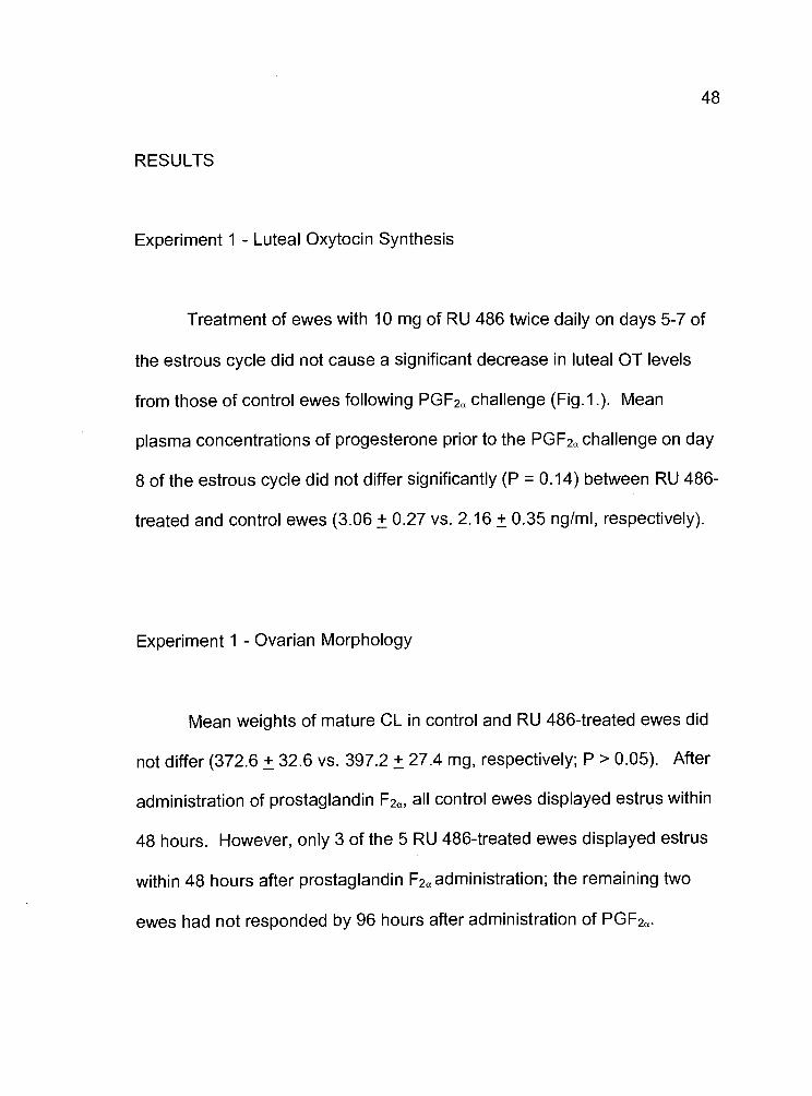
Experiment I - Luteal Oxytocin Synthesis
Treatment of ewes with 10 mg of RU 486 twice daily on days 5-7 of
the estrous cycle did not cause a significant decrease in luteal OT levels
from those of control ewes following PGF2 challenge (Fig.1 .). Mean
plasma concentrations of progesterone prior to the PGF2 challenge on day
8 of the estrous cycle did not differ significantly (P = 0.14) between RU 486-
treated and control ewes (3.06 ± 0.27 vs. 2.16 ± 0.35 ng/ml, respectively).
Experiment I Ovarian Morphology
Mean weights of mature CL in control and RU 486-treated ewes did
not differ (372.6 ± 32.6 vs. 397.2 ± 27.4 mg, respectively; P > 0.05). After
administration of prostaglandin F2a, all control ewes displayed estrus within
48 hours. However, only 3 of the 5 RU 486-treated ewes displayed estrus
within 48 hours after prostaglandin F2a administration; the remaining two
ewes had not responded by 96 hours after administration of PGF2a.

49
900
800
700
600
500 -
o 400o
300
200 - -
L-
0.--15 0 2.5 5 10 15 20 30 40
Minutes
Figure 1. Mean (± SE) plasma concentrations of oxytocin followingadministration of prostaglandin F2a on day 8 (0 mm) of the estrous cycle in
ewes treated with corn oil (n=5) or 10 mg of RU 486 (n=5) twice daily on
days 5-7.

50
Experiment 2 Luteal Oxytocin Synthesis
Treatment of ewes with 175 mg of RU 486 did not cause a significant
decrease in luteal oxytocin levels from those of the control ewes (Fig. 2).
Mean plasma concentrations of progesterone in RU 486-treated animals did
not differ from those of the control animals (0.92 ± 0.04 vs. 0.94 ± 0.07
ng/ml; respectively). After the initiation of treatments, four of five RU 486-
treated ewes exhibited another estrus, observed as early as 84 hours after
the onset of the original estrus (Fig. 3.).
Experiment 2 - Ovarian Morphology
Mean weights of mature CL in control and RU 486-treated ewes did
not differ (394.8 ± 28.8 vs. 319.5 ± 48.3 mg, respectively; P > 0.05).
Mifepristone-treated ewes contained new CL (corpora hemorrhagica) and
br preovulatory follicles (>10 mm diameter) in addition to mature CL (Table
1). Average interestrous interval for RU 486-treated ewes was 9 days
longer and was significantly different (P < 0.025) than that of the control
animals (17 ± 0.5 vs. 26.2 ± 2.9 days; Table 1).
51
450
400
350
300
0,. 250
200
150
100
50
-15 0 2.5 5 10 15 20 30 40 60
Minutes
Figure 2. Mean (± SE) plasma concentrations of oxytocin followingadministration of prostaglandin F2 on day 6 (0 mm) of the estrous cycle in
ewes treated with corn oil (n5) or 175 mg RU 486 (n=5) on days 2-5.
U)
L..'I-I
U)
(I)
0)
'4-
0dz
[Corn Oil
Lgu 486
12 24 36 48 60 72 84 96 108
Hours from onset of estrus
52
Figure 3. Effect of treatments on estrous behavior in ewes treated with cornoil (n=5) or 175 mg RU 486 (n=5) during the early luteal phase of theestrous cycle. Treatments were administered at 48 hours after the onset ofthe original estrus and continued through the treatment period. Between 84and 108 hours after the initial injection of RU 486, four of five ewesexhibited estrus a second time.
53
Table 1. Ovarian characteristics of ewes treated with RU 486
No. of Avg. (± SE) No. of Avg. (± SE) Avg. (± SE)
Group a day 6 weight of day new follicle interestrousCL 6 CL (mg) b CL diameter (mm) e interval
Control 6 394.8±28.8c 0 70.26 17.O±O.5c
Treated 9 319.5±48.3c 5 9±0.58 26229d
a N = 4 ewes/groupb Data analyzed by analysis of covariancec,d Means with a different superscript letter differ (p < 0.025)
e Two treated ewes had 4 follicles> 10 mm diameter' Interestrous interval was determined from the onset of the original
observed estrus through the onset of the next normal estrous cycle
DISCUSSION
Administration of comparatively low and high dosages of RU 486 to
ewes during the early to midluteal phase of the estrous cycle failed to affect
OT synthesis and/or secretion and did not alter systemic concentrations of
progesterone. Regardless of whether challenged on days 6 or 8 of the
cycle, both control and treated ewes responded to exogenous PGF2 with a
maximal release of OT detected within 2.5 mm after injections. The
observed response to PGF2a was similar to those previously reported by
54
Orwig et al. (1994a) and Salli et al. (2000). The systemic levels of luteal OT
were substantially lower on day 6, during the early luteal phase, than on
day 8 of the estrous cycle. Systemic OT levels after the administration of
PGF2cx during the early and midluteal phase are also consistent with those
values observed in the utero-ovarian venous blood of ewes within the same
time period (Hooper et al., 1986).
Results of this study indicate that RU 486 administration while ewes
are still in estrus and during metestrus causes a "split estrus". This split
estrus was observed in four of five ewes receiving 175 mg RU 486 (within
84 hours after the initial observed estrus). This unique occurrence was not
observed in ewes receiving the lower dose of RU 486 on days 5-7of the
estrous cycle in Experiment 1. Likewise, split estrus was not reported by
Morgan et al. (1993) who injected ewes with 2.5 mg/kg of RU 486 daily on
days 5-8 of the estrous cycle. In a concurrent study in our laboratory, 150
mg of RU 486 administered to ewes at the onset of estrus, and 24, and 48
hours later, resulted in the same phenomenon of split estrus within 96 hours
after onset of the original estrus (A. Wurst, unpublished data).
These observations of split estrus may be attributed to the
antagonistic actions of RU 486 at the level of the hypothalamus.
Mifepristone is known to be a potent antiprogestin in reproductive tissues,
as well as the central nervous system (Philbert, 1984). In studies
55
conducted with female rat hypothalami, RU 486 bound with very high
affinity, especially in the region of the preoptic nuclei (Pleim et al., 1990).
Thus, it is conceivable that RU 486 may be binding to receptors located in
the hypothalamus, blocking the negative feedback inhibition of
progesterone, and encouraging the maturation of the next follicular wave.
Campbell et al. (2000) concluded that blocking progesterone has major
effects on the LH surge mechanism that can result in ovulation of post-
estrus first wave follicles. This may be an explanation for the observed
response of those ewes receiving 175 mg of RU 486 during the early luteal
phase.
Ovarian morphology of ewes receiving 175 mg RU 486 revealed the
presence of mature CL, new sites of ovulation, as well as preovulatory
follicles occurring in various combinations on individual ovaries. It has been
observed that ewes treated with RU 486 during the early luteal phase
resulted in additional LH surges within hours after the initial ovulatory LH
surge (Campbell et al., 2000). These researchers concluded that the
subsequent LH surges resulted from normal levels of estradiol derived from
preovulatory follicles, consistent with the findings of concomitant
morphological studies.
As a result of the split estrus, the interestrous interval was extended
in ewes receiving 175 mg of RU 486. The nine day extension in RU 486-
treated ewes from the onset of the first observed estrus through the next
normal estrous cycle is likely attributed to the development and ovulation of
follicles that formed mature, functional CL. It was not until these newly
formed CL regressed that the onset of the next estrous cycle could begin.
Thus, the newly formed CL could have reset the "ovarian clock" for the
estrous cycle. Although ewes did not display a split estrus in a study
conducted by Morgan et al. (1993), by day 24 after the initial estrus RU
486-treated ewes still failed to return to estrus while the control animals had
a normal cycle of 17 days in duration. These researchers attributed this
failure to display estrus on inhibition of the progesterone receptors by RU
486, thus possibly interrupting the functional integrity of the ovarian-
endometrial luteolytic axis by which progesterone-stimulates endometrial
PGF2a synthesis.
Administration of RU 486 during the early luteal phase of the estrous
cycle appears to be a powerful antagonist at the level of the hypothalamus.
It has been suggested that progesterone may act in an autocrine fashion to
regulate its own synthesis, ultimately affecting its paracrine actions and
regulation of other hormones (Rothchild, 1981; 1996). Although the highest
dosage of RU 486 used in our study was apparently able to block the
57
negative feedback of progesterone at the level of the hypothalamo-
hypophyseal axis, it was not able to interfere with luteal function. It is
possible that the dosage was not sufficient to counteract the autocrine
effects of progesterone at the level of the ovary. Future studies must be
conducted to understand the actions of the preovulatory follicles and newly
formed CL associated with the luteal phase of the estrous cycle when
progesterone action is blocked early in the luteal phase.
SUPPRESSION OF LUTEAL OXYTOCIN AND PROGESTERONESECRETION BY EXOGENOUS GONADOTROPIN-RELEASING
HORMONE IN HEIFERS
ABSTRACT
An experiment was conducted to determine the effects of
gonadotropin-releasing hormone (GnRH)-stimulated release of luteinizing
hormone (LH) on luteal oxytocin (OT) and progesterone production in beef
heifers. Ten heifers with normal estrous cycles were assigned randomly in
equal numbers to a control and treatment group. On day 2 of the estrous
cycle (estrusday 0) heifers were injected with either physiological saline or
100 pg GnRH every 4 hours for 56 hours. Blood samples were collected 0
mm pre- and 180 mm post- GnRH challenge for progesterone analysis.
Sixty hours after the initial injection of GnRH or saline, heifers were
challenged with an i.v. injection of 500 pg prostaglandin F2a (cloprostenol)
and blood was collected at frequent intervals for OT analysis. Luteal OT
synthesis was suppressed (P < 0.01) in heifers receiving repeated
injections of GnRH compared to the saline-treated control animals.
Progesterone secretion was significantly greater in saline-treated animals
compared to GnRH-treated animals pre- and post-challenge (1.0 ± 0.06 vs.
59
0.93±0.11 ng!ml and 1.16±0.05 vs. 0.96± 0.13 ng/ml, respectively; P <
0.05).
INTRODUCTION
In the developing bovine corpus luteum (CL) transcription of the
oxytocin (OT) gene increases up to about day 3 of the estrous cycle with
maximal concentration of the nonapeptide achieved shortly thereafter
(Furuya et al., 1990; IvelI and Richter, 1984; Jones and Flint, 1988).
Luteinizing hormone (LH) appears to be the primary stimulation for OT
production by granulosa cells of preovulatory follicles (Voss and Fortune,
1991; 1992), but little is known about the factor(s) regulating the synthesis
of OT during the early luteal phase of the cycle. Because LH is luteotropic
in the cow (Wiltbank et al., 1961; Simmons and Hansel, 1964), it might be
anticipated that this gonadotropin would continue to stimulate OT
production by the developing CL. However, existing experimental evidence
suggests that brief exposure of the developing bovine or ovine CL to an
excess of LH attenuates subsequent luteal function (Rodger and
Stormshak, 1986; Slayden and Stormshak, 1990; Whitmore et al., 1996).

Although treatment with gonadotropin-releasing hormone (GnRH) to induce
the release of LH, or administration of the gonadotropin, reduced the
subsequent production of progesterone, the CL had a normal life span as
judged by the interestrous interval.
Luteinizing hormone may act indirectly via the small luteal cells
(SLC) to impact the function of the large luteal cells (LLC) because only the
former cells are endowed with a majority of the LH receptors (Fitz et al.,
1982). This mode of action of LH is supported by the data of Del Vecchio et
al. (1994) who reported that bovine SLC are able to inhibit progesterone
synthesis by perfused LLC. Apparently, the SLC produce an inhibitory
substance that acts in a paracrine fashion to suppress LLC function.
It has been demonstrated that sequential injections of GnRH
administered to beef cows during the late stages of the cycle results in a
diminished release of LH with each injection (Kinder et al., 1975). On this
basis, it was hypothesized that subjecting cows to such a treatment
regimen of GnRH injections might have a detrimental effect on luteal
function. Therefore, the present experiment was conducted to determine
whether sequential injections of GnRH into beef heifers during the formation
of the CL would alter secretion of luteal OT and progesterone.
61
MATERIALS AND METHODS
Nulliparous beef heifers were observed for estrus twice daily with a
vasectomized bull. Heifers with estrous cycles of normal duration (20 ± 2
days) were assigned randomly in equal numbers to control (n5) or
treatment (n5) groups. On day 2 of the estrous cycle, (day 0 = estrus) the
jugular vein was catheterized with a 16 gauge, 3 1/4 inch indwelling catheter
(Angiocath®, Becton Dickinson, Sandy, UT). The patency of the catheter
was maintained by subsequent injections of heparinized-saline (40 lU/mi).
Heifers received injections of either physiological saline or GnRH (100 pg
gonadorelin acetate tetrahydrate, Cystorelin®, Merial Ltd., Essex, England)
via the catheter every 4 hours for 56 hours. Samples were collected at 0
mm pre- and 180 mm post-GnRH challenge on day 4 (final GnRH injection)
for progesterone analysis. All samples were collected into 10 ml vacutainer
tubes (Becton Dickinson Vacutainer Systems, Franklin Lakes, NJ). Blood
samples were allowed to clot and were stored at 4°C for 24 hours. Sera
were separated by centrifugation (1650 x g) at 4°C for 10 mm and stored at
-20°C until analysis.
Sixty hours after the initial injection of GnRH or saline, heifers were
challenged with an i.v. injection of prostaglandmn F2 (500 pg cloprostenol)
and blood was collected at -15 and 0 mm to establish basal secretion of OT
and at 5, 10, 15, 20, 30, 40, 50, and 60 mm post-challenge. All samples
were collected into 10 ml heparinized vacutainer tubes (Becton Dickinson
Vacutainer Systems, Franklin Lakes, NJ) followed by immediate addition of
EDTA (0.5 M; 20 p1) and 1, 10 phenanthroline (5mg/mi in ethanol; 10 p1) to
block endogenous oxytocinase activity and then stored on ice. Blood
samples were centrifuged (1650 x g) at 4°C, and plasma was stored at
20°C until analysis.
All experimental procedures and protocols were reviewed and
performed in accordance with the institutional Animal Care and Use
Committee guidelines at Oregon State University.
Oxytocin Radioimmunoassay
Oxytocin was extracted from I ml of plasma and measured by RIA
using methods adapted from Schams (1983) and Abdelgadir etal. (1987),
using an OT antibody (1:7000) generously provided by Dr. Dieter Schams,
Technical University of Munich, Germany. The mean extraction efficiency
was 69% as determined by the addition of [3H] OT (4,000 cpm/ 100 p1; 2200
Ci/mmol; New England Nuclear, Boston, MA). Concentrations of OT in 100
p1 of plasma sample determined by RIA were corrected for losses due to
63
extraction. The sensitivity of the assay was 1 pg/mI. All samples were
analyzed in two consecutive assays with intra- and interassay coefficient of
variation (CV) values of 2.13 and 3.50%, respectively.
Progesterone Radioimmunoassay
Plasma concentratons of progesterone were assayed by RIA as
described by Koligan and Stormshak (1976). Plasma progesterone was
extracted from 100 p1 of plasma with benzene:hexane (1:2). To correct for
procedural loss due to extraction, [1,2,6,7-3H] progesterone (4000 cpm/100
j.il; 44.5 Ci/mmol; New England Nuclear, Boston, MA) was added to a third
tube containing an aliquot of plasma. The mean extraction efficiency was
88%. Extracted samples were quantified using the #337 anti-progesterone-
11-BSA (1:2400) provided by G.D. Niswender, Colorado State University.
All samples were analyzed in two consecutive assays with intra- and
interassay CV values of 1.9 and 8.4%, respectively. The sensitivity of the
assay was 10 pg/mI.

Statistical Analysis
Plasma concentrations of OT were analyzed by use of repeated
measures analysis of variance. Data on plasma concentrations of
progesterone were analyzed by analysis of variance for a 2x2 factorial
arrangement of groups.
RESULTS
Repeated injections of GnRH significantly (P < 0.01) reduced plasma
concentrations of luteal OT when compared to the saline-treated control
animals (Fig. 4). Serum progesterone concentrations were significantly (P
<0.05) reduced in GnRH-treated animals compared to the saline-treated
controls atO mm and 180 mm post-challenge (1 .0±0.06 vs. 0.93±0.11 ng/ml
and 1.16±0.05 vs. 0.96±0.13 ng/ml, respectively; Table 2)

65
100
90
R::c;;;ced80 -O-GnRH-Tre
70
C)
50 -
60
040
30
20
10
0
-15 0 5 10 15 20 30 40 50 60
Minutes
Figure 4. Mean (± SE) plasma concentrations of oxytocin followingprostaglandin F2a challenge (0 mm) on day 4 of the estrous cycle in heiferstreated with saline (n=5) or 100 pg GnRH (n=5) every 4 hours on days 2-4
of the estrous cycle.
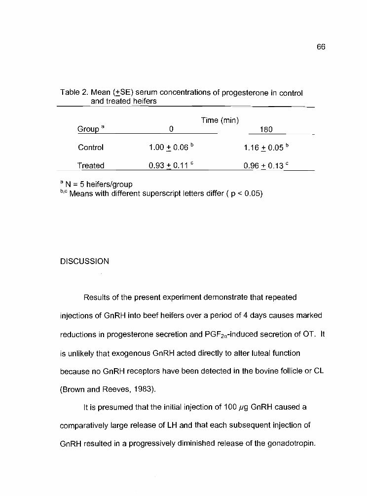
Table 2. Mean (±SE) serum concentrations of progesterone in controland treated heifers
Time (mm)Group a 0 180
Control I.00±O.O6b l.I6±O.OSb
Treated 0.93±0.11c O.96O.13c
a N = 5 heifers/groupb,c Means with different superscript letters differ ( p < 0.05)
DISCUSSION
Results of the present experiment demonstrate that repeated
injections of GnRH into beef heifers over a period of 4 days causes marked
reductions in progesterone secretion and PGF2a-induced secretion of OT. It
is unlikely that exogenous GnRH acted directly to alter Iuteal function
because no GnRH receptors have been detected in the bovine follicle or CL
(Brown and Reeves, 1983).
It is presumed that the initial injection of 100 pg GnRH caused a
comparatively large release of LH and that each subsequent injection of
GnRH resulted in a progressively diminished release of the gonadotropin.

67
Such a pituitary response to repeated injections of GnRH into cows and
anestrous ewes has been reported by Kinder et al. (1975) and Chakraborty
et al. (1974). In cattle, ovarian responses to repeated administration of 150
pg GnRH at 4 hour intervals were evaluated by Kinder et al. (1975). These
heifers were observed to have an increased mean serum concentration of
LH after the initial injection of GnRH which diminished over time with
subsequent injections of GnRH. Exposure of the ovaries to successive
releases of LH failed to affect their function. However, it should be noted
that the repeated injections of GnRH were given during days 16 to 19 of the
cycle when CL function is already waning.
The effect of the current treatment regimen on luteal function could
be due to the excessive quantities of LH to which the developing CL was
exposed. It has been reported by Rodger and Stormshak (1986) that a
single injection of GnRH administered to beef heifers on day 2 of the cycle
resulted in a significant reduction in luteal progesterone secretion. These
latter investigators reported that induced release of LH resulted in a down-
regulation of luteal LH receptors. A similar response to LH in the present
experiment could have accounted, at least in part, for the reduction in
progesterone secretion. It is also possible that exposure of the developing
CL to the excessive amounts of systemic LH may have altered the ratio of
SLC to LLC as the CL developed. Mee et al. (1993) found that an injection

of GnRH at estrus shifted the ratio of these cell types resulting in the
formation of more LLC. It is unlikely that this phenomenon occurred in our
experiment because LLC, although fewer in number then the SLC, produce
the majority of progesterone (Fitz etal., 1982; Niswender et al., 1985) and
are the source of OT (Fields and Fields, 1986; Theodosis et al., 1986)
It is possible that initial stimulations of SLC by LH caused these cells
to produce a paracrine factor that acted on the LLC to impair their function.
Del Vecchio et al. (1994) reported that the perfusate of bovine SLC
suppressed progesterone production of LLC. It is also noteworthy that the
CL is endowed with progesterone receptors (Smith et al., 1995). The
precise role of progesterone receptors in regulating luteal function has yet
to be established. Perhaps continued exposure of the developing CL to LH
over a 4 day period affected the concentration and/or function of the
progesterone receptors, and consequently the overall function of the gland.
Although luteal function was attenuated in heifers treated with GnRH,
it did not result in an abbreviated estrous cycle (control, 19.2 ± 0.48 vs.
treated, 19.5 ± 0.65 days). Thus, enough progesterone was being
produced to maintain a normal feedback effect of the steroid on estrous
behavior. Further studies need to be conducted to elucidate the exact
mechanism by which LH affects luteal OT, progesterone, and progesterone
receptor concentrations during development of the CL.

GENERAL CONCLUSIONS
The role of progesterone, in terms of regulating the uterine OTR, has
been well established (Sheldrick and Flint, 1985; Vallet et al., 1990).
However, the regulatory role (either autocrine or paracrine) progesterone
may play within the ovary remains ambiguous. Rothchild (1981; 1996) has
hypothesized that progesterone may act in an autocrine fashion to regulate
its own synthesis, ultimately affecting its paracrine actions and regulation of
other hormones. Therefore, the underlying theme of the studies presented
was to attempt to determine some link between luteal OT synthesis and/or
secretion and progesterone.
Various in vitro studies have provided evidence for a link between
progesterone and luteal OT (Fehr et al., 1987; Chandrasekher and Fortune,
1990; Lioutas et al., 1997). However, results of our study utilizing the
progesterone antagonist, RU 486, failed to affect luteal OT secretion in
ewes. Although the highest dosage of RU 486 used in our study was
apparently able to block the negative feedback of progesterone at the level
of the hypothalamo-hypophyseal axis, it was not able to interfere with luteal
function. It is possible that the dosage was not sufficient to counteract the
autocrine effects of progesterone at the level of the ovary.
70
In contrast, heifers receiving repeated administration of GnRH during
the early luteal phase resulted in a decline in progesterone and luteal OT
secretion. Perhaps because the CL is endowed with progesterone
receptors (Smith et al., 1995), continued exposure of the developing CL to
LH over a 4 day period affected the concentration and/or function of the
progesterone receptors, and consequently the overall function of the gland.
The question still remains, what link does progesterone play in
regulating luteal OT? Future studies need to be conducted to delineate
the exact role progesterone plays in regulating luteal function in the
ruminant. Experiments designed to determine the effect of progesterone
antagonists and LH-stimulation on progesterone receptor concentrations in
the CL, nuclear and membrane alike, would prove useful. Such
experiments may also help define the intracellular mechanisms that affect
the synthesis of OT upon inhibition or down-regulation of progesterone
receptors.

71
BIBLIOGRAPHY
Abdelgadir SE, Swanson LV, Oldfield JE, Stormshak F. Prostaglandin F2ainduced release of oxytocin from bovine corpora lutea in vitro. BiolReprod 1987; 37:550-555
Adams TE, Kinder JE, Chakraborty PK, Estergreen NL, Reeves JJ. Eweluteal function influenced by pulsatile administration of synthetic LH-RH/FSH-RH. Endocrinology 1975; 97:1460-1467
Al-Matubsi HY, Downing J, Jenkin G, Fairclough RJ. Stimulation of ovarianoxytocin secretion and uterine prostaglandin F2 release byexogenous progesterone early in the cycle of the ovarian auto-transplanted ewe. J Reprod Fertil 1998; 112:278-288
Ashworth AJ, Wilmut I, Springbett AJ, Webb R. Effect of an inhibitor 3t-hydroxysteroid dehydrogenase on progesterone concentrations andembryo survival in sheep. J Endocrinol 1986; 112:205-213
Bailly A, LePlage C, Rauch M, Milgrom E. Sequence specific DNA bindingof progesterone receptor to the utergiobin gene: effects of hormone,antihormone and receptor phosphorylation. EMBO J 1986; 5:3235-
3241
Berndtson AK, Weaver CJ, Fortune JE. Differential effects of oxytocin onsteroid production by bovine granulosa cells. Mol Cell Endocrinol
1996; 116:191-198
Bolden-Tiller OU, Weishons WV, Lucy MC, Smith MF. Progesteronereceptor mRNA and protein expression in bovine corpora lutea. BiolReprod 2000; 62(Suppl 1):268

72
Brinkley HJ, Wickersham EW, First, NL, Casida LE. Effect of unilateralovariectomy on the structure and function of corpora lutea of the pig.Endocrinology 1964; 74:810-815
Brown JL, Reeves JJ. Absence of specific luteinizing hormone releasinghormone receptors in ovine, bovine, and porcine ovaries. BiolReprod 1983; 29:1179-1183
Burgess KM, Jenkin G, Ralph MM, Thorburn GD. Effect of antiprogestin RU486 on uterine sensitivity to oxytocin in ewes in late pregnancy. JEndocrinol 1992; 134:353-360
Campbell BK, deSouza C, Baird DT. Progesterone as a cycle organizer:mifepristone causes ovulation of first wave follicles in sheep. JReprod Fertil 2000; (Abstr. Series) 26:4
Chakraborty PK, Adams TE, Tarnavsky GK, Reeves JJ. Serum andpituitary luteinizing hormone concentrations in ewes infused with LH-RH/FSH-RH. J Anim Sci 1974; 39:1150-1157
Chandrasekher AY, Fortune JE. Effects of oxytocin on steroidogenesis bybovine theca and granulosa cells. Endocrinology 1990; 127:926-933
Collins WE, lnskeep EK, Howland BE, Pope AL, Casida LE. Effects ofhysterectomy and corpus luteum induction on pituitary-ovarianrelationships in the ewe. J Anim Sci 1966: 25:87-91
Corner GW. On the origin of the corpus luteum of the sow from bothgranulosa and theca interna. Am J Anat 1919; 26: 117-1 83
Cosola-Smith CA, Appell LH, Abdelgadir SE, Stormshak F. Stimulation ofbovine luteal oxytocin in vitro by a phorbol ester and calciumionophore. J Anim Sci 1990; 68:2465-2470

73
Del Vecchio RP, Thibodeaux JK, Randel RD, Hansel W. Interactionsbetween large and small bovine luteal cells in a sequential perifusionco-culture system. J Anim Sd 1994; 72:963-968
Dyer RG. Oxytocin and parturition-new complications. J Endocrinol 1988;116:167-168
Fairclough RJ, Moore LG, Mc Gowan LT, Smith JF, Watkins WB. Effect ofexogenous progestins on plasma concentrations of the oxytocin-associated neurophysin and 1 3,14-dihydro-1 5-keto-prostaglandin Fin intact and ovariectomized ewes over the time of expected lutealregression. Biol Reprod 1983; 29:271-277
Fang X, Wong S, Mitchell BF. Effects of RU 486 on estrogen, progesterone,oxytocin, and their receptors in the rat uterus during late gestation.Endocrinology 1997; 138:2763-2768
Fehr S, Ivell R, Koll R, Schams D, Fields M, Richter D. Expression of theoxytocin gene in the large cells of the bovine corpus luteum. FEBSLett 1987; 210:45-50
Fields MJ, Fields PJ. Luteal neurophysin in the non-pregnant cow and ewe:immunocytochemical localization in membrane bound secretorygranules of the large luteal cells. Endocrinology 1986; 118:1723-1725
Fitz TA, Mayan MH, Sawyer HR, Niswender GD. Characterization of twosteroidogenic cell types in the ovine corpus luteum. Biol Reprod1982; 27:703-711
Fleet IR, Davis AJ, Goode JA, Hamon M, Collier RJ, Heap RB. Unilateralcontrol of ovarian oxytocin release and the facilitatory effects on IGF-I in sheep. J Reprod Fertil 1994; 100:623-628.
Flint APF, Leat WM, Sheldrick EL, Stewart HJ. Stimulation ofphosphoinositide hydrolysis by oxytocin and the mechanism bywhich oxytocin controls prostaglandin synthesis in the ovineendometrium. Biochem J 1986; 237:797-805
Flint APF, Sheldrick EL. Ovarian secretion of oxytocin is stimulated byprostaglandin. Nature 1982; 297:587-588
Flint APF, Sheldrick EL, McCann TJ, Jones DSC. Luteal oxytocin:characteristics and control of synchronous episodes of oxytocin andPGF2 secretion at luteolysis in ruminants. Dom Anim Endocrinol1990; 7:111-124
Furuya K, McArdle CA, Ivell R. The regulation of oxytocin gene expressionin early bovine luteal cells. Mo! Cell Endocrinol 1990; 70:81-88
Gilbert CL, Cripps PJ, Wathes DC. Effect of oxytocin on the pattern ofelectromyographic activity in the oviduct and uterus of the ewearound oestrus. Reprod Fertil Dev 1992; 4:193-203
Gimpi G, Fahrenholz F. The oxytocin receptor system: structure, function,and regulation. Phys Rev 2001; 81:629-683
Ginther OJ, Woody CO. Influence of exogenous progesterone and theuterus on ovarian follicles of sheep and heifers. Am J Vet Res 1970;31 :87-90
Ginther OJ, Woody CO, Mahjan S, Jankiraman K, Casida LE. Effect ofoxytocin administration on the oestrous cycle of unitlaterallyhysterectomized heifers. J Reprod Fertil 1967; 225-229

75
Griffin JE, Ojeda SR 2000. The Anterior Pituitary and Hypothalamus. In:Textbook of Endocrine Physiolociy. 4th Revised Edition. OxfordUniversity Press. New York, NY. pp 132-135
Hafez SE. Studies on the breeding season and reproduction of the ewe. JAgric Sd 1952; 42:189-265
Hartwig JH, Thelen M, Rosen A, Janmay PA, Nairn AC, Aderem A.MARCKS is an actin filament crossinking protein regulated by proteinkinase C and calcium-calmodulin. Nature 1992; 356:618-622
Hazzard T, Pinckard KL, Stormshak F. Impact of chronic treatment of eweswith estradiol-1 7 or progesterone on oxytocin receptor genetranscription and ovarian oxytocin secretion. Biol Reprod 1998;59:105-110
Hazzard T, Stormshak F. Down-regulation of oxytocin receptors andsecretion of prostaglandin F2a after chronic treatment of ewes withestradiol-1 7. Biol Reprod 1997; 56:1576-1581
Healy DL, Baulieu EE, Hodgen GD. Induction of menstration by anantiprogesterone steroid (RU 486) in primates: site of action, dose-response relationships, and hormonal effects. Fertil Steril 1983;40:253-257
Heap RB, Fleet IR, Davis AJ, Goode JA, Hamon MH, Walters DE, FlintAPF. Neurotransmitters and lymphatic-vascular transfer ofprostaglandin F2 stimulate ovarian oxytocin output in sheep. JEndocrinol 1989; 122:147-159
Hoefler WC, Holcombe DW, Blackwell JA, Hinrichs BD, Haliford DM.Reproductive responses, progesterone profiles, and ovariancharacteristics of cycling, fine-wool ewes treated with an inhibitor ofprogesterone synthesis (epostane). J Anim Sci 1986; 63:1072-1077
76
Holtori AP, Furuya K, Ivell R, McArdle CA. Oxytocin production andoxytocin mRNA levels I. Bovine large luteal cells are regulated byinsulin and IGF-l: dependence on the status of the ovarian follicle.Endocrinology 1989; 125:2612-2620
Hooper SB, Watkins WB, Thorburn GD. Oxytocin, oxytocin-associatedneurophysin, and prostaglandin F2a concentrations in the utero-ovarian vein of pregnant and nonpregnant sheep. Endocrinology1986; 119:2590-2597
lnskeep EK, Butcher RL. Local component of utero-ovarian relationships inthe ewe. J Anim Sci 1966; 25:1164-1168
Ivell R, Brackett KH, Fields MJ, Richter D. Ovulation triggers oxytocin geneexpression in the bovine ovary. FEBS Lett 1985; 190:863-267
Ivell R, Richter D. The gene for the hypothalamic peptide hormone oxytocinis highly expressed in the bovine corpus luteum: biosynthesis,structure and sequence analysis. EMBO J 1984; 3:2351-2354
Jenkin G, Gemmell RT, Thorburn GD. Induction of transient functionalluteotysis in cyclic sheep by a 3f3-hydroxysteroid dehydrogenaseinhibitor. J Endocrinol 1983; 100:61 -66
Jones DSC, Flint APF. Concentrations of oxytocin-neurophysin prohormonemRNA in the corpus luteum of sheep during the estrous cycle and inearly pregnancy. J Endocrinol 1988; 117:409-414
Jungclas B, Luck MR. Evidence for granulosa-theca interaction in thesecretion of oxytocin by bovine ovarian tissue. J Endocrinol 1986;I 09:R1-R4
77
Kinder JE, Adams TE, Chakraborty PK, Tarnavsky GK, Reeves JJ. SerumLH concentrations and ovarian activity in cows with repetitiveadministration of LH-RH/FSH-RH. J Anim Sd 1975; 41:1650-1652
Koligan KB and Stormshak F. Progesterone synthesis by ovine fetalcotyledons in vitro. J Anim Sd 1976; 42:439-443
Kooistra L, Ginther OJ. Effect of photoperiod on reproductive activity andhair in mares. Am J Vet Res 1975; 36:1413-1419
Lioutas C, Einspanier A, Kascheike B, Walther N, Ivell ft An autorcrineprogesterone positive feedback loop mediates oxytocin upregulationin bovine granulosa cells during luteinization. Endocrinology 1997;
138:5059-5062
Luck MR, Jungclas B. Catecholamines and ascorbic acid as stimutators ofbovine ovarian oxytocin secretion. J Endocrinol 1987; 114:423-430
Mee MO, Stevenson JS, Alexander BM, Sasser GR. Administration ofGnRH at estrus influences pregnancy rates, serum concentrations of
LH, FSH, estradiol-17$, pregnancy specific protein B, andprogesterone, proportion of luteal cell types and in vitro production ofprogesterone in dairy cows. J Anim Sci 1993; 75:185-198
McCracken, JA. Hormone receptor control of prostaglandin F2a secretion bythe ovine uterus. Adv Prostaglandin Thromboxane Res 1980;8:1329-1344
McCracken JA, Carison JC, Clew ME, Coding JR, Baird DT, Green K,
Samuelsson B. Prostaglandin F2a identified as the luteolytic hormonein sheep. Nature 1972; 238:129-1 34

McCracken JA, Custer EE, Eldering JA, Robinson AG. The central oxytocinpulse generator: a pacemaker for the ovarian cycle. Acta NeurobiolExp 1996; 56:819-32
Miyamoto A, Bruckmann A, vonLutzow H, Schams D. Multiple effects ofneuropeptides Y, substance P, and vasoactive intestinal polypeptideon progesterone and oxytocin release from bovine corpus luteum invitro. J Endocrinol 1993; 138:451-458
Morgan GL, Geisert RD, McCann JD, Bazer FW, Ott TL, Mirando MA,Stewart M. Failure of luteolysis and extension of the interoestrousinterval in sheep treated with the progesterone antagonistmifepristone (RU 486). J Reprod Fertil 1993; 98:451-457
Niswender GD, Schwall RH, Fitz TA, Farm CE, Sawyer HR. Regulation ofluteal function in domestic ruminants: new concepts. In: R.O. Creep,(Ed.) Rec Prog Horm Res 1985; 41:101-151
Norman, AW, Litwack GL. 1997. Hormones 2' Edition. Academic Press.San Diego, CA.
Orwig KE, Bertrand JE, Ou BR, Forsberg NE, Stormshak F. Involvement ofprotein kinase-C, calpains, and calpastatin in prostaglandin F2ainduced oxytocin secretion from the bovine corpus luteum.Endocrinology 1994a; 134:78-82
Orwig KE, Bertrand JE, Ou BR, Forsberg NE, Stormshak F.Immunochemical characterization and cellular distribution of proteinkinase C isozymes in the bovine corpus luteum. Comp BiochemPhysiol 1994b; 108:53-57
Peters AR, Lamming GE. The effect of epostane (Win32739), an inhibitor of33-hydroxysteroid dehydrogenase, on luteal function andgonadotropin secretion in cows. Anim Reprod Sci 1986; 10:91-98
79
Philbert D. 1984. RU-3486: an original multi-faceted anti-hormone in vivo.
In: Adrenal Steroid Antagonism. de Gruyter, New York, NY. pp. 77-
101
Pleim ET, Cailliau PJ, Weinstein MA, Etgen AM, Barfield RJ. Facilitation ofreceptive behavior in estrogen-primed female rats by anti-progestin,RU 486. Horm Behav 1990; 24:301-310
Rae MT, Menzies GS, Bramley TA. Bovine ovarian non-genomicprogesterone binding sites: presence in follicular and luteal cellmembranes. J Endocrinol 1998a; 159:413-427
Rae MT, Menzies GS, McNeilly AS, Woad K, Webb R, Bramley TA.Specific non-genomic, membranelocalized binding sites forprogesterone in the bovine corpus luteum. Biol Reprod 1998b;58:1394-1 406
Rahe CH, Owens RE, Fleeger JL, Newton HJ, Harms PG. Pattern ofplasma luteinizing hormone in the cyclic cow: dependence upon the
period of the cycle. Endocrinology 1980; 107:498-503
Rauch M, Loosfelt H, Philbert D, Milgrom E. Mechanism of action ofantiprogesterone, RU 486, in the rabbit endometrium: effects of RU
486 on the progesterone receptor expression of the uteroglobin
gene. EurJ Biochem 1985; 148:213-218
Richter D. Vasopressin and oxytocin are expressed as polyproteins. TrendsBiochem Sd 1983; 8:278-281.
Roberts JS, McCracken JA. Does prostaglandin F2 released from theuterus by oxytocin mediate the oxytocic action of oxytocin? BiolReprod 1976; 15: 457-463
Roberts JS, McCracken JA, Gavagon JE, Soloff MS. Oxytocin-stimulatedrelease of prostaglandin F2a from ovine endometrium in vitro:correlation with estrous cycle and oxytocin receptor binding.Endocrinology 1976; 99:1107-1114
Rodger LD, Stormshak F. Gonadotropin-releasing hormone-inducedalteration of bovine corpus luteum function. Biol Reprod 1986;35:149-1 56
Rothchild I. The regulation of the mammalian corpus luteum. Rec ProgHorm Res 1981; 37:183-298
Rothchild I. The corpus luteum revisited: are the paradoxical effects of RU486 a clue to how progesterone stimulates its own secretion? BiolReprod 1996; 55:1-4
Salli U, Supancic S, Stormshak F. Phosporylation of myristoylated alanine-rich C kinase substrate (MARCKS) protein is associated with bovineluteal cell exocytosis. Biol Reprod 2000; 63:12-20
Sampson WK. 2000. Posterior Pituitary and Water Metabolism. In:Textbook of Endocrine Physiolociy. 4th Revised Edition. OxfordUniversity Press. New York, NY. pp 163-182
Schallenberger E, Schams D, Bullermann B, Walters DL. Pulsatile secretionof gonadotrophins, ovarian steroids and ovarian oxytocin duringprostaglandin-induced regression of the corpus luteum in the cow. JReprod Fertil 1984; 71 :493-501
Schams D. Oxytocin determination by radioimmunoassay. Ill. Improvementto subpicogram sensitivity and application to blood levels in cycliccattle. Acta Endocrinol 1983: 103:180-183
sLIL'Ii
Schams D, Kruip TAM, Koll R. Oxytocin determination in steroid producing
tissues and in vitro production in ovarian follicles. Acta Endocrinol
I 985a; 109:530-536
Schams D, Lahlou-Kassi A, Glatzel P. Oxytocin concentrations in peripheralblood during the oestrous cycle and after ovariectomy in two breeds
of sheep with low and high fecundity. J Endocrinol 1982; 92:9-13
Schams D, Schallenberger E, Legros JJ. Evidence for the secretion of
immunoreactive NP I in cattle. J Reprod Fertil 1985b; 73:165-171
Schane PH, Potts GO, Creange JE. Inhibition of ovarian, placental, andadrenal steroidogenesis in the Rhesus monkey by trilostane. Fertil
Steril 1979; 32:464-467
Senger PL. 1999. The Estrous Cycle. In: Pathways to Precinancy and
Parturition. 1st Revised Edition. The Mack Printing Group-Science
Press. Ephrata, PA. pp 120-121
Sheidrick EL, Flint APF. Luteal concentrations of oxytocin decline during
early pregnancy in the ewe. J Reprod Fertil 1983; 68:477-480
Sheldrick EL, Flint APF. Endocrine control of uterine oxytocin receptors in
the ewe. J Endocrinol 1985; 106:249-258
Sheldrick EL, Flint APF. Post-translational processing of oxytocin-neurophysin prohormone in the ovine corpus luteum: activity of
peptidyl glycine ct-amidating mono-oxygenase and concentrations of
its cofactor, ascorbic acid. J Endocrinol 1989; 122:313-322

Shoupe D, Mishell DR, Lacarra M, Gutierrez E, Lahteenmaki P, Spitz IM1985. Endocrinologic effects of the antiprogesterone RU 486. In:Reproductive Biology: The antiprogestin steroid RU 486 and HumanFertility Control. Baulieu EE, Segal S (Ed.). Plenum Press. NewYork, NY. pp 285-293
Shuckovski L, Findlay JK. Activin-A inhibits oxytocin and progesteroneproduction by preovulatory bovine granulosa cells in vitro.Endocrinology 1990; 126:2222-2224
Shuckovski L, Findlay JK, Smith Al. Identification of non-covalent oxytocin-neurophysin I complex in the bovine ovary. J Endocrinol 1991;128:305-314
Simmons KR, Hansel W. Nature of the luteotropic hormone in the bovine. JAnim Sd 1964; 23:136-141
Slayden 0, Stormshak F. Suppressive action of gonadotropin-releasinghormone and luteinizing hormone on function of the developing ovinecorpus luteum. J Anim Sci 1990; 68:2425-2429
Smith GW, Gentry PC, Long DK, Bao B, Roberts RM, Smith MF.Expression of progesterone receptor mRNA within ovine post-surgefollicles and corpora lutea. Biol Reprod 1995; 52(Suppl 1):151
Stefenson A, Owman C, Sjoberg NO, Sporrong B, Walles B. Comparativestudy of the autonomic innervation of the mammalian ovary, withparticular regard to the follicular system. Cell Tissue Res 1981;215:47-62
Swann RW, O'Shaughnessy PJ, Birkett SD, Wathes DC, Porter DG,Pickering BT. Biosynthesis of oxytocin in the corpus luteum. FEBSLett 1984; 174:262-266
Theodosis DT, Wooding FBP, Sheldrick EL, Flint APF. Ultrastructurallocalization of oxytocin and neurophysin in the ovine corpus luteum.Cell Tissue Res 1986; 243:129-1 35
Vallet JL, Lamming GE, Baften M. Control of endometrial oxytocin receptorand uterine response to oxytocin by progesterone and estrogen inthe ewe. J Reprod Fertil 1990; 90:625-634
Voss AK, Fortune JE. Oxytocin secretion by bovine granulosa cells: effectsof stage of follicular development, gonadotropins and co-culture with
theca interna. Endocrinology 1991; 128:1991-1999
Voss AK, Fortune JE. Oxytocin-NP I mRNA in bovine granulosa cellsincreases after luteinizing hormone surge and is stimulated byluteinizing hormone in vitro. Endocrinology 1992; 131 :2755-2762
Voss AK, Fortune JE. Estradiol-17 8 has a biphasic effect on oxytocinsecretion by bovine granulosa cells. Biol Reprod 1993; 48:1404-1409
Waither N, Wehrenberg U, Kascheike B, Jansen M, Ivell R 1995.Regulation of oxytocin expression in the bovine corpus luteum. In:Oxytocin. Ivell R, Russell J (Ed.). Plenum Press. New York, NY. pp
79-90
Watanabe K, Yoshida R, Shimizu T, Hayaishi 0. Enzymatic formation of
prostaglandin F2a from prostaglandin H2 and D2. Purification andproperties of prostaglandin synthetase from bovine lung. J Biol Chem
1985; 260:7035-7041
Wathes DC, Denning-Kendall PA, Perks C, Brown D. Effects of stage of the
cycle and estradiol-1 7I on oxytocin synthesis by ovine granulosa
and luteal cells. Endocrinology 1992; 130:1009-1016

Wathes DC, Guldenaar SEF, Swann RW, Webb R, Porter DC, PickeringBT. A combined radioimmunoassay and immunocytochemical studyof ovarian oxytocin production during the periovulatory period in theewe. J Reprod Fertil 1986; 78: 167-183
Wathes DC, Swann RW. Is oxytocin an ovarian hormone?. Nature 1982;297:225-227
Wathes DC, Swann RW, Birkett SD, Porter DG, Pickering BT.Characterization of oxytocin, vasopressin, and neurophysin from thebovine corpus luteum. Endocrinology 1983; 113:693-698
Wathes DC, Swann RW, Pickering BT. Variations in oxytocin, vasopressin,and neurophysin concentrations in the bovine ovary during theoestrous cycle and pregnancy. J Reprod Fertil 1984; 71 :551-557
Webb R. Increasing ovulation rate and lambing rate in sheep by treatmentwith a steroid enzyme inhibitor. J Reprod Fertil 1987; 79:231-240
Whitmore DL, Bechert US, Rowe KE, Stormshak F. Luteal function inunilaterally hysterectomized ewes treated with gonadotropin-releasing hormone. J Anim Sci 1996; 74:1930-1935
Wiltbank JN, Rothlisberger JA, Zimmerman DR. Effect of human chorionicgonadotropin on maintenance of the corpus luteum and embryonicsurvival in the cow. J Anim Sci 1961; 20:827-829
Zhang L, Weston PG, Hixon JE. Role of progesterone and oestradiol in theregulation of uterine oxytocin receptors in ewes. J Reprod Fertil1992; 94:395-404
Zingg HH, Grazzini E, Breton C, Larcher A, Rozen F, Russo C, Guillon G,Mouillac B. 1998. Genomic and non-genomic mechanisms ofoxytocin receptor regulation. In: Vasopressin and Oxytocin.Molecular, Cellular, and Clinical. Adv Exp Med Biol. HH Zingg, CWBourque, DG Bichet (Ed.). Plenum Press, NY. pp 287-295