extraversion and anterior vs. posterior dmn activity during self-referential thoughts
TRANSCRIPT
ORIGINAL RESEARCH ARTICLEpublished: 07 January 2013
doi: 10.3389/fnhum.2012.00348
Extraversion and anterior vs. posterior DMN activityduring self-referential thoughtsGennady G. Knyazev*
Institute of Physiology, Siberian Branch of Russian Academy of Medical Sciences, Novosibirsk, Russia
Edited by:
Luke D. Smillie, The University ofMelbourne, Australia
Reviewed by:
Luke D. Smillie, The University ofMelbourne, AustraliaMira Chavanon, Philipps-University,Germany
*Correspondence:
Gennady G. Knyazev, Institute ofPhysiology, Siberian Branch ofRussian Academy of MedicalSciences, Timakova str., 4,Novosibirsk 630117, Russia.e-mail: [email protected]
Recent studies show that fronto-posterior electroencephalogram (EEG) spectral powerdistribution is associated with personality. Specifically, extraversion is associated withan increase of spectral power in posterior cortical regions that overlap with theposterior default mode network (DMN) hub and a decrease of spectral power inanterior regions that overlap with the anterior DMN hub. Although there is evidencethat dopaminergic neurotransmission may be involved, psychological processes thatunderlie these associations remain unclear. I hypothesize that these processes may havesomething to do with spontaneous self-referential thoughts. Specifically, I hypothesizethat in extraverts self-referential thoughts may be associated with an increase of spectralpower in the posterior DMN hub, whereas in introverts they may be associated with anincrease of spectral power in the anterior DMN hub. After spontaneous EEG registration,participants were asked to fill in a questionnaire describing their thoughts during theregistration. An item describing self-referential positive expectations (SRPE) was used tomeasure individual differences in the intensity of these processes. Source localization andindependent component analyses were applied to EEG data to reveal oscillatory activityassociated with the anterior and the posterior DMN hubs. Hierarchical regression analysisshowed a significant interaction between extraversion scores and anterior vs. posteriorDMN alpha activity in predicting individual differences in SRPE scores. In extraverts, highSRPE scores were associated with an increase of alpha power in the posterior DMN hub,whereas in introverts they were associated with an increase of alpha power in the anteriorDMN hub. Results are discussed in terms of differential involvement of the two DMNhubs in self-related reward processes in extraverts and introverts.
Keywords: extraversion, default mode network, EEG, alpha oscillations, independent component analysis
INTRODUCTIONExtraversion is one of a few major dimensions of personalitywhich consistently appear in most personality models. The view,which currently is most popular, links extraversion with the activ-ity of the brain’s dopaminergic (DA) reward system (Depue andCollins, 1999; Smillie et al., 2006). However, investigation of DAneurotransmission in humans requires either invasive measure-ments or expensive neuroimaging techniques. In this connection,the electroencephalographic (EEG) index of DA neurotransmis-sion, which has been suggested by Wacker et al. (2006), seemsvery attractive. This suggestion is based on observations of anassociation between extraversion and posterior vs. frontal EEGactivity (Hewig et al., 2004, 2006; Wacker et al., 2006). Wackeret al. (2006) demonstrated that the negative association betweenextraversion and the frontal minus parietal theta activity, whichwas observed in the placebo group, was completely reversed inthe group that received the selective DA D2 antagonist. Similareffect was observed in alpha band. This finding implies thatthe fronto- posterior distribution of spectral power may reflecttrait-like predispositions which depend on the brain DA func-tioning. This group of researchers has replicated this finding inseveral studies [see e.g., meta-analysis by Wacker et al. (2010)].Moreover, they found an association between the posterior minus
frontal slow activity on the one hand and polymorphisms of theDA D2 receptor (Koehler et al., 2011) and enzyme catechol-O-methyltransferase (Wacker and Gatt, 2010) on the other hand. Atleast one another independent group found a similar associationof extraversion with the fronto- posterior spectral power distri-bution (Knyazev, 2009, 2010; Knyazev et al., 2012a). However,these findings leave unanswered the question about psychologicalprocesses that underlie these associations.
It could be noted that the above described associations arein line with some other findings indicating that posterior cor-tical areas may be more active in extraverts, at least in somecircumstances. For example, Yuan et al. (2012) show that poste-rior cingulate cortices may mediate extraversion-related effect forpleasant stimuli, which essentially results in a decreased thresholdfor pleasant emotion and an increased threshold for unpleasantemotion. Higher scores on extraversion were found to be associ-ated with higher amplitudes of the P300 component of the ERPselicited by human faces in parietal cortical regions (Fishman et al.,2011). Phillips et al. (2012) show that monkeys who demonstratehigher levels of exploratory approach behavior have significantlygreater gray matter density in the precuneus. On the other hand,there is indirect evidence implying that prefrontal cortical regionsmight be less active in extraverts. Thus, neural valuation signals
Frontiers in Human Neuroscience www.frontiersin.org January 2013 | Volume 6 | Article 348 | 1
HUMAN NEUROSCIENCE
Knyazev Extraversion and anterior vs. posterior DMN
in the anterior cingulate cortex and functional coupling of thisregion with hippocampus and amygdala predict the degree towhich future thinking modulates individual preference functionsand reduces the rate of delay discounting (Peters and Büchel,2010); it is known that extraversion is associated with greaterdiscounting (Richards et al., 1999).
THE FRONTO- POSTERIOR SPECTRAL POWER GRADIENT AND THEDEFAULT MODE NETWORKBecause most of the associations between extraversion and pos-terior vs. frontal EEG were observed in resting conditions (butsee Knyazev, 2009), it seems reasonable to suggest that they mayrelate to psychological processes and respective brain networkswhich are most active in these conditions. Recent studies haverevealed several networks in the brain which are active in the rest-ing state. Most intriguing findings and ideas are associated withthe so-called default mode network (DMN). The DMN is a con-stellation of brain areas which decrease their activity during awide number of different goal-oriented tasks as compared to pas-sive “rest” tasks (Raichle and Snyder, 2007). Interestingly, severalDMN regions are also related to social cognition (Mitchell, 2006;Gobbini et al., 2007). In line with these findings recent studieshave revealed DMN abnormalities in autistic patients (Kennedyet al., 2006; Kennedy and Courchesne, 2008) and in patients withsocial phobia (Gentili et al., 2009). Although EEG has lower spa-tial resolution than fMRI and has no direct access to deep corticalregions (e.g., DMN’s midline cortices), it could be noted that theanterior and the posterior cortical areas, whose oscillatory activ-ity shows correlations with extraversion, overlap with the anteriorand the posterior DMN hubs, respectively. The relation of theposterior vs. anterior spectral power distribution to DMN wasalso hypothesized by Wacker et al. (2010) and Chavanon et al.(2011).
THE ANTERIOR AND THE POSTERIOR DMN HUBSThe DMN comprises a set of brain regions that are co-activatedduring passive states and show intrinsic functional correlationswith one another. However, there is clear evidence that the brainregions within the DMN contribute specialized functions thatare organized into subsystems that converge on hubs (Buckneret al., 2008). Maps of the intrinsic correlations within the defaultnetwork show that it comprises at least three interacting subsys-tems. The medial temporal lobe subsystem functions to provideinformation from prior experiences in the form of memoriesand associations that are the building blocks of mental simula-tion; the medial prefrontal cortex (MPFC) subsystem facilitatesthe flexible use of this information during the construction ofself-relevant mental simulations. These two subsystems convergeon the precuneus/posterior cingulate cortex (pC/PCC) (Buckneret al., 2008). Partial correlation network analysis suggests thatthis latter region may play a pivotal role in the DMN (Franssonand Marrelec, 2008). Indeed, PET studies have shown that themetabolic activity is higher in the pC/PCC than all other regionsduring rest (Gusnard and Raichle, 2001). It could be suggestedthat being the place of integration of prior experiences with men-tal simulations, the pC/PCC sustains a sense of self-consciousnessthat is engaged in self-referential mental thoughts during rest
(for reviews see Cavanna and Trimble, 2006; Buckner and Carroll,2007) and is commonly associated with positive emotions (Kooleet al., 2001).
THE ANTERIOR DMN HUBIt appears that social salience which reflects the relation betweenothers and oneself is processed by the MPFC (Iacoboni et al.,2004; Schmitz et al., 2004; Seger et al., 2004; Han et al., 2005;Mitchell et al., 2005a,b; Ochsner et al., 2005). MPFC is activatedduring thinking about the complex interactions among people(Buckner et al., 2008). There is ground to suggest that think-ing about the complex interactions among people is frequentlyaccompanied by negative emotion. Besides, the anterior mid-cingulate cortex is implicated in the integration of negative affect,pain, and cognitive control (Shackman et al., 2011). In general,being a part of the prefrontal cortex, the MPFC is inevitablyinvolved in conscious planning, decision making, and controlfunctions (see e.g., Luk and Wallis, 2009; Alexander and Brown,2011). Therefore, these cortices are bound to be reciprocallyrelated to motivational centers, such as amygdala and striatum(Quirk and Beer, 2006; Urry et al., 2006; Goldin et al., 2008;Ochsner and Gross, 2008). Indeed, much evidence shows that,the MPFC controls the accumbens dopamine responses to envi-ronmental challenges (e.g., Pascucci et al., 2007) and dopaminerelease in the MPFC exerts an inhibitory influence on dopaminerelease in the nucleus accumbens, whereas depletion of meso-cortical DA facilitates activation of mesoaccumbens DA release(Deutch et al., 1990; Doherty and Gratton, 1996; King et al.,1997).
THE POSTERIOR DMN HUBThe pC/PCC cortices, which constitute the posterior DMNhub, are involved in self-centered cognition (e.g., ongoing self-monitoring) and self vs. others discrimination (Vogt et al., 2006).First-person-perspective taking in social interaction and in a lan-guage task shows differential activation in the medial aspects ofthe superior parietal lobe and the right temporo-parietal junc-tion (Vogeley et al., 2001; Vogeley and Fink, 2003). PCC, theretrosplenial, and the medial parietal cortices are implicated inputting self-referential stimuli within a temporal context, link-ing them to past self-referential stimuli (Northoff et al., 2006).Transcranial magnetic stimulation over the medial parietal regioncaused a decrease in the efficiency of retrieval of previous judg-ment of mental self as compared to retrieval of judgment of other,confirming that this region may be a nodal structure in self-representation (Lou et al., 2004). Direct appraisals of self as com-pared to reflected appraisals recruited PCC (Ochsner et al., 2005).Besides, the right inferior parietal cortex and precuneus may bespecifically involved in distinguishing self-produced actions fromthose generated by others (Ruby and Decety, 2001). It shouldbe borne in mind also that the parietal cortex is activated byemotional stimuli that are not the focus of attention and are there-fore perceived mostly unconsciously (Iidaka et al., 2001; Knyazevet al., 2009). Moreover, the parietal cortex is a part of the dorsal(non-conscious) processing stream which contributes to vision-for-action (Milner and Goodale, 1995; Goodale and Milner, 2008)and participates in salience detection (Husain and Nachev, 2007).
Frontiers in Human Neuroscience www.frontiersin.org January 2013 | Volume 6 | Article 348 | 2
Knyazev Extraversion and anterior vs. posterior DMN
Non-spatial salience detection functions are particularly associ-ated with the inferior parietal lobe, which in humans consistsof novel cortical areas not shared with other primates (Husainand Nachev, 2007). Summing up, existing evidence shows thatthe anterior DMN hub is involved in mostly conscious model-ing, planning, and control functions whereas the posterior hubis involved in mostly unconscious processes that include self-representation, emotion, and salience detection. In the contextof the present study, it is interesting to note that according toGray’s (1999) theory, salience detection is the main function ofthe dopaminergic reward system.
THE ANTERIOR AND POSTERIOR DMN HUBS AND DABecause the association between extraversion and posterior vs.anterior EEG activity is mediated by dopamine, it is interestingto note that the anterior and the posterior DMN hubs appear tobe differently susceptible to dopaminergic influences. Generally,dopaminergic effects appear to be more pronounced in the pos-terior than in the anterior hub or these effects could be ofopposite directions. Many of these observations have been madeon Parkinson’s disease (PD) patients. Thus, van Eimeren et al.(2009) show that patients with mild to moderate PD (not takingmedication) and healthy controls showed comparable deactiva-tion of the MPFC, but different deactivation in the pC/PCC.Compared with controls, PD patients not only showed less deacti-vation of the pC/PCC, they even demonstrated a reversed patternof activation and deactivation. Dopamine medication appearsto restore the normal pattern of task-related deactivation in theposterior DMN hub. Thus, PD patients taking placebo only deac-tivated the ventral MPFC during a facial emotion recognitiontask. They failed to deactivate the posterior midline and lat-eral parts of DMN. After levodopa administration, this networkwas restored conjointly with the improvement of motor dysfunc-tion in PD patients (Delaveau et al., 2010). In another study,PD patients were scanned twice, once while on their DA med-ication (ON condition) and once after medication withdrawal(OFF condition). Higher activation in the precuneus was foundin the ON condition (Dusek et al., 2012). Krajcovicova et al.(2012) using the daily levodopa equivalent dose in cognitivelyunimpaired PD patients as a covariate observed an enhancedfunctional connectivity of the DMN in the posterior cingulatecortex during a cognitive task. Similar effects were observednot only in PD patients, but also in healthy older adults whocompared with younger adults showed diminished fMRI deac-tivations in pC/PCC during memory recognition. In youngeradults, greater task-induced deactivation in this region was asso-ciated with higher dopamine synthesis capacity (as measured bythe radiotracer 6-[18F]-fluoro-L-m-tyrosine). The authors sug-gest that DA system helps modulate the posterior DMN hubactivity in younger adults and that alteration to the DA systemmay contribute to age-related changes in working memory func-tion (Braskie et al., 2011). Healthy adult subjects that receivedmethylphenidate (a stimulant drug that amplifies dopaminergicsignaling in the brain) had increased deactivation during workingmemory and visual attention tasks in the insula and the PCC (butnot in the MPFC) than the group of subjects who received placebo(Tomasi et al., 2011).
Some authors observed opposite dopamine-related effects inthe posterior and the anterior DMN hubs. Thus, Tomasi et al.(2009) assessed the relationship between DA transporters (DAT,which regulate extracellular dopamine in the brain) in stria-tum (measured with positron emission tomography and [11C]cocaine used as DAT radiotracer) and brain activation and deac-tivation during a parametric visual attention task (measured withBOLD fMRI) in healthy controls. DAT availability in caudate andputamen had a negative correlation with deactivation in ventralparietal regions of the DMN (precuneus, BA7) and a positive cor-relation with deactivation in the ventral anterior cingulate gyrus(BA24/32). Similarly, Asanuma et al. (2006) show that, levodopatherapy was associated with significant metabolic increases inthe precuneus (BA7) but decreases in the MPFC. This evidenceappears to suggest that dopamine may exert inhibitory effect onthe anterior and excitatory effect on the posterior DMN hub. Theformer effect is in line with animal data showing that DA increasesthe threshold for spike firing and exerts an inhibitory action in theprefrontal cortex (Geijo-Barrientos and Pastore, 1995).
In sum, the evidence presented in the previous sectionsappears to suggest that although both the anterior and the poste-rior DMN hubs are involved in self-centered and social cognitionand are co-activated during passive states, they are associatedwith rather different functions. The anterior DMN is moreinvolved in integration, planning, and control functions, whichare mostly conscious and are reciprocally related to dopaminer-gic reward processes. The posterior DMN is more involved inself-representation and salience detection. The latter processesare mostly unconscious and are positively related to dopaminer-gic reward processes. The former processes are less and the latterprocesses are more pronounced in extraverts than in introverts.
THE PRESENT STUDYI hypothesize that the association between extraversion and theresting state posterior vs. frontal EEG activity is mediated byDMN-related spontaneous self-referential processes in such away that in more extraverted individuals these processes areassociated with an increase of spectral power in the posteriorDMN hub, whereas in more introverted individuals they couldbe associated with an increase of spectral power in the anteriorDMN hub. In this study, I aimed to obtain EEG records duringunconstrained mind-wandering and to test whether extraversionmoderates the associations between the prevalence of relevantself-referential thoughts and EEG spectral power within the ante-rior and the posterior DMN hubs. The existence of an associationbetween self-referential thoughts and EEG spectral power withinthe DMN has been shown previously (Knyazev et al., 2011,2012b). The choice of a relevant measure of extraversion and arelevant measure of self-referential thoughts was guided by thehypothesis linking these processes with dopaminergic transmis-sion and social cognition. Depue and Collins (1999) argue thatextraversion can be subdivided into two subfactors: affiliation andagency. They propose a dopaminergic basis for the agency facetof extraversion (i.e., a motivational disposition that comprisessocial dominance, enthusiasm, energy, assertiveness, ambitious-ness, and achievement striving). Keeping in mind that the DMN,which is the main focus of this study, is supposedly involved in
Frontiers in Human Neuroscience www.frontiersin.org January 2013 | Volume 6 | Article 348 | 3
Knyazev Extraversion and anterior vs. posterior DMN
self-referential processes in the context of interpersonal relation-ships (e.g., Mitchell, 2006), assertiveness appears to be the facetof extraversion which best captures both the agentic properties ofthis dimension and its projection onto the space of interpersonalrelationships. High assertiveness scorers are independent, domi-nant, and stand up for their rights. They tend to be at the center ofattention at meetings. Low scorers are humble, timid, submissive,and disinclined to take initiative in interpersonal situations, andmay be easily imposed upon (Eysenck and Wilson, 2000). Withregard to the relevant kind of self-referential thoughts, I intendedto capture an aspect of anticipation of a positive reinforcementthat is peculiar to extraverts and is supposedly mediated by thedopaminergic reward system.
METHODSSUBJECTSResting EEG data were collected in 60 healthy volunteers (32men and 28 women; age range 17–30 years, mean = 20.4, SD =2.5), mostly university students. All applicable subject protectionguidelines and regulations were followed in the conduct of theresearch in accordance with the Declaration of Helsinki. All par-ticipants gave informed consent to the study. The study has beenapproved by the Institute of Physiology ethical committee.
INSTRUMENTS AND PROCEDURESParticipants were seated in a soundproof dimly illuminated roomand did not receive any instruction. The spontaneous EEGregistration lasted about 6 min and included alternating 2 minintervals with eyes open and eyes closed. Only the eyes closedcondition was used in this study because previous research hasshown that self-referential thoughts correlate with EEG spectralpower in the eyes closed, but not in the eyes open condition(Knyazev et al., 2011). Just after the EEG registration partici-pants were asked to fill in a brief (35 items) spontaneous thoughtsquestionnaire (STQ) which described different aspects of theirstate, thoughts, and feelings during the registration. All itemswere measured on a five-point Likert scale. Factor analysis of allquestionnaire items (principal components factor analysis withvarimax rotation) showed that a four-factor solution best fittedthe data. Accordingly, four scales were created that described ner-vousness/negative emotion/lack of positive emotion (NE, exam-ple items: “felt nervous,” “experienced negative emotions,” “wascalm and relaxed”—reverse scoring, “liked the procedure”—reverse scoring, Cronbach’s alpha = 0.84); self-referential thought(SRT, example items: “thought about something pleasant that isgoing to happen to me in the near future,” “recollected episodesfrom my own life,” “most of the time, thoughts of my recentpast recurred to me,” “most of the time, I was absorbed in myprivate thoughts,” Cronbach’s alpha = 0.69); arousal level (exam-ple items: “was almost asleep”—reverse scoring, “was quiet andrelaxed”—reverse scoring, “was somewhat heated,” “was veryexcited,” Cronbach’s alpha = 0.72); attention to environment(ATT, “my attention was mostly directed to external stimuli,”“most of the time, I listened to sounds and skin sensations,”“did not pay any attention to external stimuli”—reverse scor-ing, Cronbach’s alpha = 0.65). The SRT scale (SRTS) was usedto measure individual differences in mental processes that are
presumably related to DMN activity. Besides, for the purposeof this study, I additionally used the first item from this scale(see above), which describes self-referential positive expectations(hereafter SRPE). After filling in the questionnaire subjects par-ticipated in experiments which are not described here. After theexperiments they filled in a set of personality questionnaires andwere debriefed. Facets of Extraversion were measured by respec-tive scales from the Eysenck Personality Profiler (EPP, Eysenckand Wilson, 2000; Knyazev et al., 2004). Assertiveness scale(Cronbach’s alpha = 0.78) consisted of 20 items. Example item:“Do you find it difficult to get rid of a salesperson who is persis-tent and wasting your time?”. Activity (Cronbach’s alpha = 0.73)and Sociability (Cronbach’s alpha = 0.81) scales were additionallyused in order to test the specificity of observed effects.
EEG RECORDINGEEG data were recorded using 32 silver-silver chloride electrodesmounted in an elastic cap on the positions of the international10–20 system. The signals were amplified with a multichannelbiosignal amplifier with a gain of 250 and a bandpass 0.05–70 Hz,−6 dB/octave and continuously digitized at 300 Hz. All record-ings were performed using a fronto-central electrode as groundand electronically linked mastoid electrodes as reference. Thehorizontal and vertical electrooculogram was registered simulta-neously. Electrode impedances were at or below 5 k� for all elec-trodes used in the analysis. EEG data were artifact-corrected usingICA via EEGLAB toolbox (http://www.sccn.ucsd.edu/eeglab/)retaining minimally 20 out of 30 components.
3D SOURCE RECONSTRUCTIONTo determine the cortical sources of EEG activity, sLORETA(Pascual-Marqui, 2002) was applied to the data. sLORETA usesa three-shell spherical head model registered to the digitizedTalairach and Tournoux (1988) atlas. The solution space isrestricted to cortical gray matter and parahippocampal areas.sLORETA yields images of standardized current source densityof a total of 6430 voxels at 5-mm spatial resolution. Artifact-freeepochs of 1.7 s duration were supplied for cross-spectrum cal-culation in sLORETA. The number of epochs varied in differentsubjects from 85 to 210. Subsequently current source densities ofdelta (2–4 Hz), theta (4–8 Hz), alpha (8–12 Hz), beta (12–30 Hz),and gamma (30–45 Hz) oscillations were estimated in sLORETA.The regularization factor was set at 1/100 (Congedo, 2006).
INDEPENDENT COMPONENT ANALYSISIn this study, I used the group spatial ICA, because it directlyestimates components that are consistently expressed in thepopulation, involves the least amount of user interaction andis straightforward to compare with the existing framework forgroup ICA of fMRI data (Calhoun et al., 2001). First, currentsource density estimates for the five EEG frequency bandswere calculated using sLORETA. For each frequency bandseparately, each trial’s sLORETA images were converted intothe neuroimaging informatics technology initiative (NIFTI)format using modified by the first author LOR2SPM function byPakhomov (http://www.ihb.spb.ru/∼pet_lab/L2S/L2SMain.htm).Next, group spatial ICA was applied to sLORETA images in a
Frontiers in Human Neuroscience www.frontiersin.org January 2013 | Volume 6 | Article 348 | 4
Knyazev Extraversion and anterior vs. posterior DMN
fashion that is routinely used in fMRI research. Spatial ICA wasperformed using the Group ICA for fMRI Toolbox (GIFT, Version1.3i; http://icatb.sourceforge.net/), using methods and algorithmsdescribed elsewhere (Calhoun et al., 2001, 2004). Briefly, a singleICA was performed at the group level after subject-wise dataconcatenation. Obtained independent components (ICs) wereback reconstructed to produce single-subject time courses andspatial maps from the raw data matrix (Calhoun et al., 2001). Theminimum description lengths (MDL) criterion was used to esti-mate the number of extracted components from the data. Basingon these estimates, 20 components were extracted. One-sampleT-tests in SPM8 were used to assess the statistical significance ofeach component. Each subject’s respective component image (z-score spatial map) was entered into a second-level random-effectsanalysis and assessed statistically using a threshold of PFDR = 0.05(whole-brain corrected) and minimum cluster size of 8 contigu-ous voxels. Only the components that were statistically significantacross subjects were used in further analyses.
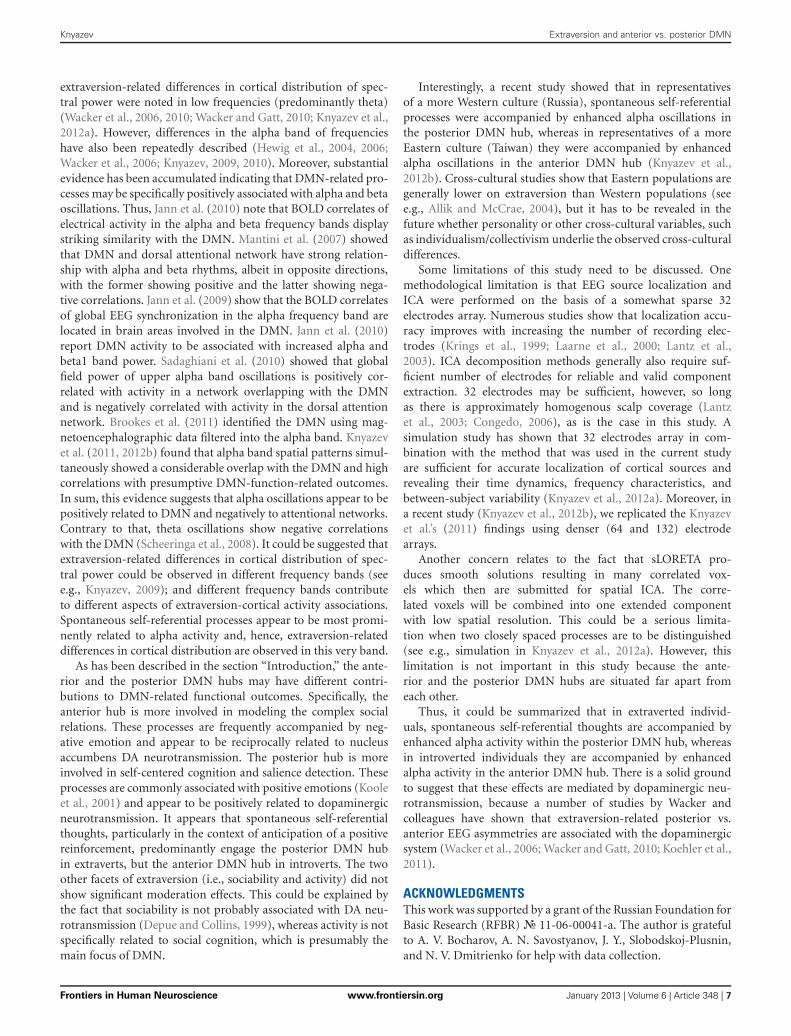
For each respective set of ICA results, ICs were spatially cor-related with an anatomically defined template and were rankedaccording to a “highest correlation” criterion with this anatomy.I created two templates using the Wake Forest Pick atlas toolbox(http://www.fmri.wfubmc.edu/) (Maldjian et al., 2004). The ante-rior DMN (ADMN) template included the medial frontal and thesuperior frontal gyrus (BAs 8/9/10) and the anterior cingulatecortex (BAs 11/24/32). The posterior DMN (PDMN) templateincluded the posterior parietal cortex (BA 7), the occipitopari-etal junction (BA 39), the posterior cingulate, and the precuneus.For the analysis of associations between SRPE scores and inter-individual variation in components’ intensity, for each of 20 ICsgenerated for each frequency band, all positive voxel values in arespective (z-score-transformed) independent component imagewere summed for each subject. These values were further used asis described below. This approach to capturing inter-individualdifferences is based on the following. After initial decompositionon all concatenated datasets at once, the components are backreconstructed in each individual subject. After that, each compo-nent is more pronounced in some subjects and may be weak orabsent in others. If a component is strongly pronounced, respec-tive brain areas will have high positive z-score values. Therefore,summing and comparing across subjects the positive values in z-transformed spatial maps allows revealing individual differencesin intensity of this component. This method is described in thepaper by Allen et al. (2011). For more details see Knyazev et al.(2011, 2012a,b).
STATISTICAL ANALYSISFor the analysis of moderation effects a combined measure ofPDMN vs. ADMN activity (hereafter P/ADMN) was created. ThePDMNIC (hereafter subscript IC denotes the independent com-ponent that showed the highest spatial correlation with a respec-tive template) and ADMNIC scores were converted to z-scoresand ADMNIC scores were subtracted from PDMNIC scores.The resulting measure represented dimension running from lowPDMN/high ADMN activity to high PDMN/low ADMN activity.
To test for moderation, regression analyses were specified forthe combination of moderator (i.e., extraversion) and factor (i.e.,
P/ADMN) as predictors of SRTS or SRPE scores. Following guide-lines on testing moderator models outlined by Baron and Kenny(1986), predictor variables were entered hierarchically in the fol-lowing order: (1) main effects for factor tested (i.e., P/ADMN)and proposed moderator variable (i.e., extraversion); (2) the two-way interaction between the factor and the moderator. To testinteractions (or moderation effects) involving continuous vari-ables, I converted all continuous variables to z-scores, followingthe suggestion by Aiken and West (1991). To gain an understand-ing of the overall pattern of the interaction, regression slopes wereplotted graphically at high (0.5 SD) and low (−0.5 SD) values ofthe moderator.
RESULTSComparison of EPP scales’ means of the current sample witha normative Russian sample (N = 576, Knyazev et al., 2004)showed no significant differences on any of the nine personal-ity characteristics. Sociability and Activity correlated positivelywith SRTS (r = 0.32, p = 0.017 and r = 0.39, p = 0.002, respec-tively) and SRPE (r = 0.34, p = 0.009 and r = 0.38, p = 0.003,respectively) and negatively with ATT (r = −0.28, p = 0.038 andr = −0.27, p = 0.039, respectively). Assertiveness did not showsignificant correlations with STQ scales.
There was a significant interaction of Assertiveness withP/ADMN in prediction of SRTS scores, B = −0.31, T(58) =−2.14, p = 0.037. There were no significant moderation effectsof Assertiveness in other frequency bands. Moderation effectsfor Activity and Sociability were not significant for all frequencybands.
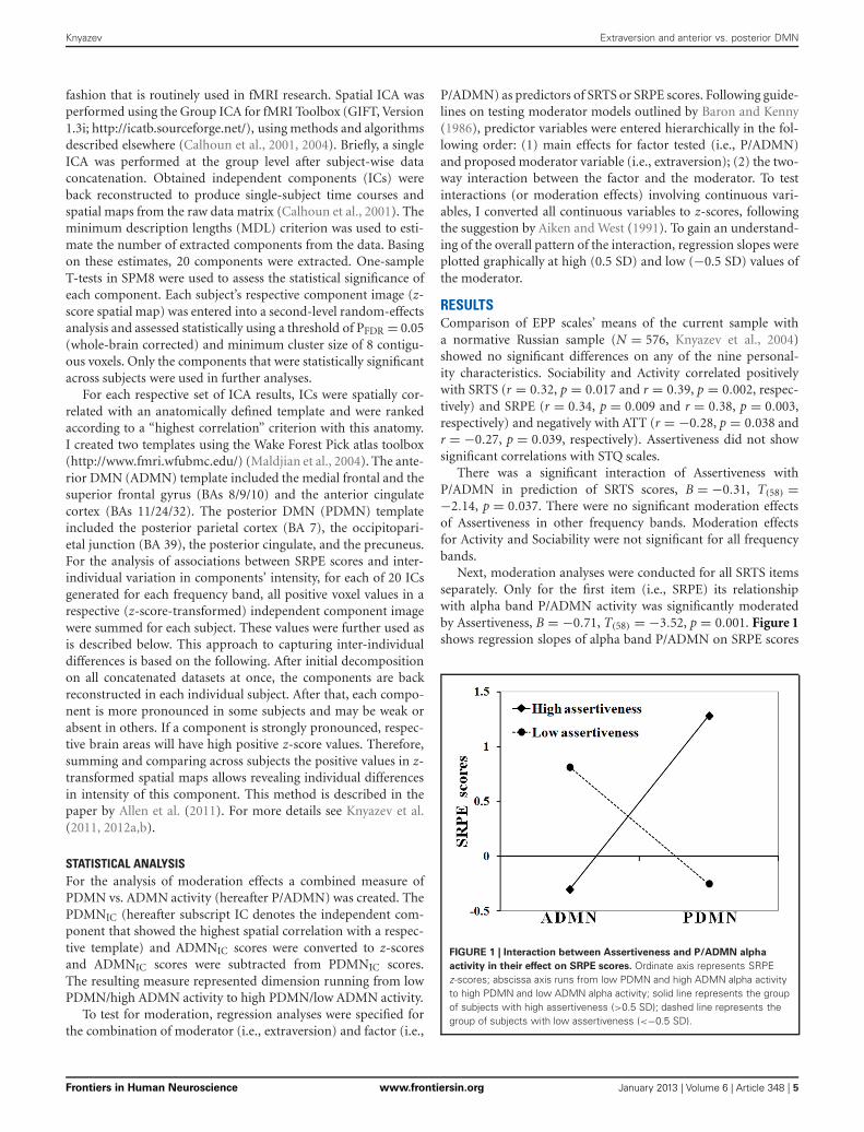
Next, moderation analyses were conducted for all SRTS itemsseparately. Only for the first item (i.e., SRPE) its relationshipwith alpha band P/ADMN activity was significantly moderatedby Assertiveness, B = −0.71, T(58) = −3.52, p = 0.001. Figure 1shows regression slopes of alpha band P/ADMN on SRPE scores
FIGURE 1 | Interaction between Assertiveness and P/ADMN alpha
activity in their effect on SRPE scores. Ordinate axis represents SRPEz-scores; abscissa axis runs from low PDMN and high ADMN alpha activityto high PDMN and low ADMN alpha activity; solid line represents the groupof subjects with high assertiveness (>0.5 SD); dashed line represents thegroup of subjects with low assertiveness (<−0.5 SD).
Frontiers in Human Neuroscience www.frontiersin.org January 2013 | Volume 6 | Article 348 | 5
Knyazev Extraversion and anterior vs. posterior DMN
in subjects with high (+0.5 SD, N = 17) and low (−0.5 SD,N = 15) Assertiveness.
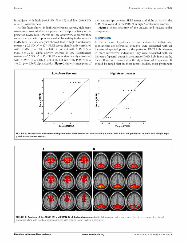
As this figure shows, in high Assertiveness scorers, high SRPEscores were associated with a prevalence of alpha activity in theposterior DMN hub, whereas in low Assertiveness scorers theywere associated with a prevalence of alpha activity in the anteriorDMN hub. Post hoc analyses showed that in high Assertivenessscorers (+0.5 SD, N = 17), SRPE scores significantly correlatedwith PDMN (r = 0.74, p = 0.001), but not with ADMN (r =0.26, p = 0.313) alpha activity, whereas in low Assertivenessscorers (−0.5 SD, N = 15), SRPE scores significantly correlatedwith ADMN (r = 0.81, p < 0.001), but not with PDMN (r =−0.01, p = 0.969) alpha activity. Figure 2 shows scatter-plots of
the relationships between SRPE scores and alpha activity in theADMN in low and in the PDMN in high Assertiveness scorers.
Figure 3 shows anatomy of the ADMN and PDMN alphacomponents.
DISCUSSIONIn line with our hypothesis, in more extraverted individuals,spontaneous self-referential thoughts were associated with anincrease of spectral power in the posterior DMN hub, whereasin more introverted individuals they were associated with anincrease of spectral power in the anterior DMN hub. In our study,these effects were observed in the alpha band of frequencies. Itshould be noted that in most recent studies, most prominent
FIGURE 2 | Scatter-plots of the relationships between SRPE scores and alpha activity in the ADMN in low (left panel) and in the PDMN in high (right
panel) Assertiveness scorers.
FIGURE 3 | Anatomy of the ADMN (A) and PDMN (B) alpha band components. Spatial maps are scaled in z-scores. The slices are presented at axialanatomical plane with numbers representing the slice position in mm relative to zero-point.
Frontiers in Human Neuroscience www.frontiersin.org January 2013 | Volume 6 | Article 348 | 6
Knyazev Extraversion and anterior vs. posterior DMN
extraversion-related differences in cortical distribution of spec-tral power were noted in low frequencies (predominantly theta)(Wacker et al., 2006, 2010; Wacker and Gatt, 2010; Knyazev et al.,2012a). However, differences in the alpha band of frequencieshave also been repeatedly described (Hewig et al., 2004, 2006;Wacker et al., 2006; Knyazev, 2009, 2010). Moreover, substantialevidence has been accumulated indicating that DMN-related pro-cesses may be specifically positively associated with alpha and betaoscillations. Thus, Jann et al. (2010) note that BOLD correlates ofelectrical activity in the alpha and beta frequency bands displaystriking similarity with the DMN. Mantini et al. (2007) showedthat DMN and dorsal attentional network have strong relation-ship with alpha and beta rhythms, albeit in opposite directions,with the former showing positive and the latter showing nega-tive correlations. Jann et al. (2009) show that the BOLD correlatesof global EEG synchronization in the alpha frequency band arelocated in brain areas involved in the DMN. Jann et al. (2010)report DMN activity to be associated with increased alpha andbeta1 band power. Sadaghiani et al. (2010) showed that globalfield power of upper alpha band oscillations is positively cor-related with activity in a network overlapping with the DMNand is negatively correlated with activity in the dorsal attentionnetwork. Brookes et al. (2011) identified the DMN using mag-netoencephalographic data filtered into the alpha band. Knyazevet al. (2011, 2012b) found that alpha band spatial patterns simul-taneously showed a considerable overlap with the DMN and highcorrelations with presumptive DMN-function-related outcomes.In sum, this evidence suggests that alpha oscillations appear to bepositively related to DMN and negatively to attentional networks.Contrary to that, theta oscillations show negative correlationswith the DMN (Scheeringa et al., 2008). It could be suggested thatextraversion-related differences in cortical distribution of spec-tral power could be observed in different frequency bands (seee.g., Knyazev, 2009); and different frequency bands contributeto different aspects of extraversion-cortical activity associations.Spontaneous self-referential processes appear to be most promi-nently related to alpha activity and, hence, extraversion-relateddifferences in cortical distribution are observed in this very band.
As has been described in the section “Introduction,” the ante-rior and the posterior DMN hubs may have different contri-butions to DMN-related functional outcomes. Specifically, theanterior hub is more involved in modeling the complex socialrelations. These processes are frequently accompanied by neg-ative emotion and appear to be reciprocally related to nucleusaccumbens DA neurotransmission. The posterior hub is moreinvolved in self-centered cognition and salience detection. Theseprocesses are commonly associated with positive emotions (Kooleet al., 2001) and appear to be positively related to dopaminergicneurotransmission. It appears that spontaneous self-referentialthoughts, particularly in the context of anticipation of a positivereinforcement, predominantly engage the posterior DMN hubin extraverts, but the anterior DMN hub in introverts. The twoother facets of extraversion (i.e., sociability and activity) did notshow significant moderation effects. This could be explained bythe fact that sociability is not probably associated with DA neu-rotransmission (Depue and Collins, 1999), whereas activity is notspecifically related to social cognition, which is presumably themain focus of DMN.
Interestingly, a recent study showed that in representativesof a more Western culture (Russia), spontaneous self-referentialprocesses were accompanied by enhanced alpha oscillations inthe posterior DMN hub, whereas in representatives of a moreEastern culture (Taiwan) they were accompanied by enhancedalpha oscillations in the anterior DMN hub (Knyazev et al.,2012b). Cross-cultural studies show that Eastern populations aregenerally lower on extraversion than Western populations (seee.g., Allik and McCrae, 2004), but it has to be revealed in thefuture whether personality or other cross-cultural variables, suchas individualism/collectivism underlie the observed cross-culturaldifferences.
Some limitations of this study need to be discussed. Onemethodological limitation is that EEG source localization andICA were performed on the basis of a somewhat sparse 32electrodes array. Numerous studies show that localization accu-racy improves with increasing the number of recording elec-trodes (Krings et al., 1999; Laarne et al., 2000; Lantz et al.,2003). ICA decomposition methods generally also require suf-ficient number of electrodes for reliable and valid componentextraction. 32 electrodes may be sufficient, however, so longas there is approximately homogenous scalp coverage (Lantzet al., 2003; Congedo, 2006), as is the case in this study. Asimulation study has shown that 32 electrodes array in com-bination with the method that was used in the current studyare sufficient for accurate localization of cortical sources andrevealing their time dynamics, frequency characteristics, andbetween-subject variability (Knyazev et al., 2012a). Moreover, ina recent study (Knyazev et al., 2012b), we replicated the Knyazevet al.’s (2011) findings using denser (64 and 132) electrodearrays.
Another concern relates to the fact that sLORETA pro-duces smooth solutions resulting in many correlated vox-els which then are submitted for spatial ICA. The corre-lated voxels will be combined into one extended componentwith low spatial resolution. This could be a serious limita-tion when two closely spaced processes are to be distinguished(see e.g., simulation in Knyazev et al., 2012a). However, thislimitation is not important in this study because the ante-rior and the posterior DMN hubs are situated far apart fromeach other.
Thus, it could be summarized that in extraverted individ-uals, spontaneous self-referential thoughts are accompanied byenhanced alpha activity within the posterior DMN hub, whereasin introverted individuals they are accompanied by enhancedalpha activity in the anterior DMN hub. There is a solid groundto suggest that these effects are mediated by dopaminergic neu-rotransmission, because a number of studies by Wacker andcolleagues have shown that extraversion-related posterior vs.anterior EEG asymmetries are associated with the dopaminergicsystem (Wacker et al., 2006; Wacker and Gatt, 2010; Koehler et al.,2011).
ACKNOWLEDGMENTSThis work was supported by a grant of the Russian Foundation forBasic Research (RFBR) № 11-06-00041-a. The author is gratefulto A. V. Bocharov, A. N. Savostyanov, J. Y., Slobodskoj-Plusnin,and N. V. Dmitrienko for help with data collection.
Frontiers in Human Neuroscience www.frontiersin.org January 2013 | Volume 6 | Article 348 | 7
Knyazev Extraversion and anterior vs. posterior DMN
REFERENCESAiken, L., and West, S. (1991).
MULTIPLE Regression: Testing andInterpreting Interactions. NewburyPark, London: Sage Publications,Inc.
Alexander, W. H., and Brown, J. W.(2011). Medial prefrontal cortex asan action-outcome predictor. Nat.Neurosci. 14, 1338–1344.
Allen, E. A., Erhardt, E. B., Wei,Y., Eichele, T., and Calhoun, V.D. (2011). Capturing inter-subjectvariability with group independentcomponent analysis of fMRI data:a simulation study. Neuroimage 59,4141–4159.
Allik, J., and McCrae, R. R. (2004).Toward a geography of personalitytraits: patterns of profiles across 36cultures. J. Cross-Cult. Psychol. 35,13–28.
Asanuma, K., Tang, C., Ma, Y., Dhawan,V., Mattis, P., Edwards, C., et al.(2006). Network modulation in thetreatment of Parkinson’s disease.Brain 129, 2667–2678.
Baron, R. M., and Kenny, D. A.(1986). The moderator-mediatorvariable distinction in socialpsycho-logical research: conceptual,strategic, and statistical consid-erations. J. Pers. Soc. Psychol. 51,1173–1182.
Braskie, M. N., Landau, S. M.,Wilcox, C. E., Taylor, S. D.,O’Neil, J. P., Baker, S. L., et al.(2011). Correlations of striataldopamine synthesis with defaultnetwork deactivations duringworking memory in youngeradults. Hum. Brain Mapp. 32,947–961.
Brookes, M. J., Woolrich, M., Luckhoo,H., Price, D., Hale, J. R., Stephenson,M. C., et al. (2011). Investigatingthe electrophysiological basisof resting state networks usingmagnetoencephalography. Proc.Natl. Acad. Sci. U.S.A. 108,16783–16788.
Buckner, R. L., Andrews-Hanna, J. R.,and Schacter, D. L. (2008). Thebrain’s default network: anatomy,function, and relevance to dis-ease. Ann. N.Y. Acad. Sci. 1124,1–38.
Buckner, R. L., and Carroll, D.C. (2007). Self-projection andthe brain. Trends Cogn. Sci. 11,49–57.
Calhoun, V. D., Adali, T., Pearlson,G. D., and Pekar, J. J. (2001). Amethod for making group infer-ences from functional MRI datausing independent componentanalysis. Hum. Brain Mapp. 14,140–151.
Calhoun, V. D., Adali, T., and Pekar, J.J. (2004). A method for comparinggroup fMRI data using independentcomponent analysis: applicationto visual, motor and visuomotortasks. Magn. Reson. Imaging 22,1181–1191.
Cavanna, A. E., and Trimble, M. R.(2006). The precuneus: a reviewof its functional anatomy andbehavioural correlates. Brain 129,564–583.
Chavanon, M. L., Wacker, J., andStemmler, G. (2011). Rostral ante-rior cingulate activity generates pos-terior versus anterior theta activ-ity linked to agentic extraversion.Cogn. Affect. Behav. Neurosci. 11,172–185.
Congedo, M. (2006). Subspace projec-tion filters for real-time brain elec-tromagnetic imaging. IEEE Trans.Biomed. Eng. 53, 1624–1634.
Delaveau, P., Salgado-Pineda, P.,Fossati, P., Witjas, T., Azulay, J. P.,and Blin, O. (2010). Dopaminergicmodulation of the defaultmodenetwork in Parkinson’s disease.Eur. Neuropsychopharmacol. 20,784–792.
Depue, R. A., and Collins, P. F.(1999). Neurobiology of the struc-ture of personality: dopamine, facil-itation of incentive motivation, andextraversion. Behav. Brain Sci. 22,491–569.
Deutch, A. Y., Clark, W. A., andRoth, R. H. (1990). Prefrontal cor-tical dopamine depletion enhancesthe responsiveness of mesolimbicdopamine neurons to stress. BrainRes. 521, 311–315.
Doherty, M. D., and Gratton, A.(1996). Medial prefrontal corti-cal D1 receptor modulation ofthe meso-accumbens dopamineresponse to stress: an electrochem-ical study in freely-behaving rats.Brain Res. 715, 86–97.
Dusek, P., Jech, R., Sieger, T., Vymazal,J., Ruzicka, E., Wackermann, J.,et al. (2012). Abnormal activity inthe precuneus during time percep-tion in parkinson’s disease: an fMRIstudy. PLoS ONE 7:e29635. doi:10.1371/journal.pone.0029635
Eysenck, H. J., and Wilson, G. D.(2000). The Eysenck PersonalityProfiler, Version 6. Worthing, UK:Psi-Press.
Fishman, I., Ng, R., and Bellugi, U.(2011). Do extraverts process socialstimuli differently from introverts?Cogn. Neurosci. 2, 67–73.
Fransson, P., and Marrelec, G. (2008).The precuneus/posterior cingulatecortex plays a pivotal role in thedefault mode network: evidence
from a partial correlation net-work analysis. Neuroimage 42,1178–1184.
Geijo-Barrientos, E., and Pastore, C.(1995). The effects of dopamine onthe subthreshold electrophysiologi-cal responses of rat prefrontal cortexneurons in vitro. Eur. J. Neurosci. 7,358–366.
Gentili, C., Ricciardi, E., Gobbini,M. I., Santarelli, M. F., Haxbye,J. V., Pietrini, P., et al. (2009).Beyond amygdala: default ModeNetwork activity differs betweenpatients with Social Phobia andhealthy controls. Brain Res. Bull. 79,409–413.
Gobbini, M. I., Koralek, A. C., Bryan, R.E., Montgomery, K. J., and Haxby,J. V. (2007). Two takes on the socialbrain: a comparison of theory ofmind tasks, J. Cogn. Neurosci. 19,1803–1814.
Goldin, P. R., McRae, K., Ramel, W.,and Gross, J. J. (2008). The neuralbases of emotion regulation: reap-praisal and suppression of nega-tive emotion. Biol. Psychiatry 63,577–586.
Goodale, M. A., and Milner, A.D. (2008). Two visual systemsre-viewed. Neuropsychologia 46,774–785.
Gray, J. A. (1999). “Cognition, emo-tion, conscious experience andthe brain,” in Handbook ofCognition and Emotion, eds T.Dalgleish and M. Power (New York,NY: John Wiley and Sons Ltd),83–102.
Gusnard, D. A., and Raichle, M. E.(2001). Searching for a baseline:functional neuroimaging and theresting human brain. Nat. Rev.Neurosci. 3, 685–694.
Han, S., Jiang, Y., Humphreys, G.W., Zhou, T., and Cai, P. (2005).Distinct neural substrates for theperception of real and virtualvisual worlds. Neuroimage 24,928–935.
Hewig, J., Hagemann, D., Seifert, J.,Naumann, E., and Bartussek, D.(2004). On the selective relationof frontal cortical asymmetryand anger-out versus anger-control. J. Pers. Soc. Psychol. 87,926–939.
Hewig, J., Hagemann, D., Seifert, J.,Naumann, E., and Bartussek, D.(2006). The relation of corticalactivity and BIS/BAS on the traitlevel. Biol. Psychol. 71, 42–53.
Husain, M., and Nachev, P. (2007).Space and the parietal cortex. TrendsCogn. Sci. 11, 30–36.
Iacoboni, M., Lieberman, M. D.,Knowlton, B. J., Molnar-Szakacs,
I., Moritz, M., Throop, C. J., et al.(2004). Watching social interactionsproduces dorsomedial prefrontaland medial parietal BOLD fMRIsignal increases compared to aresting baseline. Neuroimage 21,1167–1173.
Iidaka, T., Omori, M., Murata, T.,Kosaka, H., Yonekura, Y., Okada,T., et al. (2001). Neural interac-tion of the amygdala with the pre-frontal and temporal cortices in theprocessing of facial expressions asrevealed by fMRI. J. Cogn. Neurosci.13, 1035–1047.
Jann, K., Dierks, T., Boesch, C.,Kottlow, M., Strik, W., and Koenig,T. (2009). BOLD correlates of EEGalpha phase-locking and the fMRIdefault mode network. Neuroimage45, 903–916.
Jann, K., Kottlow, M., Dierks, T.,Boesch, C., and Koenig, T. (2010).Topographic electrophysiologicalsignatures of fMRI resting statenetworks. PLoS ONE 5:e12945. doi:10.1371/journal.pone.0012945
Kennedy, D. P., and Courchesne, E.(2008). Functional abnormalitiesof the default network during self-and other-reflection in autism.Soc. Cogn. Affect. Neurosci. 3,177–190.
Kennedy, D. P., Redcay, E., andCourchesne, E. (2006). Failingto deactivate: resting functionalabnormalities in autism. Proc. Natl.Acad. Sci. U.S.A. 103, 8275–8280.
King, D., Zigmond, M. J., andFinlay, J. M. (1997). Effects ofdopamine depletion in the medialprefrontal cortex on the stress-induced increase in extracellulardopamine in the nucleus accum-bens core and shell. Neuroscience 77,141–153.
Knyazev, G. G. (2009). Is corticaldistribution of spectral powera stable individual character-istic? Int. J. Psychophysiol. 72,123–133.
Knyazev, G. G. (2010). Antero-posterior spectral power gradientas a correlate of extraversionand behavioral inhibition. OpenNeuroimag. J. 4, 114–120.
Knyazev, G. G., Belopolsky, V. I.,Bodunov, M. V., and Wilson, G.D. (2004). The factor structureof the Eysenck personality profilerin Russia. Pers. Individ. Diff. 37,1681–1692.
Knyazev, G. G., Bocharov, A. V., andPylkova, L. V. (2012a). Extraversionand fronto-posterior EEG spectralpower gradient: an independentcomponent analysis. Biol. Psychol.89, 515–524.
Frontiers in Human Neuroscience www.frontiersin.org January 2013 | Volume 6 | Article 348 | 8
Knyazev Extraversion and anterior vs. posterior DMN
Knyazev, G. G., Savostyanov, A. N.,Volf, N. V., Liou, M., and Bocharov,A. V. (2012b). EEG correlatesof spontaneous self-referentialthoughts: a cross-cultural study. Int.J. Psychophysiol. 86, 173–181.
Knyazev, G. G., Slobodskoj-Plusnin,J. Y., and Bocharov, A. V. (2009).Event-related delta and thetasynchronization during explicitand implicit emotion processing.Neuroscience 164, 1588–1600.
Knyazev, G. G., Slobodskoj-Plusnin,J. Y., Bocharov, A. V., andPylkova, L. V. (2011). The defaultmode network and EEG alphaoscillations: an independent com-ponent analysis. Brain Res. 1402,67–79.
Koehler, S., Wacker, J., Odorfer, T.,Reif, A., Gallinat, J., Fallgatter, A.J., et al. (2011). Resting posteriorminus frontal EEG slow oscillationsis associated with extraversion andDRD2 genotype. Biol. Psychol. 87,407–413.
Koole, S. L., Dijksterhuis, A., and vanKnippenberg, A. (2001). What’s ina name: implicit self-esteem and theautomatic self. J. Pers. Soc. Psychol.80, 669–685.
Krajcovicova, L., Mikl, M., Marecek,R., and Rektorova, I. (2012). Thedefault mode network integrity inpatients with Parkinson’s diseaseis levodopa equivalent dose-dependent. J. Neural Transm. 119,443–454.
Krings, T., Chiappa, K. H., Cuffin, B.N., Cochius, J. I., Connolly, S., andCosgrove, G. R. (1999). Accuracyof EEG dipole source localizationusing implanted sources in thehuman brain. Clin. Neurophysiol.110, 106–114.
Laarne, P. H., Tenhunen-Eskelinen, M.L., Hyttinen, J. K., and Eskola, H. J.(2000). Effect of EEG electrode den-sity on dipole localization accuracyusing two realistically shaped skullresistivity models. Brain Topogr. 12,249–254.
Lantz, G., Grave de Peralta, R.,Spinelli, L., Seeck, M., and Michel,C. M. (2003). Epileptic sourcelocalization with high densityEEG: how many electrodes areneeded? Clin. Neurophysiol. 114,63–69.
Lou, H. C., Luber, B., Crupain, M.,Keenan, J. P., Nowak, M., Kjaer, T.W., et al. (2004). Parietal cortex andrepresentation of the mental self.Proc. Natl. Acad. Sci. U.S.A. 101,6827–6832.
Luk, C. H., and Wallis, J. D. (2009).Dynamic encoding of responses andoutcomes by neurons in medial
prefrontal cortex. J. Neurosci. 29,7526–7539.
Maldjian, J. A., Laurienti, P. J., andBurdette, J. H. (2004). Precentralgyrus discrepancy in electronicversions of the talairach atlas.Neuroimage 21, 450–455.
Mantini, D., Perrucci, M. G.,Del Gratta, D., Romani, G.L., and Corbetta, M. (2007).Electrophysiological signatures ofresting state networks in the humanbrain. Proc. Natl. Acad. Sci. U.S.A.104, 13170–13175.
Milner, A. D., and Goodale, M. A.(1995). The Visual Brain in Action.Oxford: Oxford University Press.
Mitchell, J. P. (2006). Mentalizingand Marr: an information pro-cessing approach to the study ofsocial cognition. Brain Res. 1079,66–75.
Mitchell, J. P., Banaji, M. R., andMacrae, C. N. (2005a). The linkbetween social cognition and self-referential thought in the medialprefrontal cortex. J. Cogn. Neurosci.17, 1306–1315.
Mitchell, J. P., Neil Macrae, C., andBanaji, M. R. (2005b). Formingimpressions of people versus inan-imate objects: social-cognitiveprocessing in the medial pre-frontal cortex. NeuroImage 26,251–257.
Northoff, G., Heinzel, A., de Greck,M., Bermpohl, F., Dobrowolny,H., and Panksepp, J. (2006). Self-referential processing in our brain –a meta-analysis of imaging stud-ies on the self. Neuroimage 31,440–457.
Ochsner, K. N., Beer, B. S., Robertson,E. R., Cooper, J. C., Gabrieli,J. D. E., Kihsltrom, J. F., et al.(2005). The neural correlates ofdirect and reflected self-knowledge.Neuroimage 28, 797–814.
Ochsner, K. N., and Gross, J. J.(2008). Cognitive emotion regula-tion. Curr. Dir. Psychol. Sci. 17,153–158.
Pascual-Marqui, R. D. (2002).Standardized low-resolutionbrain electromagnetic tomogra-phy, sLORETA): technical details.Methods Find. Exp. Clin. Pharmacol.24(Suppl. D), 5–12.
Pascucci, T., Ventura, R., Latagliata, E.C., Cabib, S., and Puglisi-Allegra,S. (2007). The medial prefrontalcortex determines the accum-bens dopamine response to stressthrough the opposing influencesof norepinephrine and dopamine.Cereb. Cortex 17, 2796—2804.
Peters, J., and Büchel, C. (2010).Episodic future thinking reduces
reward delay discounting throughan enhancement of prefrontal-mediotemporal interactions.Neuron 66, 138–148.
Phillips, K. A., Subiaulc, F., andSherwood, C. C. (2012). Curiousmonkeys have increased graymatter density in the pre-cuneus. Neurosci. Lett. 518,172–175.
Quirk, G. J., and Beer, J. S. (2006).Prefrontal involvement in theregulation of emotion: con-vergence of rat and humanstudies. Curr. Opin. Neurobiol. 16,723–727.
Raichle, M. E., and Snyder, A. Z. (2007).A default mode of brain function:a brief history of an evolving idea.Neuroimage 37, 1083–1090.
Richards, J. B., Zhang, L., Mitchell, S.H., and de Wit, H. (1999). Delay orprobability discounting in a modelof impulsive behavior: effect ofalcohol. J. Exp. Anal. Behav. 71,121–143.
Ruby, P., and Decety, J. (2001). Effect ofsubjective perspective taking duringsimulation of action: a PET inves-tigation of agency. Nat. Neurosci. 4,546–550.
Sadaghiani, S., Scheeringa, R.,Lehongre, K., Morillon, B., Giraud,A. L., and Kleinschmidt, A. (2010).Intrinsic connectivity networks,alpha oscillations, and tonicalertness: a simultaneous elec-troencephalography/functionalmagnetic resonance imaging study.J. Neurosci. 30, 10243–10250.
Scheeringa, R., Bastiaansen, M. C.M., Petersson, K. M., Oostenveld,R., Norris, D. G., and Hagoort, P.(2008). Frontal theta EEG activ-ity correlates negatively with thedefault mode network in restingstate. Int. J. Psychophysiol. 67,242–251.
Schmitz, T. W., Kawahara-Baccus,T. N., and Johnson, S. C.(2004). Metacognitive evalua-tion, self-relevance, and the rightprefrontal cortex. Neuroimage 22,941–947.
Seger, C. A., Stone, M., and Keenan, J.P. (2004). Cortical activations dur-ing judgments about the self and another person. Neuropsychologia 42,1168–1177.
Shackman, A. J., Salomons, T. V.,Slagter, H. A., Fox, A. S., Winter, J.J., and Davidson, R. J. (2011). Theintegration of negative affect, pain,and cognitive control in the cingu-late cortex. Nat. Rev. Neurosci. 12,154–167.
Smillie, L. D., Pickering, A. D., andJackson, C. J. (2006). The new
reinforcement sensitivity theory:implications for personality mea-surement. Pers. Soc. Psychol. Rev. 10,320–335.
Talairach, J., and Tournoux, P. (1988).Co-Planar Stereotaxic Atlas of theHuman Brain. New York, NY:Theme.
Tomasi, D., Volkow, N. D., Wang,G. J., Wang, R., Telang, F.,Caparelli, E. C., et al. (2011).Methylphenidate enhances brainactivation and deactivationresponses to visual attentionand working memory tasks inhealthy controls. Neuroimage 54,3101–3110.
Tomasi, D., Volkow, N. D., Wang, R.,Telang, F., Wang, G.-J., Chang,L., et al. (2009). Dopaminetransporters in striatum cor-relate with deactivation in thedefault mode network duringvisuospatial attention. PLoS ONE4:e6102. doi: 10.1371/journal.pone.0006102
Urry, H. L., van Reekum, C. M.,Johnstone, T., Kalin, N. H., Thurow,M. E., Schaefer, H. S., et al. (2006).Amygdala and ventromedialprefrontal cortex are inverselycoupled during regulation ofnegative affect and predict thediurnal pattern of cortisol secretionamong older adults. J. Neurosci. 26,4415–4425.
van Eimeren, T., Monchi, O., Ballanger,B., and Strafella, A. P. (2009).Dysfunction of the default modenetwork in Parkinson disease: afunctional magnetic resonanceimaging study. Arch. Neurol. 66,877–883.
Vogeley, K., Bussfelda, P., Newenc,A., Herrmanna, S., Happéd, F.,Falkai, P., et al. (2001). Mind read-ing: neural mechanisms of the-ory of mind and self-perspective.Neuroimage 14, 170–181.
Vogeley, K., and Fink, G. R. (2003).Neural correlates of the first-personperspective. Trends Cogn. Sci. 7,38–42.
Vogt, B. A., Vogt, L., and Laureys, S.(2006). Cytology and functionallycorrelated circuits of human poste-rior cingulate areas. Neuroimage 29,452–466.
Wacker, J., Chavanon, M. L., andStemmler, G. (2006). Investigatingthe dopaminergic basis ofextraversion in humans: a mul-tilevel approach. J. Pers. Soc.Psychol. 91, 171–187.
Wacker, J., Chavanon, M. L., andStemmler, G. (2010). Resting EEGsignatures of agentic extraversion:new results and meta-analytic
Frontiers in Human Neuroscience www.frontiersin.org January 2013 | Volume 6 | Article 348 | 9
Knyazev Extraversion and anterior vs. posterior DMN
integration. J. Res. Pers. 44,167–179.
Wacker, J., and Gatt, J. M. (2010).Resting posterior versus frontaldelta/theta EEG activity is asso-ciated with extraversion andthe COMT VAL158MET poly-morphism. Neurosci. Lett. 478,88–92.
Yuan, J., Zhang, J., Zhou, X., Yang, J.,Meng, X., Zhang, Q., et al. (2012).
Neural mechanisms underlyingthe higher levels of subjectivewell-being in extraverts: pleasantbias and unpleasant resistance.Cogn. Affect. Behav. Neurosci. 12,175–192.
Conflict of Interest Statement: Theauthor declares that the researchwas conducted in the absence of any
commercial or financial relationshipsthat could be construed as a potentialconflict of interest.
Received: 01 October 2012; accepted: 17December 2012; published online: 07January 2013.Citation: Knyazev GG (2013)Extraversion and anterior vs. posteriorDMN activity during self-referential
thoughts. Front. Hum. Neurosci. 6:348.doi: 10.3389/fnhum.2012.00348Copyright © 2013 Knyazev. This isan open-access article distributed underthe terms of the Creative CommonsAttribution License, which permits use,distribution and reproduction in otherforums, provided the original authorsand source are credited and subject to anycopyright notices concerning any third-party graphics etc.
Frontiers in Human Neuroscience www.frontiersin.org January 2013 | Volume 6 | Article 348 | 10