exceptionally preserved 450-million-year-old ordovician ostracods with brood care
TRANSCRIPT
Exceptionally Preserved
Current Biology 24, 801–806, March 31, 2014 ª2014 Elsevier Ltd All rights reserved http://dx.doi.org/10.1016/j.cub.2014.02.040
Report
450-Million-Year-Old OrdovicianOstracods with Brood Care
David J. Siveter,1,* Gengo Tanaka,2 Una C. Farrell,3
Markus J. Martin,4 Derek J. Siveter,5,6 and Derek E.G. Briggs71Department of Geology, University of Leicester,Leicester LE1 7RH, UK2BioGeos Japan Agency of Marine Science andTechnology, 2-15 Natsushima, Yokosuka City 237-0061,Kanagawa Prefecture, Japan3Biodiversity Institute, University of Kansas, Lawrence,KS 66045-7593, USA4371 Pawling Street, Watertown, NY 13601, USA5Earth Collections, University Museum of Natural History,Oxford OX1 3PW, UK6Department of Earth Sciences, University of Oxford,South Parks Road, Oxford OX1 3AN, UK7Department of Geology & Geophysics and Yale PeabodyMuseum of Natural History, Yale University, P.O. Box 208109,New Haven, CT 06520-8109, USA
Summary
Ostracod crustaceans are the most abundant fossil arthro-pods and are characterized by a long stratigraphic range.
However, their soft parts are very rarely preserved, andthe presence of ostracods in rocks older than the Silurian
period [1–5] was hitherto based on the occurrence of theirsupposed shells. Pyritized ostracods that preserve limbs
and in situ embryos, including an egg within an ovary
and possible hatched individuals, are here describedfrom rocks of the Upper Ordovician Katian Stage Lorraine
Group of New York State, including examples from thefamous Beecher’s Trilobite Bed [6, 7]. This discovery ex-
tends our knowledge of the paleobiology of ostracods bysome 25 million years and provides the first unequivocal
demonstration of ostracods in the Ordovician period,including the oldest known myodocope, Luprisca incuba
gen. et sp. nov. It also provides conclusive evidence of adevelopmental brood-care strategy conserved within
Ostracoda for at least 450 million years.
Results
Ostracods are ubiquitous today in a wide range of freshwaterand marine environments and depths. Presumed fossil exam-ples are used extensively in biostratigraphic and paleoenviron-mental studies. Most of the tens of thousands of extantostracod species are benthic or nektobenthic [8]. There isalso a minor but important pelagic fauna of ostracods thatoriginated during the Silurian period (w420–443 mega-annum[Ma]) based on both morphological and geological evidence[8–13]. Molecular phylogenetics combined with evidencefrom fossils calibrates a maximum divergence time for themonophyletic Ostracoda at about 500Ma during the Cambrianperiod [14]. Although fossil shells attributed to ostracodsoccur in countless numbers from the Ordovician period
*Correspondence: [email protected]
(w443–485 Ma) onward, shells alone are not definitiveevidence of affinity [15]: a number of other arthropod taxahave bivalved carapaces. However, fossil ostracods preser-ving soft parts are extremely rare, especially in the Paleozoic[1–5]. In situ embryos, including possible cases of brooding,are equally scarce in fossil ostracods and other invertebrates[2]. Here, we describe pyritized ostracods preserving softparts and embryos from the Ordovician Lorraine Group—astratigraphic unit that has yielded other exceptionallypreserved invertebrates [6, 7], most notably the remarkablypreserved trilobite Triarthrus from Beecher’s Trilobite Bed.The significance of this discovery is that it extends our knowl-edge of the paleobiology of the most abundant group of fossilarthropods by some 25 million years and confirms, for the firsttime, the presence of ostracods in the Ordovician period. It isthe only known fossil invertebrate species that preservesevidence of ovarian-to-juvenile brood care. This discoveryconfirms that this reproductive strategy, which is found inmyodocope ostracods today, was present in the earliest-known representatives of the group 450 million years ago.The ostracods are held by the Yale Peabody Museum of
Natural History (YPM) in New Haven, Connecticut. They werecollected from the Beecher’s Trilobite Bed site (WalcottQuarry: ‘‘Original Trilobite Bed’’ and beds 822 and 906) andJo’s Quarry in the Frankfort Shale of central New York State,and from Martin Quarry, at a slightly younger stratigraphiclevel, in the Whetstone Gulf Formation approximately 60 kmto the north (localities detailed in [6, 7]). The stratigraphicunits lie within the Maysvillian Regional Stage (Katian Stage),w450 Ma before present [16]. The ostracods are preservedon pieces of mudstone; most are internal and external molds.There are also crumpled and incomplete valves and carapacesthat are finely to coarsely pyritized. Some specimens preservepyritized limbs, embryos, and other soft tissues. The sup-posed ostracods from the Lorraine Group held by the PeabodyMuseum totalw110 specimens: YPM223936, 227953, 237106,237107, 307286–307303, 505528, 505531–505537, 506492,506506, 506521, 506532, 516164, 516170–516172, 516174–516196, 516197, 516199, 516200, 516201, 516203–516209,516211–516233, 611120–611124, 611127, 611129, 611140–611142.
Etymology
Luprisca incuba is a new genus and species assigned to theMyodocopa [17] and provisionally to the Myodocopida. Itsname alludes to the occurrence of brood care, as in theholotype (Lucina [goddess of childbirth] plus priscus [ancient]and incubatio [sitting on eggs]).
MaterialFour open carapaces with soft parts were used: YPM 307293(bed 906; Figures 1B, 1F, and 1G), YPM 307300 (holotype,Original Trilobite Bed; Figures 1A and 1D), and YPM 611120(Original Trilobite Bed; Figure 1E) from Walcott Quarry andYPM 237107 (Figures 1C and 2A–2F; Movies S1 and S2available online) from Martin Quarry. Each carapace isdeformed by flattening and partially covered by matrix. Soft
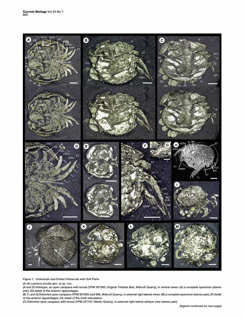
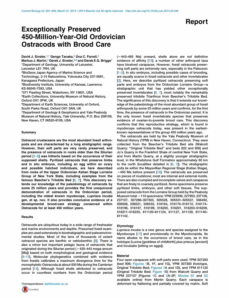
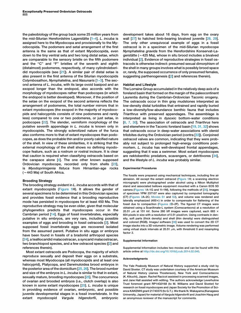
Figure 1. Ordovician and Extant Ostracods with Soft Parts
(A–G) Luprisca incuba gen. et sp. nov.
(A and D) Holotype, an open carapace with brood (YPM 307300; Original Trilobite Bed, Walcott Quarry), in ventral views: (A) a complete specimen (stereo
pair); (D) detail of the anterior appendages.
(B, F, and G) Distorted open carapace (YPM 307293; bed 906, Walcott Quarry), in external right lateral views: (B) a complete specimen (stereo pair); (F) detail
of the anterior appendages; (G) detail of the shell reticulation.
(C) Distorted open carapace with brood (YPM 237107; Martin Quarry), in external right lateral oblique view (stereo pair).
(legend continued on next page)
Current Biology Vol 24 No 7802
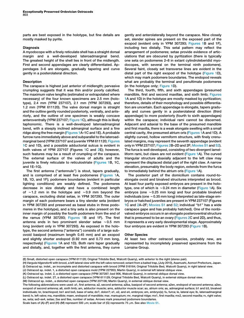
Exceptionally Preserved Ordovician Ostracods803
parts are best exposed in the holotype, but fine details aremostly masked by pyrite.
Diagnosis
Amyodocope with a finely reticulate shell has a straight dorsalmargin and a well-developed lateroadmarginal bend.The greatest height of the shell lies in front of the midlength.First and second appendages are clearly differentiated. Ap-pendages 3–6 are elongate, gradually tapering and curvegently in a posterolateral direction.
Description
The carapace is highest just anterior of midlength; pervasivecrumpling suggests that it was thin and/or poorly calcified.The maximum valve lengths (estimated or extrapolated wherenecessary) of the four known specimens are 2.5 mm (holo-type), 2.4 mm (YPM 237107), 2.1 mm (YPM 307293), and1.2 mm (YPM 611120). The valve dorsal margin is straightand the outline gently rounded posteriorly, ventrally, and ante-riorly, and the outline of one specimen is weakly concaveanteroventrally (YPM 237107; Figure 1C), although this is likelytaphonomic. There is a well-developed lateroadmarginalbend, with a steeply inclined admarginal surface and a fineridge along the freemargin (Figures 1A–1C and 1E). A probablefurrow runs immediately above and subparallel to the lateroad-marginal bend (YPM 237107 and juvenile YPM 611120; Figures1C and 1D), and a possible adductorial sulcus is evident inboth valves of YPM 237107 (Figures 1C and 2E); however,such features may be a result of crumpling of the carapace.The external surface of the valves of adults and thejuvenile is finely reticulate to reticulostriate (Figures 1B, 1C,and 1E–1G).
The first antenna (‘‘antennule’’) is stout, tapers gradually,and is comprised of at least five podomeres (Figures 1A,1B, 1D, and 1F); podomere boundaries (evident in particularlighting conditions) are weakly defined. The podomeresdecrease in size distally and have a combined lengthof w1.2 mm in the holotype and w0.9 mm beyond thecarapace, excluding distal setae, in YPM 307293. The outermargin of each podomere bears a tiny slender seta (evidentin YPM 307293 and preserved as basal stubs in three podo-meres in the holotype), and a tiny seta is also present on theinner margin of possibly the fourth podomere from the end ofthe ramus (YPM 307293; Figures 1B and 1F). The firstantenna ends in two prominent slender setae w0.5 mmlong (evident only in YPM 307293). As exposed in the holo-type, the second antenna (‘‘antenna’’) consists of a large sub-ovoid basipod (maximum length 0.45 mm) and an exopodand slightly shorter endopod (0.92 mm and 0.73 mm long,respectively) (Figures 1A and 1D). Both rami taper graduallyand distally, and, together with the first antenna, they curve
(E) Small, distorted open carapace (YPM 611120; Original Trilobite Bed, Walco
(H) Vargula hilgendorfiiwith brood, a left lateral viewwith the left valve removed
(I) Ostracod sp. indet., an incomplete and distorted carapace with brood (YPM
(J) Ostracod sp. indet. 1, a distorted open carapace mold (YPM 227953; Marti
(K) Ostracod sp. indet. 2, a distorted open carapace (YPM 307287; bed 906, W
(L) Ostracod sp. indet. 2?, a distorted open carapace (YPM 611129; Original T
(M) Ostracod sp. indet., a distorted open carapace (YPM 237106; Martin Quar
The following abbreviations were used: a1, first antenna; a2, second antenna;
exopod of second antenna; a6, sixth limb; am, adductor muscle; ams, adduct
individuals; br, brachiopod; bs1 and bs5, base of seta; do?, dolon?; e1, e2, an
bend; lv, left valve; ma, mandible; maen, endopod of mandible; mar, marsupium
se, seta; se2–se4, setae; 2se and 9se, number of setae. Arrows mark presume
Scale bars of (A)–(F) and (H)–(M) represent 500 mm; scale bar of (G) represents
gently and anterolaterally beyond the carapace. Nine closelyset, slender spines are present on the exposed part of theexopod (evident only in YPM 307293; Figures 1B and 1F),including two distally. This setal pattern may reflect thearrangement of podomeres; setae provide evidence of artic-ulations that are obscured by pyritization (there is typicallyone seta on podomeres 2–8 in extant cylindroleberidid myo-docopes, with several on the terminal ninth podomere).Several faint, closely set transverse lines are evident in thedistal part of the right exopod of the holotype (Figure 1D),which may mark podomere boundaries. The endopod revealswhat are probably the terminal and penultimate podomeres(in the holotype only; Figure 1D).The third, fourth, fifth, and sixth appendages (presumed
mandible, first and second maxillae, and sixth limb; Figures1A and 1D) in the holotype are mostly masked by pyritization;therefore, details of their morphology and possible differentia-tion are uncertain. Each appendage is elongate, tapers gradu-ally and curves gently in a posterolateral direction (thirdappendage) to more posteriorly (fourth to sixth appendages)within the carapace; individual rami cannot be discerned.Adjacent and adaxial to the base of the presumed mandibleand first maxilla, there is a weak elongate swelling with a smallcentral cavity, the presumed atrium oris (Figures 1A and 1D). Aslightly curved, hollow vermiform structure, with finely crenu-lated margins, may represent the seventh appendage (evidentonly in YPM237107; Figures 2B–2D and 2F;Movies S1 and S2).The furca is well developed, consisting of two divergent lamel-liform rami, but claws are not evident (Figure 1A). The curvedtriangular structure abaxially adjacent to the left claw mayrepresent the displaced distal part of the right claw. A narrowelevation, presumably the body region, extends from the furcato immediately behind the atrium oris (Figure 1A).The posterior part of the domicilium contains round-to-
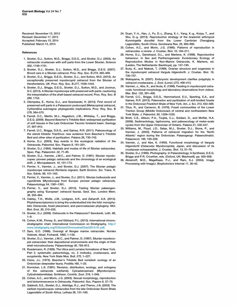
elongate ovoid and bivalved structures interpreted as brood.At least four partly exposed embryos are present in the holo-type, one of which is w0.24 mm in diameter (Figure 1A). Sixembryos (one w0.25 mm long) and four probable bivalvedindividuals (onew0.35 mm long) interpreted as late-stage em-bryos or hatched juveniles are present in YPM 237107 (Figures1C and 2A–2F; Movies S1 and S2; individual ‘‘b1’’ has a widecarapace gape and has probably hatched). One of the nonbi-valved embryos occurs in an elongate posteroventral structurethat is presumed to be an ovary (Figures 2C and 2D), and thus,it represents a very early developmental stage. Approximatelyfour embryos are evident in YPM 307293 (Figure 1B).
Other Species
At least two other ostracod species, probably new, arerepresented by incompletely preserved specimens from theLorraine Group.
tt Quarry), with anterior to the right (stereo pair).
; extant from a baited trap, (July 2010), Asamushi, Aomori Prefecture, Japan.
516164; Original Trilobite Bed, Walcott Quarry), in right lateral view.
n Quarry), in external left lateral oblique view.
alcott Quarry), in external oblique dorsal view.
rilobite Bed, Walcott Quarry), in external oblique dorsal view.
ry), in external oblique dorsal view.
a2ba, basipod of second antenna; a2en, endopod of second antenna; a2ex,
or muscle scar; ao, atrium oris; as, admarginal surface; b1 and b3, bivalved
d e4, embryos; em, embryo(s); fu, furca; le, lateral eye; lb, lateroadmarginal
; mr, marginal ridge; mx1, first maxilla; mx2, second maxilla; rv, right valve;
d podomere boundaries.
75 mm. See also Movie S1.
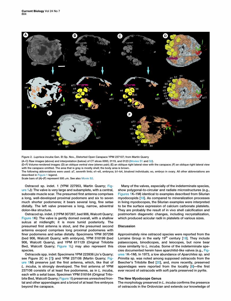
Figure 2. Luprisca incuba Gen. Et Sp. Nov., Distorted Open Carapace YPM 237107; from Martin Quarry
(A–C) Raw images (above) and interpretation (below) of CT slices 0093, 0119, and 0123 (Movies S1 and S2).
(D–F) Volume-rendered images: (D) an oblique ventral view (stereo pair); (E) an oblique right lateral view with the carapace; (F) an oblique right lateral view
with the carapace omitted. The area that is gray is mostly shell; the body area is brown.
The following abbreviations were used: a7, seventh limb; e1–e5, embryos; b1–b4, bivalved individuals; eo, embryo in ovary. All other abbreviations are
described in Figure 1 legend.
Scale bars of (A)–(F) represent 500 mm. See also Movie S2.
Current Biology Vol 24 No 7804
Ostracod sp. indet. 1 (YPM 227953, Martin Quarry; Fig-ure 1J): The valve is very large and subamplete, with a central,subovate muscle scar. The presumed first antenna comprisesa long, well-developed proximal podomere and six to sevenmuch shorter podomeres; it bears several long, fine setaedistally. The left valve preserves a long, narrow, adventraldolon-like structure.
Ostracod sp. indet. 2 (YPM307287, bed 906,Walcott Quarry;Figure 1K): The valve is gently domed overall, with a shallowsulcus at midlength; it is more tumid posteriorly. Thepresumed first antenna is stout, and the presumed secondantenna exopod comprises long proximal podomeres withfiner podomeres and setae distally. Specimens YPM 307298(bed 906, Walcott Quarry; with embryos), YPM 516199 (bed906, Walcott Quarry), and YPM 611129 (Original TrilobiteBed, Walcott Quarry; Figure 1L) may also represent thisspecies.
Ostracods spp. indet: Specimens YPM 223936 (Jo’s Quarry;see Figure 2C in [7]) and YPM 237106 (Martin Quarry; Fig-ure 1M) preserve just the first antenna, which, like that ofL. incuba, is strongly developed. The first antenna of YPM237106 consists of at least five podomeres, as in L. incuba,each with a setal base. Specimen YPM 516164 (Original Trilo-bite Bed,Walcott Quarry; Figure 1I) preserves unresolved fron-tal and other appendages and a brood of at least five embryosbeyond the carapace.
Many of the valves, especially of the indeterminate species,show polygonal-to-circular and radiate microstructures (e.g.,Figures 1K–1M) identical to examples described from Silurianmyodocopids [18]. As compared to mineralization processesin living myodocopes, the Silurian examples were interpretedto be the surface expression of calcium carbonate platelets.They are probably the result of in vivo shell calcification andpostmortem diagenetic changes, including recrystallization,which produced acicular radii in platelets of various sizes.
Discussion
Approximately nine ostracod species were reported from theLorraine Group in the early 19th century [19]. They includepalaeocopes, binodicopes, and leiocopes, but none bearclose similarity to L. incuba. Some of the indeterminate spe-cies documented herein have aparchitid-like valves (e.g., Fig-ures 1K–1M). In 1973, a low abundance of Aparchites sp. andPrimitia sp. was noted among supposed ostracods from theBeecher’s Trilobite Bed [20], and, more recently, preservedappendages were reported from the locality [6]—the firstever record of ostracods with soft parts preserved in pyrite.
The New Myodocope Genus
The morphology preserved in L. incuba confirms the presenceof ostracods in the Ordovician and extends our knowledge of
Exceptionally Preserved Ordovician Ostracods805
the paleobiology of the group back some 25 million years fromthe mid-Silurian Herefordshire Lagerstatte [1–4]. L. incuba isassigned here to the Myodocopa and provisionally to the My-odocopida. The podomere and setal arrangement of the firstantenna is the same as that of extant Myodocopida, evendown to the tiny ventral seta and two long distal setae, whichare comparable to the sensory bristle on the fifth podomereand the ‘‘C’’ and ‘‘F’’ bristles of the seventh and eighth(distalmost) podomeres, respectively, of many cylindroleberi-did myodocopids (see [21]). A similar pair of distal setae isalso present in the first antenna of the Silurian myodocopidsColymbosathon, Nymphatelina, and Nasunaris [1–3]. The sec-ond antenna of L. incuba, with its large ovoid basipod and anexopod longer than the endopod, also accords with themorphology of myodocopes rather than podocopes (in whichthe endopod is better developed). Moreover, if the position ofthe setae on the exopod of the second antenna reflects thearrangement of podomeres, the total number mirrors that inextant myodocopes (the exopod in the majority of myodoco-pids and halocyprids consists of nine podomeres and rarelyless) compared to one or two podomeres, or just setae, inpodocopes [22]. The slender, vermiform morphology of theputative seventh appendage in L. incuba is known only inmyodocopids. The strongly sclerotized nature of the furcaalso conforms more to that of extant myodocopes than podo-copes, as does the probable thin and/or poorly calcified natureof the shell. In view of these similarities, it is striking that theexternal morphology of the shell shows no defining myodo-cope feature, such as a rostrum or rostral incisure. It is clearthat caution is required when classifying ostracods based onthe carapace alone [4]. The one other known supposedOrdovician myodocope, recorded only from shells [23],is Myodoprimigenia fistuca from Hirnantian-age rocks(w443 Ma) of South Africa.
Brooding StrategyThe brooding strategy evident in L. incuba accords with that ofextant myodocopids (Figure 1H). It allows the gender ofseveral specimens to be determined in this Ordovician speciesand provides fossil evidence that a brooding reproductivemode has persisted in myodocopes for at least 450 Ma. Thisreproductive strategy may be even older, given that molecularphylogenetics predicts that myodocopes occur in theCambrian period [14]. Eggs of fossil invertebrates, especiallyputative in situ embryos, are very rare, including possibleexamples of eggs and brooding in fossil ostracods [2]. Mostsupposed fossil invertebrate eggs are recovered isolatedfrom the assumed parent. Putative in situ eggs or embryoshave been found in fossils of a bradoriid arthropod species[24], a tealliocarididmalacostracan, a syncaridmalacostracan,two branchiopod species, and a few ostracod species ([2] andreferences therein).
Most extant ostracods are Podocopa, the majority of whichreproduce sexually and deposit their eggs on a substrate,whereas most Myodocopa (all myodocopids and at least onehalocyprid), Platycopa, and Darwinuloidea brood embryos inthe posterior area of the domicilium [25, 26]. The brood numberand size of the embryos in L. incuba is similar to that in extant,sexually mature, brooding myodocopes [25]. The concurrenceof ovarian and brooded embryos (i.e., clutch overlap) is alsoknown in some extant myodocopes [25]. L. incuba is uniquein providing evidence of ovarian, embryonic, and possiblejuvenile developmental stages in a fossil invertebrate. In theextant myodocopid Vargula hilgendorfii, embryonic
development takes about 16 days, from egg on the ovarywall [27] to hatched limb-bearing bivalved juvenile [28, 29].The only other unequivocal example of eggs in a fossilostracod is in a specimen of the mid-Silurian myodocopeNymphatelina gravida from the Herefordshire Konservat-La-gerstatte (w425 Ma), whose in situ brood includes a bivalvedindividual [2]. Evidence of reproductive strategies in fossil os-tracods is otherwise indirect: presumed sexual dimorphism ofthe shell inmany groups involveswhat is possibly brood spaceor, rarely, the supposed occurrence of only presumed females,suggesting parthenogenesis ([2] and references therein).
Habitat and LifestyleThe LorraineGroup accumulated in the relatively deep axis of aforeland basin that formed on themargin of the paleocontinentLaurentia during the Cambrian-Ordovician Taconic orogeny.The ostracods occur in thin gray mudstones interpreted aslow-density distal turbidites that entrained and rapidly burieda low-diversity/low-abundance benthic fauna that includesTriarthrus with preserved appendages. The assemblage isinterpreted as living in dysoxic bottom-water conditions[6, 7, 30]. The association of ostracods and Triarthrus hereand elsewhere in the Taconic foreland basin [19, 31, 32] showsthat ostracods occur in deep-water associations with olenidtrilobites during the Ordovician period (contra [33]). Conjoinedostracod valves are common, indicating that they were prob-ably not subject to prolonged high-energy conditions post-mortem. L. incuba has well-developed frontal appendages,suggesting that it was a swimmer. Most extant myodocopesare nektobenthic predators, scavengers, or detritivores [34],and the lifestyle of L. incuba was probably similar.
Experimental Procedures
The fossils were prepared using mechanical techniques, including fine air
abrasion. All except the extant ostracod (Figure 1H; a scanning electron
micrograph) were photographed under alcohol using a Nikon Multiphot
stand and associated bellows equipment mounted with a Canon EOS 5D
camera (Figures 1A–1G and 1I–1M), following the methods of [35]. Images
of specimen YPM 237107 were also captured by computed tomography
(CT) (Figures 2A–2C; Movies S1 and S2), and volume was rendered and
laterally emphasized (4003) in order to compensate for flattening of the
fossil due to compaction (Figures 2D–2F). The figured CT images were
obtained using a ScanXmate-L system (Comscantecno) at a resolution of
20.51 mm at 151 kV. Some 260 tiff images were captured, each 683 3
604 pixels in size with a resolution of 0.51 pixel/cm. Using contrasts in den-
sity, soft parts (thick density) and shell (thin density) were distinguished
and colored (RGB). ImageJ software [36] was used to convert the 2D CT
image stacks into a 3D volumetric image. Volume rendering was performed
using virtual stack intervals at 20.51 mm, with threshold 0 and resampling
factor 2.
Supplemental Information
Supplemental Information includes two movies and can be found with this
article online at http://dx.doi.org/10.1016/j.cub.2014.02.040.
Acknowledgments
The Yale Peabody Museum of Natural History supported a study visit by
David Siveter. CT study was undertaken courtesy of the American Museum
of Natural History (James Thostenson), New York and Comscantecno
(K. Kikuchi), Japan. Rachel Racicot assisted in processing scanned images,
and Jane Hall assisted with editing. The authors acknowledge Leverhulme
Trust foremost grant RP14G0168 (to M. Williams and David Siveter) for
research on fossil myodocopes and Japan Society for the Promotion of Sci-
ence KAKENHI grant 21740370 (toG.T.). We thankN.Wakayama (Kanagawa
University, Japan) for material of Vargula hilgendorfii and JoachimHaug and
an anonymous reviewer of the manuscript for comments.
Current Biology Vol 24 No 7806
Received: November 13, 2013
Revised: December 17, 2013
Accepted: February 15, 2014
Published: March 13, 2014
References
1. Siveter, D.J., Sutton, M.D., Briggs, D.E.G., and Siveter, D.J. (2003). An
ostracode crustacean with soft parts from the Lower Silurian. Science
302, 1749–1751.
2. Siveter, D.J., Siveter, D.J., Sutton, M.D., and Briggs, D.E.G. (2007).
Brood care in a Silurian ostracod. Proc. Roy. Soc. B 274, 465–469.
3. Siveter, D.J., Briggs, D.E.G., Siveter, D.J., and Sutton, M.D. (2010). An
exceptionally preserved myodocopid ostracod from the Silurian of
Herefordshire, UK. Proc. Roy. Soc. B 277, 1539–1544.
4. Siveter, D.J., Briggs, D.E.G., Siveter, D.J., Sutton, M.D., and Joomun,
S.C. (2013). A Silurian myodocope with preserved soft-parts: cautioning
the interpretation of the shell-based ostracod record. Proc. Roy. Soc. B
280, 1752.
5. Olempska, E., Horne, D.J., and Szaniawski, H. (2012). First record of
preserved soft parts in a Palaeozoic podocopid (Metacopina) ostracod,
Cytherellina submagna: phylogenetic implications. Proc. Roy. Soc. B
279, 564–570.
6. Farrell, U.C., Martin, M.J., Hagadorn, J.W., Whiteley, T., and Briggs,
D.E.G. (2009). Beyond Beecher’s Trilobite Bed: widespread pyritization
of soft tissues in the Late Ordovician Taconic foreland basin. Geology
37, 907–910.
7. Farrell, U.C., Briggs, D.E.G., and Gaines, R.R. (2011). Paleoecology of
the olenid trilobite Triarthrus: new evidence from Beecher’s Trilobite
Bed and other sites of pyritization. Palaios 26, 730–742.
8. Horne, D.J. (2003). Key events in the ecological radiation of the
Ostracoda. Paleontol. Soc. Papers 9, 181–201.
9. Siveter, D.J. (1984). Habitats and modes of life of Silurian ostracodes.
Spec. Pap. Palaeontol. 32, 71–85.
10. Siveter, D.J., Vannier, J.M.C., and Palmer, D. (1991). Silurian myodo-
copes: pioneer pelagic ostracods and the chronology of an ecological
shift. J. Micropalaeont. 10, 151–173.
11. Perrier, V., Vannier, J., and Siveter, D.J. (2007). The Silurian pelagic
myodocope ostracod Richteria migrans. Earth Environ. Sci. Trans. R.
Soc. Edinb. 98, 151–163.
12. Perrier, V., Vannier, J., and Siveter, D.J. (2011). Silurian bolbozoids and
cypridinids (Myodocopa) from Europe: pioneer pelagic ostracods.
Palaeontology 54, 1361–1391.
13. Perrier, V., and Siveter, D.J. (2013). Testing Silurian palaeogeo-
graphy using ‘European’ ostracod faunas. Geol. Soc. London Mem.
38, 355–364.
14. Oakley, T.H., Wolfe, J.M., Lindgren, A.R., and Zaharoff, A.K. (2013).
Phylotranscriptomics to bring the understudied into the fold: monophy-
letic Ostracoda, fossil placement, and pancrustacean phylogeny. Mol.
Biol. Evol. 30, 215–233.
15. Siveter, D.J. (2008). Ostracods in the Palaeozoic? Senckenb. Leth. 88,
1–9.
16. Cohen, K.M., Finney, S., and Gibbard, P.L. (2013). International chrono-
stratigraphic chart. International Commission on Stratigraphy, http://
www.stratigraphy.org/ICSchart/ChronostratChart2013-01.pdf.
17. Sars, G.O. (1866). Oversigt af Norges marine ostracoder. Norske
Vidensk.-Akad. Forhandl. 1865, 1–130.
18. Siveter, D.J., Vannier, J.M.C., and Palmer, D. (1987). Silurian myodoco-
pid ostracodes: their depositional environments and the origin of their
shell microstructures. Palaeontology 30, 783–813.
19. Ruedemann, R. (1926). The Utica and Lorraine formations of New York.
Part 2, systematic paleontology, no. 2 mollusks, crustaceans, and
eurypterids. New York State Mus. Bull. 272, 1–227.
20. Cisne, J.L. (1973). Beecher’s Trilobite Bed revisited: ecology of an
Ordovician deepwater fauna. Postilla 160, 1–25.
21. Kornicker, L.S. (1981). Revision, distribution, ecology, and ontogeny
of the ostracode subfamily Cylcasteropinare (Myodocopina:
Cylindroleberididae). Smithson. Contrib. Zool. 319, 1–548.
22. Cohen, A.C., and Morin, J.G. (2003). Sexual morphology, reproduction
and bioluminescence in Ostracoda. Paleontol. Soc. Papers 9, 37–70.
23. Gabbott, S.E., Siveter, D.J., Aldridge, R.J., and Theron, J.N. (2003). The
earliest myodocopes: ostracodes from the late Ordovician Soom Shale
Lagerstatte of South Africa. Lethaia 36, 151–160.
24. Duan, Y.-h., Han, J., Fu, D.-j., Zhang, X.-l., Yang, X.-g., Koiya, T., and
Shu, D.-g. (2013). Reproductive strategy of the bradoriid arthropod
Kunmingella douvillei from the Lower Cambrian Chengjiang
Lagerstatte, South China. Gondwana Res. 25, 983–990.
25. Cohen, A.C., and Morin, J.G. (1990). Patterns of reproduction in
ostracodes: a review. J. Crustac. Biol. 10, 184–211.
26. Horne, D.J., Danielopol, D.L., and Martens, K. (1998). Reproductive
behaviour. In Sex and Parthenogenesis: Evolutionary Ecology of
Reproductive Modes in Non-Marine Ostracods, K. Martens, ed.
(Leiden, The Netherlands: Backhuys), pp. 157–195.
27. Ikuta, K., and Makiok, T. (1999). Ovarian structure and oogenesis in
the myodocopid ostracod Vargula hilgendorfii. J. Crustac. Biol. 19,
730–737.
28. Wakayama, N. (2007). Embryonic development clarifies polyphyly in
ostracod crustaceans. J. Zool. (Lond.) 273, 406–413.
29. Vannier, J., Abe, K., and Ikuta, K. (1998). Feeding in myodocopid ostra-
cods: functional morphology and laboratory observations from videos.
Mar. Biol. 132, 391–408.
30. Farrell, U.C., Briggs, D.E.G., Hammarlund, E.U., Sperling, E.A., and
Gaines, R.R. (2013). Paleoredox and pyritization of soft-bodied fossils
in the Ordovican Frankfort Shale of New York. Am. J. Sci. 313, 452–489.
31. Titus, R., and Cameron, B. (1976). Fossil communities of the Lower
Trenton Group (Middle Ordovician) of central and northwestern New
York State. J. Paleontol. 50, 1209–1225.
32. Brett, C.E., Allison, P.A., Tsujita, C.J., Soldani, D., and Moffat, H.A.
(2006). Sedimentology, taphonomy, and paleoecology of meter-scale
cycles from the Upper Ordovician of Ontario. Palaios 21, 530–547.
33. Williams, M., Floyd, J.D., Salas, M.J., Siveter, D.J., Stone, P., and
Vannier, J. (2003). Patterns of ostracod migration for the ‘North
Atlantic’ region during the Ordovician. Palaeogeogr. Palaeoclimatol.
Palaeoecol. 195, 193–228.
34. Vannier, J., and Abe, K. (1993). Functional morphology of Vargula
hilgendorfii (Ostacoda: Myodocopida), Japan, and discussion of its
crustacean ectoparasites. J. Crustac. Biol. 13, 51–76.
35. Siveter, D.J. (1990). Photography. In Palaeobiology: A Synthesis, D.E.G.
Briggs and P.R. Crowther, eds. (Oxford, UK: Blackwell), pp. 505–508.
36. Abramoff, M.D., Magelhaes, P.J., and Ram, S.J. (2004). Image
Processing with ImageJ. Biophotonics Internat 11, 36–42.