examining respiratory system
TRANSCRIPT
Lecture NotesExamining the Respiratory
System
W. P. Howlett
2017
Main Symptoms
Dyspnoea
Cough/Haemoptysis
Chest pain

Dyspnoea
Dyspnoea: shortness of breath
Wheeze: audible musical sounds usually expiratory
DyspnoeaGrading or Severity: mild, moderate, severe
Pattern: exertion, continuous/intermittent, rest/sleep
Time course: onset, duration, progression
Variability: diurnal, day to day, aggravating or relieving factors
Associated symptoms: cough, chest pain

Cough 1
Characteristics: type
Time course: onset & duration
Productive or non productive or dry
Sputum: what’s in it
Cough 2Type of cough: barking, harsh, productive/non productive
Pattern:continuous or intermittent, day/night
Time course: onset, duration, progression
Sputum: colour: white, pink, green, frothy, rusty, bloodyamount: a lot/little, smell/taste: odourless or foul smelling
Associated symptoms: pain, dyspnoea
Haemoptysis
Definition: coughing up of blood
Type: frank or blood stained
Degree: how much
Frequency: how often
Duration: for how long
Chest PainSite: lateral part of chest
Character: pleuritic; worse on breathing and/orcoughing, movement, sharp, stabbing
Other features:
Severity
Time Course
Aggravating & Relieving Factors
Associated factors
Previous History of pain
Past Medical History
TB/HIV: active on Rx or inactive
Chronic Resp Disease: Asthma/Wheezy/Bronchitis
Pneumonia/Pleurisy
Past history: Chest Injury/RTA, travel, childhood
chest infections
Family History
Allergies/Eczema: asthma
Respiratory Disease: TB/HIV, chronic bronchitis
Inherited risk: cystic fibrosis
Acquired risk: passive smoking
Social History
Cigs: 20 or 1 pack/day for 30 yrs = 30 pack years
Alcohol: type, quantity & duration
Occupational exposure:
Dust: mining & factories
Infections: farming, animals etc
Respiratory Examination
Undress patient: to level of the waist
Position: sitting @ angle of 45 degrees
Inspection: first from side & repeat from
front

Examining position
General InspectionAppearance: well or unwell: e.g dyspnoeic, wasting
Breathing pattern: thoracic or abdominal
Respiratory distress: dyspnoeic, wheezing or stridor
Resp Rate: normal = 14-18/min
Cyanosis: peripheral and/or central
Signs of Respiratory Distress
Respiratory distress: dyspnoeic, wheezing or stridor
Intercostal recession and/or subcostal retraction
Using accessory muscles: ali nasi, sternomastoids, scalenes
Tachypnoea: Resp Rate: >18/min
Cyanosis: peripheral and/or central
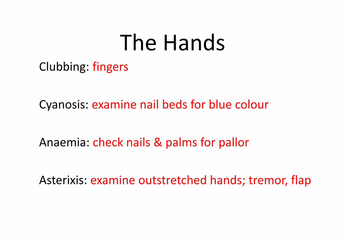
The HandsClubbing: fingers
Cyanosis: examine nail beds for blue colour
Anaemia: check nails & palms for pallor
Asterixis: examine outstretched hands; tremor, flap
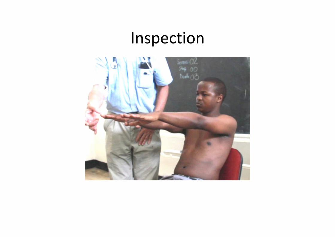
Inspection
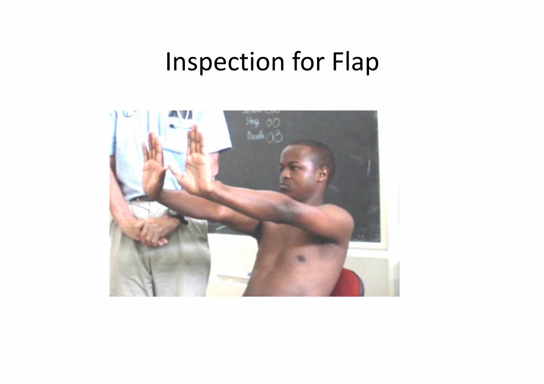
Inspection for Flap
Clubbing
1) loss of normal nail bed angle (n=<170 degrees)
2) increased nail bed fluctuancy
3) increased antero posterior curvature of nails & distal
phalanx
The Pulse
Rate: Tachycardia
Volume: Pulsus paradoxus
The Head, Face & Neck Eye lids/conjunctiva: anaemia & polycytaemia
Pupils: Horner’s Syndrome: (ptosis, miosis, enopthalmos, anhydrosis)
Tongue: cyanosis, anaemia, polycytaemia
Palate: Kaposi Sarcoma, monilia
Sinuses: tap frontal & maxillary for tenderness

Eyelids/conjunctiva
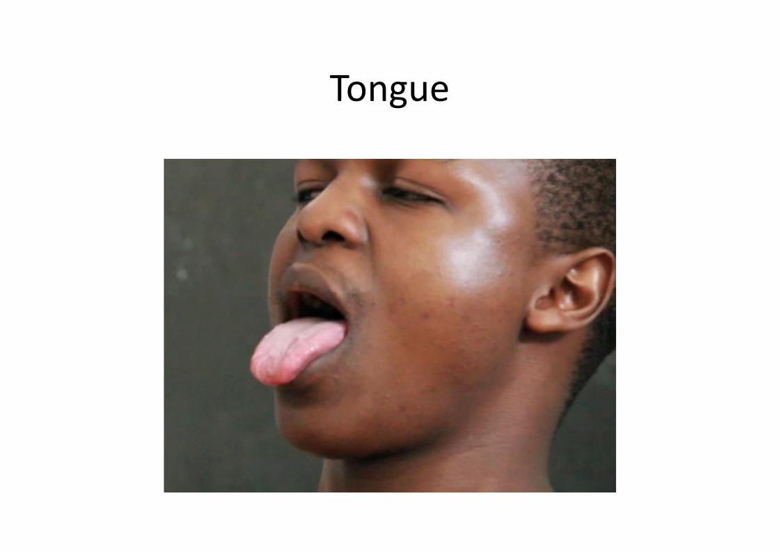
Tongue
Lymph Nodes
Supraclavicular Fossa: supraclavicular &
deep cervical neck glands
Axilla: 4 walls (ant, post, med and lat) &
apex
The Chest
Inspection
Palpation
Percussion
Auscultation
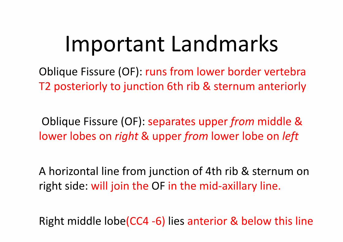
Important LandmarksOblique Fissure (OF): runs from lower border vertebra
T2 posteriorly to junction 6th rib & sternum anteriorly
Oblique Fissure (OF): separates upper from middle &
lower lobes on right & upper from lower lobe on left
A horizontal line from junction of 4th rib & sternum on
right side: will join the OF in the mid-axillary line.
Right middle lobe(CC4 -6) lies anterior & below this line
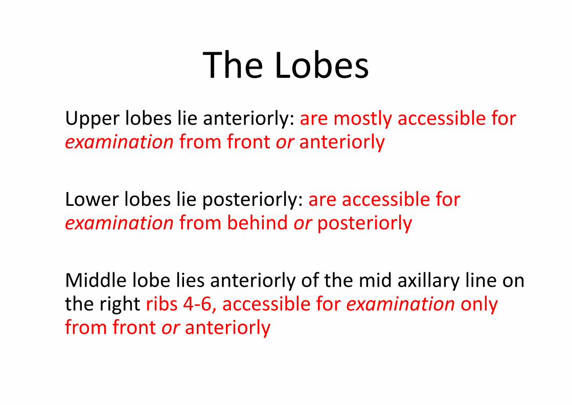
The Lobes
Upper lobes lie anteriorly: are mostly accessible for examination from front or anteriorly
Lower lobes lie posteriorly: are accessible for examination from behind or posteriorly
Middle lobe lies anteriorly of the mid axillary line on the right ribs 4-6, accessible for examination only from front or anteriorly
The Chest
Inspection
Palpation
Percussion
Auscultation
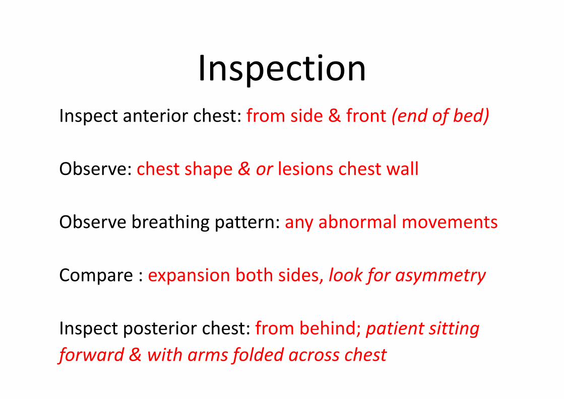
InspectionInspect anterior chest: from side & front (end of bed)
Observe: chest shape & or lesions chest wall
Observe breathing pattern: any abnormal movements
Compare : expansion both sides, look for asymmetry
Inspect posterior chest: from behind; patient sitting
forward & with arms folded across chest

Inspection from the front
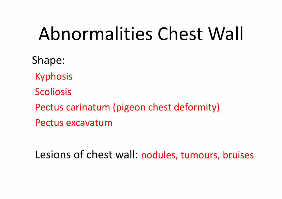
Abnormalities Chest Wall
Shape:
Kyphosis
Scoliosis
Pectus carinatum (pigeon chest deformity)
Pectus excavatum
Lesions of chest wall: nodules, tumours, bruises
The Chest
Inspection
Palpation
Percussion
Auscultation
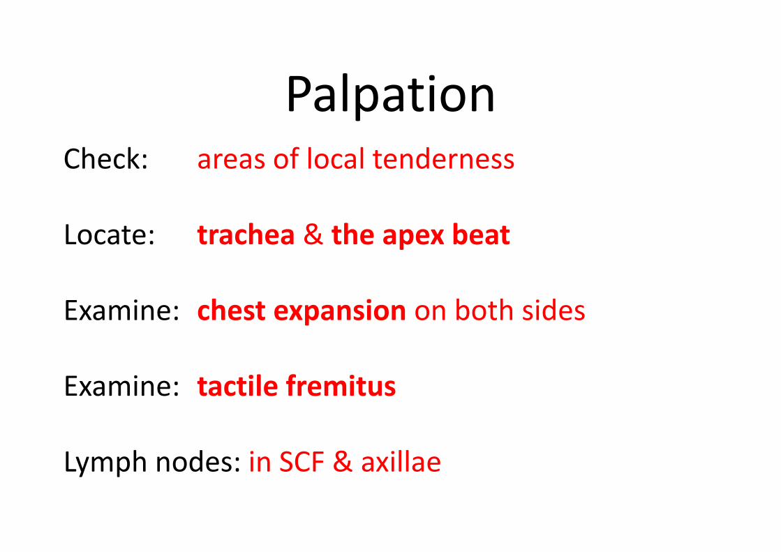
PalpationCheck: areas of local tenderness
Locate: trachea & the apex beat
Examine: chest expansion on both sides
Examine: tactile fremitus
Lymph nodes: in SCF & axillae
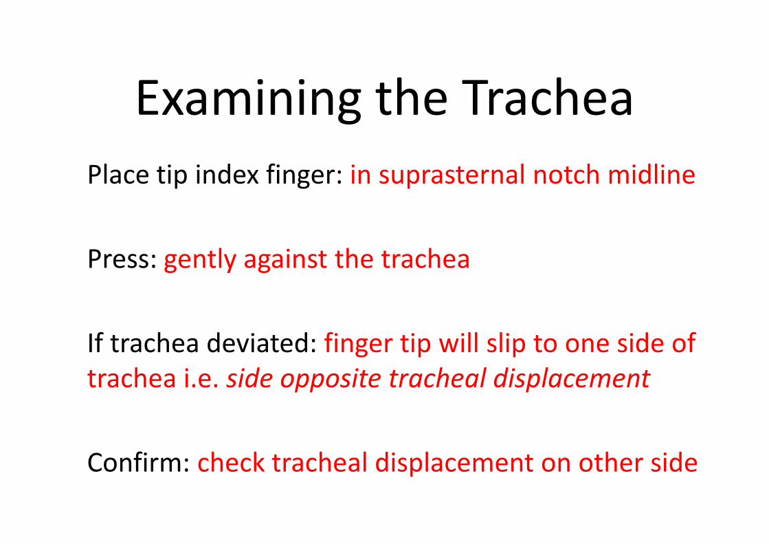
Examining the Trachea
Place tip index finger: in suprasternal notch midline
Press: gently against the trachea
If trachea deviated: finger tip will slip to one side of
trachea i.e. side opposite tracheal displacement
Confirm: check tracheal displacement on other side
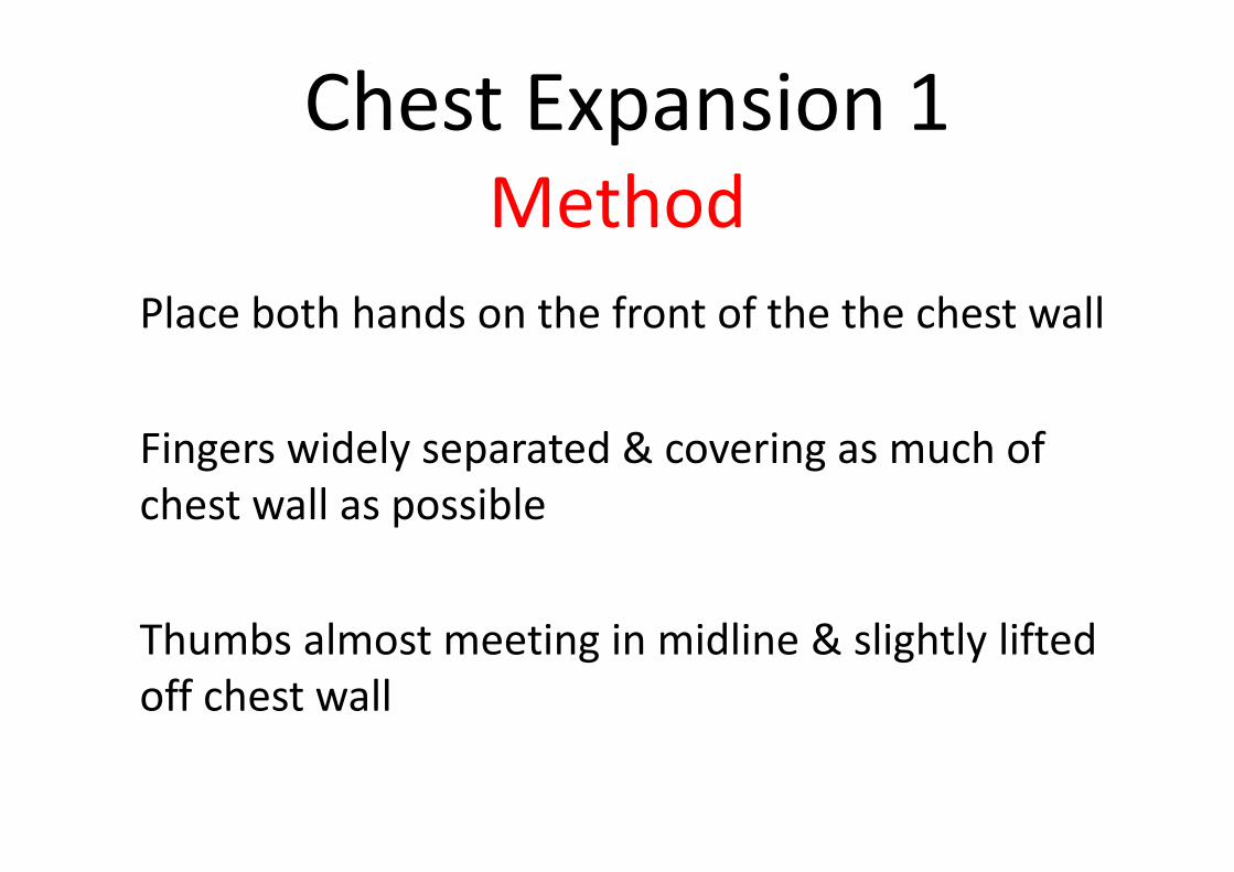
Chest Expansion 1Method
Place both hands on the front of the the chest wall
Fingers widely separated & covering as much of
chest wall as possible
Thumbs almost meeting in midline & slightly lifted
off chest wall
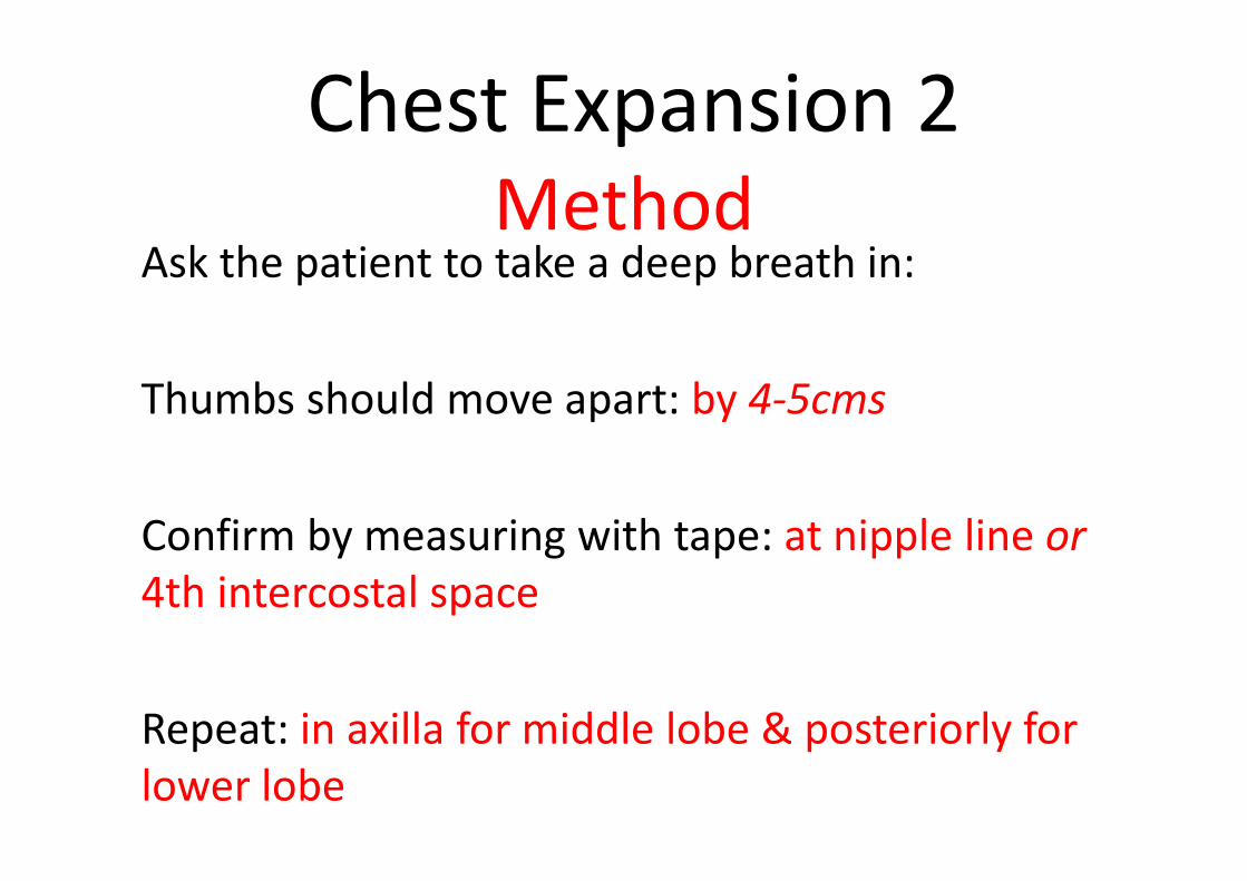
Chest Expansion 2Method
Ask the patient to take a deep breath in:
Thumbs should move apart: by 4-5cms
Confirm by measuring with tape: at nipple line or
4th intercostal space
Repeat: in axilla for middle lobe & posteriorly for
lower lobe
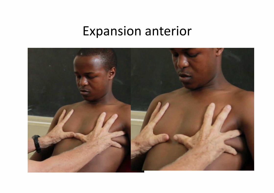
Expansion anterior
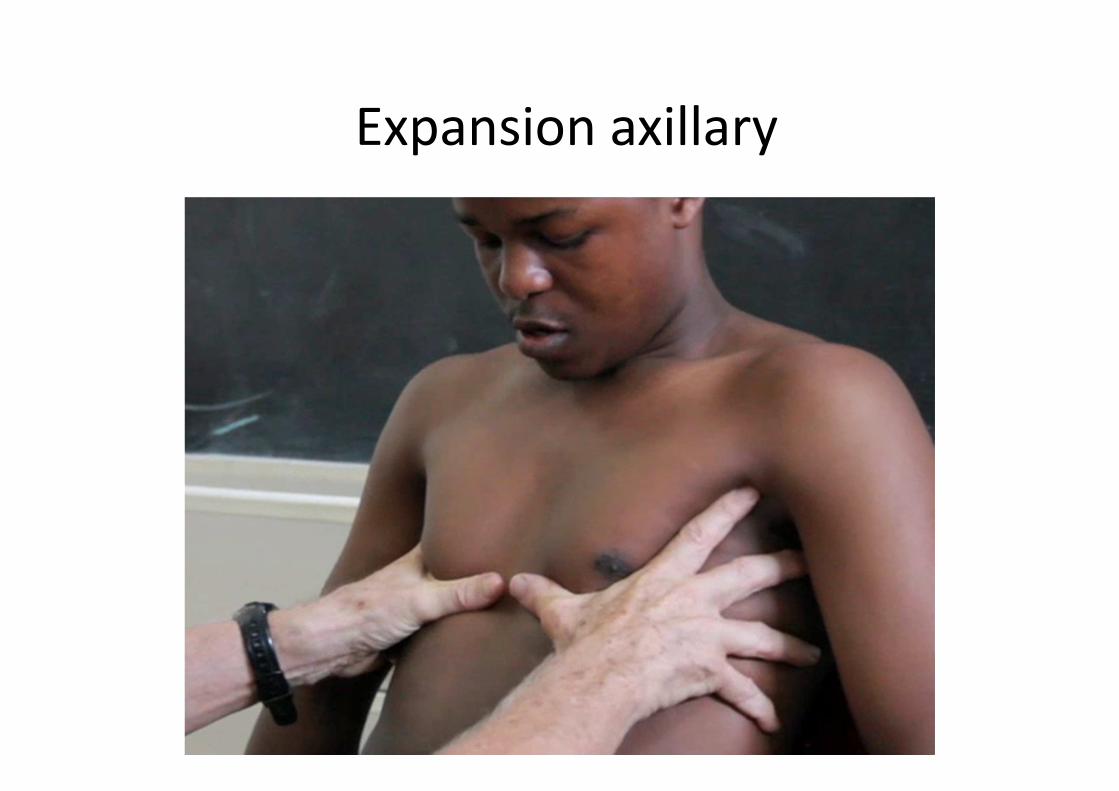
Expansion axillary
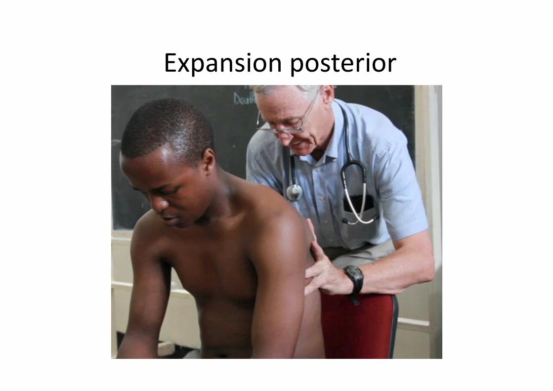
Expansion posterior

Measuring tape method

Measuring tape method
• Before inhalation • After maximum
inhalation
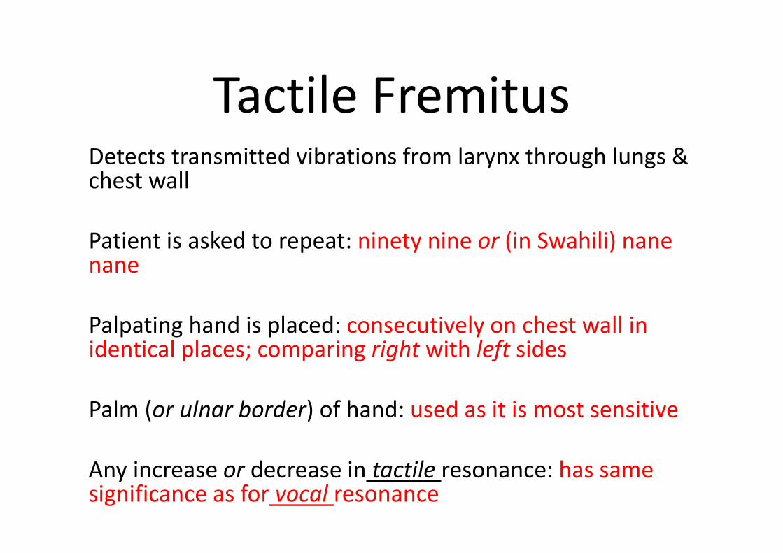
Tactile FremitusDetects transmitted vibrations from larynx through lungs & chest wall
Patient is asked to repeat: ninety nine or (in Swahili) nanenane
Palpating hand is placed: consecutively on chest wall in identical places; comparing right with left sides
Palm (or ulnar border) of hand: used as it is most sensitive
Any increase or decrease in tactile resonance: has same significance as for vocal resonance
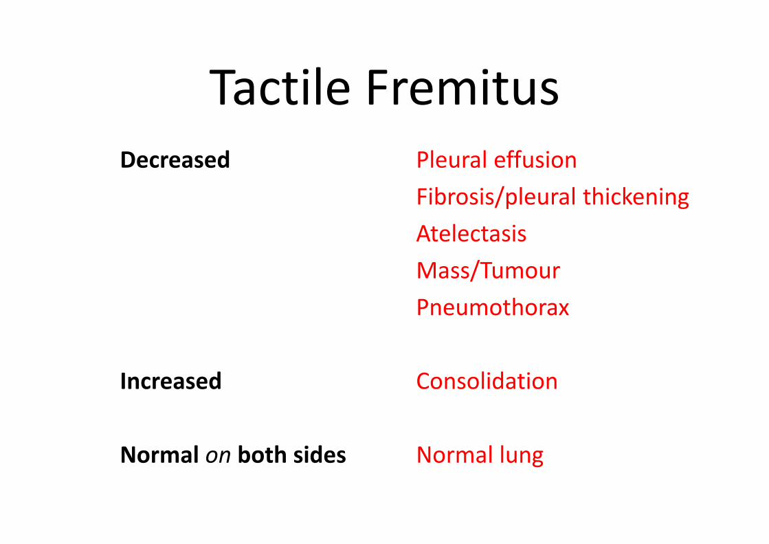
Tactile Fremitus
Decreased
Increased
Normal on both sides
Pleural effusion
Fibrosis/pleural thickening
Atelectasis
Mass/Tumour
Pneumothorax
Consolidation
Normal lung
Key Points
• Correct position of patient is sitting @ 45 degrees
• Inspection is more reliable than palpation for
detecting asymmetrical expansion of chest
• Normal position of trachea & apex indicates normal
alignment of mediastinum
• Above necessary for correct interpretation of
findings
The Chest
Inspection
Palpation
Percussion
Auscultation
PercussionPlace left hand on chest wall: fingers separated but with
middle finger in tight contact to skin
Strike second phalanx of middle finger: with tip of right
middle finger
Compare notes from: the same sites on both sides
Map out any areas of abnormality: e.g. area of dullness
Percuss: in a resonant to dull direction

Percussion 1Anterior chest wall: (2nd-6th ribs)
Clavicles: tap once each side; comparing right & left
Mid clavicular line: tap 2-3 times on each side; comparing right & left
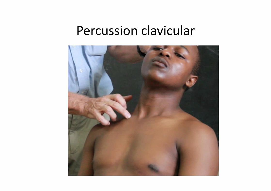
Percussion clavicular
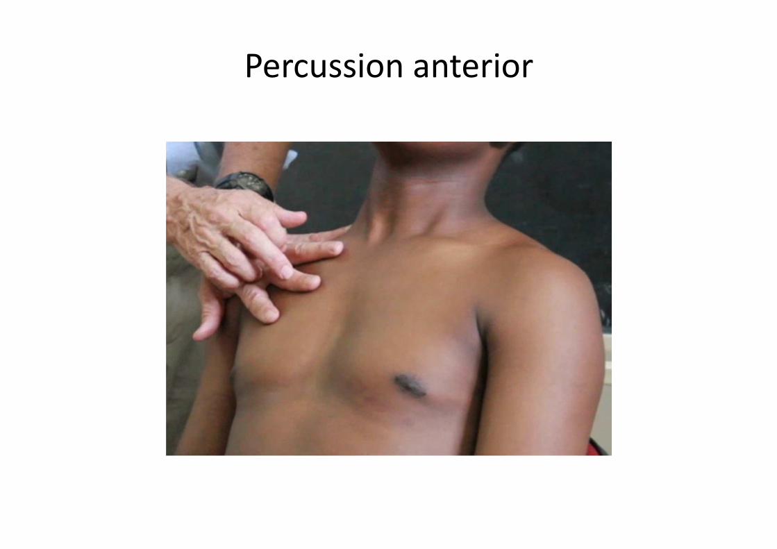
Percussion anterior
Percussion 2Lateral chest wall: (3rd-7th ribs)
Mid axillary line: tap 2-3 times on each
side; comparing right & left
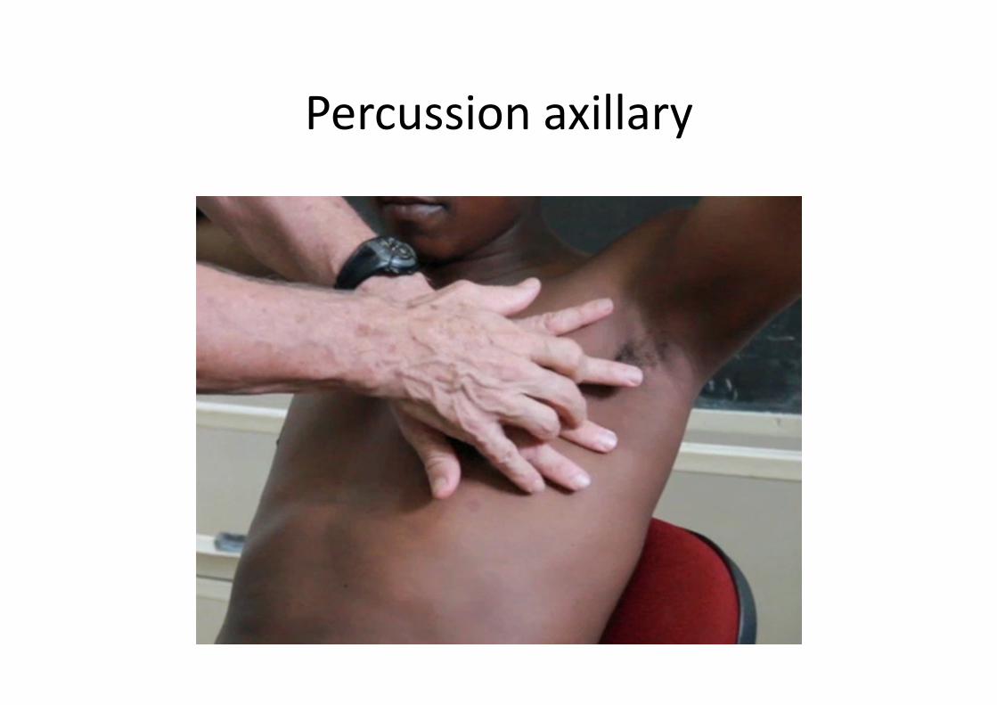
Percussion axillary
Percussion 3Posterior chest wall (apex to 11th rib)
Percuss in a: C shaped direction
Start at apex, move downwards medial to borders of scapulae & then: fanning outwards inferiorly (see video)
Tap once at apex & then repeat 5 times
on each side: comparing right & left

Percussion posterior
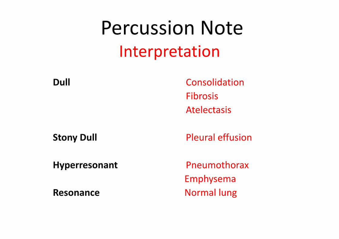
Percussion NoteInterpretation
Dull
Stony Dull
Hyperresonant
Resonance
Consolidation
Fibrosis
Atelectasis
Pleural effusion
Pneumothorax
Emphysema
Normal lung
Key Points
• Always compare the same sites on both sides of chest
• Main causes dullness: effusion, consolidation & fibrosis
• Increased resonance on one side may indicate a pneumothorax
• Increased resonance both sides usually non diagnostic
• Map out any suspected area of dullness or abnormality
The Chest
Inspection
Palpation
Percussion
Auscultation
Auscultation of lungs 1
Use Diaphragm i.e flat part of stethoscope to
listen to the lungs
Use Bell for suspected low frequency sounds: BB,
fibrosis & thin persons
Ask patient open mouth & breathe in/out: deeply
& rapidly
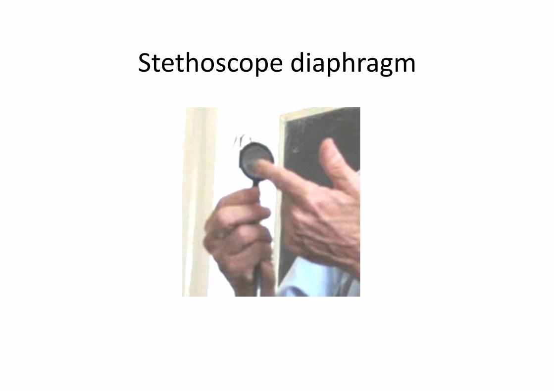
Stethoscope diaphragm
Auscultation of lungs 2Auscultate sides alternatively comparing: loudness & quality
Auscultate in both: inspiration & expiration
Ascultate at same sites: as you percussed
Auscultatory breath sounds: are either normal or abnormal
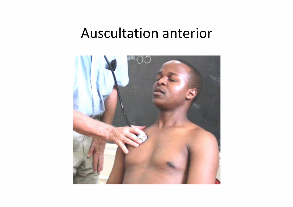
Auscultation anterior
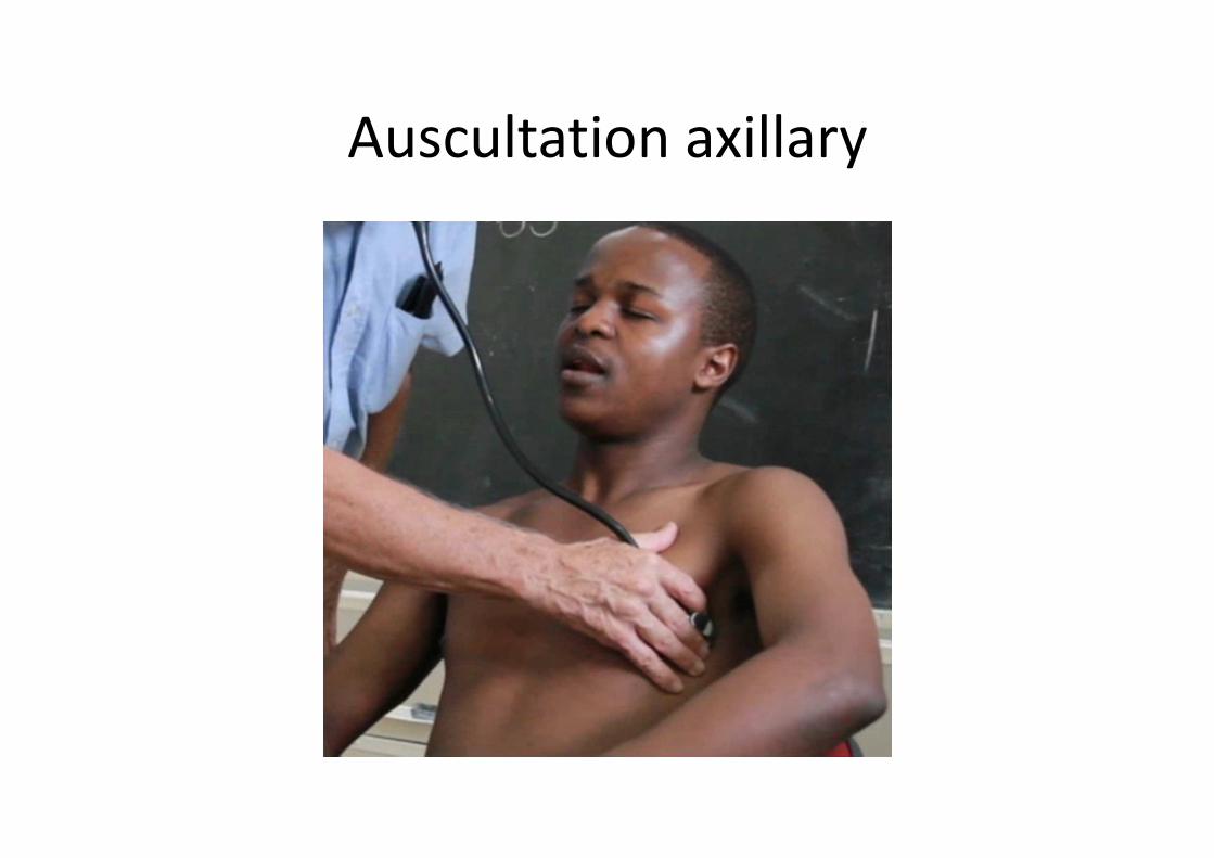
Auscultation axillary
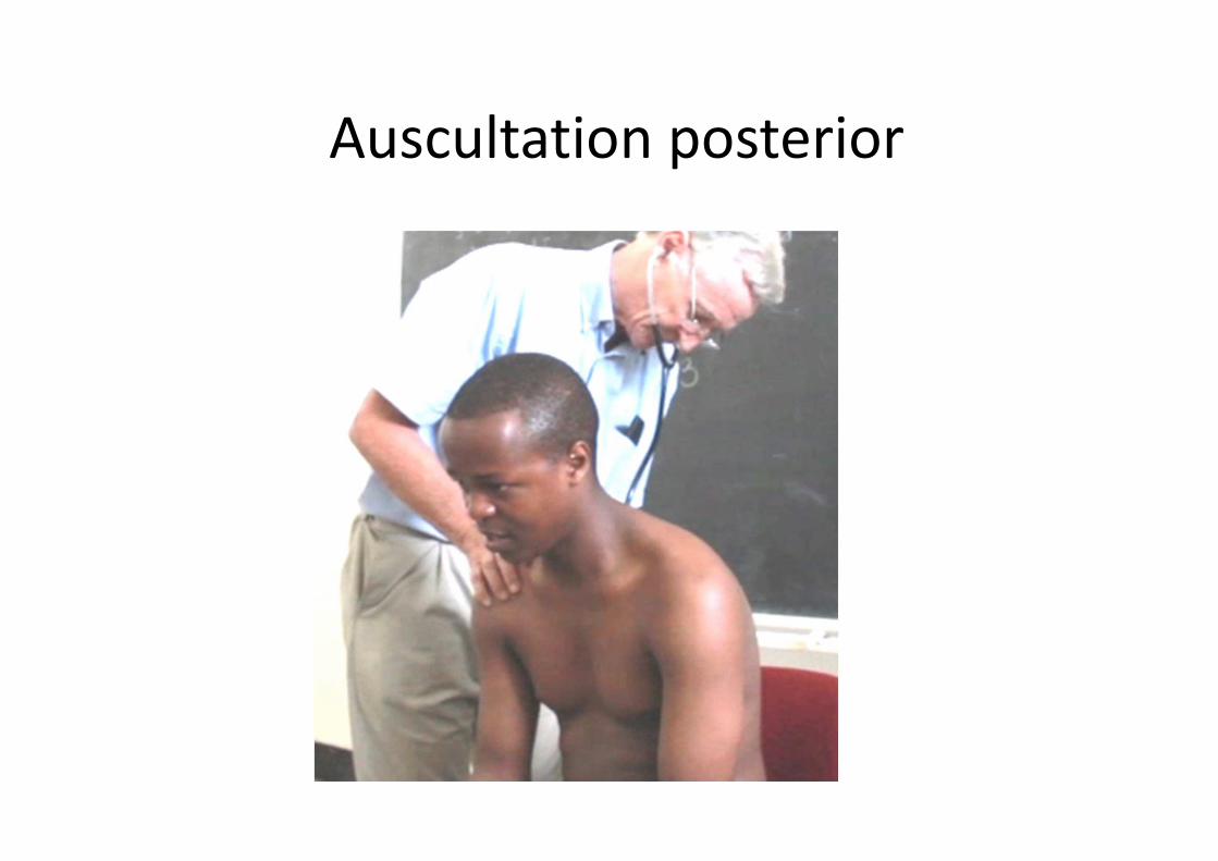
Auscultation posterior
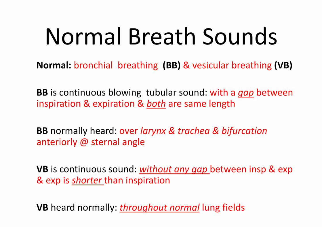
Normal Breath SoundsNormal: bronchial breathing (BB) & vesicular breathing (VB)
BB is continuous blowing tubular sound: with a gap between inspiration & expiration & both are same length
BB normally heard: over larynx & trachea & bifurcation anteriorly @ sternal angle
VB is continuous sound: without any gap between insp & exp & exp is shorter than inspiration
VB heard normally: throughout normal lung fields
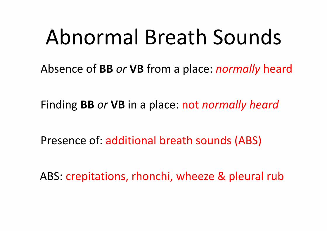
Abnormal Breath Sounds
Absence of BB or VB from a place: normally heard
Finding BB or VB in a place: not normally heard
Presence of: additional breath sounds (ABS)
ABS: crepitations, rhonchi, wheeze & pleural rub

Additional Breath Sounds
Crepitations (crackles): are interrupted small airways
sounds: classified as: fine, medium & coarse
Rhonchi: continuous high pitched musical sounds from
narrowed bronchi e.g asthma: heard with stethoscope
Wheeze: is high pitched musical sound: heard with ear
Pleural rub: is a superficial creaking, scratchy, pleural
based sound heard during insp & exp or both
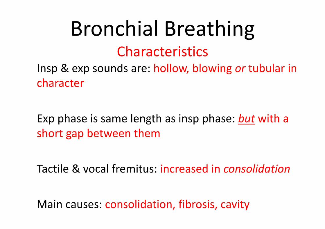
Bronchial Breathing Characteristics
Insp & exp sounds are: hollow, blowing or tubular in
character
Exp phase is same length as insp phase: but with a
short gap between them
Tactile & vocal fremitus: increased in consolidation
Main causes: consolidation, fibrosis, cavity
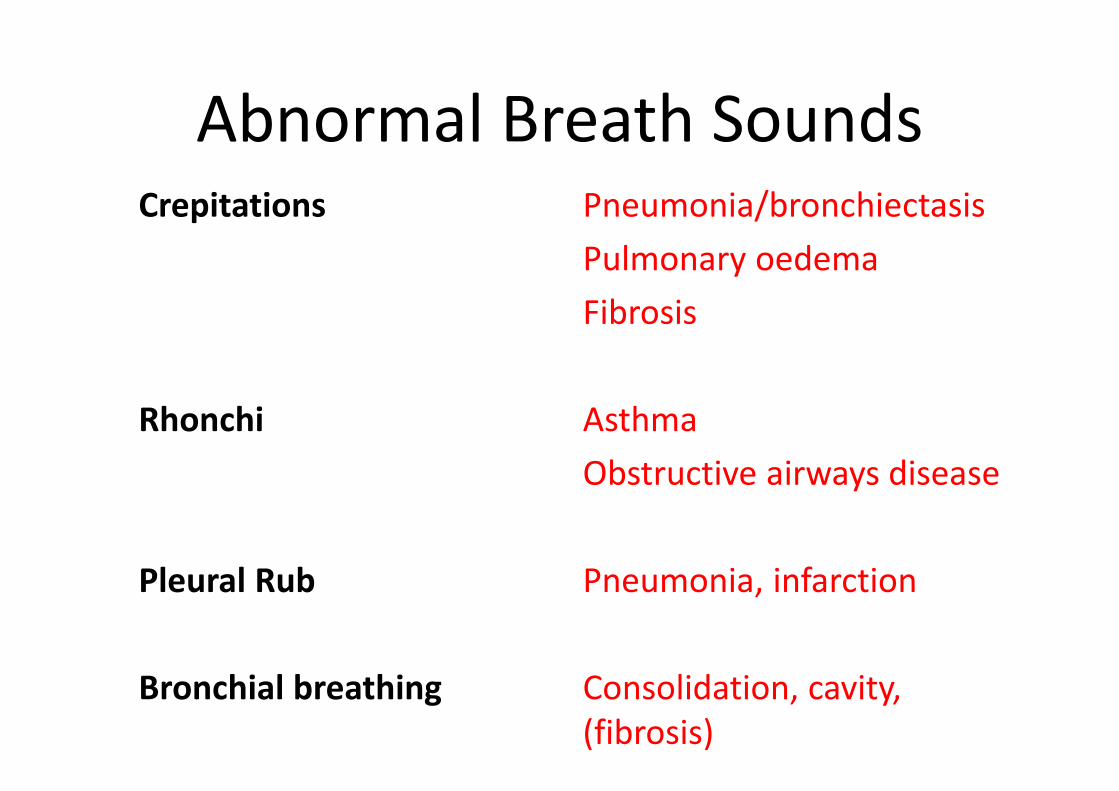
Abnormal Breath SoundsCrepitations
Rhonchi
Pleural Rub
Bronchial breathing
Pneumonia/bronchiectasis
Pulmonary oedema
Fibrosis
Asthma
Obstructive airways disease
Pneumonia, infarction
Consolidation, cavity,
(fibrosis)
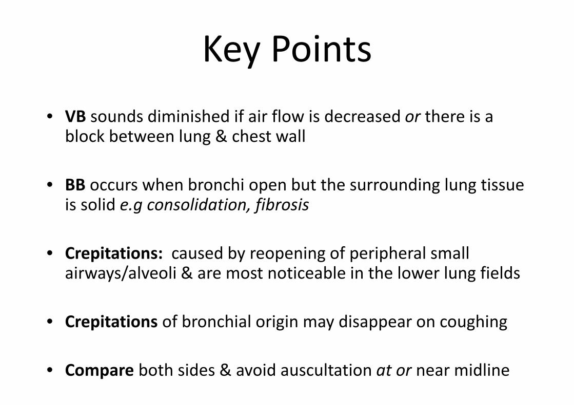
Key Points
• VB sounds diminished if air flow is decreased or there is a block between lung & chest wall
• BB occurs when bronchi open but the surrounding lung tissue is solid e.g consolidation, fibrosis
• Crepitations: caused by reopening of peripheral small airways/alveoli & are most noticeable in the lower lung fields
• Crepitations of bronchial origin may disappear on coughing
• Compare both sides & avoid auscultation at or near midline