evaluation of hearing loss in patients with graves’ disease
TRANSCRIPT
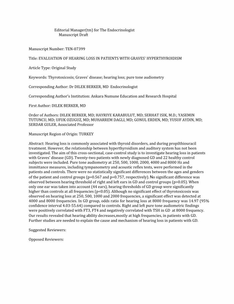
Editorial Manager(tm) for The Endocrinologist Manuscript Draft Manuscript Number: TEN-07399 Title: EVALUATION OF HEARING LOSS IN PATIENTS WITH GRAVES' HYPERTHYROIDISM Article Type: Original Study Keywords: Thyrotoxicosis; Graves' disease; hearing loss; pure tone audiometry Corresponding Author: Dr DILEK BERKER, MD Endocrinologist Corresponding Author's Institution: Ankara Numune Education and Research Hospital First Author: DILEK BERKER, MD Order of Authors: DILEK BERKER, MD; HAYRIYE KARABULUT, MD; SERHAT ISIK, M.D.; YASEMIN TUTUNCU, MD; UFUK OZUGUZ, MD; MUHARREM DAGLI, MD; GONUL ERDEN, MD; YUSUF AYDIN, MD; SERDAR GULER, Associated Professor Manuscript Region of Origin: TURKEY Abstract: Hearing loss is commonly associated with thyroid disorders, and during propilthiouracil treatment. However, the relationship between hyperthyroidism and auditory system has not been investigated. The aim of this cross-sectional, case-control study is to investigate hearing loss in patients with Graves' disease (GD). Twenty-two patients with newly diagnosed GD and 22 healthy control subjects were included. Pure tone audiometry at 250, 500, 1000, 2000, 4000 and 8000 Hz and immittance measures, including tympanometry and acoustic reflex tests, were performed in the patients and controls. There were no statistically significant differences between the ages and genders
of the patient and control groups (p=0.567 and p=0.757, respectively). o significant difference was observed between hearing threshold of right and left ears in GD and control groups (p>0.05). When only one ear was taken into account (44 ears), hearing thresholds of GD group were significantly higher than controls at all frequencies (p<0.05). Although no significant effect of thyrotoxicosis was observed on hearing loss at 250, 500, 1000 and 2000 frequencies, a significant effect was detected at 4000 and 8000 frequencies. In GD group, odds ratio for hearing loss at 8000 frequency was 14.97 (95% confidence interval 4.03-55.64) compared to controls. Right and left pure tone audiometric findings were positively correlated with FT3, FT4 and negatively correlated with TSH in GD at 8000 frequency.
Our results revealed that hearing a ility decreases,mostly at high frequencies, in patients with GD. Further studies are needed to explain the cause and mechanism of hearing loss in patients with GD. Suggested Reviewers: Opposed Reviewers:
1
EVALUATION OF HEARING LOSS IN PATIENTS WITH GRAVES’
HYPERTHYROIDISM
Dilek Berker1, MD; Hayriye Karabulut2, MD; Serhat Isik1, MD; Yasemin
Tutuncu1, MD; Ufuk Ozuguz1, MD; Muharrem Dagli3, MD; Gonul Erden4, MD;
Yusuf Aydin1, MD; Serdar Guler1, MD Associated Professor
1. Department of Endocrinology and Metabolic Diseases, Ankara Numune
Training and Research Hospital, Ankara, Turkey
2. Department of Otolaryngology, Kecioren Training and Research Hospital,
Ankara, Turkey
3. Department of Otolaryngology, Diskapi Yildirim Beyazit Training and Research
Hospital, Ankara, Turkey
4. Department of Biochemistry, Ankara Numune Training and Research
Hospital, Ankara, Turkey
Running title: Hearing Loss in Graves’ Disease
Corresponding Author: Dilek Berker
Address: 23.cad Simkent Sitesi No: 8/11 Kirkkonaklar/ Ankara, TURKEY
Phone: +903124954956
Mobile: +905056275953
Fax: +903123093398
E-mail: [email protected]
*Manuscript (REQUIRED -All Manuscript Text Pages in MS Word format, including Title Page, References and Figure Legends)
2
ABSTRACT
Hearing loss is commonly associated with thyroid disorders, and during
propilthiouracil treatment. However, the relationship between hyperthyroidism and
auditory system has not been investigated. The aim of this cross-sectional, case–
control study is to investigate hearing loss in patients with Graves’ disease (GD).
Twenty-two patients with newly diagnosed GD and 22 healthy control subjects were
included. Pure tone audiometry at 250, 500, 1000, 2000, 4000 and 8000 Hz and
immittance measures, including tympanometry and acoustic reflex tests, were
performed in the patients and controls. There were no statistically significant
differences between the ages and genders of the patient and control groups (p=0.567
and p=0.757, respectively). No significant difference was observed between hearing
threshold of right and left ears in GD and control groups (p>0.05). When only one ear
was taken into account (44 ears), hearing thresholds of GD group were significantly
higher than controls at all frequencies (p<0.05). Although no significant effect of
thyrotoxicosis was observed on hearing loss at 250, 500, 1000 and 2000
frequencies, a significant effect was detected at 4000 and 8000 frequencies. In GD
group, odds ratio for hearing loss at 8000 frequency was 14.97 (95% confidence
interval 4.03-55.64) compared to controls. Right and left pure tone audiometric
findings were positively correlated with FT3, FT4 and negatively correlated with TSH
in GD at 8000 frequency. Our results revealed that hearing ability decreases,mostly
at high frequencies, in patients with GD. Further studies are needed to explain the
cause and mechanism of hearing loss in patients with GD.
Key words: Thyrotoxicosis, Graves’ disease, hearing loss, pure tone audiometry
3
INTRODUCTION
Thyroid hormone is essential for the development of hearing (1). Prior to the onset of
hearing, thyroid hormones acting through thyroid hormone receptor-β influence
hearing by initiating myelinogenesis of the cochlea and vestibulocochlear nerve
(cranial nerve VIII) (2). Brucker-Davis et al. have estimated that the critical period of
thyroid-sensitive cochlear development in humans occur between the close of the
first trimester and the first month of life (3). Hypothyroidism caused by exposure to
thyroid-disrupting chemicals may also lead to hearing loss with cochlear and/or
auditory nerve dysfunction (4-6). It was due, in part, to a decrease in the abundance
of outer hair cells in the upper middle turn (responsible for detection of lower
frequency sounds) as well as the apical turn of the cochlea (7). In a subsequent
experiment, postnatal administration of thyroid hormone partially blocked low
frequency hearing loss (8).
Hearing loss is commonly associated with thyroid disorders in humans, including
congenital hypothyroidism, iodine deficiency, and resistance to thyroid hormone (3, 9-
11). Several cases of drug related hearing loss during propilthiouracil treatment have
been reported in patients with Graves’ disease (GD) (12-15). However, the
relationship between hyperthyroidism and auditory system has not been investigated.
In the present study, our aim was to compare hearing ability in patients with Graves’
hyperthyroidism and healthy controls using audiometric tests, and to investigate the
effects of hyperthyroidism on hearing.
MATERIALS AND METHODS
Patients
Twenty-two patients with GD, and 22 healthy control subjects were included in the
study. The diagnosis of GD was based on the clinical signs of hyperthyroidism
combined with suppressed serum thyrotropin and positive thyrotropin receptor
antibodies. Newly diagnosed patients, in whom no treatment was initiated, were
included. Informed consent was obtained from all participants. Detailed information
was obtained about possible etiological factors leading to hearing loss, such as
ototoxic drugs, exposure to noise, ear surgery, perforated tympanic membrane,
Ménière's disease, cranial trauma, metabolic diseases and systemic diseases.
Participants were excluded from the study if they had any of the following: (1)
4
otoscopic evidence of a perforated tympanic membrane or other middle-ear
pathology; (2) a flat tympanogram or absence of acoustic reflexes at 1 kHz with
contralateral stimulation; or (3) an air–bone gap of 5 dB at any frequency.
Audiometry
The initial hearing examination included otoscopy, tympanography and a complete
audiologic evaluation, including pure tone air and bone conduction audiometry and
speech audiometry. Pure tone audiometry was performed at the frequencies 250,
500, 1000, 2000, 4000 and 8000 Hz using a Clinical Audiometer Orbiter 922, V.2
(Madsen Electronics, Minnetonka, Minnesota, USA) in a sound-treated cabin. Three
pure tone average (PTA) groups were calculated: PTA1 (250 Hz), PTA2 (500, 1000
and 2000 Hz) and PTA3 (4000 and 8000 Hz). Normal middle-ear function was
defined by immittance and acoustic reflex results using a GSI Tympstar Version 2
Middle Ear Analyzer (Grason-Stadler, Inc, Milford, NH). The patients and controls
who had normal peak compliance, peak pressure, gradient, ear canal volume and
acoustic reflexes obtained by immittance measures (as defined by the American
Speech Language and Hearing Association) were included in the study.
Laboratory Assay
After an overnight fasting, blood samples were collected from all the study subjects
for the determination of serum thyroid-stimulating hormone (TSH), free
triiodothyronine (FT3), free thyroxine (FT4), and anti-TSH receptor antibody (TR-Ab)
levels. Serum TSH, FT3 and FT4 levels were evaluated using the Abbott Architect
2000 Analyzer (Abbott Diagnostics, IL, USA) and chemiluminescence microparticle
immunoassay (CMIA) method. Patients with TSH level < 0.35 µIU/mL FT3 level >
3.71 pg/mL and FT4 level >1.48 ng/dL were accepted as hyperthyroidism. TR-Ab
autoantibodies were measured using radioreceptor assay (Radim, Italy). The
reference values in our laboratory indicated that TR-Ab < 9 U/L was normal whereas
the borderline values of TR-Ab were between 9-14 U/L and positive values were > 14
U/L.
Statistical analysis
Statistical analysis was performed by statistical package for social sciences (SPSS)
version 11.5 software (SPSS Inc., Chicago, IL, United States). The normality of the
distribution of continuous variables was tested by the Shapiro Wilk test. Mean ages of
5
the patients were compared using Student’s t test. Mann Whitney U test was used for
the comparisons of the thyroid function tests and pure tone thresholds. Intra-group
comparisons were evaluated by the Wilcoxon signed rank test. Degrees of
association between continuous variables were analyzed by Spearman’s correlation
test. A p value < 0.05 was considered statistically significant.
RESULTS
There were no statistically significant differences between the ages and genders of
the patient and control groups (p=0.567 and p=0.757, respectively). While FT3 and
FT4 levels were significantly higher (p<0.001, p<0.001, respectively), TSH level was
significantly lower (p<0.001) in the GD group compared to controls (Table 1).
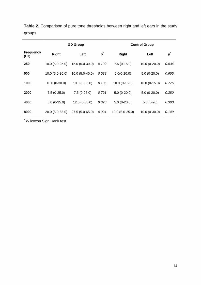
No significant difference was observed between hearing threshold of right and left
ears in GD and control groups (Table 2).
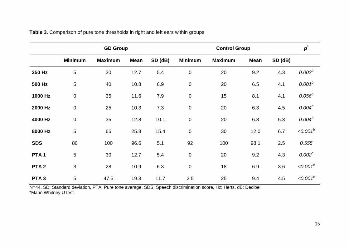
When only one ear was taken into account (44 ears), hearing thresholds of GD group
were significantly higher than controls at all frequencies (p<0.05) (Table 3). Although
no significant effect of thyrotoxicosis was observed on hearing loss at 250, 500, 1000
and 2000 frequencies, a significant effect was detected at 4000 and 8000
frequencies. In GD group, odds ratio for hearing loss at 8000 frequency was 14.97
(95% confidence interval 4.03-55.64) compared to controls.
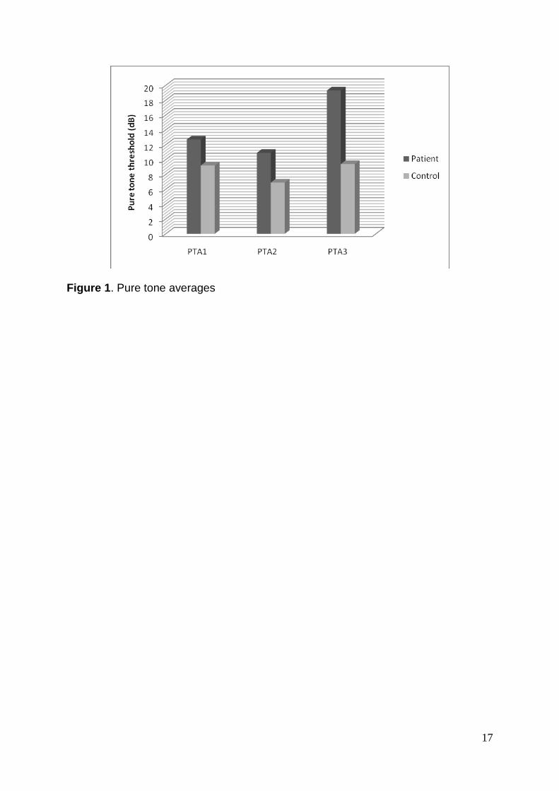
The PTA thresholds of patients and controls were significantly different in all three
PTA groups (p < 0.05). The differences were most prominent at higher frequencies
(Figure 1).
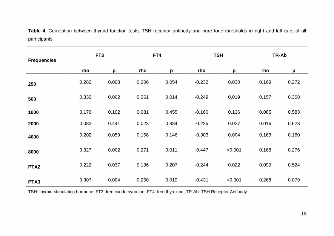
Right and left pure tone audiometric findings were positively correlated with FT3, FT4
and negatively correlated with TSH in GD at 8000 frequency.
While a statistically significant correlation was noted between audiometric findings
and TR-Ab at 4000 frequency, no correlation was found between PTA values and
TR-Ab values (Table 4).
6
DISCUSSION
A sensorineural hearing loss was observed in patients with GD compared to healthy
controls and PTA values were found to be correlated with thyroid hormone levels in
the present study.
Despite few case reports regarding sudden hearing impairment due to the use of
propilthiouracil during treatment of hyperthyroidism, hyperthyroidism-related hearing
impairment has not previously been published (12). We suggest that hearing
impairment in patients with Graves’ disease develops a result of the common effects
of several different mechanisms. Since we have noted a correlation between thyroid
hormone levels and hearing thresholds, we considered that the reason for increased
hearing thresholds was due to the metabolic effects of high thyroid hormones and
thyrotoxicosis. It is known that some of the clinical findings of hyperthyroidism result
from sympathetic over-activity due to up-regulated adrenergic receptors in some
tissues (16). The inner ear has abundant sympathetic innervation (17). Meniere’s
disease episodes may serve as an interesting model for explaining the relationship
between autonomic nervous system and hearing functions (18). Episodes of inner
ear dysfunction can be associated with emotional stress. For example, in stress
conditions normal ambient noises may seem to be unbearably loud. Stress has also
been suspected to aggravate hearing disorders. Cochlear vasoconstriction which is
likely to be under perivascular sympathetic control may occur during acoustic
exposure. It is a well known fact that the sympathetic input to the cochlea can be
either from the stellate ganglion and associated with blood vessels or from the
superior cervical ganglion, mostly independent of blood vessels (18). The protection
effects are related to the blood vessel-independent sympathetic innervations, rather
than modification in vascular tone. The former sympathetic system is associated with
norepinephrine and has been localized by immunohistochemistry outside the organ
of Corti in close association with afferents and efferents at the level of the habenula
(19).
The underlying pathogenesis of GD is the presence of antibodies against TSH
receptor. This not only results in gland hyperplasia due to thyrotropic effects of TSH,
but also promotes the formation of monoiodotyrosine and diiodotyrosine, and the
release of T3 and T4. Linkage evidence for a susceptibility gene for GD to the major
histocompatibility complex (MHC) has also been found in some populations (20-23).
7
Graves’ disease is an autoimmune disorder. All of the patients included in the study
were TR-Ab positive. Although we could not demonstrate a direct correlation between
TR-Ab levels and hearing thresholds, antibody positivity in patients and antibody
negativity in controls suggest that autoimmunity may play a role in the pathogenesis
of hearing impairment in GD. Sensorineural hearing loss may occur in many
autoimmune diseases such as systemic lupus erythematosus, rheumatoid arthritis,
autoimmune sensorineural hearing loss, relapsing polychondritis, disseminated
vasculitis, polymyalgia rheumatica and ankylosing spondylitis (24). Autoimmunity
may lead to hearing loss through a few mechanisms. Many of the autoimmune
diseases can lead to vasculitis, resulting in a variety of secondary degenerative
changes. The most widely documented effects of autoimmune diseases resulting in
sensorineural hearing loss are mediated by a vascular mechanism (25). Several
studies have demonstrated that the inner ear was the source of the antibody (26-28).
According to these studies, the inner ear is capable of responding to antigen
challenge. Harris et al. have shown a parallel rise of antibody titres over a three-week
period in guinea pigs immunized by either inner-ear or peritoneal routes of antigen
presentation (29). These studies indicate that the inner ear is an effective route of
antigen processing, which can result in the acquisition of systemic humoral immunity
as well as cellular immunity. We suggested that there might be sensorineural hearing
loss due to TR-Ab in patients with GD.
There have been no previous reports about the relationship between thyrotoxicosis
and hearing impairment. Despite certain limitations of our study, our results might
serve as preliminary findings for designing future controlled studies with large sample
size. Whether hearing impairment noted in Graves’ hyperthyroidism is caused by
autoimmunity or excessive thyroid hormone levels should be differentiated. At first, in
vitro pathophysiological and molecular animal studies are required. It seems possible
that hyperthyroidism may have some participation in the immunopathogenesis of
sensorineural hearing loss (18). It is also possible to attribute some vascular
participation to the physiopathology of sensorineural hearing loss.
CONCLUSION
Our findings suggest that hearing loss of patients with GD was sensorineural and that
their hearing level decreased mostly at high frequencies, although the pure tone
8
thresholds of patient and controls differed at all frequencies. However, further studies
are needed to explain the cause and mechanism of hearing loss in patients with GD.
9
REFERENCES
1. Rusch A, Erway LC, Oliver D, et al.: Thyroid hormone receptor β-dependent
expression of a potassium conductance in inner hair cells at the onset of hearing.
Proc Natl Acad Sci USA 1998; 95: 15758--62.
2. Knipper M, Bandtlow C, Gestwa L, et al.: Thyroid hormone affects Schwann cell
and oligodendrocyte gene expression at the glial transition zone of the VIIIth
nevre prior to cochlea function. Development 1998; 125: 3709--18.
3. Brucker-Davis F, Skarulis MC, Pikus A, et al.: Prevalence and mechanisms of
hearing loss in patients with resistance to thyroid hormone. J Clin Endocrinol
Metab 1996; 81: 2768--72.
4. Goldey ES, Kehn LS, Rehnberg GL, et al.: Effects of developmental
hypothyroidism on auditory and motor function in the rat. Toxicol Appl Pharmacol
1995; 135: 67--76.
5. Knipper M, Zinn C, Maier H, et al.: Thyroid hormone deficiency before the onset of
hearing causes irreversible damage to peripheral and central auditory systems. J
Neurophysiol 2000; 83: 3101--12.
6. Goldey ES, Kehn LS, Lau C, et al.: Developmental exposure to polychlorinated
biphenyls (Aroclor 1254) reduced circulating thyroid hormone concentrations and
causes hearing deficits in rats. Toxicol Appl Pharmacol 1995; 135: 77--88.
7. Crofton KM, Ding D, Padich R, et al.: Hearing loss following exposure during
development to polychlorinated biphenyls: a cochlear site of action. Hear Res
2000; 144: 196--204.
8. Goldey ES, Crofton KM.: Thyroxine replacement attenuates hypothyroxinemia,
hearing loss, and motor deficits following developmental exposure to Aroclor 1254
in rats. Toxicol Sci 1998; 45: 94--105.
9. Bellman SC, Davies A, Fuggle PW, et al.: Mild impairment of neuro-otological
function in early treated congenital hypothyroidism. Arch Dis Child 1996; 74: 215--
8.
10. Debruyne F, Vanderschueren-Lodeweychx M, Bastijns P.: Hearing in congenital
hypothyroidism. Audiology 1983; 22: 404--9.
10
11. Soriguer F, Millón MC, Muñoz R, et al.: The auditory threshold in a school-age
population is related to iodine intake and thyroid function. Thyroid 2000; 10: 991--
9.
12. Werneck AL, Gurgel LC, de Mello LM, et al.: Sudden sensorineural hearing loss:
a case report supporting the immunologic theory. Arq Neuropsiquiatr 2003; 61:
1018--22.
13. Thamprajamchit S, Jariengprasert C, Rajatanavin R.: Propylthiouracil-induced
sensorineural hearing loss associated with antineutrophil cytoplasmic antibodies.
Endocr Pract 2004;10: 432--7.
14. Sano M, Kitahara N, Kunikata R.: Progressive bilateral sensorineural hearing loss
induced by an antithyroid drug. ORL J Otorhinolaryngol Relat Spec 2004; 66: 281-
-5.
15. Bandyopadhyay U, Biswas K, Banerjee RK.: Extrathyroidal actions of antithyroid
thionamides. Toxicol Lett 2002; 128: 117--27.
16. Bilezekian JP, Loeb JN.: The influence of hyperthyroidism and hypothyroidism on
alpha- and beta-adrenergic receptor systems and adrenergic responsiveness.
Endocrinol Rev 1983; 4: 378--88.
17. Spoendlin H, Lichtensteiger W.: The adrenergic innervation of the labyrinth. Acta
Otolaryngol 1966; 61: 423--34.
18. Fowler EP Jr, Zeckel A.: Psychosomatic aspects of meniere's disease. J Am Med
Assoc 1952; 148: 1265--8.
19. Jones N, Fex J, Altschuler RA.: Tyrosine hydroxylase immunoreactivity identifies
possible catecholaminergic fibers in the organ of Corti. Hear Res 1987; 30: 33--8.
20. Volpe´ R.: Immunology of the thyroid. In Autoimmune disease of the endocrine
system, edited by Volpe´ R, pp. 73--239. Boca Raton, FL: CRC Press, 1990.
21. Heward JM, Allahabadia A, Daykin J, et al.: Linkage disequilibrium between the
human leukocyte antigen class II region of the major histocompatibility complex
and Graves’ disease: replication using a population case control and family-based
study. J Clin Endocrinol Metab 1998; 83: 3394--7.

11
22. Uno H, Sasazuki T, Tamai H, et al.: Two major genes, linked to HLA and Gm,
control susceptibility to Graves’ disease. Nature 1981; 292: 768--70.
23. Shields DC, Ratanachaiyavong S, McGregor AM, et al.: Combined segregation
and linkage analysis of Graves disease with a thyroid autoantibody diathesis. Am
J Hum Genet 1994; 55: 540--54.
24. Eryilmaz A, Dagli M, Karabulut H, et al.: Evaluation of hearing loss in patients
with ankylosing spondylitis. J Laryngol Otol 2007; 121: 845--9.
25. Ruckenstein MJ.: Autoimmune inner ear disease. Curr Opin Otolaryngol Head
Neck Surg 2004; 2: 426--30.
26. Harris JP.: Immunology of the inner ear. Response of the inner ear to antigen
challenge. Otolaryngol Head Neck Surg 1983; 91: 18--23.
27. Mogi G, Kawauchi H, Suruki M, et al.: Am J Otolaryngol 1985; 6: 142--7.
28. Harris JP.: Immunology of the inner ear. Evidence of local antibody production.
Ann Otol Rhinol 1984; 93: 157--62.
29. Harris JP, Woolf NK, Ryan AF.: Elaboration of systemic immunity following inner
ear immunization. Am J Otolaryngol 1985; 6: 148--52.
12
Legends
Table 1. Clinical and laboratory features of the groups
Table 2. Comparison of pure tone thresholds between right and left ears in the study
groups
Table 3. Comparison of pure tone thresholds in right and left ears within groups
Table 4. Correlation between thyroid function tests, TSH receptor antibody and pure
tone thresholds in right and left ears of all participants
Figure 1. Pure tone averages

13
Table 1. Clinical and laboratory features of the groups
Variables GD (n=22) Control (n=22) p
Age, years 36.0±11.1 37.6±8.0 0.567a
Female/Male 13/9 14/8 0.757b
FT3, pg/ml 4.7±1.4 2.6±0.4 <0.001c
FT4, ng/dl 2.0±0.7 1.2±0.6 <0.001c
TSH, μIU/ml 0.02±0.01 1.9±1.0 <0.001c
TR-Ab, IU/L 16.8±4.0 NA NA
NA= not analyzed
a Student’s t test, b Pearson Chi-square test, c Mann Whitney U test.
TSH: Thyroid-stimulating hormone; FT3: Free triiodothyronine; FT4: Free thyroxine; TR-Ab: Anti-TSH receptor antibodies
14
Table 2. Comparison of pure tone thresholds between right and left ears in the study
groups
GD Group Control Group
Frequency (Hz)
Right Left p* Right Left p
*
250 10.0 (5.0-25.0) 15.0 (5.0-30.0) 0.109 7.5 (0-15.0) 10.0 (0-20.0) 0.034
500 10.0 (5.0-30.0) 10.0 (5.0-40.0) 0.088 5.0(0-20.0) 5.0 (0-20.0) 0.655
1000 10.0 (0-30.0) 10.0 (0-35.0) 0.135 10.0 (0-15.0) 10.0 (0-15.0) 0.776
2000 7.5 (0-25.0) 7.5 (0-25.0) 0.791 5.0 (0-20.0) 5.0 (0-20.0) 0.380
4000 5.0 (0-35.0) 12.5 (0-35.0) 0.020 5.0 (0-20.0) 5.0 (0-20) 0.380
8000 20.0 (5.0-55.0) 27.5 (5.0-65.0) 0.024 10.0 (5.0-25.0) 10.0 (0-30.0) 0.149
* Wilcoxon Sign Rank test.
15
Table 3. Comparison of pure tone thresholds in right and left ears within groups
N=44, SD: Standard deviation, PTA: Pure tone average, SDS: Speech discrimination score, Hz: Hertz, dB: Decibel *Mann Whitney U test.
GD Group Control Group p*
Minimum Maximum Mean SD (dB) Minimum Maximum Mean SD (dB)
250 Hz 5 30 12.7 5.4 0 20 9.2 4.3 0.002b
500 Hz 5 40 10.8 6.9 0 20 6.5 4.1 0.001b
1000 Hz 0 35 11.6 7.9 0 15 8.1 4.1 0.056b
2000 Hz 0 25 10.3 7.3 0 20 6.3 4.5 0.004b
4000 Hz 0 35 12.8 10.1 0 20 6.8 5.3 0.004b
8000 Hz 5 65 25.8 15.4 0 30 12.0 6.7 <0.001b
SDS 80 100 96.6 5.1 92 100 98.1 2.5 0.555
PTA 1 5 30 12.7 5.4 0 20 9.2 4.3 0.002c
PTA 2 3 28 10.9 6.3 0 18 6.9 3.6 <0.001c
PTA 3 5 47.5 19.3 11.7 2.5 25 9.4 4.5 <0.001c
16
Table 4. Correlation between thyroid function tests, TSH receptor antibody and pure tone thresholds in right and left ears of all
participants
Frequencies FT3 FT4 TSH TR-Ab
rho p rho p rho p rho p
250 0.282 0.008 0.206 0.054 -0.232 0.030 0.169 0.272
500 0.332 0.002 0.261 0.014 -0.249 0.019 0.157 0.308
1000 0.176 0.102 0.081 0.455 -0.160 0.136 0.085 0.583
2000 0.083 0.441 0.023 0.834 -0.235 0.027 0.016 0.623
4000 0.202 0.059 0.156 0.146 -0.303 0.004 0.163 0.160
8000 0.327 0.002 0.271 0.011 -0.447 <0.001 0.168 0.276
PTA2 0.222 0.037 0.136 0.207 -0.244 0.022 0.099 0.524
PTA3 0.307 0.004 0.250 0.019 -0.431 <0.001 0.268 0.079
TSH: thyroid-stimulating hormone; FT3: free triiodothyronine; FT4: free thyroxine; TR-Ab: TSH Receptor Antibody
17
Figure 1. Pure tone averages
Dear editor,
We are sending our manuscript to your journal after all the authors have seen and
approved the final form of it. This original report has not been published elsewhere
before nor is it being considered simultaneously in another journal. The following
address can be used for any kind of negotiations. There is no conflinct of interest
between authors. We do look forward to hearing from you soon.
Best regards
Corresponding Author: Dilek Berker, MD
Mailing Address: 23.Cad Simkent Sitesi No:8/11 Kırkkonaklar/ Ankara
Phone: +903125084732
Fax: +903123093398
Email: [email protected]
Cover Letter

Copyright Transfer Agreement


*Financial Disclosure Agreement