evaluation of continuous low dose rate versus acute single high dose rate radiation combined with...
TRANSCRIPT
Evaluation of continuous low dose rate versus acute single highdose rate radiation combined with oncolytic viral therapy forprostate cancer
CHUNYAN LIU1, YONGGANG ZHANG2, MINZHI MAGGIE LIU1, HAOMING ZHOU2, WASIM H.CHOWDHURY1, SHAWN E. LUPOLD1, TED L. DEWEESE2, and RONALD RODRIGUEZ1
1 James Brady Urological Institute, The Johns Hopkins University School of Medicine, Baltimore,Maryland, USA2 Department of Radiation Oncology & Molecular Radiation Sciences, The Johns HopkinsUniversity School of Medicine, Baltimore, Maryland, USA
AbstractPurpose—Conditionally Replicative Adenovirus (CRAd) has been previously demonstrated toaugment the activity of radiation, resulting in synergy of cell kill. However, previous modelscombining radiation with CRAd have not focused on the methods of radiation delivery.
Materials and methods—We model the combination of a novel prostate-specific CRAd, Ad5PSE/PBN E1A-AR (Ad5: adenovirus 5; PSE: prostate-specific enhancer; PBN: rat probasinpromoter; E1A: early region 1A; AR: androgen receptor), with radiation delivered both acutelyand continuously, in an effort to better mimic the potential clinical modes of prostate cancerradiotherapy.
Results—We demonstrate that pre-treatment of cells with acute single high dose rate (HDR)radiation 24 hours prior to viral infection results in significantly enhanced viral replication andvirus-mediated cell death. In addition, this combination causes increased level of γ-H2AX(Phosphorylated histone protein H2AX on serine 139), a marker of double-stranded DNA damageand an indirect measure of nuclear fragmentation. In contrast, continuous low dose rate (LDR)radiation immediately following infection of the same CRAd results in no enhancement of viralreplication, and only additive effects in virus-mediated cell death.
Conclusions—These data provide the first direct assessment of the real-time impact of radiationon viral replication and the first comparison of the effect of radiation delivery on the efficacy ofCRAd virotherapy. Our data demonstrate substantial differences in CRAd efficacy based on themode of radiation delivery.
Keywordsadenocarcinoma of prostate; radiosensitisation; oncolytic viral therapy; prostate-specificconditionally replicative adenovirus; acute single high dose rate radiation; continuous low doserate radiation
© 2010 Informa UK Ltd.Correspondence: Ron Rodriguez, MD, PhD, James Brady Urological Institute, Johns Hopkins University School of Medicine, 600 N.Wolfe Street, Marburg Room 205A, Baltimore, MD 21287-2101, USA. Tel: +1 410 614 4442. Fax: + 1 443 287 [email protected] of interest: The authors report no conflicts of interest. The authors alone are responsible for the content and writing ofthe paper.
NIH Public AccessAuthor ManuscriptInt J Radiat Biol. Author manuscript; available in PMC 2011 May 13.
Published in final edited form as:Int J Radiat Biol. 2010 March ; 86(3): 220–229. doi:10.3109/09553000903419338.
NIH
-PA Author Manuscript
NIH
-PA Author Manuscript
NIH
-PA Author Manuscript

IntroductionRadiation therapy is a common treatment for adenocarcinoma of the prostate (PCa);however, despite significant improvements in delivery technologies, many patients developrecurrence after treatment with curative intent (Nguyen and Zietman 2007). In its mostsimplistic terms, radiation can be delivered from an external source (i.e., external beamradiation therapy or EBRT) or from an internal source, such as a radioactive seed. When theradiation is delivered externally, it is typically fractionated into multiple high dose rate(HDR) fractions. When the radiation is delivered through the permanent instillation ofradioactive seeds, the radiation exposure is continuous but at a low dose rate (LDRbrachytherapy). Because of the ease of treatment and recovery, LDR brachytherapy has beengaining popularity as the CaPSURE (Cancer of the Prostate Strategic Urologic ResearchEndeavor) database has reported an increase in the use of LDR brachytherapy from 3.1–12.0% in the past decade (Cooperberg et al. 2004).
The total dose of radiation given by these strategies is limited by the potential for damage tosurrounding tissues (Teh et al. 2004). Despite continuously improving technology, someprostate cancer cells survive. This radiation-resistance may be due to the unusually slowgrowth rate of prostate cancers, compared with other cancers, or to alterations in cellularpathways such as DNA damage repair, cell cycle, or apoptosis. Recently, there have beensignificant advances in circumventing this radiation-resistance by combining radiationtherapy with radiation sensitisers, such as certain chemotherapies (Maggiorella et al. 2003,Lebedeva et al. 2007, Shewach and Lawrence 2007). While successful, one of the importantlimitations has been the concomitant radiation sensitisation of non-cancerous tissues. Itwould be more advantageous to establish approaches that provide tissue-specific radiationsensitisation. Adenoviral gene therapy has this potential because it can be highly tailored toprostate tissues through tissue-specific promoters. We have previously described prostate-specific CRAd gene therapy, using the promoter of Prostate-Specific Antigen (PSA) tocontrol the E1A adenoviral gene (Rodriguez et al. 1997). The persistently active status of thePSA promoter in most advanced prostate cancers, evident by the presence of continuouslyrising serum PSA, makes the use of the PSA promoter an attractive strategy for all stages ofPCa (Wu et al. 2001, Zegarra-Moro et al. 2002, Chen et al. 2004). The original vector(CN706, also known as CG7060) and the subsequent vector (CV787, also known asCG7870, in which E1A and E1B (early region 1B) are under the control of two separateprostate-specific promoters) have been applied to several prostate cancer clinical trials, inwhich they were each used alone for treatment of locally-recurrent PCa following radiationor meta-static PCa through intravenous delivery (DeWeese et al. 2001, Small et al. 2006). Inall trials, there has been compelling evidence of viral mediated therapeutic effect. We haverecently modified the prostate-specific CRAd concept by inserting a fusion of the viral E1Agene with the androgen receptor (Hoti et al. 2007). The E1A-AR chimera overcomes amutual inhibition effect of viral E1A and host AR, makes the virus more androgenresponsive, tissue-specific, and therapeutically effective. We utilise this newest generationof prostate-specific CRAd virotherapy (Ad5 PSE/PBN E1A-AR) in combination with twodifferent models of radiation therapy to assess the oncolytic activity in vitro.
Several studies have demonstrated that oncolytic adenoviral gene therapy can enhance thetherapeutic effect of radiation. However, there have only been a few reports on targeting thiscombination therapy to a specific tissue. Moreover, all of these have been with EBRT (Chenet al. 2001, Advani et al. 2006, Idema et al. 2007). To date there have been no studies toevaluate the efficacy of combining oncolytic gene therapy with continuous LDR radiation.Since LDR brachytherapy is minimally invasive and well tolerated, we reasoned this mightbe a superior method for combination with CRAd therapy, as both radioactive seeds andCRAd could be delivered directly into the prostate under a single anesthetic. Here we
LIU et al. Page 2
Int J Radiat Biol. Author manuscript; available in PMC 2011 May 13.
NIH
-PA Author Manuscript
NIH
-PA Author Manuscript
NIH
-PA Author Manuscript

compare the effects of combining a prostate-specific CRAd, Ad5 PSE/PBN E1A-AR, withboth acute HDR radiation and continuous LDR radiation. Our results indicate that these tworadiation strategies do not provide equal benefit when combined with CRAd gene therapy;the combination with acute HDR radiation appears to be most efficacious. Nevertheless,both combination strategies do result in a significant benefit.
Materials and methodsCell culture
LNCaP, PC3, and DU145, from American Type Culture Collection (ATCC, Manassas, VA,USA), and LAPC4 (obtained from Dr John Isaac laboratory at Johns Hopkins UniversitySchool of Medicine, MD, USA) were cultured in RPMI-1640 medium (Roswell ParkMemorial Institute-1640 medium) (Cellgro Mediatech, Herndon, VA, USA) supplementedwith 10% fetal bovine serum (FBS, Gemini Bio-Products, West Sacramento, CA, USA), andmaintained at passages 50–60. LNCaP (ATCC® Number: CRL-1740™) and LAPC4, humanPCa cell lines that produce PSA and possess a mutated but functional androgen receptor or awild-type androgen receptor respectively, were used as the target cell lines. PC3 (ATCC®
Number: CRL-2698™) and DU145 (ATCC® Number: HTB-81™), human PCa cell lines thatlack the androgen receptor and do not produce PSA, were used as control cell lines for ARresponsiveness. HT29 (ATCC® Number: HTB-38™), a colon cancer cell line, and U2OS(ATCC® Number: HTB-96™), an osteosarcoma cell line, were used as control cell lines fortissue specificity. Both were maintained according to the manufacture’s protocols. Viruspackaging line DPL-S11 was maintained in Dulbecco’s Modified Eagle Medium (DMEM,Cellgro Mediatech, Herndon, VA, USA) with 5% FBS and 200 μg/ml G418 (Mediatech,Manassas, VA, USA). It was derived from helper cell line Per.C6 (Fallaux et al. 1998) byselection for diphtheria toxin resistance, and expresses a previously characterisedmembrane-bound anti-fiber single chain antibody as a pseudo-receptor (van Beusechem etal. 2002). The 293HEK cell line (Quantum Biotechnologies, Laval, Quebec, Canada) wasused for viral titration. It was maintained in DMEM with 10% FBS. All media weresupplemented with 1% Penicillin-Streptomycin (Invitrogen Corporation, Carlsbad, CA,USA). All cells were maintained at 37°C in an atmosphere containing 5% CO2.
Viruses and their dosageAd5 PSE/PBN E1A-AR (Hoti et al. 2007) was generated in the AdEasy system (a gift fromDr Bert Vogelstein laboratory at Johns Hopkins University School of Medicine) throughrecombination of line-arised shuttle plasmid RpS-PSE-PBN-E1A-AR (RpS: Rodriguezplasmid shuttle) with pAdEasy-1 in DPL-S11 cells. The E1A-AR chimera includes wild-type AR. Adeno-X-LacZ, a recombinant, ΔE1/ΔE3 adenovirus that encodes β-galactosidase(Clontech, Mountain View, CA, USA) was used as a control virus. Both viruses wereemployed at doses ranging from 0.5–5 multiplicities of infection (MOI). FFIG (Ad5 FiberIRES GFP) (a gift from Dr Gary Ketner laboratory at Johns Hopkins University School ofPublic Health), a replication-defective reporter virus that links green fluorescent protein(GFP) expression to fiber gene (which is part of the major late transcription unit) productionthrough the use of an internal ribosome entry site (IRES) (Hoti et al. 2007), was used at 30MOI. Viral infection was performed on 48-well plates with 2 × 10⁴ cells per well.
Large-scale viral purification was performed using CsCl2 (Invitrogen) gradientultracentrifuge and stored in dialysis buffer containing 15 mM Tris (pH 7.8), 2 mM MgCl2and 5% sucrose. The titer of the viral stocks was determined using the Adeno-X™ RapidTiter Kit and 293HEK cells. All viral stocks were tested for wild-type replication competentadenovirus (RCA) background generated by homologous recombination, using quantitative
LIU et al. Page 3
Int J Radiat Biol. Author manuscript; available in PMC 2011 May 13.
NIH
-PA Author Manuscript
NIH
-PA Author Manuscript
NIH
-PA Author Manuscript

PCR with primers against wild-type E1A promoter. The RCA content of all the viruses wasat undetectable level.
In vitro irradiation: Timing and dosageAcute single HDR radiation was performed 24 h prior to viral infection at doses of 2 Gy or 6Gy (0.67 Gy/min) (Gammacell 40 137Cs irradiator, Atomic Energy Commission of Canada,Ltd, Mississauga, Ontario, Canada); un-irradiated control cells were seeded and infected atthe same time. Continuous LDR radiation (25 cGy/h in an incubated 137Cs irradiator,custom-built) was performed as described (DeWeese et al. 1998). Cell culture plates wereplaced in a sealed chamber filled with 5% CO2 and maintained at 37°C in the LDR irradiatorfor 24 h (up to 6 Gy) or 8 h (up to 2 Gy) immediately after viral infection; un-irradiatedcontrol cells were seeded and infected at the same time.
Cell viabilityCell viability was measured using MTT ((3-(4,5-Dimethylthiazol-2-yl)- 2,5-diphenyltetrazolium bromide, a tetrazole) Cell Proliferation Assay kit (ATCC) five, seven,nine or 12 days post-treatment. Growth media was removed and replaced with MTT solution10-fold diluted with fresh media; after incubation for 3 h at 37°C, detergent reagent providedin the kit was added to dissolve the purple precipitates. Absorbance was recorded at 570 nmusing Spectramax M2e fluorescence plate reader (Molecular Devices, Sunnyvale, CA,USA). At each time point, percentage of cell survival after respective treatment wascalculated by comparison to the growth of untreated cells, i.e., optical density (OD) oftreated cells/OD of untreated cells.
Real time viral replication influenced by radiationTo observe the reciprocal effect of radiation on viral replication, FFIG was co-infected withAd5 PSE/PBN E1A-AR. GFP expression of FFIG is a direct measure of CRAd replication(Hoti et al. 2007). The level of GFP at five, seven and nine days post-treatment was recordedusing Spectramax M2e fluorescence plate reader (Molecular Devices) (excitation at 485 nm,and emission at 535 nm). Considering the significant cell death after treatment, the GFPreading was normalised to percentage of surviving cells (GFP reading/cell). The data wasconfirmed with viral output/input assay to avoid bias in data collection.
Viral output/input assayLNCaP and LAPC4 cells infected with Ad5 PSE/PBN E1A-AR virus (2 MOI) in thepresence and absence of radiation were pelleted and lysed to release viruses at the end ofseven days post-treatment. The amount of released viruses was measured by titration using293HEK cells. The ‘amplification ratio’ of a virus produced from an infected cell (Output)to the amount originally used to infect the cells (Input) were determined and plotted as viraloutput/input ratio.
Viral uptake influenced by radiationTo observe the effect of radiation on viral uptake, AdTrack virus (a gift from Dr BertVogelstein laboratory at Johns Hopkins University School of Medicine) that is driven byCMV (Cytomegalovirus) promoter and expresses GFP (He et al. 1998) was employed.LNCaP cells irradiated with 6 Gy of acute single HDR radiation were infected with AdTrack24 h after irradiation. The level of GFP 24, 48, and 72 h post-infection was recorded usingthe above mentioned fluorescence plate reader. The GFP data are plotted as GFP foldincrease relative to the mock-infected cells (signal/background).
LIU et al. Page 4
Int J Radiat Biol. Author manuscript; available in PMC 2011 May 13.
NIH
-PA Author Manuscript
NIH
-PA Author Manuscript
NIH
-PA Author Manuscript

Western blot analysisLNCaP cells were infected with Ad5 PSE/PBN E1A-AR virus (2 MOI) on 100 mm culturedishes. Cells were washed with 1 × PBS, scraped, lysed on ice using RIPA buffer (buffer forradio immunoprecipitation assay) (Pierce Biotechnology, Rockford, IL, USA) supplementedwith fresh protease inhibitor cocktail (Complete, EDTA-free, Roche Applied Science,Indianapolis, IN, USA), sonicated and centrifuged to remove the cell debris five and sevendays post-treatment. Protein concentration was assessed using BCA (bicinchoninic acid)Protein Assay Kit (Pierce Biotechnology). 10–20 μg of protein was heated at 95°C andloaded on 4–15% SDS-PAGE (sodium dodecyl sulfate polyacrylamide gel electrophoresis)gels (Bio-Rad, Hercules, CA, USA). Gels were run for 2 h until the loading dyes reached tothe bottom, and transferred to PVDF (Polyvinylidene Fluoride) (Bio-Rad) membrane. Pre-stained molecular weight markers (Rainbow Marker; Bio-Rad) were run in the same gels forcomparison of molecular weight and estimation of transfer efficiency. The membranes wereblocked with 1 × TBS (Tris-Buffered Saline) including 0.01% Tween 20 and 5% milk for 1h and then probed with primary antibodies, i.e., a rabbit polyclonal antibody against γ-H2AX(working concentration 0.1 μg/ml) (Upstate Biotechnology, Billerica, MA, USA) or a rabbitmonoclonal antibody against β-actin (1:5000 dilution) (Sigma, St Louis, MO, USA)overnight at 4°C. Secondary mouse anti-rabbit antibodies labeled with horseradishperoxidase were subsequently applied. Films were developed after incubation for 1–5 minwith the horseradish peroxidase substrate provided in the enhanced chemiluminescence kit(Amersham, Piscataway, NJ, USA).
Statistical analysisStatistical analysis was performed on Graph Pad Prism 5.0, running on an IBM compatiblecomputer, using the Windows XP operating system. Comparisons for paired data wereanalysed using the Student’s t-test. Statistical significance was defined as a p-value <0.05and was denoted in each of the figures by an asterisk. Expected total cell survival in Figure 1was calculated as (1 − cell death by radiation × cell death by CRAd).
ResultsAcute single HDR radiation versus continuous LDR radiation combined with oncolyticadenovirus
To evaluate the effect of combining CRAd virotherapy with radiation delivery, two in vitroradiation models were established. The first model is acute administration of radiation,followed by virotherapy. This model is most analogous to clinical HDR radiation. Themodel is intended to provide information most relevant for either EBRT, where totalradiation therapy is fractionated at HDRs, or HDR brachytherapy, where a HDR radioactivesource is instilled via catheters into the prostate for short intervals. In contrast, we alsoperformed combination of CRAd virotherapy with a continuous LDR mode of radiation. Inthis case the model is most analogous to LDR brachytherapy, where radioactive seeds andviruses would be placed/or injected simultaneously. In this set of experiments we applied acontinuous LDR radiation source immediately after infection using a custom made 137Csirradiator at 25 cGy/h. Cell viability for the individual therapies as well as the twocombinations was assessed five, seven and nine days post-treatment using MTT assay.
For the acute HDR studies, an acute single HDR radiation (6 Gy or 2 Gy at a rate of 0.67Gy/min) was delivered to cells 24 h prior to viral infection. This timing was chosen based onour comparison of resulting combined cytotoxicity from experiments where radiation wasdelivered 24 h prior to infection versus irradiation 72 h post-infection (data not shown) andpreviously published reports (Qian et al. 2005, Egami et al. 2008). Ad5 PSE/PBN E1A-ARwas infected at doses ranging from 0.5–5 MOI. For LNCaP cells, as shown in Figure 1a, 6
LIU et al. Page 5
Int J Radiat Biol. Author manuscript; available in PMC 2011 May 13.
NIH
-PA Author Manuscript
NIH
-PA Author Manuscript
NIH
-PA Author Manuscript

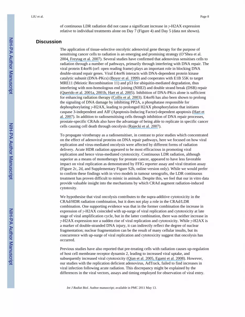
Gy of acute single HDR radiation resulted in 45% cell death at Day 9, and virotherapy (Ad5PSE/PBN E1A-AR) at an MOI of 2 resulted in 10% cell death. However, when radiationand virotherapy were combined at these doses, cell death on Day 9 increased significantly to90% (p <0.05). This observed value of cell death is much greater than the calculated additivedeath rate of 50%. In this experiment, even though virus alone killed only 10% cells on Day9, at the end of 12 days, it killed 90% of cells (Figure 1a). Combinations of other doses ofCRAd (0.5 MOI and 5 MOI) and radiation (2 Gy) showed similar results (data not shown).
On the other hand, in Figure 1b, continuous LDR radiation at the same dose of 6 Gy resultedin 46% and 66% cell death seven and nine days post-radiation, whereas virotherapy with anMOI of 2 resulted in 29% and 87% death of LNCaP cells seeded at the same time with theradiation-treated cells over the same period of time. (It is important to note that the enhancedcytotoxicity of the CRAd monotherapy in the LDR experiment, in comparison to the HDRexperiment, is due to the shorter time gap between cell plating and viral infection).However, when radiation and CRAd were combined at these doses, cell death increasedsignificantly to 63% (p <0.05) and 92%, respectively, at the end of the same period post-radiation. In this case, the combined therapies resulted in an additive cytotoxic effect. Thesedata demonstrate that the combination of CRAd with acute single HDR radiation, performed24 h prior to viral infection, was superior to continuous LDR radiation performedimmediately after viral infection. However, over time both combination therapies achieved~90% cell kill in these in vitro models. Combinations of other doses of CRAd (0.5 MOI and5 MOI) and radiation (2 Gy) showed similar results (data not shown). Another AR positivePCa cell line, LAPC4, was also tested for the combination of radiation and CRAdvirotherapy, and similar data were obtained (Supplementary Figure S1, online version only).
Reciprocal effect of radiation on viral replicationTo interrogate the differences in cytotoxicity between the two combination therapies, westudied the effects of radiation dose rate and the timing of radiation delivery on viralreplication. A special replication-incompetent reporter virus (FFIG) was used to assess viralreplication (Hoti et al. 2007). In this virus, the major late promoter (MLP) is used to driveGFP expression. Since the MLP is only active during the final stages of viral replication, thisreporter virus provides an indirect readout of viral replication when combined with areplication-competent virus such as Ad5 PSE/PBN E1A-AR.
FFIG was co-infected with the Ad5 PSE/PBN E1A-AR to evaluate viral replication in realtime. For these studies, GFP reading was normalised to the percentage of living cells asdetermined using MTT assay. As shown in Figure 2a, MLP induction increased 11-fold inLNCaP cells pre-treated with acute single HDR radiation 24 h prior to infection, whereasnormal viral replication in un-irradiated LNCaP cells showed only a two-fold induction ofthe MLP in the same period of time. The difference in viral replication was statisticallysignificant (p <0.05). This increase in MLP induction at a later stage of the time coursecoincided with the sudden increase in cell death shown in Figure 1a, indicating that therewas a significant viral outburst on Day 9.
To confirm the result from FFIG reporter assay, a second experiment was performed toevaluate viral output titers (a direct measurement of replication), under these sameconditions, over seven days. As shown in Figure 2b, we confirmed a two-fold greater viraltiter in the irradiated cells compared to un-irradiated LNCaP cells. This increase wasstatistically significant (p <0.05) and is consistent with the findings from the FFIG reporterstudies at Day 7 (Figure 2a).
In similar studies with LDR radiation, there did not appear to be a radiation-inducedenhancement of viral replication using the FFIG reporter assay (Figure 2c). In identical viral
LIU et al. Page 6
Int J Radiat Biol. Author manuscript; available in PMC 2011 May 13.
NIH
-PA Author Manuscript
NIH
-PA Author Manuscript
NIH
-PA Author Manuscript

output titration experiments, it was surprising to find that significantly less virus (p <0.05)was produced after continuous LDR radiation treatment when compared to untreated control(Figure 2d). Under these conditions, where there was not a significant increase in viralnumber, it appears that CRAd oncolysis contributed less to total host cell death whencombined with LDR radiation (Figure 1b).
LAPC4 was also employed for the comparison of the reciprocal effect of the two radiationmodels on viral replication, and similar data were obtained (Supplementary Figure S2,online version only).
These results indicate that the mode of radiation delivery can affect viral replication, andthat the difference in replication may account for the disparity in the efficacy of acute HDRradiation versus LDR radiation in this experimental system.
It appeared that CRAd replicated to a greater extent and caused more significant cytotoxicityin the LDR experimental system compared to in HDR experiments when it was used asmonotherapy (Figures 1 and 2). This can be explained by the difference in the experimentalsettings, i.e., there was a shorter time gap (24 h) between cell seeding and viral infection inLDR experiments compared to that in the HDR experiments (48 h). Indeed it seems thatphysiological status of cells at the time of viral infection may affect viral entry. It is reportedthat oncolytic Herpes Simplex Virus has better penetration when cancer cells are induced togo through apoptosis (Nagano et al. 2008). In our study, freshly seeded cells in LDRexperiments might be more prone to viral penetration, which led to increased replication andsubsequent cytotoxicity.
Viral uptake is not upregulated by acute single HDR radiationTo investigate whether the increase in viral replication following acute single HDR radiationwas due to enhanced viral uptake, a replication-deficient reporter virus AdTrack, whichconstitutively expresses GFP, was employed. LNCaP was infected with 2 MOI of AdTrack24 h following 2 and 6 Gy of acute single HDR radiation and the GFP expression wasassessed 24, 48, and 72 h after infection using the fluorescent plate reader. As shown inFigure 3, GFP level of AdTrack in cells pre-treated with radiation at a dose of 6 Gy was notsignificantly different from the one of un-irradiated cells 24, 48, and 72 h after infection. Wehave carefully counted the number of cells expressing GFP in every time points (data notshown), and have drawn the same conclusion from that set of data. The same was true whencells were treated with 2 Gy acute single HDR radiation (data not shown). This resultdemonstrated that acute single HDR radiation treatment 24 h prior to infection did notsignificantly affect viral uptake.
Viral/acute single HDR radiation combination results in increased expression of γ-H2AXTo further investigate the mechanism contributing to the difference in the cytotoxicitybetween the two combination therapies, we evaluated their distinctive response to DNAdamage. We chose to measure the expression level of γ-H2AX, which is a marker of double-stranded DNA injury and also an indirect reflection of nuclear fragmentation and cell death.As shown in Figure 4, combination treatment of LNCaP with 2 MOI of Ad5 PSE/PBN E1A-AR 24 h after 6 Gy acute single HDR radiation resulted in increased expression of γ-H2AXcompared to the virus and radiation treatments individually. This effect was seen on Day 7(Figure 4), but not Day 5 (data not shown), following respective treatment, suggesting thatthese differences occur later during viral amplification cycles. We hypothesise that theconcurrence of enhanced DNA damage was probably the result of viral oncolysis, which ledto increased nuclear DNA fragmentation and subsequently increased phosphorylation ofhistone H2AX. On the contrary, combination treatment of the same MOI of virus with 6 Gy
LIU et al. Page 7
Int J Radiat Biol. Author manuscript; available in PMC 2011 May 13.
NIH
-PA Author Manuscript
NIH
-PA Author Manuscript
NIH
-PA Author Manuscript
of continuous LDR radiation did not cause a significant increase in γ-H2AX expressionrelative to individual treatments alone on Day 7 (Figure 4) and Day 5 (data not shown).
DiscussionThe application of tissue-selective oncolytic adenoviral gene therapy for the purpose ofsensitising cancer cells to radiation is an emerging and promising strategy (O’Shea et al.2004, Freytag et al. 2007). Several studies have confirmed that adenovirus sensitises cells toradiation through a number of pathways, primarily through interfering with DNA repair. Theviral protein E4orf6 (orf: open reading frame) plays an important role in blocking DNAdouble-strand repair genes. Viral E4orf6 interacts with DNA-dependent protein kinasecatalytic subunit (DNA-PKcs) (Boyer et al. 1999) and cooperates with E1B 55K to targetMRE11 (Meiotic Recombination 11) and p53 for ubiquitin-mediated degradation, thusinterfering with non-homologous end joining (NHEJ) and double strand break (DSB) repair(Querido et al. 2001a, 2001b, Hart et al. 2005). Inhibition of DNA-PKcs alone is sufficientfor enhancing radiation therapy (Collis et al. 2003). E4orf6 has also been shown to prolongthe signaling of DNA damage by inhibiting PP2A, a phosphatase responsible fordephosphorylating γ-H2AX, leading to prolonged H2AX phosphorylation that initiatescaspase 3-independent and AIF (Apoptosis-Inducing Factor)-dependent apoptosis (Hart etal. 2007). In addition to radiosensitising cells through inhibition of DNA repair processes,prostate-specific CRAds also have the advantage of being able to replicate in specific cancercells causing cell death through oncolysis (Rajecki et al. 2007).
To propagate virotherapy as a radiosensitiser, in contrast to prior studies which concentratedon the effect of adenoviral proteins on DNA repair pathways, here we focused on how viralreplication and virus-mediated oncolysis were affected by different forms of radiationdelivery. Acute HDR radiation appeared to be most efficacious in promoting viralreplication and hence virus-mediated cytotoxicity. Continuous LDR radiation, althoughsuperior as a means of monotherapy for prostate cancer, appeared to have less favorableimpact on viral replication as demonstrated by FFIG reporter assay and viral titration assay(Figure 2c, 2d, and Supplementary Figure S2b, online version only). While we would preferto confirm these findings with in vivo models in tumour xenografts, the LDR continuoustreatment has proven difficult to mimic in animals. Despite this, we feel that our in vitro dataprovide valuable insight into the mechanisms by which CRAd augment radiation-inducedcytotoxity.
We hypothesise that viral oncolysis contributes to the supra-additive cytotoxicity in theCRAd/HDR radiation combination, but it does not play a role in the CRAd/LDRcombination. One supporting evidence was that in the former combination the increase inexpression of γ-H2AX coincided with up-surge of viral replication and cytotoxicity at latestage of viral amplification cycle, but in the latter combination, there was neither increase inγ-H2AX expression nor a sudden rise of viral replication and cytotoxicity. While γ-H2AX isa marker of double-stranded DNA injury, it can indirectly reflect the degree of nuclearfragmentation; nuclear fragmentation can be the result of many cellular insults, but itsconcurrence with up-surge of viral replication and cytotoxicity suggest that oncolysis hasoccurred.
Previous studies have also reported that pre-treating cells with radiation causes up-regulationof host cell membrane receptor dynamin 2, leading to increased viral uptake, andsubsequently increased viral cytotoxicity (Qian et al. 2005, Egami et al. 2008). However,our studies with the replication deficient adenovirus, AdTrack, failed to find increases inviral infection following acute radiation. This discrepancy might be explained by thedifferences in the viral vectors, assays and timing employed for observation of viral entry.
LIU et al. Page 8
Int J Radiat Biol. Author manuscript; available in PMC 2011 May 13.
NIH
-PA Author Manuscript
NIH
-PA Author Manuscript
NIH
-PA Author Manuscript

Nonetheless, it remains to be determined whether radiation-enhanced replication is due toup-regulation of viral entry, activation of viral proteins, or alteration of host cellularenvironment.
To confirm that the enhanced cytotoxicity demonstrated in the combination of CRAdvirotherapy and acute radiation was specifically mediated by tissue-specific viral replication,we compared the cell kill caused by the CRAd virotherapy and a control virus Adeno-X-LacZ in LNCaP, LAPC4, PC3, DU145, HT29, and U2OS cells following 6 Gy of HDRradiation treatment performed 24 h prior to infection. As shown in Supplementary Figure S3(online version only), we found that it was our prostate-specific CRAd (Ad5 PSE/PBN E1A-AR), but not Adeno-X-LacZ, that caused enhanced cell death in LNCaP and LAPC4, andthere was no significantly CRAd-mediated cell death in AR-negative PCa cells (PC3 andDU145) and non-prostate cancer cells (HT29 and U2OS).
In summary, our study demonstrates that prostate-specific CRAd is capable of enhancing thetherapeutic effects of radiation for the treatment of AR positive PCa. In this experimentalsystem, radiation appeared to enhance viral replication, but only when the radiation wasgiven at a HDR 24 h prior to infection. This enhancement of viral replication appears to bethe dominant mechanism contributing to the supra-additive effect of combination therapywith acute single HDR radiation, compared to combination therapy with LDR radiation. Toour knowledge, this is the first demonstration of a direct effect on viral replication byradiation and also the first comparison of LDR radiation to HDR radiation, when combinedwith CRAd virotherpy. These data lay a clear foundation for the translation of combinationCRAd virotherapy with radiation therapy, suggesting that clinical protocols utilising thecombination are best done with either EBRT or HDR brachytherapy.
Supplementary MaterialRefer to Web version on PubMed Central for supplementary material.
AcknowledgmentsThe authors would like to thank Marikki Laiho, MD, PhD, Danny Song, MD, and Gary Ketner, PhD (The JohnsHopkins University School of Medicine) for helpful discussions and advices, and Tarana Kudrolli, MS, and NasirHoti, PhD (The Johns Hopkins University School of Medicine) for technical assistance during the course of thesestudies. This project has been supported in whole or in part through the Prostate Cancer SPORE (5 P50 CA58236),DOD, Prostate Cancer Consortium (DAMD17-03-2-033), and Prostate Cancer Foundation (private philanthropy).
ReferencesAdvani SJ, Mezhir JJ, Roizman B, Weichselbaum RR. ReVOLT: Radiation-enhanced viral oncolytic
therapy. International Journal of Radiation Oncology Biology Physics. 2006; 66:637–646.Boyer J, Rohleder K, Ketner G. Adenovirus E4 34k and E4 11k inhibit double strand break repair and
are physically associated with the cellular DNA-dependent protein kinase. Virology. 1999;263:307–312. [PubMed: 10544104]
Chen CD, Welsbie DS, Tran C, Baek SH, Chen R, Vessella R, Rosenfeld MG, Sawyers CL. Moleculardeterminants of resistance to antiandrogen therapy. Nature Medicine. 2004; 10:33–39.
Chen Y, DeWeese T, Dilley J, Zhang Y, Li Y, Ramesh N, Lee J, Pennathur-Das R, Radzyminski J,Wypych J, Brignetti D, Scott S, Stephens J, Karpf DB, Henderson DR, Yu DC. CV706, a prostatecancer-specific adenovirus variant, in combination with radiotherapy produces synergistic antitumorefficacy without increasing toxicity. Cancer Research. 2001; 61:5453–5460. [PubMed: 11454691]
Collis SJ, Swartz MJ, Nelson WG, DeWeese TL. Enhanced radiation and chemotherapy-mediated cellkilling of human cancer cells by small inhibitory RNA silencing of DNA repair factors. CancerResearch. 2003; 63:1550–1554. [PubMed: 12670903]
LIU et al. Page 9
Int J Radiat Biol. Author manuscript; available in PMC 2011 May 13.
NIH
-PA Author Manuscript
NIH
-PA Author Manuscript
NIH
-PA Author Manuscript

Cooperberg MR, Lubeck DP, Meng MV, Mehta SS, Carroll PR. The changing face of low-riskprostate cancer: Trends in clinical presentation and primary management. Journal of ClinicalOncology. 2004; 22:2141–2149. [PubMed: 15169800]
DeWeese TL, Shipman JM, Dillehay LE, Nelson WG. Sensitivity of human prostatic carcinoma celllines to low dose rate radiation exposure. Journal of Urology. 1998; 159:591–598. [PubMed:9649298]
DeWeese TL, van der Poel H, Li S, Mikhak B, Drew R, Goemann M, Hamper U, DeJong R, DetorieN, Rodriguez R, Haulk T, DeMarzo AM, Piantadosi S, Yu DC, Chen Y, Henderson DR, CarducciMA, Nelson WG, Simons JW. A phase I trial of CV706, a replication-competent, PSA selectiveoncolytic adenovirus, for the treatment of locally recurrent prostate cancer following radiationtherapy. Cancer Research. 2001; 61:7464–7472. [PubMed: 11606381]
Egami T, Ohuchida K, Mizumoto K, Onimaru M, Toma H, Nishio S, Nagai E, Matsumoto K,Nakamura T, Tanaka M. Radiation enhances adenoviral gene therapy in pancreatic cancer viaactivation of cytomegalovirus promoter and increased adenovirus uptake. Clinical Cancer Research.2008; 14:1859–1867. [PubMed: 18347189]
Fallaux FJ, Bout A, van der Velde I, van den Wollenberg DJ, Hehir KM, Keegan J, Auger C, CramerSJ, van Ormondt H, van der Eb AJ, Valerio D, Hoeben RC. New helper cells and matched earlyregion 1-deleted adenovirus vectors prevent generation of replication-competent adenoviruses.Human Gene Therapy. 1998; 9:1909–1917. [PubMed: 9741429]
Freytag SO, Movsas B, Aref I, Stricker H, Peabody J, Pegg J, Zhang Y, Barton KN, Brown SL, Lu M,Savera A, Kim JH. Phase I trial of replication-competent adenovirus-mediated suicide genetherapy combined with IMRT for prostate cancer. Molecular Therapy. 2007; 15:1016–1023.[PubMed: 17375076]
Hart LS, Ornelles D, Koumenis C. The adenoviral E4orf6 protein induces atypical apoptosis inresponse to DNA damage. Journal of Biological Chemistry. 2007; 282:6061–6067. [PubMed:17172468]
Hart LS, Yannone SM, Naczki C, Orlando JS, Waters SB, Akman SA, Chen DJ, Ornelles D,Koumenis C. The adenovirus E4orf6 protein inhibits DNA double strand break repair andradiosensitizes human tumor cells in an E1B-55K-independent manner. Journal of BiologicalChemistry. 2005; 280:1474–1481. [PubMed: 15507430]
He TC, Zhou S, da Costa LT, Yu J, Kinzler KW, Vogelstein B. A simplified system for generatingrecombinant adenoviruses. Proceedings of the National Academy of Sciences of the USA. 1998;95:2509–2514. [PubMed: 9482916]
Hoti N, Li Y, Chen CL, Chowdhury WH, Johns DC, Xia Q, Kabul A, Hsieh JT, Berg M, Ketner G,Lupold SE, Rodriguez R. Androgen receptor attenuation of Ad5 replication: Implications for thedevelopment of conditionally replication competent adenoviruses. Molecular Therapy. 2007;15:1495–1503. [PubMed: 17565351]
Idema S, Geldof AA, Dirven CM, van der Jagt M, Gerritsen WR, Vandertop WP, Lamfers ML.Evaluation of adenoviral oncolytic effect on glioma spheroids by 18F-DG positron-emissiontomography. Oncology Research. 2007; 16:471–477. [PubMed: 18196871]
Lebedeva IV, Emdad L, Su ZZ, Gupta P, Sauane M, Sarkar D, Staudt MR, Liu SJ, Taher MM, Xiao R,Barral P, Lee SG, Wang D, Vozhilla N, Park ES, Chatman L, Boukerche H, Ramesh R, Inoue S,Chada S, Li R, De Pass AL, Mahasreshti PJ, Dmitriev IP, Curiel DT, Yacoub A, Grant S, Dent P,Senzer N, Nemunaitis JJ, Fisher PB. mda-7/IL-24, novel anticancer cytokine: Focus on bystanderantitumor, radiosensitization and antiangiogenic properties and overview of the phase I clinicalexperience (Review). International Journal of Oncology. 2007; 31:985–1007. [PubMed:17912425]
Maggiorella L, Deutsch E, Frascogna V, Chavaudra N, Jeanson L, Milliat F, Eschwege F, Bourhis J.Enhancement of radiation response by roscovitine in human breast carcinoma in vitro and in vivo.Cancer Research. 2003; 63:2513–2517. [PubMed: 12750274]
Nagano S, Perentes JY, Jain RK, Boucher Y. Cancer cell death enhances the penetration and efficacyof oncolytic herpes simplex virus in tumors. Cancer Research. 2008; 68:3795–3802. [PubMed:18483263]
Nguyen PL, Zietman AL. High-dose external beam radiation for localized prostate cancer: Currentstatus and future challenges. Cancer Journal. 2007; 13:295–301.
LIU et al. Page 10
Int J Radiat Biol. Author manuscript; available in PMC 2011 May 13.
NIH
-PA Author Manuscript
NIH
-PA Author Manuscript
NIH
-PA Author Manuscript

O’Shea CC, Johnson L, Bagus B, Choi S, Nicholas C, Shen A, Boyle L, Pandey K, Soria C, Kunich J,Shen Y, Habets G, Ginzinger D, McCormick F. Late viral RNA export, rather than p53inactivation, determines ONYX-015 tumor selectivity. Cancer Cell. 2004; 6:611–623. [PubMed:15607965]
Qian J, Yang J, Dragovic AF, Abu-Isa E, Lawrence TS, Zhang M. Ionizing radiation-inducedadenovirus infection is mediated by Dynamin 2. Cancer Research. 2005; 65:5493–5497. [PubMed:15994918]
Querido E, Blanchette P, Yan Q, Kamura T, Morrison M, Boivin D, Kaelin WG, Conaway RC,Conaway JW, Branton PE. Degradation of p53 by adenovirus E4orf6 and E1B55K proteins occursvia a novel mechanism involving a Cullin-containing complex. Genes & Development. 2001a;15:3104–3117. [PubMed: 11731475]
Querido E, Morrison MR, Chu-Pham-Dang H, Thirlwell SW, Boivin D, Branton PE. Identification ofthree functions of the adenovirus e4orf6 protein that mediate p53 degradation by the E4orf6-E1B55K complex. Journal of Virology. 2001b; 75:699–709. [PubMed: 11134283]
Rajecki M, Kanerva A, Stenman UH, Tenhunen M, Kangasniemi L, Sarkioja M, Ala-Opas MY,Alfthan H, Sankila A, Rintala E, Desmond RA, Hakkarainen T, Hemminki A. Treatment ofprostate cancer with Ad5/3Delta24hCG allows non-invasive detection of the magnitude andpersistence of virus replication in vivo. Molecular Cancer Therapeutics. 2007; 6:742–751.[PubMed: 17308070]
Rodriguez R, Schuur ER, Lim HY, Henderson GA, Simons JW, Henderson DR. Prostate attenuatedreplication competent adenovirus (ARCA) CN706: A selective cytotoxic for prostate-specificantigen-positive prostate cancer cells. Cancer Research. 1997; 57:2559–2563. [PubMed: 9205053]
Shewach DS, Lawrence TS. Antimetabolite radiosensitizers. Journal of Clinical Oncology. 2007;25:4043–4050. [PubMed: 17827452]
Small EJ, Carducci MA, Burke JM, Rodriguez R, Fong L, van Ummersen L, Yu DC, Aimi J, Ando D,Working P, Kirn D, Wilding G. A phase I trial of intravenous CG7870, a replication-selective,prostate-specific antigen-targeted oncolytic adenovirus, for the treatment of hormone-refractory,metastatic prostate cancer. Molecular Therapy. 2006; 14:107–117. [PubMed: 16690359]
Teh BS, Amosson CM, Mai WY, McGary J, Grant WH 3rd, Butler EB. Intensity modulated radiationtherapy (IMRT) in the management of prostate cancer. Cancer Investigation. 2004; 22:913–924.[PubMed: 15641489]
van Beusechem VW, Grill J, Mastenbroek DC, Wickham TJ, Roelvink PW, Haisma HJ, Lamfers ML,Dirven CM, Pinedo HM, Gerritsen WR. Efficient and selective gene transfer into primary humanbrain tumors by using single-chain antibody-targeted adenoviral vectors with native tropismabolished. Journal of Virology. 2002; 76:2753–2762. [PubMed: 11861842]
Wu L, Matherly J, Smallwood A, Adams JY, Billick E, Belldegrun A, Carey M. Chimeric PSAenhancers exhibit augmented activity in prostate cancer gene therapy vectors. Gene Therapy.2001; 8:1416–1426. [PubMed: 11571582]
Zegarra-Moro OL, Schmidt LJ, Huang H, Tindall DJ. Disruption of androgen receptor functioninhibits proliferation of androgen-refractory prostate cancer cells. Cancer Research. 2002;62:1008–1013. [PubMed: 11861374]
LIU et al. Page 11
Int J Radiat Biol. Author manuscript; available in PMC 2011 May 13.
NIH
-PA Author Manuscript
NIH
-PA Author Manuscript
NIH
-PA Author Manuscript
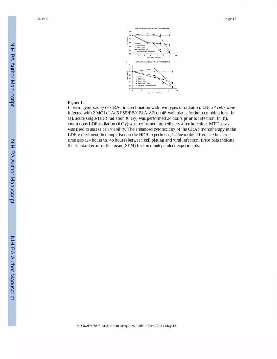
Figure 1.In vitro cytotoxicity of CRAd in combination with two types of radiation. LNCaP cells wereinfected with 2 MOI of Ad5 PSE/PBN E1A-AR on 48-well plates for both combinations. In(a), acute single HDR radiation (6 Gy) was performed 24 hours prior to infection. In (b),continuous LDR radiation (6 Gy) was performed immediately after infection. MTT assaywas used to assess cell viability. The enhanced cytotoxicity of the CRAd monotherapy in theLDR experiment, in comparison to the HDR experiment, is due to the difference in shortertime gap (24 hours vs. 48 hours) between cell plating and viral infection. Error bars indicatethe standard error of the mean (SEM) for three independent experiments.
LIU et al. Page 12
Int J Radiat Biol. Author manuscript; available in PMC 2011 May 13.
NIH
-PA Author Manuscript
NIH
-PA Author Manuscript
NIH
-PA Author Manuscript
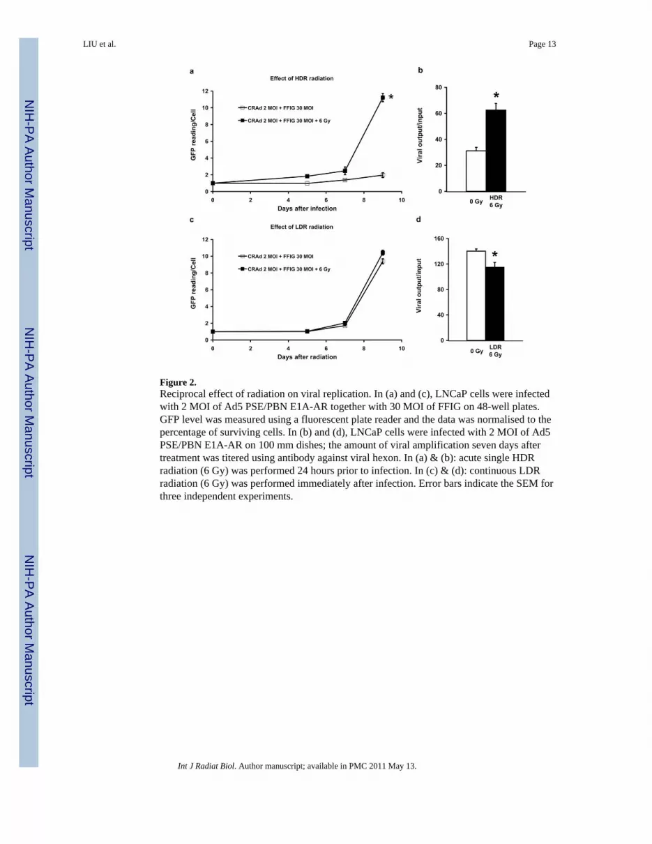
Figure 2.Reciprocal effect of radiation on viral replication. In (a) and (c), LNCaP cells were infectedwith 2 MOI of Ad5 PSE/PBN E1A-AR together with 30 MOI of FFIG on 48-well plates.GFP level was measured using a fluorescent plate reader and the data was normalised to thepercentage of surviving cells. In (b) and (d), LNCaP cells were infected with 2 MOI of Ad5PSE/PBN E1A-AR on 100 mm dishes; the amount of viral amplification seven days aftertreatment was titered using antibody against viral hexon. In (a) & (b): acute single HDRradiation (6 Gy) was performed 24 hours prior to infection. In (c) & (d): continuous LDRradiation (6 Gy) was performed immediately after infection. Error bars indicate the SEM forthree independent experiments.
LIU et al. Page 13
Int J Radiat Biol. Author manuscript; available in PMC 2011 May 13.
NIH
-PA Author Manuscript
NIH
-PA Author Manuscript
NIH
-PA Author Manuscript
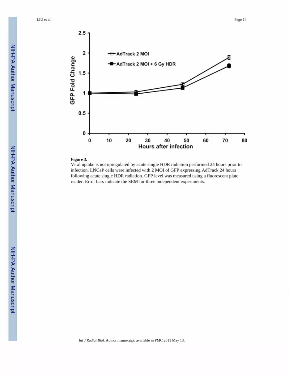
Figure 3.Viral uptake is not upregulated by acute single HDR radiation performed 24 hours prior toinfection. LNCaP cells were infected with 2 MOI of GFP expressing AdTrack 24 hoursfollowing acute single HDR radiation. GFP level was measured using a fluorescent platereader. Error bars indicate the SEM for three independent experiments.
LIU et al. Page 14
Int J Radiat Biol. Author manuscript; available in PMC 2011 May 13.
NIH
-PA Author Manuscript
NIH
-PA Author Manuscript
NIH
-PA Author Manuscript

Figure 4.Viral/acute single HDR radiation combination causes increased expression of double-stranded DNA damage marker γ-H2AX. LNCaP cells were infected with 2 MOI of Ad5PSE/PBN E1A-AR on 100 mm dishes either 24 hours following acute single HDR radiation(6 Gy) (as control, un-irradiated cells were seeded and infected at the same time, denotedas a) or immediately followed by continuous LDR radiation (6 Gy) (as control, un-irradiatedcells were seeded and infected at the same time, denoted as b). Whole cell extracts werecollected seven days post-treatment. For Western blotting, 10–20 μg of whole cell extractwas loaded on 4–15% acrylamide gels.
LIU et al. Page 15
Int J Radiat Biol. Author manuscript; available in PMC 2011 May 13.
NIH
-PA Author Manuscript
NIH
-PA Author Manuscript
NIH
-PA Author Manuscript