epstein-barr virus latent membrane protein 1 trans-activates mir155 transcription through the nf-ib...
TRANSCRIPT
6608–6619 Nucleic Acids Research, 2008, Vol. 36, No. 20 Published online 21 October 2008doi:10.1093/nar/gkn666
Epstein–Barr virus latent membrane protein 1trans-activates miR-155 transcription throughthe NF-iB pathwayGraziana Gatto, Annalisa Rossi, Daniela Rossi, Sven Kroening, Stefano
Bonatti and Massimo Mallardo*
Department of Biochemistry and Medical Biotechnologies, University of Naples Federico II, Naples, Italy
Received August 8, 2008; Revised September 15, 2008; Accepted September 21, 2008
ABSTRACT
The Epstein–Barr virus (EBV)-encoded latent mem-brane protein-1 (LMP1), a functional homologue ofthe tumor necrosis factor receptor family, substan-tially contributes to EBV’s oncogenic potential byactivating nuclear factor-iB (NF-iB). miR-155 is anoncogenic miRNA critical for B-cell maturation andimmunoglobulin production in response to antigen.We report that miR-155 expression is much higher inEBV-immortalized B cells than in EBV-negativeB cells. LMP1, but not LMP2, up-regulated theexpression of miR-155, when transfected in EBV-negative B cells. We analyzed two putative NF-iBbinding sites in the miR-155 promoter; both sitesrecruited NF-iB complex, in nuclear extract fromEBV-immortalized cells. The exogenous expressionof LMP1, in EBV-negative background, is temporallycorrelated to induction of p65 with binding on bothNF-iB sites and with miR-155 overexpression. Theinduction of p65 binding together with increasedRNA polymerase II binding, confirms that LMP1-mediated activation of miR-155 occurs transcrip-tionally. In reporter assays, miR-155 promoter lack-ing NF-iB binding sites was no longer activatedby LMP1 expression and an intact AP1 site isneeded to attain maximum activation. Finally, wedemonstrate that LMP1-mediated activation ofmiR-155 in an EBV-negative background correlateswith reduction of protein PU.1, which is a possiblemiR target.
INTRODUCTION
Epstein–Barr virus (EBV), which infects over 90% of theadults, appears to have evolved to exploit the normal biol-ogy of B-cell development in order to persist as a life-longasymptomatic infection. However, EBV can contribute tooncogenesis. Indeed, it is frequently found in Burkitt’slymphoma, Hodgkin’s lymphoma, nasopharyngeal carci-noma and lymphoproliferative diseases in immune-suppressed individuals (1,2). EBV-related oncogenesis isprincipally associated with latency during which only alimited subset of the full repertoire of viral genes are tran-scribed. In addition to protein-coding genes, the non-coding RNAs EBER1, EBER2 and a set of EBV-encodedmicroRNAs (miRNAs) are expressed in all three forms oflatency (2). Of the genes expressed during viral latency inEBV-associated diseases, LMP1 is the one most impli-cated in tumor formation (3,4). LMP1 is invariablyexpressed in Burkitt’s lymphoma, it is required for EBV-mediated transformation of lymphocytes in vitro and ittransforms rodent fibroblasts. Moreover, transgenic miceexpressing LMP1 under the control of an immunoglobulinpromoter develop B lymphocyte tumors more frequentlythan wild-type mice (3).
LMP1 is a six-transmembrane constitutively active sig-naling molecule that functionally mimics members of thecellular tumor necrosis factor receptor (TNFR) family.While the transmembrane domains are required for aggre-gation and constitutive activation of LMP1 (5–8), twocytoplasmic domains, i.e. the C-terminal activator regions1 and 2 (CTAR-1 and -2), are critical for the transformingproperties of LMP1 (9–12). Together, these signalingdomains act through cellular TNFR-associated factors
The authors wish it to be known that, in their opinion, the first two authors should be regarded as joint First Authors
*To whom correspondence should be addressed. Tel: +39 081 7463627; Fax: +39 081 7463205; Email: [email protected] address:Sven Kroening, Universitatsklinikum Erlangen, Medizinische Klinik 4, Erlagen, DE, Germany
� 2008 The Author(s)This is an Open Access article distributed under the terms of the Creative Commons Attribution Non-Commercial License (http://creativecommons.org/licenses/by-nc/2.0/uk/) which permits unrestricted non-commercial use, distribution, and reproduction in any medium, provided the original work is properly cited.
by guest on Novem
ber 30, 2013http://nar.oxfordjournals.org/
Dow
nloaded from
(TRAFs) and other cell-signaling molecules to activate,three transcription factors, namely, nuclear factor-kB(NF-kB), activator transcription factor-2 (ATF)-2 andAP-1, via c-Jun N-terminal kinase (JNK) (8–12). Bymanipulating these cell-signaling pathways, LMP1 affectshost cellular processes that regulate cell proliferation,migration and apoptosis, thereby contributing to cellularimmortalization and tumorigenesis.
The miR-155 is processed from a primary transcript,known as ‘BIC’ (B-cell integration cluster), whoseupstream region was originally found to be a frequentsite of integration of the avian leucosis virus in lymphomas(13). Transgenic mouse studies demonstrated that B-celltargeted expression of BIC leads to the developmentof B-cell malignancies (14). Furthermore, a number ofmiRNA profiling studies have shown elevated levels ofmiR-155 in a wide array of cancers including lymphomas(14–19). miR-155 is one of the miRNAs most frequentlyimplicated in cancers. It plays a critical role in lymphocyteactivation in vivo (20,21) and is induced by a variety ofimmune cell stimuli, i.e. toll-like receptor (TLR) ligands,TNF-a, interferon-beta (IFN-b) and antigens [B-cellreceptor (BCR) engagement] (16,22). The mechanism bywhichmiR-155 is regulated after TLR and IFN signaling inmacrophages is still unknown. Here we show that LMP1upregulates the expression of miR-155mainly by activatingthe NF-kB pathway, which suggests that miR-155 cancooperate in the EBV-mediated transformation of B cells.
MATERIALS AND METHODS
Cell culture
EBV-negative human B cells (DeFew) and EBV-immortalized human B cells MC3 (kindly provided byProf. Giuseppe Scala, University ‘Magna Grecia’Catanzaro) Devozione, Cap (kindly provided by DrGiuseppina Ruggiero, University ‘Federico II’, Naples),were cultured in RPMI 1640 medium (Invitrogen,Carlsbad, CA, USA) supplemented with 10% fetalbovine serum (Invitrogen) and 2mM glutamine. Murineembryonic fibroblasts (MEFs) and HEK 293 referred to asLinX, stably transfected with the helper vector, [LinX cellline, Openbiosystem, (http://www.openbiosystems.com/RNAi/LinX/)] were cultured in DMEM medium(GIBCO-Invitrogen) supplemented with 10% fetalbovine serum (Invitrogen) and 2mM glutamine.
RNA preparation and northern blot
Total RNA was extracted using Trizol reagent(Invitrogen) according to supplier’s protocol. Northernblot analysis was carried out on 15 mg of total RNAs.Briefly, all RNA samples were dissolved in loadingbuffer [0.05% bromophenol blue, 0.05% cyanol, 5%Ficoll (type 400), 80% formamide and 7M urea], boiledfor 5min at 958C and loaded onto 15% polyacrylamidegel under denaturizing conditions [15% acrylamide-bis-acrylamide 19/1, 45mM Tris, 45mM boric acid, 1mMEDTA pH 8, 7M urea, 0.01% N,N,N0,N0-tetramethyl-ethylenediamine (TEMED) and 0.1% ammonium persul-fate (APS)]. Samples were resolved by electrophoresis for
90min at 150V and transferred onto nylon membranes(Hybond N+, Amersham/GE Healthcare, LittleChalfont, UK) by capillary blot. The nylon membraneswere then equilibrated in 1M NaCl and pre-hybridizedin 6� SSC, 5� Denhart’s solution (1� Denhart’s solu-tion=0.1% Ficoll, 0.1% polyvinyl pyrrolydone and0.1% bovine serum albumin), 5 mg/ml of shearedsalmon sperm ds-DNA (Ambion, Austin TX, USA) at428C for 2 h. After pre-hybridization, 1� 106CPM/ml of[g32P-ATP] radiolabeled oligonucleotide probe was addedand the hybridization carried out overnight at 428C. Themembranes were washed twice in 2� SSC, 1% SDS at428C for 30min and exposed either by autoradiographyor by phosphorimage screen (Amersham/GE Healthcare).The signals were quantified with image-scanning or byImage-J software analysis.
Western blot analysis
The cell pellets were resuspended in 1ml of lysis buffer[10mM HEPES, pH 7.4, 150mM KCl, 1mM EDTA pH8, 1% Triton X-100, 1mM DTT, 1mM orthovadanate,1mM NaF and protease inhibitor cocktail (Roche, Basel,Switzerland)] at 48C for 20min. Then, 40 mg of proteins, ofeach sample, were resuspended in Laemmli buffer (23) andresolved by SDS–PAGE (running gel: 0.4M Tris, pH 8.8,12% acrylamide/bis-acrylamide 37/1, 0.1% SDS, 0.01%TEMED, 0.1% APS; staking gel: 0.07M Tris pH 6.8,5% acrylamide/bis-acrylamide 37/1, 0.1% SDS, 0.01%TEMED, 0.1% APS). The proteins were transferredonto nitrocellulose membranes (Hybond ECL, Ame-sham/GE Healthcare) by electroblotting. Membraneswere blocked in 5% of ‘non-fat milk’ (BioRad, HerculesCA, USA) 0.5% BSA in phosphate buffer solution for 2 hat room temperature and immunoblotted with monoclo-nal antibody against LMP1 (kindly provided by Dr DongYun Lee, McArdle Laboratory for Cancer Research, Uni-versity of Wisconsin-Madison), mouse polyclonal anti-body against g-tubulin (Sigma Chemical Company, StLouis MO, USA) mouse monoclonal antibody againstp65 (SantaCruz, CA, USA), rabbit polyclonal antibodyagainst IkBa (SantaCruz), rabbit polyclonal against H3(SantaCruz) or rabbit polyclonal antibody against PU.1(Santa Cruz). Specific secondary antibodies (Sigma Che-mical Company), horse-radish peroxidase conjugated,were used for protein detection by enhanced chemilumi-nescence (ECL, Amesham/GE Healthcare) followed byautoradiography (Hyperfilm ECL, Amesham/GE Health-care). We used the Image-J software for densitometricanalysis.
Cloning and mutagenesis of BIC/miR-155 promoter
The human BIC/miR-155 promoter region extending from1783 to 1, from 1380 to 1, from 1065 to 1 relative to thestart site was isolated from MC3 genomic DNA by PCRusing the following primers:Fw AGCTGAGCTCGAAAGGTCACCCTAGAATTG(�1783),Fw AGCTGAGCTCGATCTGGCACATGGTAAATG(�1380),
Nucleic Acids Research, 2008, Vol. 36, No. 20 6609
by guest on Novem
ber 30, 2013http://nar.oxfordjournals.org/
Dow
nloaded from
Fw AGCTGAGCTCCAGTCACATGTTGATGAGGC(�1065),Rev CATGAAGCTTATCCGCTCCCTTCCCGAG(+1).The isolated fragments were digested with SacI and
HindIII and cloned into SacI and HindIII cutpGL3basic (Promega, Madison, WI, USA). The entirepromoter region was sequenced and no discrepancieswere identified relative to the Genebank genomic sequence(http://www.ncbi.nlm.nih.gov/Genbank/).Mutagenesis of the pLuc1380 and pLuc1783 reporter
plasmid was carried out with the ‘Expand LongTemplate PCR system’ (Roche) and the following oligo-nucleotides: 50-GTA AAT TAA GTA CTA TGC TCGAGC CAG CTC TGA CAT G-30 and 50-CAT AGTACT TAA TTT ACA GAT GGC TCA GGT TGGTTA AG-30 (NF-kB1), Fw 50-CAACCTAGAATGAGAAATGCTCGAGTCAGAAAGGCATTGTAGG-30 andRev 50-CATTTCTCATTCTAGGTTGAACTATACCTCCCTTCTCCCAGTG-30 (NF-kB2), 50-GGC GCC TGGTCG GTT ATC TCG AGC AAG TGA GTT ATAAAA-30 and 50-ATA ACC GAC CAG GCG CCT TTTCTG CAA CCC-30 (AP-1). In each case, the core tran-scription factor binding site was replaced by a XhoI restric-tion site. Mutations were initially screened by digestingwith XhoI and then verified by sequence analysis.
Reporter analysis
For each reporter plasmid, 2� 106 MEFs were distributedin 6 cm dish plates containing 5ml RPMI1640 (Invitrogen)10% FBS (Cambrex, East Rutherford, NJ, USA), 2mMglutamine. FuGene (Roche) of 6 ml were added to 300 ml ofDMEM and incubated for 5min at room temperature.Each reporter vector of 0.5mg plus 0.2 mg of b-galactosi-dase vector were then added to the mixture, the tubes wereshacked and incubated at room temperature for 15min.For each transfection, the mixtures were then added toeach plate and plates were incubated at 378C, 5% CO2
for 48 h. Cells were harvested 48 h later and assayed forluciferase activity. Results are presented as average ofthree independent experiments.For each reporter plasmid, 2� 106 DeFew were distrib-
uted in 6 cm dish plates containing 5ml RPMI1640(Invitrogen) 10% FBS (Cambrex), 4mM glutamine.FuGene (Roche) of 8 ml were added to 300 ml of DMEMand incubated for 5min at room temperature. Each repor-ter vector of 1 mg plus 0.5 mg of b-galactosidase vector werethen added to the mixture, the tubes were shacked andincubated at room temperature for 15min. For each trans-fection, the mixtures were then added to each plate andplates were incubated at 378C, 5% CO2. After 6 h eachsample was split in two, and one of them was treatedwith 10 ng/ml of phorbol myristoylated acetate (PMA).Cells were harvested 48 h later and assayed for luciferaseactivity. Results are presented as average of three indepen-dent experiments.
Chromatin immunoprecipitation assay
MC3 or DeFew cells (5� 106 cells) were exposed to 1%formaldehyde for 10min at 378C to obtain protein–DNA
cross-linking. The nuclear fraction was sonicated to yieldchromatin fragments of 200–1000 bp; 5% of the totalvolume was removed from each sample and used asthe input fraction. Chromatin was pre-cleared by pre-incubation with a DNA salmon sperm/protein A-agarose50% slurry (Upstate, Temecula, CA, USA) for 1 h at 48C.The agarose was centrifuged, and the pre-cleared chroma-tin supernatant was incubated with the antibodies (3 mg)against RNA pol II (mAb anti RNA polymerase II, ActiveMotif, Rixensart, Belgium), p50 or p65 (pAb anti p50 orpAb anti p65, kindly provided by Dr N. Rice, FrederickCancer Research and Development Center, Frederick,MD, USA), against histone 3 (pAb anti H3, SantaCruz),overnight at 48C. The protein–DNA-antibody complexeswere collected by the addition of the salmon sperm DNA–protein A-agarose (2 h at 48C) and washed, and protein–DNA cross-linking was reversed (4 h at 658C). DNA waspurified by phenol/chloroform extraction and ethanol pre-cipitation and aliquots (25%) of the purified materialsunderwent PCR (5min at 948C, 1min at 948C, 1min at528C, 1min at 728C for 40 cycles, 5min at 728C).
Preparation of nuclear extracts and electrophoresismobility shift assay
MC3 cells (5- to 6� 106 cells) were washed with cold phos-phate buffered saline and harvested. The cell pellet wasresuspended in extraction buffer containing 10mMHEPES, pH 7.9, 10mM KCl, 1.5mM MgCl2, 0.1mMEGTA, 0.5mM DTT, 0.5mM PMSF, 10 mg/ml aprotinin,10 mg/ml leupeptin, passed through a needle, kept on icefor 45min, and centrifuged (15min at 14 000 r.p.m. at48C). The nuclear pellet was then resuspended in highsalt extraction buffer containing 10mM HEPES, pH 7.9,0.4mM NaCl, 1.5mM MgCl2, 0.1mM EGTA, 0.5mMDTT, 0.5mM phenylmethylsulfonyl fluoride, 10 mg/mlaprotinin, 10 mg/ml leupeptin and incubated for 45minat 48C. The nuclear extract supernatant was obtained bycentrifugation (30min at 14 000 r.p.m. at 48C), proteinconcentration was determined and 5 mg aliquots werestored at �808C until used. Double-stranded syntheticoligonucleotides were radiolabeled using [g32P]ATP(3000Ci mmol–1; Amersham Biosciences) and T4 polynu-cleotide kinase (Fermentas, Burlington, ON, Canada).The binding reaction was carried out for 20min at roomtemperature with 5 mg of nuclear proteins in 10% glycerol,60mM KC1, 1mM EDTA, 1mM dithiothreitol and0.250 mg/ml poly[dI–dC] containing 50 000 cpm of radiola-beled probe and a 80-fold molar excess of unlabeled com-petitor oligonucleotide when indicated. For supershiftexperiments, 4 mg of specific anti-p65 or anti-p50 wereadded to the binding reaction and incubated for 30min,after which we added the radiolabeled probe. DNA–protein complexes were separated by 5% non-denaturingpolyacrylamide gel and revealed by either autoradiogra-phy or phosphorimage screen (Amersham Biosciences).
Transient transfection
DeFew cells (3� 106 cells) were centrifuged, washed inPBS and resuspended in 300 ml of electroporationmedium (RPMI 1640 medium, 20% FBS and 2mM
6610 Nucleic Acids Research, 2008, Vol. 36, No. 20
by guest on Novem
ber 30, 2013http://nar.oxfordjournals.org/
Dow
nloaded from
Glutamine). Cells were mixed with 20 mg of plasmidexpressing LMP1 (pcDNA3-LMP1), or LMP2(pcDNA3-LMP2) or both and subsequently transferredin electroporation cuvette. Cells were electroporatedtwice at 220V, 960 mFa and incubated 5min on ice. Cellswere plated in 10ml of RPMI 1640 medium, 10%FBS and 2mM glutamine for 2 days. After 2 days, theywere harvested and assayed for miR-155 and LMP1expression.
Viral generation, infection and time course
The retroviral vector LMP1pBABE-puro was kindly pro-vided by Prof. Eiji Hara (University of Tokushima,Japan). LinX cell line, stably transfected with the helpervector, were grown in DMEM medium 10% FBS, 2mMglutamine and 100 mg/ml hygromicin. Transfections withLMP1pBABEpuro or pBABEpuro (empty vector) were asfollows: 6 cm plate of semi-confluent (about 1� 106 cells)cells were transfected and 7.5 mg of DNA vector incubatedin 20 ml of FuGene and incubated in 4ml of medium with-out antibiotic for 24 h. After incubation, the medium washarvested, replaced with 4ml of fresh medium and theLinX cells incubated for 24 h for a second round of viralgeneration.
DeFew cells (4� 106 cells) were centrifuged, washed inPBS and resuspended in 4ml of virus containing mediumplus 4 mg/ml of Polybrene (Sigma). The infection was per-formed by spinoculation, namely by centrifugation at3500 r.p.m. for 90min at 208C. After centrifugation, thecells were resuspended in 10ml of RPMI and incubatedat 378C, 5% CO2. Twenty-four hours later, the cellsunderwent a second round of infection. After 48 h, cellswere treated with 1 mg/ml of puromicine to start selectionof positive clones.
For the time course, DeFew cells (4� 106 cells) werecentrifuged, washed in PBS and resuspended in 12ml ofvirus containing medium plus 12 mg/ml of Polybrene(Sigma). The infection was performed by spinoculation,namely by centrifugation at 3500 r.p.m. for 90min at208C. After centrifugation, the cells were resuspended in10ml of RPMI and incubated at 378C, 5% CO2. Twenty-four hours later, 1 mg/ml of puromocine was added to cellsto start selection. Cells were harvested at 6, 24, 48 and 72 hafter infection and assayed for miR-155 and LMP1expression.
Transient transfection ofMC3
MC3 cells (3� 106 cells) were centrifuged, washed in PBSand resuspended in 300 ml of electroporation medium(RPMI 1640 medium, 20% FBS and 2mM glutamine).Cells were mixed in a tube with 20 mg of pRc/CMV-HA-IkBa-S32/36A (kindly provided by Prof. Ileana Quinto,University Magna Grecia, Catanzaro) and subsequentlyput in electroporation cuvette. Cells were electroporatedtwice at 220V, 960 mFa and incubated 5min on ice. Cellswere plated in 10ml of RPMI 1640 medium, 10% FBSand 4mM glutamine for 2 days. After 2 days, they wereharvested and assayed for miR-155, p65 and IkBaexpression.
RESULTS
The expression of mir-155 is regulated by LMP1
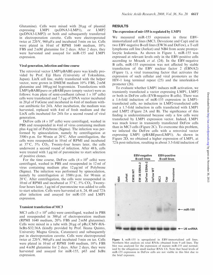
We measured miR-155 expression in three EBV-immortalized cell lines (MC3, Devozione and Cap) and intwo EBV-negative B-cell lines (EW36 and DeFew), a T-celllymphoma cell line (Jurkat) and NB4 from acute promye-locytic leukemia. As shown in Figure 1, miR-155 wasexpressed at relevant levels only in the EBV-positive cellsaccording to Mrazek et al. (24). In the EBV-negativeB cells, miR-155 expression was not affected by stabletransfection of the EBV nuclear factor 2 (EBNA2)(Figure 1), a viral transacting factor that activates theexpression of such cellular and viral promoters as theHIV-1 long terminal repeat (25) and the interleukin-6promoter (26).To evaluate whether LMP1 induces miR activation, we
transiently transfected a vector expressing LMP1, LMP2or both in DeFew cells (EVB-negative B cells). There wasa 1.6-fold induction of miR-155 expression in LMP1-transfected cells, no induction in LMP2-transfected cellsand a 1.7-fold induction in cells transfected with LMP1and LMP2 (Figure 2A and B). The significance of thisfinding is underestimated because only a few cells weretransfected by LMP1 expression vector. Indeed, LMP1was much lower in transiently transfected DeFew cellsthan in MC3 cells (Figure 2C). To overcome this problem,we infected the DeFew cells with a retroviral vectorexpressing LMP1 (pBABEpuroLMP1). As shown inFigure 2D, we obtained a higher expression of LMP1, at72 h post-infection, resulting in about 3.5-fold induction of
Figure 1. miR-155 is upregulated in EBV-immortalized cell lines.Northern blot analysis on total RNAs obtained from 9 cell lines. Theblot was analyzed for the expression of mature miR-155 and normal-ized by the ubiquitous small nuclear RNA U6. Note that basal levels ofmiR-155 expression in DeFew cells are not visible in this blot due tothe brief exposure.
Nucleic Acids Research, 2008, Vol. 36, No. 20 6611
by guest on Novem
ber 30, 2013http://nar.oxfordjournals.org/
Dow
nloaded from
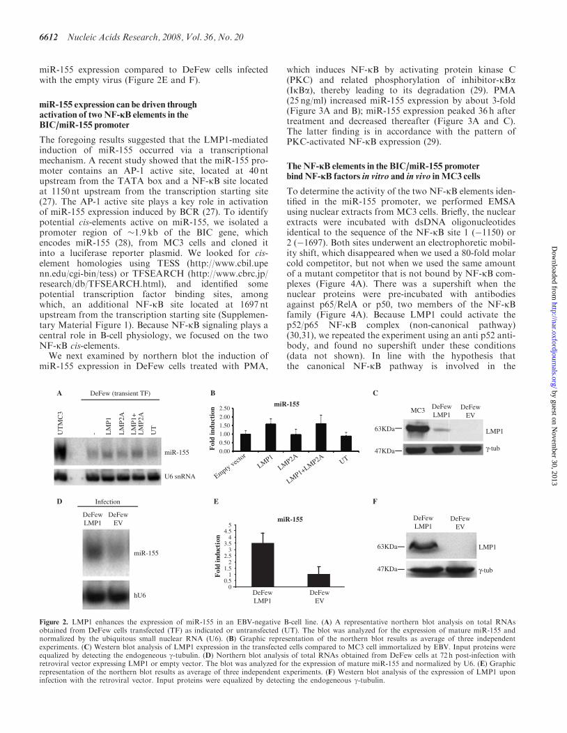
miR-155 expression compared to DeFew cells infectedwith the empty virus (Figure 2E and F).
miR-155 expression can be driven throughactivation of two NF-iB elements in theBIC/miR-155 promoter
The foregoing results suggested that the LMP1-mediatedinduction of miR-155 occurred via a transcriptionalmechanism. A recent study showed that the miR-155 pro-moter contains an AP-1 active site, located at 40 ntupstream from the TATA box and a NF-kB site locatedat 1150 nt upstream from the transcription starting site(27). The AP-1 active site plays a key role in activationof miR-155 expression induced by BCR (27). To identifypotential cis-elements active on miR-155, we isolated apromoter region of �1.9 kb of the BIC gene, whichencodes miR-155 (28), from MC3 cells and cloned itinto a luciferase reporter plasmid. We looked for cis-element homologies using TESS (http://www.cbil.upenn.edu/cgi-bin/tess) or TFSEARCH (http://www.cbrc.jp/research/db/TFSEARCH.html), and identified somepotential transcription factor binding sites, amongwhich, an additional NF-kB site located at 1697 ntupstream from the transcription starting site (Supplemen-tary Material Figure 1). Because NF-kB signaling plays acentral role in B-cell physiology, we focused on the twoNF-kB cis-elements.We next examined by northern blot the induction of
miR-155 expression in DeFew cells treated with PMA,
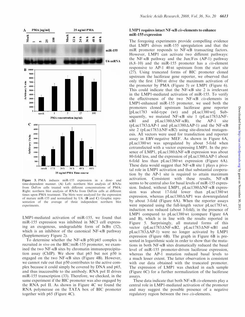
which induces NF-kB by activating protein kinase C(PKC) and related phosphorylation of inhibitor-kBa(IkBa), thereby leading to its degradation (29). PMA(25 ng/ml) increased miR-155 expression by about 3-fold(Figure 3A and B); miR-155 expression peaked 36 h aftertreatment and decreased thereafter (Figure 3A and C).The latter finding is in accordance with the pattern ofPKC-activated NF-kB expression (29).
The NF-iB elements in the BIC/miR-155 promoterbind NF-iB factors in vitro and in vivo inMC3 cells
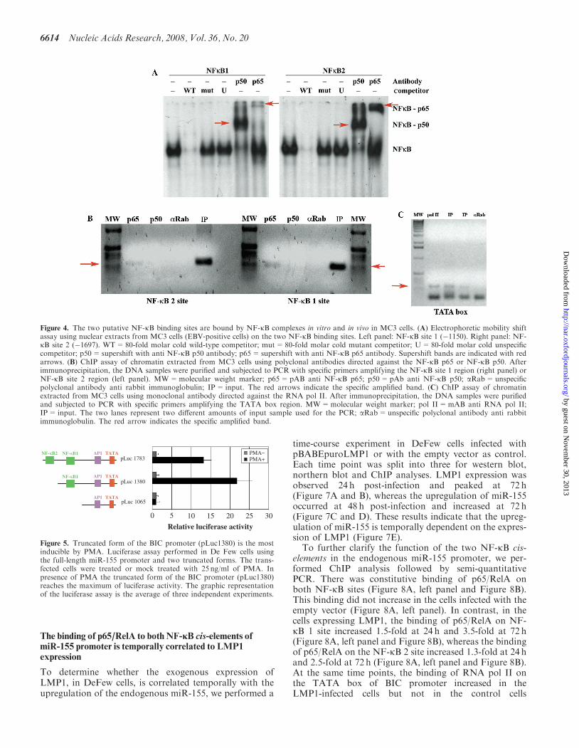
To determine the activity of the two NF-kB elements iden-tified in the miR-155 promoter, we performed EMSAusing nuclear extracts from MC3 cells. Briefly, the nuclearextracts were incubated with dsDNA oligonucleotidesidentical to the sequence of the NF-kB site 1 (�1150) or2 (�1697). Both sites underwent an electrophoretic mobil-ity shift, which disappeared when we used a 80-fold molarcold competitor, but not when we used the same amountof a mutant competitor that is not bound by NF-kB com-plexes (Figure 4A). There was a supershift when thenuclear proteins were pre-incubated with antibodiesagainst p65/RelA or p50, two members of the NF-kBfamily (Figure 4A). Because LMP1 could activate thep52/p65 NF-kB complex (non-canonical pathway)(30,31), we repeated the experiment using an anti p52 anti-body, and found no supershift under these conditions(data not shown). In line with the hypothesis thatthe canonical NF-kB pathway is involved in the
U6 snRNA
UT
MC
3
- LM
P1
LM
P2A
LM
P1+
LM
P2A
UT
DeFew (transient TF)
miR-155
A
miR-155
0.00
0.50
1.00
1.50
2.00
2.50
Fol
d in
duct
ion
Empty vector
LMP1
LMP2A
LMP1+LMP2AUT
B
miR-155
hU6
DeFewLMP1
DeFewEV
InfectionD
miR-155
DeFewLMP1
DeFewEV
0.50
11.5
22.5
33.5
44.5
5
Fol
d in
duct
ion
E
C
LMP1
γ-tub
63KDa
47KDa
DeFewLMP1
DeFewEV
MC3
F
LMP1
γ-tub
63KDa
47KDa
DeFewLMP1
DeFewEV
Figure 2. LMP1 enhances the expression of miR-155 in an EBV-negative B-cell line. (A) A representative northern blot analysis on total RNAsobtained from DeFew cells transfected (TF) as indicated or untransfected (UT). The blot was analyzed for the expression of mature miR-155 andnormalized by the ubiquitous small nuclear RNA (U6). (B) Graphic representation of the northern blot results as average of three independentexperiments. (C) Western blot analysis of LMP1 expression in the transfected cells compared to MC3 cell immortalized by EBV. Input proteins wereequalized by detecting the endogeneous g-tubulin. (D) Northern blot analysis of total RNAs obtained from DeFew cells at 72 h post-infection withretroviral vector expressing LMP1 or empty vector. The blot was analyzed for the expression of mature miR-155 and normalized by U6. (E) Graphicrepresentation of the northern blot results as average of three independent experiments. (F) Western blot analysis of the expression of LMP1 uponinfection with the retroviral vector. Input proteins were equalized by detecting the endogeneous g-tubulin.
6612 Nucleic Acids Research, 2008, Vol. 36, No. 20
by guest on Novem
ber 30, 2013http://nar.oxfordjournals.org/
Dow
nloaded from
LMP1-mediated activation of miR-155, we found thatmiR-155 expression was inhibited in MC3 cell express-ing an exogenous, undegradable form of IkBa (32),which is an inhibitor of the canonical NF-kB pathway(Supplementary Figure 2).
To determine whether the NF-kB p50/p65 complex isrecruited in vivo on the BIC/miR-155 promoter, we exam-ined the two NF-kB sites by chromatin immunoprecipita-tion assay (ChIP). We show that p65 but not p50 isengaged on the two NF-kB sites (Figure 4B). However,we cannot rule out that p50 contributes to the active com-plex because it could simply be covered by DNA and p65,and thus inaccessible to the antibody. RNA pol II drivesmiR-155 transcription (33). Therefore, we checked, in thesame experiment if the BIC promoter was also engaged bythe RNA pol II. As shown in Figure 4C we found theRNA polymerase on the TATA box of BIC promotertogether with p65 (Figure 4C).
LMP1 requires intact NF-iB cis-elements to enhancemiR-155 expression
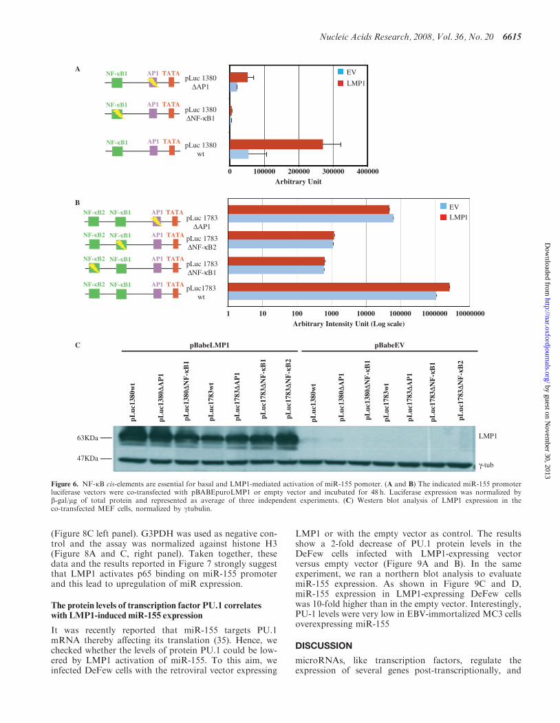
The foregoing experiments provide compelling evidencethat LMP1 drives miR-155 upregulation and that themiR promoter responds to NF-kB transacting factors.However, LMP1 can activate two different pathways:the NF-kB pathway and the Jun/Fos (AP-1) pathway(6,8–10) and the miR-155 promoter has a cis-elementresponsive to AP-1 40 nt upstream from the start site(27). Using truncated forms of BIC promoter clonedupstream the luciferase gene reporter, we observed thatonly the first 1380 nt drive the maximum activation ofthe promoter by PMA (Figure 5) or LMP1 (Figure 6).This could indicate that the NF-kB site 2 is irrelevantin the LMP1-mediated activation of miR-155. To verifythe effectiveness of the two NF-kB cis-elements inLMP1-enhanced miR-155 promoter, we used both thepromoters cloned upstream luciferase gene reporter[pLuc1783 wild-type (wt) and pLuc1380wt]. Sub-sequently, we mutated NF-kB site 1 (pLuc1783�NF-kB1 and pLuc1380�NF-kB), the AP-1 site(pLuc1783�AP-1 and pLuc1380�AP-1) and the NF-kBsite 2 (pLuc1783�NF-kB2) using site-directed mutagen-esis. All vectors were used for transfection and reporterassay in EBV-negative MEF. As shown in Figure 6A,pLuc1380wt was upregulated by about 5-fold whencotransfected with a vector expressing LMP1. In the pre-sence of LMP1, pLuc1380�NF-kB expression was about80-fold less, and the expression of pLuc1380�AP-1 about6-fold less than pLuc1380wt expression (Figure 6A).These data would suggest that NF-kB site 1 plays a pivo-tal role in LMP1 activation and that substantial coopera-tion by the AP-1 site is required to attain maximumactivation. Consistent with these results, NF-kBseems to be central also for basal levels of miR-155 expres-sion. Indeed, without LMP1, pLuc1380�NF-kB expres-sion was about 17-fold lower than pLuc1380wtexpression, whereas pLuc1380�AP1 was downregulatedby about 3-fold (Figure 6A). When the reporter assayswere repeated using the full-length vector pLuc1783wt,induction was reduced (about 3-fold), in the presence ofLMP1 compared to pLuc1380wt (compare Figure 6Aand B), which is in line with the results reported inFigure 5. Surprisingly, all mutated forms of thisvector (pLuc1783�NF-kB2, pLuc1783�NF-kB1 andpLuc1783�AP-1) were no longer activated by LMP1expression (Figure 6B). The graph in Figure 6B is pre-sented in logarithmic scale in order to show that the muta-tions in both NF-kB sites dramatically reduced the basallevel of miR-155 promoter-driven luciferase expression,whereas the AP-1 mutation reduced basal levels toa much lesser extent. The latter observation is consistentwith our data obtained with the truncated promoter.The expression of LMP1 was checked in each sample(Figure 6C) for a further normalization of the luciferaseassays.These data indicate that both NF-kB cis-elements play a
central role in LMP1-mediated activation of the promoterand may suggest the possible presence of a negativeregulatory region between the two cis-elements.
Figure 3. PMA induces miR-155 expression in a dose- andtime-dependent manner. (A) Left: northern blot analysis of RNAsfrom DeFew cells treated with different concentrations of PMA;Right: northern blot analysis of RNAs from DeFew cells at differenttimes upon PMA treatment. The blots were analyzed for the expressionof mature miR-155 and normalized by U6. (B and C) Graphic repre-sentation of the average of three independent northern blotexperiments.
Nucleic Acids Research, 2008, Vol. 36, No. 20 6613
by guest on Novem
ber 30, 2013http://nar.oxfordjournals.org/
Dow
nloaded from
The binding of p65/RelA to both NF-iB cis-elements ofmiR-155 promoter is temporally correlated to LMP1expression
To determine whether the exogenous expression ofLMP1, in DeFew cells, is correlated temporally with theupregulation of the endogenous miR-155, we performed a
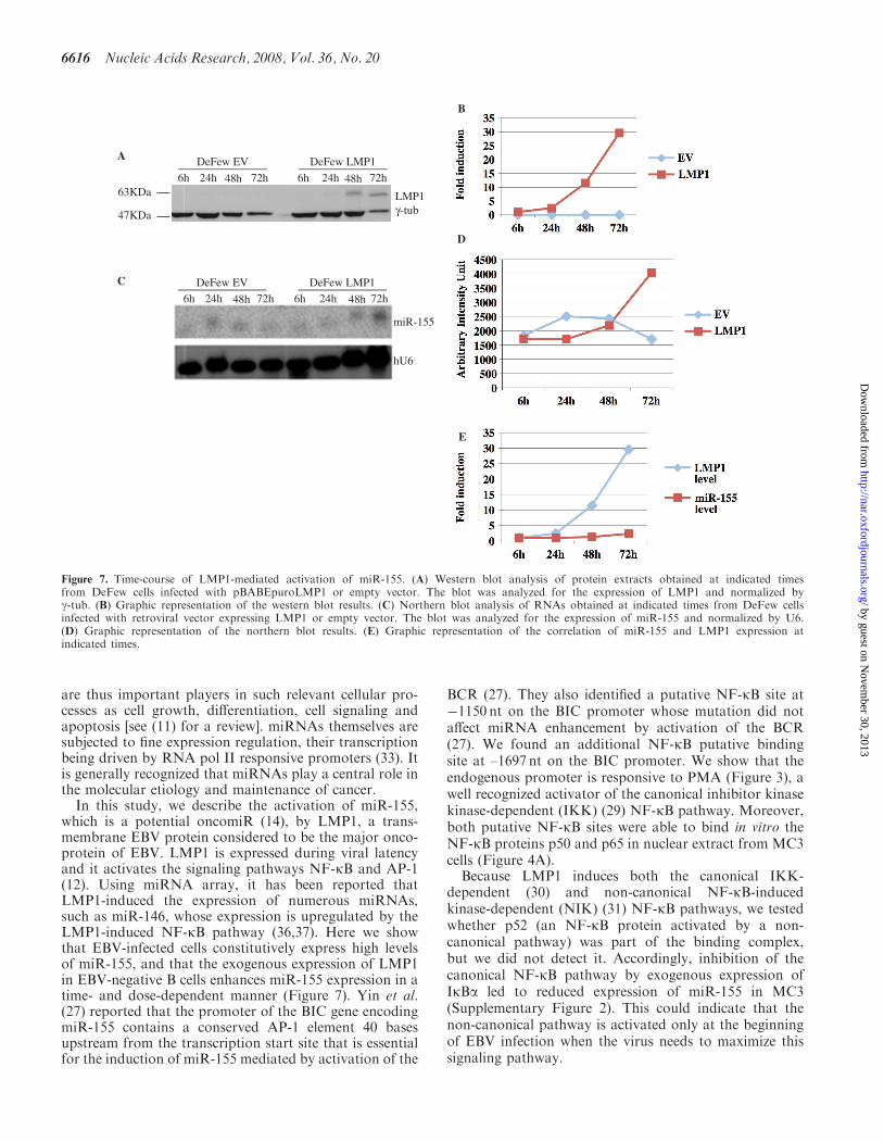
time-course experiment in DeFew cells infected withpBABEpuroLMP1 or with the empty vector as control.Each time point was split into three for western blot,northern blot and ChIP analyses. LMP1 expression wasobserved 24 h post-infection and peaked at 72 h(Figure 7A and B), whereas the upregulation of miR-155occurred at 48 h post-infection and increased at 72 h(Figure 7C and D). These results indicate that the upreg-ulation of miR-155 is temporally dependent on the expres-sion of LMP1 (Figure 7E).
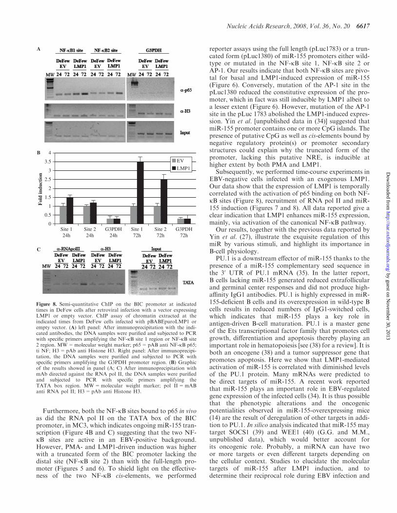
To further clarify the function of the two NF-kB cis-elements in the endogenous miR-155 promoter, we per-formed ChIP analysis followed by semi-quantitativePCR. There was constitutive binding of p65/RelA onboth NF-kB sites (Figure 8A, left panel and Figure 8B).This binding did not increase in the cells infected with theempty vector (Figure 8A, left panel). In contrast, in thecells expressing LMP1, the binding of p65/RelA on NF-kB 1 site increased 1.5-fold at 24 h and 3.5-fold at 72 h(Figure 8A, left panel and Figure 8B), whereas the bindingof p65/RelA on the NF-kB 2 site increased 1.3-fold at 24 hand 2.5-fold at 72 h (Figure 8A, left panel and Figure 8B).At the same time points, the binding of RNA pol II onthe TATA box of BIC promoter increased in theLMP1-infected cells but not in the control cells
Figure 4. The two putative NF-kB binding sites are bound by NF-kB complexes in vitro and in vivo in MC3 cells. (A) Electrophoretic mobility shiftassay using nuclear extracts from MC3 cells (EBV-positive cells) on the two NF-kB binding sites. Left panel: NF-kB site 1 (�1150). Right panel: NF-kB site 2 (�1697). WT=80-fold molar cold wild-type competitor; mut=80-fold molar cold mutant competitor; U=80-fold molar cold unspecificcompetitor; p50=supershift with anti NF-kB p50 antibody; p65=supershift with anti NF-kB p65 antibody. Supershift bands are indicated with redarrows. (B) ChIP assay of chromatin extracted from MC3 cells using polyclonal antibodies directed against the NF-kB p65 or NF-kB p50. Afterimmunoprecipitation, the DNA samples were purified and subjected to PCR with specific primers amplifying the NF-kB site 1 region (right panel) orNF-kB site 2 region (left panel). MW=molecular weight marker; p65=pAB anti NF-kB p65; p50=pAb anti NF-kB p50; aRab=unspecificpolyclonal antibody anti rabbit immunoglobulin; IP= input. The red arrows indicate the specific amplified band. (C) ChIP assay of chromatinextracted from MC3 cells using monoclonal antibody directed against the RNA pol II. After immunoprecipitation, the DNA samples were purifiedand subjected to PCR with specific primers amplifying the TATA box region. MW=molecular weight marker; pol II=mAB anti RNA pol II;IP= input. The two lanes represent two different amounts of input sample used for the PCR; aRab=unspecific polyclonal antibody anti rabbitimmunoglobulin. The red arrow indicates the specific amplified band.
NF-κB1NF-κB2 AP1 TATA
NF-κB1 AP1 TATA
AP1 TATA
pLuc 1783
pLuc 1380
pLuc 1065
Relative luciferase activity
50 10 15 20 25 30
PMA−PMA+
Figure 5. Truncated form of the BIC promoter (pLuc1380) is the mostinducible by PMA. Luciferase assay performed in De Few cells usingthe full-length miR-155 promoter and two truncated forms. The trans-fected cells were treated or mock treated with 25 ng/ml of PMA. Inpresence of PMA the truncated form of the BIC promoter (pLuc1380)reaches the maximum of luciferase activity. The graphic representationof the luciferase assay is the average of three independent experiments.
6614 Nucleic Acids Research, 2008, Vol. 36, No. 20
by guest on Novem
ber 30, 2013http://nar.oxfordjournals.org/
Dow
nloaded from
(Figure 8C left panel). G3PDH was used as negative con-trol and the assay was normalized against histone H3(Figure 8A and C, right panel). Taken together, thesedata and the results reported in Figure 7 strongly suggestthat LMP1 activates p65 binding on miR-155 promoterand this lead to upregulation of miR expression.
The protein levels of transcription factor PU.1 correlateswith LMP1-induced miR-155 expression
It was recently reported that miR-155 targets PU.1mRNA thereby affecting its translation (35). Hence, wechecked whether the levels of protein PU.1 could be low-ered by LMP1 activation of miR-155. To this aim, weinfected DeFew cells with the retroviral vector expressing
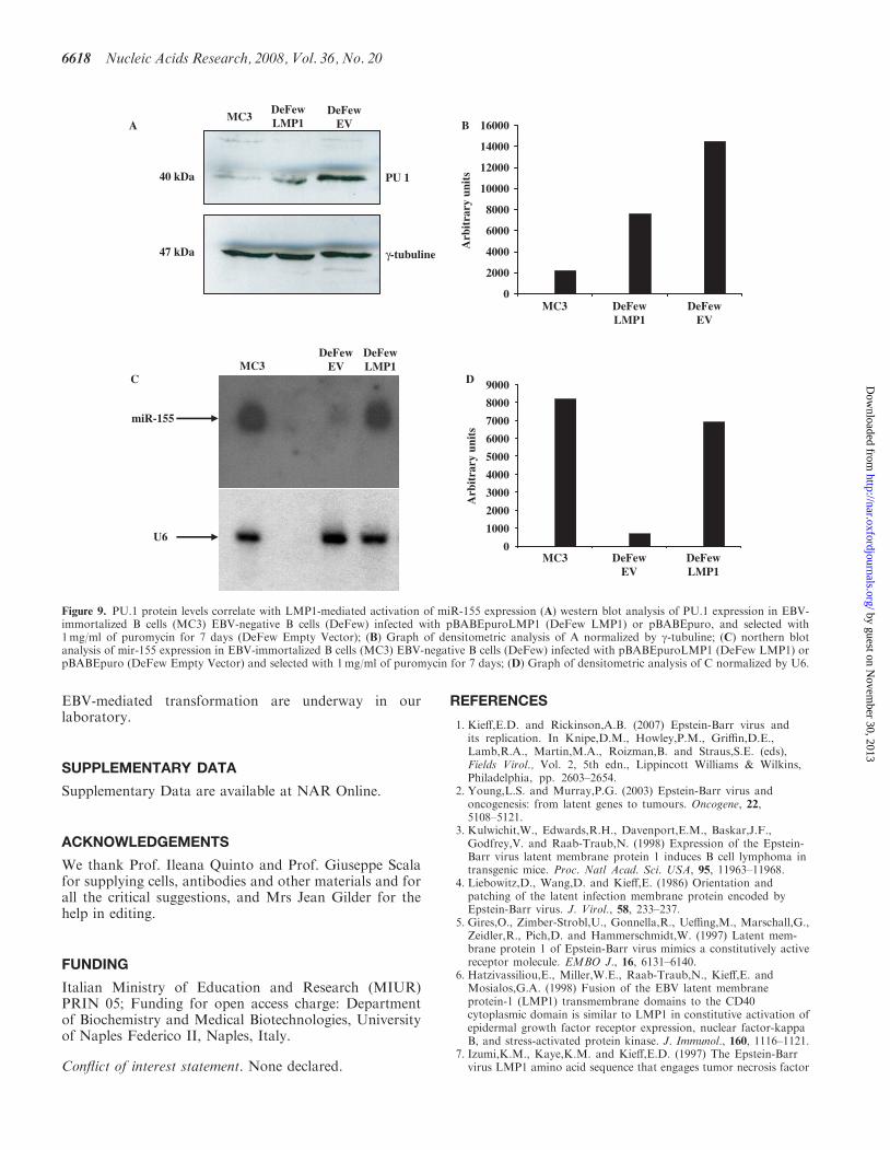
LMP1 or with the empty vector as control. The resultsshow a 2-fold decrease of PU.1 protein levels in theDeFew cells infected with LMP1-expressing vectorversus empty vector (Figure 9A and B). In the sameexperiment, we ran a northern blot analysis to evaluatemiR-155 expression. As shown in Figure 9C and D,miR-155 expression in LMP1-expressing DeFew cellswas 10-fold higher than in the empty vector. Interestingly,PU-1 levels were very low in EBV-immortalized MC3 cellsoverexpressing miR-155
DISCUSSION
microRNAs, like transcription factors, regulate theexpression of several genes post-transcriptionally, and
NF-kB1NF-kB2 AP1 TATA
NF-kB1NF-kB2 AP1 TATA
NF-kB1NF-kB2 AP1 TATA
NF-kB1NF-kB2 AP1 TATA
pLuc 1783∆AP1
pLuc1783wt
pLuc 1783∆NF-κB1
pLuc 1783 ∆NF-κB2
pLuc
1380
wt
pLuc
1380
DAP
1
pLuc
1380
DNF
-kB
1
pLuc
1783
wt
pLuc
1783
DNF
-kB
1
pLuc
1783
DNF
-kB
2
pLuc
1783
DAP
1
pLuc
1380
wt
pLuc
1380
DAP
1
pLuc
1380
DNF
-kB
1
pLuc
1783
DNF
-kB
1
pLuc
1783
DNF
-kB
2
pLuc
1783
DAP
1
pLuc
1783
wt
pBabeLMP1 pBabeEV
AP1 TATA
pLuc 1380∆AP1
pLuc 1380wt
pLuc 1380∆NF-κB1
NF-kB1 AP1 TATA
NF-kB1
NF-kB1
AP1 TATA
A
B
C
LMP1
γ-tub
Arbitrary Intensity Unit (Log scale)
Arbitrary Unit
63KDa
47KDa
0
1 10 100 1000 10000 100000 1000000 10000000
100000 200000 300000 400000
EVLMP1
EV
LMP1
Figure 6. NF-kB cis-elements are essential for basal and LMP1-mediated activation of miR-155 pomoter. (A and B) The indicated miR-155 promoterluciferase vectors were co-transfected with pBABEpuroLMP1 or empty vector and incubated for 48 h. Luciferase expression was normalized byb-gal/mg of total protein and represented as average of three independent experiments. (C) Western blot analysis of LMP1 expression in theco-transfected MEF cells, normalized by gtubulin.
Nucleic Acids Research, 2008, Vol. 36, No. 20 6615
by guest on Novem
ber 30, 2013http://nar.oxfordjournals.org/
Dow
nloaded from
are thus important players in such relevant cellular pro-cesses as cell growth, differentiation, cell signaling andapoptosis [see (11) for a review]. miRNAs themselves aresubjected to fine expression regulation, their transcriptionbeing driven by RNA pol II responsive promoters (33). Itis generally recognized that miRNAs play a central role inthe molecular etiology and maintenance of cancer.In this study, we describe the activation of miR-155,
which is a potential oncomiR (14), by LMP1, a trans-membrane EBV protein considered to be the major onco-protein of EBV. LMP1 is expressed during viral latencyand it activates the signaling pathways NF-kB and AP-1(12). Using miRNA array, it has been reported thatLMP1-induced the expression of numerous miRNAs,such as miR-146, whose expression is upregulated by theLMP1-induced NF-kB pathway (36,37). Here we showthat EBV-infected cells constitutively express high levelsof miR-155, and that the exogenous expression of LMP1in EBV-negative B cells enhances miR-155 expression in atime- and dose-dependent manner (Figure 7). Yin et al.(27) reported that the promoter of the BIC gene encodingmiR-155 contains a conserved AP-1 element 40 basesupstream from the transcription start site that is essentialfor the induction of miR-155 mediated by activation of the
BCR (27). They also identified a putative NF-kB site at�1150 nt on the BIC promoter whose mutation did notaffect miRNA enhancement by activation of the BCR(27). We found an additional NF-kB putative bindingsite at –1697 nt on the BIC promoter. We show that theendogenous promoter is responsive to PMA (Figure 3), awell recognized activator of the canonical inhibitor kinasekinase-dependent (IKK) (29) NF-kB pathway. Moreover,both putative NF-kB sites were able to bind in vitro theNF-kB proteins p50 and p65 in nuclear extract from MC3cells (Figure 4A).
Because LMP1 induces both the canonical IKK-dependent (30) and non-canonical NF-kB-inducedkinase-dependent (NIK) (31) NF-kB pathways, we testedwhether p52 (an NF-kB protein activated by a non-canonical pathway) was part of the binding complex,but we did not detect it. Accordingly, inhibition of thecanonical NF-kB pathway by exogenous expression ofIkBa led to reduced expression of miR-155 in MC3(Supplementary Figure 2). This could indicate that thenon-canonical pathway is activated only at the beginningof EBV infection when the virus needs to maximize thissignaling pathway.
LMP1γ-tub
6h 24h 48h 72h63KDa
47KDa
DeFew LMP1DeFew EV
6h 24h 48h 72h
6h 24h 48h 72h
DeFew LMP1DeFew EV
6h 24h 48h 72h
miR-155
hU6
A
C
B
D
E
Figure 7. Time-course of LMP1-mediated activation of miR-155. (A) Western blot analysis of protein extracts obtained at indicated timesfrom DeFew cells infected with pBABEpuroLMP1 or empty vector. The blot was analyzed for the expression of LMP1 and normalized byg-tub. (B) Graphic representation of the western blot results. (C) Northern blot analysis of RNAs obtained at indicated times from DeFew cellsinfected with retroviral vector expressing LMP1 or empty vector. The blot was analyzed for the expression of miR-155 and normalized by U6.(D) Graphic representation of the northern blot results. (E) Graphic representation of the correlation of miR-155 and LMP1 expression atindicated times.
6616 Nucleic Acids Research, 2008, Vol. 36, No. 20
by guest on Novem
ber 30, 2013http://nar.oxfordjournals.org/
Dow
nloaded from
Furthermore, both the NF-kB sites bound to p65 in vivoas did the RNA pol II on the TATA box of the BICpromoter, in MC3, which indicates ongoing miR-155 tran-scription (Figure 4B and C) suggesting that the two NF-kB sites are active in an EBV-positive background.However, PMA- and LMP1-driven induction was higherwith a truncated form of the BIC promoter lacking thedistal site (NF-kB site 2) than with the full-length pro-moter (Figures 5 and 6). To shield light on the effective-ness of the two NF-kB cis-elements, we performed
reporter assays using the full length (pLuc1783) or a trun-cated form (pLuc1380) of miR-155 promoters either wild-type or mutated in the NF-kB site 1, NF-kB site 2 orAP-1. Our results indicate that both NF-kB sites are pivo-tal for basal and LMP1-induced expression of miR-155(Figure 6). Conversely, mutation of the AP-1 site in thepLuc1380 reduced the constitutive expression of the pro-moter, which in fact was still inducible by LMP1 albeit toa lesser extent (Figure 6). However, mutation of the AP-1site in the pLuc 1783 abolished the LMP1-induced expres-sion. Yin et al. [unpublished data in (34)] suggested thatmiR-155 promoter contains one or more CpG islands. Thepresence of putative CpG as well as cis-elements bound bynegative regulatory protein(s) or promoter secondarystructures could explain why the truncated form of thepromoter, lacking this putative NRE, is inducible athigher extent by both PMA and LMP1.Subsequently, we performed time-course experiments in
EBV-negative cells infected with an exogenous LMP1.Our data show that the expression of LMP1 is temporallycorrelated with the activation of p65 binding on both NF-kB sites (Figure 8), recruitment of RNA pol II and miR-155 induction (Figures 7 and 8). All data reported give aclear indication that LMP1 enhances miR-155 expression,mainly, via activation of the canonical NF-kB pathway.Our results, together with the previous data reported by
Yin et al. (27), illustrate the exquisite regulation of thismiR by various stimuli, and highlight its importance inB-cell physiology.PU.1 is a downstream effector of miR-155 thanks to the
presence of a miR-155 complementary seed sequence inthe 30 UTR of PU.1 mRNA (35). In the latter report,B cells lacking miR-155 generated reduced extrafollicularand germinal center responses and did not produce high-affinity IgG1 antibodies. PU.1 is highly expressed in miR-155-deficient B cells and its overexpression in wild-type Bcells results in reduced numbers of IgG1-switched cells,which indicates that miR-155 plays a key role inantigen-driven B-cell maturation. PU.1 is a master geneof the Ets transcriptional factor family that promotes cellgrowth, differentiation and apoptosis thereby playing animportant role in hematopoiesis [see (38) for a review]. It isboth an oncogene (38) and a tumor suppressor gene thatpromotes apoptosis. Here we show that LMP1-mediatedactivation of miR-155 is correlated with diminished levelsof the PU.1 protein. Many mRNAs were predicted tobe direct targets of miR-155. A recent work reportedthat miR-155 plays an important role in EBV-regulatedgene expression of the infected cells (34). It is thus possiblethat the phenotypic alterations and the oncogenicpotentialities observed in miR-155-overexpressing mice(14) are the result of deregulation of other targets in addi-tion to PU.1. In silico analysis indicated that miR-155 maytarget SOCS1 (39) and WEE1 (40) (G.G. and M.M.,unpublished data), which would better account forits oncogenic role. Probably, a miRNA can have twoor more targets or even different targets depending onthe cellular context. Studies to elucidate the moleculartargets of miR-155 after LMP1 induction, and todetermine their reciprocal role during EBV infection and
0
0.5
1
1.5
2
2.5
3
3.5
4
Site 124h
Site 224h
G3PDH24h
Site 172h
Site 272h
G3PDH72h
Fol
d in
duct
ion
EV
LMP1
A
B
C
Figure 8. Semi-quantitative ChIP on the BIC promoter at indicatedtimes in DeFew cells after retroviral infection with a vector expressingLMP1 or empty vector. ChIP assay of chromatin extracted at theindicated times from DeFew cells infected with pBABEpuroLMP1 orempty vector. (A) left panel: After immunoprecipitation with the indi-cated antibodies, the DNA samples were purified and subjected to PCRwith specific primers amplifying the NF-kB site 1 region or NF-kB site2 region. MW=molecular weight marker; p65=pAB anti NF-kB p65;ti NF; H3=pAb anti Histone H3. Right panel: After immunoprecipi-tation, the DNA samples were purified and subjected to PCR withspecific primers amplifying the G3PDH promoter region. (B) Graphicof the results showed in panel (A; C) After immunoprecipitation withmAb directed against the RNA pol II, the DNA samples were purifiedand subjected to PCR with specific primers amplifying theTATA box region. MW=molecular weight marker; pol II=mABanti RNA pol II; H3=pAb anti Histone H3.
Nucleic Acids Research, 2008, Vol. 36, No. 20 6617
by guest on Novem
ber 30, 2013http://nar.oxfordjournals.org/
Dow
nloaded from
EBV-mediated transformation are underway in ourlaboratory.
SUPPLEMENTARY DATA
Supplementary Data are available at NAR Online.
ACKNOWLEDGEMENTS
We thank Prof. Ileana Quinto and Prof. Giuseppe Scalafor supplying cells, antibodies and other materials and forall the critical suggestions, and Mrs Jean Gilder for thehelp in editing.
FUNDING
Italian Ministry of Education and Research (MIUR)PRIN 05; Funding for open access charge: Departmentof Biochemistry and Medical Biotechnologies, Universityof Naples Federico II, Naples, Italy.
Conflict of interest statement. None declared.
REFERENCES
1. Kieff,E.D. and Rickinson,A.B. (2007) Epstein-Barr virus andits replication. In Knipe,D.M., Howley,P.M., Griffin,D.E.,Lamb,R.A., Martin,M.A., Roizman,B. and Straus,S.E. (eds),Fields Virol., Vol. 2, 5th edn., Lippincott Williams & Wilkins,Philadelphia, pp. 2603–2654.
2. Young,L.S. and Murray,P.G. (2003) Epstein-Barr virus andoncogenesis: from latent genes to tumours. Oncogene, 22,5108–5121.
3. Kulwichit,W., Edwards,R.H., Davenport,E.M., Baskar,J.F.,Godfrey,V. and Raab-Traub,N. (1998) Expression of the Epstein-Barr virus latent membrane protein 1 induces B cell lymphoma intransgenic mice. Proc. Natl Acad. Sci. USA, 95, 11963–11968.
4. Liebowitz,D., Wang,D. and Kieff,E. (1986) Orientation andpatching of the latent infection membrane protein encoded byEpstein-Barr virus. J. Virol., 58, 233–237.
5. Gires,O., Zimber-Strobl,U., Gonnella,R., Ueffing,M., Marschall,G.,Zeidler,R., Pich,D. and Hammerschmidt,W. (1997) Latent mem-brane protein 1 of Epstein-Barr virus mimics a constitutively activereceptor molecule. EMBO J., 16, 6131–6140.
6. Hatzivassiliou,E., Miller,W.E., Raab-Traub,N., Kieff,E. andMosialos,G.A. (1998) Fusion of the EBV latent membraneprotein-1 (LMP1) transmembrane domains to the CD40cytoplasmic domain is similar to LMP1 in constitutive activation ofepidermal growth factor receptor expression, nuclear factor-kappaB, and stress-activated protein kinase. J. Immunol., 160, 1116–1121.
7. Izumi,K.M., Kaye,K.M. and Kieff,E.D. (1997) The Epstein-Barrvirus LMP1 amino acid sequence that engages tumor necrosis factor
MC3DeFewLMP1
DeFewEV
DeFewLMP1
DeFewEV
MC3 DeFewLMP1
DeFewEV
A B
C
miR-155
U6
Arb
itra
ry u
nits
Arb
itra
ry u
nits
DMC3
MC3 DeFewLMP1
DeFewEV
PU 1
γ-tubuline
40 kDa
47 kDa
16000
14000
12000
10000
8000
6000
4000
2000
0
9000
8000
7000
6000
5000
4000
3000
2000
1000
0
Figure 9. PU.1 protein levels correlate with LMP1-mediated activation of miR-155 expression (A) western blot analysis of PU.1 expression in EBV-immortalized B cells (MC3) EBV-negative B cells (DeFew) infected with pBABEpuroLMP1 (DeFew LMP1) or pBABEpuro, and selected with1mg/ml of puromycin for 7 days (DeFew Empty Vector); (B) Graph of densitometric analysis of A normalized by g-tubuline; (C) northern blotanalysis of mir-155 expression in EBV-immortalized B cells (MC3) EBV-negative B cells (DeFew) infected with pBABEpuroLMP1 (DeFew LMP1) orpBABEpuro (DeFew Empty Vector) and selected with 1mg/ml of puromycin for 7 days; (D) Graph of densitometric analysis of C normalized by U6.
6618 Nucleic Acids Research, 2008, Vol. 36, No. 20
by guest on Novem
ber 30, 2013http://nar.oxfordjournals.org/
Dow
nloaded from
receptor associated factors is critical for primary B lymphocytegrowth transformation. Proc. Natl Acad. Sci. USA, 94, 1447–1452.
8. Izumi,K.M. and Kieff,E.D. (1997) The Epstein-Barr virus oncogeneproduct latent membrane protein 1 engages the tumor necrosisfactor receptor-associated death domain protein to mediate Blymphocyte growth transformation and activate NF-kappaB.Proc. Natl Acad. Sci. USA, 94, 12592–12597.
9. Kaye,K.M., Izumi,K.M., Mosialos,G. and Kieff,E. (1995) TheEpstein-Barr virus LMP1 cytoplasmic carboxy terminus is essentialfor B-lymphocyte transformation; fibroblast cocultivation comple-ments a critical function within the terminal 155 residues. J. Virol.,69, 675–683.
10. Eliopoulos,A.G., Blake,S.M., Floettmann,J.E., Rowe,M. andYoung,L.S. (1999) Epstein-Barr virus-encoded latent membraneprotein 1 activates the JNK pathway through its extreme C termi-nus via a mechanism involving TRADD and TRAF2. J. Virol., 73,1023–1035.
11. Eliopoulos,A.G. and Young,L.S. (1998) Activation of the cJunN-terminal kinase (JNK) pathway by the Epstein-Barr virus-encoded latent membrane protein 1 (LMP1). Oncogene, 16,1731–1742.
12. Mosialos,G., Birkenbach,M., Yalamanchili,R., VanArsdale,T.,Ware,C. and Kieff,E. (1995) The Epstein-Barr virus transformingprotein LMP1 engages signaling proteins for the tumor necrosisfactor receptor family. Cell, 80, 389–399.
13. Clurman,B.E. and Hayward,W.S. (1989) Multiple proto-oncogeneactivations in avian leukosis virus-induced lymphomas, evidence forstage-specific events. Mol. Cell Biol., 9, 2657–2664.
14. Costinean,S., Zanesi,N., Pekarsky,Y., Tili,E., Volinia,S.,Heerema,N. and Croce,C.M. (2006) Pre-B cell proliferation andlymphoblastic leukemia/high-grade lymphoma in E(mu)-miR155transgenic mice. Proc. Natl Acad. Sci. USA, 103, 7024–7029.
15. Kluiver,J., Poppema,S., de Jong,D., Blokzijl,T., Harms,G.,Jacobs,S., Kroesen,B.J. and van den Berg,A. (2005) BIC andmiR-155 are highly expressed in Hodgkin, primary mediastinaland diffuse large B cell lymphomas. J. Pathol., 207, 243–249.
16. den Berg,A., Kroesen,B.J., Kooistra,K., de Jong,D., Briggs,J.,Blokzijl,T., Jacobs,S., Kluiver,J., Diepstra,A., Maggio,E. et al.(2003) High expression of B-cell receptor inducible gene BIC in allsubtypes of Hodgkin lymphoma. Gene Chromosome Canc., 37,20–28.
17. Volinia,S., Calin,G.A., Liu,C.G., Ambs,S., Cimmino,A.,Petrocca,F., Visone,R., Iorio,M., Roldo,C., Ferracin,M. et al.(2006) A microRNA expression signature of human solid tumorsdefines cancer gene targets. Proc. Natl Acad. Sci. USA, 103,2257–2261.
18. Tam,W., Hughes,S.H., Hayward,W.S. and Besmer,P. (2002) Avianbic, a gene isolated from a common retroviral site in avian leukosisvirus-induced lymphomas that encodes a noncoding RNA, coop-erates with c-myc in lymphomagenesis and erythroleukemogenesis.J. Virol., 76, 4275–4286.
19. Yanaihara,N., Caplen,N., Bowman,E., Seike,M., Kumamoto,K.,Yi,M., Stephens,R.M., Okamoto,A., Yokota,J., Tanaka,T. et al.(2006) Unique microRNA molecular profiles in lung cancerdiagnosis and prognosis. Cancer Cell, 9, 189–198.
20. Thai,T.H., Calado,D.P., Casola,S., Ansel,K.M., Xiao,C., Xue,Y.,Murphy,A., Frendewey,D., Valenzuela,D., Kutok,J.L. et al. (2007)Regulation of the germinal center response by microRNA-155.Science, 316, 604–608.
21. Rodriguez,A., Vigorito,E., Clare,S., Warren,M.V., Couttet,P.,Soond,D.R., van Dongen,S., Grocock,R.J., Das,P.P., Miska,E.A.et al. (2007) Requirement of bic/microRNA-155 for normal immunefunction. Science, 316, 608–611.
22. O’Connell,R.M., Taganov,K.D., Boldin,M.P., Cheng,G. andBaltimore,D. (2007) MicroRNA-155 is induced during themacrophage inflammatory response. Proc. Natl Acad. Sci. USA,104, 1604–1609.
23. Laemmli,U.K. (1970) Cleavage of structural proteins during theassembly of the head of bacteriophage T4. Nature, 227, 680–685.
24. Mrazek,J., Kreutmayer,S.B., Grasser,F.A., Polacek,N. andHuttenhofer,A. (2007) Subtractive hybridization identifies noveldifferentially expressed ncRNA species in EBV-infected human Bcells. Nucleic Acids Res., 35, e73.
25. Scala,G., Quinto,I., Ruocco,M.R., Mallardo,M., Ambrosino,C.,Squitieri,B., Tassone,P. and Venuta,S. (1993) Epstein-Barr virusnuclear antigen 2 transactivates the long terminal repeat of humanimmuno-deficiency virus type 1. J. Virol., 67, 2853–2861.
26. Scala,G., Quinto,I., Ruocco,M.R., Arcucci,A., Mallardo,M.,Caretto,P., Forni,G. and Venuta,S. (1990) Expression of anexogenous interleukin 6 gene in human Epstein Barr virus B cellsconfers growth advantage and in vivo tumorigenicity. J. Exp. Med.,172, 61–68.
27. Yin,Q., Wang,X., McBride,J., Fewell,C. and Flemington,E. (2008)B-cell Receptor Activation Induces BIC/miR-155 Expressionthrough a Conserved AP-1 Element. J. Biol. Chem., 283, 2654–2662.
28. Tam,W. (2001) Identification and characterization of human BIC, agene on chromosome 21 that encodes a noncoding RNA. Gene, 274,157–167.
29. Kang,C.D., Han,C.S., Kim,K.W., Do,I.R., Kim,C.M., Kim,S.H.,Lee,E.Y. and Chung,B.S. (1998) Activation of NF-kappaB mediatesthe PMA-induced differentiation of K562 cells. Cancer Lett., 132,99–106.
30. Luftig,M., Yasui,T., Soni,V., Kang,M.S., Jacobson,N., Cahir-McFarland,E., Seed,B. and Kieff,E. (2004) Epstein-Barr virus latentinfection membrane protein 1 TRAF-binding site induces NIK/IKKalpha-dependent noncanonical NF-kappaB activation. Proc. NatlAcad. Sci. USA, 101, 141–6. Epub 2003 Dec 22.
31. Eliopoulos,A.G., Caamano,J.H., Flavell,J., Reynolds,G.M.,Murray,P.G., Poyet,J.L. and Young,L.S. (2003) Epstein-Barrvirus-encoded latent infection membrane protein 1 regulates theprocessing of p100 NF-kappaB2 to p52 via an IKKgamma/NEMO-independent signalling pathway. Oncogene, 22, 7557–7569.
32. Brown,K., Gerstberger,S., Carlson,L., Franzoso,G. andSiebenlist,U. (1995) Control of I kappa B-alpha proteolysis by site-specific, signal-induced phosphorylation. Science, 267, 1485–1488.
33. Lee,Y., Kim,M., Han,J., Yeom,K.H., Lee,S., Baek,S.H. andKim,V.N. (2004) MicroRNA genes are transcribed by RNApolymerase II. Embo. J., 23, 4051–4060.
34. Yin,Q., McBride,J., Fewell,C., Lacey,M., Wang,X., Lin,Z.,Cameron,J. and Flemington,E.K. (2008) MicroRNA-155 is anEpstein-Barr virus-induced gene that modulates Epstein-Barrvirus-regulated gene expression pathways. J. Virol., 82, 5295–5306.
35. Vigorito,E., Perks,K.L., Abreu-Goodger,C., Bunting,S., Xiang,Z.,Kohlhaas,S., Das,P.P., Miska,E.A., Rodriguez,A., Bradley,A. et al.(2007) microRNA-155 regulates the generation of immunoglobulinclass-switched plasma cells. Immunity, 27, 847–859.
36. Cameron,J.E., Yin,Q., Fewell,C., Lacey,M., McBride,J., Wang,X.,Lin,Z., Schaefer,B.C. and Flemington,E.K. (2008) Epstein-Barrvirus latent membrane protein 1 induces cellular microRNA miR-146a, a modulator of lymphocyte signaling pathways. J. Virol., 82,1946–1958.
37. Motsch,N., Pfuhl,T., Mrazek,J., Barth,S. and Grasser,F.A. (2007)Epstein-Barr virus-encoded latent membrane protein 1 (LMP1)induces the expression of the cellular microRNA miR-146a. RNABiol., 4, 131–137.
38. Oikawa,T., Yamada,T., Kihara-Negishi,F., Yamamoto,H.,Kondoh,N., Hitomi,Y. and Hashimoto,Y. (1999) The role of Etsfamily transcription factor PU.1 in hematopoietic cell differentia-tion, proliferation and apoptosis. Cell Death Differ., 6, 599–608.
39. Wormald,S. and Hilton,D.J. (2007) The negative regulatory roles ofsuppressor of cytokine signaling proteins in myeloid signalingpathways. Curr. Opin Hematol., 1, 9–15.
40. Kellogg,D.R. (2003) Wee1-dependent mechanisms requiredfor coordination of cell growth and cell division. J. Cell Sci.,116 (Pt 24), 4883–4890.
Nucleic Acids Research, 2008, Vol. 36, No. 20 6619
by guest on Novem
ber 30, 2013http://nar.oxfordjournals.org/
Dow
nloaded from