epidemiology of disappearing plasmodium vivax malaria: a case study in rural amazonia
TRANSCRIPT

Epidemiology of Disappearing Plasmodium vivaxMalaria: A Case Study in Rural AmazoniaSusana Barbosa1., Amanda B. Gozze1., Nathalia F. Lima1, Camilla L. Batista1, Melissa da Silva Bastos1,
Vanessa C. Nicolete1, Pablo S. Fontoura1, Raquel M. Goncalves1, Susana Ariane S. Viana1, Maria
Jose Menezes1, Kezia Katiani G. Scopel2, Carlos E. Cavasini3, Rosely dos Santos Malafronte4, Monica
da Silva-Nunes5, Joseph M. Vinetz6,7, Marcia C. Castro8, Marcelo U. Ferreira1*
1 Department of Parasitology, Institute of Biomedical Sciences, University of Sao Paulo, Sao Paulo, Sao Paulo, Brazil, 2 Department of Parasitology, Microbiology and
Immunology, Institute of Biological Sciences, Federal University of Juiz de Fora, Juiz de Fora, Minas Gerais, Brazil, 3 Department of Dermatologic, Infectious and Parasitic
Diseases, Faculty of Medicine of Sao Jose do Rio Preto, Sao Jose do Rio Preto, Sao Paulo, Brazil, 4 Institute of Tropical Medicine of Sao Paulo, University of Sao Paulo, Sao
Paulo, Sao Paulo, Brazil, 5 Center of Health Sciences and Sports, Federal University of Acre, Rio Branco, Acre, Brazil, 6 Division of Infectious Diseases, Department of
Medicine, University of California San Diego, La Jolla, California, United States of America, 7 Alexander von Humboldt Institute of Tropical Medicine and Faculty of
Sciences, Department of Cellular and Molecular Sciences, Laboratory of Research and Development, Universidad Peruana Cayetano Heredia, Lima, Peru, 8 Department of
Global Health and Population, Harvard School of Public Health, Boston, Massachusetts, United States of America
Abstract
Background: New frontier settlements across the Amazon Basin pose a major challenge for malaria elimination in Brazil.Here we describe the epidemiology of malaria during the early phases of occupation of farming settlements in Remansinhoarea, Brazilian Amazonia. We examine the relative contribution of low-density and asymptomatic parasitemias to the overallPlasmodium vivax burden over a period of declining transmission and discuss potential hurdles for malaria elimination inRemansinho and similar settings.
Methods: Eight community-wide cross-sectional surveys, involving 584 subjects, were carried out in Remansinho over 3years and complemented by active and passive surveillance of febrile illnesses between the surveys. We used quantitativePCR to detect low-density asexual parasitemias and gametocytemias missed by conventional microscopy. Mixed-effectsmultiple logistic regression models were used to characterize independent risk factors for P. vivax infection and disease.
Principal Findings/Conclusions: P. vivax prevalence decreased from 23.8% (March–April 2010) to 3.0% (April–May 2013),with no P. falciparum infections diagnosed after March–April 2011. Although migrants from malaria-free areas were atincreased risk of malaria, their odds of having P. vivax infection and disease decreased by 2–3% with each year of residencein Amazonia. Several findings indicate that low-density and asymptomatic P. vivax parasitemias may complicate residualmalaria elimination in Remansinho: (a) the proportion of subpatent infections (i.e. missed by microscopy) increased from43.8% to 73.1% as P. vivax transmission declined; (b) most (56.6%) P. vivax infections were asymptomatic and 32.8% of themwere both subpatent and asymptomatic; (c) asymptomatic parasite carriers accounted for 54.4% of the total P. vivaxbiomass in the host population; (d) over 90% subpatent and asymptomatic P. vivax had PCR-detectable gametocytemias;and (e) few (17.0%) asymptomatic and subpatent P. vivax infections that were left untreated progressed to clinical diseaseover 6 weeks of follow-up and became detectable by routine malaria surveillance.
Citation: Barbosa S, Gozze AB, Lima NF, Batista CL, Bastos MdS, et al. (2014) Epidemiology of Disappearing Plasmodium vivax Malaria: A Case Study in RuralAmazonia. PLoS Negl Trop Dis 8(8): e3109. doi:10.1371/journal.pntd.0003109
Editor: Mauricio Martins Rodrigues, Federal University of Sao Paulo, Brazil
Received April 24, 2014; Accepted July 11, 2014; Published August 28, 2014
Copyright: � 2014 Barbosa et al. This is an open-access article distributed under the terms of the Creative Commons Attribution License, which permitsunrestricted use, distribution, and reproduction in any medium, provided the original author and source are credited.
Data Availability: The authors confirm that all data underlying the findings are fully available without restriction. The original data files, in either SPSS, STATA orR format, are available from the authors, upon request, to all researchers interested in reproducing the analyses presented here or in carrying out new analyses,provided that appropriate ethical clearance is obtained. Requests for adata access should be submitted to the Ethical Committee for Research with Human Beings(Portuguese acronym, CEPSH), Institute of Biomedical Sciences, University of Sao Paulo, e-mail: [email protected], telephone 55-11-3091-7733, homepage: http://www3.icb.usp.br/corpoeditorial/index.php?option = com_content&view = article 677.
Funding: This research was supported by research grants from the National Institute of Allergy and Infectious Diseases (NIAID), National Institutes of Health(NIH), United States of America (U19 AI089681, D43TW007120, and K24AI068903 to JMV) and the Fundacao de Amparo a Pesquisa do Estado de Sao Paulo(FAPESP, 2009/52729-9 to MUF), Brazil. SB (2013/23770-6), ABG (2009/12180-8), RMG (2010/51938-0), and MCC (2013/17259-7) were or are currently supported byFAPESP. NFL, VCN, PSF are supported by scholarships from the Conselho Nacional de Desenvolvimento Cientıfico e Tecnologico (CNPq) of Brazil, which alsoprovides a senior researcher scholarship to MUF and CLB is supported by a scholarship from the Coordenacao de Aperfeicoamento de Pessoal de Nıvel Superior(CAPES), Brazil. The funders had no role in study design, data collection and analysis, decision to publish, or preparation of the manuscript.
Competing Interests: The authors have declared that no competing interests exist.
* Email: [email protected]
. These authors contributed equally to this work.
PLOS Neglected Tropical Diseases | www.plosntds.org 1 August 2014 | Volume 8 | Issue 8 | e3109

Introduction
Malaria is one of the major tropical infectious diseases for which
decades of intensive control efforts have met with only partial
success in Brazil [1]. With nearly 243,000 slide-confirmed
infections, this country contributed 52% of all malaria cases
reported in the Region of the Americas and the Caribbean in 2012
[2]. Most transmission in Brazil occurs in open mining enclaves,
logging camps and farming settlements across the Amazon Basin,
a region that currently accounts for 99.9% of the country-wide
malaria burden [3].
Since the early 1970s, official and informal colonization projects
in densely forested areas of Amazonia have attracted migrant
farmers from the malaria-free South and Southeast regions,
originating a number of new frontier agricultural settlements [4,5].
Initial land clearing for slash-and-burn agriculture and extensive
logging can induce major changes in vector biology, by creating or
expanding mosquito breeding habitats, as well as in vector species
composition, with a marked increase in the abundance of the
highly competent local malaria vector Anopheles darlingi [6–10].
Not surprisingly, recent frontier settlements, where ongoing
deforestation and the immigration of non-immune pioneers favor
transmission, constitute malaria hotspots until these communities
become more stable and endemicity declines [6].
Plasmodium falciparum and P. vivax infections are widespread
across Amazonia, with rare and focal P. malariae transmission
[11–14]. A clear change has been recently observed in the relative
proportion of the two main species. Similar proportions of slide-
confirmed infections were due to P. falciparum and P. vivax until
1990, but transmission of the latter species maintained an upward
trend while that of P. falciparum declined steadily throughout the
next decade [15]. Plasmodium vivax now accounts for 85% of the
malaria burden in Brazil [2]. These trends may be explained by
factors such as the presence of dormant liver stages (hypnozoites)
and the early circulation of sexual stages (gametocytes) in
peripheral blood, which may render P. vivax less responsive than
P. falciparum to available control strategies based on early
diagnosis and treatment of blood-stage infections [16,17].
Here we describe the epidemiology of malaria and associated
risk factors during the early phases of occupation of frontier
agricultural settlements in the Amazon Basin of Brazil. We
observed a major decline in P. vivax prevalence, with vanishing P.falciparum transmission, over 3 years of malaria surveillance. Risk
of both infection and P. vivax-related disease decreased with
increasing cumulative exposure to malaria, consistent with anti-
parasite and anti-disease immunity being acquired by this
population. We discuss the challenges of controlling and elimi-
nating malaria, especially that caused by the resilient parasite P.vivax, in low-endemicity areas where most infections are
asymptomatic and parasite densities are often below the detection
threshold of conventional microscopy.
Methods
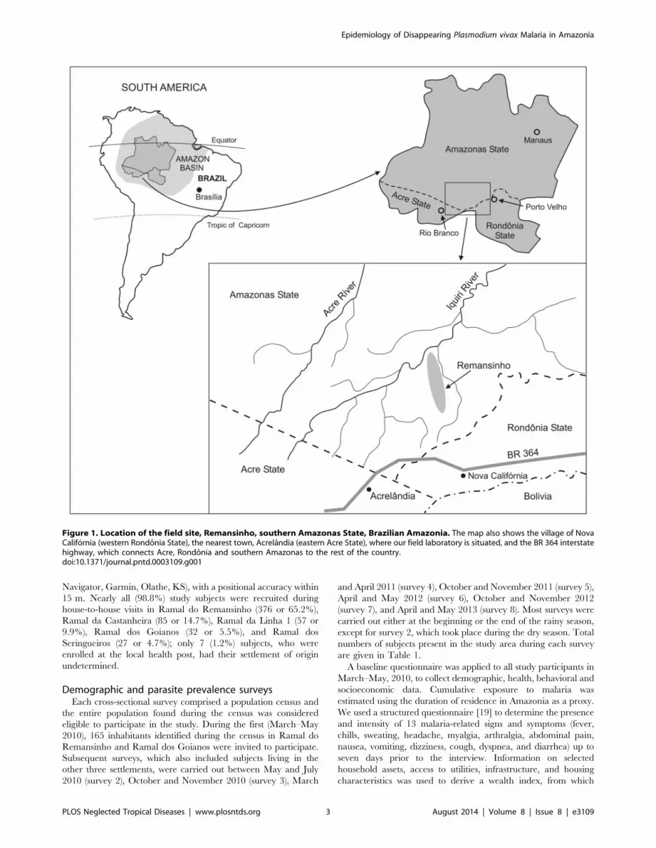
Study areaOnce a sparsely populated rubber tapper settlement (seringal)
situated in southern Amazonas state, northwestern Brazil,
Remansinho (average population, 260) now comprises five
farming settlements (Figure 1). The main settlement is situated
along the final 40 km of the Ramal do Remansinho, a 60 km-long
unpaved road originating from the BR-364 interstate highway,
while the other four are situated along secondary roads (known as
Ramal da Linha 1, Ramal da Castanheira, Ramal dos
Seringueiros, and Ramal dos Goianos) originating from this main
unpaved road (Figure S1). The farming settlements along Ramal
da Linha 1 and Ramal da Castanheira were opened in the late
1990s, whereas the colonization of the other areas started only in
2007. Most houses have complete or incomplete wooden walls and
thatched roofs; just a few of them have brick walls and are covered
with asbestos, cement or zinc shingles. With a typical equatorial
humid climate (annual average temperature, 26.4uC), Reman-
sinho receives most rainfall between November and March
(annual average, 2,318 mm), but malaria transmission occurs
year-round. The main local malaria vector is the highly
anthropophilic and exophilic An. darlingi [18]. Most families
currently living in Remansinho have resettled from other areas
within Amazonia, and are now involved in subsistence agriculture
and logging. There is a single government-run health post in
Remansinho, which provides free malaria diagnosis and treat-
ment, but a small proportion of locally acquired infections are
diagnosed and treated in the nearest village (Nova California;
population, 2,600), situated along the BR-364 highway, about
60 km south of Remansinho (Figure 1). There is no electricity or
piped water supply in the area.
Study design and populationA population-based prospective cohort study was initiated in
March 2010 to estimate the prevalence and incidence of malaria
parasite carriage in Remansinho, by combining microscopy and
molecular diagnosis, and to characterize risk factors for malaria
infection and clinical disease in the local population. This ongoing
study comprises periodic cross-sectional malaria prevalence
surveys of the entire population, every four months between
March 2010 and March 2011 and every six months thereafter,
complemented with clinical and laboratory surveillance of febrile
illness episodes between the cross-sectional surveys.
Here we analyze data collected from March 2010 to May 2013.
During this period, we enrolled 584 participants belonging to 205
households. Dwellings were geo-localized using a hand-held 12-
chanel global positioning system (GPS) receiver (eTrex Personal
Author Summary
Despite decades of control efforts, malaria remains a majorpublic health concern in Brazil. A large proportion of the243,000 cases diagnosed per year originate from areas ofrecent colonization in the densely forested Amazon Basin.This population-based longitudinal study addresses theepidemiology of malaria during the early stages ofcolonization of frontier settlements in Remansinho area,rural Amazonia. We documented a major decline in theprevalence of P. vivax infection, from 23.8% to 3.0%,between March–April 2010 and April–May 2013. Up to73.1% of the P. vivax infections were missed by microscopyas malaria transmission declined and most (56.6%) of theseinfections caused no clinical signs or symptoms. Few(17.0%) asymptomatic P. vivax infections that were leftuntreated eventually progressed to clinical disease, be-coming detectable by routine malaria surveillance, over 6weeks of follow-up. Moreover, nearly all P. vivax infectionsthat were undetected by microscopy had gametocytes,the parasite’s blood stages responsible for malaria trans-mission to mosquito vectors, detected by molecularmethods. These findings indicate that apparently healthycarriers of low-density parasitemias, who are often missedby conventional microscopy, contribute significantly toongoing P. vivax transmission and may further complicateresidual malaria elimination in Remansinho and similarendemic settings.
Epidemiology of Disappearing Plasmodium vivax Malaria in Amazonia
PLOS Neglected Tropical Diseases | www.plosntds.org 2 August 2014 | Volume 8 | Issue 8 | e3109
Navigator, Garmin, Olathe, KS), with a positional accuracy within
15 m. Nearly all (98.8%) study subjects were recruited during
house-to-house visits in Ramal do Remansinho (376 or 65.2%),
Ramal da Castanheira (85 or 14.7%), Ramal da Linha 1 (57 or
9.9%), Ramal dos Goianos (32 or 5.5%), and Ramal dos
Seringueiros (27 or 4.7%); only 7 (1.2%) subjects, who were
enrolled at the local health post, had their settlement of origin
undetermined.
Demographic and parasite prevalence surveysEach cross-sectional survey comprised a population census and
the entire population found during the census was considered
eligible to participate in the study. During the first (March–May
2010), 165 inhabitants identified during the census in Ramal do
Remansinho and Ramal dos Goianos were invited to participate.
Subsequent surveys, which also included subjects living in the
other three settlements, were carried out between May and July
2010 (survey 2), October and November 2010 (survey 3), March
and April 2011 (survey 4), October and November 2011 (survey 5),
April and May 2012 (survey 6), October and November 2012
(survey 7), and April and May 2013 (survey 8). Most surveys were
carried out either at the beginning or the end of the rainy season,
except for survey 2, which took place during the dry season. Total
numbers of subjects present in the study area during each survey
are given in Table 1.
A baseline questionnaire was applied to all study participants in
March–May, 2010, to collect demographic, health, behavioral and
socioeconomic data. Cumulative exposure to malaria was
estimated using the duration of residence in Amazonia as a proxy.
We used a structured questionnaire [19] to determine the presence
and intensity of 13 malaria-related signs and symptoms (fever,
chills, sweating, headache, myalgia, arthralgia, abdominal pain,
nausea, vomiting, dizziness, cough, dyspnea, and diarrhea) up to
seven days prior to the interview. Information on selected
household assets, access to utilities, infrastructure, and housing
characteristics was used to derive a wealth index, from which
Figure 1. Location of the field site, Remansinho, southern Amazonas State, Brazilian Amazonia. The map also shows the village of NovaCalifornia (western Rondonia State), the nearest town, Acrelandia (eastern Acre State), where our field laboratory is situated, and the BR 364 interstatehighway, which connects Acre, Rondonia and southern Amazonas to the rest of the country.doi:10.1371/journal.pntd.0003109.g001
Epidemiology of Disappearing Plasmodium vivax Malaria in Amazonia
PLOS Neglected Tropical Diseases | www.plosntds.org 3 August 2014 | Volume 8 | Issue 8 | e3109

Ta
ble
1.
Nu
mb
er
of
mal
aria
lin
fect
ion
sd
iag
no
sed
by
con
ven
tio
nal
mic
rosc
op
y(C
M)
and
qu
anti
tati
vere
al-t
ime
PC
R(q
PC
R),
acco
rdin
gto
the
pre
sen
ceo
rab
sen
ceo
fm
alar
ia-
rela
ted
sym
pto
ms,
du
rin
g8
con
secu
tive
cro
ss-s
ect
ion
alsu
rve
ysin
the
po
pu
lati
on
of
Re
man
sin
ho
,B
razi
l(2
01
0–
13
).
Cro
ss-s
ect
ion
al
surv
ey
12
34
56
78
Sy
mp
tom
sS
pe
cie
sC
Mq
PC
RC
Mq
PC
RC
Mq
PC
RC
Mq
PC
RC
Mq
PC
RC
Mq
PC
RC
Mq
PC
RC
Mq
PC
R
Ye
sP
.fa
lcip
aru
m0
20
10
30
10
00
00
00
0
P.
viva
x1
02
05
11
35
12
91
21
21
31
1
Mix
ed
01
00
01
00
00
00
00
00
No
.te
ste
d6
36
25
45
24
54
23
43
14
03
82
22
16
56
54
64
3
No
P.
falc
ipa
rum
16
11
00
14
01
00
00
00
00
P.
viva
x1
01
61
01
78
14
21
04
10
03
11
04
Mix
ed
01
01
06
00
00
00
00
00
No
.te
ste
d9
68
91
45
13
51
66
15
61
82
16
81
64
15
91
56
15
61
34
13
41
29
12
6
To
tal
P.
falc
ipa
rum
18
11
10
17
02
00
00
00
00
P.
viva
x2
03
61
52
81
11
93
12
13
22
15
24
15
Mix
ed
02
01
07
00
00
00
00
00
No
.te
ste
d1
59
15
01
99
18
72
11
19
82
16
19
92
04
19
71
78
17
71
99
19
91
75
16
9
To
tal
po
pu
lati
on
(%te
ste
d)
16
5(9
6.3
)2
76
(72
.1)
27
0(7
8.1
)2
76
(78
.3)
29
0(7
0.3
)2
38
(74
.8)
25
7(7
7.4
)2
21
(79
.2)
Pre
vale
nce
%P
.fa
lcip
aru
m0
.65
.30
.55
.90
.08
.60
.01
.00
.00
.00
.00
.00
.00
.00
.00
.0
P.
viva
x1
2.6
24
7.5
15
.05
.29
.61
.46
.06
.41
1.2
0.6
2.8
1.0
2.0
0.6
3.0
Mix
ed
0.0
1.3
0.0
0.5
0.0
3.5
0.0
0.0
0.0
0.0
0.0
0.0
0.0
0.0
0.0
0.0
Dat
es
of
cro
ss-s
ect
ion
alsu
rve
ysw
ere
:su
rve
y1
1,M
arch
–M
ay,2
01
0;s
urv
ey
2,M
ay–
July
,20
10
;su
rve
y3
,Oct
ob
er–
No
vem
be
r,2
01
0;s
urv
ey
4,M
arch
–A
pri
l,2
01
1;s
urv
ey
5,O
cto
be
r–N
ove
mb
er,
20
11
;su
rve
y6
,Ap
ril–
May
,20
12
;su
rve
y7
,O
cto
be
r–N
ove
mb
er,
20
12
;su
rve
y8
,A
pri
l–M
ay,
20
13
.P
oly
eth
yle
ne
be
d-n
ets
tre
ate
dw
ith
2%
pe
rme
thri
n(O
lyse
tN
et)
we
red
istr
ibu
ted
toth
ee
nti
rest
ud
yp
op
ula
tio
nin
Au
gu
st,
20
12
.d
oi:1
0.1
37
1/j
ou
rnal
.pn
td.0
00
31
09
.t0
01
Epidemiology of Disappearing Plasmodium vivax Malaria in Amazonia
PLOS Neglected Tropical Diseases | www.plosntds.org 4 August 2014 | Volume 8 | Issue 8 | e3109

socioeconomic status was estimated. We combined discrete (i. e.,
yes or no) ownership information (for power generator, chainsaw,
radio, sofa set, shotgun, bicycle, car, motorcycle, and well) and
continuous data (i. e., total number of items, for beds, rooms and
bedrooms present in the household, and number of pigs, cattle,
chickens, ducks, and horses owned). Principal component analysis,
carried out using statistical software STATA 12.1, was used to
weight each variable [20]. The first principal component
explained 18% of the variability and gave the greatest weights to
ownership of beds, number of rooms, number of bedrooms, sofa
set, and chickens. Lowest weights were given to ownership of
horses, ducks or cattle. The scores were summed to give a wealth
index for each household. Wealth indices were then used to stratify
households into quartiles in increasing order (first quartile, 25%
poorest). A shorter version of the baseline questionnaire was used
in all subsequent cross-sectional surveys to update demographic
and clinical data.
All inhabitants in the study area aged more than 3 months were
invited to contribute either venous (5-ml) or finger-prick blood
samples for malaria diagnosis, irrespective of any clinical
symptoms, Duffy blood group genotyping, and other laboratory
assays, such as hemoglobin measurements and ABO and Rh blood
group typing. The participation rates ranged between 96.3% in
survey 1 (159 of 165 inhabitants) and 70.3% in survey 5 (204 of
290) (Table 1). Nearly all study participants provided venous
blood samples in all but one survey; the exception was survey 3,
during which finger-prick capillary blood was preferentially
collected from all participants for logistic reasons. Reasons for
not providing blood samples included temporary absence from the
study area, age below 3 months, inability to perform venous
puncture, and refusal to participate. Given the high mobility of the
study population, only 21 subjects (3.6% of the study population)
contributed blood samples in all cross-sectional surveys; 529
subjects (90.6%) participated in two or more surveys. All study
participants, either symptomatic or not, who provided either
venous or finger-prick blood samples during cross-sectional surveys
and had malaria diagnosis confirmed by onsite microscopy were
treated according to the malaria therapy guidelines published by
the Ministry of Health of Brazil in 2010 [21]. Briefly, P. vivaxinfections were treated with chloroquine (total dose, 25 mg of
base/kg over 3 days) and primaquine (0.5 mg of base/kg/day for
7 days), while P. falciparum infections were treated with a fixed-
dose combination of artemether (2–4 mg/kg/day) and lumefan-
trine (12–24 mg/kg/day) for 3 days. Infections that were missed
by onsite microscopy but later confirmed by polymerase chain
reaction (PCR) were left untreated because the results of molecular
diagnosis were not available at the time of the cross-sectional
surveys.
Malaria surveillance between cross-sectional surveysTo quantify clinical malaria episodes diagnosed between the
cross-sectional surveys, we examined all records of slide-confirmed
infections diagnosed between March 2010 and November 2013 at
the government-run health posts in Remansinho and in the
nearest village, Nova California. Local malaria control personnel
performed both active and passive detection of febrile cases during
the study period. Blood samples were collected and examined for
malaria parasites whenever febrile subjects visited the health posts
in Remansinho or Nova California or were found during monthly
house-to-house visits carried out by field health workers in
Remansinho. This strategy is assumed to detect virtually all
clinical malaria episodes in cohort subjects between the cross-
sectional surveys, since there are no other public or private
facilities providing laboratory diagnosis of malaria in the area.
Microscopic diagnosis is required to obtain antimalarial drugs in
Brazil, which are distributed free of charge by the Ministry of
Health and cannot be purchased in local drugstores.
Laboratory methodsLaboratory diagnosis of malaria was based on microscopic
examination of thick smears and PCR. A total of 1,541 thick blood
smears were stained with Giemsa in our field laboratory in
Acrelandia (120 km southwest of Remansinho). At least 100 fields
were examined for malaria parasites, under 10006magnification,
by two experienced microscopists, before a slide was declared
negative. We additionally used quantitative real-time PCR (qPCR)
that target the 18S rRNA genes [22] to detect and quantify P.vivax and P. falciparum blood stages in 1,476 clinical samples
(Methods S1). Because microscopy is poorly sensitive for detecting
circulating gametocytes [23], we used a quantitative reverse
transcriptase PCR (qRT-PCR) that targets pvs25 gene transcripts
[24,25] to detect and quantify mature gametocytes in 55
laboratory-confirmed P. vivax infections diagnosed during cross-
sectional surveys 4, 5, and 6 (Methods S1).
Since co-infection with multiple parasite clones has been
suggested to either increase or reduce the risk of clinical
falciparum malaria, we sought to determine whether the presence
of multiple-clone P. vivax infections was associated with malaria-
related morbidity. To this end, we amplified two highly
polymorphic single-copy markers, msp1F1 (a variable domain of
the merozoite surface protein-1 gene) [26] and MS16 (a P. vivax-
specific microsatellite DNA marker with degenerate trinucleotide
repeats) [27], using the nested PCR protocols of Koepfli and
colleagues [28]. DNA samples from 85 qPCR-confirmed P. vivaxinfections (all of them isolated from venous blood samples) were
tested for the presence of multiple clones; 47 were from
asymptomatic and 38 from symptomatic parasite carriers. PCR
products were analyzed by capillary electrophoresis on an
automated DNA sequencer ABI 3500 (Applied Biosystems), and
their lengths (in bp) and relative abundance (peak heights in
electropherograms) were determined using the commercially
available GeneMapper 4.1 (Applied Biosystems) software. The
minimal detectable peak height was set to 200 arbitrary
fluorescence units. We scored two alleles at a locus when the
minor peak was .33% the height of the predominant peak.
Plasmodium vivax infections were considered to contain multiple
clones if one or both loci showed more than one allele.
Since Duffy blood group polymorphisms modulate the ability of
P. vivax merozoites to invade human red blood cells (reviewed by
[29]), we used TaqMan assays (Applied Biosystems) to genotype
two major Duffy polymorphisms: the T-33C substitution in the red
blood cell-specific GATA1 transcription factor binding motif
(rs2814778), which suppresses Duffy expression on the erythrocyte
surface (Fy phenotype, associated with FY*BES allele homozygoz-
ity), and the G125A polymorphism (rs12075), which defines the
FY*B (wild-type) and FY*A (mutated) alleles associated with the
Fyb and Fya phenotypes, respectively. The primers and probes
(labelled with VIC and FAM) were designed and synthesized by
Applied Biosystems (assay ID, C__15769614_10 and
C__2493442_10) [30]. We used a Step One Plus Real-Time
PCR System (Applied Biosystems) for genotyping, with a template
denaturation step at 95uC for 10 min, followed by 40 cycles of
15 sec at 95uC and 1 min at 60uC, with a final step at 60uC for
30 sec. DNA samples from 487 study participants were genotyped.
Clinical definitionsA laboratory-confirmed malarial infection was defined as any
episode of parasitemia detected by thick-smear microscopy,
Epidemiology of Disappearing Plasmodium vivax Malaria in Amazonia
PLOS Neglected Tropical Diseases | www.plosntds.org 5 August 2014 | Volume 8 | Issue 8 | e3109

qPCR, or both. Subpatent or submicroscopic infections were
defined as infections confirmed by qPCR but missed by
microscopy. We defined clinical malaria as a laboratory-confirmed
infection, irrespective of the parasite density, diagnosed in a
subject reporting one or more of the 13 signs and symptoms
investigated, at or up to seven days before the interview. No
attempt was made to calculate pyrogenic thresholds of parasit-
emias in our heterogeneous group of study participants. Subjects
with laboratory-confirmed infection, irrespective of the parasite
density, who reported no signs or symptoms at or up to seven days
prior to the interview, were classified as asymptomatic carriers of
malaria parasites.
Statistical analysisA database was created with SPSS 17.0 software (SPSS Inc.,
Chicago, IL) and all subsequent analyses were performed with R
statistical software [31]. For the purposes of explanatory data
analysis, proportions were compared using standard x2, Mantel-
Haenzel x2 for stratified data, or x2 tests for linear trends.
Correlations between parasite densities, which had an over-
dispersed distribution in the population, and other continuous
variables were evaluated using the nonparametric Spearman
correlation. Median parasitemias were compared with the
nonparametric Mann-Whitney U test. Statistical significance was
defined at the 5% level and 95% confidence intervals (CI) were
estimated whenever appropriate.
Separate regression models were built to describe independent
associations between potential risk factors and two outcomes: (a) P.vivax infection and (b) clinical (i. e., symptomatic) vivax malaria.
Due to the small number of P. falciparum infections detected in
the community no attempt was made to characterize risk factors
for infection with this species. Dependent variables were assumed
to follow a binomial distribution with a logit link function, being
fitted with a logistic regression. We considered the nested structure
of the data, intrinsic to the study design, when building regression
models; we have repeated observations (up to 8 observations over
3 years of study; grouping variable, ‘‘survey’’) nested within
subjects (grouping variable, ‘‘individual’’) who are clustered within
households (grouping variable, ‘‘household’’). This clustered
sampling scheme introduces dependency among observations that
can affect model parameter estimates. Consequently, we used
mixed-effects regression models that include the grouping
variables as random factors to handle nested observations.
Our modeling strategy further considered the hierarchical levels
of independent variables (Methods S1). The effects of distal
determinants, such as demographic, social and environmental
factors, on malaria risk are often not direct, but mediated by more
proximate determinants, such as occupational and behavioral
factors [32]. Variables within each level of determination were
introduced in the model in a stepwise approach, and only those
that were associated with the outcome at a significance level of at
least 20% were retained. Most subjects with missing observations
were excluded from the final model, except those with missing
values for the following four variables: Duffy genotype, wealth
index, whether bedroom windows were left open at night, and
main occupation. These were maintained in the model by creating
a new missing-value category. All models were adjusted for the
timing of the survey (months elapsed since the beginning of the
study in March 2010). Three variables in the model were time-
dependent: age, years of residence in Amazonia, and timing of the
survey. The final models comprised 1,242 observations from 442
individuals grouped into 159 households (outcome: P. vivaxinfection), and 1,237 observations from 438 individuals grouped
into 158 households (outcome: vivax malaria).
Alternative logistic models, which excluded Duffy-negative
subjects (88 observations from 31 subjects), examined the
association between Duffy-positive genotypes (FY*AFY*BES,
FY*AFY*A, FY*AFY*B, FY*BFY*BE, and FY*BFY*B) and
risk of P. vivax infection and vivax malaria. To account for the
hypothesis that age at the beginning of exposure to malaria affects
the rate at which antimalarial immunity is acquired by migrants
[33], we further tested for an interaction between age and years of
residence in Amazonia. In addition, we fitted mixed-effects
Poisson regression models to the data, but the random-effects
variances associated with the estimates were substantially higher
than those obtained with the logistic models described above. As a
consequence, here we only present results derived from the logistic
regression analysis.
In addition, we used a mixed-effects Cox proportional hazards
model [34] to compare the risk of slide-positive vivax malaria
between the surveys in two sub-cohorts of asymptomatic subjects:
(a) carriers of subpatent P. vivax infections at baseline that were
left untreated and (b) control subjects who were parasite-negative
at baseline by both microscopy and qPCR. Subjects who were
symptomatic but parasite-negative at baseline were excluded from
the uninfected sub-cohort because they might harbor ongoing low-
grade infections, causing malaria-related symptoms, which were
missed by our laboratory methods. At each survey, eligible study
participants were assigned to either sub-cohort and followed up
until the next survey at which their clinical and infection status was
reassessed. Time at risk was defined as either the interval between
two consecutive surveys in which the subjects participated (the first
survey in the pair was defined as the baseline survey) or the
interval between the baseline survey and the date when subjects
left the study, whatever came first. Analysis was adjusted for
subjects’ age (stratified as ,15 years and $15 years), Duffy blood
group negativity, and years of residence in Amazonia. The
clustering of repeated observations within individuals was modeled
as a random effect [34].
As required for all observational studies published by PLoSNeglected Tropical Diseases, this article includes the STROBE
(STrengthening the Reporting of OBservational studies in
Epidemiology) checklist to document its compliance with
STROBE guidelines (Checklist S1).
Ethics statementStudy protocols were approved in early 2010 by the Institutional
Review Board of the University Hospital of the University of Sao
Paulo (1025/10) and by the National Human Research Ethics
Committee of the Ministry of Health of Brazil (551/2010). The
ethical clearance has been renewed annually by the Institutional
Review Board of the University Hospital of the University of Sao
Paulo. Written informed consent was obtained from all study
participants or their parents/guardians.
Results
Subject characteristics and prevalence of malariainfection
Of 584 people living in Remansinho who participated in at least
one cross-sectional survey, 333 (57.0%) were male and 251
(43.0%) were female, with a median age of 23.0 years. Nearly all
(94.3%) adult subjects aged more than 18 years were migrants,
42.2% of them originating from malaria-free areas outside
Amazonia. Only 31 subjects (6.4%) were homozygous FY*BES
carriers, with the P. vivax-refractory Duffy-negative (Fy) pheno-
type; 127 (26.1%) had the Fya phenotype (70 FY*AFY*BES
heterozygotes and 57 FY*A FY*A homozygotes), 142 (29.2%) had
Epidemiology of Disappearing Plasmodium vivax Malaria in Amazonia
PLOS Neglected Tropical Diseases | www.plosntds.org 6 August 2014 | Volume 8 | Issue 8 | e3109

the FyaFyb phenotype (FY*A FY*B heterozygotes), and 187
(38.4%) had the Fyb phenotype (91 FY*BFY*BES heterozygotes
and 96 FY*B FY*B homozygotes).
Polyethylene bed-nets treated with 2% permethrin (Olyset Net,
Sumitomo Chemical, London, United Kingdom) were distributed,
free of charge, to the entire study population in August 2012, as a
component of malaria control activities in Brazilian Amazonia. In
October–November 2012 (survey 7), 74.4% of the study partic-
ipants reported having slept the previous night under an Olyset
net; the corresponding figure for April–May 2013 (survey 8) was
84.5%. No other insecticide-treated bed nets were available in the
community.
A total of 1,541 blood samples were examined for malaria
parasites by microscopy, qPCR, or both. Of these, 141 (9.1%)
were positive (by one or both methods) for P. vivax, 40 (2.6%) for
P. falciparum and 10 (0.6%) for both species. Over the entire
study period, 191 (12.4%) samples examined tested positive for
malaria parasites; 10 P. vivax and 2 P. falciparum infections were
only diagnosed by microscopy, since DNA samples were not
available for qPCR or qPCR yielded negative results. In addition,
61.8% of all infections diagnosed by qPCR, regardless of the
infecting species, and 49.6% of the qPCR-confirmed single-species
P. vivax infections, were missed by conventional microscopy and
thus defined as subpatent. The last P. falciparum infections in
Remansinho were diagnosed (by qPCR only) in March–April
2011.
These figures, however, changed over time. The numbers of
malaria infections, either symptomatic or not, diagnosed by
conventional microscopy and qPCR in each cross-sectional survey
are shown in Table 1. The proportions of qPCR-confirmed single-
species P. vivax infections that were subpatent varied widely
across surveys, ranging from 73.1% in the surveys with the lowest
P. vivax prevalence rates (surveys 4, 6, 7, and 8 combined; 26
qPCR-confirmed infections) to 43.8% in those with the highest
prevalence rates (surveys 1, 2, 3, and 5 combined; 105 qPCR-
confirmed infections; Yates’ corrected x2 = 6.02, 1 degree of
freedom [df], P = 0.014). The numbers of P. falciparum and
mixed-species infections were too small for a similar comparison.
Microscopy thus had a better diagnostic performance for vivax
malaria when overall parasite prevalence rates were higher,
consistent with a recent meta-analysis of P. falciparum data
showing lower proportions of submicroscopic infections in areas
with greater malaria transmission [35].
Malaria-related symptoms, parasite density, andgametocytes
Overall, 17.1% of the study subjects (ranging between 12.3% in
survey 6 and 39.6% in survey 1) interviewed during the cross-
sectional surveys reported one or more malaria-related signs and
symptoms up to seven days prior to the interview (Table 1).
However, reported clinical signs and symptoms were neither
sensitive nor specific for malaria diagnosis. On the one hand,
almost two thirds (64.5%) of all qPCR-confirmed malaria
infections by any species, and 56.6% of those due to P. vivax,
were asymptomatic; on the other hand, only 26.7% of subjects
reporting symptoms had a malaria infection (by any species)
confirmed by microscopy, qPCR, or both. All carriers of mixed-
species infections (all of them confirmed by qPCR but missed by
microscopy) were asymptomatic (Table 1).
Most P. vivax-infected subjects harbored few parasites, with
densities estimated by qPCR on 129 samples ranging between 2.1
and 38,390 parasites/mL (median, 49.1 parasites/mL; interquartile
range, 10.0–483.1 parasites/mL; data were missing for 2 qPCR-
confirmed infections). We found no evidence for decreasing P.
vivax densities with increasing cumulative exposure to malaria in
this population. In fact, individual P. vivax parasitemias did not
show a negative correlation with the subjects’ length of residence
in Amazonia, a proxy of cumulative exposure to malaria
(Spearman correlation coefficient rs = 20.046, P = 0.600), or with
their age (rs = 20.068, P = 0.427). We next tested whether
differences in the diagnostic sensitivity of conventional microscopy
across cross-sectional surveys might be explained by higher
average parasite densities found at times of increased malaria
transmission [35]. Parasitemias appeared slightly higher in qPCR-
positive samples (P. vivax only) obtained during surveys 1,2,3 and
5 (high prevalence), with a median of 55.7 parasites/mL
(interquartile range, 10.4–597.6 parasites/mL; n = 103), than in
those obtained during surveys 4,6,7, and 8 (low prevalence), with a
median of 19.8 parasites/mL (interquartile range, 5.8–65.4
parasites/mL; n = 26), although the difference did not reach
statistical significance (Mann-Whitney U test, P = 0.057).
The proportion of symptomatic P. vivax infections correlated
positively with increasing parasite density (x2 for trend = 7.99, 1 df,
P,0.005). Only 30.6% of the subjects carrying less than 10
parasites/mL, but 73.9% of those carrying more than 1,000
parasites/mL, reported one or more malaria-related symptoms
(Figure 2). Consistent with previous observations from Amazonia
[36,37], more than half (53.9%) of the asymptomatic infections
with this species confirmed by qPCR were missed by conventional
microscopy (Table 1). Overall, 32.8% of the 131 single-species,
qPCR-confirmed P. vivax infections for which complete data were
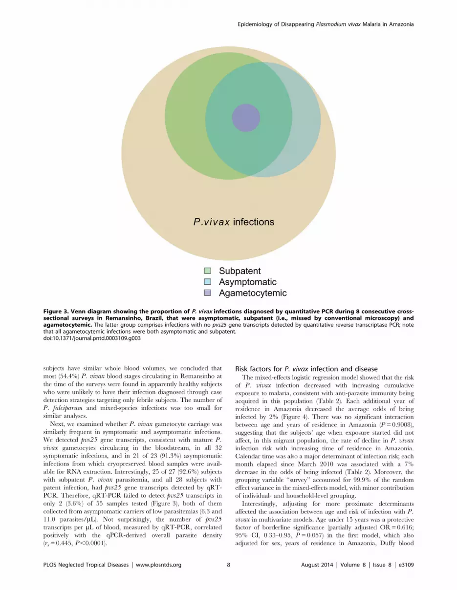
available were both subpatent and asymptomatic (Figure 3). Only
one P. vivax infection was diagnosed by qPCR, but missed by
conventional microscopy, among 88 samples collected from Duffy-
negative study participants during the 8 cross-sectional surveys.
The only reported symptom during this subpatent P. vivaxinfection in a Duffy-negative subject was a chronic myalgia;
parasite density was very low (9.9 parasites/mL of blood).
To estimate the relative contribution of asymptomatic parasite
carriage to the total P. vivax biomass in the host population, we
summed up all individual qPCR-derived P. vivax densities and
calculated the fraction corresponding to asymptomatic infections.
Assuming that, on average, asymptomatic and symptomatic
Figure 2. Proportion of P.vivax infections diagnosed during 8consecutive cross-sectional surveys in Remansinho, Brazil, thatwere symptomatic (black bar segments) and asymptomatic(white bar segments) according to parasite density estimatedby quantitative PCR. The bar widths are proportional to the numberof cases within each parasite density class. A total of 129 P. vivaxinfections were classified according to the presence of symptoms andparasite density.doi:10.1371/journal.pntd.0003109.g002
Epidemiology of Disappearing Plasmodium vivax Malaria in Amazonia
PLOS Neglected Tropical Diseases | www.plosntds.org 7 August 2014 | Volume 8 | Issue 8 | e3109
subjects have similar whole blood volumes, we concluded that
most (54.4%) P. vivax blood stages circulating in Remansinho at
the time of the surveys were found in apparently healthy subjects
who were unlikely to have their infection diagnosed through case
detection strategies targeting only febrile subjects. The number of
P. falciparum and mixed-species infections was too small for
similar analyses.
Next, we examined whether P. vivax gametocyte carriage was
similarly frequent in symptomatic and asymptomatic infections.
We detected pvs25 gene transcripts, consistent with mature P.vivax gametocytes circulating in the bloodstream, in all 32
symptomatic infections, and in 21 of 23 (91.3%) asymptomatic
infections from which cryopreserved blood samples were avail-
able for RNA extraction. Interestingly, 25 of 27 (92.6%) subjects
with subpatent P. vivax parasitemia, and all 28 subjects with
patent infection, had pvs25 gene transcripts detected by qRT-
PCR. Therefore, qRT-PCR failed to detect pvs25 transcripts in
only 2 (3.6%) of 55 samples tested (Figure 3), both of them
collected from asymptomatic carriers of low parasitemias (6.3 and
11.0 parasites/mL). Not surprisingly, the number of pvs25transcripts per mL of blood, measured by qRT-PCR, correlated
positively with the qPCR-derived overall parasite density
(rs = 0.445, P,0.0001).
Risk factors for P. vivax infection and diseaseThe mixed-effects logistic regression model showed that the risk
of P. vivax infection decreased with increasing cumulative
exposure to malaria, consistent with anti-parasite immunity being
acquired in this population (Table 2). Each additional year of
residence in Amazonia decreased the average odds of being
infected by 2% (Figure 4). There was no significant interaction
between age and years of residence in Amazonia (P = 0.9008),
suggesting that the subjects’ age when exposure started did not
affect, in this migrant population, the rate of decline in P. vivaxinfection risk with increasing time of residence in Amazonia.
Calendar time was also a major determinant of infection risk; each
month elapsed since March 2010 was associated with a 7%
decrease in the odds of being infected (Table 2). Moreover, the
grouping variable ‘‘survey’’ accounted for 99.9% of the random
effect variance in the mixed-effects model, with minor contribution
of individual- and household-level grouping.
Interestingly, adjusting for more proximate determinants
affected the association between age and risk of infection with P.vivax in multivariate models. Age under 15 years was a protective
factor of borderline significance (partially adjusted OR = 0.616;
95% CI, 0.33–0.95, P = 0.057) in the first model, which also
adjusted for sex, years of residence in Amazonia, Duffy blood
Figure 3. Venn diagram showing the proportion of P. vivax infections diagnosed by quantitative PCR during 8 consecutive cross-sectional surveys in Remansinho, Brazil, that were asymptomatic, subpatent (i.e., missed by conventional microscopy) andagametocytemic. The latter group comprises infections with no pvs25 gene transcripts detected by quantitative reverse transcriptase PCR; notethat all agametocytemic infections were both asymptomatic and subpatent.doi:10.1371/journal.pntd.0003109.g003
Epidemiology of Disappearing Plasmodium vivax Malaria in Amazonia
PLOS Neglected Tropical Diseases | www.plosntds.org 8 August 2014 | Volume 8 | Issue 8 | e3109

group genotype, months elapsed since the beginning of the study,
and wealth index quartiles. However, after adjusting for main
occupation, the effect of age on infection risk became non-
significant (fully adjusted OR = 1.169; 95% CI, 0.63–2.19,
P = 0.624). These results indicate that young age per se is not
protective, but young subjects are less likely to engage in activities
such as logging and fishing in the fringes of the rain forest, which
are potentially associated with increased risk of infection (Table 2).
Not surprisingly, Duffy-negativity emerged as a protective factor
against P. vivax infection in this community (Table 2). However,
additional logistic regression models including only Duffy-positive
subjects showed that, compared to FY*A FY*B heterozygotes,
neither FY*A FY*BES heterozygotes (OR = 0.864; 95% CI, 0.43–
1.73) and FY*A FY*A homozygotes (OR = 0.921; 95% CI, 0.48–
1.75) were protected against P. vivax infection, nor FY*B FY*BES
heterozygotes (OR = 1.226; 95%, 0.69–2.17) and FY*B FY*Bhomozygotes (OR = 0.588; 95% CI, 0.32–1.09) were at increased
risk of infection. These results are consistent with a protective role
of FY*BES heterozygosity, but not of FY*A allele carriage, against
P. vivax infection in this population.
The risk of clinical P. vivax malaria decreased with increasing
cumulative exposure to malaria (Table 2); each additional year of
residence in Amazonia decreased the odds of having vivax malaria
by 3%, again with no significant interaction between age and
length of residence in Amazonia (P = 0.863). These findings are
consistent with similar exposure-dependent rates of acquisition of
anti-parasite and anti-disease immunity in this community.
Calendar time was the only other major determinant of malaria
risk; each month elapsed since the beginning of the study was
associated with an 8% decrease in the odds of having clinical vivax
malaria (Table 2). Due to the small sample size, Duffy-negativity
emerged as a protective factor of borderline significance
(OR = 0.16, 95% CI, 0.02–1.29, P = 0.084) against clinical vivax
malaria.
Asymptomatic P. vivax carriage and subsequent risk ofclinical malaria
In Brazil, malaria is only treated if blood smear microscopy is
positive; subpatent malaria parasitemia as determined with qPCR
is not accepted as the basis for treatment. Of 53 asymptomatic
subpatent P. vivax infections diagnosed at baseline, 9 (17.0%)
progressed to clinical malaria over the following 6 weeks, being
diagnosed by onsite microscopy and treated (Figure 5). During this
6-week period, only 2.5% of the subjects in the uninfected cohort
experienced an episode of slide-confirmed vivax malaria, but at
the end of the follow-up period similar proportions of subjects in
each sub-cohort had experienced vivax malaria episodes con-
firmed by microscopy (Figure 5). A Cox proportional hazards
model revealed no significant difference, between the two sub-
cohorts, in overall risk of vivax malaria episodes, after controlling
Figure 4. Correlation between length of residence in Amazonia(in years), a proxy of cumulative exposure to malaria, and theprobability of having a P. vivax infection (continuous red line)and a clinical vivax malaria episode (continuous black line).Lines represent median individual probabilities derived from the final(fully adjusted) mixed-effects logistic regression models; the shadedarea surrounding the lines represent interquartile ranges.doi:10.1371/journal.pntd.0003109.g004
Table 2. Factors associated with Plasmodium vivax infection and clinical vivax malaria, as revealed by multiple logistic regressionanalysis, in Remansinho, Brazil (2010–13).
Plasmodium vivax infection Clinical vivax malaria
ORa (95% CIb) P value ORa (95% CIb) P value
Duffy blood group phenotype
Positive 1 (reference) 1 (reference)
Negative 0.08 (0.01–0.60) 0.0142 0.16 (0.02–1.29) 0.0849
Missing information 0.80 (0.32–2.00) 0.6362 0.41 (0.05–3.32) 0.4015
Length of residence in Amazonia (years) 0.98 (0.97–1.00) 0.0238 0.97 (0.94–0.99) 0.0082
Time since beginning of study (months) 0.93 (0.90–0.96) ,0.0001 0.92 (0.87–0.98) 0.0062
Main Occupation
Housekeeping 1 (reference) 1 (reference)
Agriculture and forestry 0.83 (0.52–1.32) 0.4325 0.97 (0.48–2.00) 0.9473
Students and minors 0.39 (0.20–0.76) 0.0058 0.26 (0.09–0.74) 0.0125
aOR = adjusted odds ratio.bCI = confidence interval.doi:10.1371/journal.pntd.0003109.t002
Epidemiology of Disappearing Plasmodium vivax Malaria in Amazonia
PLOS Neglected Tropical Diseases | www.plosntds.org 9 August 2014 | Volume 8 | Issue 8 | e3109
for potential confounders (hazard ratio = 1.07; 95% CI, 0.52–
2.22, P = 0.840). Most subpatent asymptomatic infections
cleared spontaneously (or, at least, became undetectable by
qPCR), since only 5 of 44 (11.4%) carriers who remained
untreated were again P. vivax-positive in the next survey.
Therefore, few asymptomatic and subpatent P. vivax infec-
tions eventually became patent and symptomatic (and there-
fore detectable by routine malaria surveillance) over the
following weeks. We conclude that untreated, low-density,
and asymptomatic P. vivax parasitemias may persist for
several weeks without progressing to clinical disease, and thus
constitute a major infectious reservoir for continued transmis-
sion in the community.
Multiple-clone P. vivax infections and malaria-relatedillness
By typing two highly polymorphic markers, we found more than
one genetically distinct clone in 25 of 85 (29.4%) P. vivaxinfections analyzed. Although multiple-clone infections were more
frequent in symptomatic (13 of 38, 34.1%) than asymptomatic (12
of 47, 25.5%) carriers, this difference did not reach statistical
significance (Yates’ corrected x2 = 0.762, 1 df, P = 0.382). Because
average P. vivax densities were lower in asymptomatic infections
and detecting minority clones may be more difficult in samples
with low-level parasitemias, we re-analyzed the data after
stratifying parasite densities into quartiles. Again, stratified analysis
yielded negative results (Mantel-Haenzel x2 = 0.004, 1 df,
P = 0.991). Therefore there was no observable association between
multiplicity of P. vivax infection and the presence of symptoms in
this community.
Discussion
This longitudinal study in newly opened frontier settlements
provides further evidence that carriers of low-density parasitemias,
who are often missed by conventional microscopy, contribute
significantly to ongoing P. vivax transmission in rural Amazonia.
Results from this and other studies in Amazonia [12–14,36,37]
challenge the often persisting view that subjects in low malaria
transmission settings are unlikely to harbor low parasitemias, due
to the lack of acquired immunity. To the contrary, average
parasite densities decreased, with higher proportions of P. vivaxinfections being missed by microscopy, as malaria prevalence
decreased in the community. Interestingly, our findings for P.vivax are consistent with a recent meta-analysis of 106 P.falciparum prevalence studies worldwide that combined micros-
copy and molecular methods [35]. Because the risk of P. vivaxinfection (confirmed by microscopy, qPCR, or both) correlated
negatively with cumulative exposure to malaria, we suggest that
our study population has developed over time some degree of anti-
parasite immunity, in line with recent findings from traditional
riverine communities in Amazonia [13,37]. Finally, we show that
nearly all subpatent blood-stage P. vivax infections comprise
mature gametocytes detected by a highly sensitive molecular
technique [24]. We thus conclude that subpatent infections
constitute a major P. vivax reservoir in rural Amazonia and
possibly in other low-transmission settings.
Our findings also challenge classical views regarding asymp-
tomatic infections in low-endemicity populations. Prior to the
molecular diagnosis era, nearly all laboratory-confirmed episodes
of malarial infection, even those with low parasite densities, were
thought to elicit clinical disease in pioneer settlements across the
Amazon Basin [38–40]. More recent surveys, however, demon-
strated that subclinical infections are common in agricultural
settlements [12,14] and traditional riverine communities
[13,36,37,41], but most of them are missed by microscopy.
Interestingly, the high proportion of infections found to be
asymptomatic in the present study must be interpreted as a
conservative estimate. We may have misclassified some episodes of
parasite carriage in subjects reporting any of the 13 symptoms
investigated, which may or may not be caused by the current
infection, as symptomatic malaria infections, overestimating the
proportion of symptomatic infections. Not surprisingly, however,
we found very low P. vivax densities in most subclinical infections
in Remansinho. Conventional microscopy missed 54% of them,
suggesting that previous microscopy-based studies failed to detect
asymptomatic parasite carriage in rural Amazonians because they
missed a large proportion of low-density infections. Mathematical
models identified asymptomatic infections as a crucial target for P.falciparum malaria eradication efforts in Africa [42], but no
similar analyses are available for other endemic areas and other
human malaria parasite species [43]. The following findings argue
for a major role of asymptomatic infections in maintaining
ongoing P. vivax transmission in Remansinho: (a) apparently
healthy subjects accounted for half of the total P. vivax biomass
found in the local population, (b) nearly all asymptomatic
infections comprised mature gametocytes, and (c) few untreated
asymptomatic infections became symptomatic (and thus detectable
by routine surveillance) over the next few weeks of follow-up. We
were unable to measure the average duration of untreated,
asymptomatic infections in our population; there is a recent
estimate of 194 days of duration for untreated P. falciparuminfections in Ghana [44], but no comparable estimate is currently
available for P. vivax. Specific studies to quantify the transmis-
sibility of subpatent parasitemia to vector mosquitos via direct and
Figure 5. Kaplan-Meier estimates of the proportion of P. vivax-infected (continuous black line) and uninfected (continuousred line) asymptomatic subjects who remained free of slide-confirmed clinical vivax malaria over the follow-up period.Dashed lines represent the respective 95% confidence intervals. Thesmall vertical tick-marks indicate the occurrence of a slide positive caseof P.vivax, corresponding to the right censoring of the individualsurvival time. A Cox proportional hazards model revealed no significantdifference, between the two groups, in overall risk of vivax malariaepisodes, after controlling for potential confounders (hazard ra-tio = 1.07; 95% CI, 0.52–2.22, P = 0.840).doi:10.1371/journal.pntd.0003109.g005
Epidemiology of Disappearing Plasmodium vivax Malaria in Amazonia
PLOS Neglected Tropical Diseases | www.plosntds.org 10 August 2014 | Volume 8 | Issue 8 | e3109

membrane feeding assays are ongoing (JMV and colleagues,
unpublished data).
Who are at risk of malaria in Remansinho? Migrants from
malaria-free areas (54.5% of the adults in the community)
constitute a major risk group, with each year of residence in
Amazonia decreasing their risk of P. vivax infection and clinical
vivax malaria by 2–3%. In some Amazonian communities,
malaria has been associated with forest-related activities such as
logging, fishing and mining, which typically involve young male
adults [12,39,45,46]. However, we show that housekeeping and
forest-related activities were associated with similar risks for
infection and disease in Remansinho. We hypothesize that nearly
all adolescents and adults of both genders engage to some extent in
farming activities, especially harvesting, in the forest fringes close
to their dwellings, although only young males are often involved in
logging and land clearing in more densely forested areas. We are
currently using high-resolution satellite images to measure the
distance between dwellings and forest fringes to further explore the
association between proximity to the forest environment and risk
of malaria in Remansinho. Interestingly, malaria transmission
appears to be relatively homogeneous across all settlements in the
area, equally affecting the poorest and least poor people of both
sexes, with no differences in risk according to main house
characteristics. Whether the vectorial capacity of An. darlingi is
spatially homogeneous is a key question to be answered by
ongoing vector biology studies in this site.
Detection of gametocytes, through pvs25 gene transcripts, in
nearly all qPCR-confirmed P. vivax infections tested is somewhat
surprising, since recent studies have found much lower proportions
of gametocyte-positive infections in Southeast Asia [47,48] and
Papua New Guinea [49]. Since gametocytes comprise only 2% of
circulating blood stages [50], microscopists are likely to miss
gametocytes in population-based studies where low-density infec-
tions are often sampled [23]. Furthermore, we argue that even
molecular methods may be poorly sensitive if suboptimal
techniques for sample storage and RNA extraction are used
under field conditions. For RNA isolation, we cryopreserved
venous blood samples at 270uC or in liquid nitrogen a few hours
after collection, since our previous attempts to amplify pvs25transcripts from RNA isolated from classic FTA microcards
(Wathman), QIAcards (Qiagen), 903 protein saver cards (What-
man), and 3MM filter papers (Whatman) impregnated with P.vivax-infected blood and kept at ambient temperature had all
failed [24]. Storing filter papers impregnated with blood in TRizol
reagent (Qiagen) may improve RNA yield, but almost two thirds of
the bloodspots from PCR-confirmed P. vivax infections tested by
Wampfler and colleagues [49] were negative for pvs25 transcripts
by TaqMan assays, despite previous TRizol reagent treatment.
Long-term asymptomatic carriage of P. falciparum has been
suggested to protect against subsequent malaria-related disease in
Africa [51,52], possibly by reducing the risk of superinfection with
more virulent strains. An explanation for this finding is premu-
nition, originally defined by Sergent and Parrot (1935) as the
protection against new infections resulting from immune responses
to the existing infection [53]. Alternatively, ongoing blood-stage
infection might arrest the development of subsequently inoculated
sporozoites in the liver. Such an inhibition of superinfection
appears to be mediated by the iron regulatory hormone hepcidin,
produced in response to blood-stage parasitemia [54]. However,
an opposite effect (i.e., increased risk of subsequent disease in
asymptomatic P. falciparum carriers) has also been described,
suggesting that a proportion of asymptomatic infections will
eventually reach the host’s pyrogenic threshold [55]. Here we
found no significant association between asymptomatic carriage of
low-density P. vivax infection and protection from subsequent
malaria morbidity, suggesting that treating individuals with
asymptomatic P. vivax infections would not render them more
vulnerable to clinical malaria over the next few weeks or months.
Although we have identified challenges for malaria control that
are not currently addressed by routine surveillance, malaria
transmission in Remansinho has declined dramatically over 3
years of surveillance, and P. falciparum was found only during the
first four surveys. Factors that may have contributed to this decline
include drastic environmental changes resulting from logging and
land clearing for farming, variation in climate, the widespread use
of insecticide-treated bed-nets since August 2012, and the
implementation of research activities in the area.
To address the first two hypotheses, we are now analyzing high-
resolution satellite images to track environmental changes over
time. Consistent with the third hypothesis, two studies have
provided evidence that insecticide-treated bed-nets are effective for
malaria control in Amazonia. The first was a case-control study in
Colombia that showed more than 50% reduction in malaria,
relative to no net use, although the advantage of impregnated over
non-impregnated nets was not statistically significant [56]. The
second study, a randomized trial of lambdacyhalothrin- versus
placebo-treated nets in the Amazonas State of Venezuela, showed
a protective efficacy of 55% [57]. Whether insecticide-tread bed-
nets alone can reduce malaria incidence rates throughout the
Amazon Basin remains uncertain, mostly due to the highly
variable biting behavior of An. darlingi across the region [58],
with strong evidence of significant blood-fed and exophilic host-
seeking behavior [59–61]. In addition, the decline in transmission
in Remansinho preceded the distribution of bednets. Finally, the
presence of a research team continuously working in the area for
over 3 years may affect positively both diagnostic and treatment
practices. The external slide revision routinely carried out by our
team provides an example of intervention that may have enhanced
the diagnostic skills of local microscopists. Moreover, active case
detection during 8 consecutive surveys allowed for the early
diagnosis and prompt treatment of several slide-positive asymp-
tomatic infections that would have been missed by routine passive
surveillance.
Eliminating residual foci when malaria is nearly disappearing,
but remains entrenched in a few hotspots, is the next major goal in
Remansinho and many other similar endemic settings. Case
detection strategies in areas approaching malaria elimination often
target only subjects presenting with fever or with a history of
recent fever, who are screened for malaria parasites by conven-
tional microscopy or rapid diagnostic tests (RDT) and receive
prompt antimalarial treatment if found to be infected [62]. These
strategies overlook asymptomatic infections that might be detected
by periodic cross-sectional surveys of the entire population at risk
[63], as we did in Remansinho. Nevertheless, the cost-effectiveness
of mass blood surveys for detecting and treating these residual
infections decreases proportionally as malaria transmission
declines, since: (a) large populations must be screened to diagnose
relatively few asymptomatic carriers, and (b) diagnostic techniques
available for large-scale use, such as microscopy and RDT, are not
sensitive enough to detect low-grade infections that are typical of
residual malaria settings [64]. As an alternative, we are currently
testing a reactive case detection strategy that has been tailored for
the relapsing parasite P. vivax to detect new infections in the
neighborhood of malaria cases diagnosed by routine surveillance
in frontier settlements similar to Remansinho. Evaluating this and
other strategies of active surveillance to cope with asymptomatic
infections in residual P. vivax foci is a top research priority in the
context of current malaria elimination efforts worldwide.
Epidemiology of Disappearing Plasmodium vivax Malaria in Amazonia
PLOS Neglected Tropical Diseases | www.plosntds.org 11 August 2014 | Volume 8 | Issue 8 | e3109

Supporting Information
Figure S1 Map showing the five human settlements within
Remansinho area. Open circles are approximate locations of the
households with study subjects.
(TIF)
Figure S2 Conceptual hierarchical framework used to evaluate
risk factors for P. vivax infection and clinical vivax malaria in
mixed effects logistic regression models.
(TIF)
Checklist S1 STROBE (STrengthening the Reporting of
OBservational studies in Epidemiology) checklist. As required for
all observational studies published by PLoS Neglected TropicalDiseases, this paper includes the STROBE checklist to document
its compliance with STROBE guidelines.
(DOC)
Methods S1 Detailed description of the molecular methods used
to detect malaria parasites and gametocytes, as well as of the
hierarchical approach used to model risk factors for P. vivax
infection and clinical vivax malaria. This supplement includes
Figure S2.
(DOCX)
Acknowledgments
We thank all inhabitants in Remansinho for their enthusiastic participation
in this study; Dr. Carla Roberta O. Carvalho and Dr. Mauro R. Tucci
(University of Sao Paulo) for clinical support; Dr. Ariel M. Silber
(University of Sao Paulo) and Dr. Cristiana F. Alves de Brito (Fiocruz)
for laboratory support during fieldwork; Cleide F. Nunes and Eusueli
Arraes da Silva for onsite microscopic diagnosis of malaria; and Marcio C.
Santana, Andrecresa N. Duarte, and Francisco Naildo C. Leitao for overall
support during fieldwork.
Author Contributions
Conceived and designed the experiments: ABG MdSN JMV MCC MUF.
Performed the experiments: NFL CLB MdSB. Analyzed the data: SB ABG
MCC MUF. Contributed to the writing of the manuscript: SB JMV MCC
MUF. Carried out field work: SB ABG CLB VCN PSF RMG SASV MJM
KKGS CEC RdSM MdSN MCC MUF.
References
1. Barreto ML, Teixeira MG, Bastos FI, Ximenes RA, Barata RB, et al. (2011)
Successes and failures in the control of infectious diseases in Brazil: social and
environmental context, policies, interventions, and research needs. Lancet
377:1877–89.
2. World Health Organization (2013) World Malaria Report 2013. Geneva, World
Health Organization, 284 pp. Available: http://who.int/malaria/publications/
world_malaria_report_2013/report/en/
3. da Silva-Nunes M, Moreno M, Conn JE, Gamboa D, Abeles S, et al. (2012)
Amazonian malaria: asymptomatic human reservoirs, diagnostic challenges,
environmentally driven changes in mosquito vector populations, and the
mandate for sustainable control strategies. Acta Trop 121: 281–91.
4. Sawyer D (1993) Economic and social consequences of malaria in new
colonization projects in Brazil. Soc Sci Med 37:1131–6
5. Confalonieri UE, Margonari C, Quintao AF (2014) Environmental change and
the dynamics of parasitic diseases in the Amazon. Acta Trop 129: 33–41.
6. Castro MC, Monte-Mor RL, Sawyer DO, Singer BH (2006) Malaria risk on the
Amazon frontier. Proc Natl Acad Sci USA 103: 2452–7.
7. Norris DE (2004) Mosquito-borne diseases as a consequence of land use change.
EcoHealth 1: 19–24.
8. Vittor AY, Gilman RH, Tielsch J, Glass G, Shields T, et al. (2006) The effect of
deforestation on the human-biting rate of Anopheles darlingi, the primary vector
of falciparum malaria in the Peruvian Amazon. Am J Trop Med Hyg 74: 3–11.
9. Vittor AY, Pan W, Gilman RH, Tielsch J, Glass G, et al. (2009) Linking
deforestation to malaria in the Amazon: characterization of the breeding habitat
of the principal malaria vector, Anopheles darlingi. Am J Trop Med Hyg 81: 5–
12.
10. Hahn MB, Gangnon RE, Barcellos C, Asner GP, Patz JA (2014) Influence of
deforestation, logging, and fire on malaria in the Brazilian Amazon. PLoS One
9: e85725.
11. Scopel KKG, Fontes CJ, Nunes AC, Horta MF, Braga EM (2005) High
prevalence of Plamodium malariae infections in a Brazilian Amazon endemic
area (Apiacas-Mato Grosso State) as detected by polymerase chain reaction.
Acta Trop 90: 61–4.
12. da Silva-Nunes M, Codeco CT, Malafronte RS, da Silva NS, Juncansen C, et al.
(2008) Malaria on the Amazonian frontier: transmission dynamics, risk factors,
spatial distribution, and prospects for control. Am J Trop Med Hyg 79: 624–35.
13. Ladeia-Andrade S, Ferreira MU, de Carvalho ME, Curado I, Coura JR (2009)
Age-dependent acquisition of protective immunity to malaria in riverine
populations of the Amazon Basin of Brazil. Am J Trop Med Hyg 80: 452–9.
14. da Silva NS, da Silva-Nunes M, Malafronte RS, Menezes MJ, D’Arcadia RR,
et al. (2010) Epidemiology and control of frontier malaria in Brazil: lessons from
community-based studies in rural Amazonia. Trans R Soc Trop Med Hyg 104:
343–50.
15. Oliveira-Ferreira J, Lacerda MV, Brasil P, Ladislau JL, Tauil PL, et al. (2010)
Malaria in Brazil: an overview. Malar J 9: 115.
16. Sattabongkot J, Tsuboi T, Zollner GE, Sirichaisinthop J, Cui L (2004)
Plasmodium vivax transmission: chances for control? Trends Parasitol 20:
192–8.
17. Shanks GD (2012) Control and elimination of Plasmodium vivax. Adv Parasitol
80: 301–41.
18. Sinka ME, Rubio-Palis Y, Manguin S, Patil AP, Temperley WH, et al. (2010)
The dominant Anopheles vectors of human malaria in the Americas: occurrence
data, distribution maps and bionomic precis. Parasit Vectors 3: 72.
19. da Silva-Nunes M, Ferreira MU (2007) Clinical spectrum of uncomplicatedmalaria in semi-immune Amazonians: beyond the ‘‘symptomatic’’ vs ‘‘asymp-
tomatic’’ dichotomy. Mem Inst Oswaldo Cruz 102: 341–7.
20. Filmer D, Pritchett LH (2001) Estimating wealth effects without expendituredata–or tears: an application to educational enrollments in states of India.
Demography 38: 115–32.
21. Ministry of Health of Brazil (2010) Practical guidelines for malaria therapy [inPortuguese]. Brasılia, Ministry of Health of Brazil, 2010. Available: http://
bvsms.saude.gov.br/bvs/publicacoes/guia_pratico_malaria.pdf
22. Kimura M, Kaneko O, Liu Q, Zhou M, Kawamoto F, et al. (1997)Identification of the four species of human malaria parasites by nested PCR
that targets variant sequences in the small subunit rRNA gene. Parasitol Int 46:91–5.
23. Bousema T, Drakeley C (2011) Epidemiology and infectivity of Plasmodiumfalciparum and Plasmodium vivax gametocytes in relation to malaria controland elimination. Clin Microbiol Rev 24: 377–410.
24. Lima NF, Bastos MS, Ferreira MU (2012) Plasmodium vivax: reverse
transcriptase real-time PCR for gametocyte detection and quantitation inclinical samples. Exp Parasitol 132: 348–54.
25. Bharti AR, Chuquiyauri R, Brouwer KC, Stancil J, Lin J, et al. (2006)
Experimental infection of the neotropical malaria vector Anopheles darlingi byhuman patient-derived Plasmodium vivax in the Peruvian Amazon. Am J Trop
Med Hyg 75: 610–6.
26. Imwong M, Pukrittayakamee S, Gruner AC, Renia L, Letourneur F, et al.(2005) Practical PCR genotyping protocols for Plasmodium vivax using Pvcs and
Pvmsp1. Malar J 4: 20.
27. Karunaweera ND, Ferreira MU, Hartl DL, Wirth DF (2007) Fourteenpolymorphic microsatellite DNA markers for the human malaria parasite
Plasmodium vivax. Mol Ecol Notes 7: 172–5.
28. Koepfli C, Mueller I, Marfurt J, Goroti M, Sie A, et al. (2009) Evaluation ofPlasmodium vivax genotyping markers for molecular monitoring in clinical
trials. J Infect Dis 199: 1074–80.
29. Zimmerman PA, Ferreira MU, Howes RE, Mercereau-Puijalon O (2013) Redblood cell polymorphism and susceptibility to Plasmodium vivax. Adv Parasitol
81: 27–76.
30. Kempinska-Podhorodecka A, Knap O, Drozd A, Kaczmarczyk M, ParafiniukM, et al. (2012) Analysis for genotyping Duffy blood group in inhabitants of
Sudan, the fourth cataract of the Nile. Malar J 11: 115.
31. R Core Team (2013) R: A language and environment for statistical computing.R Foundation for Statistical Computing, Vienna, Austria. Available at: http://
www.R-project.org/
32. Victora CG, Huttly SR, Fuchs SC, Olinto MT (1997) The role of conceptualframeworks in epidemiological analysis: a hierarchical approach. Int J Epide-
miol 26: 224–7
33. Baird JK (1995) Host age as a determinant of naturally acquired immunity toPlasmodium falciparum. Parasitol Today 11: 105–11.
34. Vaida F, Xu R (2000) Proportional hazards model with random effects. Stat
Med 19: 3309–24.
35. Okell LC, Bousema T, Griffin JT, Ouedraogo AL, Ghani AC, et al. (2012)
Factors determining the occurrence of submicroscopic malaria infections and
their relevance for control. Nat Commun 3: 1237.
36. Alves FP, Durlacher RR, Menezes MJ, Krieger H, Silva LH, et al. (2002) High
prevalence of asymptomatic Plasmodium vivax and Plasmodium falciparuminfections in native Amazonian populations. Am J Trop Med Hyg 66: 641–8.
Epidemiology of Disappearing Plasmodium vivax Malaria in Amazonia
PLOS Neglected Tropical Diseases | www.plosntds.org 12 August 2014 | Volume 8 | Issue 8 | e3109

37. Roshanravan B, Kari E, Gilman RH, Cabrera L, Lee E, et al. (2003) Endemic
malaria in the Peruvian Amazon region of Iquitos. Am J Trop Med Hyg 69: 45–
52.
38. Prata A, Urdaneta M, McGreevy PB, Tada MS (1988) Infrequency of
asymptomatic malaria in an endemic area in Amazonas, Brazil. Rev Soc Bras
Med Trop 21: 51–4.
39. Camargo LM, Ferreira MU, Krieger H, de Camargo EP, da Silva LP (1994)
Unstable hypoendemic malaria in Rondonia (western Amazon region, Brazil):
epidemic outbreaks and work-associated incidence in an agro-industrial rural
settlement. Am J Trop Med Hyg 51: 16–25.
40. Camargo LM, dal Colletto GM, Ferreira MU, Gurgel SM, Escobar AL, et al.
(1996) Hypoendemic malaria in Rondonia (Brazil, western Amazon region):
seasonal variation and risk groups in an urban locality. Am J Trop Med Hyg 55:
32–8.
41. Branch OL, Casapia WM, Gamboa DV, Hernandez JN, Alava FF, et al. (2005)
Clustered local transmission and asymptomatic Plasmodium falciparum and
Plasmodium vivax malaria infections in a recently emerged, hypoendemic
Peruvian Amazonian community. Malar J 4: 27.
42. Aguas R, White LJ, Snow RW, Gomes MG (2008) Prospects for malaria
eradication in sub-Saharan Africa. PLoS One 3: e1767.
43. Lindblade KA, Steinhardt L, Samuels A, Kachur SP, Slutsker L (2013) The
silent threat: asymptomatic parasitemia and malaria transmission. Expert Rev
Anti Infect Ther 11: 623–39.
44. Felger I, Maire M, Bretscher MT, Falk N, Tiaden A, et al. (2012) The dynamics
of natural Plasmodium falciparum infections. PLoS One 7: e45542.
45. Marques AC (1987) Human migration and the spread of malaria in Brazil.
Parasitol Today 3: 166–70.
46. Parker BS, Paredes Olortegui M, Penataro Yori P, Escobedo K, Florin D, et al.
(2013) Hyperendemic malaria transmission in areas of occupation-related travel
in the Peruvian Amazon. Malar J 12: 178.
47. Beurskens M, Mens P, Schallig H, Syafruddin D, Asih PB, et al. (2009)
Quantitative determination of Plasmodium vivax gametocytes by real-time
quantitative nucleic acid sequence-based amplification in clinical samples.
Am J Trop Med Hyg 81: 366–9.
48. Douglas NM, Simpson JA, Phyo AP, Siswantoro H, Hasugian AR, et al. (2013)
Gametocyte dynamics and the role of drugs in reducing the transmission
potential of Plasmodium vivax. J Infect Dis 208: 801–12.
49. Wampfler R, Mwingira F, Javati S, Robinson L, Betuela I, et al. (2013)
Strategies for detection of Plasmodium species gametocytes. PLoS One 8:
e76316.
50. McKenzie FE, Wongsrichanalai C, Magill AJ, Forney JR, Permpanich B, et al.
(2006) Gametocytemia in Plasmodium vivax and Plasmodium falciparuminfections. J Parasitol 92: 1281–5.
51. Bereczky S, Liljander A, Rooth I, Faraja L, Granath F, et al.(2007) Multiclonal
asymptomatic Plasmodium falciparum infections predict a reduced risk ofmalaria disease in a Tanzanian population. Microbes Infect 9: 103–10.
52. Males S, Gaye O, Garcia A (2008) Long-term asymptomatic carriage of
Plasmodium falciparum protects from malaria attacks: a prospective studyamong Senegalese children. Clin Infect Dis 46: 516–22.
53. Perignon JL, Druilhe P (1994) Immune mechanisms underlying the premunitionagainst Plasmodium falciparum malaria. Mem Inst Oswaldo Cruz 89 Suppl 2:
51–3.
54. Portugal S, Carret C, Recker M, Armitage AE, Goncalves LA, et al. (2011)Host-mediated regulation of superinfection in malaria. Nat Med 17: 732–7.
55. Njama-Meya D, Kamya MR, Dorsey G (2004) Asymptomatic parasitaemia as arisk factor for symptomatic malaria in a cohort of Ugandan children. Trop Med
Int Health 9: 862–8.56. Alexander N, Rodriguez M, Perez L, Caicedo JC, Cruz J, et al. (2005) Case-
control study of mosquito nets against malaria in the Amazon region of
Colombia. Am J Trop Med Hyg 73: 140–8.57. Magris M, Rubio-Palis Y, Alexander N, Ruiz B, Galvan N, et al. (2007)
Community-randomized trial of lambdacyhalothrin-treated hammock nets formalaria control in Yanomami communities in the Amazon region of Venezuela.
Trop Med Int Health 12: 392–403.
58. Zimmerman RH, Voorham J (1997) Use of insecticide-impregnated mosquitonets and other impregnated materials for malaria control in the Americas. Rev
Panam Salud Publica 2: 18–25.59. Tadei WP, Santos JMMd, Costa WLdS, Scarpassa VM (1988) Biologia de
Anofelinos Amazonicos. XII. Ocorrencia de especies de Anopheles, dinamica datransmissao e controle da malaria na zona urbana de Ariquemes (Rondonia).
Rev Inst Med Trop Sao Paulo 30: 221–251.
60. Klein TA, Lima JBP (1990) Seasonal distribution and biting patterns ofAnopheles mosquitoes in Costa Marques, Rondonia, Brazil. J Am Mosq Control
Assoc 6: 700–707.61. Lourenco-de-Oliveira R, Guimaraes AEG, Arle M, Silva TF, Castro MG, et al.
(1989) Anopheline species, some of their habits and relation to malaria in
endemic areas of Rondonia State, Amazon region of Brazil. Mem Inst OswaldoCruz 84: 501–514.
62. World Health Organization (2012) Disease surveillance for malaria elimination.An operational manual. Geneva: World Health Organization, WHO Press. 48
p. Available: http://whqlibdoc.who.int/publications/2012/9789241503334_eng.pdf
63. Sturrock HJ, Hsiang MS, Cohen JM, Smith DL, Greenhouse B, et al. (2013)
Targeting asymptomatic malaria infections: active surveillance in control andelimination. PLoS Med 10: e1001467.
64. Stresman G, Kobayashi T, Kamanga A, Thuma PE, Mharakurwa S, et al.(2012) Malaria research challenges in low prevalence settings. Malar J 11: 353.
Epidemiology of Disappearing Plasmodium vivax Malaria in Amazonia
PLOS Neglected Tropical Diseases | www.plosntds.org 13 August 2014 | Volume 8 | Issue 8 | e3109