ephrin-b reverse signaling is required for formation of strictly contralateral auditory brainstem...
TRANSCRIPT

Ephrin-B Reverse Signaling is Required for Formation of StrictlyContralateral Auditory Brainstem Pathways
Candace Y. Hsieh1,*, Paul A. Nakamura1,*, Samantha O. Luk1, Ilona J. Miko1, MarkHenkemeyer2, and Karina S. Cramer11 Department of Neurobiology and Behavior, University of California, Irvine, Irvine, CA 926972 Department of Developmental Biology, Kent Waldrep Center for Basic Research on NerveGrowth and Regeneration, University of Texas Southwestern Medical Center, Dallas, TX75390-9133
AbstractSpecificity in the projections from the mammalian ventral cochlear nucleus (VCN) is essential forsound localization. Globular bushy cells project from the VCN to the medial nucleus of thetrapezoid body (MNTB) on the contralateral, but not the ipsilateral side of the brainstem,terminating in large synaptic endings known as calyces of Held. The precision in this pathway iscritical for the computation of interaural intensity differences, which are used in soundlocalization. The mechanisms underlying the development of this projection are not completelyunderstood. In this study, we tested the role of Eph receptor tyrosine kinases and their ephrinligands in limiting the VCN-MNTB projection to the contralateral side. We found that mice withnull mutations in EphB2 and EphB3 had normal contralateral VCN-MNTB projections, yet theseprojections also had significant numbers of aberrant collateral branches in the ipsilateral MNTB.These aberrant branches ended in calyceal terminations in MNTB. Similar ipsilateral projectionswere seen in mice with mutations in ephrin-B2. In both of these mouse lines, ipsilateralprojections formed concurrently with normal contralateral projections, and were not eliminatedlater in development. However, mice with mutations that affected only the intracellular domain ofEphB2 had normal, strictly contralateral VCN-MNTB projections. Expression studies showed thatEphB2 is expressed in VCN axons and ephrin-B2 is expressed in MNTB. Together, these datasuggest that EphB2-ephrin-B2 reverse signaling is required to prevent the formation of ipsilateralVCN-MNTB projections, and that this signaling operates non-cell autonomously.
KeywordsMNTB; Auditory; Brainstem; Axon Guidance; Development; Synaptogenesis
IntroductionThe mammalian auditory brainstem requires precise neuronal connectivity to extractinformation used in sound localization. One key pathway computes interaural leveldifference (ILD), used primarily to localize high frequency sounds (Casseday and Neff,1973). In this pathway, axons from globular bushy cells in the ventral cochlear nucleus(VCN) project to the contralateral, but not ipsilateral, medial nucleus of the trapezoid body
Corresponding Author: Department of Neurobiology and Behavior, University of California, Irvine, 2205 McGaugh Hall, Irvine, CA92697-4550, [email protected].*These authors contributed equally to this work.
NIH Public AccessAuthor ManuscriptJ Neurosci. Author manuscript; available in PMC 2011 January 1.
Published in final edited form as:J Neurosci. 2010 July 21; 30(29): 9840–9849. doi:10.1523/JNEUROSCI.0386-10.2010.
NIH
-PA Author Manuscript
NIH
-PA Author Manuscript
NIH
-PA Author Manuscript

(MNTB; Cant and Casseday, 1986; Glendenning et al., 1992), ending in large terminalsknown as calyces of Held. MNTB neurons in turn send inhibitory projections to the lateralsuperior olive (LSO), which also receives a tonotopically matched excitatory projectionfrom ipsilateral VCN (Boudreau and Tsuchitani, 1968). VCN and MNTB axons converge onLSO neurons, where relative levels of excitation versus inhibition provide the basis for thecomputation of ILDs (Irvine, 1986; Sanes, 1990; Tollin, 2003). The strictly contralateralprojection from VCN to MNTB is thus critical to the function of the ILD circuit.
The integrity of the VCN-MNTB pathway requires specific axon pathfinding duringdevelopment. VCN axons first grow past the ipsilateral MNTB without innervating it, andthen arrive at the contralateral MNTB prior to birth in mammals (Kil et al., 1995; Hoffpauiret al., 2006). At early postnatal ages, small branches of these axons enter the contralateralMNTB (Kandler and Friauf, 1993; Kil et al., 1995) and form functional synapticconnections (Hoffpauir et al., 2006; Rodriguez-Contreras et al., 2008; Hoffpauir et al.,2009). However, the molecular cues underlying selective innervation of MNTB neurons bya calyx from only the contralateral VCN have not been identified.
Several families of axon guidance molecules may contribute to the specificity of the VCN-MNTB pathway. In this study, we evaluated the roles of Eph receptor tyrosine kinases andtheir membrane-associated ephrin ligands. These proteins mediate attractive or repulsiveinteractions in axon guidance and synaptic plasticity (Wilkinson, 2001; Davy and Soriano,2005; Flanagan, 2006; Egea and Klein, 2007; Pasquale, 2008; Klein, 2009). Eph receptorsinclude the A and B classes, which generally interact with ephrin-A and ephrin-B ligands,respectively (Lai and Ip, 2009), with a few exceptions (Gale et al., 1996; Himanen et al.,2004). Eph/ephrin signaling is bidirectional, so that upon binding, signaling occurs in boththe Eph-bearing cell and the ephrin-bearing cell (Henkemeyer et al., 1996; Klein, 2009).Forward signaling is mediated by tyrosine kinase actvity of Eph receptors, while reversesignaling is mediated by ephrins, with the extracellular domain of the Eph receptor acting asa ligand (Holland et al., 1996; Bruckner et al., 1997).
Key roles for EphB signaling in auditory circuit development and function have beenpreviously reported (Cramer, 2005; Cramer et al., 2006; Miko et al., 2007; Miko et al.,2008). Here, we have examined the projections from VCN to MNTB in mice with mutantEphB proteins. Our results suggest that mutations resulting in impaired reverse signaling areassociated with significant numbers of abnormal projections from VCN to ipsilateralMNTB. These findings suggest that ephrin-B reverse signaling normally restricts VCNaxons to contralateral MNTB.
Materials and MethodsAnimals
In this study we used EphB2null;EphB3null, EphB2lacZ;EphB3null and ephrin-B2lacZ
transgenic mouse lines on a CD-1/129 background and wild type CD-1 mice. Animals (n =196) were studied at ages P0–P28. The EphB2null;EphB3null and EphB2lacZ;EphB3null micediffer in the mutant alleles of the EphB2 gene (Cowan et al., 2000). Both mouse lines have anull mutation in EphB3. While the EphB2null allele lacks the EphB2 protein, the EphB2lacZ
mutant allele encodes a fusion protein containing the extracellular, transmembrane, andjuxtamembrane domains of EphB2 with β-galactosidase replacing the intracellular domain.The mutant protein permits reverse, but not forward signaling. In ephrin-B2lacZ mice,(Dravis et al., 2004), the mutant allele encodes a membrane-bound ephrin-B2-β-galactosidase fusion protein that lacks the cytoplasmic domain and permits forward but notreverse signaling. Only ephrin-B2lacZ/+ and ephrin-B2+/+ mice were used in this study, asthe ephrin-B2lacZ/lacZ mutation is lethal on or before P0. For the EphB2null;EphB3null and
Hsieh et al. Page 2
J Neurosci. Author manuscript; available in PMC 2011 January 1.
NIH
-PA Author Manuscript
NIH
-PA Author Manuscript
NIH
-PA Author Manuscript

EphB2lacZ;EphB3null mouse lines, we used mice that were homozygous mutant,heterozygous, or wild type for the EphB2 gene and homozygous mutant for EphB3. Allprocedures were approved by the University of California, Irvine, Institutional Animal Careand Use Committee.
For PCR genotyping, mice were anesthetized using isoflurane and DNA was extracted fromtail samples with 100% isopropanol after an intervening lysis step. DNA was thenresuspended in 50–100 μl sterile water and stored at 4° C. The PCR reaction was performedin a total volume of 15 μl (100 mg/μl DNA, 20 μM of each primer, 1 mM PCR NucleotideMix [Roche], 1 U/μl Taq polymerase [Promega] with the appropriate reaction buffer) andwas performed on DNA from each animal. A positive control (a known heterozygote) andnegative control (PCR mix without DNA) were included in every gel. The following primerswere used for PCR:
EphB2;EphB3 mutant mice (Henkemeyer et al., 1996)—The genotype in theEphB2null;EphB3null mouse line was determined using three primers: N1 PCR A (5′-ACGATT GCC TAG GCT CTT GGA GTA G-3′), N1 PCR B (5′-GGG TAC ATC TCA GTGGTA GAA TG-3′) and Neo3′ PCR (5′-GTC AGT TTC ATA GCC TGA AGA ACG-3′). Gelelectrophoresis yielded a wild type PCR product of 600 bp, and an EphB2null mutantproduct of 270 bp.
The EphB2lacZ;EphB3null mouse line was genotyped using three different primers: N2 PCR1 (5′-CAC AAG TCA TTT TTG CCA CTC TAG-3′), N2 PCR 2 (5′-TAA AAC GAC GGGATC ATC GCG AGC C-3′) and N2 PCR 6 (5′-AGC CAT GGT ACC TTG AGG CATTTG-3′). The wild type product is 450 bp and the EphB2lacZ mutant product is 400 bp.
The presence of the EphB3null mutation was determined using two separate PCR reactions,one wild type reaction and one mutant reaction. The wild type reaction uses two primers:Sek4 44 (5′-CCA GCA ACG CCG TGT GAC CTG TG-3′) and Sek4 45 (5′-ACC AGGGAG CTG GTC TAG GTG GG-3′). The product is 800 bp. The mutant reaction uses twoprimers: Sek4 43 (5′-GCT CCC GAT TCG CAG CGC ATC G-3′) and Sek4 44 (sameprimer used for wild type reaction). The product is 550 bp.
Ephrin-B2lacZ mice (Dravis et al., 2004)—Three oligonucleotides (LZ, EB2-1, andEB2-2N) containing the sequences AGGCGATTAAGTTGGGTAACG,TCTGTCAAGTTCGCTCTGAGG, and CTTGTAGTAAATGTTGGCAGGACT were used(Operon Biotechnologies). Gel electrophoresis of PCR product revealed a 500 bp band forwild type ephrin-B2 allele and a 400 bp band for the ephrin-B2lacZ allele.
EphB2 and ephrin-B2 immunohistochemistryFor EphB2 immunohistochemistry, the primary antibody (Zymed/Invitrogen) was a rabbitpolyclonal antiserum generated against a synthetic peptide derived from the juxtamembraneregion of the mouse EphB2 protein, used at 5 μg/mL. For ephrin-B2 immunohistochemistry,the primary antibody (Neuromics) was a goat polyclonal antiserum generated against theextracellular domain of the mouse ephrin-B2 protein, used at 10 μg/mL. Wild type micewere perfused with 4% paraformaldehyde (PFA) and brainstem tissue was sectionedcoronally at 16 μm on a cryostat (Leica Microsystems). Slides were immersed in 0.03%H2O2 in methanol, rinsed in TBST (0.05% Triton X-100 in 0.1 M Tris buffered saline, pH7.4) and incubated in blocking solution containing TBST with 3% normal goat serum (forEphB2) or normal rabbit serum (for ephrin-B2). Slides were then incubated overnight inprimary antibody in 1% serum in TBST, rinsed, incubated with a biotinylated anti-rabbit oranti-goat secondary antibody (for EphB2 and ephrin-B2, respectively; Vector Laboratories)diluted in 1% serum in TBST, rinsed and incubated in ABC solution (Vector Laboratories).
Hsieh et al. Page 3
J Neurosci. Author manuscript; available in PMC 2011 January 1.
NIH
-PA Author Manuscript
NIH
-PA Author Manuscript
NIH
-PA Author Manuscript
Slides were reacted with 3,3′-diaminobenzidine (DAB) and then dehydrated in a gradedseries of ethanol to xylene, and coverslipped with DPX. To facilitate identification ofanatomical borders of brainstem nuclei, thionin staining was performed on every thirdsection. Negative control sections were processed with the above protocol except that theprimary antibody was omitted.
To assess whether expression varied with the frequency axis of MNTB, the optical density(OD) was measured in immunolabeled sections from P4 and P12 wild type mice using NIHImageJ. MNTB was divided into 5 equal parts and OD was measured in each of thosedivisions, encompassing the full mediolateral extent of each MNTB. Values werenormalized to the background level for each section. Linear regression analysis was used totest whether OD varied systematically along the mediolateral axis.
StereologyThe cell density and volume of MNTB were measured in thionin stained sections from P12wild type and ephrin-B2 lacZ/+ mice. To measure cell density, boxes with fixed size wereplaced in rostral and caudal sections of MNTB and the number of nuclei within each boxwas counted for each section. Measurements from sections were then averaged together toobtain a single value for each animal. The Cavalieri method was used to obtain MNTBvolume estimates. Briefly, MNTB area was measured using Openlab software (Improvision)in alternate sections. MNTB area measurements were summed, multiplied by 2 (the sectionspacing), and multiplied by the section thickness (50 μm) to obtain an MNTB volume foreach animal. T-tests were used to determine whether cell density and volume differedsignificantly between genotypes.
Neuroanatomical labelingTo visualize the projections from VCN to MNTB, axons from VCN were labeled in twoways: with lipophilic dyes in fixed tissue or with dextran dyes in fresh tissue slices made inan acute in vitro preparation. In fixed tissue, we used the lipophilic dye NeuroVue Red (PTIResearch, Inc., Exton, PA), as previously described (Hsieh and Cramer, 2006; Hsieh et al.,2007). After perfusion with 4% PFA, the cerebellum was carefully dissected away so thatVCN was clearly visible. With fine forceps, a small piece of NeuroVue Red filter paper(100–200 μm2) was placed in VCN. To allow dye transport, brainstems were incubated in4% PFA at 37° C for 10–14 days. Coronal vibratome sections were then cut at 100 μm,mounted onto slides, and coverslipped with Glycergel mounting medium (Dako).
In fresh tissue, we used dextran dyes in acute brainstem slices maintained in vitro. First,mice were euthanized with an overdose of isoflurane and perfused transcardially withartificial cerebrospinal fluid (ACSF; 130 mM NaCl, 3 mM KCl, 1.2 mM KH2PO4, 2.4 mMCaCl2, 1.3 mM MgSO4, 20 mM NaHCO3, 3 mM HEPES, and 10 mM glucose saturatedwith 95% O2/5% CO2). Brainstems were quickly removed and thick brainstem slices (0.5–1.0 mm) were cut and maintained in oxygenated ACSF. Dextran amine dyes (Rhodamine orAlexa 488, 3,000 MW; Invitrogen) were used in a 6.25% solution containing 0.4% TritonX-100 in PBS. Brainstems were injected either unilaterally with a single dye or bilaterallywith two different dyes. Dye solutions were delivered to VCN using pressure injectionthrough a pulled micropipette attached to a Picospritzer. Current was subsequently applied atthe dye injection site. Dye was allowed to transport for 1–3 hours. Tissue was fixedovernight in 4% PFA in PBS at 4°C, rinsed, then sectioned on a vibratome at 100 μm,mounted on slides and coverslipped with Glycergel mounting medium.
In some cases we delivered small deposits of rhodamine dextran amine (RDA) orhorseradish peroxidase (HRP) in order to label small numbers of identifiable individual
Hsieh et al. Page 4
J Neurosci. Author manuscript; available in PMC 2011 January 1.
NIH
-PA Author Manuscript
NIH
-PA Author Manuscript
NIH
-PA Author Manuscript
axons in P6–28 ephrin-B2lacZ/+ mice. Using a pulled glass pipette coated with concentratedRDA or HRP, three to four deposits were made in the ventral acoustic stria, justventromedial to the VCN. Dye was allowed to transport for 2 hours and brainstems werefixed in 4% PFA at 4°C overnight. Brainstems were sectioned coronally at 150–200 μm on avibratome, reacted for DAB (for HRP deposits), mounted on slides, and coverslipped.
VCN axon tracing and synaptic marker immunofluorescenceTo test whether aberrant terminations expressed synaptic markers, we used in vitro dyelabeling with RDA in the fresh tissue slice preparation described above, combined withimmunolabeling of synaptic terminal proteins. RDA was applied unilaterally in brainstemsobtained from P3–4, P9–10 and P25–26 mice. Brainstems were then fixed overnight in 4%PFA at 4°C and were sectioned at 50 μm on a microtome or 25 μm on a cryostat (P3–4).Tissue sections containing MNTB were then processed for synapsin I or SV2immunofluorescence. Sections were incubated in blocking solution containing 4% normalgoat serum and 0.5% Triton-X100 in PBS for 1 hour at room temperature. We used primaryantibody generated against the presynaptic protein synapsin I (Abcam) or SV2. The SV2antibody developed by Kathleen M. Buckley was obtained from the Developmental StudiesHybridoma Bank developed under the auspices of the NICHD and maintained by theUniversity of Iowa, Department of Biology, Iowa City, EA 52242. Primary antibodies wereused at 0.2 μg/mL (synapsin I) or 1 μg/mL (SV2) in blocking solution, and tissue wasincubated at 4°C overnight. Sections were rinsed with blocking solution and then incubatedin a secondary antibody solution containing either goat anti-rabbit Alexa Fluor 488(Invitrogen; for synapsin I reactivity) or goat anti-mouse Alexa Fluor 405 or 488 (for SV2reactivity) diluted at 2 μg/mL in blocking solution for 2 hours at room temperature. Sectionswere rinsed, mounted on slides, coverslipped, and imaged.
ImagingImages of brainstem tissue were obtained with either a Zeiss Axiocam digital cameraoperated with Openlab software (Improvision) or a custom-made video-rate two-photonlaser scanning microscope, as described previously (Nguyen et al., 2001; Stutzmann et al.,2003; Stutzmann and Parker, 2005). Briefly, excitation was provided by trains of 100 fspulses at 750–800 nm from a Chameleon Ti:sapphire laser (Coherent, Inc.). The laser beamwas scanned at 30 frames per second and focused through a 20x water-immersion objective[numerical aperture (NA) 1.2], or 40x oil-immersion objective (NA 1.3) on an uprightmicroscope (Olympus BX51WIF, Olympus America, Inc.). Emitted fluorescence light wasdetected by photomultipliers (Hamamatsu) to derive a video signal that was captured andanalyzed using SlideBook 5.0 (Intelligent Imaging Innovations, Inc.). Z-stacks wereacquired at 0.5–1.0 μm per plane, rendered in 3-D, and exported as TIFF files for furtheranalysis. Images obtained with either microscope were then prepared for figure layout withAdobe CS4 software (Adobe Systems).
Data analysisFor each mouse genotype, we evaluated whether the VCN-MNTB projection was restrictedto the contralateral MNTB. To enable comparisons between mouse lines, we corrected forvariations in the size of dye placement or volume of dextran dye injections as previouslydescribed (Hsieh and Cramer, 2006; Hsieh et al., 2007). Specifically, for all sections fromeach mouse we counted dye-labeled calyceal terminations in the MNTB ipsilateral to thedye placement (MNTBi), and divided that count by the total number of calycealterminations in the MNTB contralateral to the dye placement (MNTBc). This ratio, the I/Cratio, was calculated for each animal, and sorted by genotype. To determine differences inthe I/C ratio between different mouse genotypes, we used a Wilcoxon/Kruskal-Wallis RankSum test.
Hsieh et al. Page 5
J Neurosci. Author manuscript; available in PMC 2011 January 1.
NIH
-PA Author Manuscript
NIH
-PA Author Manuscript
NIH
-PA Author Manuscript

ResultsEphB2 and ephrin-B2 expression in developing auditory brainstem
To determine the roles of EphB2 and ephrin-B2 in the development of the VCN-MNTBpathway, we first surveyed the expression of these proteins in wild type brainstems at agesP0–P21. Immunohistochemical labeling over this range of ages revealed complementaryexpression patterns. At P0, EphB2 was lightly expressed in VCN and VCN axons (Fig. 1A–B). Ephrin-B2 expression was not seen in VCN and MNTB at P0 (Fig. 1C–D). At P4 EphB2expression increased in VCN neurons (Fig. 1E) and VCN axons (Fig. 1F). Ephrin-B2expression was observed in VCN and MNTB cell bodies at this age (Fig. 1G–H). At P12,EphB2 and ephrin-B2 expression in VCN remained heavy (Fig. 1I and 1K, respectively),while EphB2 VCN axon staining substantially diminished (Fig. 1J). Expression of ephrin-B2in MNTB was still apparent at P12 (Fig. IL). At P21, expression of EphB2 and ephrin-B2 inVCN and MNTB was undetectable (Fig 1M–P). Together, these results indicate that EphB2is expressed in VCN axons and ephrin-B2 is expressed in MNTB neurons in the VCN-MNTB pathway at early postnatal ages, with a peak in expression at about P12 and a declinethereafter.
These expression studies showed that ephrin-B2, but not EphB2, is expressed in MNTBduring development. Because Eph proteins in many cases have graded expression thatfacilitates the formation of topographic projections (McLaughlin and O’Leary, 2005;Flanagan, 2006), we examined the expression of ephrin-B2 along the tonotopic(mediolateral) axis of MNTB. OD measurements were made in ephrin-B2 immunolabeledMNTB from P4 and P12 wild type mice. These measurements were made from sequentialbinned regions across the mediolateral extent of MNTB at both ages. Linear regressionanalysis showed that optical density did not vary with mediolateral position in MNTB at P4(r2 = 0.019, p > 0.5; n = 3 animals) or at P12 (r2 = 0.015, p > 0.5; n = 4; SupplementalFigure 1).
Effects of EphB2, EphB3 and ephrin-B2 mutationsTo determine the roles of EphB2, EphB3 and ephrin-B2 in the development of strictlycontralateral brainstem projections, we examined brainstem projections at P10–14, just priorto a time when sound evoked responses have adult-like properties (Sonntag et al., 2009) andthe VCN-MNTB projection is nearly mature (Kandler and Friauf, 1993; Kil et al., 1995;Ford et al., 2009). We unilaterally labeled VCN in mice from the EphB2null;EphB3null,EphB2lacZ;EphB3null and ephrin-B2lacZ mouse lines using NeuroVue Red dye placement infixed tissue and then evaluated labeled terminations in MNTBc and MNTBi (see schematicdiagram in Figure 2). We counted labeled calyces in MNTBi and MNTBc and obtained an I/C ratio (see Materials and Methods) that normalizes for inter-animal variations in theamount of dye delivered to VCN, and thus allows for comparisons between animal groups.
In wild type animals, VCN projections terminated in identifiable calyces in MNTBc (Fig.2A) but not MNTBi (Fig. 2B), yielding a very low I/C ratio of 0.046 ± 0.010 (n = 16animals). Similarly, we found that in EphB2+/+;EphB3−/− mice, VCN projections terminatedonly in MNTBc, yielding an I/C ratio of 0.0281 ± 0.09 (n = 9; data not shown). A Wilcoxon/Kruskal-Wallis test revealed no significant difference between wild type mice and thesemice with only null mutations of EphB3 (p = 0.27). However, in EphB2+/−;EphB3−/− andEphB2−/−;EphB3−/− mice, there were clearly visible abnormal projections from VCN toMNTBi (Fig. 2D) in addition to a normal, robust projection from VCN to MNTBc (Fig. 2C).These ipsilateral projections terminated in structures that morphologically resembled thecalyx of Held. I/C ratios for these groups were 0.061 ± 0.022 (n = 12) and 0.141 ± 0.030 (n= 5), respectively (Fig. 2E), indicating that the abnormal ipsilateral projection amounted to
Hsieh et al. Page 6
J Neurosci. Author manuscript; available in PMC 2011 January 1.
NIH
-PA Author Manuscript
NIH
-PA Author Manuscript
NIH
-PA Author Manuscript
about 10% of the contralateral projection. I/C ratios for EphB2−/−;EphB3−/− mice weresignificantly higher than EphB2+/+;EphB3−/−mice (p < 0.014). These data suggest thatEphB2, alone or together with EphB3, is required to prevent formation of ipsilateral VCN-MNTB projections.
We assessed brainstem projections in mice from the EphB2lacZ;EphB3null line (Fig. 2F–H).We found that all three genotypes examined, EphB2+/+;EphB3−/−, EphB2lacZ/+;EphB3−/−
and EphB2lacZ/lacZ;EphB3−/−, had normal projections from VCN terminating in MNTBc,and few discernable projections to MNTBi. Figure 3F and 3G show labeling in anEphB2lacZ/lacZ;EphB3−/− mouse. The I/C ratios for EphB2+/+;EphB3−/−,EphB2lacZ/+;EphB3−/− and EphB2lacZ/lacZ;EphB3−/− were 0.025 ± 0.025 (n = 5), 0.010 ±0.006 (n = 9) and 0.000 ± 0.000 (n = 3) (Fig. 2H). These values were not significantlydifferent from one another (p = 0.568), suggesting that the intracellular domain of theEphB2 receptor, and forward signaling through this region, are not necessary for normaltargeting of VCN axons to MNTB.
We then examined the ephrin-B2lacZ mouse line to further investigate the role of reversesignaling in strictly contralateral projections in the auditory brainstem (Fig. 2I–K). Wefound that in ephrin-B2+/+ mice, there were very few projections from VCN to MNTBi (datanot shown), producing a very low I/C ratio (0.046 ± 0.010; n = 16). In contrast, ephrin-B2lacZ/+ mice had numerous calyces in MNTBi (Fig. 2J) in addition to the normalprojections to MNTBc (Fig. 2I). The I/C ratio in ephrin-B2lacZ/+ mice (0.138 ± 0.028; n =12) was significantly higher than in ephrin-B2+/+ mice (p < 0.001; Fig. 2K). The ephrin-B2lacZ/+ mice had I/C ratios similar to those obtained from EphB2−/−;EphB3−/− micedescribed above (p = 0.60). The results from these three transgenic mouse lines show thatthe absence of reverse signaling between EphB2 and ephrin-B2 is strongly associated withthe robust appearance of aberrant ispilateral calyces in MNTB.
Aberrant and normal projections develop simultaneouslyWe examined EphB2null;EphB3null and ephrin-B2lacZ mouse lines at earlier time points todetermine whether the aberrant projections from VCN to MNTBi form concurrently withnormal projections from VCN to MNTBc (Fig. 3). We used in vitro labeling to visualizeprojections from VCN at P3–5, a time when calyces are first beginning to form in MNTB(Hoffpauir et al., 2006).
In P3–4 wild type animals (Fig. 3A–D), projections from VCN to MNTBc appeared as smallbranches emanating from the ventral acoustic stria (Fig. 3A) or as immature calycealstructures (Fig. 3C). These calyx-like terminations strongly co-localized with thepresynaptic protein SV2 (Supplemental Figure 2; n = 12). In MNTBi at this age, weobserved collateral axon branches (Fig. 3B, D), but these ipsilateral branches did not havecalyceal morphology and did not co-localize with SV2 (Supplemental Figure 2). At P5, weno longer observed small branches in MNTBc or MNTBi and only observed calycealterminations in MNTBc (data not shown).
Similar to the results seen in wild type mice, labeling of the VCN-MNTB pathway in bothtransgenic mouse lines at P3–4 (Fig. 3E–H) revealed non-calyceal axonal branchesemanating from the ventral acoustic stria in both MNTBc (Fig. 3E) and MNTBi (Fig. 3F).However, unlike the results in wild type mice, labeling of the VCN-MNTB pathwayrevealed calyceal terminations in both MNTBc (Fig. 3G) and MNTBi (Fig. 3H). Theserudimentary calyceal terminations were also present at P5 in both MNTBc and MNTBi (datanot shown). EphB2+/+;EphB3−/− mice, like wild type mice, had few if any aberrant calycealterminations in MNTBi (I/C ratio = 0.006 ± 0.006; n = 11), and significantly less thanEphB2−/−;EphB3−/− mice (Fig 3I; I/C ratio = 0.206 ± 0.087; n = 3; p < 0.004). Likewise, in
Hsieh et al. Page 7
J Neurosci. Author manuscript; available in PMC 2011 January 1.
NIH
-PA Author Manuscript
NIH
-PA Author Manuscript
NIH
-PA Author Manuscript

ephrin-B2+/+ mice, there were very few aberrant calyceal terminations in MNTBi (I/C ratio= 0.017 ± 0.008; n = 9) but there were significantly more projections to MNTBi in ephrin-B2lacZ/+ mice (Fig 3J; I/C ratio = 0.073 ± 0.018; n = 11; p < 0.022). Thus, at the first ageswhere calyces can be detected, both aberrant projections and normal projections arise at thesame time in mutant EphB2−/−;EphB3−/− and ephrin-B2lacZ/+ mice.
Aberrant projections persist after hearing onsetWe next determined whether aberrant projections from VCN to MNTBi persist or areeliminated later in development. We used bilateral dextran amine labeling of projectionsfrom VCN to the MNTB in acute brainstem slices from P20 and older, ages when hearingonset has occurred and when projections are considered mature. To visualize both normaland aberrant projections to a single MNTB, we injected one VCN with Alexa 488 dextranamine and the other VCN with RDA (Fig. 4). We found that, similar to results from earlierages, EphB2−/−;EphB3−/− (Fig. 4A) and ephrin-B2lacZ/+ (Fig. 4B) mice have significantlygreater numbers of ipsilateral projections to MNTB than wild type mice.
We quantified calyceal projections using the I/C ratio for both mouse lines at these olderages. Within the EphB2null; EphBnull line (Fig. 4C), EphB2+/+;EphB3−/− mice lackedprojections from VCN to MNTBi (I/C ratio = 0.005 ± 0.002; n = 17); likewise, heterozygousEphB2+/−;EphB3−/− mice had very few, if any, ipsilateral calyces (I/C ratio = 0.004 ± 0.003;n = 6). However, numerous calyces were seen in MNTBi in EphB2−/−;EphB3−/− mice (Fig.4A; I/C ratio = 0.112 ± 0.020; n = 3) and the I/C ratio was significantly greater thanEphB2+/+;EphB3−/− mice (p < 0.0049). We compared EphB2−/−;EphB3−/− I/C ratios at thisage to ratios from P3–5 and P10–14 double mutants and found no significant difference (p =0.417), indicating that the aberrant projections from VCN to MNTBi persist at later ages.
We performed a similar dual labeling study in the ephrin-B2lacZ mouse line at P20 and older(Fig. 4D). Like EphB2−/−;EphB3−/− mice, ephrin-B2lacZ/+ mice retained their aberrantipsilateral VCN-MNTB projections. Figure 4B shows a mature, ipsilateral calyx adjacent tocontralateral axons and a normal calyx in an ephrin-B2lacZ/+ mouse. The mean I/C ratio inephrin-B2lacZ/+ mice was 0.184 ± 0.038 (n = 3), which was significantly greater than thatobtained in ephrin-B2+/+ mice (I/C ratio = 0.009 ± 0.005; n = 3; p < 0.049). BothEphB2−/−;EphB3−/− and ephrin-B2-lacZ/+ mice thus maintain their ipsilateral calyces atolder ages.
In all of the mice examined using bilateral labeling at early and late ages, we found no casesin which an individual MNTB neuron received both ipsilateral and contralateral calyces,suggesting that mutant mice retain the ability to establish only a single VCN input perMNTB neuron. This observation suggests that ipsilateral VCN-MNTB projections inmutants thus replace the normal contralateral projection. An alternative possibility is that theipsilateral projections may rescue MNTB neurons that would normally undergo cell death. Ifso, we would predict that mutants with bilateral VCN-MNTB projections would have agreater total number of MNTB neurons. To test this possibility, we compared the celldensity and volume of thionin-stained MNTB in wild type and ephrin-B2 lacZ/+ mice. Wefound that ephrin-B2 lacZ/+ mice did not differ from wild type mice in MNTB cell density (p> 0.1; n = 3 for each group; Supplemental Figure 3). Moreover, the total volume of MNTBdid not differ between ephrin-B2 lacZ/+ mice and wild type littermates (p > 0.1; n = 6 wildtype mice and 4 ephrin-B2 lacZ/+ mice; Supplemental Figure 3). Together, these data suggestthat the number of MNTB neurons in these mutant mice is not different from wild typemice. It is thus unlikely that MNTB neuron number is altered by the presence of ipsilateralVCN-MNTB projections in these mice.
Hsieh et al. Page 8
J Neurosci. Author manuscript; available in PMC 2011 January 1.
NIH
-PA Author Manuscript
NIH
-PA Author Manuscript
NIH
-PA Author Manuscript

In order to evaluate the origin of axons that form ipsilateral projections to MNTB, we madesmall injections to label individual axons in ephrin-B2 lacZ/+ mice (Supplemental Figure 4).Of 10 cases in which individual axons that projected to MNTBi could be unambiguouslyresolved, 7 could be followed back to parent branches that projected horizontally toward themidline, consistent with the possibility that ipsilateral calyces arise from branches of thenormal, contralateral VCN-MNTB projection. In contrast, 3 axons were seen that projectedonly to ipsilateral MNTB, with no branch projecting toward the midline (SupplementalFigure 4).
Ipsilateral calyces express presynaptic markersThe calyceal terminations of ipsilateral VCN-MNTB projections appear morphologicallyvery similar to the normal, contralateral termination. To assess the possibility that theseaberrant ipsilateral calyces form synaptic contacts in MNTBi, we used synapsin I or SV2immunofluorescence together with RDA axon labeling in animals P9 and older. RDAlabeled ipsilateral calyces (Fig. 5A) in a P9 EphB2−/−; EphB3−/− mouse co-localized withsynapsin I immunofluorescence (Fig. 5B–C). Similarly, ipsilateral projections in a P26ephrin-B2lacZ/+ mouse (Fig. 5D) co-localized with SV2 immunofluorescence (Fig. 5E–F).All of the labeled ipsilateral calyces examined (n = 10 mice) in both genotypes wereimmunopositive for presynaptic markers, similar to the normal, functional synapses seen inMNTBc.
DiscussionWe evaluated the role of EphB signaling in restricting the VCN-MNTB projection to thecontralateral side of the brainstem. We found that when calyces form in MNTB (P0–P4),EphB2 is expressed in VCN axons and ephrin-B2 expression in MNTB increases. Mice withmutations restricted to the intracellular domain of EphB2 showed no abnormality in VCNaxon targeting. In contrast, EphB2−/−; EphB3−/− and ephrin-B2lacZ/+ mice had significantnumbers of ipsilateral, calyceal VCN-MNTB projections. In these mutants, VCN-MNTBiprojections emerged during early stages of development, and persisted through later ages.Some of these aberrant ipsilateral calyces arose from axons that projected only ipsilaterally,while others arose from collateral branches of axons that also projected contralaterally.Furthermore, these ipsilateral calyceal terminations expressed the synaptic markers SV2 andsynapsin I, suggesting that they form functional connections.
Our results, summarized in Figure 6, suggest that ipsilateral calyceal projections arenormally prevented by reverse signaling through ephrin-B2. Our expression data suggestthat this reverse signaling is elicited in MNTB neurons by VCN axons that express EphB2.Taken together, our findings suggest that reverse signaling acts non-cell-autonomously toprevent ipsilateral calyces from forming.
Formation of contralateral VCN-MNTB projectionsVCN axons normally grow through ipsilateral MNTB without forming connections, andsubsequently make contacts only with contralateral MNTB neurons. One possibleexplanation for this target specificity is that when axons first encounter the ipsilateralMNTB, they are repelled, or lack sufficient attraction. Previous work has shown that VCNaxons are strongly attracted to the midline during early brainstem development (Poe andBrunso-Bechtold, 1998). Specifically, netrin-1 is expressed at the midline of the brainstemduring the initial growth of VCN axons, which express the netrin receptor DCC; mutationsin either of these genes results in a failure of the ventral acoustic stria to form (Howell et al.,2007). As in the spinal cord (Stein and Tessier-Lavigne, 2001), VCN axon attraction to
Hsieh et al. Page 9
J Neurosci. Author manuscript; available in PMC 2011 January 1.
NIH
-PA Author Manuscript
NIH
-PA Author Manuscript
NIH
-PA Author Manuscript

netrin might diminish after axons reach the midline, permitting axon growth towardcontralateral MNTB.
In our studies, all genotypes had substantial contralateral VCN-MNTB projections. EphB2and ephrin-B2 are thus not necessary for growth across the midline, or for formation ofcontralateral calyces. Instead, mutations in EphB2 and ephrin-B2 resulted in a significantnumber of projections from VCN to ipsilateral MNTB. Our results suggest that, duringnormal development, these molecules prevent the formation of ipsilateral VCN-MNTBprojections.
Formation of ipsilateral VCN-MNTB projections in mutant miceEphrin-B2 expression is evident in MNTB only after P0, when VCN axons have grown pastMNTBi, crossed the midline, and arrived at MNTBc (Kil et al., 1995; Hoffpauir et al.,2006). EphB2-ephrin-B2 signaling could commence at this time during the initial contactsbetween VCN growth cones and MNTB cells. This interaction might normally result in anenvironment that inhibits formation of ipsilateral contacts. In mutants, reduced inhibitioncould promote the induction or maintenance of ipsilateral sprouts. Additionally, it couldalter the likelihood that existing ipsilateral branches form calyceal terminations in MNTB.
EphB signaling promotes synapse maturation (Henkemeyer et al., 2003; Kayser et al., 2006;Kayser et al., 2008; Lim et al., 2008; McClelland et al., 2009) and could thus influence theability of VCN axon branches in ipsilateral MNTB to make synaptic contacts. However, ourdata show that a decrease in signaling enhances formation of synaptic contacts betweenVCN axon branches and ipsilateral MNTB, contradicting EphB functions in other systems.Moreover, mutant mice appear to have normal contralateral terminations. It is thus likelythat mutant phenotypes arise instead from increased ipsilateral axon branching or increasedstability of these ipsilateral branches. In support of this possibility, several studies havedemonstrated that EphB signaling influences axon branching, and this signaling can promoteeither branch growth (Hindges et al., 2002) or retraction (Xu and Henkemeyer, 2009).
Origins of ipsilateral projectionsIpsilateral calyceal projections have previously been described under two othercircumstances. In the first, unilateral cochlear deafferentation induces sprouting of the intactVCN axons to form ipsilateral calyces (Kitzes et al., 1995; Russell and Moore, 1995; Hsiehet al., 2007). These projections can be induced at later postnatal ages when the branchesobserved at P3–4 are no longer present in ipsilateral MNTB (Hsieh and Cramer, 2006). Inthe second, null mutations of Robo3 cause VCN axons to grow only to the ipsilateral MNTBand form calyceal terminations without reaching the midline (Renier et al., 2010). Theseresults suggest that ipsilateral calyceal projections may result from branches of normal,contralateral projections, or as direct exclusive projections from VCN to ipsilateral MNTB.
Both types of projection in EphB2 or ephrin-B2 mutants could potentially underlie ourfinding that reverse signaling through ephrin-B2 is needed to prevent the formation ofipsilateral VCN-MNTB projections. Our observations of single axons suggest that bothtypes of projection occur in mice with mutations in EphB2 or ephrin-B2. Strictly ipsilateralprojections might arise in mutants if axons experience increased attraction during theirinitial growth through MNTBi. An interesting possibility that may account for our diverseobservations in single axons is that mutant VCN-MNTBi axons may extend or retract theircontralateral branches during the postnatal period. Further studies examining how theseaxons change over time are thus needed to characterize fully the source of ipsilateral VCN-MNTB projections in these mutants.
Hsieh et al. Page 10
J Neurosci. Author manuscript; available in PMC 2011 January 1.
NIH
-PA Author Manuscript
NIH
-PA Author Manuscript
NIH
-PA Author Manuscript

Effects of EphB2-ephrin-B2 signalingA role for reverse signaling in the normal targeting of VCN-MNTB axons is supported bythe observation of aberrant ipsilateral calyces in ephrin-B2lacZ/+ mice, in which reversesignaling is reduced but forward signaling is normal. The number of aberrant, ipsilateralprojections in these mice was similar to that seen in EphB2−/−; EphB3−/− mutants.Moreover, the EphB2lacZ mutation, which permits reverse but not forward signaling, has noeffect. These data, together with expression patterns, suggest that interactions between VCNaxons and MNTB cells normally permit reverse signaling in the MNTB target cell, and thisreverse signaling results in an environment that non-cell-autonomously limits the formationof ipsilateral calyces (see Figure 6). In the absence of reverse signaling, ipsilateral VCNaxon branches would encounter a permissive environment in MNTB.
Our results suggest that normal signaling between VCN axons and MNTB inhibits theformation of ipsilateral calyces. This finding is consistent with findings after unilateraldeafferentation (Kitzes et al., 1995; Russell and Moore, 1995; Hsieh and Cramer, 2006;Hsieh et al., 2007), in which VCN-MNTB projections might be attracted toward MNTBcells after inhibitory signals induced by intact projections are removed. Interestingly, inmice lacking the Eph receptor EphA4, which binds to ephrin-B2 (Gale et al., 1996), lesion-induced ipsilateral projections are more numerous (Hsieh et al., 2007), consistent with theidea that Eph protein signaling limits ipsilateral projections.
There are several possible ways in which EphB2-ephrin-B2 binding could alter signalsaffecting formation of ipsilateral connections. EphB2-ephrin-B2 binding is part of acomplex set of interactions mediated by the large family of Eph proteins. Relative levels ofEph signaling are significant determinants of axon trajectories for EphA proteins (Brown etal., 2000; Reber et al., 2004). Moreover, the function of Eph proteins as attractants orrepellants varies with concentration and may result from concentration-dependentdifferences in oligomerization (Stein et al., 1998; Huynh-Do et al., 1999; Hansen et al.,2004). Mutations in EphB2 or ephrin-B2 could thus shift the balance of repulsion andattraction in the VCN-MNTB pathway so that a significant fraction of VCN neurons projectto MNTBi and remain.
An alternative possibility is that axons that contact MNTB cells might, through reversesignaling, result in production of increased activity of repulsive molecules, either throughpost-translational modifications or altered gene expression. Mutations that impair reversesignaling would reduce the level of repulsion in MNTB and permit growth of ipsilateralprojections. The characterization of proteins whose activity is modified by reverse ephrin-B2 signaling in MNTB, and the effects of these proteins on ipsilateral VCN branches,remain to be determined.
Selective effects on ipsilateral VCN-MNTB projectionsThe robust effects of EphB2 and ephrin-B2 mutations raise the question of how theseproteins might normally act to selectively prevent these ipsilateral VCN-MNTB projections.An interesting possibility is that ipsilateral branches might express EphB2 in differentamounts than contralateral branches. In support of this possibility is the observation thatinterstitial branches of avian retinal ganglion cells are selectively influenced by ephrin-B1,possibly due to selective subcellular targeting of EphB receptors (or their mRNA) to thesebranches (McLaughlin et al., 2003). Indeed, Eph protein synthesis has been demonstrated inaxons and can result in expression in focal regions within axons (Brittis et al., 2002).
Ipsilateral VCN axon branches receive guidance cues that are distinct from thoseencountered by contralateral axon segments, largely because they do not encounter themidline. Moreover, normally occurring ipsilateral branches form after the contralateral
Hsieh et al. Page 11
J Neurosci. Author manuscript; available in PMC 2011 January 1.
NIH
-PA Author Manuscript
NIH
-PA Author Manuscript
NIH
-PA Author Manuscript

branches have reached the contralateral superior olivary complex (Howell et al., 2007). Thusexposure to distinct environments would enable distinct responses to cues that determine thecompetence of axon branches to form terminations in MNTB.
Supplementary MaterialRefer to Web version on PubMed Central for supplementary material.
AcknowledgmentsThis work was supported by grants NIH R01 NIDCD 005771, NIH F31DC010092, NIH P30DC008369, and NSFIOS-0642346. The authors are grateful to Dr. Leonard Kitzes and Matthew J. Korn for helpful comments on themanuscript.
Literature CitedBoudreau JC, Tsuchitani C. Binaural interaction in the cat superior olive S segment. J Neurophysiol
1968;31:442–454. [PubMed: 5687764]Brittis PA, Lu Q, Flanagan JG. Axonal protein synthesis provides a mechanism for localized
regulation at an intermediate target. Cell 2002;110:223–235. [PubMed: 12150930]Brown A, Yates PA, Burrola P, Ortuno D, Vaidya A, Jessell TM, Pfaff SL, O’Leary DD, Lemke G.
Topographic mapping from the retina to the midbrain is controlled by relative but not absolutelevels of EphA receptor signaling. Cell 2000;102:77–88. [PubMed: 10929715]
Bruckner K, Pasquale EB, Klein R. Tyrosine phosphorylation of transmembrane ligands for Ephreceptors. Science 1997;275:1640–1643. [PubMed: 9054357]
Cant NB, Casseday JH. Projections from the anteroventral cochlear nucleus to the lateral and medialsuperior olivary nuclei. J Comp Neurol 1986;247:457–476. [PubMed: 3722446]
Casseday JH, Neff WD. Localization of pure tones. J Acoust Soc Am 1973;54:365–372. [PubMed:4759008]
Cowan CA, Yokoyama N, Bianchi LM, Henkemeyer M, Fritzsch B. EphB2 guides axons at themidline and is necessary for normal vestibular function. Neuron 2000;26:417–430. [PubMed:10839360]
Cramer KS. Eph proteins and the assembly of auditory circuits. Hear Res 2005;206:42–51. [PubMed:16080997]
Cramer KS, Cerretti DP, Siddiqui SA. EphB2 regulates axonal growth at the midline in the developingauditory brainstem. Dev Biol 2006;295:76–89. [PubMed: 16626680]
Davy A, Soriano P. Ephrin signaling in vivo: look both ways. Dev Dyn 2005;232:1–10. [PubMed:15580616]
Dravis C, Yokoyama N, Chumley MJ, Cowan CA, Silvany RE, Shay J, Baker LA, Henkemeyer M.Bidirectional signaling mediated by ephrin-B2 and EphB2 controls urorectal development. DevBiol 2004;271:272–290. [PubMed: 15223334]
Egea J, Klein R. Bidirectional Eph-ephrin signaling during axon guidance. Trends Cell Biol2007;17:230–238. [PubMed: 17420126]
Flanagan JG. Neural map specification by gradients. Curr Opin Neurobiol 2006;16:59–66. [PubMed:16417998]
Ford MC, Grothe B, Klug A. Fenestration of the calyx of Held occurs sequentially along the tonotopicaxis, is influenced by afferent activity, and facilitates glutamate clearance. J Comp Neurol2009;514:92–106. [PubMed: 19260071]
Gale NW, Holland SJ, Valenzuela DM, Flenniken A, Pan L, Ryan TE, Henkemeyer M, Strebhardt K,Hirai H, Wilkinson DG, Pawson T, Davis S, Yancopoulos GD. Eph receptors and ligandscomprise two major specificity subclasses and are reciprocally compartmentalized duringembryogenesis. Neuron 1996;17:9–19. [PubMed: 8755474]
Hsieh et al. Page 12
J Neurosci. Author manuscript; available in PMC 2011 January 1.
NIH
-PA Author Manuscript
NIH
-PA Author Manuscript
NIH
-PA Author Manuscript

Glendenning KK, Baker BN, Hutson KA, Masterton RB. Acoustic chiasm V: inhibition and excitationin the ipsilateral and contralateral projections of LSO. J Comp Neurol 1992;319:100–122.[PubMed: 1317390]
Hansen MJ, Dallal GE, Flanagan JG. Retinal axon response to ephrin-as shows a graded,concentration-dependent transition from growth promotion to inhibition. Neuron 2004;42:717–730. [PubMed: 15182713]
Henkemeyer M, Itkis OS, Ngo M, Hickmott PW, Ethell IM. Multiple EphB receptor tyrosine kinasesshape dendritic spines in the hippocampus. J Cell Biol 2003;163:1313–1326. [PubMed: 14691139]
Henkemeyer M, Orioli D, Henderson JT, Saxton TM, Roder J, Pawson T, Klein R. Nuk controlspathfinding of commissural axons in the mammalian central nervous system. Cell 1996;86:35–46.[PubMed: 8689685]
Himanen JP, Chumley MJ, Lackmann M, Li C, Barton WA, Jeffrey PD, Vearing C, Geleick D,Feldheim DA, Boyd AW, Henkemeyer M, Nikolov DB. Repelling class discrimination: ephrin-A5binds to and activates EphB2 receptor signaling. Nat Neurosci 2004;7:501–509. [PubMed:15107857]
Hindges R, McLaughlin T, Genoud N, Henkemeyer M, O’Leary DD. EphB forward signaling controlsdirectional branch extension and arborization required for dorsal-ventral retinotopic mapping.Neuron 2002;35:475–487. [PubMed: 12165470]
Hoffpauir BK, Grimes JL, Mathers PH, Spirou GA. Synaptogenesis of the calyx of Held: rapid onsetof function and one-to-one morphological innervation. J Neurosci 2006;26:5511–5523. [PubMed:16707803]
Hoffpauir BK, Marrs GS, Mathers PH, Spirou GA. Does the brain connect before the periphery candirect? A comparison of three sensory systems in mice. Brain Res 2009;1277:115–129. [PubMed:19272365]
Holland SJ, Gale NW, Mbamalu G, Yancopoulos GD, Henkemeyer M, Pawson T. Bidirectionalsignalling through the EPH-family receptor Nuk and its transmembrane ligands. Nature1996;383:722–725. [PubMed: 8878483]
Howell DM, Morgan WJ, Jarjour AA, Spirou GA, Berrebi AS, Kennedy TE, Mathers PH. Molecularguidance cues necessary for axon pathfinding from the ventral cochlear nucleus. J Comp Neurol2007;504:533–549. [PubMed: 17701984]
Hsieh CY, Cramer KS. Deafferentation induces novel axonal projections in the auditory brainstemafter hearing onset. J Comp Neurol 2006;497:589–599. [PubMed: 16739167]
Hsieh CY, Hong CT, Cramer KS. Deletion of EphA4 enhances deafferentation-induced ipsilateralsprouting in auditory brainstem projections. J Comp Neurol 2007;504:508–518. [PubMed:17702003]
Huynh-Do U, Stein E, Lane AA, Liu H, Cerretti DP, Daniel TO. Surface densities of ephrin-B1determine EphB1-coupled activation of cell attachment through alphavbeta3 and alpha5beta1integrins. Embo J 1999;18:2165–2173. [PubMed: 10205170]
Irvine, DRF. A review of the structure and function of auditory brainstem processing mechanisms. In:Ottoson, D., editor. Sensory Physiology. Berlin: Springer Verlag; 1986. p. 1-279.
Kandler K, Friauf E. Pre- and postnatal development of efferent connections of the cochlear nucleus inthe rat. J Comp Neurol 1993;328:161–184. [PubMed: 8423239]
Kayser MS, Nolt MJ, Dalva MB. EphB receptors couple dendritic filopodia motility to synapseformation. Neuron 2008;59:56–69. [PubMed: 18614029]
Kayser MS, McClelland AC, Hughes EG, Dalva MB. Intracellular and trans-synaptic regulation ofglutamatergic synaptogenesis by EphB receptors. J Neurosci 2006;26:12152–12164. [PubMed:17122040]
Kil J, Kageyama GH, Semple MN, Kitzes LM. Development of ventral cochlear nucleus projections tothe superior olivary complex in gerbil. J Comp Neurol 1995;353:317–340. [PubMed: 7751434]
Kitzes LM, Kageyama GH, Semple MN, Kil J. Development of ectopic projections from the ventralcochlear nucleus to the superior olivary complex induced by neonatal ablation of the contralateralcochlea. J Comp Neurol 1995;353:341–363. [PubMed: 7751435]
Klein R. Bidirectional modulation of synaptic functions by Eph/ephrin signaling. Nat Neurosci2009;12:15–20. [PubMed: 19029886]
Hsieh et al. Page 13
J Neurosci. Author manuscript; available in PMC 2011 January 1.
NIH
-PA Author Manuscript
NIH
-PA Author Manuscript
NIH
-PA Author Manuscript
Lai KO, Ip NY. Synapse development and plasticity: roles of ephrin/Eph receptor signaling. Curr OpinNeurobiol 2009;19:275–283. [PubMed: 19497733]
Lim BK, Matsuda N, Poo MM. Ephrin-B reverse signaling promotes structural and functional synapticmaturation in vivo. Nat Neurosci 2008;11:160–169. [PubMed: 18193042]
McClelland AC, Sheffler-Collins SI, Kayser MS, Dalva MB. Ephrin-B1 and ephrin-B2 mediate EphB-dependent presynaptic development via syntenin-1. Proc Natl Acad Sci U S A 2009;106:20487–20492. [PubMed: 19915143]
McLaughlin T, O’Leary DD. Molecular gradients and development of retinotopic maps. Annu RevNeurosci 2005;28:327–355. [PubMed: 16022599]
McLaughlin T, Hindges R, Yates PA, O’Leary DD. Bifunctional action of ephrin-B1 as a repellent andattractant to control bidirectional branch extension in dorsal-ventral retinotopic mapping.Development 2003;130:2407–2418. [PubMed: 12702655]
Miko IJ, Henkemeyer M, Cramer KS. Auditory brainstem responses are impaired in EphA4 andephrin-B2 deficient mice. Hear Res 2008;235:39–46. [PubMed: 17967521]
Miko IJ, Nakamura PA, Henkemeyer M, Cramer KS. Auditory brainstem neural activation patterns arealtered in EphA4- and ephrin-B2-deficient mice. J Comp Neurol 2007;505:669–681. [PubMed:17948875]
Nguyen QT, Callamaras N, Hsieh C, Parker I. Construction of a two-photon microscope for video-rateCa(2+) imaging. Cell Calcium 2001;30:383–393. [PubMed: 11728133]
Pasquale EB. Eph-ephrin bidirectional signaling in physiology and disease. Cell 2008;133:38–52.[PubMed: 18394988]
Poe BH, Brunso-Bechtold JK. Directed outgrowth from a subset of cochlear nucleus fibers in acollagen-gel matrix. Brain Res Dev Brain Res 1998;105:153–157.
Reber M, Burrola P, Lemke G. A relative signalling model for the formation of a topographic neuralmap. Nature 2004;431:847–853. [PubMed: 15483613]
Renier N, Schonewille M, Giraudet F, Badura A, Tessier-Lavigne M, Avan P, De Zeeuw CI, ChedotalA. Genetic dissection of the function of hindbrain axonal commissures. PLoS Biol2010;8:e1000325. [PubMed: 20231872]
Rodriguez-Contreras A, van Hoeve JS, Habets RL, Locher H, Borst JG. Dynamic development of thecalyx of Held synapse. Proc Natl Acad Sci U S A 2008;105:5603–5608. [PubMed: 18375766]
Russell FA, Moore DR. Afferent reorganisation within the superior olivary complex of the gerbil:development and induction by neonatal, unilateral cochlear removal. J Comp Neurol1995;352:607–625. [PubMed: 7722003]
Sanes DH. An in vitro analysis of sound localization mechanisms in the gerbil lateral superior olive. JNeurosci 1990;10:3494–3506. [PubMed: 2172478]
Sonntag M, Englitz B, Kopp-Scheinpflug C, Rubsamen R. Early postnatal development ofspontaneous and acoustically evoked discharge activity of principal cells of the medial nucleus ofthe trapezoid body: an in vivo study in mice. J Neurosci 2009;29:9510–9520. [PubMed:19641114]
Stein E, Tessier-Lavigne M. Hierarchical organization of guidance receptors: silencing of netrinattraction by slit through a Robo/DCC receptor complex. Science 2001;291:1928–1938. [PubMed:11239147]
Stein E, Lane AA, Cerretti DP, Schoecklmann HO, Schroff AD, Van Etten RL, Daniel TO. Ephreceptors discriminate specific ligand oligomers to determine alternative signaling complexes,attachment, and assembly responses. Genes Dev 1998;12:667–678. [PubMed: 9499402]
Stutzmann GE, Parker I. Dynamic multiphoton imaging: a live view from cells to systems. Physiology(Bethesda) 2005;20:15–21. [PubMed: 15653835]
Stutzmann GE, LaFerla FM, Parker I. Ca2+ signaling in mouse cortical neurons studied by two-photonimaging and photoreleased inositol triphosphate. J Neurosci 2003;23:758–765. [PubMed:12574404]
Tollin DJ. The lateral superior olive: a functional role in sound source localization. Neuroscientist2003;9:127–143. [PubMed: 12708617]
Wilkinson DG. Multiple roles of EPH receptors and ephrins in neural development. Nat Rev Neurosci2001;2:155–164. [PubMed: 11256076]
Hsieh et al. Page 14
J Neurosci. Author manuscript; available in PMC 2011 January 1.
NIH
-PA Author Manuscript
NIH
-PA Author Manuscript
NIH
-PA Author Manuscript

Xu NJ, Henkemeyer M. Ephrin-B3 reverse signaling through Grb4 and cytoskeletal regulatorsmediates axon pruning. Nat Neurosci 2009;12:268–276. [PubMed: 19182796]
Hsieh et al. Page 15
J Neurosci. Author manuscript; available in PMC 2011 January 1.
NIH
-PA Author Manuscript
NIH
-PA Author Manuscript
NIH
-PA Author Manuscript
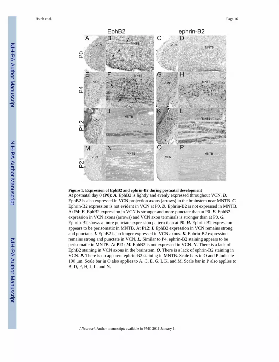
Figure 1. Expression of EphB2 and ephrin-B2 during postnatal developmentAt postnatal day 0 (P0): A. EphB2 is lightly and evenly expressed throughout VCN. B.EphB2 is also expressed in VCN projection axons (arrows) in the brainstem near MNTB. C.Ephrin-B2 expression is not evident in VCN at P0. D. Ephrin-B2 is not expressed in MNTB.At P4: E. EphB2 expression in VCN is stronger and more punctate than at P0. F. EphB2expression in VCN axons (arrows) and VCN axon terminals is stronger than at P0. G.Ephrin-B2 shows a more punctate expression pattern than at P0. H. Ephrin-B2 expressionappears to be perisomatic in MNTB. At P12: I. EphB2 expression in VCN remains strongand punctate. J. EphB2 is no longer expressed in VCN axons. K. Ephrin-B2 expressionremains strong and punctate in VCN. L. Similar to P4, ephrin-B2 staining appears to beperisomatic in MNTB. At P21: M. EphB2 is not expressed in VCN. N. There is a lack ofEphB2 staining in VCN axons in the brainstem. O. There is a lack of ephrin-B2 staining inVCN. P. There is no apparent ephrin-B2 staining in MNTB. Scale bars in O and P indicate100 μm. Scale bar in O also applies to A, C, E, G, I, K, and M. Scale bar in P also applies toB, D, F, H, J, L, and N.
Hsieh et al. Page 16
J Neurosci. Author manuscript; available in PMC 2011 January 1.
NIH
-PA Author Manuscript
NIH
-PA Author Manuscript
NIH
-PA Author Manuscript

Figure 2. Effects of EphB2, EphB3 and ephrin-B2 mutations on axonal projections from VCN toMNTBThe schematic diagram shows the orientation of brainstem sections and dye labelingprotocol. For all panels in this figure, dye originates on the right side of the image, in theVCN outside the field of view. The rectangle indicates the portion of the section in whichlabeling will be evaluated. Labeling in fixed tissue was performed at P10–14 in wild typeand transgenic mice. A. In wild type mice, dye labeling of VCN results in labeled calyces inMNTBc. Arrows indicate some of the labeled calyceal terminations. B. No aberrantprojections are seen in MNTBi. C. In EphB2−/−;EphB3−/− mice, VCN-MNTBc projectionsare normal and terminate in identifiable calyces (arrows). D. However, there are alsoabnormal VCN projections to MNTBi that terminate in calyx-like morphology (arrows). E.A ratio of the number of ipsilateral calyces to the number of contralateral calyces (I/C ratio)reveals that EphB2−/−;EphB3−/−mice have significantly more aberrant projections (*) thanEphB2+/+;EphB3−/− or EphB2+/−;EphB3−/− mice. F. In EphB2lacZ/lacZ;EphB3−/− mice,VCN-MNTBc projections are normal and terminate in identifiable calyces (arrows). G. Noaberrant projections are seen in MNTBi. H. There are no significant differences in I/C ratiosfor EphB2+/+;EphB3−/−, EphB2lacZ/+;EphB3−/− and EphB2lacZ/lacZ;EphB3−/− mice. I. Inephrin-B2lacZ/+mice, VCN-MNTBc projections appear normal and terminate in identifiablecalyces (arrows). J. However, there are aberrant projections from VCN to MNTBi (arrows).K. I/C ratios reveal significantly more aberrant projections (*) in ephrin-B2lacZ/+ thanephrin-B2+/+mice. Asterisks indicate p values < 0.05. Scale bar in J also applies to A–D, F–G and I and indicates 50 μm.
Hsieh et al. Page 17
J Neurosci. Author manuscript; available in PMC 2011 January 1.
NIH
-PA Author Manuscript
NIH
-PA Author Manuscript
NIH
-PA Author Manuscript

Figure 3. Aberrant projections develop at the same time as normal projectionsThese images were obtained from P3 and P4 EphB2-wildtype and EphB2-null mice. Bothmice have a null mutation in EphB3 that does not affect the VCN-MNTBc projection. In thewild type: A. RDA labeling in a P3 wild type mouse reveals small axon branches emergingfrom the ventral acoustic stria in MNTBc. B. Similarly, small branches are seen in MNTBi.In A and B, arrows indicate labeled axon branches. C. RDA labeling in a wild type mouse atP4 reveals an immature calyceal termination (arrow) in MNTBc. D. Branches in MNTBi donot have calyceal terminations. In the mutant: E. RDA labeling in a P3 EphB2 null mutantreveals small branches emerging from the ventral acoustic stria in MNTBc. F. Similarly,branches are seen in MNTBi. G. RDA labeling of a P4 EphB2 null mutants reveals animmature calyceal termination in MNTBc. H. In these mutants, abnormal projections withimmature calyceal terminations are present in MNTBi. In G and H, arrows indicaterudimentary calyceal terminations. I. I/C ratios from the EphB2null;EphB3null mouse lineindicate that even at this young age (P4–5), there are significantly more aberrant projections(*) from VCN to MNTBi in EphB2−/−;EphB3−/− than EphB2+/+;EphB3−/− orEphB2+/−;EphB3−/− mice. J. I/C ratios from the ephrin-B2lacZ mouse line indicate
Hsieh et al. Page 18
J Neurosci. Author manuscript; available in PMC 2011 January 1.
NIH
-PA Author Manuscript
NIH
-PA Author Manuscript
NIH
-PA Author Manuscript

significantly more aberrant projections in ephrin-B2lacZ/+ than ephrin-B2+/+mice. Asterisksindicate p values < 0.05. Scale bar in H also applies to A–G and indicates 10 μm.
Hsieh et al. Page 19
J Neurosci. Author manuscript; available in PMC 2011 January 1.
NIH
-PA Author Manuscript
NIH
-PA Author Manuscript
NIH
-PA Author Manuscript
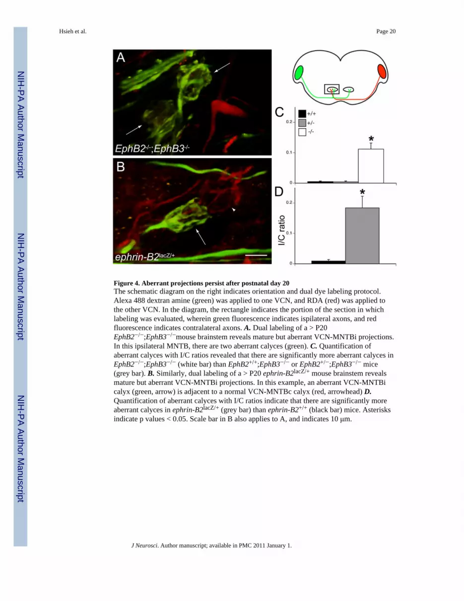
Figure 4. Aberrant projections persist after postnatal day 20The schematic diagram on the right indicates orientation and dual dye labeling protocol.Alexa 488 dextran amine (green) was applied to one VCN, and RDA (red) was applied tothe other VCN. In the diagram, the rectangle indicates the portion of the section in whichlabeling was evaluated, wherein green fluorescence indicates ispilateral axons, and redfluorescence indicates contralateral axons. A. Dual labeling of a > P20EphB2−/−;EphB3−/−mouse brainstem reveals mature but aberrant VCN-MNTBi projections.In this ipsilateral MNTB, there are two aberrant calyces (green). C. Quantification ofaberrant calyces with I/C ratios revealed that there are significantly more aberrant calyces inEphB2−/−;EphB3−/− (white bar) than EphB2+/+;EphB3−/− or EphB2+/−;EphB3−/− mice(grey bar). B. Similarly, dual labeling of a > P20 ephrin-B2lacZ/+ mouse brainstem revealsmature but aberrant VCN-MNTBi projections. In this example, an aberrant VCN-MNTBicalyx (green, arrow) is adjacent to a normal VCN-MNTBc calyx (red, arrowhead) D.Quantification of aberrant calyces with I/C ratios indicate that there are significantly moreaberrant calyces in ephrin-B2lacZ/+ (grey bar) than ephrin-B2+/+ (black bar) mice. Asterisksindicate p values < 0.05. Scale bar in B also applies to A, and indicates 10 μm.
Hsieh et al. Page 20
J Neurosci. Author manuscript; available in PMC 2011 January 1.
NIH
-PA Author Manuscript
NIH
-PA Author Manuscript
NIH
-PA Author Manuscript

Figure 5. Aberrant ipsilateral calyces express presynaptic markersA. In this mutant ipsilateral MNTB (P9 EphB2−/−;EphB3−/−), RDA labeling reveals anaberrant calyx (arrow). B. Expression of synapsin I in the same section as in A. C. Merge ofpanels A and B indicates that the RDA-labeled aberrant calyx is also positive for synapsin I(yellow). D. In a different mutant ipsilateral MNTB (P26 ephrin-B2lacZ/+) RDA labelingreveals axons (red) and two aberrant calyces (arrows). E. SV2 immunofluorescence in thesame section as in D. F. Merge of panels D and E indicates that the RDA-labeled calyces(arrows) are also positive for SV2 (yellow). Scale bar in F also applies to A–E, and indicates20 μm.
Hsieh et al. Page 21
J Neurosci. Author manuscript; available in PMC 2011 January 1.
NIH
-PA Author Manuscript
NIH
-PA Author Manuscript
NIH
-PA Author Manuscript

Figure 6. Summary of results suggesting that reverse signaling through ephrin-B2 in MNTBneurons prevents the formation of ipsilateral calycesGenotype is indicated across the top row, and phenotype is indicated across the bottom row.A. In wild type mice, signaling occurs bidirectionally, as both the receptor (pink) and theligand (blue) are fully expressed. These mice have a normal phenotype (bottom), in whichVCN projects to contralateral, but not ipsilateral, MNTB. B. In EphB2−/− mice a nullmutation eliminates the EphB2 protein entirely, so signaling does not occur in either forwardor reverse direction. Our data indicate that these mice have a significant number of abnormalipsilateral VCN-MNTB projections. C. EphB2lacZ/lacZ mice have β-galactosidase replacingthe intracellular portion of the EphB2 receptor. Thus, they cannot signal in the forwarddirection, but reverse signaling functions normally. These mice have normal, contralateralVCN-MNTB projections. D. Ephrin-B2lacZ/+ mice have intact forward signaling but reversesignaling through ephrin-B2 is reduced. These mice have abnormal ipsilateral VCN-MNTBprojections.
Hsieh et al. Page 22
J Neurosci. Author manuscript; available in PMC 2011 January 1.
NIH
-PA Author Manuscript
NIH
-PA Author Manuscript
NIH
-PA Author Manuscript