enrichment and isolation of microbial strains degrading
TRANSCRIPT
Vol. 14(27), pp. 2216-2226, 8 July, 2015 DOI:10.5897/AJB2011. 3980 Article Number: 26FFF6054087 ISSN 1684-5315 Copyright © 2015 Author(s) retain the copyright of this article http://www.academicjournals.org/AJB
African Journal of Biotechnology
Full Length Research Paper
Enrichment and isolation of microbial strains degrading bioplastic polyvinyl alcohol and time course study of
their degradation potential
Rajashree Patil and U. S. Bagde*
Department of Life Sciences, Microbiology Laboratory, University of Mumbai, Vidyanagari, Mumbai, 400078, India.
Received 7 December, 2011 Accepted 8 May, 2014
Polyvinyl alcohol (PVA) degrading bacterial strains were isolated from various environmental sites rich in plastic wastes by using the enrichment culture technique. Among the various isolated strains, the selected potent PVA degrading bacterial strains were tentatively characterized as Bacillus and Pseudomonas sp. The time course of the PVA degradation potential of the characterized strains in growth media containing PVA as a major carbon source was evaluated using the spectropho-tometric assay method. This was done by determining the residual PVA remaining in the culture media, increase in cell growth and change in pH of the media over a period of twenty days. The ultimate biodegradation (mineralization) of PVA to its mineral constituents CO2 and H2O was determined by the CO2 evolution test. The strain characterized as Bacillus sp. showed 65% of PVA degradation as determined by spectrophotometric assay and 45.4% of mineralization of PVA over a period of 20 days. The strain characterized as Pseudomonas sp. showed 42% of PVA degradation as determined by spectrophoto-metric assay and 28.9% of mineralization of PVA over a period of 20 days. Key words: Polyvinyl alcohol (PVA) degrading bacteria, isolation, ultimate biodegradation, mineralization.
INTRODUCTION Use of biodegradable, or single use disposable items as a replacement of inert synthetic plastics is gaining wide scale recognition due to its potential in overcoming, or at least reducing issues associated with the management of the post consumption status of synthetic plastics waste. Among the many others biodegradable polymers polyvinyl alcohol (PVA) is attracting increasing attention for its application in the production of environmental-
friendly plastic items (Corti et al., 2002; Tang and Alavi, 2011; Qui and Netravali, 2012). PVA is a vinyl polymer where the main chain is joined by only one carbon-carbon linkage (-CH2-CHOH-)n. This linkage is the same as those of typical plastics, such as polyethylene, polypropylene and polystyrene. PVA is a water soluble polymer but also has thermo plasticity and can be molded in various shapes such as container and films (Shimao,
*Corresponding author. E-mail. [email protected]
Author(s) agree that this article remain permanently open access under the terms of the Creative Commons Attribution License 4.0
International License
2001). Biodegradability of this material is mainly due to the presence of the hydroxyl group, which leads to its water solubility and susceptibility to oxidation (Kawai, 1995). Due to these properties, biodegradable plastic based on PVA gains popularity among biodegradable plastics, and is widely used in packaging and agricultural mulch film (Bastioli et al., 1993). Also, types of plastic formulated with PVA are available in the market with several trade names such as Akwa Tears, Alcotex, Alvyl, Aracet, Cipoviol, Celvol, Elvanol, Gelvatol, Ivalon, Solvar, Sumitex, Vinol, etc. Moreover, new and high production capacities of PVA-based plastics are opening in the Republic of Korea, India and South-East Asia (Flieger et al., 2003). In particular, four major segments of PVA consumption include wrap sizing, paper coating, adhesives, films and biodegradable PVA items for example mulching films, laundry bags, etc. (Chiellini et al.,1999).
Widespread application of this polymer as biode-gradable plastic has evoked research to understand the fate of this polymer in nature due to microbial degra-dation. Several groups of scientists have reported microbial degradation of PVA. Suzuki et al. (1973) showed for the first time that PVA was completely degraded and utilized by the bacterial strain Pseudomonas O-3, which can use it as the sole source of carbon and energy. An overview of literature cited on PVA biodegradation indicated that many of the research groups reported members of the genus Pseudomonadaceae as the key group providing useful organisms for accomplishing PVA degradation (Hoffmann et al., 2003).
The enrichment culture technique employing PVA as the sole source of carbon and energy is the most extensively used method for isolation of these groups of microorganisms. In the present study, isolation of PVA degrading microbial strains using the enrichment culture technique was carried out from various samples collected from different sites polluted with plastic waste in Mumbai, India. The major objective of the work was to isolate potent PVA degrader which can then be selectively applied to enhance biodegradation of PVA accumulated after its post-consumption. The potential PVA degraders were selected from various isolates and further characterized. The rate of PVA degradation by selected strains in PVA containing growth media were evaluated using spectrophotometric assay and the CO2 evolution test. MATERIALS AND METHODS
Collection of microbial source samples from different environmental sites
Samples from various sites were collected to be used as a microbial source for enrichment of PVA degrading microorganisms. Soil
Patil and Bagde 2217 samples were collected from the garden area near the campus of the University of Mumbai, India, and from a dumping ground used to dump municipal solid waste, near Kalyan city, India. Industrial
effluent samples were collected from effluent drainage near Gharda Chemicals Ltd, Dombivili, India and from the sea creek, near Century Rayon Ltd. Shahad, India. The sea sediments and sea water samples were collected from sites highly polluted with plastic waste from one of the beaches of Mumbai, India. Polymer sample
PVA (M.W. 125000) in powdered form was purchased from S. D. Fine Chemicals, Mumbai, India. Enrichment of PVA degrading microbial stains
Environmental samples collected from different sites rich in plastic waste were used as a source of microbial inoculum for enrichment
culture. Each solid sample 1 to 2 g and liquid sample 1 ml was diluted to 10 and 9 ml using normal saline and used as inoculum for the enrichment culture in the ratio of 1 ml to 100 ml of mineral salt vitamin media (MSV). 1000 mL MSV medium in distilled water contained: PVA, 5.0 g; (NH4)2SO4, 1.0 g; KH2PO4, 1.0 g; K2HPO4, 8.0 g; MgSO4.7H2O, 0.2 g; NaCl, 0.1 g; CaCl2.2H2O, 0.02 g; FeSO4, 0.01 g; Na2MoO4.2H2O, 0.5 mg; MnSO4, 0.5 mg; Inositol, 0.2 mg; p-amino benzoic acid, 0.2 mg; pyridoxine, 0.4 mg; thiamine, 2.0 µg; biotine, 2.0 µg; vitamin B, 120.5 µg; DW, 1000 ml; pH 7. The
medium used was the same one that was used previously by Suzuki et al. (1973). A solid medium was prepared by adding 20 g of agar agar powder to 1000 ml of the MSV medium before autoclaving. Isolation and characterization of PVA degrading microbial strains
A portion of enrichment culture was diluted adequately with sterile saline and spread on the nutrient agar plates. Incubation was carried out at 30°C for 48 h. Individual colonies formed on nutrient agar were picked and tested for their ability to grow on a solid MSV medium containing emulsified PVA, where the medium was fortified with a supplement and without any supplement such as 0.1% yeast extract and 0.1% glucose. Pure cultures of the PVC degrading bacteria were obtained by repeated sub-culturing of the isolated
colonies on the same medium. The selected isolates were assigned codes such as PVA 1, PVA 2 etc. for further study. The selected isolates were characterized up to species level using Bergey’s manual of determinative bacteriology (Krieg and Holt, 1984). Optimization of media used for degradation study
For isolation and degradation studies, various compositions of the MSV containing PVA as a primary source of carbon and energy were supplemented with an additional co-substrate, such as yeast extract and glucose at a final concentration of 0.1%.
Preparation of microbial cell suspension for various degradation studies
The suspension of microbial cells used in various degradation studies was grown in a nutrient broth for 18 h at 30°C. The cells were harvested from the culture by centrifugation at 4500 rpm for
2218 Afr. J. Biotechnol. 15 min (Sorvall RC 5B Plus, Kendo, Newtown, USA). After discarding the supernatant, the cell pellet was suspended in normal saline and centrifuged at 4500 rpm. The same procedure was
repeated twice. The washed cell pellet was re-suspended in the medium used for the degradation experiment. Determination of PVA degradation by spectrophotometric assay
The kinetics of PVA degradation by the selected isolated strain was studied by determining the residual concentration of PVA left in the growth media at regular intervals of time using spectrophotometric assay. The test was performed in a 500 ml Erlenmeyer flask containing 250 ml of MSV-PVA media containing 0.5% PVA supplemented with 0.1 % yeast extract (Hi Media India). 5% of washed microbial cell suspension (approximately 3.1 × 10
8 cells/ml)
was used as an inoculum for the test. The test flasks were incubated at 30°C in a shaker incubator (Neolab India) at 180 rpm along with control flasks. Two controls were maintained in the
present experiment, one with MSV-PVA medium without test cultures and a second MSV medium without PVA and inoculated with a suspension of the test culture. At each test interval, the cell growth was estimated by reading the optical density at 600 nm. Simultaneously, 10 ml of the sample was taken from each culture flask for analysis of residual PVA concentration. Samples were centrifuged at 4500 rpm for 20 min at 4°C (Sorvall RC 5B Plus, Kendo, Newtown, USA). The resulting supernatants were filter sterilized through the Sartorius filter (Sartorius, Germany) and the
filtrates were used for analysis of residual PVA concentration. Controls were treated in the same way. Absence of microbial contaminants was checked by optical microscopy before any determinations were made. After appropriate dilution, residual PVA concentration in the culture filtrate was estimated according to Finley (1961) using spectrophotometric assay. The assay is based on a green color produced by the reaction of PVA with iodine in the presence of boric acid.
In a properly diluted 1 ml of culture filtrate 0.75 ml of 4% boric acid and 0.15 ml of potassium iodide (KI) solution (12.7 g of iodine and 25 g KI were dissolved in distilled water and a 1000 ml solution was made). The solution was mixed well and allowed to equilibrate for 30 min at room temperature. The mixture was diluted to a volume of 2.5 ml with distilled water and analyzed at 660 nm using Shimadzu UV-Vis double beam spectrophotometer (Shimadzu Ltd.). The blank was treated in the same way. The amount of PVA in the filtrate was determined using a standard calibration curve.
The calibration curve was prepared by using a range of standard PVA solution from 10-100 µg/ml in distilled water. Color development and absorbance measurements were carried out following procedure described above. A standard calibration curve was plotted as absorbance 600 nm against PVA concentration. All the measurements were made in triplicate.
PVA degradation study by carbon dioxide (CO2) production test
PVA biodegradation was determined by the CO2 production test as per the general guidelines of ISO 14855 (1999) and ASTM D5338 (1998). The medium used for assay of CO2 production was the same as that given in the OECD (2001) guidelines for the testing of chemicals. The apparatus bio meter flask used in the present study was as described by Reich and Bartha (1977), and Yabannavar and Bartha (1993, 1994). For measurement of CO2 production, the main compartment of the bio meter flask was amended with 100 ml of mineral medium with 0.1% of PVA along with 5% washed cell
suspension of the selected isolated strain. The CO2 produced during the metabolic activity was absorbed in a solution of barium hydroxide Ba (OH) 2 and subsequently determined by titration using
0.05N HCl where the amount of CO2 produced was calculated from the amount of residual base remaining in the absorption tube. The mineralization was expressed as a percentage of the theoretical CO2 (ThCO2) produced, computed from the total carbon content of the samples. During the test period, flasks were incubated at room temperature in the dark. The stopcock was periodically opened for exchange of air. At each test interval (1, 7, 14, .21, 28, 35, 42, 49, 56, 63 and 70), Ba (OH) 2 from the side arm was removed for analysis of residual Ba (OH) 2. The amount of un-reacted Ba(OH)2 in the sample was with 0.05 N HCI control containing an inoculated medium without any test compounds, was evaluated for CO2 production to determine endogenous metabolism of the test culture. A control, containing an un-inoculated medium with test substance was also used for determining the CO2 evolved due to non-biological degradation. The amount of CO2 evolved from the control flask was subtracted from the corresponding experimental flask. The percentage biodegradation was calculated from the cumulative
amount of CO2 released during the entire test period.
RESULTS
Isolation of PVA degrading microbial strains
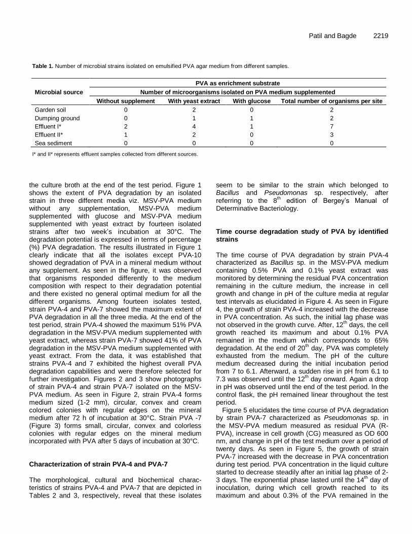
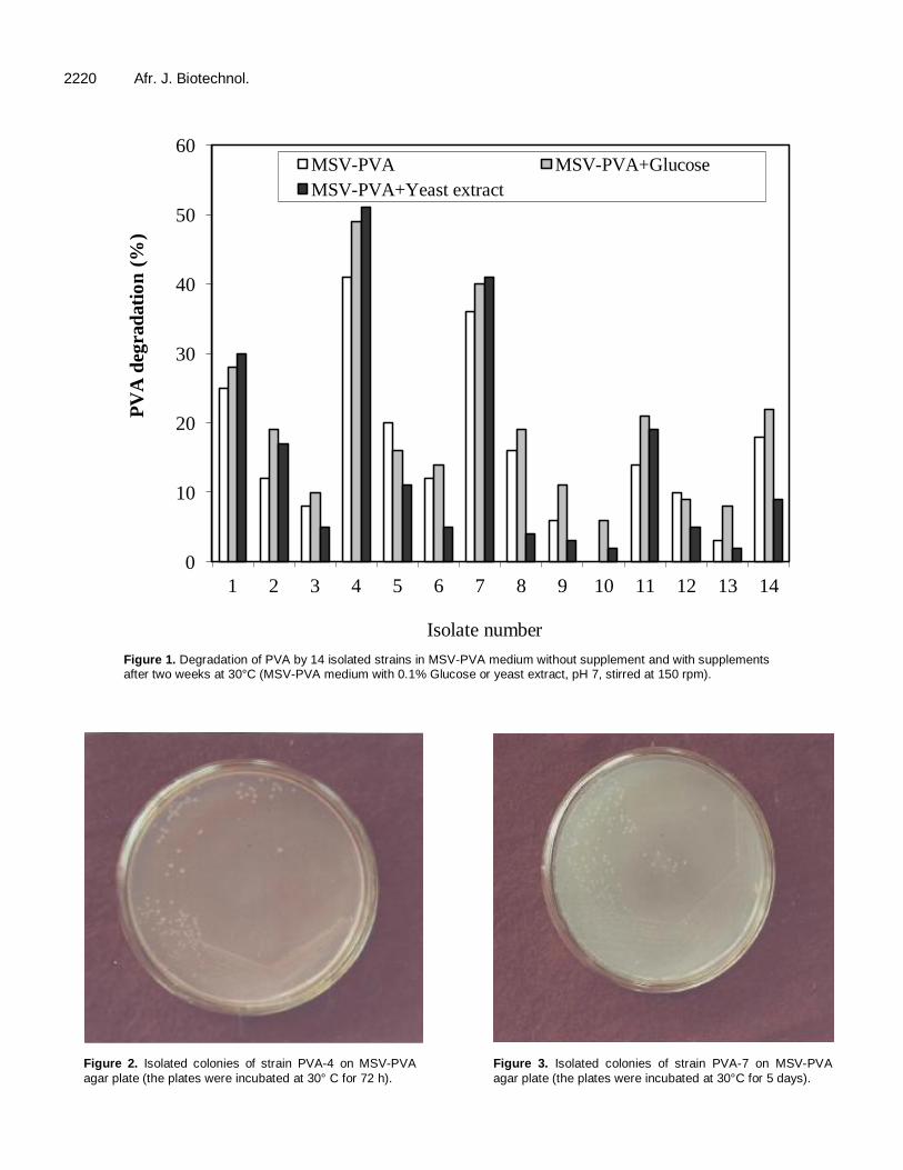
PVA degrading microbial strains were isolated using the enrichment culture technique. Samples collected from different microbial sources were subjected to repeated enrichment in MSV-PVA containing PVA as the sole source of carbon and energy. From enrichment cultures incorporated with PVA and incubated for two weeks at 30°C, 30 morphologically different bacterial isolates were obtained on the nutrient agar. These strains were then tested for their ability to grow on MSV-PVA without any supplementation and with supplements. Among these thirty isolates, only fourteen strains were able to grow on MSV agar medium containing PVA. Table 1 shows the total number of organisms isolated from a different source on the MSV-PVA agar medium without supplementation and with supplements from an enrichment culture containing PVA as the enrichment substrate. Table 1 reveals that the PVA degrading micro-organism inhabited different environments. However, industrial effluent samples harbored the maximum number of microorganisms as compared to the other microbial sources. Observation from Table 1 also reveals that only three strains were able to grow on the MSV-PVA agar medium without any supplementation. The maximum number of isolates could grow in the presence of the yeast extract. The 14 isolates obtained were numbered from strain PVA-1 to PVA-14. Screening of 14 different PVA degrading isolates for their PVA degradation potential was carried out by determining the extent of PVA degraded by an individual isolated strain in the MSV-PVA medium supplemented without and with 0.1% yeast extract and 0.1% glucose. PVA degradation in various degradation media was determined by analyzing the residual PVA concentration remaining in
Patil and Bagde 2219
Table 1. Number of microbial strains isolated on emulsified PVA agar medium from different samples.
Microbial source
PVA as enrichment substrate
Number of microorganisms isolated on PVA medium supplemented
Without supplement With yeast extract With glucose Total number of organisms per site
Garden soil 0 2 0 2
Dumping ground 0 1 1 2
Effluent I* 2 4 1 7
Effluent II* 1 2 0 3
Sea sediment 0 0 0 0
I* and II* represents effluent samples collected from different sources.
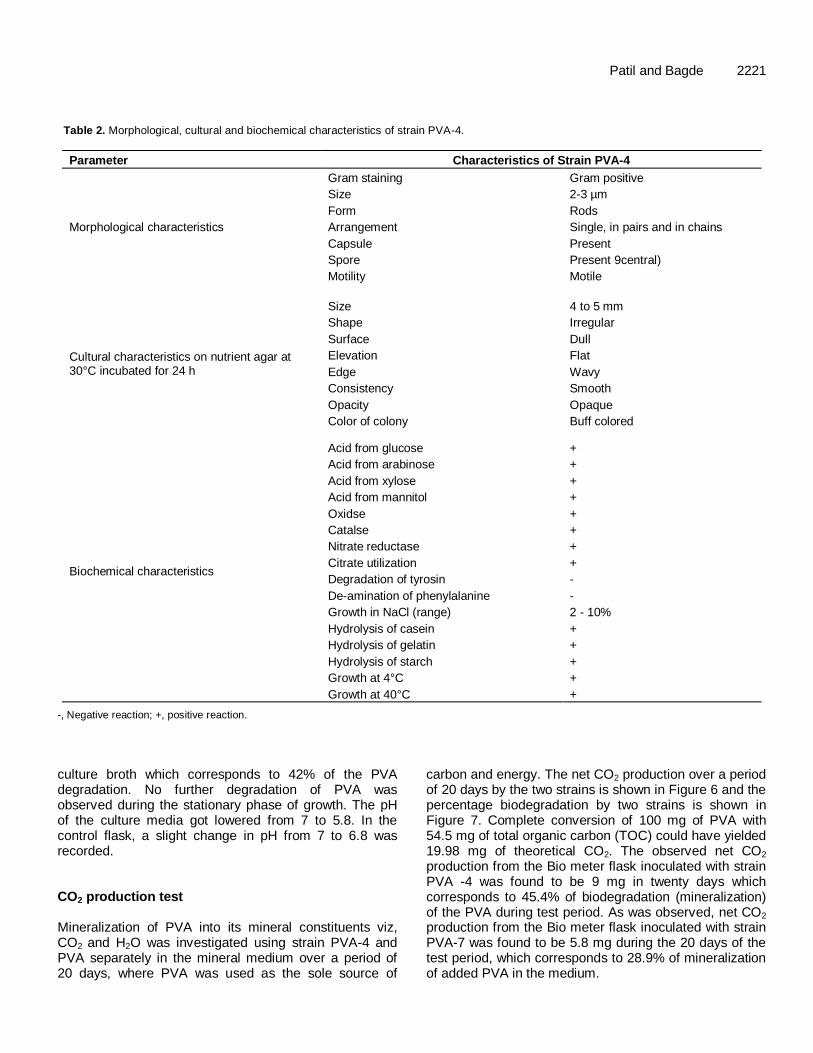
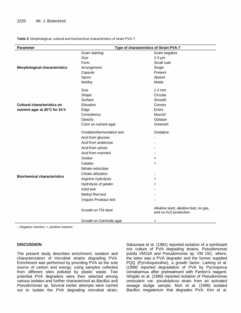
the culture broth at the end of the test period. Figure 1 shows the extent of PVA degradation by an isolated strain in three different media viz. MSV-PVA medium without any supplementation, MSV-PVA medium supplemented with glucose and MSV-PVA medium supplemented with yeast extract by fourteen isolated strains after two week’s incubation at 30°C. The degradation potential is expressed in terms of percentage (%) PVA degradation. The results illustrated in Figure 1 clearly indicate that all the isolates except PVA-10 showed degradation of PVA in a mineral medium without any supplement. As seen in the figure, it was observed that organisms responded differently to the medium composition with respect to their degradation potential and there existed no general optimal medium for all the different organisms. Among fourteen isolates tested, strain PVA-4 and PVA-7 showed the maximum extent of PVA degradation in all the three media. At the end of the test period, strain PVA-4 showed the maximum 51% PVA degradation in the MSV-PVA medium supplemented with yeast extract, whereas strain PVA-7 showed 41% of PVA degradation in the MSV-PVA medium supplemented with yeast extract. From the data, it was established that strains PVA-4 and 7 exhibited the highest overall PVA degradation capabilities and were therefore selected for further investigation. Figures 2 and 3 show photographs of strain PVA-4 and strain PVA-7 isolated on the MSV-PVA medium. As seen in Figure 2, strain PVA-4 forms medium sized (1-2 mm), circular, convex and cream colored colonies with regular edges on the mineral medium after 72 h of incubation at 30°C. Strain PVA -7 (Figure 3) forms small, circular, convex and colorless colonies with regular edges on the mineral medium incorporated with PVA after 5 days of incubation at 30°C. Characterization of strain PVA-4 and PVA-7 The morphological, cultural and biochemical charac-teristics of strains PVA-4 and PVA-7 that are depicted in Tables 2 and 3, respectively, reveal that these isolates
seem to be similar to the strain which belonged to Bacillus and Pseudomonas sp. respectively, after referring to the 8
th edition of Bergey’s Manual of
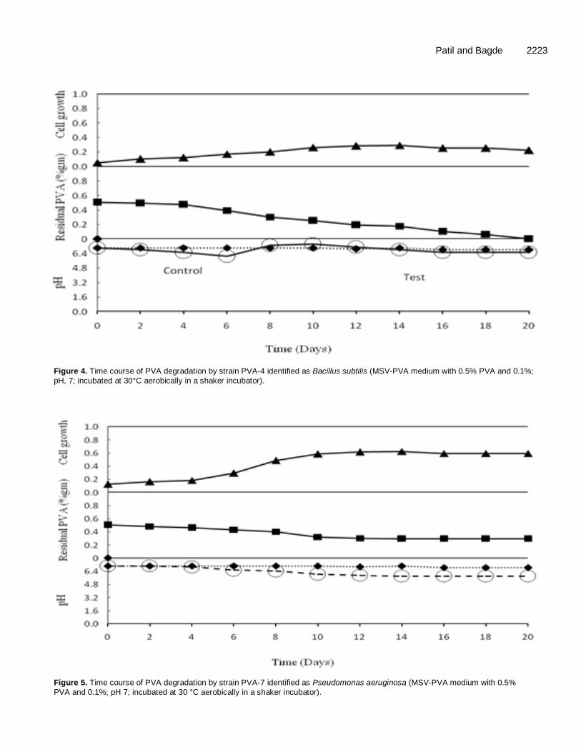
Determinative Bacteriology. Time course degradation study of PVA by identified strains The time course of PVA degradation by strain PVA-4 characterized as Bacillus sp. in the MSV-PVA medium containing 0.5% PVA and 0.1% yeast extract was monitored by determining the residual PVA concentration remaining in the culture medium, the increase in cell growth and change in pH of the culture media at regular test intervals as elucidated in Figure 4. As seen in Figure 4, the growth of strain PVA-4 increased with the decrease in PVA concentration. As such, the initial lag phase was not observed in the growth curve. After, 12
th days, the cell
growth reached its maximum and about 0.1% PVA remained in the medium which corresponds to 65% degradation. At the end of 20
th day, PVA was completely
exhausted from the medium. The pH of the culture medium decreased during the initial incubation period from 7 to 6.1. Afterward, a sudden rise in pH from 6.1 to 7.3 was observed until the 12
th day onward. Again a drop
in pH was observed until the end of the test period. In the control flask, the pH remained linear throughout the test period.
Figure 5 elucidates the time course of PVA degradation by strain PVA-7 characterized as Pseudomonas sp. in the MSV-PVA medium measured as residual PVA (R-PVA), increase in cell growth (CG) measured as OD 600 nm, and change in pH of the test medium over a period of twenty days. As seen in Figure 5, the growth of strain PVA-7 increased with the decrease in PVA concentration during test period. PVA concentration in the liquid culture started to decrease steadily after an initial lag phase of 2-3 days. The exponential phase lasted until the 14
th day of
inoculation, during which cell growth reached to its maximum and about 0.3% of the PVA remained in the
2220 Afr. J. Biotechnol.
Figure 1. Degradation of PVA by 14 isolated strains in MSV-PVA medium without supplement and with supplements after two weeks at 30°C (MSV-PVA medium with 0.1% Glucose or yeast extract, pH 7, stirred at 150 rpm).
Figure 2. Isolated colonies of strain PVA-4 on MSV-PVA
agar plate (the plates were incubated at 30° C for 72 h).
Figure 3. Isolated colonies of strain PVA-7 on MSV-PVA
agar plate (the plates were incubated at 30°C for 5 days).
0
10
20
30
40
50
60
1 2 3 4 5 6 7 8 9 10 11 12 13 14
PV
A d
egra
dati
on
(%
)
Isolate number
MSV-PVA MSV-PVA+Glucose
MSV-PVA+Yeast extract
Patil and Bagde 2221
Table 2. Morphological, cultural and biochemical characteristics of strain PVA-4.
Parameter Characteristics of Strain PVA-4
Morphological characteristics
Gram staining Gram positive
Size 2-3 µm
Form Rods
Arrangement Single, in pairs and in chains
Capsule Present
Spore Present 9central)
Motility Motile
Cultural characteristics on nutrient agar at 30°C incubated for 24 h
Size 4 to 5 mm
Shape Irregular
Surface Dull
Elevation Flat
Edge Wavy
Consistency Smooth
Opacity Opaque
Color of colony Buff colored
Biochemical characteristics
Acid from glucose +
Acid from arabinose +
Acid from xylose +
Acid from mannitol +
Oxidse +
Catalse +
Nitrate reductase +
Citrate utilization +
Degradation of tyrosin -
De-amination of phenylalanine -
Growth in NaCl (range) 2 - 10%
Hydrolysis of casein +
Hydrolysis of gelatin +
Hydrolysis of starch +
Growth at 4°C +
Growth at 40°C +
-, Negative reaction; +, positive reaction.
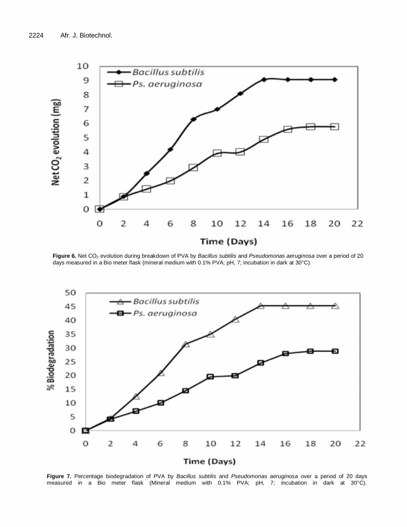
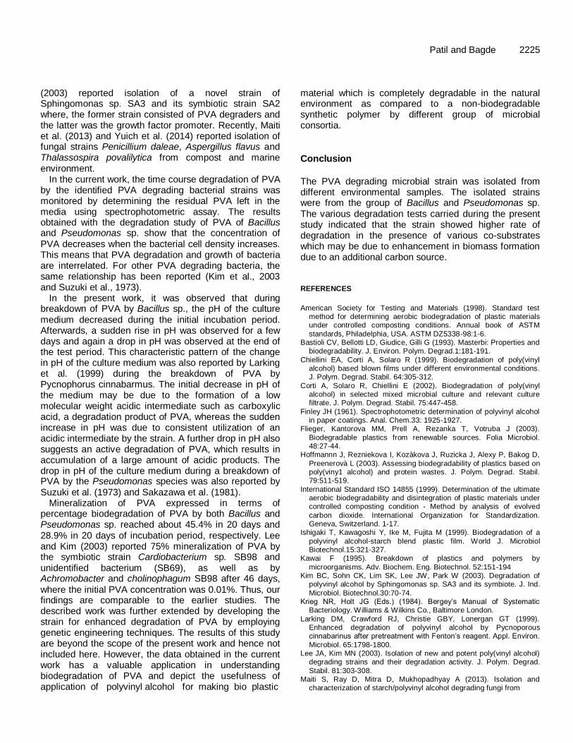
culture broth which corresponds to 42% of the PVA degradation. No further degradation of PVA was observed during the stationary phase of growth. The pH of the culture media got lowered from 7 to 5.8. In the control flask, a slight change in pH from 7 to 6.8 was recorded. CO2 production test Mineralization of PVA into its mineral constituents viz, CO2 and H2O was investigated using strain PVA-4 and PVA separately in the mineral medium over a period of 20 days, where PVA was used as the sole source of
carbon and energy. The net CO2 production over a period of 20 days by the two strains is shown in Figure 6 and the percentage biodegradation by two strains is shown in Figure 7. Complete conversion of 100 mg of PVA with 54.5 mg of total organic carbon (TOC) could have yielded 19.98 mg of theoretical CO2. The observed net CO2 production from the Bio meter flask inoculated with strain PVA -4 was found to be 9 mg in twenty days which corresponds to 45.4% of biodegradation (mineralization) of the PVA during test period. As was observed, net CO2
production from the Bio meter flask inoculated with strain PVA-7 was found to be 5.8 mg during the 20 days of the test period, which corresponds to 28.9% of mineralization of added PVA in the medium.
2220 Afr. J. Biotechnol. Table 3. Morphological, cultural and biochemical characteristics of strain PVA-7.
Parameter Type of characteristics of Strain PVA-7
Morphological characteristics
Gram staining Gram negative
Size 2-3 µm
Form Small rods
Arrangement Single
Capsule Present
Spore Absent
Motility Motile
Cultural characteristics on
nutrient agar at 30°C for 24 h
Size 1-2 mm
Shape Circular
Surface Smooth
Elevation Convex
Edge Entire
Consistency Mucoid
Opacity Opaque
Color on nutrient agar Greenish
Biochemical characteristics
Oxidation/fermentation test Oxidative
Acid from glucose -
Acid from arabinose -
Acid from xylose -
Acid from mannitol -
Oxidse +
Catalse +
Nitrate reductase
Citrate utilization -
Arginine hydrolysis +
Hydrolysis of gelatin +
Indol test -
Methyl Red test -
Vogues Proskaur test -
Growth on TSI slant Alkaline slant, alkaline butt, no gas, and no H2S production
Growth on Cetrimide agar +
-, Negative reaction; +, positive reaction.
DISCUSSION The present study describes enrichment, isolation and characterization of microbial strains degrading PVA. Enrichment was performed by providing PVA as the sole source of carbon and energy, using samples collected from different sites polluted by plastic waste. Two potential PVA degraders were then selected among various isolates and further characterized as Bacillus and Pseudomonas sp. Several earlier attempts were carried out to isolate the PVA degrading microbial strain.
Sakazawa et al. (1981) reported isolation of a symbioant mix culture of PVA degrading strains, Pseudomonas putida VM15A and Pseudomonas sp. VM 15C, where, the latter was a PVA degrader and the former supplied PQQ (Pyrroloquinoline), a growth factor. Larking et al. (1999) reported degradation of PVA by Pycnoporus cinnabarinus after pretreatment with Fenton’s reagent. Ishigaki et al. (1999) reported isolation of Pseudomonas vesicularis var. povalolyticus strain from an activated sewage sludge sample. Mori et al. (1996) isolated Bacillus megaterium that degrades PVA. Kim et al.
Patil and Bagde 2223
Figure 4. Time course of PVA degradation by strain PVA-4 identified as Bacillus subtilis (MSV-PVA medium with 0.5% PVA and 0.1%;
pH, 7; incubated at 30°C aerobically in a shaker incubator).
Figure 5. Time course of PVA degradation by strain PVA-7 identified as Pseudomonas aeruginosa (MSV-PVA medium with 0.5%
PVA and 0.1%; pH 7; incubated at 30 °C aerobically in a shaker incubator).
2224 Afr. J. Biotechnol.
Figure 6. Net CO2 evolution during breakdown of PVA by Bacillus subtilis and Pseudomonas aeruginosa over a period of 20
days measured in a Bio meter flask (mineral medium with 0.1% PVA; pH, 7; incubation in dark at 30°C).
Figure 7. Percentage biodegradation of PVA by Bacillus subtilis and Pseudomonas aeruginosa over a period of 20 days measured in a Bio meter flask (Mineral medium with 0.1% PVA; pH, 7; incubation in dark at 30°C).
(2003) reported isolation of a novel strain of Sphingomonas sp. SA3 and its symbiotic strain SA2 where, the former strain consisted of PVA degraders and the latter was the growth factor promoter. Recently, Maiti et al. (2013) and Yuich et al. (2014) reported isolation of fungal strains Penicillium daleae, Aspergillus flavus and Thalassospira povalilytica from compost and marine environment.
In the current work, the time course degradation of PVA by the identified PVA degrading bacterial strains was monitored by determining the residual PVA left in the media using spectrophotometric assay. The results obtained with the degradation study of PVA of Bacillus and Pseudomonas sp. show that the concentration of PVA decreases when the bacterial cell density increases. This means that PVA degradation and growth of bacteria are interrelated. For other PVA degrading bacteria, the same relationship has been reported (Kim et al., 2003 and Suzuki et al., 1973).
In the present work, it was observed that during breakdown of PVA by Bacillus sp., the pH of the culture medium decreased during the initial incubation period. Afterwards, a sudden rise in pH was observed for a few days and again a drop in pH was observed at the end of the test period. This characteristic pattern of the change in pH of the culture medium was also reported by Larking et al. (1999) during the breakdown of PVA by Pycnophorus cinnabarmus. The initial decrease in pH of the medium may be due to the formation of a low molecular weight acidic intermediate such as carboxylic acid, a degradation product of PVA, whereas the sudden increase in pH was due to consistent utilization of an acidic intermediate by the strain. A further drop in pH also suggests an active degradation of PVA, which results in accumulation of a large amount of acidic products. The drop in pH of the culture medium during a breakdown of PVA by the Pseudomonas species was also reported by Suzuki et al. (1973) and Sakazawa et al. (1981).
Mineralization of PVA expressed in terms of percentage biodegradation of PVA by both Bacillus and Pseudomonas sp. reached about 45.4% in 20 days and 28.9% in 20 days of incubation period, respectively. Lee and Kim (2003) reported 75% mineralization of PVA by the symbiotic strain Cardiobacterium sp. SB98 and unidentified bacterium (SB69), as well as by Achromobacter and cholinophagum SB98 after 46 days, where the initial PVA concentration was 0.01%. Thus, our findings are comparable to the earlier studies. The described work was further extended by developing the strain for enhanced degradation of PVA by employing genetic engineering techniques. The results of this study are beyond the scope of the present work and hence not included here. However, the data obtained in the current work has a valuable application in understanding biodegradation of PVA and depict the usefulness of application of polyvinyl alcohol for making bio plastic
Patil and Bagde 2225 material which is completely degradable in the natural environment as compared to a non-biodegradable synthetic polymer by different group of microbial consortia.
Conclusion
The PVA degrading microbial strain was isolated from different environmental samples. The isolated strains were from the group of Bacillus and Pseudomonas sp. The various degradation tests carried during the present study indicated that the strain showed higher rate of degradation in the presence of various co-substrates which may be due to enhancement in biomass formation due to an additional carbon source. REFERENCES
American Society for Testing and Materials (1998). Standard test
method for determining aerobic biodegradation of plastic materials under controlled composting conditions. Annual book of ASTM
standards, Philadelphia, USA. ASTM DZ5338-98:1-6. Bastioli CV, Bellotti LD, Giudice, Gilli G (1993). Masterbi: Properties and
biodegradability. J. Environ. Polym. Degrad.1:181-191.
Chiellini EA, Corti A, Solaro R (1999). Biodegradation of poly(vinyl alcohol) based blown films under different environmental conditions. J. Polym. Degrad. Stabil. 64:305-312.
Corti A, Solaro R, Chiellini E (2002). Biodegradation of poly(vinyl alcohol) in selected mixed microbial culture and relevant culture filtrate. J. Polym. Degrad. Stabil. 75:447-458.
Finley JH (1961). Spectrophotometric determination of polyvinyl alcohol in paper coatings. Anal. Chem.33: 1925-1927.
Flieger, Kantorova MM, Prell A, Rezanka T, Votruba J (2003).
Biodegradable plastics from renewable sources. Folia Microbiol. 48:27-44.
Hoffmannn J, Rezniekova I, Kozàkova J, Ruzicka J, Alexy P, Bakog D,
Preenerovà L (2003). Assessing biodegradability of plastics based on poly(viny1 alcohol) and protein wastes. J. Polym. Degrad. Stabil. 79:511-519.
International Standard ISO 14855 (1999). Determination of the ultimate aerobic biodegradability and disintegration of plastic materials under controlled composting condition - Method by analysis of evolved
carbon dioxide. International Organization for Standardization. Geneva, Switzerland. 1-17.
Ishigaki T, Kawagoshi Y, Ike M, Fujita M (1999). Biodegradation of a
polyvinyl alcohol-starch blend plastic film. World J. Microbiol Biotechnol.15:321-327.
Kawai F (1995). Breakdown of plastics and polymers by
microorganisms. Adv. Biochem. Eng. Biotechnol. 52:151-194 Kim BC, Sohn CK, Lim SK, Lee JW, Park W (2003). Degradation of
polyvinyl alcohol by Sphingomonas sp. SA3 and its symbiote. J. Ind.
Microbiol. Biotechnol.30:70-74. Krieg NR, Holt JG (Eds.) (1984). Bergey’s Manual of Systematic
Bacteriology. Williams & Wilkins Co., Baltimore London.
Larking DM, Crawford RJ, Christie GBY, Lonergan GT (1999). Enhanced degradation of polyvinyl alcohol by Pycnoporous cinnabarinus after pretreatment with Fenton’s reagent. Appl. Environ.
Microbiol. 65:1798-1800. Lee JA, Kim MN (2003). Isolation of new and potent poly(vinyl alcohol)
degrading strains and their degradation activity. J. Polym. Degrad.
Stabil. 81:303-308. Maiti S, Ray D, Mitra D, Mukhopadhyay A (2013). Isolation and
characterization of starch/polyvinyl alcohol degrading fungi from

2226 Afr. J. Biotechnol.
aerobic compost environment. International Biodeterioration & Biodegradation. 82:9-12.
Mori T, Sakimoto M, Kagi T, Sakai T (1996). Isolation and characterization of strain of Bacillus megaterium that degrades
poly(vinyl alcohol). Biosci. Biotechnol. Biochem. 60:330-332. OECD (2001). OECD Guidelines for the Testing of Chemical: Ready
Biodegradability - CO2 evolution in sealed vessels (Headspace test). Organization for Economic Co-operation and Development, France. 1-16.
Qui K, Netravali AN (2012). Fabrication and characterization of biodegradable composites based on micro fibrillated cellulose and polyvinyl alcohol. J. Composites Sci and Tech. 72(13):1588-1594.
Reich M, Bartha R (1977). Degradation and mineralization of a polybutene film-mulch by the synergistic action of sunlight and soil microbes. Soil. Sci. 124:177-180.
Sakazawa C, Shimao M, Taniguchi Y, Kato N (1981). Symbiotic utilization of polyvinyl alcohol by mixed culture. Appl. Environ. Microbiol. 41:261-267.
Shimao M. (2001). Biodegradation of plastics. Curr. Opin. Biotechnol. 12:242-247.
Suzuki T, Ichihara Y, Yamada M, Tonomura K (1973). Some
characteristic of Pseudomonas O-3 which utilizes polyvinyl alcohol.
Agric. Boil.Chem. 37:747-756.
Yabannavar AV, Bartha R (1994). Methods for assessment of Biodegradability of plastic films in soil. Appl. Environ. Microbiol. 60:3608-3614.
Yabannavar A, Bartha R (1993). Biodegradability of some food packaging materials in soil. Soil Biol. Biochem. 25: 1469 - 1475.
Tang X, Alavi S (2011). Recent advances in starch, polyvinyl alcohol
based polymer blends, nanocomposites and their biodegradability. Carbohydrate Polymers. 85(1):7–16.
Yuich N, Masaki Y, Masayuki M (2014). Thalassospira povalilytica sp.
nov., a polyvinyl alcohol-degrading marine bacterium. J. International journal of systematic and evolutionary microbiology. 1466-5026.