enhanced oromucosal delivery of progesterone via hexosomes
TRANSCRIPT

Research Paper
Enhanced Oromucosal Delivery of Progesterone Via Hexosomes
Nitin K. Swarnakar,1 Vikas Jain,1 Vaibhav Dubey,1 Dinesh Mishra,1 and N.K. Jain1,2
Received June 3, 2007; accepted July 10, 2007; published online September 8, 2007
Purpose. Formulation and characterization of progesterone loaded hexosomes employing a novel
method for oromucosal delivery.
Method. Hexosomes were prepared employing a method in which ethanolic solution of lipid phase
(monolein and oleic acid) was vortexed with aqueous phase (surfactant solution) and characterized for
particle size, morphology and internal structure. FT-IR and confocal laser scanning microscopy (CLSM)
were performed to investigate the possible mechanism and penetration pathway of hexosomes within the
mucosa.
Results. Hexosomes exhibited anisotropy, hexagonal shape and nanometric size (251.2T1.8 nm). Internal
structure confirmed by X-ray diffraction peaks with spacing ratio of ¾1:¾3:¾4 proved two-dimensional
hexagonal arrangements. Entrapment efficiency of system was greater than 95%. In vitro release studies
revealed an enhanced transmucosal flux (4.67T0.14 mg cmj2 hj1) and decreased lag time (1.54 h) across
albino rabbit mucosa. FT-IR and CLSM of treated mucosa shows lipid extraction phenomena as well as
structural irregularities within intercellular lipids respectively. These irregularities can function as
Fvirtual channels_ facilitating hexosome_s penetration.
Conclusion. Developed hexosomes formulation exhibited high entrapment efficiency, high permeability
and better stability on storage, thus proposing itself a novel carrier for enhanced oromucosal delivery of
progesterone.
KEY WORDS: hexosomes; oromucosal delivery; permeation enhancement; progesterone.
INTRODUCTION
To reach the drug into systemic circulation buccal routeis an attractive approach of administration. It provides amuch milder environment for drug absorption as well asprotects drugs from undergoing gastrointestinal degradationand hepatic first-pass metabolism (1). The mucosal lining ofthe oral cavity is richly vascularized and more accessible forthe administration and removal of a dosage form (2). Themembrane of oral cavity have 170 cm2 total surface area fordrug absorption (3), including õ50 cm2 of non-keratinizedtissues (4). Buccal mucosa represents low enzymatic activityas compared to other mucosal routes (5) which seems to beadvantageous while administering proteins and peptides.Additionally, buccal drug delivery has a high patientacceptability compared to other non-oral routes of drugadministration.
Progesterone, named for its progestational role in main-taining pregnancy, is traditionally regarded as a Bfemalehormone^, playing a key role in generating female-typicalbehaviors such as lordosis and maternal behavior. In females,
the ovary (and/or placenta) is the primary source of thedynamic levels of plasma progesterone that exist during theestrous cycle and over the course of pregnancy and lactation(6,7). It has also shown its therapeutic efficacy in treatmentof many disorders including hormone replacement therapy inthe menopausal woman, as well as a natural agent inestablishment and maintenance of pregnancy (8,9).
Being so efficacious, the major problem with its thera-peutic use is selection of a proper route of administration.Oral intake is still the most convenient approach but requireslarge doses, an appropriate vehicle, and an increased surfacearea (micronization) to achieve reasonable blood levels (10),however rapid and extensive intestinal metabolism preventsadequate absorption (11). Alternative routes of administra-tion of progesterone include intramuscular, vaginal andtopical. Several studies indicate that topically applied pro-gesterone is absorbed transdermally (12,13) although, thereremains considerable uncertainty as to the resultant proges-terone levels in target tissues (14,15). O_Leary et al. (16)showed no change in plasma concentration of topicallyadministered progesterone, while salivary progesterone levelswere elevated in both pre- and post-menopausal women.Thus, topically applied progesterone appears to exert biolog-ical activity, there remains uncertainty about its pattern ofuptake, metabolism and subsequent tissue distribution (17).Vaginal progesterone delivery has also been proposed butchanges in epithelial thickness during menstrual cycle maychange its bioavailability (18).
2223 0724-8741/07/1200-2223/0 # 2007 Springer Science + Business Media, LLC
Pharmaceutical Research, Vol. 24, No. 12, December 2007 (# 2007)DOI: 10.1007/s11095-007-9409-y
1 Pharmaceutics Research Laboratory, Department of Pharmaceuti-
cal Sciences, Dr. Hari Singh Gour University, Sagar (M.P.), 470003,
India.2 To whom correspondence should be addressed. (e-mail: jnarendr@
yahoo.co.in)
Our present study focuses on preparation and charac-terization of hexosomes employing a newer method formucosal delivery of progesterone and studying the possiblemechanism of penetration of these carriers. The reversedhexagonal phase (honeycomb structure) comprises hexago-nally close-packed infinite water layers covered by a surfactantmonolayer (19,20). Earlier, lyotropic liquid crystalline phasesbearing various therapeutic agents were prepared and theywere shown to exhibit sustained release properties (21–23).
Hexosomal dispersion were prepared and characterizedfor their particle size, morphology, stability, entrapmentefficiency, release profile and internal structure. confocallaser scanning microscopy (CLSM) and FT-IR were per-formed for studying the extent of penetration and hexoso-mal–mucosal interaction and thus proposing mechanism ofpenetration of hexosomal dispersion.
MATERIALS AND METHODS
Materials and Reagents
Monoolein 99% [GMO], was purchased from Sigma-Aldrich (St. Louis, MO USA), Oleic acid, from CDH (India),Pluronic F-68, from Himedia (India) and Rhodamine B (RB)from Molecular Probes (Eugene, Oregon, USA). Progester-one (99%) was received as a gift sample from M/s SunPharmaceuticals Ltd. (Vadodara, India). All chemicals usedwere of AR grade, solvents were of HPLC grade and tripledistilled water was used for all experiments.
Preparation of Dispersion
The method employed in preparation of hexosomedispersion was a slight modification of procedure reportedby Spicer et al. (24). It involves dilution of an isotropic liquidmade up of 50% lipid phase (GMO and oleic acid, 60:40 w/w)and 50% ethanol with a polymer solution (1%) in water andvortexing at 3,200 rpm for 120 s to form a colloidal dispersionof hexosome. The final concentrations were 5% lipid, 5%ethanol and 1% Pluronic F-68. To attain the equilibrium,dispersion was stored undisturbed in dark at room temper-ature for about 2 weeks.
For drug-loaded dispersion, the desired amount ofprogesterone (1% w/w wrt lipid) was dissolved in absoluteethanol and mixed with hydrophobic part whilst for RBloaded dispersion, the dye was dissolved in diluting solvent.
Characterization of Dispersion
Visual Inspection
After one week of preparation the dispersions wereassessed optically for color, turbidity, and presence foraggregates.
Light Microscopy
A Polarizing microscope (Nikon Eclipse LV 100, Japan)was used with and without crossed polarizer or differentialinterference contrast at magnification of �100 and �450attached with Q imaging micro-publisher, 3.3 R TV camera.
Photon Correlation Spectroscopy
The particle size of hexosomal dispersion was investi-gated using a Zetasizer DTS ver. 4.10 (Malvern Instruments,UK). The sample of dispersions was diluted 1:9 v/v withdeionized water. Measurements were carried out at 25-C atan angle of 90- with a run time of 60 s. The meanhydrodynamic diameter was evaluated by method of cumu-lant. The viscosity and refractive index were assumed thesame as pure water (0.89 cp and 1.330, respectively).
Transmission Electron Microscopy (TEM)
Transmission electron microscope (Philips Morgagni268, Eindhoven Netherlands) with MegaView III FW camerawas used as a visualizing aid for hexosomes. Samples werenegatively stained with 2% aqueous solution of uranylacetate. The sample was placed on the 400-mesh carboncoated grids and allowed to stand at room temperature for90 s. Excess sample was removed by using filter paper. Then10 ml of staining solution was placed on the grid, allowed tostand for 60 s and drained. The specimens were viewed underthe microscope at 10–100 k-fold enlargements at an acceler-ating voltage of 60.0 kV.
Nephelometric Measurements
Turbidity of hexosome formulations were determinedusing a nephelometer (Superfit, Ambala, India) takingphosphate buffer saline (pH 6.8) as blank.
X-ray Diffraction (XRD) Measurements
The internal structure of hexosomes was investigated bysmall angle XRD. To obtain a clear XRD pattern thedispersion had to be concentrated by centrifugation. In thismethod 1 mm internal diameter quartz capillary was filledwith dispersion and sealed with epoxy resin. X-ray generator(FLCU 4KE, Bruker, Germany) operating at 40 kV and45 mA used to produce Cu Ka X-rays (1.542 A). Three hoursof exposure time was taken to analyze sample. X-raydiffraction data was obtained by general area detectordiffraction system at 25-C.
Percent Drug Loading
The amount of progesterone loaded in to the dispersionwas determined with Centricon\ (YM-100, Millipore Corp.,Bedford, MA) as described previously by Chung et al. (25)and the amount of drug present in the filtrate was assayedthrough HPLC.
Percent drug loading ¼ Entrapped drug mgð ÞTotal drug added mgð Þ � 100
HPLC Analysis
The high performance liquid chromatography techniquedescribed by Pucci et al. (26) was used to separate andestimate progesterone. The quantitative determination of
2224 Swarnaker et al.
progesterone was performed by HPLC using a mixture of 2-propanol-phosphate buffer (pH 2.5; 30 mM) with an apparentpH of 3.0 (1:1, v/v) as mobile phase at a flow rate of 2.0 ml/minby LC 10-AT vp pump (Shimadzu, Japan). Twenty microlitersof injection volume was eluted in LUNA 54, C18, 4.6�150 mm,column (Phenomenex, USA) at room temperature. The columneluant was monitored at 245 nm using SPD–M10A vp diodearray UV detector (Shimadzu, Japan), progesterone peaks werewell separated from that of internal standard (Indomethacin)with a retention time of 4.8 min. A calibration curve with aconcentration range from 50–500 ng/ml was used to measure theprogesterone concentration of samples and to validate analyticaltechnique. Internal standard was utilized to better resolve thepeaks obtained in chromatogram.
Physical Stability of Dispersion
The ability of hexosomal dispersion to retain the drug(i.e., drug-retentive behavior) was assessed by keeping thedispersion at 25T2-C (room temperature; RT) for a period of16 weeks (27). The dispersion was kept in sealed ampoules(10 ml capacity) after flushing with nitrogen. Samples werewithdrawn periodically and analyzed for the drug content byHPLC. Stability of hexosome was also assessed by themeasurement of size and structure over time using photoncorrelation spectroscopy along with TEM visualization afternegative staining with uranyl acetate as described above.
Viscous Vehicle Preparation
A weighed amount of Carbopol 934P (1% w/w) wasdispersed in deionized water and stored (12 h) for swelling.Then adequate amount of triethanolamine up to pH 7 wasadded to produce gel (27). The obtained gel was diluted withsuitable amount of hexosomal dispersion loaded with pro-gesterone and progesterone–water suspension. Final ratio ofdispersion and gel was 2:1 w/w and final drug concentrationsin the formulations were 0.066%.
Ex Vivo Studies
FT-IR Study
Infrared spectroscopy was performed on FT-IR 8400 S(Shimadzu, Columbia USA) was employed as a tool to studythe transbuccal permeation of formulation. The rabbit wassacrificed; buccal mucosa was excised and washed withnormal saline. The underlying tissue was removed from themucosa with surgical scissors, and epithelium was separatedcarefully from most of the connective tissue using a scalpel.The slice thickness was around 700 mm. All investigationswere performed after approval by the Institutional ethicalcommittee of Dr. H. S. Gour University, Sagar.
For FT-IR study, separated mucosa was incubated for 4h with 500 mg formulation, washed with PBS and dried in adesiccator. Dried mucosa (3 mm2) was mixed with potassiumbromide and compressed into a disc ready for analysis. Theobtained spectrum was normalized by using the quantitativesoftware of the system. For comparison, the lipid extractedmucosa was obtained by treating it in chloroform/methanolsolutions (1:1) for 2 h and then solvents were evaporated (28).
Confocal Laser Scanning Microscopy
The penetration pathway of RB loaded hexosomesthrough mucosa was assessed using CLS microscopy. Theprobe loaded dispersion was separated from unentrappedprobe by utilizing Centricon\ and then about 100 mg ofhexosomal dispersion was applied for 8 h on the mucosalsurface of 5–6 week old albino rabbit. The rabbit wassacrificed; buccal mucosa was excised and washed withnormal saline. The excised mucosa was gently teased offany adhering tissue and cut out in the pieces of 1 mm2 andtested for probe penetration. Optical excitations were carriedout with a 488 nm argon laser beam and fluorescenceemission was detected above 460 nm for RB. The followingparameters were set up before the experiment and were notchanged throughout the measurements: pin hole size,electron gain, neutral density filters and background levels.
Ex Vivo Release Study
For Ex vivo release studies fresh samples of excisedbuccal mucosa of rabbit was employed and mounted on alocally fabricated Franz-type diffusion cell (effective perme-ation area=3.14 cm2, receptor compartment volume=12 ml).To establish Bsink condition^ and permeant solubilizationwater–ethanol solution (80: 20 v/v) was employed and filledin the receptor compartment. The solution was stirred (500rpm) with the help of a magnetic stirrer (Expo India Ltd.,Mumbai, India) and thermostated at 37T1-C during all theexperiments (29, 30). Five hundred milligrams of gelformulation was applied on exposed mucosa and donorcompartment sealed to avoid evaporation. Each experimentwas run in triplicate for 12 h (n=3). Samples (100 ml) werecollected at different time intervals (t=0, 1, 2, 3... 12 h), andreplaced with fresh solution. Progesterone content in sampleswas analyzed using HPLC.
Calculation of Permeation Parameters
The cumulative amount of drug permeated per unit areawas plotted as a function of time, the steady-state permeationrate (Jss) and lag time (LT, h) were calculated from the slopeand X-intercept of the linear portion, respectively. Diffusioncoefficient (D, cm2/h) was calculated from the followingequation:
LT ¼ H2�
6D
Where H=thickness of rabbit mucosa; the enhancementratio (ER) was calculated from following equation:
ER ¼ Transmucosal flux from formulation
Transmucosal flux from plain drug suspension
Statistical Analysis
Data are expressed as the meanTstandard deviation (SD)of the mean and statistical analysis was carried out employingthe Student_s t test using the software Prism (Graph Pad). Avalue of P<0.005 was considered statistically significant.
2225Enhanced Oromucosal Delivery of Progesterone via Hexosomes
RESULTS
Preparation of Dispersion
In this preparation ethanol served as a hydrotrope,which reduces the viscosity and increases solubility of lipidwhen mixed. The isotropic liquid upon dilution with poly-meric solution forms liquid crystalline dispersion spontane-ously by a presumed homogeneous nucleation process (24).Oleic acid had been incorporated to increase the triglyceridecontent and hence to alter or bring about lattice modification.
Characterization of Dispersion
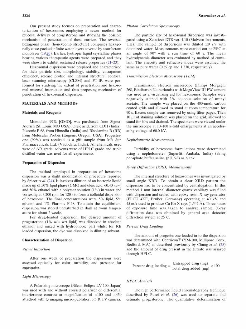
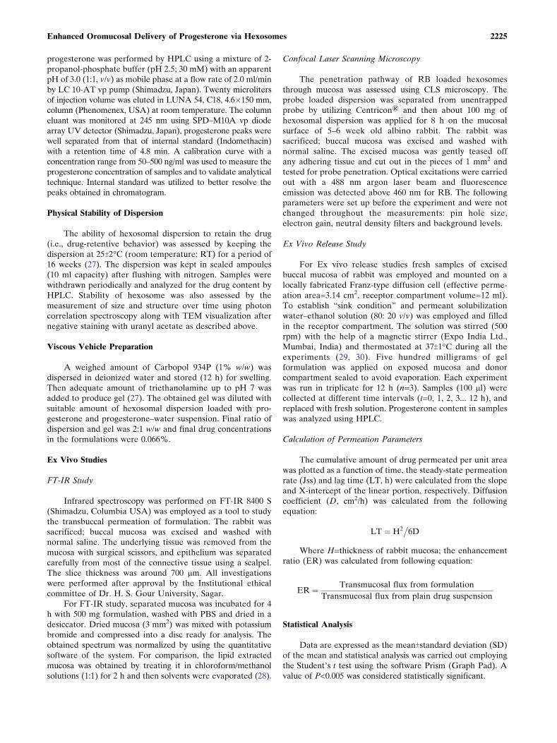
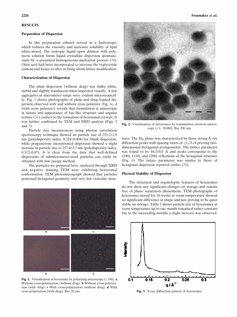
The plain dispersion (without drug) was milky white,turbid and slightly translucent when inspected visually. A fewaggregates in micrometer range were evident microscopical-ly. Fig. 1 shows photographs of plain and drug loaded dis-persion observed with and without cross polarizer. Fig. 1c, d(with cross polarizer) reveals that formulation is anisotropicin nature and appearance of fan like structure and angulartexture (31) confers to the formation of hexosomal crystals. Itwas further confirmed by TEM and XRD analysis (Figs. 2and 3).
Particle size measurement using photon correlationspectroscopy technique showed an particle size of 251.2T1.8nm (polydispersity index, 0.324T0.004) for blank dispersion,while progesterone incorporated dispersion showed a slightincrease in particle size to 257.4T2.3 nm (polydispersity index,0.312T0.07). It is clear from the data that well-defineddispersions of submicrometer-sized particles can easily beobtained with low energy method.
The particles we prepared here, analyzed through XRDand negative staining TEM were exhibiting hexosomalconformation. TEM photomicrograph showed that particlespossessed hexagonal geometry and very few vesicular struc-
tures. The HII phase was characterized by three strong X-raydiffraction peaks with spacing ratios of ¾1:¾3:¾4 proving two-dimensional hexagonal arrangements. The lattice parameterwas found to be 64.2T0.3 A and peaks correspond to the(100), (110), and (200) reflections of the hexagonal structure(Fig. 3). The lattice parameter was similar to those ofhexagonal dispersion reported earlier (32).
Physical Stability of Dispersion
The structural and organoleptic features of hexosomesdo not show any significant changes on storage and remainfree of phase separation phenomena. TEM photographs ofhexosomes stored for 16 weeks at room temperature showedno significant difference in shape and size proving to be quietstable on storage. Table I shows particle size of hexosomes atroom temperature up to one month remained rather constantbut in the succeeding months a slight increase was observed.
Fig. 1. Visualization of hexosome by polarizing microscope (�100). a
Without cross-polarization (without drug). b Without cross-polariza-
tion (with drug). c With cross-polarization (without drug). d With
cross-polarization (with drug). Bar 20 mm.
Fig. 2. Visualization of hexosomes by transmission electron micros-
copy (�1, 10,000). Bar 200 nm.
Fig. 3. X-ray diffraction pattern of hexosomes.
2226 Swarnaker et al.
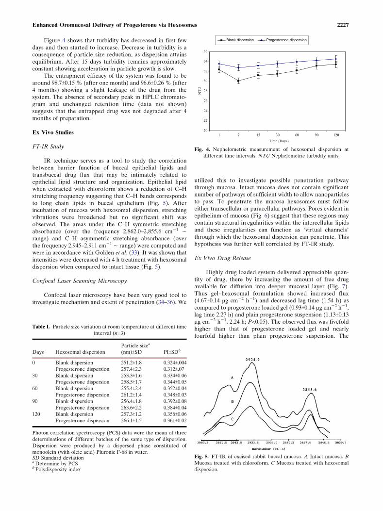
Figure 4 shows that turbidity has decreased in first fewdays and then started to increase. Decrease in turbidity is aconsequence of particle size reduction, as dispersion attainsequilibrium. After 15 days turbidity remains approximatelyconstant showing acceleration in particle growth is slow.
The entrapment efficacy of the system was found to bearound 98.7T0.15 % (after one month) and 96.6T0.26 % (after4 months) showing a slight leakage of the drug from thesystem. The absence of secondary peak in HPLC chromato-gram and unchanged retention time (data not shown)suggests that the entrapped drug was not degraded after 4months of preparation.
Ex Vivo Studies
FT-IR Study
IR technique serves as a tool to study the correlationbetween barrier function of buccal epithelial lipids andtransbuccal drug flux that may be intimately related toepithelial lipid structure and organization. Epithelial lipidwhen extracted with chloroform shows a reduction of C–Hstretching frequency suggesting that C–H bands correspondsto long chain lipids in buccal epithelium (Fig. 5). Afterincubation of mucosa with hexosomal dispersion, stretchingvibrations were broadened but no significant shift wasobserved. The areas under the C–H symmetric stretchingabsorbance (over the frequency 2,862.0–2,855.6 cmj1 õrange) and C–H asymmetric stretching absorbance (overthe frequency 2,945–2,911 cmj1 õ range) were computed andwere in accordance with Golden et al. (33). It was shown thatintensities were decreased with 4 h treatment with hexosomaldispersion when compared to intact tissue (Fig. 5).
Confocal Laser Scanning Microscopy
Confocal laser microscopy have been very good tool toinvestigate mechanism and extent of penetration (34–36). We
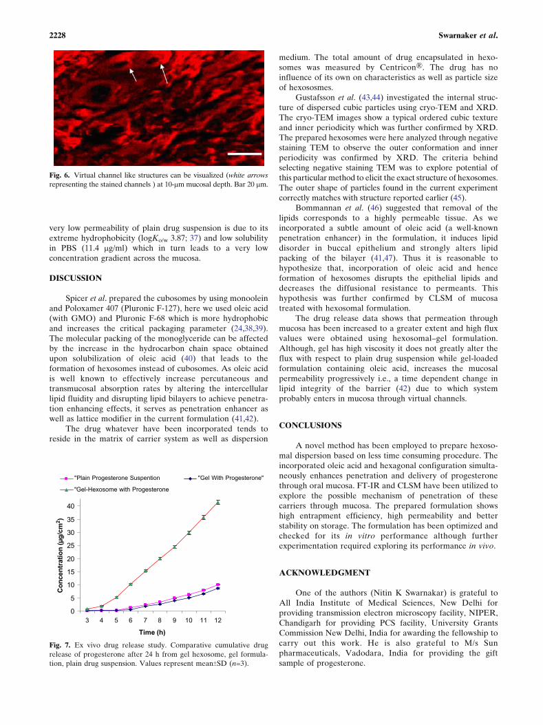
utilized this to investigate possible penetration pathwaythrough mucosa. Intact mucosa does not contain significantnumber of pathways of sufficient width to allow nanoparticlesto pass. To penetrate the mucosa hexosomes must followeither transcellular or paracellular pathways. Pores evident inepithelium of mucosa (Fig. 6) suggest that these regions maycontain structural irregularities within the intercellular lipidsand these irregularities can function as Fvirtual channels_through which the hexosomal dispersion can penetrate. Thishypothesis was further well correlated by FT-IR study.
Ex Vivo Drug Release
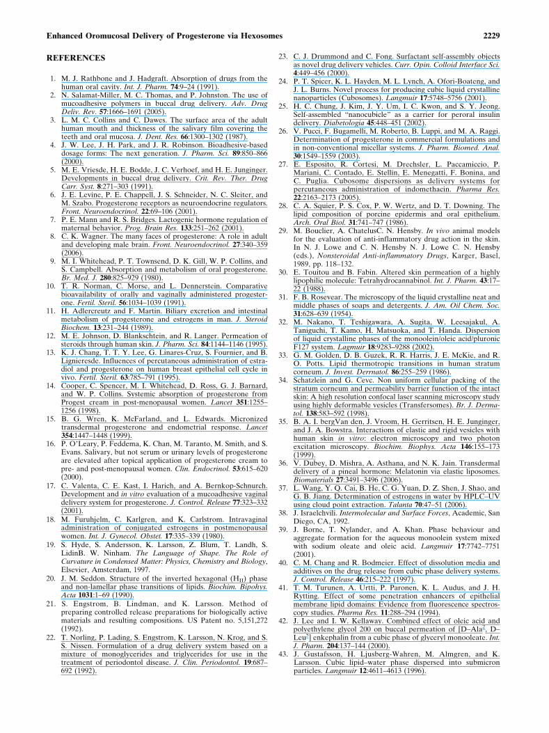
Highly drug loaded system delivered appreciable quan-tity of drug, there by increasing the amount of free drugavailable for diffusion into deeper mucosal layer (Fig. 7).Thus gel–hexosomal formulation showed increased flux(4.67T0.14 mg cmj2 hj1) and decreased lag time (1.54 h) ascompared to progesterone loaded gel (0.93T0.14 mg cmj2 hj1,lag time 2.27 h) and plain progesterone suspension (1.13T0.13mg cmj2 hj1, 2.24 h; P<0.05). The observed flux was fivefoldhigher than that of progesterone loaded gel and nearlyfourfold higher than plain progesterone suspension. The
Table I. Particle size variation at room temperature at different time
interval (n=3)
Days Hexosomal dispersion
Particle sizea
(nm)TSD PITSDb
0 Blank dispersion 251.2T1.8 0.324T.004
Progesterone dispersion 257.4T2.3 0.312T.07
30 Blank dispersion 253.3T1.6 0.334T0.06
Progesterone dispersion 258.5T1.7 0.344T0.05
60 Blank dispersion 255.4T2.4 0.352T0.04
Progesterone dispersion 261.2T1.4 0.348T0.03
90 Blank dispersion 256.4T1.8 0.392T0.08
Progesterone dispersion 263.6T2.2 0.384T0.04
120 Blank dispersion 257.3T1.2 0.356T0.06
Progesterone dispersion 266.1T1.5 0.361T0.02
Photon correlation spectroscopy (PCS) data were the mean of three
determinations of different batches of the same type of dispersion.
Dispersion were produced by a dispersed phase constituted of
monoolein (with oleic acid) Pluronic F-68 in water.SD Standard deviationa Determine by PCSb Polydispersity index
20
22
24
26
28
30
32
34
36
1 7 15 30 60 90 120
Time (Days)
NT
U
Blank dispersion Progesterone dispersion
Fig. 4. Nephelometric measurement of hexosomal dispersion at
different time intervals. NTU Nephelometric turbidity units.
Fig. 5. FT-IR of excised rabbit buccal mucosa. A Intact mucosa. B
Mucosa treated with chloroform. C Mucosa treated with hexosomal
dispersion.
2227Enhanced Oromucosal Delivery of Progesterone via Hexosomes
very low permeability of plain drug suspension is due to itsextreme hydrophobicity (logKo/w 3.87; 37) and low solubilityin PBS (11.4 mg/ml) which in turn leads to a very lowconcentration gradient across the mucosa.
DISCUSSION
Spicer et al. prepared the cubosomes by using monooleinand Poloxamer 407 (Pluronic F-127), here we used oleic acid(with GMO) and Pluronic F-68 which is more hydrophobicand increases the critical packaging parameter (24,38,39).The molecular packing of the monoglyceride can be affectedby the increase in the hydrocarbon chain space obtainedupon solubilization of oleic acid (40) that leads to theformation of hexosomes instead of cubosomes. As oleic acidis well known to effectively increase percutaneous andtransmucosal absorption rates by altering the intercellularlipid fluidity and disrupting lipid bilayers to achieve penetra-tion enhancing effects, it serves as penetration enhancer aswell as lattice modifier in the current formulation (41,42).
The drug whatever have been incorporated tends toreside in the matrix of carrier system as well as dispersion
medium. The total amount of drug encapsulated in hexo-somes was measured by Centricon\. The drug has noinfluence of its own on characteristics as well as particle sizeof hexososmes.
Gustafsson et al. (43,44) investigated the internal struc-ture of dispersed cubic particles using cryo-TEM and XRD.The cryo-TEM images show a typical ordered cubic textureand inner periodicity which was further confirmed by XRD.The prepared hexosomes were here analyzed through negativestaining TEM to observe the outer conformation and innerperiodicity was confirmed by XRD. The criteria behindselecting negative staining TEM was to explore potential ofthis particular method to elicit the exact structure of hexosomes.The outer shape of particles found in the current experimentcorrectly matches with structure reported earlier (45).
Bommannan et al. (46) suggested that removal of thelipids corresponds to a highly permeable tissue. As weincorporated a subtle amount of oleic acid (a well-knownpenetration enhancer) in the formulation, it induces lipiddisorder in buccal epithelium and strongly alters lipidpacking of the bilayer (41,47). Thus it is reasonable tohypothesize that, incorporation of oleic acid and henceformation of hexosomes disrupts the epithelial lipids anddecreases the diffusional resistance to permeants. Thishypothesis was further confirmed by CLSM of mucosatreated with hexosomal formulation.
The drug release data shows that permeation throughmucosa has been increased to a greater extent and high fluxvalues were obtained using hexosomal–gel formulation.Although, gel has high viscosity it does not greatly alter theflux with respect to plain drug suspension while gel-loadedformulation containing oleic acid, increases the mucosalpermeability progressively i.e., a time dependent change inlipid integrity of the barrier (42) due to which systemprobably enters in mucosa through virtual channels.
CONCLUSIONS
A novel method has been employed to prepare hexoso-mal dispersion based on less time consuming procedure. Theincorporated oleic acid and hexagonal configuration simulta-neously enhances penetration and delivery of progesteronethrough oral mucosa. FT-IR and CLSM have been utilized toexplore the possible mechanism of penetration of thesecarriers through mucosa. The prepared formulation showshigh entrapment efficiency, high permeability and betterstability on storage. The formulation has been optimized andchecked for its in vitro performance although furtherexperimentation required exploring its performance in vivo.
ACKNOWLEDGMENT
One of the authors (Nitin K Swarnakar) is grateful toAll India Institute of Medical Sciences, New Delhi forproviding transmission electron microscopy facility, NIPER,Chandigarh for providing PCS facility, University GrantsCommission New Delhi, India for awarding the fellowship tocarry out this work. He is also grateful to M/s Sunpharmaceuticals, Vadodara, India for providing the giftsample of progesterone.
Fig. 6. Virtual channel like structures can be visualized (white arrows
representing the stained channels ) at 10-mm mucosal depth. Bar 20 mm.
0
5
10
15
20
25
30
35
40
3 4 5 6 7 8 9 10 11 12
Time (h)
Co
ncen
trati
on
(µ
g/c
m2)
"Plain Progesterone Suspention "Gel With Progesterone"
"Gel-Hexosome with Progesterone
Fig. 7. Ex vivo drug release study. Comparative cumulative drug
release of progesterone after 24 h from gel hexosome, gel formula-
tion, plain drug suspension. Values represent meanTSD (n=3).
2228 Swarnaker et al.
REFERENCES
1. M. J. Rathbone and J. Hadgraft. Absorption of drugs from thehuman oral cavity. Int. J. Pharm. 74:9–24 (1991).
2. N. Salamat-Miller, M. C. Thomas, and P. Johnston. The use ofmucoadhesive polymers in buccal drug delivery. Adv. DrugDeliv. Rev. 57:1666–1691 (2005).
3. L. M. C. Collins and C. Dawes. The surface area of the adulthuman mouth and thickness of the salivary film covering theteeth and oral mucosa. J. Dent. Res. 66:1300–1302 (1987).
4. J. W. Lee, J. H. Park, and J. R. Robinson. Bioadhesive-baseddosage forms: The next generation. J. Pharm. Sci. 89:850–866(2000).
5. M. E. Vriesde, H. E. Bodde, J. C. Verhoef, and H. E. Junginger.Developments in buccal drug delivery. Crit. Rev. Ther. DrugCarr. Syst. 8:271–303 (1991).
6. J. E. Levine, P. E. Chappell, J. S. Schneider, N. C. Sleiter, andM. Szabo. Progesterone receptors as neuroendocrine regulators.Front. Neuroendocrinol. 22:69–106 (2001).
7. P. E. Mann and R. S. Bridges. Lactogenic hormone regulation ofmaternal behavior. Prog. Brain Res. 133:251–262 (2001).
8. C. K. Wagner. The many faces of progesterone: A role in adultand developing male brain. Front. Neuroendocrinol. 27:340–359(2006).
9. M. I. Whitehead, P. T. Townsend, D. K. Gill, W. P. Collins, andS. Campbell. Absorption and metabolism of oral progesterone.Br. Med. J. 280:825–929 (1980).
10. T. R. Norman, C. Morse, and L. Dennerstein. Comparativebioavailability of orally and vaginally administered progester-one. Fertil. Steril. 56:1034–1039 (1991).
11. H. Adlercreutz and F. Martin. Biliary excretion and intestinalmetabolism of progesterone and estrogens in man. J. SteroidBiochem. 13:231–244 (1989).
12. M. E. Johnson, D. Blankschtein, and R. Langer. Permeation ofsteroids through human skin. J. Pharm. Sci. 84:1144–1146 (1995).
13. K. J. Chang, T. T. Y. Lee, G. Linares-Cruz, S. Fournier, and B.Lignieresde. Influences of percutaneous administration of estra-diol and progesterone on human breast epithelial cell cycle invivo. Fertil. Steril. 63:785–791 (1995).
14. Cooper, C. Spencer, M. I. Whitehead, D. Ross, G. J. Barnard,and W. P. Collins. Systemic absorption of progesterone fromProgest cream in post-menopausal women. Lancet 351:1255–1256 (1998).
15. B. G. Wren, K. McFarland, and L. Edwards. Micronizedtransdermal progesterone and endometrial response. Lancet354:1447–1448 (1999).
16. P. O_Leary, P. Feddema, K. Chan, M. Taranto, M. Smith, and S.Evans. Salivary, but not serum or urinary levels of progesteroneare elevated after topical application of progesterone cream topre- and post-menopausal women. Clin. Endocrinol. 53:615–620(2000).
17. C. Valenta, C. E. Kast, I. Harich, and A. Bernkop-Schnurch.Development and in vitro evaluation of a mucoadhesive vaginaldelivery system for progesterone. J. Control. Release 77:323–332(2001).
18. M. Furuhjelm, C. Karlgren, and K. Carlstrom. Intravaginaladministration of conjugated estrogens in postmenopausalwomen. Int. J. Gynecol. Obstet. 17:335–339 (1980).
19. S. Hyde, S. Andersson, K. Larsson, Z. Blum, T. Landh, S.LidinB. W. Ninham. The Language of Shape. The Role ofCurvature in Condensed Matter: Physics, Chemistry and Biology,Elsevier, Amsterdam, 1997.
20. J. M. Seddon. Structure of the inverted hexagonal (HII) phaseand non-lamellar phase transitions of lipids. Biochim. Bipohys.Acta 1031:1–69 (1990).
21. S. Engstrom, B. Lindman, and K. Larsson. Method ofpreparing controlled release preparations for biologically activematerials and resulting compositions. US Patent no. 5,151,272(1992).
22. T. Norling, P. Lading, S. Engstrom, K. Larsson, N. Krog, and S.S. Nissen. Formulation of a drug delivery system based on amixture of monoglycerides and triglycerides for use in thetreatment of periodontol disease. J. Clin. Periodontol. 19:687–692 (1992).
23. C. J. Drummond and C. Fong. Surfactant self-assembly objectsas novel drug delivery vehicles. Curr. Opin. Colloid Interface Sci.4:449–456 (2000).
24. P. T. Spicer, K. L. Hayden, M. L. Lynch, A. Ofori-Boateng, andJ. L. Burns. Novel process for producing cubic liquid crystallinenanoparticles (Cubosomes). Langmuir 17:5748–5756 (2001).
25. H. C. Chung, J. Kim, J. Y. Um, I. C. Kwon, and S. Y. Jeong.Self-assembled Bnanocubicle^ as a carrier for peroral insulindelivery. Diabetologia 45:448–451 (2002).
26. V. Pucci, F. Bugamelli, M. Roberto, B. Luppi, and M. A. Raggi.Determination of progesterone in commercial formulations andin non-conventional micellar systems. J. Pharm. Biomed. Anal.30:1549–1559 (2003).
27. E. Esposito, R. Cortesi, M. Drechsler, L. Paccamiccio, P.Mariani, C. Contado, E. Stellin, E. Menegatti, F. Bonina, andC. Puglia. Cubosome dispersions as delivery systems forpercutaneous administration of indomethacin. Pharma Res.22:2163–2173 (2005).
28. C. A. Squier, P. S. Cox, P. W. Wertz, and D. T. Downing. Thelipid composition of porcine epidermis and oral epithelium.Arch. Oral Biol. 31:741–747 (1986).
29. M. Bouclier, A. ChatelusC. N. Hensby. In vivo animal modelsfor the evaluation of anti-inflammatory drug action in the skin.In N. J. Lowe and C. N. Hensby N. J. Lowe C. N. Hensby(eds.), Nonsteroidal Anti-inflammatory Drugs, Karger, Basel,1989, pp. 118–132.
30. E. Touitou and B. Fabin. Altered skin permeation of a highlylipophilic molecule: Tetrahydrocannabinol. Int. J. Pharm. 43:17–22 (1988).
31. F. B. Rosevear. The microscopy of the liquid crystalline neat andmiddle phases of soaps and detergents. J. Am. Oil Chem. Soc.31:628–639 (1954).
32. M. Nakano, T. Teshigawara, A. Sugita, W. Leesajakul, A.Taniguchi, T. Kamo, H. Matsuoka, and T. Handa. Dispersionof liquid crystalline phases of the monoolein/oleic acid/pluronicF127 system. Lagmuir 18:9283–9288 (2002).
33. G. M. Golden, D. B. Guzek, R. R. Harris, J. E. McKie, and R.O. Potts. Lipid thermotropic transitions in human stratumcorneum. J. Invest. Derrnatol. 86:255–259 (1986).
34. Schatzlein and G. Cevc. Non uniform cellular packing of thestratum corneum and permeability barrier function of the intactskin: A high resolution confocal laser scanning microscopy studyusing highly deformable vesicles (Transfersomes). Br. J. Derma-tol. 138:583–592 (1998).
35. B. A. I. bergVan den, J. Vroom, H. Gerritsen, H. E. Junginger,and J. A. Bowstra. Interactions of elastic and rigid vesicles withhuman skin in vitro: electron microscopy and two photonexcitation microscopy. Biochim. Biophys. Acta 146:155–173(1999).
36. V. Dubey, D. Mishra, A. Asthana, and N. K. Jain. Transdermaldelivery of a pineal hormone: Melatonin via elastic liposomes.Biomaterials 27:3491–3496 (2006).
37. L. Wang, Y. Q. Cai, B. He, C. G. Yuan, D. Z. Shen, J. Shao, andG. B. Jiang. Determination of estrogens in water by HPLC–UVusing cloud point extraction. Talanta 70:47–51 (2006).
38. J. Israelchvili. Intermolecular and Surface Forces, Academic, SanDiego, CA, 1992.
39. J. Borne, T. Nylander, and A. Khan. Phase behaviour andaggregate formation for the aqueous monoolein system mixedwith sodium oleate and oleic acid. Langmuir 17:7742–7751(2001).
40. C. M. Chang and R. Bodmeier. Effect of dissolution media andadditives on the drug release from cubic phase delivery systems.J. Control. Release 46:215–222 (1997).
41. T. M. Turunen, A. Urtti, P. Paronen, K. L. Audus, and J. H.Rytting. Effect of some penetration enhancers of epithelialmembrane lipid domains: Evidence from fluorescence spectros-copy studies. Pharma Res. 11:288–294 (1994).
42. J. Lee and I. W. Kellaway. Combined effect of oleic acid andpolyethylene glycol 200 on buccal permeation of [D–Ala2, D–Leu5] enkephalin from a cubic phase of glyceryl monooleate. Int.J. Pharm. 204:137–144 (2000).
43. J. Gustafsson, H. Ljusberg-Wahren, M. Almgren, and K.Larsson. Cubic lipid–water phase dispersed into submicronparticles. Langmuir 12:4611–4613 (1996).
2229Enhanced Oromucosal Delivery of Progesterone via Hexosomes

44. J. Gustafsson, H. Ljusberg-Wahren, M. Almgren, and K. Larsson.Submicron particles of reversed lipid phases in water stabilized bya nonionic amphiphilic polymer. Langmuir 13:6964–6971 (1997).
45. J. Barauskas, M. Johnsson, and F. Tiberg. Self-assembled lipidsuperstructures: Beyond vesicles and liposomes. Nano Lett.5:1615–1619 (2005).
46. D. Bommannan, R. O. Potts, and R. H. Guy. Examination ofstratum corneum barrier function in vivo by infrared spectros-copy. J. Invest. Dermatol. 95:403–408 (1990).
47. G. M. Golden, J. E. McKie, and R. O. Potts. Role of stratumcorneum lipid fluidity in transdermal drug flux. J. Pharm. Sci.76:25–28 (1987).
2230 Swarnaker et al.