engineering stem cells into organs: topobiological transformations demonstrated by beak, feather,...
TRANSCRIPT

5 ____________________________________________________________________________
Engineering Stem Cells into Organs:Topobiological Transformations Demonstratedby Beak, Feather, and Other EctodermalOrgan Morphogenesis
Cheng‐Ming Chuong, Ping Wu, Maksim Plikus, Ting‐Xin Jiang, andRandall Bruce WidelitzDepartment of Pathology, Keck School of Medicine
University of Southern California, Los Angeles, California 90033
I. Introduction
II. Between Stem Cells and Organs
III. Topobiological Transformation Events in Epithelial Organ Formation
IV. Feather Morphogenesis
A. Development
B. Topobiology of Multiprimordium Organs
C. Evolution
V. Beak Morphogenesis
A. Development
B. Topology of Multicomponent Organs
C. Evolution
VI. Topobiology of Other Organs
VII. Topographic Specificity of Multiprimordia Organs
VIII. Integration of Stem Cells and Organs to Reach the Level of System Biology
Acknowledgments
References
To accomplish regenerative medicine, several critical issues in stem cell
biology have to be solved, including the identification of sources, the
expanding population, building them into organs, and assimilating them to
the host. Although many stem cells can now diVerentiate along certain
lineages, knowledge on how to use them to build organs lags behind. Here
we focus on topobiological events that bridge this gap, for example, the
regulation of number, size, axes, shape, arrangement, and architecture
during organogenesis. Rather than reviewing detail molecular pathways
known to disrupt organogenesis when perturbed, we highlight conceptual
questions at the topobiological level and ask how cellular and molecular
mechanisms can work to explain these phenomena. The avian integument is
used as the Rosetta stone because the molecular activities are linked to
organ forms that are visually apparent and have functional consequences
Current Topics in Developmental Biology, Vol. 72 0070-2153/06 $35.00Copyright 2006, Elsevier Inc. All rights reserved. 237 DOI: 10.1016/S0070-2153(05)72005-6

during evolution with fossil records and extant diversity. For example, we
show that feather pattern formation is the equilibrium of stochastic
interactions among multiple activators and inhibitors. Although morpho-
gens and receptors are coded by the genome, the result is based on the
summed physical‐chemical properties on the whole cell’s surface and is self‐organizing. For another example, we show that developing chicken and
duck beaks contain diVerently configured localized growth zones (LoGZs)
and can modulate chicken beaks to phenocopy diverse avian beaks in nature
by altering the position, number, size, and duration of LoGZs. DiVerentorgans have their unique topology and we also discuss shaping mechanisms
of liver and diVerent ways of branching morphogenesis. Multi‐primordium
organs (e.g., feathers, hairs, and teeth) have additional topographic
specificities across the body surface, an appendage field, or within an
appendage. Promises and problems in reconstitute feather/hair follicles and
other organs are discussed. Finally, simple modification at the topobiolo-
gical level may lead to novel morphology for natural selection at the
evolution level. � 2006, Elsevier Inc.
I. Introduction
One of the most fundamental questions in biology is how the single dimen-
sion genomic codes are transformed into three‐dimensional forms that are
even able to morph temporally. As the genomics of diVerent organisms are
gradually completed, in the post‐genomic age, we need to learn more about
how the molecular events are translated to biological structures and how
cells are arranged in time and space to build an organ. In the last decade,
many secreted regulatory pathways (e.g., sonic hedgehog [Shh], bone mor-
phogenic protein [BMP], and Wnt) were identified and developmental biol-
ogists gained a lot of new understanding and insight into the morphogenetic
processes in development and diseases (Hogan and Kolodziej, 2002; Moon
et al., 2004; Scott, 2000; Tickle, 2003). However, as we all analyzed molecu-
lar pathways more, we gradually grew less satisfied that we could disrupt
organ formation by misexpressing certain molecular pathways but did not
know how the molecular pathways work together to build an organ. We
have the ability to dissect molecular pathways and we know certain molecu-
lar pathways are essential, yet we do not know enough to assemble them into
organs (Fig. 1).
Maybe we should also look at a more global level in order to strive for
integration of multiple molecular and cellular pathways. Maybe it is time to
revisit the topobiology concept. As Dr. Gerald M. Edelman (1988a) muses,
‘‘While the triumph of molecular biology answers the question on the chemi-
cal nature of genes and how hereditary traits are transmitted, it does not fully
238 Chuong et al.

answer the question on how genes determine traits.’’ He felt that ‘‘it is very
diYcult to account for the forms, patterns or shapes of complex animals
simply by extrapolating from the rules governing the shape of proteins,’’
and therefore turned to ‘‘the other side of biology,’’ hence the birth of
‘‘topobiology.’’ He defined topobiology as the ‘‘place dependent molecular
interactions at the cell surface’’ (Edelman, 1988a). He emphasized the funda-
mental importance of cell proliferation, adhesion, migration, death, and
diVerentiation, and particularly the links of cell collectives by cell adhesion
molecules and the regulation of these links. A single cell is capable of prolifer-
ation, migration, shape changes, apoptosis, and diVerentiation, but cell adhe-sion, epithelial sheet morphogenesis, and tissue interactions require cell
Figure 1 Levels of organ formation. From molecules to the organism, there are diVerent levels
of interactions. Each level is important and interdependent but also operates with diVerent
principles.
5. Topobiology of Stem Cells and Organogenesis 239

collectives. The topobiology concept focuses on multicellular activities to
examine how multipotential stem cells are organized into tissues and organs,
with particular architectures, sizes, and shapes.
The advent of genomics provides a ‘‘dictionary’’ of molecules, but we still
lack the syntax of how this information is used. New understanding has been
gained for studying molecular interactions, enhancer regulations, and path-
way activities. These molecular events are integrated at the cellular level
(Fig. 1). The basic information is genetically determined because the num-
bers of adhesion molecules or morphogen receptors on the cell membrane
are predetermined by the genome; however, the interaction among these cells
is a physicochemical phenomenon. Tissue and organ organization and struc-
ture reflect an equilibrium of thousands of chemical reactions within a
particular physical constraint. The importance of physicochemical phenom-
ena at this level has been pointed out previously (Kiskowski et al., 2004;
Newman and Frisch, 1979; Oster et al., 1985). However, major research
eVorts and hence progress has been at the molecular and cellular level. The
concept of topobiology did not get the attention it deserves and the para-
meters for topobiology remain mostly elusive. This knowledge is even more
urgent now as we start to work on stem cells and hope to build an organ for
regenerative medicine.
To understand how an organ is built, our laboratory has been using the
avian integument as the Rosetta stone. Avian feathers and beaks are good
models because the end points show distinct morphologies with functional
consequences. Their evolution occurs through a series of novel topobiologi-
cal events, which add evolutionary novelties that can be selected out by the
environment. The accessibility of avian embryos and regenerating feather
follicles provides excellent opportunities for tackling cellular and molecular
events experimentally (Brown et al., 2003). Thus, they are excellent models
to further develop the concept of topobiology. In this chapter, we first
identify gaps that need to be bridged in stem cell biology and introduce
progress that has been made in the topobiology of epithelial organs. The
work on feather organogenesis has been of intense interest because of the
many newly excavated feather‐related fossils from northern China, and our
eVort to link molecular findings with these intermediate ‘‘proto‐feather’’morphologies (reviewed in Prum and Brush, 2002; Chuong et al., 2003;
Sawyer and Knapp, 2003). The beak is used because the diverse beak shapes
in Galapagos finches inspired Darwin’s Evolution Theory. The break-
through by Tabin’s and our group (Abzhanov et al., 2004; Wu et al.,
2004a) was praised in the accompanying Science commentary, which said
‘‘Darwin will be pleased’’ (Pennisi, 2004). These works are examples de-
monstrating how natural selection engineers organ forms on a grand scale of
hundreds of millions of years in the context of ‘‘Evo‐Devo.’’ We then briefly
apply the topobiology concept to mammalian ectodermal organogenesis,
240 Chuong et al.

liver shaping, lung branching, etc. We also discuss the regional specificity
issue that we must face in engineering organs. At the end of this chapter,
we reflect on how understanding these principles may contribute to the
engineering of stem cells. With this progress, we can further develop the
topobiology concept to mean ‘‘bioinformation generated by topology‐dependent molecular expression and cellular behavior.’’
II. Between Stem Cells and Organs
Stem cell biology has emerged as an important new discipline of translation-
al research in the context of regenerative medicine. Several issues are impor-
tant in stem cell biology research. They are (A) identifying sources of stem
cells, (B) expanding stem cell populations while maintaining their properties,
(C) engineering stem cells to form the tissue/organ desired, and (D) having
the engineered tissues/organs assimilate into the host. For the first issue, the
research at this stage has been on embryonic stem cells and identifying
possible sources of adult stem cells (Fuchs and Segre, 2000; Lako et al.,
2002; Li and Xie, 2005; Toma et al., 2005). Somatic nuclei transfer technol-
ogy has allowed the progress of therapeutic cloning. For the second issue,
scientists have worked on culture conditions and found some promising
clues. For instance, Wnt has been found to help expand hematopoietic stem
cells (Reya et al., 2003).
The third issue is how to engineer these cells to organ‐like structures andbe useful for the host. This has proven to be of diVerent diYculty levels for
diVerent types of organs. For hematopoietic cells, multiple blood cell types
float in the bloodstream without being organized into a particular form and
can function in response to cytokines. This lack of structural organization
makes blood a relatively easy organ to work with, and as a result, hemato-
poietic stem cells have already been used successfully in clinical practice. The
next level is to have engineered tissues that secrete needed extracellular
factors required to alleviate disease conditions, such as insulin from pancre-
atic beta cells for diabetes (Efrat, 2004; Lumelsky et al., 2001) or dopamine‐secreted neurons for Parkinson’s disease (Snydeer and Olanow, 2005). The
next challenging level is to be able to produce certain shapes suitable for
functional morphology. For example, it is now possible to induce chondro‐diVerentiation from mesenchymal cells in culture, but it is still very diYcult
to have these cells form the right contours on a cartilage or bone element.
The use of a biodegradable polymer scaVold to generate auricular‐shapedcartilage (Shieh et al., 2004) can facilitate the process when a better solution
is not available. It would be best to find out how nature performs morpho-
genesis in development, but even nature ‘‘forgets’’ how to do it during
regeneration in the adult; during the body’s eVort to regenerate in response
5. Topobiology of Stem Cells and Organogenesis 241


to osteoarthritis, bone spurs form, which cause more damage. Even if we can
have a functional tissue/organ entity, we still have to learn how to make
them connect with the host. For example, a group of beating cardiomyocytes
have to coordinate the motion of the whole myocardium and a group of
transplanted neurons has to be connected with other parts of the brain.
Finally, stem cell–derived organs have to survive without being rejected by
the host immune system or competed out by the native cells. Therefore,
while stem cell engineering holds promise, there are many challenges before
the knowledge is translated to clinical applications.
The focus of this chapter is on the third issue: how to engineer stem cells to
form the tissue/organ desired. Suppose current stem cell research reaches a
stage at which we have enough stem cells that can be induced to form
diVerent diVerentiated phenotypes. How do we direct them to form organs?
We need to position ourselves to answer these questions. Developmental
biology used to be considered a basic science operating in an ivory tower.
Now scientists appreciate that tissue engineering and developmental biology
are two sides of the same coin: When nature does it, it is developmental
biology; when humans do it, it is stem cell engineering. The best way to
engineer stem cells is to learn how to guide them in nature’s way.
III. Topobiological Transformation Events in EpithelialOrgan Formation
Here we use topological transformation to mean the conversion from one cell
collective configuration to the other. It does not entirely fit the definition in
mathematics, but we use the term to emphasize the geometric aspect of tissue
morphogenesis: the forming and dissolution of cell groups, the shifting
arrangement, the making and elimination of boundaries, the orientations,
etc. In fact, the creation or removal of boundaries or breaking of epithelial
sheet makes them topologically nonequivalent. The formation of epithelial
organs involves topological transformation of a two‐dimensional (2D) epi-
thelial sheet into diVerent structures (Fig. 2A). In ectodermal organ forma-
tion, they can evaginate out to form bumplike configurations (e.g., scale),
some with elaborate surface (e.g., molar), protrusions (e.g., canine, claw),
Figure 2 Topobiological transformation events during epithelial organ formation. (A) A
prototype animal with ectodermal and endodermal organs. Although these epithelial organs
appear diverse, they share similar morphogenesis‐related signaling pathways and topobiological
principles (modified from Chuong, 1998). The molecular basis of epithelial appendage
morphogenesis. (B) Types of topobiological transformation events. These events are meaningful
only at the level of cell groups (epithelial sheet, mesenchymal condensations), not at the single
cell level. We need to learn more about how molecular mechanisms contribute to these events.
5. Topobiology of Stem Cells and Organogenesis 243

elongated filaments (e.g., hair), some with hierarchical branches (e.g., feath-
ers), etc. They can also invaginate to form tubes (e.g., sweat glands), some
with branching (e.g., salivary glands, mammary glands), follicles (e.g., hair,
feather), etc. (Chuong, 1998). In the endoderm, similar topological transfor-
mations occur in the gut. Regional specialization of epithelia leads to the
formation of the stomach, intestines, lungs, liver, and pancreas, which form
by budding from the gastrointestinal tract during embryonic development.
These apparently diVerent epithelial organs actually share similar topo-
biological transformation events (i.e., an event that changes the topological
configuration of cells before and after it happens). The involved molecular
mechanisms have begun to be understood. Some examples are given
(Fig. 2B).
Layer formation: In this event, randomly arranged epithelial cells start to
join with each other. The progeny of cell proliferation remains in the same
sheet as the axial orientation of mitosis within the 2D plane. Epithelial cell
adhesion molecules such as E‐cadherin were first shown to have this function
(Nagafuchi et al., 1987).
Stratification: Some mitosis becomes asymmetric with a mitotic axis becom-
ing perpendicular to the epithelial sheet. The daughter cells remaining in the
basal layer can still proliferate (the beginning of stem cells), while the other
daughter cells, now postmitotic, start to pile up, forming multiple layers.
Stratification enables the epithelia to form a multilayered barrier, protecting
the organism from its environment, and allows functional diversification.
Activation of the p63 pathway is involved in the stratification process
(Koster et al., 2004; Koster and Roop, 2004). p63 is expressed early in the
epidermal lineage when cells are still forming a single layer (Green et al.,
2003; Koster et al., 2004). p63‐null mice fail to form stratified epithelial
derivatives (Mills et al., 1999).
Convergent extension: Convergent extension allows a change of shape of
epithelial sheets by cell rearrangements. Lateral and medial cells become
polarized and then the lateral cells intercalate between the medial cells,
causing an extension along the anteroposterior axis (Keller, 2002). This
process was originally shown to be responsible for gastrulation in Xenopus
and zebrafish (Keller, 1986), gut elongation in sea urchins (Ettensohn, 1985;
Hardin and Cheng, 1986), the formation of the avian primitive streak (Wei
and Mikawa, 2000), and shaping of the avian neural plate (Schoenwolf,
1991; Schoenwolf and Alvarez, 1989). It is likely to be a fundamental
topological transformation process involved in other organ formation.
Signaling along the noncanonical Wnt pathway is likely to be involved.
Invagination: Invagination of epithelial tissues is seen in the organization of
the neuroepithelium in Xenopus (Schoenwolf and Alvarez, 1989). It also
244 Chuong et al.

plays a critical role in tooth formation (Jernvall and ThesleV, 2000). Theactivation of Wnt/�‐catenin and the suppression of BMP by noggin leads to
an invagination of the epithelial placode to initiate hair follicle formation
(Jamora et al., 2003).
Tube formation: Tube formation can occur through rearrangements of
epithelial cells to form a lumen within an elongated cell cord. Tubular
structures can form in many ways. An epithelial sheet can curl and seal itself
to form a tube. This occurs during neural tube formation (Colas and
Schoenwolf, 2001). This involves cell shape changes forming a narrow apical
region and a broad basal region. Tubes can also form by budding out from
an epithelial surface. The lung is thought to branch out in this manner
(Hogan and Kolodziej, 2002; Metzger and Krasnow, 1999). A mass of cells
can invaginate to form a central cavity, as occurs during salivary gland
formation (Melnick and Jaskoll, 2000). Apoptosis may play a role in this
mechanism (Coucouvanis and Martin, 1995). In angiogenesis, hemangio-
blasts form an aggregate called blood islands. The inner cells become
hematopoietic stem cells while the outer cells become angioblasts, which
go on to multiply and diVerentiate into endothelial cells forming the blood
vessels. So cords of hemangioblasts hollow out to form a tube (reviewed in
Baron, 2003).
Branching: Branching is used to increase the surface area for interactions
with the environment, be it internal or external. Branching involves the
splitting of the long axis into two. While the end results can be quite similar,
they can be generated from very diVerent mechanisms. It can be generated
by diVerential growth or death. The process is seen in lung and mammary
gland morphogenesis (see Section VI, later in this chapter), as well as in
feather barb branching.
Condensations and decondensations: This involves increased cell adhesion
that brings out a group of highly compacted cells, or the reverse of this
process. Not only physically does a cell collective form or dissolve, but there
are also changes of cell properties due to signaling initiated by cell contacts.
The formation of dermal condensations is a very early step in feather
formation (Chuong and Edelman, 1985a; Jiang and Chuong, 1992). The
regulation of this process leads to periodic pattern formation (see Section
VII, later in this chapter). The migration of neural crest cells is a good
physiological example of epithelial–mesenchymal transformation (Kang
and Svoboda, 2005).
Fusion: When two cell collectives meet, the epithelial can remain as two
entities with a surface boundary in between, or the boundary disappears
and two cell collectives fuse into one. This may occur through epithelial–
mesenchymal transformation (Kang and Svoboda, 2005) or may involve
apoptosis.
5. Topobiology of Stem Cells and Organogenesis 245

IV. Feather Morphogenesis
Feathers on the bird body show hierarchical branch patterns (Prum and
Dyck, 2003). The major types of avian feathers include contour feathers,
remiges, rectrices, downy feathers, etc. (Lucas and Stettenheim, 1972).
A typical avian feather consists of a shaft (rachis) and barbs. The barbs
are composed of a shaft (ramus) and many smaller branches (barbules)
(Fig. 3A). DiVerent feathers show variations in symmetry. Down feathers
are radially symmetric. Their rachis is absent or very short. Contour feathers
have a weak bilateral symmetry. Flight feathers are bilaterally symmetric
and some become bilaterally asymmetric (see later discussion) (Fig. 5).
A contour feather can have a distal pennaceous region and a proximal
plumulaceous region, so the feather can help the integument function for
contour/communication display with the distal portion but maintain
warmth with its proximal plumulaceous portion (Fig. 9C). The plumulac-
eous regions are made of similarly shaped barbules both proximal and distal
to the ramus. They are loose and fluVy. The pennaceous regions are made of
groove‐shaped proximal barbules and hook‐shaped distal barbules. There-
fore, the distal barbules of a barb interlock with the proximal barbules of the
barb above, forming a feather vane using a Velcro‐like mechanism.
A. Development
During avian embryonic development, feather formation starts with a pla-
code, which is composed of elongated epithelia accompanied with dermal
condensations (Sengel, 1976; Wu et al., 2004b). These feather primordia
elongate and protrude out to form feather buds, topologically transforming
a 2D flat epidermis into a three‐dimensional (3D) structure (Chuong and
Edelman, 1985b) (Fig. 3C). Feather buds are originally radially symmetric
but soon acquire anteroposterior polarity through interactions with the
epithelium. Feathers then start to elongate and develop a proximal‐distalaxis. Feathers form follicles that oVer advantages over skin appendages that
do not, such as scales. The follicular structure protects the epithelial stem
cells and dermal papillae. Localization of the stem cells within a protected
environment enables regeneration through natural feather molting cycles
and induction by plucking. New cell proliferation at the follicle base pushes
the more diVerentiated portions of the feather filament to the distal end.
Feather filaments go through epithelial invaginations and evaginations to
form the barb ridges, which precede the formation of the barbs and barbules.
The barb ridges further diVerentiate into the barbule plates, axial plates, and
marginal plates. Barbule plate cells later keratinize to become the feather
structure, while marginal plate and axial plate cells undergo apoptosis, die,
246 Chuong et al.
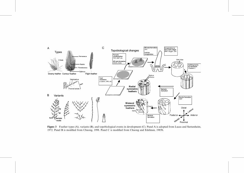
Figure 3 Feather types (A), variants (B), and topobiological events in development (C). Panel A is adopted from Lucas and Stettenheim,
1972. Panel B is modified from Chuong, 1998. Panel C is modified from Chuong and Edelman, 1985b.

and become spaces (Chang et al., 2004) (Fig. 4). The central pulp undergoes
apoptosis, allowing the feathers to unfold and assume their characteristic flat
shapes, transforming a 3D cylinder back to a 2D plane. Topobiological
transformation events are listed in the boxes in Fig. 3C. In each process,
signaling molecules are used in diVerent ways (reviewed in Widelitz et al.,
2003; Jiang et al., 2004; Wu et al., 2004b, and references within), and some
Figure 4 Pattern‐forming processes that regulate the number and size of multiple primordia
within a field. Panel B is from Jiang et al., 1999. Part of it is from Jiang et al., 1994, 2004.
248 Chuong et al.

(e.g., BMP, Shh) are used repetitively in diVerent contexts in the so called
co‐optive use of signaling modules (Harris et al., 2002).
With so many topological parameters involved, tuning of some of these
parameters can lead to diVerent feather shapes (Prum and Williamson,
2001), generating the diverse feather shapes in nature. The range of feather
variants can be appreciated in Bartels (2003) and the interesting photos in
Extraordinary Chickens (Green‐Armytage, 2000). Schematic examples of
these variants can be seen in Fig. 3B. To obtain diVerent feather shapes,
one can simply change the relative length of the rachis, barbs, and barbules.
For example, in Fig. 3A, the middle one represents the fluVy contour feath-
ers of an ostrich, the right one is a strong flight feather of an eagle, and the
left represents the contour feathers on the trunk of pheasants and the natal
down. The one on the right represents the scalelike feathers of a penguin in
which the rachis is enlarged while barbs and barbules are miniaturized.
There are also the spectacular peacock tail contour feathers, and the many
unusual decorative feathers found on birds of paradise.
An interesting point is that they are all keratinocytes built into diVerentarchitectures. The variations do not just exist among diVerent avian species
but can exist in the same individual. Furthermore, the epidermal stem cells
can be guided by the dermal papilla to form diVerent feather types in
diVerent skin regions (Cohen and Espinasse, 1961; our unpublished data).
B. Topobiology of Multiprimordium Organs
Some organs are made of multiple primordia. Each primordium can be
considered as one organ, but they work together as a functional unit. This
can be seen often in integument organs such as teeth, hairs, feathers, etc. All
teeth have to work together to serve the function of breaking up food.
Feathers in a tract also have to work together. A single feather does not
permit flight, but together multiple pennaceous feathers can connect to form
a feather vane, as discussed earlier. While cells diVerentiate, the topology
(i.e., the number, shape, size, and arrangement of individual primordium) is
crucial for the way that particular organs work and provides a new level of
functional integration and variation.
Feathers are laid out in exquisite patterns on the surface of the chicken
embryo. These regular patterns have inspired scientists to think about how
such regular patterns arise (Held, 1992). In general, one category of model
considers that the fates of cells are predetermined by their position, whether
the molecular coordinates exist in the form of specific enhancer sequences or
as a morphogen gradient (Fig. 4). The other category considers the major
driving force is based on physicochemical phenomena. The reaction–
diVusion mechanism has been used to describe periodic patterning in
5. Topobiology of Stem Cells and Organogenesis 249

inanimate objects and in living systems (Gierer and Meinhardt, 1972; Jung
et al., 1998; Moore et al., 1998; Nagorcka and Mooney, 1985; Turing, 1952).
In reaction–diVusion, random fluctuations in molecular expression become
amplified to form peaks and valleys. These, however, are unstable. The
peaks and valleys were later postulated to be maintained and propagated
through chemical interactions or mechanical forces. Meinhardt and Gierer
(1974, 2000) proposed that some molecules distributed by a reaction–
diVusion mechanism might stimulate the production of the periodic struc-
tures (activators) while some suppress their synthesis (inhibitors) through
autocatalysis and cross‐catalysis. Activators also have the ability to further
stimulate the production of activators and induce the production of inhibi-
tors. Based on these models and our experimental results (Jiang et al., 1999,
2004; Jung et al., 1998), we propose a model for feather pattern formation. It
consists of the following events. (1) Competent cells without specific identity
are distributed in the field and move randomly. (2) Extracellular activators
and inhibitors governed by a reaction–diVusion mechanism diVuse in the
field. (3) Cells respond to activators and inhibitors stochastically and the
results are manifested in changes of cell adhesion. (4) Cell cluster formations
(dermal condensations) are reversible initially, then become committed once
a threshold is reached. (5) The pattern reached is the result of competitive
equilibrium. If the system is reset without changing any parameter, the
pattern with similar topology will reappear, but it will not be identical to
the original pattern.
If feather patterns are predetermined, scrambling the cells should not
change their fates. The feather reconstitution model (Jiang et al., 1999)
oVered an opportunity to test this, because it allowed us to recombine a
fixed‐sized epithelium with diVerent numbers of mesenchymal cells. When
increasing numbers of mesenchymal cells were used, we could expect either
the same number of primordia with increased size or the same size of
primordia with increased numbers of primordia (Fig. 4B). Experimental
results show that for mesenchymal cells derived from the same region, the
feather primordia were always the same size. When mesenchymal cell density
was below the threshold, no primordia formed. At lower mesenchymal cell
density, primordia appeared in random positions, not as aborted rows of a
hexagonal lattice. As more cells were added, the number of primordia
increased until they reached a maximal packing density, and feathers ap-
peared to be arranged in a hexagonal pattern. However, this hexagonal
pattern is a result of maximal packaging, not a consequence of preset
molecular codes or positional values.
Thus, the feather precursor cells at this stage are truly stem cells; they
can become either bud or interbud cells. The size, number, and spacing of
feather primordia can be regulated by altering the properties of cells or the
microenvironment (Jiang et al., 1999; Shen et al., 2004). To help patients,
250 Chuong et al.

dermatologists can implant hair follicles one by one into the alopecic scalp.
We can foresee if all these parameters can be set right, the delivered stem
cells should be able to self‐organize into multiple hair follicles as they do
during embryonic morphogenesis.
C. Evolution
During the morphological transformation from reptiles to birds, new chal-
lenges were imposed on early birds to reengineer themselves from a tetrapod
form mainly living on the land to a smaller bipedal animal with wings to live
in the sky. The Jehol Biota spreading in northern China is unique because it
contains unique features and many plants and animals are preserved in
outstanding condition (Zhou et al., 2003). It is particularly valuable for the
analysis of the evolution of birds because birds evolved from reptiles during
this period (Chatterjee, 1997; Chiappe, 1995; Feduccia, 1999). Early research
suggested that feathers evolved from an elongation of scales enlisted for
protection. It was then subdivided over time to form pennaceous and then
plumulaceous feather types (Regal, 1975) (Fig. 5, Model 1). Thus, the order
of formation is scales ! elongated scales ! the vanelike scale plates !partial pennaceous vanes with an rachis like central axis ! bilaterally
symmetric feathers ! plumulaceous barbs ! radially symmetric downy
feathers (also see Wu et al., 2004b). From the developmental and molecular
studies, Prum (1999), Prum and Brush (2002), and us (Chuong et al., 2000;
Yu et al., 2002) propose that the order of formation is buds ! follicle !cylindrical feather filaments ! splitting to form radially symmetrically
arranged barbs ! radially symmetric downy feathers with plumulaceous
barbules. By topologically changing the slanting angles of barb ridge orga-
nization, a rachis is created and the other lineage can lead to bilaterally
symmetric plumulaceous feathers! bilaterally symmetric pennaceous vanes
! bilaterally asymmetric vanes (Fig. 5, Model 2). This is also the order
observed in development. In a broad sense of ontogeny repeating phylogeny,
this probably occurred in evolution too. Indeed, a series of fossils were
discovered representing intermediate forms of feathers or featherlike
appendages from the Jehol Biota of China.
Furthermore, considering the topology of epithelium and mesenchyme,
the scale is diVerent from feathers (Chuong et al., 2003; Prum, 1999) (Fig. 6).
The scale dermis remains in the adult, and both anterior and posterior sides
of scales are equivalent to the suprabasal side of the epidermis (Fig. 5, Model
1a). In contrast, in the developing feather follicles, the cylindrical feather
filament surrounds the mesenchymal pulp with the basement membrane
facing inside. Upon maturation, apoptosis of the pulp epithelium and shed-
ding of the feather sheath allows the feathers to open. Thus, the anterior and
5. Topobiology of Stem Cells and Organogenesis 251

posterior side of the feather vane originally faces the suprabasal and basal
layer, respectively (Chang et al., 2003) (Fig. 5, Model 1). An elongated scale
may show branches and may be called a ‘‘non‐avian feather’’ (Jones et al.,
2000) but is not an avian feather.
From these results, a set of criteria have been developed to define the true
avian feathers (Chuong et al., 2003). It includes (1) possessing actively
proliferating cells in the proximal follicle for a proximodistal growth mode;
(2) forming hierarchical branches of rachis, barbs, and barbules, with barbs
that can be bilaterally or radially symmetric, formed by diVerential cell
death; (3) having a follicle structure, with a mesenchyme core during devel-
opment; (4) when this matures, it consists of epithelia without a mesenchyme
core with two sides of the vane facing the previous basal and suprabasal
Figure 5 Models on feather evolution. Model 1 proposes elongated scales as the origin of the
feather (modified from Regal., 1975). Model 2 proposes that a series of novel topobiological
transformation events, as evolution novelties, transform epidermal buds into complex feathers.
Panels 1a and 2a are cross‐sections.
252 Chuong et al.

layers, respectively; and (5) having epithelial stem cells and the dermal
papilla in the follicle, which maintains the ability to molt and regenerate.
Work in molecular biology laboratories has allowed us to start to identify
molecular pathways involved in each of these processes (Harris et al., 2002; Yu
et al., 2004) (Fig. 6). We have developed a novel feather plucking/regeneration
model to misexpress genes in the regenerating feather stem cells (Yu et al.,
2002). This allows us to gauge the contribution of eachmolecular pathway.We
showed that BMP promotes rachis formation while noggin promotes barb
branch formation. Shh is important to set up the spacing between barbs (Chang
et al., 2004). Harris et al. (2002) also showed that BMP2 and Shhmediate barb
ridge formation and have developed an activator/inhibition model to explain
the branch patterning (Harris et al., 2005).
Recently, we identified feather stem cells and found they assume a ring
configuration in the collar region. Interestingly, the ring is horizontally
placed in radial symmetric downy but tilted anterior‐posteriorly (A‐P) in
bilaterally symmetric flight feathers (Yue et al., 2005). Furthermore, an A‐PWnt 3a gradient was identified, and flattening out the Wnt gradient experi-
mentally caused bilaterally symmetric feathers to become radially symmetric
(Yue et al., 2006). These results provide supports for the hypothesis that
diverse feather forms can be generated by topobiological modulation of stem
cells, rather than specific molecular blueprints. Putting previous works to-
gether (Prum, 1999; Chuong et al., 2000), we can summarize Evo‐Devo of
feathers as the following. First, the formation of feather follicles made stem
cells and growth zone cells shift proximally to a protected environment and
also allowed continuous growth and molting. Second, the feather filament
branch became barbs, forming downys which are efficient in thermal regula-
tion. Third, topological alterations of stem cell configuration allowed the
formation of rachis and bilateral symmetry. Fourth, asymmetric barbules
formed that interweaved barbs into a vane, enabling the birds to develop
flight. Thus, a series of topobiological transformation events opened the
entire sky for the Aves class. In a way, the sky niche is the best ‘‘patent
award’’ given to birds for their successful evolutionary novelties.
V. Beak Morphogenesis
The recruitment of forelimbs as wings allowed a newly found mobility
resulting from flight and opened vast ecomorphological possibilities. How-
ever, this came at a cost because animals now needed to develop a new
feeding mechanism without the use of arms. This exerted selection pressures
on the evolving structure of the face; a strong, lightweight, and eVectivefeeding apparatus had to evolve. Furthermore, the beak had to show an
ability to evolve through adaptive radiation to diVerent ecoenvironments.
5. Topobiology of Stem Cells and Organogenesis 253

Figure 6 Molecular shaping of the beak. (A) Diverse beak shapes and the basic design of
beaks. By positioning localized growth zone in diVerent numbers and positions, the beak can
become diVerent shapes.
254 Chuong et al.

The results are the amazing transformation of the snout into a large range of
beak topologies adapted to diVerent ecological niches (Zweers et al., 1997).At the global scale, it involves a reptile snout–bird beak transformation. At
the finer scale, it involves the fine‐tuning of Galapagos finches that inspired
Darwin’s Evolution Theory (Grant, 1986). At the developmental level, how
are the diVerent shapes of beaks produced (Fig. 6A)?
A. Development
The embryonic chick face is composed of multiple facial prominences
(reviewed by Francis‐West, et al., 1998, 2003; Helms and Schneider, 2003)
(Fig. 6B). Mesenchymal processes covered by epithelium surround the de-
veloping mouth. These prominences grow out together to form the face. The
upper beak is formed from the frontal nasal mass (FNM) and MXP on the
side. Lateral nasal masses have only smaller contributions and are not
emphasized here. The lower beak is derived from the paired mandibular
prominences (MDPs), which contain the two Meckel’s cartilages. Cellular
fate tracing with DiI labeling illustrates that cell populations centered
around the nasal pits, the midline of the paired MDPs, and at sites of fusion
contribute most to the overall expansion (McGonnell et al., 1998). These
data suggest that there are specific localized growth zones in these originally
nearly round prominences. When the beak forms, FNM and MDPs assume
an elongated shape, while MXPs remain short and ball‐like. These develop-ing facial prominences change shape substantially in developing stages,
leading to the formation of primary and secondary palates. Therefore, the
final shape and size of each prominence is the combination of the diVuserandom growth and the directed localized growth in that prominence.
Growth and morphogenesis of the prominences must be tightly coordinated
to obtain the final distinct configuration of the face.
Experiments show that the identity of facial prominences are specified
early in the neural crest stage (Couly et al., 2002; Noden, 1983) and are
coordinated by signaling molecules (Francis‐West et al., 2003). An elegant
experiment by transplanting duck crest into quail embryos (forming duail)
and quail crest into duck embryos (quack) shows the beak morphology is in
accord to the origin of the cephalic neural crest (Schneider and Helms, 2003).
The identity of an MXP can be respecified to an FNM by a combination of
noggin and retinoic acid (Lee et al., 2001). BMP, fibroblast growth factor
(FGF), Shh, and Hox are involved in the formation of these prominences
(Ashique et al., 2002a,b; Barlow and Francis‐West, 1997; Creuzet et al.,
2002; Helms and Schneider, 2003; Hu et al., 2003; Hu and Helms, 1999;
Richman et al., 1997; Wilke et al., 1997). An epithelial region in the FNM
with juxtaposed FGF8/Shh was shown to induce beak outgrowth (Hu et al.,
5. Topobiology of Stem Cells and Organogenesis 255

2003). Indeed FGF8/Shh were shown to induce cranial chondrogenesis
in vitro and in vivo (Abzhanov and Tabin, 2004).
Although the facial morphology is determined by the crest cells (Schneider
and Helms, 2003), we are interested in how chicken and duck faces develop
diVerently in the late stages of morphogenesis. We showed that there are
localized mesenchymal cell proliferative zones (LoGZ) in the FNM. In both
chickens and ducks, there were two LoGZ at lateral FNM at (chicken H&H)
stage 26. They converged into one in the chicken but remained as two in the
duck. We showed that this region is enriched with BMP4 and further showed
that BMP4 is involved in mediating LoGZ activity (Wu et al., 2004a)
(Fig. 6C). Independently, Dr. Tabin’s group pursued Galapagos Island finch
beaks directly. Using cDNA library subtraction, they also found the main
candidate for beak diversity is BMP4. They went on to use chickens to show
that BMP4 is functionally involved (Abzhanov et al., 2004). The concept is
that a special activity may not be based on the presence or absence of a
signaling molecule. Rather, the configuration of signaling molecule ex-
pressing cell clusters is important. This is further demonstrated in the cleft
primary palate chicken mutant in which the abnormality is due to the failure
of FGF8 to become restrictively expressed, not the absence or mutation of
FGF8 (MacDonald et al., 2004). Therefore, BMP is likely to be the major
mediator of beak growth, while other morphoregulatory molecules can act
on the BMP pathway and in this way adjust its activity and, therefore, the
shape of the beak. How the messages in the chicken or duck neural crest cells
are translated into the topological diVerences of localized growth zones in
the FNM remains to be investigated.
B. Topology of Multicomponent Organs
One unique aspect of the beak is that it represents a paradigm of ‘‘complex
morphogenesis’’ in which an organ is made from multiple components, in
contrast to ‘‘simple morphogenesis’’ in which the whole organ is sculpted
from one primordium. Comparing the limb bud with facial morphogenesis,
the limb bud is a paradigm of ‘‘simple morphogenesis.’’ Developmental
biologists have learned a lot of the molecular mechanisms of limb morpho-
genesis in the last decade (Capdevila and Izpisua Belmonte, 2001; Dudley
and Tabin, 2000; Niswander, 2003; Tickle, 2003). Through careful analyses
of many laboratories, we now learned how molecular pathways (FGF, Shh,
Hox, Wnt, etc.) are involved in apical ectodermal ridge (AER), zone of
polarizing activity (ZPA), and dorsal‐ventral patterning that work together
to shape the limb from a single primordium.
In contrast, the beak is made from the coordinated growth of multiple
facial prominences. We try to define the following three categories of growth
256 Chuong et al.

activities during beak morphogenesis: (1) Concerted ‘‘overall growth activ-
ities’’ are responsible for the global expansion of the face, (2) ‘‘diVuse growthzone,’’ the dispersed mesenchymal growth in each prominence contributes to
diVerent dimensions of the face, (3) the ‘‘localized growth zone’’ (LoGZ),
which focuses on the temporospatial growth activities within individual
prominences, molding specific shapes out of one prominence (Fig. 6). There
appears to be a global overall growth activity in all facial prominences, and
yet each facial prominence has its distinct localized growth zone. Some
facial prominences have multiple LoGZs. Thus, for the beak of each bird,
a unique facial configuration emerges from the undulating landscape of
global growth activities with peaks and valleys fine‐tuned by LoGZs and
localized apoptotic zones.
Complex morphogenesis oVers more opportunities to generate morpho-
logical diversity (Fig. 6A), but the complex process is also prone to errors, as
seen in the high incidences of cleft palate/lips due to lack of coordination of
cellular events (MacDonald et al., 2004). We can speculate a giant beak as
seen in the Toucan may be produced when the ‘‘overall growth activity’’ is
high. By increasing the ‘‘diVuse growth activity’’ in the maxilla or mandible
alone, asymmetrically bigger upper/lower beaks may be generated, as seen in
parrots and pelicans. By adjusting the configurations of ‘‘LoGZs,’’ flat beaks
like those in ducks or vertical beaks like those seen in the seagulls may be
produced. By positioning the LoGZ in a horizontal or oblique angle, beaks
may grow straight as in the duck or curved as in the eagle. By sustaining the
activity of a focused LoGZ, a long sharp beak as seen in the crane can be
produced. The molecular bases of these interesting beak designs remain to
be investigated.
C. Evolution
How do we define an ‘‘avian beak?’’ An avian beak requires the formation of
a horny sheath, loss of teeth, and the modification of the maxilla and
mandibles into unique shapes. From the reptile to bird, the toothed jaws
were gradually transformed into beaks. Indeed, in reptiles, beaks were seen
in Psittacosaurus (a beaked dinosaur) and even in today’s turtles. During the
evolution of the beak, the trend is the gradual reduction and eventual loss
of teeth, coupled with the formation of the horny sheath by thickened
epidermal diVerentiation (Feduccia, 1999). Some Mesozoic birds existed
representing intermediate stages (Fig. 7).
Archaeopteryx had uniform reptilian teeth in both its upper and its lower
jaw. Longirostravis (125 million years ago) had a very long and slender
rostrum and signs of the presence of a horny sheath (Hou et al., 2004).
Ten small and conical‐shaped teeth are arranged in pairs and preserved in
5. Topobiology of Stem Cells and Organogenesis 257

the distal snout. As this is the earliest wading bird, the preservation of teeth
in the anterior snout may have facilitated securing its prey. The arboreal
Confuciusornis is likely to be among the early birds that have formed a real
beak with a complete loss of teeth in both of the upper and the lower beak
(Hou et al., 1996). The diversity of beaks is shaped by diet and reflects
adaptive radiation (Feduccia, 1999; Lucas and Stettenheim, 1972). Darwin’s
finches in the Galapagos Islands are derived from a common ancestor and
have evolved diVerent sizes and shapes of beaks. The variation is subject to
natural selection and environmental changes (Grant, 1986). In other birds,
seed eaters such as chickens, quails, and pigeons have conical beaks. Ducks
have soft, leathery, and flattened beaks for filtering food from the mud and
water (Lucas and Stettenheim, 1972). Hawks have curved upper beaks for
raptorial tearing.
To summarize beak morphogenesis, we have learned that beaks are made
of the same diVerentiation materials (bone, horny sheath), but they form
diverse shapes in diVerent species. The diVerent shapes are based on diVerent
Figure 7 Evolution of beaks. DiVerent shapes of snout from reptiles, Mesozoic birds, and
today’s birds are represented.
258 Chuong et al.

topobiologically arranged cellular activities. By varying the proportion of
the width, depth, and length, diVerent dimensions, and their angles, the
architecture of the beak is laid down. By modulating the number, size, and
positions of LoGZs, the beak can be further shaped (Fig. 6). We have
learned that BMP pathway members, agonists and antagonists, may work
as molecular candidates mediating the formation of a spectrum of morphol-
ogies for selection. Our experimental study with chickens showed that we
can indeed produce beaks phenocopying those in nature by modulating
diVerent developmental steps (Wu et al., 2004a). It is likely that the diversi-
fication of beak shapes was achieved by modulating prototypical molecular
modules during the evolution of the beak. We now know that the BMP4
pathway is involved and can start by studying molecules related to this
pathway.
VI. Topobiology of Other Organs
Similar topobiological events take place in other organs as well. To continue
the discussion of the integument, we have applied this concept to analyze the
eVect of tilting the balance of BMP activity on the formation of various
integument organs. We used K14 to drive the expression of noggin in the
basal layer of the integument. Ectodermal organ formation shares induction,
morphogenesis, diVerentiation, and regenerative phases. Because K14‐induced expression of noggin suppressed BMP activity at diVerent stages
of integument organ formation, the consequences are diVerent (Plikus et al.,2004). When BMP is suppressed at the induction stage, the number of hair
follicles increases. When BMP is suppressed at the morphogenesis stage, the
size of the genitals is increased. Suppressing BMP also causes conversion of
sweat glands and meibomian glands into hairs. Moderate reduction of
BMP activity in claw morphogenesis causes splitting of claw growth zone
into multiple small growth zones and hence multiple nail plates. Complete
suppression converts claw regions into epidermis. In addition, molar teeth
change cusp shapes and sizes (Plikus et al., 2005). Thus, the change of
phenotypes can be appreciated in the context of morphoregulation
(Edelman, 1988b). Since the changes of number, size, and shape here are
relatively minor, we also asked whether these should be considered true
pathology (pathology only if it is nonfunctional) or if they may be pheno-
typic variations that may be useful someday if the environment changes
(Plikus et al., 2004). Topobiological analyses also have been used to analyze
the change of cell adhesion during hair follicle morphogenesis (Muller‐Rover et al., 1999). Invagination of hair placodes also has been success-
fully explained by increased expression of noggin and �‐catenin (Jamora
et al., 2003).
5. Topobiology of Stem Cells and Organogenesis 259
Among the visceral organs, the liver has a unique morphology with an
asymmetric apex growing out from the liver lobes. We showed that initially
there are diVuse growth activities and that BrdU‐labeled cells are distributed
all over the developing liver primordia in embryonic day 4 (E4) chicken
embryos. At E7, proliferating cells become limited to the outermost layer of
the developing liver primordia. The duration of this stage determines the
overall size of the liver. At E8, the proliferative zones become localized to the
apex and a few regions in the outer margin to allow expansion in those
specific regions, producing unique liver shapes (Suksaweang et al., 2004)
(Fig. 8). �‐Catenin mediates growth zone activity, and diVerent liver
morphologies are produced when �‐catenin is overexpressed or suppressed
(Suksaweang et al., 2004). As the liver primordia become mature toward the
center, the hepatoblasts start to organize into a unique hepatic architecture,
from layers to clusters, acini configuration, and hepatic cords.
In the lung, formation of branches increases the surface area for air sac/
endothelial contact and is essential for its function. Branching occurs at the
growing tips. Retinoic acid induces the expression of FGF10 (Desai et al.,
2004). Epithelial Shh helps to restrict the expression of mesenchymal FGF10.
FGF10 defects lead to tracheobronchial truncations. BMP4 further restricts
FGF10 expression along the proximal‐distal axis (AVolter et al., 2003;
Bellusci et al., 1996). Through a feedback loop, FGF10 increases BMP4
expression levels. It is thought that Shh present at the growing tip down-
regulates FGF10 in the center, eVectively splitting the field and inducing lung
branching. Transforming growth factor‐�1 (TGF‐�1) is also expressed at
branch sites and proximal regions of the branches. It promotes the deposition
of extracellular matrix molecules and is believed to inhibit branching.
In the mammary gland, branching is largely dependent on matrix metal-
loproteinases. Branching occurs at the terminal end‐buds but also can occur
along the side of the ducts by budding. As in the lung, branching of mouse
mammary glands 1, 2, 3, and 5 appears to be dependent upon FGF10
expression (Mailleux et al., 2002). The epithelial ducts are surrounded by
myoepithelial cells and a dense stroma containing connective tissues and
fibroblasts. Hormonal stimulation during estrous cycles leads to expanded
growth and branching followed by regression during involution. Levels of
Msx1 and possibly Msx2 drop during lactation and return during involution
(Phippard et al., 1996), showing their possible regulation by hormones.
In contrast, branching of feather barbs occurs via a diVerent mechanism.
The feather filament cylinder forms first, and then cells between barb ridges
go through apoptosis to sculpt out the spaces (Chang et al., 2004) (Fig. 3).
This is similar to digit separation in the limb. Thus, similar organ morphol-
ogies may be achieved through totally diVerent topobiological mechanisms.
It should also be pointed out that in some organs, the end points of
organogenesis can be chemical reactions (e.g., liver) or electric activities
260 Chuong et al.

(e.g., brain). The topobiology concept was originally applied to brain func-
tion (Edelman, 1988a). For these, the topological arrangements are also
important because they provide the essential anatomical constraints for cell
groups to interact and connect. We chose integument organs because the
consequence is obvious and helpful for us to decipher the topobiological
principles.
Figure 8 Topobiological events in liver development. Stippled region: growth zone. The
growth zone is changed from diVuse, to outer layer of developing primordia, to selected region
of growing liver (from Suksaweang et al., 2004).
5. Topobiology of Stem Cells and Organogenesis 261

VII. Topographic Specificity of Multiprimordia Organs
Themultiplicity of certain ectodermal organs allows regional specification for
diverse functions. The regional specificity can be considered at diVerenthierarchical levels: (1) across the whole body surface, (2) across an appendage
field, and (3) within one appendage organ.
The regional specificity across the body surface can be appreciated clearly in
humans. In our facial skin, eyebrows, lips, palms, soles, nails, etc., diVerentskin regions have fundamentally similar skin and skin appendage structures,
but with topological variations for specialized functions (Chuong, 1998;
Chuong et al., 2002). The mouse appears furry and the regional diVerencesdo not appear to be as apparent. We can see clear diVerences in vibrissae, tail
skin, footpads, claws (Plikus et al., 2004; Plikus et al., 2006; Sundberg, 1994).
Though not very obvious, there are also dorsal‐ventral diVerences (Candilleet al., 2004) and primary/secondary hair diVerences (Botchkarev et al., 2002).In other mammals, these diVerences can be exaggerated and diVerent hairfollicles respond diVerently to seasonal changes. The regional specificity is
very clear in birds. There are downy feathers, contour feathers, flight feathers,
tail feathers, scales, claws, beaks, combs, etc. (Lucas and Stettenheim, 1972)
(Fig. 9A). Every small region is specialized to make the best use of the skin.
Yet these regional diversifications are the results of evolutionary novelty and
natural selection. The ‘‘proto‐feathered’’ dinosaurs, Sinornithosaurus, about120 million years ago had similar ‘‘proto‐feathers’’ all over the body without
much appreciable regional specificity (Chen et al., 1998; Xu et al., 2001).
What are the molecular bases of these regional specificities? Classic tissue
recombination experiments implied that the determinants are in the mesen-
chyme, if the epidermal cells maintain ‘‘stem cell’’ properties, competent in
its multipotentiality and not irreversibly committed (Fig. 10, the bidirection-
al arrows in the epidermal cell column). DiVerences in dorsal and ventral
dermal progenitors have been defined (Fliniaux et al., 2004a), yet the molec-
ular basis remains elusive. We have earlier observed Hox proteins expressed
diVerently in diVerent body regions of the developing feather buds and have
suggested the Hox code hypothesis for the regional specificity of the skin
(Chuong et al., 1990). The diVerent Hox expression patterns observed in
human dermal fibroblasts derived from diVerent body regions are consistent
with this hypothesis (Chang et al., 2002). The involvement of Tbx15 in the
dorsal/ventral mouse coat is another exciting advance (Candille et al., 2004).
With genome availability and microarray technology, a topographical
mapping of skin regions over the body surface will provide insight to help
zoom in on the molecular basis of regional specificity. This control of the
specificity is also critical to regulating the type of ectodermal organs one may
obtain from stem cells (Fig. 10).
262 Chuong et al.

Figure 9 Topographic regional specificities. (A) Regional specificity across the body surface is illustrated in diVerent species of
birds. They also fly in diVerent modes with diVerent wing shapes. (B) Regional specificity across an appendage field is best
demonstrated by the array of primary remiges on the wing. (C) Intraappendage regional specificity is best demonstrated by
contour feathers on the trunk. (Panel A is from Feduccia, 1999.)
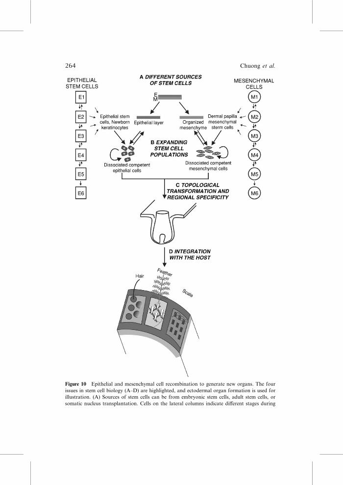
Figure 10 Epithelial and mesenchymal cell recombination to generate new organs. The four
issues in stem cell biology (A–D) are highlighted, and ectodermal organ formation is used for
illustration. (A) Sources of stem cells can be from embryonic stem cells, adult stem cells, or
somatic nucleus transplantation. Cells on the lateral columns indicate diVerent stages during
264 Chuong et al.
In the bird, the body regions are established by dividing the body surface
into diVerent fields or tracts during development (Dhouailly et al., 2004;
Jiang et al., 2004; Sengel, 1976). By having multiple feathers in one feather
tract, another level of topobiological specificity is possible across the feather
tract. There are diVerent modes of flight based on diVerent wing shapes
(Feduccia, 1999) (Fig. 9A). The shape of the wing is made by the combina-
tion of the 20–30 flight feathers (remiges). Their relative lengths form the
contour of the wing. Because the length of the feather shaft is a function of the
duration of the growth phase (like the anagen phase of the hair cycle), the
shape of the wing becomes the spatial layout of multiple flight feathers from
the medial to the lateral regions of the wing (in which the midline of the body
is the medial; one can also consider this as the proximal‐distal axis of the limb
bud), eachwith its own temporal cycle regulation, but together add up to form
a distinct shape of the wing. Another level of complexity is imposed on top of
this array of flight feathers: the medial/lateral bilateral asymmetry (again,
here we use the body axis, not the feather rachis as the reference point).
According to aerodynamic engineering, the feather in the most lateral wing
is most bilateral asymmetric, with the lateral vane much narrower than the
medial vane (Fig. 9B). This feature was used to judge whether a fossil bird is a
good flyer (Feduccia, 1999). Birds that give up flight (e.g., on isolated islands)
soon lose this level of asymmetry over several generations. Two aspects of
interest pertain to the molecular basis of this process: one is by what topo-
biological mechanism lateral/medial asymmetry is produced from the bilater-
ally symmetric flight feathers; the other is how this molecular activity can be
displayed in a graduated medial‐lateral fashion.In mammals, the diVerences of hair follicles within a domain are not
clearcut. There are hair whorls on human scalp, which indicate a relation-
ship among hair follicles during development (Plikus and Chuong, 2004). In
frizzled‐6–null mice, there are also disoriented hair follicles leading to vari-
able whorls and tufts, suggesting a role of frizzled‐6 in hair follicle orienta-
tions (Guo et al., 2004). In some adult mutant mice, clear and shifting
alopecic domains are observed on the surface of mouse body (Ma et al.,
progression of stem cells. The downward arrows mean diVerentiation. The reverse arrows mean
de‐diVerentiation, which eventually disappears, meaning that cells are fully committed and their
fates cannot be reversed anymore. (B) Cell populations are expanded with the idea that the stem
cell properties, self‐renewal and pluripotentiality, will not be lost or deregulated to become
tumors. (C) Competent epithelial stem cells and regional specific mesenchymal cells are
combined in the proper environment to generate organs. If everything is set right, they can self‐organize in normal morphogenesis. In tissue engineering, we need to learn these principles and
the regulation of specificity. (D) A single feather follicle would not be too useful if it is not
connected to other parts of the body and coordinated as part of the system (Fig. 1, 10D).
Ectodermal organs have to be connected with other systems via angiogenesis, myogenesis, and
neurogenesis to be fully integrated with the organism.
5. Topobiology of Stem Cells and Organogenesis 265

2003; Suzuki et al., 2003). However, these are due to problems of cyclic
alopecia in which hair filaments are dislodged from the follicle at a specific
time of hair cycle (Ma et al., 2003). These are problems of hair cycling (Sten
and Paus, 2001), not regional specificity. Tooth fields have similar types of
topological modulations to generate diVerent sizes and shapes of incisors,
canines, and molars (Jernvall and ThesleV, 2000; Plikus et al., 2005). Thesespecializations do not exist in most reptiles or Mesozoic birds (Hou et al.,
2003, 2004).
There are further regional variations within a single appendage organ. For
example, the graded topological modulation of feathers can be seen in
contour feathers. In the trunk, the functions of each feather are further
divided along the proximal‐distal axis. The distal region is made of pennac-
eous barbs (for contouring or communication), and the proximal domain is
made of plumulaceous barbs (for thermal insulation) (Fig. 9C). Further-
more, the ratio of plumulaceous versus pennaceous regions changes gradu-
ally among adjacent feathers in the same feather tract, reflecting the need of
diVerent body parts to make the best balance between preserving body
temperatures and streamlining body shapes. Such regional specific modula-
tion of organ morphology makes the most eVective use of every keratino-
cyte. In other organs, this type of sophisticated modification among cell
groups may also exist (e.g., diVerent brain regions, cortex laminations,
neuronal circuits) (Edelman, 1988b). Yet the feather is a good model because
it lays out all topological arrangements clearly: The barbule represents a row
of 10–20 keratinocytes connected in a head‐to‐tail fashion.
VIII. Integration of Stem Cells and Organs to Reach theLevel of System Biology
We now come back to the stem cell issue. In the beginning, we emphasized
that there are four types of issues that stem cell biology have to solve to
achieve the goal of regenerative medicine (Fig. 10A–D). Using the skin as an
example, progress has led to new understanding in the interfollicular epider-
mal stem cells (Watt, 2002) and hair bulge stem cells (Morris et al., 2004;
Tumbar et al., 2004) (Fig. 10A). We have learned the importance of the
niche in regulating stem cell homeostasis (Fig. 10B). We also have learned
that, to a limit, these epidermal progenitors can be dediVerentiated and
transdiVerentiated. Indeed it is most interesting to observe the conversion
of part of the scales into feathers, amniotic membranes into feathers and
hairs (Fliniaux et al., 2004b), sweat glands/meibomian glands into hairs
(Plikus et al., 2004), and even adult cornea epithelium into hairs (Pearton
et al., 2005). Research in genetic and epigenetic regulation should shed more
light on the control of cellular phenotypes.
266 Chuong et al.

Suppose this research bears fruit and we are able to form an organ; how
then do we direct it to become part of the host and function in a useful
manner? One ideal situation is to have competent epidermal stem cells and
induce mesenchymal cells incubated in a microenvironment with proper
chemical signaling and topological setting, and then let them self‐organize(Fig. 10). This type of approach was pioneered in Moscona’s cell aggregate
approaches to form feathers, retina, lentoid, livers, etc. (e.g., Garber et al.,
1968; Vardimon et al., 1988). In these aggregates, a quite remarkable degree
of histogenesis and chemical diVerentiation was achieved in the 3D aggre-
gates, yet their topological relationships are random. We constrained disso-
ciated feather mesenchymal cells into a 2D configuration and put on top a
competent epithelia sheet. With this topological arrangement, we were able to
obtain a reconstituted skin with an array of evenly spaced and oriented
feather follicles (Jiang et al., 1999; our unpublished data). In themouse, Lichti
et al. (1995) mixed a population of competent epidermal and dermal cells in a
chamber that was transplanted on a nude mouse. The cells sort out to form
hair follicles. This procedure was simplified and improved to generate exoge-
nous hair organs that are supported by the host can can cycle (Zheng et al.,
2005). This is very good progress, albeit the hair filaments point to the center
of the aggregates, forming a cyst. We still have to make the topobiological
events right before stem cell engineering can be applied to humans.
Stem cell biology is just at its dawn. There are many critical issues to be
solved and knowledge from multiple disciplines to be integrated. Assuming
we could have access to sources of stem cells and know, to a certain level,
how to induce their diVerentiations someday soon, here we focus on the
issue of guiding stem cells into organs. We identify the fundamental and
practical importance of topobiological events in building the architecture or
an organ. We turn to Nature to learn how she solves the simple to complex
designs of ectodermal organs. Using feather and beak morphogenesis to
decipher the principles, we observe a succession of topobiological transfor-
mation events, taking the epithelia from a flat sheet to more and more
complex structures.
Some of these topobiological principles are likely to be in operation in
other organogeneses as well. These processes are important in development
and morphological evolution and have to be considered in tissue engineer-
ing. There may be a long way to go, but the process is exciting and the best is
yet to come.
Acknowledgments
This work is supported by grants from the National Institutes of Health (CMC, AR42177,
47364, 47364; RW, CA83716). C.M.C. thanks Dr. G. M. Edelman for his inspirations.
5. Topobiology of Stem Cells and Organogenesis 267

References
Abzhanov, A., Protas, M., Grant, B. R., Grant, P. R., and Tabin, C. J. (2004). Bmp4 and
morphological variation of beaks in Darwin’s finches. Science 305, 1462–1465.
Abzhanov, A., and Tabin, C. J. (2004). Shh and Fgf8 act synergistically to drive cartilage
outgrowth during cranial development. Dev. Biol. 273, 134–148.
AVolter, M., Bellusci, S., Itoh, N., Shilo, B., Thiery, J. P., and Werb, Z. (2003). Tube or not
tube: Remodeling epithelial tissues by branching morphogenesis. Dev. Cell. 4, 11–18.
Ashique, A. M., Fu, K., and Richman, J. M. (2002a). Signalling via type IA and type IB
bone morphogenetic protein receptors (BMPR) regulates intramembranous bone
formation, chondrogenesis and feather formation in the chicken embryo. Int. J. Dev. Biol.
46, 243–253.
Ashique, A. M., Fu, K., and Richman, J. M. (2002b). Endogenous bone morphogenetic proteins
regulate outgrowth and epithelial survival during avian lip fusion.Development 129, 4647–4660.
Barlow, A. J., and Francis‐West, P. H. (1997). Ectopic application of recombinant BMP‐2 and
BMP‐4 can change patterning of developing chick facial primordia. Development 124,
391–398.
Baron, M. H. (2003). Embryonic origins of mammalian hematopoiesis. Exp. Hematol. 31,
1160–1169.
Bartels, T. (2003). Variations in the morphology, distribution, and arrangement of feathers in
domesticated birds. J. Exp. Zoolog. B Mol. Dev. Evol. 298, 91–108.
Bellusci, S., Henderson, R., Winnier, G., Oikawa, T., and Hogan, B. L. (1996). Evidence from
normal expression and targeted misexpression that bone morphogenetic protein (Bmp‐4)plays a role in mouse embryonic lung morphogenesis. Development 122, 1693–1702.
Botchkarev, V. A., Botchkareva, N. V., Sharov, A. A., Funa, K., Huber, O., and Gilchrest,
B. A. (2002). Modulation of BMP signaling by noggin is required for induction of the
secondary (nontylotrich) hair follicles. J. Invest. Dermatol. 118, 3–10.
Brown, W. R., Hubbard, S. J., Tickle, C., and Wilson, S. A. (2003). The chicken as a model for
large‐scale analysis of vertebrate gene function. Nat. Rev. Genet. 4, 87–98.
Candille, S. I., Van Raamsdonk, C. D., Chen, C., Kuijper, S., Chen‐Tsai, Y., Russ, A.,
Meijlink, F., and Barsh, G. S. (2004). Dorsoventral patterning of the mouse coat by Tbx15.
PLoS Biol. 2, 30–42.
Capdevila, J., and Izpisua Belmonte, J. C. (2001). Patterning mechanisms controlling vertebrate
limb development. Annu. Rev. Cell Dev. Biol. 17, 87–132.
Chang, H. Y., Chi, J. T., Dudoit, S., Bondre, C., van de Rijn, M., Botstein, D., and Brown,
P. O. (2002). Diversity, topographic diVerentiation, and positional memory in human
fibroblasts. Proc. Natl. Acad. Sci. USA 99, 12877–12882.
Chang, C.‐H., Yu, M., Wu, P., Jiang, T.‐X., Yu, H.‐S., Widelitz, R. B., and Chuong, C.‐M.
(2004). Sculpting skin appendages out of epidermal layers via temporally and spatially
regulated apoptotic events. J. Invest. Dermatol. 122, 1348–1355.
Chatterjee, S. (1997). ‘‘The Rise of Birds.’’ John Hopkins University Press, Baltimore.
Chen, P. J., Dong, Z. M., and Shen, S. N. (1998). An exceptionally well‐preserved theropod
dinosaur from the Yixian Formation of China. Nature 391, 147–152.
Chiappe, L. M. (1995). The First 85 million years of Avian Evolution. Nature 378, 349–355.
Chuong, C.‐M., Oliver, G., Ting, S., Jegalian, B., Chen, H. M., and De Robertis, E. M. (1990).
Gradient of homeoproteins in developing feather buds. Development 110, 1021–1030.
Chuong, C.‐M. (1998). ‘‘Molecular Basis of Epithelial Appendage Morphogenesis.’’ Landes
Bioscience, Austin, Texas.
Chuong, C.‐M., and Edelman, G. M. (1985a). Expression of cell adhesion molecules in
embryonic induction. I. Morphogenesis of nestling feathers. J. Cell Biol. 101, 1009–1026.
268 Chuong et al.

Chuong, C.‐M., and Edelman, G. M. (1985b). Expression of cell adhesion molecules in
embryonic induction. II. Morphogenesis of adult feathers. J. Cell Biol. 101, 1027–1043.
Chuong, C.‐M., Chodankar, R., Widelitz, R. B., and Jiang, T. X. (2000). Evo‐devo of feathers
and scales: Building complex epithelial appendages. Curr. Opin. Dev. Gen. 10, 449–456.
Chuong, C.‐M., NickoloV, B. J., Elias, P. M., Goldsmith, L. A., Macher, E., Maderson, P. A.,
Sundberg, J. P., Tagami, H., Planka, P. M., Thestrup‐Pederson, K., Bernard, B. A,,
Schroder, J. M., Dotto, P., Chang, C. M., Williams, M. L., Feingold, K. R., Kling, L. E.,
Klingman, A. M., Rees, J. L., and Christophers, E. (2002). What is the ‘‘true’’ function of
skin. Exp. Dermatol. 11, 159–187.
Chuong, C.‐M., Wu, P., Zhang, F. C., Xu, X., Yu, M., Widelitz, R. B., Jiang, T. X., and Hou,
L. (2003). Adaptation to the sky: Defining the feather with integument fossils fromMesozoic
China and experimental evidence from molecular laboratories. J. Exp. Zoolog. B Mol. Dev.
Evol. 298, 42–56.
Cohen, J., and Espinasse, P. G. (1961). On the normal and abnormal development of the
feather. J. Embryol. Exp. Morphol. 9, 223–251.
Colas, J. F., and Schoenwolf, G. C. (2001). Towards a cellular and molecular understanding of
neurulation. Dev. Dyn. 221, 117–145.
Coucouvanis, E., and Martin, G. R. (1995). Signals for death and survival: A two‐stepmechanism for cavitation in the vertebrate embryo. Cell 83, 279–287.
Couly, G., Creuzet, S., Bennaceur, S., Vincent, C., and Le Douarin, N. M. (2002). Interactions
between Hox‐negative cephalic neural crest cells and the foregut endoderm in patterning the
facial skeleton in the vertebrate head. Development 129, 1061–1073.
Creuzet, S., Couly, G., Vincent, C., and Le Douarin, N. M. (2002). Negative eVect of Hox gene
expression on the development of the neural crest‐derived facial skeleton. Development 129,
4301–4313.
Desai, T. J., Malpel, S., Flentke, G. R., Smith, S. M., and Cardoso, W. V. (2004). Retinoic acid
selectively regulates Fgf10 expression and maintains cell identity in the prospective lung field
of the developing foregut. Dev. Biol. 273, 402–415.
Dhouailly, D., Olivera‐Martinez, I., Fliniaux, I., Missier, S., Viallet, J. P., and Thelu, J. (2004).
Skin field formation: Morphogenetic events. Int. J. Dev. Biol. 48, 85–91.
Dudley, A. T., and Tabin, C. J. (2000). Constructive antagonism in limb development. Curr.
Opin. Genet. Dev. 10, 387–392.
Edelman, G. M. (1988a). ‘‘Topobiology: An Introduction to Molecular Biology,’’ pp. 1–56.
Basic Books, New York, NY.
Edelman, G. M. (1988b). Morphoregulatory molecules. Biochemistry 27, 3533–3543.
Efrat, S. (2004). Regulation of insulin secretion: Insights from engineered beta‐cell lines. Ann.NY Acad. Sci. 1014, 88–96.
Ettensohn, C. A. (1985). Gastrulation in the sea urchin embryo is accompanied by the
rearrangement of invaginating epithelial cells. Dev. Biol. 112, 383–390.
Feduccia, A. (Ed.) (1999). In ‘‘The Origin and Evolution of Birds’’, 2 Ed., pp. 93–137. Yale
University Press, New Haven.
Fliniaux, I., Viallet, J. P., and Dhouailly, D. (2004a). Ventral vs. dorsal chick dermal progenitor
specification. Int. J. Dev. Biol. 48, 103–106.
Fliniaux, I., Viallet, J. P., Dhouailly, D., and Jahoda, C. A. (2004b). Transformation of amnion
epithelium into skin and hair follicles. DiVerentiation 72, 558–565.
Francis‐West, P. H., Ladher, R., Barlow, A., and Graveson, A. (1998). Signalling interactions
during facial development. Mech. Dev. 75, 3–28.
Francis‐West, P. H., Robson, L., and Evans, D. J. (2003). Craniofacial development: The tissue
and molecular interactions that control development of the head. Adv. Anat. Embryol. Cell.
Biol. 169(III‐VI), 1–138.Fuchs, E., and Segre, J. A. (2000). Stem cells: A new lease on life. Cell 100, 143–155.
5. Topobiology of Stem Cells and Organogenesis 269

Garber, B., Kollar, E. J., and Moscona, A. A. (1968). Aggregation in vivo of dissociated cells. 3.
EVect of state of diVerentiation of cells on feather development in hybrid aggregates of
embryonic mouse and chick skin cells. J. Exp. Zool. 168, 455–472.
Gierer, A., and Meinhardt, H. (1972). A theory of biological pattern formation. Kybernetik 12,
30–39.
Grant, P. R. (1986). In ‘‘Ecology and Evolution of Darwin’s Finches’’ (P. R. Grant, Ed.),
pp. 1–492. Princeton University Press, Princeton.
Green, H., Easley, K., and Iuchi, S. (2003). Marker succession during the development of
keratinocytes from cultured human embryonic stem cells. Proc. Natl. Acad. Sci. USA 100,
15625–15630.
Green‐Armytage, S. (2000). ‘‘Extraordinary Chickens.’’ Harry N. Abrams, Inc., New York.
Guo, N., Hawkins, C., and Nathans, J. (2004). Frizzled6 controls hair patterning in mice. Proc.
Natl. Acad. Sci. USA 101, 9277–9281.
Hardin, J. D., and Cheng, L. Y. (1986). The mechanism and mechanics of archenteron
elongation during sea urchin gastrulation. Dev. Biol. 115, 490–501.
Harris, M. P., Fallon, J. F., and Prum, R. O. (2002). Shh‐Bmp2 signaling module and the
evolutionary origin and diversification of feathers. J. Exp. Zool. 294, 160–176.
Harris, M. P., Williamson, S., Fallon, J. F., Meinhardt, H., and Prum, R. O. (2005). Molecular
evidence for an activator‐inhibitor mechanism in development of embryonic feather
branching. Proc. Natl. Acad. Sci. USA 102, 11734–11739.
Held, L. I. (1992). ‘‘Models for Embryonic Periodicity.’’ Karger, New York.
Helms, J.A, and Schneider, R. A. (2003). Cranial skeletal biology. Nature 423, 326–331.
Hogan, B. L., and Kolodziej, P. A. (2002). Organogenesis: Molecular mechanisms of
tubulogenesis. Nat. Rev. Genet. 3, 513–523.
Hou, L. H, Chiappe, L., Zhang, F. C., and Chuong, C.‐M. (2004). New early cretaceous fossil
from China documents a novel trophic specialization for Mesozoic birds. Naturwissenschaf-
ten 91, 22–25.
Hou, L. H., Chuong, C. M., Yang, A., Zeng, X. L., and Hou, J. F. (2003). ‘‘Fossil Birds of
China.’’ Yunnan Science and Technology, China.
Hou, L. H., Martin, L. D., Zhou, Z., and Feduccia, A. (1996). Early adaptive radiation of birds:
Evidence from fossils from northeastern China. Science 274, 1164–1167.
Hu, D., and Helms, J. A. (1999). The role of sonic hedgehog in normal and abnormal
craniofacial morphogenesis. Development 126, 4873–4884.
Hu, D., Marcucio, R. S., and Helms, J. A. (2003). A zone of frontonasal ectoderm regulates
patterning and growth in the face. Development 130, 1749–1758.
Jamora, C., DasGupta, R., Kocieniewski, P., and Fuchs, E. (2003). Links between signal
transduction, transcription and adhesion in epithelial bud development.Nature. 422, 317–322.
Jernvall, J., and ThesleV, I. (2000). Reiterative signaling and patterning during mammalian
tooth morphogenesis. Mech. Dev. 92, 19–29.
Jiang, T.‐X., and Chuong, C.‐M. (1992). Mechanism of feather morphogenesis: I. Analyses with
antibodies to adhesion molecules tenascin, N‐CAM and integrin. Dev. Biol. 150, 82–98.
Jiang, T.‐X., Jung, H. S., Widelitz, R. B., and Chuong, C.‐M. (1999). Self organization of
periodic patterns by dissociated feather mesenchymal cells and the regulation of size, number
and spacing of primordia. Development 126, 4997–5009.
Jiang, T.‐X., Wideltz, R. B., Shen, W. M., Will, P., Wu, D. Y., Lin, C. M., Jung, J. S., and
Chuong, C.‐M. (2004). Integument pattern formation involves genetic and epigenetic
controls operated at diVerent levels: Feather arrays simulated by a digital hormone model.
Int. J. Dev. Biol. 48, 117–136.
Jones, T. D., Ruben, J. A., Martin, L. D., Kurochkin, E. N., Feduccia, A., Maderson, P. F.,
Hillenius, W. J., Geist, N. R., and Alifanov, V. (2000). Nonavian feathers in a late Triassic
archosaur. Science 288, 2202–2205.
270 Chuong et al.

Jung, H. S., Francis‐West, P. H., Widelitz, R. B., Jiang, T. X., Ting‐Berreth, S., Tickle, C.,Wolpert, L., and Chuong, C. M. (1998). Local inhibitory action of BMPs and their
relationships with activators in feather formation: Implications for periodic patterning. Dev.
Biol. 196, 11–23.
Kang, P., and Svoboda, K. K. (2005). Epithelial‐mesenchymal transformation during
craniofacial development. J. Dent. Res. 84, 678–690.
Keller, R. (1986). The cellular basis of amphibian gastrulation. In ‘‘The Cellular Basis of
Morphogenesis. Developmental Biology: A comprehensive Synthesis’’ (L. Browder, Ed.),
pp. 241–327. Plenum Press, New York.
Keller, R. (2002). Shaping the vertebrate body plan by polarized embryonic cell movements.
Science 298, 1950–1954.
Kiskowski, M. A., Alber, M. S., Thomas, G. L., Glazier, J. A., Bronstein, N. B., Pu, J., and
Newman, S. A. (2004). Interplay between activator‐inhibitor coupling and cell‐matrix
adhesion in a cellular automaton model for chondrogenic patterning. Dev. Biol. 271,
372–387.
Koster, M. I., Kim, S., Mills, A. A., De Mayo, F. J., and Roop, D. R. (2004). p63 is the
molecular switch for initiation of an epithelial stratification program. Genes Dev. 18,
126–131.
Koster, M. I., and Roop, D. R. (2004). p63 and epithelial appendage development.
DiVerentiation 72, 364–370.
Lako, M., Armstrong, L., Cairns, P. M., Harris, S., Hole, N., and Jahoda, C. A. (2002). Hair
follicle dermal cells repopulate the mouse haematopoietic system. J. Cell Sci. 115, 3967–3974.
Lee, S.H, Fu, K. K., Hui, J. N., and Richman, J. M. (2001). Noggin and retinoic acid transform
the identity of avian facial prominences. Nature. 414, 909–912.
Li, L., and Xie, T. (2005). Stem cell niche: Structure and function. Annu. Rev. Cell Dev. Biol. 21,
605–631.
Lichti, U., Scandurro, A. B., Kartasova, T., Rubin, J. S., La Rochelle, W., and Yuspa, S. H.
(1995). Hair follicle development and hair growth from defined cell populations grafted onto
nude mice. J. Invest. Dermatol. 104, 43S–44S.
Lucas, A. M. and Stettenheim, P. R. (Eds.) (1972). ‘‘Avian Anatomy: Integument. Agriculture
Handbook 362: Agricultural Research Services.’’ US Department of Agriculture,
Washington, D.C.
Lumelsky, N., Blondel, O., Laeng, P., Velasco, I., Ravin, R., and McKay, R. (2001).
DiVerentiation of embryonic stem cells to insulin‐secreting structures similar to pancreatic
islets. Science 292, 1389–1394.
Ma, L., Liu, J., Wu, T., Plikus, M., Jiang, T.‐X, Liu, Y.‐H., Muller‐Rover, S., Peters, H.,
Maxson, R., Maas, R., and Chuong, C.‐M. (2003). Cyclic alopecia in Msx2 mutants: Defects
in hair cycling and hair shaft diVerentiation. Development 130, 379–389.
MacDonald, M.E, Abbott, U. K., and Richman, J. M. (2004). Upper beak truncation in
chicken embryos with the cleft primary palate mutation is due to an epithelial defect in the
frontonasal mass. Dev. Dyn. 230, 335–349.
Mailleux, A. A., Spencer‐Dene, B., Dillon, C., Ndiaye, D., Savona‐Baron, C., Itoh, N., Kato,
S., Dickson, C., Thiery, J. P., and Bellusci, S. (2002). Role of FGF10/FGFR2b signaling
during mammary gland development in the mouse embryo. Development 129, 53–60.
McGonnell, I. M., Clarke, J. D., and Tickle, C. (1998). Fate map of the developing chick face:
Analysis of expansion of facial primordia and establishment of the primary palate. Dev. Dyn.
212, 102–118.
Meinhardt, H., and Gierer, A. (2000). Pattern formation by local self‐activation and lateral
inhibition. Bioessays. 22, 753–760.
Meinhardt, H., and Gierer, A. (1974). Applications of a theory of biological pattern formation
based on lateral inhibition. J. Cell Sci. 15, 321–346.
5. Topobiology of Stem Cells and Organogenesis 271

Melnick, M., and Jaskoll, T. (2000). Mouse submandibular gland morphogenesis: A paradigm
for embryonic signal processing. Crit. Rev. Oral Biol. Med. 11, 199–215.
Metzger, R. J., and Krasnow, M. A. (1999). Genetic control of branching morphogenesis.
Science 284, 1635–1639.
Mills, A. A., Zheng, B., Wang, X. J., Vogel, H., Roop, D. R., and Bradley, A. (1999). p63 is a
p53 homologue required for limb and epidermal morphogenesis. Nature 398, 708–713.
Moon, R. T., Kohn, A. D., De Ferrari, G. V., and Kaykas, A. (2004). WNT and beta‐cateninsignalling: Diseases and therapies. Nat. Rev. Genet. 5, 691–701.
Moore, G. P., Jackson, N., Isaacs, K., and Brown, G. (1998). Pattern and morphogenesis in
skin. J. Theor. Biol. 191, 87–94.
Morris, R. J., Liu, Y., Marles, L., Yang, Z., Trempus, C., Li, S., Lin, J. S., Sawicki, J. A., and
Cotsarelis, G. (2004). Capturing and profiling adult hair follicle stem cells. Nat. Biotechnol.
22, 411–417.
Muller‐Rover, S., Tokura, Y., Welker, P., Furukawa, F., Wakita, H., Takigawa, M., and Paus,
R. (1999). E‐ and P‐cadherin expression during murine hair follicle morphogenesis and
cycling. Exp. Dermatol. 8, 237–246.
Nagafuchi, A., Shirayoshi, Y., Okazaki, K., Yasuda, K., and Takeichi, M. (1987).
Transformation of cell adhesion properties by exogenously introduced E‐cadherin cDNA.
Nature 329, 341–343.
Nagorcka, B. N., and Mooney, J. R. (1985). The role of a reaction‐diVusion system in the
initiation of primary hair follicles. Theor. Biol. 114, 243–272.
Newman, S. A., and Frisch, H. L. (1979). Dynamics of skeletal pattern formation in developing
chick limb. Science 205, 662–668.
Niswander, L. (2003). Pattern formation: Old models out on a limb.Nat. Rev. Genet. 4, 133–143.
Noden, D. M. (1983). The role of the neural crest in patterning of avian cranial skeletal,
connective, and muscle tissues. Dev. Biol. 96, 144–165.
Oster, G. F., Murray, J. D., and Maini, P. K. (1985). A model for chondrogenic condensations
in the developing limb: The role of extracellular matrix and cell tractions. J. Embryol. Exp.
Morphol. 89, 93–112.
Pearton, D. J., Yang, Y., and Dhouailly, D. (2005). TransdiVerentiation of corneal epithelium
into epidermis occurs by means of a multistep process triggered by dermal developmental
signals. Proc. Natl. Acad. Sci. USA 102, 3714–3719.
Pennisi, E. (2004). Bonemaking protein shapes beaks of Darwin’s finches. Science 305, 1383.
Phippard, D. J., Weber‐Hall, S. J., Sharpe, P. T., Naylor, M. S., Jayatalake, H., Maas, R., Woo,
I., Roberts‐Clark, D., Francis‐West, P. H., Liu, Y. H., Maxson, R., Hill, R. E., and Dale,
T. C. (1996). Regulation of Msx‐1, Msx‐2, Bmp‐2 and Bmp‐4 during foetal and postnatal
mammary gland development. Development 122, 2729–2737.
Plikus, M., and Chuong, C.‐M. (2004). Making waves with hairs. J. Invest. Dermatol. 122,
VII–IX.
Plikus, M. V., Sundberg, J. P., and Chuong, C. M. (2006). Morphogenesis of skin and its
appendages. In ‘‘The Mouse in Biomedical Research.’’ Academic Press, New York.
Plikus, M., Wang, W. P., Liu, J., Wang, X., Jiang, T. X., and Chuong, C.‐M. (2004). Morpho‐regulation of ectodermal organs: Integument pathology and phenotypic variations in K14‐Noggin engineered mice through modulation of bone morphogenic protein pathway. Am. J.
Pathol. 164, 1099–1114.
Plikus, M. V., Zeichner‐David, M., Mayer, J. A., Reyna, J., Bringas, P., Thewissen,
J. G. M., Snead, M. L., Chai, Y., and Chuong, C. M. (2005). ‘‘Morphoregulation of
teeth: Modulating the number, size, shape and diVerentiation by tuning Bmp activity.’’
Evolut. Dev. 7, 440–457.
Prum, R. O. (1999). Development and evolutionary origin of feathers. J. Exp. Zool. 285,
291–306.
272 Chuong et al.

Prum, R.O, and Brush, A. H. (2002). The evolutionary origin and diversification of feathers. Q.
Rev. Biol. 77, 261–295.
Prum, R. O., and Dyck, J. (2003). A hierarchical model of plumage: Morphology, development,
and evolution. J. Exp. Zoolog. B Mol. Dev. Evol. 298, 73–90.
Prum, R. O., and Williamson, S. (2001). Theory of the growth and evolution of feather shape.
J. Exp. Zool. 291, 30–57.
Regal, P. J. (1975). The evolutionary origin of feathers. Q. Rev. Biol. 50, 35–66.
Reya, T., Duncan, A. W., Ailles, L., Domen, J., Scherer, D. C., Willert, K., Hintz, L., Nusse,
R., and Weissman, I. L. (2003). A role for Wnt signalling in self‐renewal of haematopoietic
stem cells. Nature 423, 409–414.
Richman, J. M., Herbert, M., Matovinovic, E., and Walin, J. (1997). EVect of fibroblast growth
factors on growth of facial mesenchyme. Dev. Biol. 189, 135–147.
Sawyer, R. H., and Knapp, L. W. (2003). Avian skin development and the evolutionary origin
of feathers. J. Exp. Zoolog. B Mol. Dev. Evol. 298, 57–72.
Schneider, R. A., and Helms, J. A. (2003). The cellular and molecular origins of beak
morphology. Science. 299, 565–568.
Schoenwolf, G. C., and Alvarez, I. S. (1989). Roles of neuroepithelial cell rearrangement and
division in shaping of the avian neural plate. Development 106, 427–439.
Schoenwolf, G. C. (1991). Cell movements driving neurulation in avian embryos. Development
Suppl. 2, 157–168.
Scott, M. P. (2000). Development: The natural history of genes. Cell 100, 27–40.
Sengel, P. (1976). ‘‘Morphogenesis of Skin.’’ Cambridge University Press, Cambridge.
Shen, W.‐M., Will, P., Galstyan, A., and Chuong, C.‐M. (2004). Hormone‐inspired self‐organization and distributed control of robotic swarms. Auton. Robots 17, 93–105.
Shieh, S. J., Terada, S., and Vacanti, J. P. (2004). Tissue engineering auricular reconstruction:
In vitro and in vivo studies. Biomaterials 25, 1545–1557.
Snyder, B. J., and Olanow, C. W. (2005). Stem cell treatment for Parkinson’s disease: An update
for 2005. Curr. Opin. Neurol. 18, 376–385.
Stenn, K. S., and Paus, R. (2001). Controls of hair follicle cycling. Physiol. Rev, 81, 449–494.
Suksaweang, S., Lin, C. M., Jiang, T. X., Hughes, M. W., Widelitz, R. B., and Chuong, C.‐M.
(2004). Morphogenesis of chicken liver: Identification of localized growth zones and the role
of beta‐catenin/Wnt in size regulation. Dev. Biol. 266, 109–122.
Sundberg, J. P. (Ed.) (1994). ‘‘Handbook of Mouse Mutations with Skin and Hair
Abnormalities: Animal Models and Biomedical Tools.’’ CRC Press, Boca Raton, Florida.
Suzuki, N., Hirata, M., and Kondo, S. (2003). Traveling stripes on the skin of a mutant mouse.
Proc. Natl. Acad. Sci. USA 100, 9680–9685.
Tickle, C. (2003). Patterning systems—fromone end of the limb to the other.Dev. Cell 4, 449–458.
Toma, J. G., McKenzie, I. A., Bagli, D., and Miller, F. D. (2005). Isolation and
characterization of multipotent skin‐derived precursors from human skin. Stem Cells. 23,
727–737.
Tumbar, T., Guasch, G., Greco, V., Blanpain, C., Lowry, W. E., Rendl, M., and Fuchs, E.
(2004). Defining the epithelial stem cell niche in skin. Science 303, 359–363.
Turing, A. M. (1952). The chemical basis of morphogenesis. Phil. Trans. R. Soc. B 237, 37–72.
Vardimon, L., Fox, L. L., Degenstein, L., and Moscona, A. A. (1988). Cell contacts are
required for induction by cortisol of glutamine synthetase gene transcription in the retina.
Proc. Natl. Acad. Sci. USA 85, 5981–5985.
Watt, F. M. (2002). The stem cell compartment in human interfollicular epidermis. J. Dermatol.
Sci. 28, 173–180.
Wei, Y., and Mikawa, T. (2000). Formation of the avian primitive streak from spatially
restricted blastoderm: Evidence for polarized cell division in the elongating streak.
Development 127, 87–96.
5. Topobiology of Stem Cells and Organogenesis 273

Widelitz, R. B., Jiang, T. X., Yu, M., Shen, T., Shen, J.‐Y., Wu, P., Yu, Z., and Chuong, C.‐M.
(2003). Molecular biology of feather morphogenesis: A testable model for evo‐devo research.
J. Exp. Zool. 298B, 109–122.
Wilke, T. A., Gubbels, S., Schwartz, J., and Richman, J. M. (1997). Expression of fibroblast
growth factor receptors (FGFR1, FGFR2, FGFR3) in the developing head and face. Dev.
Dyn. 210, 41–52.
Wu, P., Jiang, T. X., Suksaweang, S., Widelitz, R. B., and Chuong, C.‐M. (2004a). Molecular
shaping of the beak. Science 305, 1465–1466.
Wu, P., Hou, L., Plikus, M., Hughes, M., Scehnet, J., Suksaweang, S., Widelitz, R., Jiang,
T. X., and Chuong, C.‐M. (2004b). Evo‐devo of amniote integuments and appendages. Int.
J. Dev. Biol. 48, 249–270.
Xu, X., Zhou, Z., and Prum, R. O. (2001). Branched integumental structures in
Sinornithosaurus and the origin of feathers. Nature 410, 200–204.
Yu, M., Wu, P., Widelitz, R. B., and Chuong, C.‐M. (2002). The morphogenesis of feathers.
Nature 420, 308–312.
Yu, M., Yue, Z., Wu, P., Wu, D. Y., Mayer, J. A., Medina, M., Widelitz, R. B., Jiang, T. X.,
and Chuong, C.‐M. (2004). The biology of feather follicles. Int. J. Dev. Biol. 48, 181–191.
Yue, Z., Jiang, T.‐X., Wildelitz, R. B., and Chuong, C.‐M. (2005). Mapping stem cell activities
in the feather follicle. Nature 438, 1026–1029.
Yue, Z., Jiang, T.‐X., Wildelitz, R. B., and Chuong, C.‐M. (2006). Wnt 3a gradient converts
radial to bilateral feather symmetry via topological arrangement of epithelia. Proc. Natl.
Acad. Sci. USA. In press.
Zheng, Y., Du, X., Wang, W., Boucher, M., Parimoo, S., and Stenn, K. (2005). Organogenesis
from dissociated cells: Generation of mature cycling hair follicles from skin‐derived cells.
J. Invest. Dermatol. 124, 867–876.
Zhou, Z., Barrett, P. M., and Hilton, J. (2003). An exceptionally preserved lower cretaceous
ecosystem. Nature 421, 807–814.
Zweers, G.A, Vanden Berge, J. C., and Berkhoudt, H. (1997). Evolutionary pattern of avian
trophic diversification. Zoology 100, 25–57.
274 Chuong et al.