emerginginfectiousdiseasesvol1... - bio-nica.info
TRANSCRIPT


Emerging Infectious DiseasesEmerging Infectious Diseases is published monthly by the
National Center for Infectious Diseases, Centers for DiseaseControl and Prevention, 1600 Clifton Road, Mailstop D61,Atlanta, GA 30333, USA. Telephone 404-639-1960, fax 404-639-1954, email [email protected].
The opinions expressed by authors contributing to this journaldo not necessarily reflect the opinions of the Centers for DiseaseControl and Prevention or the institutions with which the authorsare affiliated.
All material published in Emerging Infectious Diseases is inthe public domain and may be used and reprinted without specialpermission; proper citation, however, is required.
Use of trade names is for identification only and does notimply endorsement by the Public Health Service or by the U.S.Department of Health and Human Services.
∞ Emerging Infectious Diseases is printed on acid-free paper that meetsthe requirements of ANSI/NISO 239.48-1992 (Permanence of Paper)
EDITORIAL STAFFFounding EditorJoseph E. McDade, Rome, Georgia, USAManaging Senior EditorPolyxeni Potter, Atlanta, Georgia, USAAssociate EditorsCharles Ben Beard, Ft. Collins, Colorado, USADavid Bell, Atlanta, Georgia, USAJay C. Butler, Anchorage, Alaska, USACharles H. Calisher, Ft. Collins, Colorado, USAStephanie James, Bethesda, Maryland, USABrian W.J. Mahy, Atlanta, Georgia, USANina Marano, Atlanta, Georgia, USAMartin I. Meltzer, Atlanta, Georgia, USADavid Morens, Bethesda, Maryland, USAJ. Glenn Morris, Baltimore, Maryland, USAMarguerite Pappaioanou, St. Paul, Minnesota, USATanja Popovic, Atlanta, Georgia, USAPatricia M. Quinlisk, Des Moines, Iowa, USAGabriel Rabinovich, Buenos Aires, ArgentinaJocelyn A. Rankin, Atlanta, Georgia, USADidier Raoult, Marseilles, FrancePierre Rollin, Atlanta, Georgia, USADavid Walker, Galveston, Texas, USAJ. Todd Weber, Atlanta, Georgia, USAHenrik C. Wegener, Copenhagen, Denmark Copy EditorsAngie Frey, Thomas Gryczan, Ronnie Henry, Anne Mather, Carol Snarey ProductionReginald Tucker, Ann Jordan, Maureen MarshallEditorial AssistantSusanne Justice
EDITORIAL BOARDDennis Alexander, Addlestone Surrey, United KingdomMichael Apicella, Iowa City, Iowa, USAPaul Arguin, Atlanta, Georgia, USABarry J. Beaty, Ft. Collins, Colorado, USAMartin J. Blaser, New York, New York, USADavid Brandling-Bennet, Washington, D.C., USADonald S. Burke, Baltimore, Maryland, USAArturo Casadevall, New York, New York, USAKenneth C. Castro, Atlanta, Georgia, USAThomas Cleary, Houston, Texas, USAAnne DeGroot, Providence, Rhode Island, USAVincent Deubel, Shanghai, ChinaEd Eitzen, Washington, D.C., USADuane J. Gubler, Honolulu, Hawaii, USARichard L. Guerrant, Charlottesville, Virginia, USAScott Halstead, Arlington, Virginia, USADavid L. Heymann, Geneva, SwitzerlandSakae Inouye, Tokyo, JapanCharles King, Cleveland, Ohio, USAKeith Klugman, Atlanta, Georgia, USATakeshi Kurata, Tokyo, JapanS.K. Lam, Kuala Lumpur, MalaysiaBruce R. Levin, Atlanta, Georgia, USAMyron Levine, Baltimore, Maryland, USAStuart Levy, Boston, Massachusetts, USAJohn S. MacKenzie, Perth, AustraliaTom Marrie, Edmonton, Alberta, CanadaBan Mishu-Allos, Nashville, Tennessee, USAJohn E. McGowan, Jr., Atlanta, Georgia, USAPhilip P. Mortimer, London, United KingdomFred A. Murphy, Galveston, Texas, USABarbara E. Murray, Houston, Texas, USAP. Keith Murray, Ames, Iowa, USAStephen Ostroff, Honolulu, Hawaii, USARosanna W. Peeling, Geneva, SwitzerlandDavid H. Persing, Seattle, Washington, USARichard Platt, Boston, Massachusetts, USAMario Raviglione, Geneva, SwitzerlandLeslie Real, Atlanta, Georgia, USADavid Relman, Palo Alto, California, USANancy Rosenstein, Atlanta, Georgia, USAConnie Schmaljohn, Frederick, Maryland, USATom Schwan, Hamilton, Montana, USAIra Schwartz, Valhalla, New York, USATom Shinnick, Atlanta, Georgia, USABonnie Smoak, Bethesda, Maryland, USARosemary Soave, New York, New York, USAP. Frederick Sparling, Chapel Hill, North Carolina, USAJan Svoboda, Prague, Czech RepublicBala Swaminathan, Atlanta, Georgia, USARobert Swanepoel, Johannesburg, South AfricaPhillip Tarr, St. Louis, Missouri, USATimothy Tucker, Cape Town, South AfricaElaine Tuomanen, Memphis, Tennessee, USAJohn Ward, Atlanta, Georgia, USADavid Warnock, Atlanta, Georgia, USAMary E. Wilson, Cambridge, Massachusetts, USA
Emerging Infectious Diseases • www.cdc.gov/eid • Vol. 12, No. 4, April 2006
A Peer-Reviewed Journal Tracking and Analyzing Disease Trends pages 543–718
EDITOR-IN-CHIEFD. Peter Drotman
www.cdc.gov/eid
PerspectivesLow Risk for Epidemics after Geophysical Disasters . . . . . . . . . . . . . .543N. Floret et al.Short-term risk for epidemics after geophysical disasters is very low.
Potential Arbovirus Emergence and Implications . . . . . . . . . . . . . . . . . . .549 E.A. Gould et al.Climate change can cause arthropodborne diseasesto emerge.
Zoonoses, Links between Human and Veterinary Medicine . . . . . . . . . . . . .556L.H. KahnGreater collaboration is needed between humanand veterinary medicine to better control zoonoses.
Human Influenza Surveillance: the Demand To Expand . . . . . . . . . . . . . .562S.P. LayneThe potential of avian A/H5N1 to cause a globalhuman pandemic is uncertain because it cannot bepredicted with current knowledge.
Prospects for Universal Influenza Virus Vaccine . . . . . . . . . . . . .569W. Gerhard et al.A vaccine less sensitive to the antigenic evolution ofthe virus is a feasible goal.
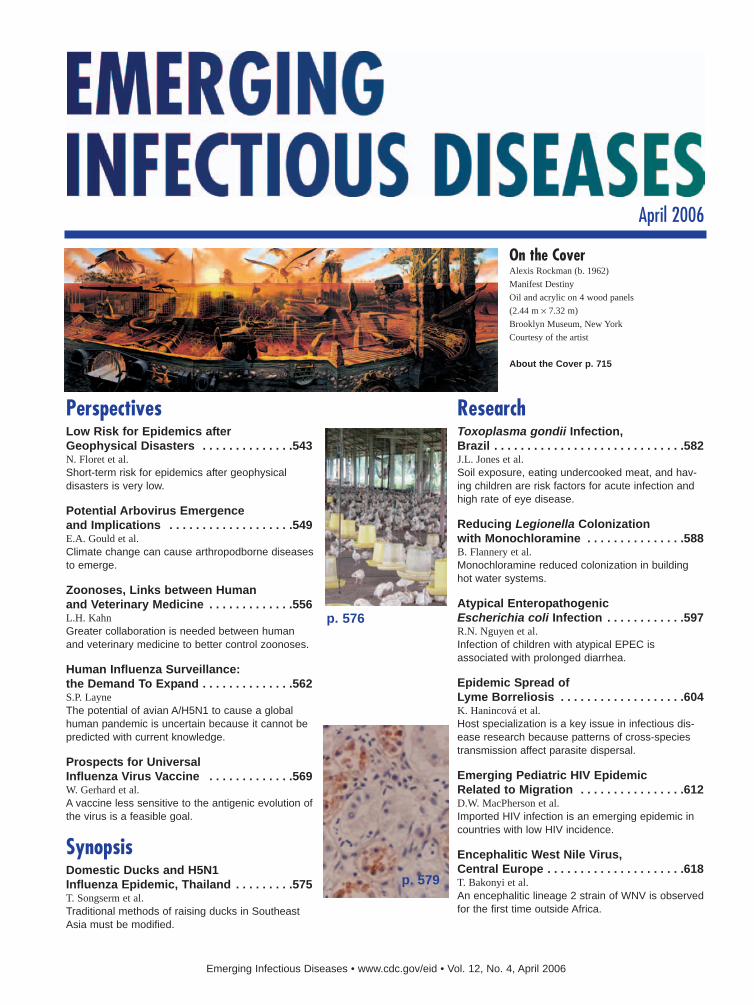
SynopsisDomestic Ducks and H5N1 Influenza Epidemic, Thailand . . . . . . . . .575T. Songserm et al.Traditional methods of raising ducks in SoutheastAsia must be modified.
ResearchToxoplasma gondii Infection, Brazil . . . . . . . . . . . . . . . . . . . . . . . . . . . . .582J.L. Jones et al.Soil exposure, eating undercooked meat, and hav-ing children are risk factors for acute infection andhigh rate of eye disease.
Reducing Legionella Colonization with Monochloramine . . . . . . . . . . . . . . .588B. Flannery et al.Monochloramine reduced colonization in buildinghot water systems.
Atypical Enteropathogenic Escherichia coli Infection . . . . . . . . . . . .597R.N. Nguyen et al. Infection of children with atypical EPEC is associated with prolonged diarrhea.
Epidemic Spread of Lyme Borreliosis . . . . . . . . . . . . . . . . . . .604K. Hanincová et al.Host specialization is a key issue in infectious dis-ease research because patterns of cross-speciestransmission affect parasite dispersal.
Emerging Pediatric HIV Epidemic Related to Migration . . . . . . . . . . . . . . . .612D.W. MacPherson et al.Imported HIV infection is an emerging epidemic incountries with low HIV incidence.
Encephalitic West Nile Virus, Central Europe . . . . . . . . . . . . . . . . . . . . .618T. Bakonyi et al.An encephalitic lineage 2 strain of WNV is observedfor the first time outside Africa.
On the CoverAlexis Rockman (b. 1962) Manifest DestinyOil and acrylic on 4 wood panels (2.44 m × 7.32 m) Brooklyn Museum, New YorkCourtesy of the artist
About the Cover p. 715
April 2006
p. 579
p. 576
Emerging Infectious Diseases • www.cdc.gov/eid • Vol. 12, No. 4, April 2006

Another DimensionBedside Manners . . . . . . . . . . . . . . . . . . .623C. Wiseman
Trypanosoma cruzi in Mexico . . . . . . . .624J.G. Estrada-Franco et al.Seroanalysis of parasite circulation in dogs can helpidentify T. cruzi infection in humans.
Contrasting Pediatric and AdultMRSA Isolates . . . . . . . . . . . . . . . . . . . . .631M.Z. David et al.Children may share a reservoir of MRSA strainsthat have an antimicrobial drug resistance profiledistinct from that of adults.
Microarray Identification of Influenza Viruses . . . . . . . . . . . . . . . . . . .638Z. Wang et al.Resequencing microarrays rapidly identify influenzaviruses.
Animals as Sentinels of Bioterrorism Agents . . . . . . . . . . . . . . . .647P. Rabinowitz et al.Pets, wildlife, or livestock could provide early warning.
Economic Impact of Lyme Disease . . . .653X. Zhang et al.Since 1975, Lyme disease has become the mostcommon vectorborne inflammatory disease in theUnited States.
1951 Influenza Epidemic . . . . . . . . . . . . .661C. Viboud et al.Death rates were substantially higher for Englandand Canada than for the United States.
Dispatches669 HIV Transmission in Correctional
FacilityA. Macher et al.
672 Fatal Human Infection withRickettsia rickettsiiJ.E. Zavala-Castro et al.
675 Borna Disease Virus HostsM. Hilbe et al.
678 Tularemia Outbreak, Bulgaria,1997–2005T. Kantardjiev et al.
681 Avian Influenza H5N1 in Domestic CatT. Songserm et al.
684 Cryptosporidiosis and Apple CiderB.G. Blackburn et al.
687 Long-term Follow-up for MDR-TBS.S. Shin et al.
689 Immune Restoration Disease inHIV PatientN.E. Jenkins et al.
692 Differential Diagnosis of ViralHemorrhagic FeversG. Palacios et al.
Commentary696 Atypical EPEC: Typical
Pathogens?J.P. Nataro
Letters697 Computer-assisted Telephone
Interview Techniques (Replies)698 Lack of Transmission of Vaccinia
Virus700 Discrimination between H5 Avian
Influenza A Viruses702 Rift Valley Fever in Goats,
Cameroon703 HIV-1 CRF07_BC Infections,
Injecting Drug Users, Taiwan705 Chlamydialike Organisms and
Atherosclerosis707 Maculopathy in Dengue Fever707 Pulmonary Tuberculosis in SARS,
China709 Tetanus in Injecting Drug Users,
United Kingdom711 Henipavirus in Pteropus vampyrus
Bats, Indonesia
Book Reviews713 AIDS in Asia: A Continent in Peril714 Biological Weapons Defense
Corrections714 Vol. 12, No. 2
News & NotesAbout the Cover
715 Manifesting Ecologic andMicrobial Connections
p.681
p. 673
April 2006
Emerging Infectious Diseases • www.cdc.gov/eid • Vol. 12, No. 4, April 2006

After geophysical disasters (i.e., earthquakes, volcaniceruptions, tsunamis), media reports almost always stressthe risk for epidemics; whether this risk is genuine hasbeen debated. We analyzed the medical literature and datafrom humanitarian agencies and the World HealthOrganization from 1985 to 2004. Of >600 geophysical dis-asters recorded, we found only 3 reported outbreaks relat-ed to these disasters: 1 of measles after the eruption ofPinatubo in Philippines, 1 of coccidioidomycosis after anearthquake in California, and 1 of Plasmodium vivax malar-ia in Costa Rica related to an earthquake and heavy rain-fall. Even though the humanitarian response may play arole in preventing epidemics, our results lend support to theepidemiologic evidence that short-term risk for epidemicsafter a geophysical disaster is very low.
Natural disasters are defined as “a disruption of humanecology which exceeds the community’s capacity to
adjust, so that outside assistance is needed” (1). Their clas-sifications are geophysical (earthquakes, volcanic erup-tions, tsunamis), hydrometeorologic (floods and windstorms), and geomorphologic (landslides). When coveringthese events, media outlets almost always mention the riskfor epidemics that could raise the death toll well above analready staggering number of victims. According to theCenters for Disease Control and Prevention (CDC), an epi-demic is the occurrence of more cases of disease thanexpected in a given area or among a specific group of per-sons over a particular period of time. For many, the wordepidemic is associated with large numbers of deaths andpoor living conditions, such as those that sometimes occurin refugee camps (2). The term outbreak is synonymouswith epidemic and is sometimes preferred because it maynot evoke the sensationalism associated with the word epi-demic.
In addition to the media, other outlets draw attention tothe risk for epidemics. In a letter published 3 weeks afterthe earthquake in Bam, Iran, in December 2004, the WorldHealth Organization (WHO) warned that potential out-
breaks of cholera, typhoid fever, malaria, and leishmania-sis were a major concern (3). WHO also issued a warningabout the risk for epidemics that could develop after the2004 tsunami: “There is an immediate INCREASEDRISK of waterborne diseases, i.e., cholera, typhoid fever,shigellosis and hepatitis A and E…. Outbreaks of these dis-eases could occur at any moment” (4). The high risk forepidemics in areas affected by the tsunami was also point-ed out by several papers published during the weeks afterthe disaster (5,6). Responding to WHO announcements,humanitarian agencies invested effort, time, personnel, andmoney in gearing up for potential epidemics, and consid-erable stocks of antimicrobial drugs, rehydration fluids forcholera patients, and vaccines were sent to the field.
However, not all experts support these alarming predic-tions. Some experts hold that disasters do not usually resultin disease outbreaks but may increase disease transmissionunder certain circumstances (e.g., fecal contamination ofwater, spread of respiratory diseases in evacuation camps)(7). A similar point of view was published by VanRooyenand Leaning (8) and by de Ville de Goyet (9), who spokeof the myths propagated after disasters, some of whichlead to an overestimation of the risk for epidemics.
No article has systematically reviewed published reportsdealing with epidemics after geophysical disasters. The roleplayed by outbreaks of infectious diseases in causing illnessafter geophysical disasters must be identified so that prior-ities can be defined and resources can be appropriately allo-cated. A systematic review of medical literature could helpanswer the question, “Is the risk for epidemics high after ageophysical disaster?” Consequently, we analyzed medicalliterature of the past 20 years and data provided by severalwebsites and databases that compile outbreak alert mes-sages and situation reports after disasters.
Materials and Methods
Literature ReviewWe screened Medline for articles that described out-
breaks and epidemics, in both English and French, pub-lished from January 1985 through December 2004. We
Negligible Risk for Epidemics afterGeophysical Disasters
Nathalie Floret,*† Jean-François Viel,*† Frédéric Mauny,*† Bruno Hoen,*† and Renaud Piarroux*†
Emerging Infectious Diseases • www.cdc.gov/eid • Vol. 12, No. 4, April 2006 543
*University Hospital of Besançon¸ Besançon, France; and†University of Franche-Comté, Besançon, France
used the following search terms: (natural disaster* ORseism* OR earthquake* OR volcano* OR tsunami*) AND(infectious disease* OR communicable disease* OR epi-demic* OR outbreak* OR vector-borne disease* ORarboviruses OR cholera OR malaria OR dengue OR WestNile virus OR Rift Valley fever OR hepatitis OR lep-tospirosis OR typhoid fever OR measles OR shigellosisOR scrub typhus OR plague OR diarrhea). We first select-ed all articles related to a specific earthquake, volcaniceruption, or tsunami, and then we examined them for anyquantitative data on at least 1 infectious disease.
Screening Databases on the InternetData on epidemics and geophysical disasters were col-
lected from the following databases: Emergency DisastersData Base (Em-Dat, www.em-dat.net), WHO websites(http://who.int/), Disease Outbreak News (http://who.int/csr/don/en/), Centers for Disease Control and Prevention(http://www.cdc.gov/), Morbidity and Mortality WeeklyReport (http://www.cdc.gov/mmwr/), and the Pan AmericaHealth Organization (http://www.paho.org). Researchfocused on events that occurred from January 1985through December 2004. For disasters that were responsi-ble for >100 deaths, we systematically screened reports ofhumanitarian agencies available on Reliefweb (http://reliefweb.int/).
Results
Literature ReviewAlthough we found 233 articles in the Medline database
related to our query, only 18 (7.7%) actually reported oninfectious disease data collected after geophysical disas-ters. Common respiratory tract infections and diarrheawere the most frequently reported diseases. After the Bamearthquake in December 2003, a survey of 75,586 dis-placed persons described the main health problemsencountered (10). Respiratory tract infections (mainlyupper respiratory tract infections) were most frequentlyencountered; 11,320 cases were seen in the 10 weeks afterthe disaster. Researchers attributed the high number of res-piratory infections to the freezing winter nights. Diarrheawas commonly diagnosed (1,224 cases with 174 cases ofbloody diarrhea), but Vibrio cholerae infection was notobserved. Similar findings were reported after the Chi-Chiearthquake in Taiwan in September 1999. An epidemiolog-ic survey conducted in shelters showed that acute respira-tory infections and acute gastroenteritis were the mostcommon illnesses reported (11). They increased during thefirst 4 weeks, were significantly higher than those in unaf-fected neighboring counties, and then declined to baselinelevels afterwards. An increase in gastrointestinal and respi-ratory infections also followed the 2001 earthquakes in El
Salvador (12). Woersching and Snyder. conducted a 32-question survey in 100 households (594 persons) severelyaffected by the earthquakes. These researchers found that30% of households assessed experienced ≥1 case of upperrespiratory infection, and 22% experienced ≥1 case ofdiarrheal disease. This study also showed a high frequencyof skin infections (31% of households). In a figure, authorsreported 6 cases of cholera but did not state whether thediagnosis was biologically confirmed (no cases of cholerawere officially identified in El Salvador in 2001) (13). Anincrease in respiratory and intestinal tract infections wasalso reported after the eruption of the Cerro Negro volcanoin Nicaragua in April 1992, although this increase was notdeclared an epidemic (14). An assessment of the healthconsequences of the disaster showed that acute diarrheawas 6 times more frequent after the eruption, and medicalconsultations for acute respiratory disease were 3.6 timesmore frequent than before.
Two studies were performed to assess medical recordsof inpatients hospitalized during the first 15 days after theHanshin-Awaji earthquake in January 1995 (15,16).Among infectious diseases, pneumonia was the most fre-quent illness diagnosed in inpatients (13%–21% accordingto the 2 surveys). An increased number of inpatients werealso recorded in Papua New Guinea after the tsunami inJuly 1998 (17). However, no outbreak of communicabledisease occurred.
A few studies investigated the prevalence of somepathogens in persons living in shelters. After the earth-quake in Turkey in August 1999, an analysis of 1,468 stoolcultures taken from persons with diarrhea showed 92%negative results; the most frequently isolated pathogenswere Shigella spp. (4.9%). Phenotypic and genotypic com-parisons of strains showed no cloning among the Shigellastrains (18). Another study was conducted to determine theinfluence of the earthquake on patient admittance to theoutpatient dermatology clinic. The incidence of skin infec-tions was higher after the earthquake than it was in thesame period 1 year later (19). A third study was performedto assess the prevalence of hepatitis A and E among chil-dren living in camps in northwestern Turkey (20).Hepatitis A and E virus seroprevalence was higher in thecamps around Golyaka (68.8% and 17.2%, respectively)than in camps around Düzce (44.4% and 4.7%). Theauthors suggested that these differences were possiblyrelated to delays in obtaining toilet facilities and pipedwater. After the earthquake in Colombia in January 1999,a parasitologic survey was performed in transitory housingcamps from January 2000 to July 2001 (21). A high preva-lence of Giardia spp. (60%) was found in stool specimensof 217 randomly selected children, and this prevalence wassignificantly associated with the use of communal toiletsinstead of individual toilets and with drinking municipal
PERSPECTIVE
544 Emerging Infectious Diseases • www.cdc.gov/eid • Vol. 12, No. 4, April 2006

water instead of water from individual tanks. The authorsalso stated that no outbreak of diarrhea, dengue fever, ormalaria had occurred.
Only 3 articles reported outbreaks after a geophysicaldisaster. An outbreak of malaria (due to Plasmodiumvivax) was reported after an earthquake in April 1991 inCosta Rica (22). From June 1991 through May 1992, atotal of 3,597 cases were recorded, compared to 549 and681 cases for the same period during the 2 preceding years.Even though heavy rainfall occurred in August 1991,authors suggested that the earthquake may have played arole. An outbreak of coccidioidomycosis was describedafter the 1994 earthquake in Northridge, California. Theattack rate reached 30 cases per 100,000 inhabitants.According to the authors, being in a dust cloud and theamount of time spent in a dust cloud were associated withan increased risk of diagnosis (23). An outbreak of measlesoccurred after the eruptions of Mt. Pinatubo in June 1991.By August, many children of the Aeta tribe, who usuallylived in isolation on the slopes of Pinatubo, had died inevacuation centers. The death toll reached 349 in the first12 weeks, accounting for a death rate of 26/10,000 by theseventh week after the eruption (24–26). Deaths werecaused by measles (31%), diarrhea (29%), and respiratoryinfections (22%). Living conditions were extremely diffi-cult in camps: tents provided only minimal shelter from theelements, and evacuees experienced extremely hot daysand cold, damp nights (26). Malnutrition and lack of basicsanitation also contributed to high death rates among chil-dren (24).
Database ResearchFrom 1985 to 2004, 516 earthquakes, 89 volcano erup-
tions, and 16 tidal waves or tsunamis (including theDecember 2004 tsunami) were identified in the Em-Datdatabase. Sixty-three of these geophysical disasters wereresponsible for >100 deaths each, and 26 of them wereresponsible for ≥1,000 deaths (Table). Most of them (55 of63) were reported on the ReliefWeb site. However, only 21descriptions included medical data that covered at least the3-month period after the disaster. Only 1 outbreak wasreported: 19 cases of Crimean-Congo hemorrhagic fever,including 12 fatal cases, occurring in mid-March 1998 in avillage in the district of Rustaq, Afghanistan, where anearthquake had occurred in February 1998. This outbreakwas not caused by the earthquake but was detected becauseof epidemiologic surveillance that was implemented afterthe earthquake. No outbreak was reported after the otherdisasters, even in reports published up to 3 months afterthe events.
Among alert messages reported on the WHO outbreaknews website, >300 concerned new outbreaks detectedfrom 1997 to 2004 (we could not access previous WHO
archives), and 90 of these concerned cholera outbreaks. Wealso found 779 epidemics reported in Em-Dat from 1985 to2004. However, only 1 outbreak (of Crimean-Congo hem-orrhagic fever [previously mentioned]) occurred in an areaaffected by a recent geophysical disaster.
DiscussionAlthough >600 geophysical disasters were recorded in
the 20-year period we studied, we found no report in themedical literature in which major epidemics occurred intheir wake. Only 2 outbreaks, one of Coccidioides immitisinfection and the other of measles, could clearly be relatedto a preceding disaster (23–26). Since this result is at vari-ance with the fact that iterative warning messages arebroadcast after each disaster, we enlarged our search of thepast 20 years by checking for alert messages from variousinstitutional disease control databases and by screeningreports available on Reliefweb.
The lack of reported epidemics in all the sources weanalyzed begs an essential question: If epidemics can beexpected to occur after a geophysical disaster, why arethey almost never detected or reported? In general, epi-demiologic studies are rarely conducted after disasters, andwhen they are, their methods are open to criticism. Mostinvestigations only use cross-sectional survey methodswithout any reference to baseline status or control areas(27). In remote, rural areas of developing countries and inareas affected by war, surveillance systems are often notfunctioning, and an epidemic may go unnoticed. In addi-tion, medical humanitarian agencies mainly focus onshort-term assistance to affected persons, and most volun-teers and experts usually leave the area within 3 months(1). At that time, basic sanitation facilities and access tobasic hygiene may still be unavailable because of econom-ic consequences of the disaster, and some affected victimsmay have to stay in camps and shelters for prolonged peri-ods. Given the flaws of epidemiologic surveys describedabove, the hypothesis that unreported outbreaks occur aconsiderable time after the onset of a disaster must beexamined. However, for some diseases, such as cholera,meningitis, and dengue, a large-scale outbreak would like-ly be detected by local health authorities or by humanitar-ian agencies working after the emergency phase. In thatcase, WHO would be notified or a field report would bemade, even though the outbreak might not be reported in amedical journal.
Many arguments are usually presented to show that ageophysical disaster is a high-risk situation for epidemics.First, water and sanitation systems may be destroyed dur-ing the disaster, increasing the risk for outbreaks of water-borne diseases. However, natural disasters do not importdiseases, and even in areas where a given disease isendemic, the worst-case scenario does not always occur
Low Risk for Epidemics after Geophysical Disasters
Emerging Infectious Diseases • www.cdc.gov/eid • Vol. 12, No. 4, April 2006 545

(9). Cholera is endemic around the Bay of Bengal, butcases of cholera are not constantly diagnosed in each vil-lage around the bay. Even if brackish water in the estuar-ies is an environmental reservoir for V. cholerae, toxigenicbacteria do not necessarily spread from them, should atsunami occur. Many ecologic, sociologic, and seasonalfactors are involved in the emergence of V. cholerae, andthese factors rarely converge (28). Another surprisingassertion is that tsunamis increase water sources for mos-quitoes and therefore enhance the risk for vectorborne dis-ease outbreaks. Water is an essential component of themosquito environment. The characteristics of the waterhabitat, whether it is running or standing, clean or pollut-ed, fresh or brackish, shaded or sunlit, permanent or inter-mittent, are the predominant factors determining whichspecies of mosquito breed in it. Transient, polluted saltwater generated by a tsunami will not sustain most speciesinvolved in transmission of dengue fever and malaria(29).
Second, natural disasters arguably lead to populationdisplacement, formation of camps, overcrowding, andtherefore, propitious circumstances for an epidemic.Settlements for victims of natural disasters, however, arenot synonymous with refugee camps created to cope withcomplex emergencies (e.g., war, oppression, famine). Insuch complex emergencies, refugees may live for a long
time in overcrowded conditions with a poor water supplyand bad sanitary facilities. Usually refugees have beenmalnourished for weeks or even months before they reachthe camps. Conditions like this in Goma, Zaire, producedepidemics of cholera, shigellosis, and meningitis, whichcaused thousands of deaths (30). For natural disasters, theshock is short-term, and communities can cope with prob-lems more easily; predisaster health and nutrition status arebetter than in complex emergencies. The camps are oftenmuch smaller, which limits the spread of pathogens; accessto food, safe water, and sanitary facilities is usually better;and most people stay only a few days or weeks.Nevertheless, crowded conditions and, in some cases, coldweather, favor the transmission of airborne diseases. Thefirst response in preventing an outbreak of respiratory dis-ease is to provide adequate shelter as soon as possible toinjured persons and to prevent overcrowding. In our study,however, measles outbreaks were far less frequent thanexpected. Early implementation of immunization cam-paigns probably has a protective effect, and vaccination isrecommended each time nonimmunized populations aremoved to camps. Vaccination against influenza is not rec-ommended even though it is a highly contagious diseaseand has a shorter incubation period than measles.Surprisingly, despite the lack of immunization campaignswe observed in our study, we never found any report of an
PERSPECTIVE
546 Emerging Infectious Diseases • www.cdc.gov/eid • Vol. 12, No. 4, April 2006
influenza epidemic whose spread was aided by a precedinggeophysical disaster.
The “fact” that dead bodies are a potential cause of epi-demics after a disaster is also almost always broadcastafter major disasters. This “fact” is a myth, and deprivingsurvivors of appropriate burial ceremonies for their rela-tives may administer yet another blow to already injured orweakened persons (9,31,32). The only situation in whichhandling corpses is a risk is during epidemics of infectiousdiseases such as cholera. Even in these situations, no rea-son exists to totally deprive families from honoring theirdead if they follow certain precautions (33).
Our results, in line with those of Noji and de Ville deGoyet, lend support to the epidemiologic evidence that nohigh, short-term risk for epidemics follows a geophysicaldisaster. While most medical topics are usually discussedin small task groups of highly specialized experts, thedebate about risk for epidemics after natural disasters isusually conducted by the mass media. The news industryis prone to emphasizing more dramatic and simplistic mes-sages, and unjustified warnings will likely continue to bespread on the basis of an approximate assessment of risks.To respond more effectively to the needs of victims of nat-ural disasters, the public, mass media, humanitarian organ-izations, and policymakers must be accurately informedregarding what actions are effective and what actions arefutile.
Dr Floret is a public health physician at the UniversityHospital of Besançon, France. Her research interests are in healthrisk assessment for environmental pollutants, particularly dioxins.
References
1. Lechat MF. The epidemiology of health effects of disasters.Epidemiol Rev. 1990;12:192–8.
2. Goma Epidemiologic Group. Public health impact of Rwandanrefugee crisis: what happened in Goma, Zaire, in July 1994. Lancet.1995;345:339–44.
3. Zarocostas J. WHO praises Bam response but warns of disease.Lancet. 2004;363:218.
4. World Health Organization. South Asia tsunami situation report 4[monograph on the Internet]. 2005 Jan 2 [cited 2006 Feb 15].Available from http://www.who.int/hac/crises/international/asia_tsunami/sitrep/04/en/index.html
5. Moszynski P. Disease threatens millions in wake of tsunami. BMJ.2005;330:59.
6. Vogel G. Indian Ocean tsunami. Using scientific assessments to staveoff epidemics. Science. 2005;307:345.
7. Noji EK. Public health issues in disasters. Crit Care Med. 2005;33(1Suppl):S29–33.
8. VanRooyen M, Leaning J. After the tsunami—facing the publichealth challenges. N Engl J Med. 2005;352:435–8.
9. de Ville de Goyet C. Stop propagating disaster myths. Lancet.2000;356:762–4.
10. Akbari ME, Farshad AA, Asadi-Lari M. The devastation of Bam: anoverview of health issues 1 month after the earthquake. PublicHealth. 2004;118:403–8.
11. Chen KT, Chen WJ, Malilay J, Twu SJ. The public health response tothe Chi-Chi earthquake in Taiwan, 1999. Public Health Rep. 2003;118:493–9.
12. Woersching JC, Snyder AE. Earthquakes in El Salvador: a descriptivestudy of health concerns in a rural community and the clinical impli-cations—part II. Disaster Manag Response. 2004;2:10–3.
13. Woersching JC, Snyder AE. Earthquakes in El Salvador: a descriptivestudy of health concerns in a rural community and the clinical impli-cations—part I. Disaster Manag Response. 2003;1:105–9.
14. Malilay J, Real MG, Ramirez Vanegas A, Noji E, Sinks T. Publichealth surveillance after a volcanic eruption: lessons from CerroNegro, Nicaragua, 1992. Bull Pan Am Health Organ.1996;30:218–26.
15. Matsuoka T, Yoshioka T, Oda J, Tanaka H, Kuwagata Y, Sugimoto H,et al. The impact of a catastrophic earthquake on morbidity rates forvarious illnesses. Public Health. 2000;114:249–53.
16. Tanaka H, Oda J, Iwai A, Kuwagata Y, Matsuoka T, Takaoka M, et al.Morbidity and mortality of hospitalized patients after the 1995Hanshin-Awaji earthquake. Am J Emerg Med. 1999;17:186–91.
17. Asari Y, Koido Y, Nakamura K, Yamamoto Y, Ohta M. Analysis ofmedical needs on day 7 after the tsunami disaster in Papua NewGuinea. Prehospital Disaster Med. 2000;15:9–13.
18. Vahaboglu H, Gundes S, Karadenizli A, Mutlu B, Cetin S, Kolayli F,et al. Transient increase in diarrheal diseases after the devastatingearthquake in Kocaeli, Turkey: results of an infectious disease sur-veillance study. Clin Infect Dis. 2000;31:1386–9.
19. Bayramgurler D, Bilen N, Namli S, Altinas L, Apaydin R. The effectsof 17 August Marmara earthquake on patient admittances to our der-matology department. J Eur Acad Dermatol Venereol. 2002;16:249–52.
20. Sencan I, Sahin I, Kaya D, Oksuz S, Yildirim M. Assessment of HAVand HEV seroprevalence in children living in post-earthquake campsfrom Duzce, Turkey. Eur J Epidemiol. 2004;19:461–5.
21. Lora-Suarez F, Marin-Vasquez C, Loango N, Gallero M, Torres E,Gonzalez MM, et al. Giardiasis in children living in post-earthquakecamps from Armenia (Colombia). BMC Public Health. 2002;2:5.
22. Saenz R, Bissel RA, Paniagua F. Post-disaster malaria in Costa Rica.Prehospital Disaster Med. 1995;10:154–60.
23. Schneider E, Hajjeh RA, Spiegel RA, Jibson RW, Harp EL, MarshallGA, et al. A coccidioidomycosis outbreak following the Northridge, Calif, earthquake. JAMA. 1997;277:904–8.
24. Centers for Disease Control and Prevention. Surveillance in evacua-tion camps after the eruption of Mt. Pinatubo, Philippines. MMWRSurveill Summ. 1992;41:9–12. Erratum in MMWR Surveill Summ.1992;41:963.
25. Magpantay RL, Abellanosa IP, White ME, Dayrit MM. Measlesamong Aetas in evacuation centers after volcanic eruption.International Scientific Conference on Mt. Pinatubo; Department ofForeign Affairs, Manila; 1992 May 27–31. p. 33.
26. Banzon Bautista C. The Mount Pinatubo disaster and the people ofcentral Luzon [monograph on the Internet]. 1999 Jun 10 [cited 2006Feb 15]. Available from http://pubs.usgs.gov/pinatubo/cbautist
27. Logue JN, Evans Melick M, Hansen H. Research issues and direc-tions in the epidemiology of health effects of disasters. EpidemiolRev. 1981;3:140–62.
28. Sack RB, Siddique AK, Longini IM, Nizam A, Yunus M, Islam MS,et al. 4-year study of the epidemiology of Vibrio cholerae in four ruralareas of Bangladesh. J Infect Dis. 2003;187:96–101.
29. Briët O, Galappaththy G, Konradsen F, Amerasinghe P, AmerasingheF. Maps of the Sri Lanka malaria situation preceding the tsunami andkey aspects to be considered in the emergency phase and beyond.Malar J. 2005;4:8.
Low Risk for Epidemics after Geophysical Disasters
Emerging Infectious Diseases • www.cdc.gov/eid • Vol. 12, No. 4, April 2006 547

30. Baxter P, Ancia A. Human health and vulnerability in the Nyriagongovolcano crisis, DR Congo Jun 2002 [monograph on the Internet].2002 Jun [cited 2006 Feb 15]. Available from http://www.reliefweb.int/rw/rwb.nsf/AllDocsByUNID/302be587c8df7c39c1256be2002cf5cc
31. Thieren M, Guitteau R. Identifying cadavers following disasters:why? Disasters. 2000;80:1–2.
32. Morgan O. Infectious disease risks from dead bodies following natu-ral disasters. Rev Panam Salud Publica. 2004;15:307–12.
33. Piarroux R. Cholera: epidemiology and transmission. Experiencefrom several humanitarian interventions in Africa, Indian Ocean andCentral America [article in French]. Bull Soc Pathol Exot.2002;95:345–50.
Address for correspondence: Renaud Piarroux, Service de Parasitologieet Mycologie, Hôpital Jean Minjoz, 25000 Besançon, France; fax: 33-381-668-914; email: [email protected]
PERSPECTIVE
548 Emerging Infectious Diseases • www.cdc.gov/eid • Vol. 12, No. 4, April 2006
Searchpast issues

Arboviruses have evolved a number of strategies tosurvive environmental challenges. This review examinesthe factors that may determine arbovirus emergence, pro-vides examples of arboviruses that have emerged into newhabitats, reviews the arbovirus situation in western Europein detail, discusses potential arthropod vectors, andattempts to predict the risk for arbovirus emergence in theUnited Kingdom. We conclude that climate change is prob-ably the most important requirement for the emergence ofarthropodborne diseases such as dengue fever, yellowfever, Rift Valley fever, Japanese encephalitis, Crimean-Congo hemorrhagic fever, bluetongue, and African horsesickness in the United Kingdom. While other arboviruses,such as West Nile virus, Sindbis virus, Tahyna virus, andLouping ill virus, apparently circulate in the UnitedKingdom, they do not appear to present an imminent threatto humans or animals.
More than 550 arboviruses have been identified (1).Many are mammalian and avian pathogens, 4 of
which (West Nile virus [WNV], Usutu virus, Sindbis virus[SINV], and Tahyna virus) circulate in the UnitedKingdom in resident and migratory birds (2,3). The onlyzoonotic arboviruses isolated from field material in theUnited Kingdom are the flavivirus Louping ill virus (LIV)(4) and the bunyavirus Uukuniemi virus (5). LIV is includ-ed in the tickborne encephalitis virus (TBEV) complex thatincludes Kyasanur Forest disease virus and Alkhurmavirus. These viruses can cause encephalitis and hemor-rhagic disease (6). Other human pathogens in the genusFlavivirus that will be discussed in this review include yel-low fever virus (YFV), dengue virus (DENV), Israelturkey meningoencephalomyelitis virus, and Bagaza virus.European human and animal pathogens in the genusAlphavirus include SINV, Ockelbo virus, and
Chikungunya virus and in the family Bunyaviridae, sand-fly fever Naples virus (often referred to as Toscana virus),sandfly fever Sicilian virus, Crimean-Congo hemorrhagicfever virus (CCHFV), Inkoo virus, and Tahyna virus,which is widespread throughout Europe. Rift Valley fevervirus (RVFV) and Nairobi sheep disease virus (NSDV)could be introduced to Europe from Africa through animaltransportation. Finally, the family Reoviridae contains avariety of animal arbovirus pathogens, including blue-tongue virus and African horse sickness virus, both knownto be circulating in Europe. This review considers whetherany of these pathogenic arboviruses are likely to emergeand cause disease in the United Kingdom in the foresee-able future.
Factors That May Determine Arbovirus Emergence
Transmission of arboviruses between invertebrates andvertebrates imposes constraints on evolution and dispersal,which are reflected in their phylogenetic relationships (7).However, the influence of modern life on arbovirus emer-gence cannot be overemphasized. The following humanactivities may influence arbovirus emergence: 1) increasedtransportation of animals, humans, plants, arthropods, andother materials; 2) increased outdoor leisure activities; 3)reduced or nonexistent arthropod control programs; 4)deforestation; 5) reforestation; 6) land reclamation; 7)altered farming practices; 8) urbanization programs; 9)irrigation projects, including building dams or creatingreservoirs and lakes; 10) military activities; 11) movementof military personnel and local populations in war zones;12) natural disasters, such as flooding; and 13) the earlyeffects of climate change (8).
Examples of Emerging Tickborne ArbovirusesTBEV complex viruses rarely cause disease in indige-
nous forest animals but may emerge as pathogens whenthey infect introduced species. Members of the complex
Potential Arbovirus Emergence andImplications for the United Kingdom
Ernest Andrew Gould,* Stephen Higgs,† Alan Buckley,* and Tamara Sergeevna Gritsun*
Emerging Infectious Diseases • www.cdc.gov/eid • Vol. 12, No. 4, April 2006 549
*Centre for Ecology and Hydrology Oxford, Oxford, UnitedKingdom; and †University of Texas Medical Branch, Galveston,Texas, USA

have evolved and dispersed westward across Asian andEuropean forests during the past 3–5 millenia. This disper-sion is driven by Ixodes spp. vectors that inhabit the moistforest undergrowth. Nymphal ticks may infect newlyhatched larvae when they co-feed on forest animals. Sincethese vertebrates do not become sick or have detectableviremia, this direct method of vector infection is known asnonviremic transmission. In contrast, LIV and relatedviruses in Spain, Turkey, Greece, and nearby regions pro-duce viremia and fatal encephalomyelitis in domesticatedanimals when infectious ticks feed on them (9). These ani-mals have no genetic resistance or immunity to the TBEV-related viruses that have emerged from the forests (8).
Kyasanur Forest disease virus emerged in Karnataka,India, in 1957 after forests were cleared for urbanizationand farmland reclamation. Arboreal monkeys scavengingon the exposed forest floor became infested with KyasanurForest disease virus–infected ticks from the undergrowth.Local inhabitants and veterinary scientists who examineddead and dying monkeys also became infected. A closelyrelated virus, Alkhurma virus, emerged in Saudi Arabia in1992, causing hemorrhagic disease in butchers who han-dled sheep imported into Saudi Arabia for the Hajj (10).Alkhurma virus was detected in ticks removed fromimported sheep, but the country from which these sheeporiginated has not been identified.
CCHFV emerges sporadically in Africa, Asia, andEurope. This arbovirus may be transmitted to animals byticks, especially members of the Hyalomma genus.Emergence of CCHFV largely depends on the transporta-tion of livestock such as cattle and goats, on which infect-ed ticks feed. Most cases of hemorrhagic disease occur inhumans involved with the livestock industry. Since >21tick species are present in the United Kingdom, and sinceviruses such as CCHFV can infect a wide variety of ticks,the introduction and emergence of CCHFV are not incon-ceivable.
NSDV (family Bunyaviridae, genus Nairovirus) istransmitted to sheep by Rhipicephalus appendiculatus andcauses fever and gastroenteritis; the infection is often fatal.NSDV is distributed widely throughout Africa (1) but notelsewhere. However, the closely related Ganjam virus isfound in India, which may imply that NSDV has beentransported in ticks on animals imported to India fromAfrica. Both NSDV and Ganjam virus produce febrile ill-ness with polyarthritis in humans.
Examples of Emerging MosquitoborneArboviruses
YFV circulates among simians and Aedes andHemagogus spp. mosquitoes in the forest canopy and adja-cent savannah regions of Africa and South America. InAfrica, nonhuman primates do not become ill from YFV
infection, presumably because the virus has adapted to thesimian hosts. Nevertheless, human yellow fever epidemicsdo occur in Africa because the virus emerges sporadicallyfrom the jungles and savannah into rural and urban areas,where most of the human population is susceptible. InSouth American jungles, moribund monkeys are usuallyfound immediately before local epidemics of yellow fever,implying that the virus recently encroached into the region.YFV was introduced into South America on ships thatsailed from Africa during the past few centuries (8). ManyEuropean and North American ports observed cases of yel-low fever. In some cases, the virus was carried on sailingships from Cuba to Europe, causing localized outbreakswhere the ships docked (11). Infections in Swansea(Wales) and St. Nazaire (France) resulted from the bite ofAedes aegypti mosquitoes released from the newly arrivedships. However, the disease immediately disappearedwhen the local temperature dropped and the tropical mos-quitoes died. Thus, such viruses are unlikely to emerge inthe United Kingdom.
The 4 DENV serotypes emerged more recently thanYFV (8) and can generate epidemics in humans withoutdepending on a sylvatic cycle in the jungles. Therefore, incontrast to YFV, which is maintained in the jungles ofAfrica and South America, DENV continues to be dis-persed by humans and mosquitoes throughout the tropicsand subtropics worldwide (8,12). Presumably, as Ae.aegypti and Ae. albopictus continue to spread, the geo-graphic distribution of these viruses will also expand.
Since birds are highly mobile and can travel great dis-tances in a relatively short time, they play a role inarbovirus dispersal. SINV, for example, is carried annual-ly between South Africa and Scandinavia by migratingbirds (12). SINV is rarely associated with disease inAfrica. However, when cases of arthritis, polyarthritis, andfever associated with rash were recorded in Scandinavia,the antigenically closely related Ockelbo virus was isolat-ed. Presumably, Ockelbo virus evolved from introducedSINV and subsequently became established in a localtransmission cycle. In the New World, St. Louis encephali-tis virus is also carried long distances by migrating birds.It was probably introduced into South America from Africaduring the past few centuries and gradually dispersed bymigrating birds to North America (13). More recently,WNV was introduced into the New York area, probably in1999. The virus became established in birds and was rap-idly dispersed north to Canada; south to Florida, Mexico,and the Caribbean; and west, reaching California in 2004(http://www.cdc.gov/ncidod/dvbid/westnile/). The WNVthat was introduced into New York was related to anEgypt/Israel strain that is also present in the Volga RiverDelta. Despite its high virulence in birds (particularlycorvids), horses, and alligators in North America, WNV
PERSPECTIVE
550 Emerging Infectious Diseases • www.cdc.gov/eid • Vol. 12, No. 4, April 2006

circulates relatively harmlessly in the Old World, presum-ably reflecting co-evolution of virus and host over a longperiod.
Other emerging flaviviruses include Usutu virus, whichappeared unexpectedly in Austria in 2000 (14), killingbirds in Vienna and surrounding areas. Previously, Usutuvirus had only been found in Africa. Israel turkey menin-goencephalitis virus, isolated in Israel, is phylogeneticallyclosely related to the African Bagaza virus (7). Thus, Israelturkey meningoencephalitis virus may represent the bird-borne version of Bagaza virus.
RVFV (family Bunyaviridae, genus Phlebovirus) is usu-ally transmitted by Aedes spp. mosquitoes and occasional-ly infects livestock, producing epizootics with high deathrates in sheep and other ruminants. Humans in areas whereRVFV is epizootic may also become infected. Until 2000,RVFV was most commonly found only in eastern, western,and southern Africa. However, in September 2000, an out-break was reported in Saudi Arabia and subsequently inYemen, representing the first cases identified outside Africaand showing the potential for global dispersal.
Bluetongue virus (family Reoviridae, genus Orbivirus)is transmitted to domestic and wild ruminants by culicoidemidges. Bluetongue virus epidemics can threaten entirelivestock populations and were first described in SouthAfrica after Merino sheep were introduced from Europe inthe late 18th century. The virus subsequently dispersedglobally, recently reemerging in Italy, Spain, and Portugal
(15), most likely through the transportation of ruminants.Whether bluetongue virus will emerge in the UnitedKingdom likely depends on the effects of climate changeon vector dispersal.
Arboviruses Most Likely To Circulate in the United Kingdom
The English Channel and North Sea are not barriers tomigrant birds, bats, or commercial transportation systems.Therefore, most mainland European arboviruses could beintroduced and established in the United Kingdom. TheTable presents a summary of all recognized zoonotic mos-quitoborne and tickborne arboviruses circulating in west-ern Europe. While Uukuniemi virus has not been shown tobe a human pathogen, it circulates widely in Europe, andseropositivity in humans has been demonstrated manytimes. YFV, DENV, RVFV, Ganjam virus, and NSDV areexcluded from the Table because they only arise as import-ed viruses. Only LIV and Uukuniemi virus have been iso-lated from field specimens in the United Kingdom,probably because of limited investigation, although theother viruses in the Table have been isolated in Europe.Serologic evidence obtained from bird sera (2) implies thatat least 3 additional arboviruses, WNV, Usutu virus, andSINV, circulate in the United Kingdom. Currently, TBEVhas not dispersed west beyond the Alsace forests in France(29). LIV, Spanish sheep encephalomyelitis virus, andGreek goat encephalomyelitis virus, on the other hand,
Potential Arbovirus Emergence and Implications
Emerging Infectious Diseases • www.cdc.gov/eid • Vol. 12, No. 4, April 2006 551

represent more recent lineages of western European andFar Eastern TBEV that have emerged on the moorlandsafter the introduction of sheep and goats for grazing (8).
Three alphaviruses are listed in the Table, SINV,Semliki Forest virus, and Chikungunya virus. SINV hasbeen isolated from field material in Scandinavia, Estonia,Czechoslovakia, and Italy. This virus is widespreadthroughout the Old World. In Albania, seroconversion toChikungunya virus was detected in 5 patients with mildflulike symptoms (12); other findings of antibodies toChikungunya virus have been reported, but evidence ofcross-reactivity with other alphaviruses was not alwaystested. Among the remaining zoonotic arboviruses includ-ed in the Table, Tahyna virus is the most common. Thisvirus is widespread throughout western Europe in humansand many nonhuman mammalian species and birds (12).Inkoo virus has only been found in Scandinavia, but fewinvestigations have taken place elsewhere. While investi-gations for Batai virus have not been extensive, this virusis also widespread throughout western Europe (12).
Probability That Arboviruses Will Emerge as Epidemic Pathogens
Tickborne ArbovirusesMany arboviruses emerged relatively recently (30) and
have therefore had little time to be dispersed to new envi-ronments, such as the United Kingdom. Although birdscarry millions of ticks north and south annually betweenAfrica and Europe, tickborne African bunyaviruses andflaviviruses have not yet appeared as notable pathogens inthe United Kingdom. European, Siberian, and Far Easternstrains of TBEV have been isolated in Estonia, probablyintroduced by migratory birds (31). However, they havenot caused substantial disease in these regions. TBEV isless virulent than LIV for sheep (6) and might, therefore,become less dominant than LIV if it were introduced intothe sheep-grazing moorlands of the United Kingdom.Whether such introduced viruses would emerge mayreflect the level of contact between humans and farm ani-mals. Although ticks are less susceptible to the effects ofenvironmental disruption, the specific region into whichTBEV-carrying ticks are introduced might determinewhether they survived and then reproduced. The UnitedKingdom has an abundant supply of Ixodes ticks, but therisk that virulent Far Eastern TBEV would be introduced,become established, and emerge as an endemic pathogenin UK forests is probably low. Alternatively, in the case ofCCHFV, the fact that this virus has been isolated from ticksin Greece, the Balkans, and western Russia in the VolgaDelta cannot be ignored. Other tickborne virus pathogensare presumably subject to the same qualifications.
Mosquito-, Sandfly- and Midge-transmittedArboviruses
When one takes into account the information in theTable and the serologic evidence of antibodies againstSINV, WNV, Usutu virus, and Tahyna virus in sera ofmigrant and resident UK birds, the likelihood is high thatmosquitoborne arboviruses, particularly those associatedwith ornithophilic mosquitoes, such as Culex spp., circu-late, albeit harmlessly, in the United Kingdom. Otherarboviruses circulating in Europe that are not recognized inthe United Kingdom include Toscana virus, bluetonguevirus, and African horse sickness virus, which was proba-bly introduced into Spain after the importation of infectedhorses.
Tahyna virus, which causes encephalitis in humans, iswidespread across western Europe (Table). Birds and rab-bits are vertebrate hosts. Since the movement of rabbitsbetween the United Kingdom and mainland Europe isextensive, and since many bird migratory routes fromAfrica pass over the United Kingdom, Scandinavia, andmainland western Europe, Tahyna virus is most likely toappear, if not emerge, in the United Kingdom.
Why has none of these viruses yet emerged as a notice-able pathogen in the United Kingdom? Perhaps they have,and we do not realize it. Recent reports state that <40% ofhuman deaths from viral encephalitis are attributed to aspecific etiologic agent in the United Kingdom (32).Moreover, no systematic studies have been conducted todetermine the extent to which Tahyna virus is circulating,even though the virus is known to be present in UK birds(3). In addition, on the basis of serologic tests (2), a SINV-like virus also circulates among birds in the UnitedKingdom, possibly vectored by Culex mosquitoes. InScandinavia, this virus causes rash, fever, and acute pol-yarthritis in humans.
Necessary Arthropods for Arbovirus EmergenceAt least 21 species of hard ticks and 3 argasids are rec-
ognized in mainland Britain and its islands (33). Most par-asitize mammals, but at least 12 prefer birds, and 2 areregularly found on reptiles. In many parts of the UnitedKingdom, LIV has been isolated from I. ricinus ticks,which are believed to maintain LIV in the wild by non-viremic transmission during co-feeding (34). Other tickspecies, such as Ornithodorus maritimus and I. uriae,transmit bunyaviruses, nairoviruses, and orbivirusesamong seabirds (5,35) that inhabit the cliffs around the UKcoastline. However, these seabird-tickborne viruses do notcause disease in mammals in the United Kingdom.Nevertheless, the United Kingdom has a wide range ofticks that can transmit arboviruses.
At least 33 mosquito species are endemic in the UnitedKingdom (36), but their susceptibility to infection by
PERSPECTIVE
552 Emerging Infectious Diseases • www.cdc.gov/eid • Vol. 12, No. 4, April 2006

arboviruses and ability to replicate and transmit them tovertebrate hosts have not been assessed. At least 9 of thesemosquito species have been linked to transmission ofWNV (37). Many related viruses cause encephalitic infec-tions and are transmitted in nature between birds and mos-quitoes, particularly Culex spp. Six of the 33 endemicmosquito species feed on humans and birds, which makesthem potential bridge vectors (36). Eleven of the remain-ing 33 species bite humans. Clearly, the United Kingdomhas a wide range of potential mosquito vectors. Moreover,if DENV vector species that survive in colder tempera-tures, such as Ae. albopictus, become established, theUnited Kingdom may be more vulnerable to outbreaks ofdengue fever.
Several pathogenic mosquitoborne viruses have beenisolated from animals, mosquitoes, and humans in Europeand Scandinavia (Table). The same viruses are likely also toexist in the United Kingdom, but why do these mosquito-borne viruses not cause disease? Possible explanationsinclude the following: 1) the level of disease in the UnitedKingdom is low and therefore missed by surveillance sys-tems; 2) the climate rarely satisfies the requirements forefficient arbovirus transmission by mosquitoes; 3) residentarthropods in the United Kingdom have low competencefor arboviruses; 4) low-level immunity has been establishedamong mammalian and avian species involved in virus cir-culation; and 5) viruses are circulating, but nonviremictransmission of the virus is occurring between mosquitoesco-feeding on nonsusceptible vertebrate hosts (38).
Some species of Culicoides (biting midges) are vectorsof animal diseases, such as bluetongue virus (e.g.,Culicoides imicola and C. variipennis) and African horsesickness. According to Campbell and Pelham-Clinton (39),41 Culicoides spp. occur in the United Kingdom; dominantspecies include C. impunctatus in Scotland and C. obsole-tus in southern England. In Scotland, 37 species occur, ≈20of which are mammalophilic, but only 5 of which bitehumans. In the highlands of Scotland, C. impunctatus canreach pest proportions and is associated with “nuisance bit-ing.” However, bluetongue virus and African horse sick-ness virus are not present in the United Kingdom, andbecause its climate is cooler than that of southern Europe,they are not likely to become established in the near future(http://www.defra.gov.uk/animalh/diseases/notifiable/dis-ease/bluetongue.htm).
Sandflies are not known to be endemic in the UnitedKingdom, but they are present in all Mediterranean coun-tries. As climate change takes effect, sandflies are expect-ed to become established in the United Kingdom. Thus,sandfly fever/Toscana virus might emerge later in thiscentury.
Finally, direct transmission of arboviruses between ver-tebrates (i.e., not requiring an arthropod vector) has been
recognized for many years. For example, LIV and TBEVmay be transmitted through the milk of sheep and goats(6). Moreover, evidence is accumulating that mosquito-borne arboviruses can be transmitted among vertebratespecies by routes additional to those involving arthropods(40). While invertebrates almost certainly contribute toarbovirus transmission in northern Europe, the relativelylower numbers and density of mosquitoes in these regionsmight limit their efficiency in disease transmission. Thesealternative nonvectored modes of transmission could inpart explain how such viruses might circulate between ver-tebrates in environments previously considered suboptimalfor efficient circulation of arboviruses.
ConclusionArboviruses rarely cause disease in their maintenance
hosts or vectors; consequently, viremic blood is not avail-able for vectorborne transmission. Arboviruses have there-fore developed other transmission strategies to ensure theirsurvival. For years, the favorite mechanisms to explainvirus survival in the absence of disease were vertical trans-mission, overwintering, or persistent infection in thearthropod or vertebrate host. We now recognize that theprocess of nonviremic transmission between co-feedingticks or co-feeding mosquitoes, and perhaps ticks co-feed-ing with mosquitoes, is a powerful force for virus trans-mission even in the presence of immunity. Therefore, thisrepertoire of transmission strategies can ensure arbovirusperpetuation in the absence of disease.
Armed with this versatility, arboviruses can adapt tovirtually any situation. Most arboviruses have evolved asylvatic existence that is infrequently associated with dis-ease in vertebrate hosts. However, in the presence of sus-ceptible hosts, an environment that favors rapid arthropodamplification, an efficient mechanism for dispersal, and asuitable climate for transmission, DENV, RVFV, Japaneseencephalitis virus, and YFV may cause epidemic outbreaksof immense proportions.
This variety of critical factors must come together at anappropriate time for an arbovirus to emerge as an epidem-ic pathogen, a situation that has rarely happened in theUnited Kingdom. Of course, if climate change has theeffects that experts predict it will, the situation couldchange during the 21st century. In conclusion, despitecompelling evidence that potentially highly pathogenicarboviruses are circulating or, alternatively, are beingintroduced annually into the United Kingdom, no immi-nent threat exists that they will cause epidemic outbreaksof serious concern to public or environmental health.
Dr Gould is a senior research fellow in the Centre forEcology and Hydrology Oxford. He has been an arbovirologist
Potential Arbovirus Emergence and Implications
Emerging Infectious Diseases • www.cdc.gov/eid • Vol. 12, No. 4, April 2006 553

since 1968, although his interests include several other mam-malian and avian viruses.
References
1. Karabatsos N. International catalogue of arthropod-borne viruses. 3rded. San Antonio (TX): American Society for Tropical Medicine andHygiene; 1985.
2. Buckley A, Dawson A, Moss SR, Hinsley SA, Bellamy PE, GouldEA. Serological evidence of West Nile virus, Usutu virus and Sindbisvirus infection of birds in the UK. J Gen Virol. 2003;84:2807–17.
3. Chastel C, Couatarmanac’h A, Le Lay G, Guiguen C, Linn I, HardyE, et al. Infections à arbovirus chez let petits mammifères du parcd’Amorique (Bretagne) et de la région d’Exeter (Grand Bretagne):enquêtes sérologiques comparatives. Bulletin de la Société Françaisede Parasitologie. 1985:79–82.
4. Pool WA, Brownlee A, Wilson DR. The etiology of louping ill. JCompar Pathol. 1930;43:253–65.
5. Moss SR, Nuttall PA. Isolation of orbiviruses and uukuviruses frompuffin ticks. Acta Virol. 1985;29:158–61.
6. Gritsun TS, Nuttall PA, Gould EA. Tick-borne flaviviruses. AdvVirus Res. 2003;61:317–71.
7. Gaunt MW, Sall AA, Lamballerie X, Falconar AK, Dzhivanian TI,Gould EA. Phylogenetic relationships of flaviviruses correlate withtheir epidemiology, disease association and biogeography. J GenVirol. 2001;82:1867–76.
8. Gould EA, de Lamballerie X, Zanotto PM, Holmes EC. Origins, evo-lution, and vector/host co-adaptations within the genus Flavivirus. In:Chambers TJ, Monath TM, editors. The flaviviruses: current molecu-lar aspects of evolution, biology, and disease prevention. London:Academic Press; 2003. p. 277–314.
9. McGuire K, Holmes EC, Gao GF, Reid HW, Gould EA. Tracing theorigins of louping ill virus by molecular phylogenetic analysis. J GenVirol. 1998;79:981–8.
10. Charrel RN, Zaki AM, Attoui H, Fakeeh M, Billoir F, Yousef AI, etal. Complete coding sequence of the Alkhurma virus, a tick-borne fla-vivirus causing severe hemorrhagic fever in humans in Saudi Arabia.Biochem Biophys Res Commun. 2001;287:455–61.
11. Smith CE, Gibson ME. Yellow fever in South Wales, 1865. Med Hist.1986;30:322–40.
12. Lundstrom JO. Mosquito-borne viruses in Western Europe: a review.J Vector Ecol. 1999;24:1–39.
13. Gould EA, Moss SR, Turner SL. Evolution and dispersal ofencephalitic flaviviruses. Arch Virol. 2004(Suppl):65–84.
14. Weissenbock H, Kolodziejek J, Url A, Lussy H, Rebel-Bauder B,Nowotny N. Emergence of Usutu virus, an African mosquito-borneflavivirus of the Japanese encephalitis virus group, Central Europe.Emerg Infect Dis. 2002;8:652–6.
15. Purse BV, Mellor PS, Rogers DJ, Samuel AR, Mertens PP, Baylis M.Climate change and the recent emergence of bluetongue in Europe.Nat Rev Microbiol. 2005;3:171–81.
16. Francy DB, Jaenson TGT, Lundstrom JO, Schildt E-B, Espmark A,Henrrikson B, et al. Ecologic studies of mosquitoes and birds as hostsof Ockelbo virus in Sweden, and isolation of Inkoo and Batai virusesfrom mosquitoes. Am J Trop Med Hyg. 1989;41:355–63.
17. Traavik T, Mehl R, Wiger R. The first tick-borne encephalitis virusisolates from Norway. Acta Pathol Microbiol Scand B.1978;86:253–5.
18. Juricova Z, Pinowski J, Literak I, Hahm KH, Romanowski J.Antibodies to alphavirus, flavivirus, and bunyavirus arboviruses inhouse sparrows (Passer domesticus) and tree sparrows (P. montanus)in Poland. Avian Dis. 1998;42:182–5.
19. Kunz C, Buckley SM. Antibodies in man against Tahyna and Lumboviruses determined by hemagglutination-inhibition and tissue cultureneutralization tests. Am J Trop Med Hyg. 1964;13:738–41.
20. Hubálek Z, Halouzka J. West Nile fever– a remeerging mosquito-borne viral disease in Europe. Emerg Infect Dis. 1999; 5:643–50.
21. Hannoun C, Chatelain J, Krams S, Guillon JC. Isolation, in Alsace, ofthe tick encephalitis virus (arbovirus, group B). C R Acad Sci HebdSeances Acad Sci D. 1971;272:766–8.
22. Filipe AR. Antibodies against arboviruses of wild birds in Portugal.Arch Gesamte Virusforsch. 1971;35:395–8.
23. Filipe AR. Serological survey for antibodies to arboviruses in thehuman population of Portugal. Trans R Soc Trop Med Hyg.1974;68:311–4.
24. Chastel C, Launay H, Rogues G, Beaucournu JC. Arbovirus infec-tions in Spain: serological survey on small mammals. Bull Soc PatholExot Filiales. 1980;73:384–90.
25. Bardos V, Sefcovicova L. The presence of antibodies neutralizingTahyna virus in the sera of inhabitants of some European, Asian,African and Australian countries. J Hyg Epidemiol MicrobiolImmunol. 1961;5:501–4.
26. Vesenjak-Hirjan J. Arboviruses in Yugoslavia. In: Vesenjak-Hirjan J,Porterfield JS, Arslanagic E, editors. Arboviruses in theMediterranean countries. Stuttgart: Gustav Fischer Verlag; 1980. p.165–77.
27. Draganescu N, Girjabu E, Iftimovici R, Totescu E, Iacobescu V,Tudor G, et al. Investigations on the presence of antibodies toalphaviruses, flaviviruses, bunyavirus and Kemerovo virus in humansand some domestic animals. Virologie. 1978;29:107–11.
28. Antoniadis A, Alexiou-Daniel S, Malissovas N, Doutsos J, PolyzoniT, LeDuc JW, et al. Seroepidemiological survey for antibodies toarboviruses in Greece. Arch Virol. 1990(Suppl 1):277–85.
29. Chatelain J, Hannoun C, Rodhain F, Chatelain J, Hannoun C, SalmonAM, et al. Ecology of indigenous arboviruses in Alsace. Tick CentralEuropean encephalitis. I.—Complex Ixodes ricinus—bank voles.II.—Study of bank voles population immunity. III.—Virologic resultsin bank voles population (author’s transl.). Rev Epidemiol SantePublique. 1979;27:277–99.
30. Zanotto PM, Gould EA, Gao GF, Harvey PH, Holmes EC. Populationdynamics of flaviviruses revealed by molecular phylogenies. ProcNatl Acad Sci U S A. 1996;93:548–53.
31. Golovljova I, Vene S, Sjolander KB, Vasilenko SK, Plyusnin A,Lundkvist A. Characterization of tick-borne encephalitis virus fromEstonia. J Med Virol. 2004;74:580–8.
32. Davison KL, Crowcroft NS, Ramsay ME, Brown DWG, Andrews NJ.Viral encephalitis in England 1989–1998: What did we miss? EmergInfect Dis. 2003;9:234–40.
33. Hendry GAF, Ho-Yen D. Ticks—a lay guide to a human hazard.Edinburgh (Scotland): Mercat Press; 1998.
34. Jones LD, Gaunt M, Hails RS, Laurenson K, Hudson PJ, Reid H, etal. Transmission of louping ill virus between infected and uninfectedticks co-feeding on mountain hares. Med Vet Entomol.1997;11:172–6.
35. Spence RP, Harrap KA, Nuttall PA. The isolation of Kemerovo grouporbiviruses and Uukuniemi group viruses of the family Bunyaviridaefrom Ixodes uriae ticks from the Isle of May, Scotland. Acta Virol.1985;29:129–36.
36. Medlock JM, Snow KR, Leach S. Potential transmission of West Nilevirus in the British Isles: an ecological review of candidate mosquitobridge vectors. Med Vet Entomol. 2005;19:2–21.
37. Higgs S, Snow KR, Gould EA. The potential for West Nile virus toestablish outside of its natural range: a consideration of potentialmosquito vectors in the United Kingdom. Trans R Soc Trop MedHyg. 2004;98:82–7.
38. Higgs S, Schneider BS, Vanlandingham DL, Klingler KA, Gould EA.Non-viremic transmission of West Nile virus. Proc Natl Acad Sci U SA. 2005;102:8871–4.
39. Campbell JA, Pelham-Clinton EC. A taxonomic review of the Britishspecies of Culicoides latreille (Diptera, Ceratopogonidae). Proc REnt Soc Lond. 1960;67B:181–302.
PERSPECTIVE
554 Emerging Infectious Diseases • www.cdc.gov/eid • Vol. 12, No. 4, April 2006
40. Komar N, Langevin S, Hinten S, Neneth N, Edwards E, Hettler D, etal. Experimental infection of North American birds with the NewYork strain of West Nile virus. Emerg Infect Dis. 2003;9:311–27.
Address for correspondence: Earnest Andrew Gould, CEH Oxford,Mansfield Rd, Oxford OX1 3SR, UK; fax: 0044-1865-281696; email:[email protected]
Potential Arbovirus Emergence and Implications
Emerging Infectious Diseases • www.cdc.gov/eid • Vol. 12, No. 4, April 2006 555
Search

Many of the emerging infectious diseases, includingthose caused by bioterrorist agents, are zoonoses. Sincezoonoses can infect both animals and humans, the medicaland veterinary communities should work closely together inclinical, public health, and research settings. In the clinicalsetting, input from both professions would improve assess-ments of the risk-benefit ratios of pet ownership, particular-ly for pet owners who are immunocompromised. In publichealth, human and animal disease surveillance systems areimportant in tracking and controlling zoonoses such asavian influenza virus, West Nile virus, and foodbornepathogens. Comparative medicine is the study of diseaseprocesses across species, including humans. Physician andveterinarian comparative medicine research teams shouldbe promoted and encouraged to study zoonotic agent-hostinteractions. These efforts would increase our understand-ing of how zoonoses expand their host range and would,ultimately, improve prevention and control strategies.
Zoonoses are diseases that can be transmitted from wildand domestic animals to humans and are public health
threats worldwide. Because these diseases come from ani-mals, prevention and control strategies need to be innova-tive and require the combined efforts of many fields. Forexample, closer collaborations are needed between veteri-narians, physicians, and public health professionals in 3areas: individual health, population health, and compara-tive medicine research. In the individual health setting,assessing the potential for zoonotic disease transmissionfrom animals to humans should include input from bothphysicians and veterinarians, especially for patients at highrisk such as those who are immunocompromised. In popu-lation health, zoonotic disease threats should be addressedthrough surveillance systems that include domestic andwild animal and human populations, which would helplead to effective control measures. Since physicians andveterinarians would be the key professionals to recognizeand report outbreaks, enhanced communications between
hospital epidemiologists, veterinarians, and local publichealth officials would not only help expedite a localresponse, but also help identify whether unusual diseasesor outbreaks involving animals and humans were related orseparate events. In the research setting, collaborationbetween physicians and veterinarians in comparative med-icine would improve our understanding of zoonotic agent-host interactions.
Individual Health CollaborationsAt the individual health level, zoonotic diseases are a
concern for all who live or work with animals. This risk isespecially problematic for persons, such as companion ani-mal owners, who are immunocompromised. Grant andOlsen found that physicians are generally not comfortablediscussing the role of animals in the transmission ofzoonoses and would prefer that veterinarians play a role(1). However, most patients do not view veterinarians as asource of information for human health. The authors foundthat only 21% of HIV patients asked their veterinariansabout the health risks of pet ownership (1).
Zoonotic risks from companion animals are not limitedto those living with HIV. One patient who was takinginfliximab for longstanding rheumatoid arthritis becameinfected with Cryptococcus neoformans after cleaning acockatiel’s cage the week before hospitalization (2).Human lymphocytic choriomeningitis virus (LCMV)infection is associated with pet rodents and also causesserious infections in immunocompromised persons (3).These risks extend beyond the pet owners and can involvethe recipients of the animal owners’ donated organs. Forexample, LCMV has been responsible for the deaths of 3organ transplant recipients who received their organs fromdonors who had owned infected pet rodents (3).
Exotic or unusual pets can pose a risk to the healthy.Salmonellosis developed in 4 children, 1 mother, and an80-year-old woman after exposure to small pet turtles (4).Salmonellosis has also been associated with pet rodents.For example, during the summer of 2004, two young chil-
Confronting Zoonoses, LinkingHuman and Veterinary Medicine
Laura H. Kahn*
PERSPECTIVE
556 Emerging Infectious Diseases • www.cdc.gov/eid • Vol. 12, No. 4, April 2006
*Princeton University, Princeton, New Jersey, USA

dren became seriously ill with salmonellosis shortly aftertheir families purchased pet rodents (5). A national searchof the PulseNet National Salmonella Database fromDecember 2003 to October 2004 found 28 matchinghuman-case isolates of Salmonella enterica serovarTyphimurium. Of the 22 patients who were interviewed,13 (59%) had been exposed to rodents during the 8 daysbefore onset of illness (5).
Exotic pets can introduce pathogens previouslyunknown in the North American continent. For example,the 2003 monkeypox outbreak in the Midwestern UnitedStates originated after imported African rodents infectedprairie dogs in pet distribution facilities (6). Laboratory-confirmed monkeypox developed in 35 persons (6). Noone died, but the outbreak required vaccinating 30 personswith smallpox vaccine, 23 because of potential occupa-tional exposure (6).
Occupational risks for exposure to zoonotic diseasesare a concern for persons such as farmers, meatpackers,and pet shop employees who work with animals. Forexample, Streptococcus suis can cause meningitis or occa-sionally fulminant sepsis in pig farmers (7,8)Campylobacter infection is an occupational risk for pack-ers in poultry factories, and Streptobacillus moniliformiscan be an occupational risk for pet shop employees (9,10)
These examples illustrate that living and working withanimals can impact human health at the individual level.Veterinarians who treat animals that suddenly become illwith confirmed infections should assess the risk forzoonotic potential and inform the animals’ owners accord-ingly. From a medical-legal standpoint, veterinarians areobligated to do this, but the extent to which they shouldinform animal owners and ensure that they seek medicalattention varies depending on the circumstances (11). Theseverity of the risk for zoonotic disease as well as the levelof understanding by the animal owner in question wouldneed to be considered (11). For example, the veterinarianmay merely advise potentially exposed persons to seekmedical attention or may strenuously urge and ensure thatthe person receives medical attention immediately.However, veterinarians’ roles in assessing risk for potentialzoonotic disease transmission could extend beyond thislevel of involvement.
Risk-benefit ratios for ongoing animal exposure couldbe weighed and discussed by both veterinarians and physi-cians. The roles in these veterinary-physician relationshipswould need to be established from the start so that the vet-erinarians would not be at risk of appearing to practicemedicine. For example, veterinarians could provide anassessment of an animal’s health status to a physicianwhose patient is immunocompromised and insists on keep-ing his or her companion animal. Since companion animalownership has psychologic and physiologic benefits, this
type of collaboration and cooperation between the 2 pro-fessions would be invaluable to patients. The veterinarianwould provide regular checkups to the companion animalto ensure that its health status is closely monitored. In theoccupational setting, regular veterinary monitoring of allinvolved animals’ health may not be possible; however, ifa worker were immunocompromised, then a carefulassessment should be made about his or her continuing thatline of work. Veterinary input might be helpful in these dif-ficult decisions. Joint medical and veterinary medicalworkshops on zoonotic risks to human health could helpforge ties and facilitate opportunities to establish thesetypes of collaborative efforts.
Population Health CollaborationsRecognizing whether human and animal outbreaks
were simultaneous would provide important informationfor identifying the causative pathogens and developingcontrol strategies. For example, physicians treating the ini-tial West Nile virus (WNV) patients in New York City in1999 might have benefited if they knew that for the previ-ous month and concurrently, veterinarians in the surround-ing area had been seeing dozens of dying crows withneurologic symptoms similar to those of the affectedhumans (12). Depending on the state, animal disease sur-veillance can be fragmented. For example, in New York,human and animal rabies are the responsibility of local andstate health departments, livestock are overseen by thestate agriculture agency, and wildlife is the responsibilityof the state environmental agency (12).
In New York, no local or state agency assumed fullresponsibility for the large wildlife die-off investigation in1999 since which agency was responsible was not initiallyclear (12). This situation hindered communicationsbetween the veterinarians, public health officials, andphysicians who were involved in the outbreak response atthe local level. As an emergency, short-term measure, vet-erinarians could have expressed their concerns directly tothe hospital epidemiologists in the area to be on the look-out for a possible human impact from an unknown diseasethat was causing widespread severe neurologic symptomsand death in wild birds. Such rapid, direct communicationbetween veterinarians and physician epidemiologists couldbe particularly important in states in which local publichealth agencies either do not exist or are not involved inzoonotic disease reporting or investigation.
In some states, animal disease reporting and responseare state level functions and are separate from human pub-lic health. I contacted state veterinarians in all 50 statesabout their states’ animal disease reporting requirements.State veterinarians from 8 (19%) of the 43 respondingstates replied that veterinarians are required to contact theirlocal public health agencies directly about reportable
Zoonoses, Links between Human and Veterinary Medicine
Emerging Infectious Diseases • www.cdc.gov/eid • Vol. 12, No. 4, April 2006 557

zoonotic diseases. Of these, 2 require reports of rabiesonly. Names and contact information were obtained fromthe US Department of Agriculture Animal and Plant HealthInspection Service website (http://www.aphis.usda.gov/vs/sregs/official.html) and the Council of State and TerritorialEpidemiologists Point of Contact Veterinarians website(http://www.cste.org/). State agencies such as departmentsof agriculture, environment, or boards of animal health arethe usual primary recipients of animal disease reports.However, these agencies may not have the resources toconduct animal disease prevention and control activities atthe local level. In addition, in the case of departments ofagriculture, their mission, historically, has been to promoteagriculture, not necessarily to control infectious diseases inall types of animals.
Animal disease reporting and oversight are splitbetween different agencies in some states. This is the situ-ation at the federal level and has prompted a recentNational Academy of Sciences report to recommend that afederal-level, centralized coordinating mechanism beestablished to improve collaboration and cooperationamong all the players in animal health oversight, includingindustry and local, state, and federal agencies (13). A sim-ilar mechanism for improving communication and collab-oration across state agencies, such as between state animalhealth and public health veterinarians, would be importantsince evidence suggests that veterinarians preferentiallyreport to more “animal-centric” state agencies.
For example, the Alaska Department of Health andSocial Services and the Alaska Department ofEnvironmental Conservation (DEC) mailed a laboratoryusage and needs assessment survey to all 200 licensed vet-erinarians in Alaska. Of the 140 who responded, 95% stat-ed that they would report to the state veterinarian at DEC,4% to the state department of health, and 1% to the USDepartment of Agriculture when asked, “Who would youcontact if you suspected or diagnosed a reportable animaldisease?” (R. Gerlach, pers. comm.).
In 2004, I surveyed 4,144 randomly selected licensedveterinarians in 4 states: New Hampshire, New Jersey, NewYork, and Pennsylvania. When asked, “Which governmentagency would you first notify if your companion animal orlivestock patient had an unusual infectious disease?” thelargest percentage of the 1,070 respondents chose “StateAgriculture Agency.” Some veterinarians, ≈10% for com-panion animals and 14% for livestock, would skip the stateand local agencies altogether and notify a federal agency.Twenty-eight percent of the veterinarians did not know iftheir community had a local public health agency. The sur-vey did not include questions about wildlife. Veterinarians’names and addresses were obtained from each state’slicensing boards except for New York State, which pro-hibits access to this information. For New York State,
names and addresses were obtained from the AmericanVeterinary Medical Association (Tables 1 and 2).
In addition to working with state officials during seri-ous zoonotic outbreaks, veterinarians should also commu-nicate and collaborate with local public health officials.During the 1999 WNV outbreak, the presumptive diag-noses for the initial human cases included Guillain-Barrésyndrome, encephalitis, meningitis, and aspiration pneu-monia. (14). Public health officials assumed the cause ofthe outbreak was St. Louis encephalitis (SLE) until a vet-erinary pathologist at the Bronx Zoo linked the animal andhuman outbreaks (12). She realized that crows and otherbirds ordinarily resistant to SLE were dying, so the agentwas not likely SLE. Her work helped set the stage for thediscovery of WNV in the Western Hemisphere (12).
At the population level, zoonotic pathogens cause food-borne, waterborne, and arthropodborne disease outbreaks.These pathogens include Salmonella, Escherichia coliO157:H7, Cryptosporidium, yellow fever virus, andBorrelia burgdorferi (15). Many of the category A, B, andC bioterrorist agents, such as Bacillus anthracis, Yersiniapestis, Francisella tularensis, Coxiella burnetii, and Nipahvirus, cause zoonoses (16,17).
The magnitude of the problem of zoonoses illustrateswhy the efforts of medicine, veterinary medicine, and pub-lic health need to overlap. Taylor and others identified1,415 infectious agents and found that 868 (61%) could betransmitted between animals and humans (18). They foundthat zoonotic diseases were twice as likely to be associat-ed with emerging or newly discovered infections than non-zoonotic pathogens and that viruses and protozoa were thezoonotic pathogens most likely to emerge. RNA viruses, inparticular, have been identified as highly likely to emerge(19). These agents include WNV, avian influenza virus,hantavirus, and severe acute respiratory syndrome–associ-ated coronavirus.
Joint surveillance of animal and human zoonotic diseaseoutbreaks is already reaping benefits worldwide. For exam-ple, recognition of the first human case of H5N1 avianinfluenza in Hong Kong in 1997 was facilitated by the sur-veillance of ducks, geese, and chickens in southern Chinaduring the preceding decades (20). On the domestic front,in 1999, the Centers for Disease Control and Preventionestablished ArboNET, a cooperative surveillance systemthat monitors the geographic spread of WNV in humans,mosquitoes, birds, and other animals in response to the out-break of WNV disease (21). ArboNET has provided aninvaluable system for tracking the disease’s spread andseverity across the United States, identifying early WNVactivity, and justifying continuing support for mosquitocontrol (22). These types of surveillance systems should becontinued and expanded to include other serious zoonoticdiseases such as plague and tularemia.
PERSPECTIVE
558 Emerging Infectious Diseases • www.cdc.gov/eid • Vol. 12, No. 4, April 2006
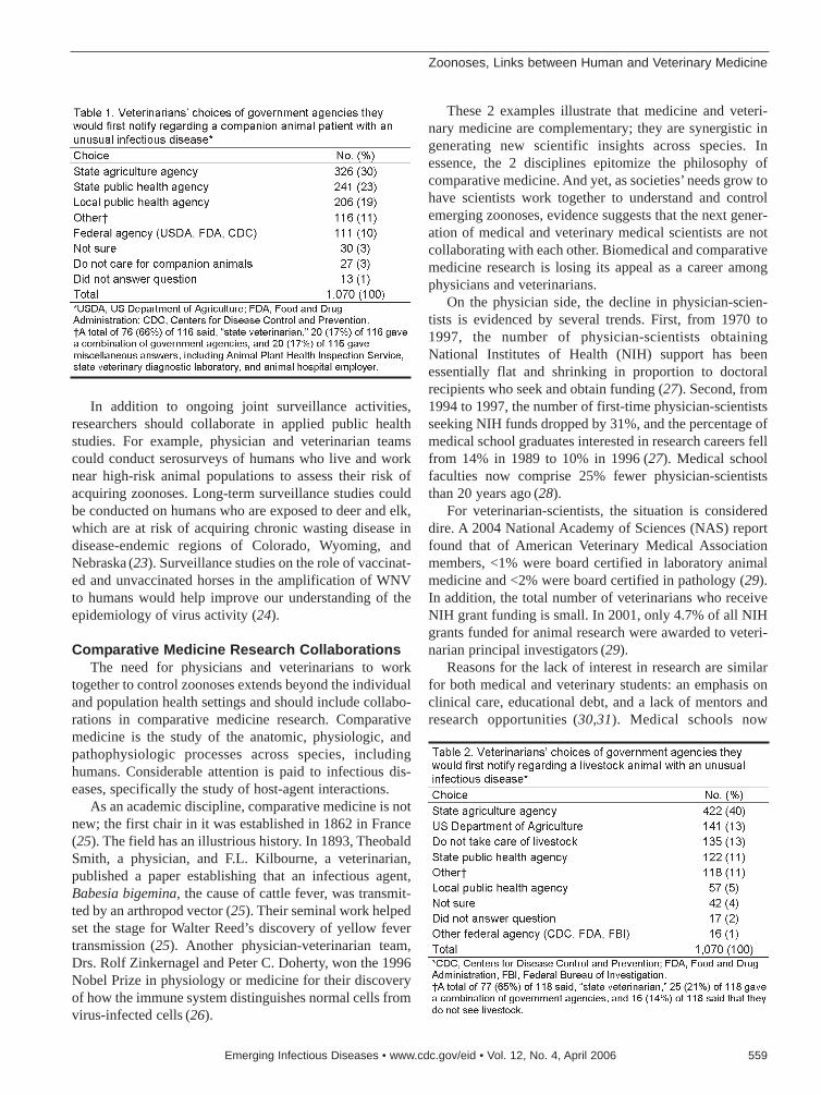
In addition to ongoing joint surveillance activities,researchers should collaborate in applied public healthstudies. For example, physician and veterinarian teamscould conduct serosurveys of humans who live and worknear high-risk animal populations to assess their risk ofacquiring zoonoses. Long-term surveillance studies couldbe conducted on humans who are exposed to deer and elk,which are at risk of acquiring chronic wasting disease indisease-endemic regions of Colorado, Wyoming, andNebraska (23). Surveillance studies on the role of vaccinat-ed and unvaccinated horses in the amplification of WNVto humans would help improve our understanding of theepidemiology of virus activity (24).
Comparative Medicine Research CollaborationsThe need for physicians and veterinarians to work
together to control zoonoses extends beyond the individualand population health settings and should include collabo-rations in comparative medicine research. Comparativemedicine is the study of the anatomic, physiologic, andpathophysiologic processes across species, includinghumans. Considerable attention is paid to infectious dis-eases, specifically the study of host-agent interactions.
As an academic discipline, comparative medicine is notnew; the first chair in it was established in 1862 in France(25). The field has an illustrious history. In 1893, TheobaldSmith, a physician, and F.L. Kilbourne, a veterinarian,published a paper establishing that an infectious agent,Babesia bigemina, the cause of cattle fever, was transmit-ted by an arthropod vector (25). Their seminal work helpedset the stage for Walter Reed’s discovery of yellow fevertransmission (25). Another physician-veterinarian team,Drs. Rolf Zinkernagel and Peter C. Doherty, won the 1996Nobel Prize in physiology or medicine for their discoveryof how the immune system distinguishes normal cells fromvirus-infected cells (26).
These 2 examples illustrate that medicine and veteri-nary medicine are complementary; they are synergistic ingenerating new scientific insights across species. Inessence, the 2 disciplines epitomize the philosophy ofcomparative medicine. And yet, as societies’ needs grow tohave scientists work together to understand and controlemerging zoonoses, evidence suggests that the next gener-ation of medical and veterinary medical scientists are notcollaborating with each other. Biomedical and comparativemedicine research is losing its appeal as a career amongphysicians and veterinarians.
On the physician side, the decline in physician-scien-tists is evidenced by several trends. First, from 1970 to1997, the number of physician-scientists obtainingNational Institutes of Health (NIH) support has beenessentially flat and shrinking in proportion to doctoralrecipients who seek and obtain funding (27). Second, from1994 to 1997, the number of first-time physician-scientistsseeking NIH funds dropped by 31%, and the percentage ofmedical school graduates interested in research careers fellfrom 14% in 1989 to 10% in 1996 (27). Medical schoolfaculties now comprise 25% fewer physician-scientiststhan 20 years ago (28).
For veterinarian-scientists, the situation is considereddire. A 2004 National Academy of Sciences (NAS) reportfound that of American Veterinary Medical Associationmembers, <1% were board certified in laboratory animalmedicine and <2% were board certified in pathology (29).In addition, the total number of veterinarians who receiveNIH grant funding is small. In 2001, only 4.7% of all NIHgrants funded for animal research were awarded to veteri-narian principal investigators (29).
Reasons for the lack of interest in research are similarfor both medical and veterinary students: an emphasis onclinical care, educational debt, and a lack of mentors andresearch opportunities (30,31). Medical schools now
Zoonoses, Links between Human and Veterinary Medicine
Emerging Infectious Diseases • www.cdc.gov/eid • Vol. 12, No. 4, April 2006 559

emphasize primary care and care for the underserved, andwhile certainly important, this shift in priorities has been atthe expense of encouraging biomedical research careers.
Veterinary schools have shifted their focus from com-parative medicine research and livestock medicine to com-panion animal medicine to meet societal demand (32).However, similar to the situation with medical schools,this shift has caused fewer numbers of veterinary studentsto pursue research careers. In addition, comparative medi-cine programs have been shifting from a research to serv-ice orientation that limits veterinarians’ researchinvolvement to being primarily caretakers for laboratoryanimals (32).
This shift in comparative medicine orientation has dis-couraged many veterinary students from pursuing careersin research and hinders research on emerging zoonosesfrom diverse animal hosts.
What can be done? Although NIH has begun a roadmapto improve biomedical research into the 21st century,nowhere does the plan mention comparative medicine andthe importance of veterinary involvement, which wouldcertainly fit into its goals of promoting interdisciplinaryresearch and new pathways to discovery (33). An NASreport recognizes the need for the roadmap initiative toaddress this issue and recommends creating integrated vet-erinary research through joint interagency collaborativeprograms at NIH (34).
One way to achieve this would be to offer jointly spon-sored comparative medicine research grants from both theNational Center for Research Resources (NCRR) andNational Institute of Allergy and Infectious Diseases(NIAID). With NIAID’s emphasis on zoonoses and cross-species investigations, comparative medicine researchwould fit in well with its mission of research on bioterror-ist agents, emerging infectious diseases, and immunology.The NCRR and NIAID could offer research grants to med-ical and veterinary medical research teams that are promot-ing collaborative projects on zoonoses.
A second NAS report addressing this issue recom-mends that federal agencies involved in human and animalresearch coordinate their efforts. Jointly funded integratedand comprehensive animal health research programsshould be established to ensure that veterinary and medicalscientists work together as collaborators domestically andinternationally (13).
Encouraging more veterinary school graduates to pur-sue careers in research is critical if partnerships are to bedeveloped. A third NAS report recommends that the num-ber of veterinarians serving as principal investigatorsshould increase (29). This could be accomplished byincreasing the number of NCRR-funded T32 traininggrants and making them available to persons who want toenter research training programs immediately after gradu-
ation from veterinary school (29). Finally, another way toencourage more veterinary students to pursue researchcareers would be for the National Institute of GeneralMedical Sciences (NIGMS) to offer research training pro-grams to them analogous to those offered to medical stu-dents. Currently, NIGMS research training programs areonly open to holders of MD and PhD degrees (35).
DiscussionSince zoonoses are diseases of animals that can infect
humans, veterinarians, physicians, and public health offi-cials need to work more closely together to control, pre-vent, and understand them. In the individual health setting,collaborative input from both veterinarians and physicianswould help assess a patient’s potential zoonotic diseaserisks from animal exposure. For high-risk immunocompro-mised patients, these collaborative efforts could be tremen-dously important, not only for their personal well-beingbut also for their livelihoods.
Regarding population health, reporting of animal dis-eases varies considerably from state to state. Some stateshave 1 agency responsible for all animal disease reportingwhile others split the reports between various agencies.However, in many states, animal disease surveillanceappears to be largely a state level function. In few states,local public health agencies are expected to receivezoonotic disease reports directly from veterinarians. Ifcontrolling zoonotic diseases is to be improved, greatercommunication and collaboration between veterinarians,physicians, and public health officials at the local level areneeded. One NAS report recommends a federal levelmechanism to promote greater collaboration among all theplayers involved in animal health (13); similar mecha-nisms could also be considered in states.
Joint disease surveillance efforts, which are proving tobe extremely useful in the tracking of zoonoses, includeArboNet for WNV surveillance, the NationalAntimicrobial Resistance Monitoring System for entericbacteria surveillance, and FoodNet for the population-based surveillance of foodborne pathogens (36,37). Theseprograms should continue to be supported, and new sur-veillance programs for other serious zoonoses should bedeveloped. Medical, veterinary, and public health schoolsshould offer courses on zoonotic risks to human health thatintegrate all 3 perspectives.
Society would benefit if more collaborative compara-tive medicine research projects were conducted by physi-cians and veterinarians to investigate zoonotic agent-hostinteractions. Among the many ways to promote these proj-ects are multiagency-sponsored comparative medicineresearch grants and more training grants for veterinariansinterested in careers in research. These efforts wouldincrease our understanding of how zoonoses expand their
PERSPECTIVE
560 Emerging Infectious Diseases • www.cdc.gov/eid • Vol. 12, No. 4, April 2006

host range and would, ultimately, improve prevention andcontrol strategies.
AcknowledgmentsThe author thanks Nina Marano for her invaluable com-
ments and suggestions.
The research for this work was supported by the JosiahMacy Jr. Foundation of New York City.
Dr Kahn is a general internist and a member of the researchstaff in the Program on Science and Global Security in theWoodrow Wilson School of Public and International Affairs,Princeton University. Her research interests include public healthpolicy and emergency preparedness.
References
1. Grant S, Olsen CW. Preventing zoonotic diseases in immunocompro-mised persons: the role of physicians and veterinarians. Emerg InfectDis. 1999;5:159–63.
2. Shrestha RK, Stoller JK, Honari G, Procop GW, Gordon SM.Pneumonia due to Cryptococcus neoformans in a patient receivinginfliximab: possible zoonotic transmission from a pet cockatiel.Respir Care. 2004;49:606–8.
3. Centers for Disease Control and Prevention. Update: interim guid-ance for minimizing risk for human lymphocytic choriomeningitisvirus infection associated with pet rodents. MMWR Morb MortalWkly Rep. 2005;54:799–801.
4. Centers for Disease Control and Prevention. Salmonellosis associat-ed with pet turtles—Wisconsin and Wyoming, 2004. MMWR MorbMortal Wkly Rep. 2005;54:223–6.
5. Centers for Disease Control and Prevention. Outbreak of multidrug-resistant Salmonella typhimurium associated with rodents purchasedat retail pet stores—United States, December 2003–October 2004.MMWR Morb Mortal Wkly Rep. 2005;54:429–33.
6. Centers for Disease Control and Prevention. Update: multistate out-break of monkeypox—Illinois, Indiana, Kansas, Missouri, Ohio, andWisconsin, 2003. MMWR Morb Mortal Wkly Rep. 2003;52:642–6.
7. Perseghin P, Bezzi G, Troupioti P, Gallina M. Streptococcus suismeningitis in an Italian blood donor. Lancet. 1995;346:1305–6.
8. Bartelink AK, van Kregten E. Streptococcus suis as threat to pig-farmers and abattoir workers. Lancet. 1995;346:1707.
9. Wilson IG. Airborne Campylobacter infection in a poultry worker:case report and review of the literature. Commun Dis Public Health.2004;7:349–53.
10. Centers for Disease Control and Prevention. Fatal rat-bitefever⎯Florida and Washington, 2003. MMWR Morb Mortal WklyRep. 2005;53:1198–202.
11. Tannenbaum J. Medical-legal aspects of veterinary public health inprivate practice. Semin Vet Med Surg (Small Anim). 1991;6:175–85.
12. US General Accounting Office. West Nile virus outbreak. Lessons forpublic health preparedness. Washington: The Office; 2000. GAO/HEHS-00-180.
13. National Academy of Sciences. Animal health at the crossroads: pre-venting, detecting, and diagnosing animal diseases. Washington:National Academy Press; 2005.
14. Asnis DS, Conetta R, Teizeira AA, Waldman G, Sampson BA. TheWest Nile outbreak of 1999 in New York: the Flushing Hospital expe-rience. Clin Infect Dis 2000;30:413–8.
15. Chomel BB. New emerging zoonoses: a challenge and an opportuni-ty for the veterinary profession. Comp Immunol Microbiol. InfectDis. 1998;21:1–14.
16. Davis RG. The ABCs of bioterrorism for veterinarians, focusing oncategory A agents. J Am Vet Med Assoc. 2004;224:1084–95.
17. Davis RG. The ABCs of bioterrorism for veterinarians, focusing oncategory B and C agents. J Am Vet Med Assoc. 2004;224:1096–104.
18. Taylor LH, Latham SM, Woolhouse MEJ. Risk factors for human dis-ease emergence. Philos Trans. R Soc Lond B Biol Sci.2001;356:983–9.
19. Cleaveland S, Laurenson MK, Taylor LH. Diseases of humans andtheir domestic mammals: pathogen characteristics, host range and therisk of emergence. Philos Trans R Soc Lond B Biol Sci.2001;356:991–9.
20. Shortridge KF, Peiris JSM, Guan Y. The next influenza pandemic:lessons from Hong Kong. J Appl Microbiol. 2003;94:70S–9S.
21. Marfin AA, Peterson LR, Eidson M, Miller J, Hadler J, Farello C, etal. Widespread West Nile virus activity, eastern United States, 2000.Emerg Infect Dis. 2001;7:730–5.
22. O’Leary DR, Marfin AA, Montgomery SP, Kipp AM, Lehman JA,Biggerstaff BJ, et al. The epidemic of West Nile virus in the UnitedStates, 2002. Vector Borne Zoonotic Dis. 2004;4:61–70.
23. Belay ED, Maddox RA, Williams ES. Chronic wasting disease andpotential transmission to humans. Emerg Infect Dis. 2004;10:977–84.
24. Castillo-Olivares, J, Wood J. West Nile virus infection of horses. VetRes. 2004;35:467–83.
25. Wilkinson L. Animals and disease. An introduction to the history ofcomparative medicine. Cambridge (UK): Cambridge UniversityPress; 1992.
26. Zinkernagel RM, Doherty PC. Immunological surveillance againstaltered self components by sensitized T lymphocytes in lymphocyticchoriomeningitis. Nature. 1974; 251:547–8.
27. Rosenberg LE. Physician-scientists—endangered and essential.Science. 1999;283:331–2.
28. Varki A, Rosenberg LE. Emerging opportunities and career paths forthe young physician-scientist. Nat Med. 2002;8:437–9.
29. National Academy of Sciences. National need and priorities for vet-erinarians in biomedical research. Washington: National AcademyPress; 2004.
30. Neilson EG. The role of medical school admissions committees in thedecline of physician-scientists. J Clin Invest. 2003;111:765–7.
31. Barthold SW. Biomedical research and veterinarians: where’s Waldo?Comp Med. 2002;52:95–6.
32. Barthold SW. Musings of a Connecticut Yankee in King Arthur’scourt: antemortem analysis of the veterinary profession. J Vet MedEduc. 2005;32:306–13.
33. Zerhouni E. The NIH roadmap. Science. 2003;302:63–72.34. National Research Council. Critical needs for research in veterinary
science. Washington: National Academy Press; 2005.35. National Institute of General Medical Sciences. Training and Careers.
[cited 2006 Jan 23]. Available from http://www.nigms.nih.gov/fund-ing/trngmech.html#new_emphasis
36. Centers for Disease Control and Prevention. NARMS website. [cited2006 Jan 23]. Available from http://www.cdc.gov/narms/
37. Centers for Disease Control and Prevention. FoodNet website. [cited2006 Jan 23]. Available from http://www.cdc.gov/foodnet/
Address for correspondence: Laura H. Kahn, Research Staff, Program onScience and Global Security, Woodrow Wilson School of Public andInternational Affairs, Princeton University, 221 Nassau St, Second Floor,Princeton, NJ 08542, USA; fax: 609-258-3661; email: [email protected]
Zoonoses, Links between Human and Veterinary Medicine
Emerging Infectious Diseases • www.cdc.gov/eid • Vol. 12, No. 4, April 2006 561
The World Health Organization Influenza Program isone of the best developed and longest running infectiousdisease surveillance systems that exists. It maintains aworldwide watch of influenza’s evolution to assist deliveryof appropriately formulated vaccines in time to blunt sea-sonal epidemics and unpredictable pandemics. Despite theprogram’s success, however, much more is possible withtoday’s advanced technologies. This article summarizesongoing human influenza surveillance activities worldwide.It shows that the technology to establish a high-throughputlaboratory network that can process and test influenzaviruses more quickly and more accurately is available. Italso emphasizes the practical public health and scientificapplications of such a network.
Influenza strikes persons in developing and industrializedcountries alike and is capable of killing healthy persons
of all ages. Among the hardest hit are infants <1 year ofage and adults >65. During any given year, influenza epi-demics kill 500,000–1,000,000 persons globally, and anunpredictable pandemic is capable of killing millions (1).Yet death rate statistics alone do not capture the full impactof influenza; it causes many hospitalizations, secondarybacterial pneumonias, and middle ear infections in infantsand young children (2). Worldwide, literally tons ofantimicrobial drugs are used to treat these complications,and the economic consequences are enormous. For largepopulations, the only way to deter influenza is to adminis-ter vaccines targeted against ever-mutating strains (3).
Current SurveillanceThe World Health Organization (WHO) Influenza
Program was established in 1952 to assist with publichealth threats associated with influenza. Today, its networkof 112 national centers in 83 countries collects ≈160,000samples each year from 600 to 1,200 million persons withinfluenza. As shown in Figure 1, the centers screen sam-
ples for influenza viruses and type- (A versus B) and sub-type- (e.g., A/H1N1, A/H3N2) relevant samples (4).Certain influenza-positive samples are then forwarded to 1of 4 WHO collaborating centers for further immunologicand genetic characterizations. Twice a year, WHO organiz-es a formal meeting with its collaborating center directorsto review information on circulating influenza strains. Thisadvisory committee identifies circulating strains that newvaccine formulations should target. Because influenza epi-demics peak during the winter months, the committeeoffers its recommendations in February and September forthe Northern and Southern Hemispheres, respectively. Thefindings are then reviewed by national health authoritieswho approve, and occasionally amend, implementation ofthe recommended vaccine strains (5). Surveillance forinfluenza requires global and national monitoring for bothvirus and disease activity to determine when, where, andwhich influenza viruses are circulating in the United Statesand globally, to determine the intensity and impact ofinfluenza activity on defined health outcomes and identifyunusual or severe outbreak, and to detect the emergence ofnovel influenza viruses that may cause a pandemic.
The Centers for Disease Control and Prevention’s(CDC) WHO Collaborating Center for Reference andResearch on Influenza supplies standardized reagents andtest kits to all national centers for detecting influenza A andB strains, subtyping strains, and determining whether sam-ple strains are immunologically related to recent vaccinestrains. Typical circulating strains and atypical ones thatappear to differ from vaccine strains are forwarded to 1 of4 collaborating centers (in the United States, UnitedKingdom, Australia, and Japan) for further characteriza-tion (5). During the 2004–2005 influenza season, CDC’scenter in Atlanta received ≈3,500 strains from domesticand foreign surveillance. A partial summary of laboratorymethods used is outlined below.
In most situations, 6–10 serum samples are used tocompare sample strains against vaccine and referencestrains, and during any given influenza season, >99% of
Human Influenza Surveillance:the Demand to Expand
Scott P. Layne*
PERSPECTIVE
562 Emerging Infectious Diseases • www.cdc.gov/eid • Vol. 12, No. 4, April 2006
*University of California at Los Angeles, Los Angeles,California,USA

sample strains are successfully matched by routine hemag-glutination inhibition (HI) assays. Laboratory workersreplicate sample strains in cell cultures or fertile eggs andadjust the resulting viral stocks to a standard hemagglu-tinin titer. They then use HI assays to determine whethersample strains are immunologically related (i.e., cross-reactive) to recent vaccine strains. Typing sera are added towells in a series of 2-fold dilutions, and after the reactionsstabilize, laboratory workers score assay wells (positive ornegative) by looking for agglutinated erythrocytes (posi-tive) that do not form buttons at the bottom of plates ver-sus non-agglutinated cells (negative) that do form buttons.For sample strains that behave as variants, laboratoryworkers inject them into ferrets to produce a strain-specif-ic antiserum. When the new antiserum is ready, HI assaysare again performed as above. If the new sera show signif-icant gaps in cross-reactivity (usually defined as a 4-folddifference between sample and vaccine strains), they areincorporated into the routine laboratory set and used tolook for new epidemic strains. Examples of strains thatwere identified as variants include A/H3N2/Sydney, whichspread rapidly among persons in 1997, and A/H5N1/HongKong, which jumped from fowl to humans in 1997. Duringthe 2004–2005 influenza season, ≈3,500 HI assays wereperformed.
During the 2004–2005 influenza season, >600 cleavedhemagglutinin (HA1) domain sequences were analyzed. Inthis process, laboratory workers extract viral RNA fromsamples, convert viral RNA to cDNA, amplify cDNA withpolymerase chain reaction (PCR) primers, and analyzeDNA products with capillary array sequencers. They thencompare sample strain sequences against vaccine and ref-
erence strain sequences to determine their phylogeneticrelationships.
During the 2004–2005 influenza season, ≈350 neu-raminidase (NA) sequences were analyzed to determinetheir phylogenetic relationships. For this process, laborato-ry workers follow similar preparative steps used for HA1and then use multiple PCR primers to sequence the entiregene segment.
During the 2004–2005 influenza season, ≈3,500 singlenucleotide polymorphism (SNP) profiles were analyzedfrom circulating strains. For this analysis, laboratory work-ers follow similar preparative steps used for HA1 and thenuse pyrosequencing to detect SNP that confer resistance tothe antiviral drugs amantadine and rimantadine.
During the 2004–2005 influenza season, ≈120 wholegenomes were sequenced from circulating strains. In thisprocess, laboratory workers extract viral RNA from sam-ples, convert viral RNA to cDNA, amplify cDNA withPCR primers, and analyze DNA products with capillaryarray sequencers. Amplification of all 8 gene segments(PB1, PB2, PA, HA, NP, NA, M1/M2, NS1/NEP) that havea combined length of ≈13.6 kb requires ≈30 type- and sub-type-specific PCR primers.
These activities give ≈6 months to vaccine manufactur-ers on either side of the equator for scale up, production,and distribution. These activities also give healthcare serv-ices another 3 months to administer the ≈250 million dosesof trivalent vaccine that are used globally. Despite itssophistication and scale, however, the WHO InfluenzaProgram has several shortcomings (6).
First, surveillance gaps exist in many parts of the worldfor a variety of reasons, including limited funding, lack ofinfrastructure support for surveillance teams and preserv-ing influenza samples, and intentional underreporting atthe national level (7). Second, current laboratory methodsfor characterizing influenza are time-consuming and labor-intensive and, as a result, relatively few viral strains under-go definitive phenotyping and genotyping assays (5,8,9).During the 2004–2005 influenza season, for example, the4 WHO collaborating centers analyzed ≈6,000 strains, rep-resenting only 1 sample per 100,000 influenza casesworldwide (Figure 1). Far fewer strains from domesticpoultry and swine or from wild aquatic birds, which arethought to serve as precursors for pandemic strains, areanalyzed comprehensively in a given year (10).
Third, with current methods, it can take weeks tomonths to generate laboratory data on influenza samplesand understand their significance (Appendix availableonline at http://www.cdc.gov/ncidod/EID/vol12no04/05-1198.htm#app). Such prolonged times can impede vaccinestrain selection activities. For example, the vaccine admin-istered throughout North America in 1997 providedinadequate protection against the A/H3N2 Sidney strain
Human Influenza Surveillance: the Demand to Expand
Emerging Infectious Diseases • www.cdc.gov/eid • Vol. 12, No. 4, April 2006 563
Figure 1. Global influenza surveillance for the 2004–2005 season.Respiratory samples were collected from persons with febrile res-piratory illness worldwide. Approximately 15% of samples wereinfluenza positive. Note that only some of the type A viruses weresubtyped. Data are from the Centers for Disease Control andPrevention website (http://www.cdc.gov/flu/weekly/weekl-yarchives2004-2005/04-05summary.htm).

that spread rapidly from Asia and Australia (11). Publichealth officials ascribed the poor match between circulat-ing strains and vaccine strains to many factors including 1)less than optimal surveillance, 2) time required to prepareand ship isolates, 3) lag time in laboratory testing with cur-rent manual methods, and 4) rapid spread of the Sydneyvariant.
Although the program has a remarkably good trackrecord, it did not detect the Sydney variant in time toinclude it in the vaccine before the epidemic. Failure todetect an emerging influenza virus could prove disastrousshould it be a novel strain with pandemic potential (12).The 1918 influenza pandemic is the biggest infectious dis-ease catastrophe on record, topping even the medievalBlack Death. Within months after the initial outbreak, theA/H1N1 virus struck 500 million persons and killed 40–50million worldwide when the total population was only 2billion (13). Subsequent pandemics, brought about by ashift to A/H2N2 in 1957 and A/H3N2 in 1968, had farlower death rates.
Between March and May of 1997, an outbreak of avianA/H5N1 in Hong Kong killed a child who was otherwisehealthy and thousands of chickens (14). For the next 6months, no new cases appeared. Between November andDecember of 1997, avian A/H5N1 infected 17 people andkilled 5 of them (15,16). Confronted with a case-fatalityratio of 33%, health authorities took quick action andopted to destroy all 1.5 million chickens in Hong Kong. Ittook nearly 6 months to identify the index case; eventstranspired so quickly that manual laboratory methods wereunable to generate all of the information that was needed.
Since 2004, avian A/H5N1 has caused additional out-breaks throughout Asia, resulting in >60 human deaths inVietnam, Thailand, Cambodia, and Indonesia and thedestruction of 150 million birds (17). In 2005, through acombination of wild bird migrations and farming practices,highly pathogenic avian influenza spread to northernChina, Mongolia, Tibet, Kazakhstan, and Russia (18). Atthe time of this writing, it had further spread to severalEuropean countries (Turkey, Romania, and Greece) andthreatened to spread to other continents, including Africaand North America through avian flyways.
The potential of avian A/H5N1 to cause a global humanpandemic is uncertain because it cannot be predicted withcurrent knowledge (19). Nevertheless, the anticipated eco-nomic, social, and political consequences are enormous(20,21). Therefore, we face a compelling demand toexpand the current influenza surveillance system (6,22).
High-throughput NetworkIn August 2004, the US Department of Health and
Human Services released a draft of its Pandemic InfluenzaResponse and Preparedness Plan. The plan’s surveillance
annex offered specific recommendations for systemenhancements and next steps (5). Many of these enhance-ments could be achieved by developing a high-throughputlaboratory network that would expand the capabilities ofthe existing WHO collaborating centers on influenza(6,22). With such enhancements, WHO national centerswould be able to collect samples from people with febrilerespiratory illnesses, record epidemiologic observations,and send samples directly to the high-throughput network.At each site, high-throughput automated systems wouldcollaborate, and epidemiologic observations and testresults would appear in the laboratory’s web-enabled data-base for analysis within days (23). Internet-based capabil-ities would allow WHO national centers to examine theirown data and improve surveillance in an iterative manner(Figure 2). In tracking changes in epidemic strains, thenew system would facilitate nonbiased proportional sam-pling of persons with febrile respiratory illnesses in an iter-ative fashion. In detecting the emergence of novel strainswith pandemic potential, the new system would facilitatethe use of rapid and more sensitive methods.
The plan integrates available biologic, engineering, andinformatic technologies into a networked capability and
PERSPECTIVE
564 Emerging Infectious Diseases • www.cdc.gov/eid • Vol. 12, No. 4, April 2006
Figure 2. Flow chart for utilizing the high-throughput laboratorynetwork.

makes them available through the Internet (23). Influenzais well suited to this approach because of its obvious pub-lic health implications, but also because a well-establishedinfrastructure that includes global surveillance, standard-ized laboratory methods, surveillance-based recommenda-tions, and targeted vaccines already exists (6,22). Keyelements are shown in Figure 2.
Platform-independent software that facilitates influenzasurveillance would be provided by the high-throughputnetwork (23). Internet-based tools would manage laborato-ry access, epidemiologic questionnaires, testing instruc-tions, sample submission, data analysis, and dataprivileges. The release of data to other users or entitieswould be controlled by the submitting organization orauthority. More importantly, the WHO committee andnational health authorities that recommend and reviewvaccine strains and antiviral drugs would have access to allthe data.
On average, 1 of every 6 samples collected from per-sons with febrile respiratory illnesses contains influenza Aor B viruses (24). The remainder contain other viral andbacterial pathogens. To expedite data collection, surveil-lance teams could use influenza dipsticks to screen sam-ples on the spot. Several companies make diagnostic kitsfor influenza A and B; although these tests have certaindisadvantages (limited sensitivity and specificity ofimmunoassays), their underlying technologies can formthe basis for improved influenza screening and samplingdevices (2). Such influenza dipsticks—or even portablePCR-based assays—would make it easier for teams in thefield to screen out negative samples and focus on docu-menting epidemiologic information on positive ones.
An epidemiologic questionnaire would be provided tosurveillance teams, with a menu that covers key questions(23). What are the collection date and location? Who is thehost (human versus animal)? What is the age of the host?What is the physiologic source of the sample? What is theobserved or reported severity of illness? What is theobserved or reported outcome of illness? Which influenzavaccine or antiviral agents were administered? What arethe likely exposures? Is there any recent travel history?The questionnaire would run on inexpensive handhelddevices (e.g., personal digital assistants) or cell phones.Bar codes would be used to link samples to their corre-sponding questionnaires. Completed questionnaires wouldbe sent by email to the high-throughput laboratory net-work, where questionnaire and laboratory data would formthe basis for seeking associations on factors that influencevirulence, transmissibility, and host range (19).
Current high-throughput automated laboratory systemsare capable of operating 24 hours a day. At each networkedsite, epidemiologic questionnaires and instructions wouldarrive by the Internet, and bar coded samples would arrive
by air freight. Larger sites could operate systems for geno-typing, phenotyping, replicating, and archiving influenzaviruses. Smaller sites could operate systems for genotyp-ing and archiving viruses. In serving as resources, each sitewould provide reagents and supplies for analyzing allinfluenza subtypes. They would also perform controlassays on a daily basis and maintain a quality assuranceprogram, the documentation of which would be stored inthe database. Automated laboratory methods would buildupon manual methods currently in use and, because theycan reduce working (liquid) volumes by at least 5- to 10-fold, they would enable economies of scale (23,25).
Genotyping systems would have flexibility to sequenceall 8 RNA segments or any individual segment frominfluenza viruses. The various steps performed wouldinclude transcription viral RNA into cDNA, selection ofoptimal PCR primers, amplification of DNA by PCR, andanalysis by capillary array DNA sequencers (26–28).Influenza viruses are often propagated in cell cultures orembryonated eggs to obtain enough viral RNA forsequencing. Newer methods that avoid this growth step,however, would facilitate direct high-throughput analysisof native samples from surveillance, including active sam-ples preserved by cold chain as well as inactive samplespreserved by ethanol fixation (29).
Phenotyping systems would conduct HI and neu-raminidase inhibition (NI) assays. HI assays are easilyadaptable to automation, but they require relatively largequantities of virus and typing sera (8). To overcome thisdrawback, automated methods that use flow cytometry areunder development (30). They work by attaching mono-clonal or polyclonal typing sera to a set of color-coded(multiplexed) beads and detecting the interaction ofinfluenza with such beads. A high-throughput system thatperforms HI assays in parallel with flow cytometer–basedassays, for example, may offer the best means to test andvalidate improved influenza serotyping methods. NIassays are also easily adaptable to automation, particularlynewer ones that use a chemiluminescent sialic acid sub-strate instead of a fluorogenic substrate (31). They work bymixing substrate with neuraminidase from sample strainsand measuring the chemiluminescent signal over time.When performed over a range of inhibitor or antiviral drug(oseltamivir and zanamivir) concentrations, they enablethe determination of the 50% inhibitory concentration(IC50) for individual drugs and strains (32).
A replicating system would verify that influenza A andB antigens are present in samples and set aside negativeones (33). Positive samples would be injected into cell cul-tures or embryonated eggs and, several days later, auto-matically harvested, assayed for HA titers, and adjusted touniform concentrations. A part of this fresh stock wouldthen go to the long-term storage system.
Human Influenza Surveillance: the Demand to Expand
Emerging Infectious Diseases • www.cdc.gov/eid • Vol. 12, No. 4, April 2006 565

Archiving systems would store influenza samples foran extended time. The archiving system would take stocksfrom the replication system and place them into modularbar-coded storage containers, which would then be placedinto freezers. Every step in the storage and retrievalprocess would be recorded by bar code scanners and man-aged by an inventory tracking program (23).
Expanded SurveillanceInfluenza virus evolves through a combination of point
mutations (drifts) and reassortment events (shifts) in itsgene segments. For vaccine strain selection, laboratorymethods for characterizing influenza have focused prima-rily on changes in hemagglutinin and neuraminidase (andto a much lesser extent on the M2 ion channel protein)because immunity against these surface proteins is protec-tive (2). The emphasis on immune-inducing proteins isclearly practical, but it may overlook changes in theremaining gene segments (26).
Whole-genome sequencing and phylogenetic analysisof 156 A/H3N2 viruses that infected humans in New Yorkfrom 1999 to 2004 shows 2 substantial findings (28). Thefirst is that multiple influenza strains co-circulated inhumans over time, with strains falling into 1 of 3 distinctclades. The second is that mixing between these cladesoccurred over short intervals of time, resulting in at least 4reassortment events among the strains analyzed (28). Onesuch reassortment event (shift), rather than a point muta-tion (drift), appears to explain the emergence of theA/Fujian/411/2002-like strain that caused an epidemic dur-ing the 2003–2004 influenza season. Similar findings onA/H3N2 viruses that infected humans in Australia andNew Zealand from 2003 to 2004 have also been reported(27). In this instance, swaps of neuraminidase and 3 inter-nal genes (NS1/NEP, NP, M1/M2) were found. Both inde-pendent findings show that influenza A virus is lessrestricted than previously believed and that reassortmentevents can occur without warning. Altogether, these newfindings underscore the importance of rapid, whole-genome analysis for future influenza surveillance (28).
The high-throughput laboratory network would giverise to 3 domains of associated data from surveillance (34).Epidemiologic data would pertain to dates, locations,hosts, outcomes, histories, and exposures. Genotypic datawould pertain to the exact sequence of nucleotides in all 8viral RNA segments. Phenotypic date would pertain toimmunologic pedigrees (HI titers) and antiviral drug sensi-tivities (IC50) of sample strains. Some practical publichealth and scientific uses of such organized data follow.
New influenza vaccines are often introduced after 3 cri-teria have been met (25). First, a new strain is identified bylaboratory-based methods. Second, geographic spread ofthe new strain is associated with human illness. Third, the
most recent influenza vaccine stimulates a reducedimmunologic response to the new strain. Given these crite-ria, the high-throughput laboratory network would help in2 ways. It would provide faster information for vaccinestrain selection, potentially saving 1–2 months in vaccinedelivery. It would also continuously monitor for the emer-gence of escaping influenza strains and guide critical deci-sions to update pandemic vaccines or use them incombination with limited supplies of antiviral drugs (19).Researchers and drug companies are developing modernmethods (based on reverse genetics and cell cultures, forexample) to manufacture influenza vaccines that could cutdelivery times in half (2,35). Within the next few years,these new methods, in combination with a high-throughputnetwork, could save additional vaccine delivery time andsave lives (6,19).
Fifteen hemagglutinin (H1–H15) and 9 neuraminidase(N1–N9) subtypes diverge by as much as 50% in theiroverall amino acid composition. Within each subtype,smaller amino acid substitutions (drifts) that enableinfluenza viruses to evade preexisting immunity exist(2,3). Although sequencing influenza viruses is useful forunderstanding viral mixing and evolution, it cannot delin-eate how immunologic (i.e., drift and shift) variants relateto one another at the amino acid and RNA coding levels.To develop such understanding, a large base of phenotyp-ic data must be associated with its corresponding genotyp-ic data. For each receptor subtype, phenotypic data wouldconsist of HI titers and genotypic data would consist ofRNA sequences from the same virus. Building a roughassociation matrix would be the first step in understandinghow variants relate to one another at the amino acid andRNA levels.
Subsequently, a more complete association matrixwould be used to develop models that could predictwhether viral strains are immunologically related fromsequences alone. Such efforts could help develop influen-za vaccines that protect against a wider range of variantsand establish a more fundamental molecular basis forinfluenza surveillance (25).
Researchers have proposed using antiviral drugs suchas oseltamivir to halt an avian influenza outbreak inhumans (36,37). The strategy would require stockpilingmillions of doses and administering them to persons in theepicenter and surrounding areas within weeks. Immediaterecognition of the outbreak and rapid surveillance to deter-mine its size would be essential. Drug-resistant avianinfluenza viruses would likely emerge at some point, rep-resenting a potential threat to emergency control efforts,and health authorities would need real-time information onwhere the viruses were found and how many of them exist-ed (38). Such emergency interventions would generatethousands of samples for laboratory analysis within days.
PERSPECTIVE
566 Emerging Infectious Diseases • www.cdc.gov/eid • Vol. 12, No. 4, April 2006

Given current laboratory surge capacity, a high-throughputlaboratory network may be the only feasible means to meetthe challenge.
ImplementationThe spreading avian A/H5N1 outbreak poses serious
threats to the health, economy, and security of the world(22). It has motivated political leaders and health officialsto increase financial support for influenza surveillance andto seek agreements and incentives that promote informa-tion sharing and international cooperation (39). Effectivemeasures will require real-time, accurate, and comprehen-sive information to make rapid public health decisions.
The first 2 sites in a high-throughput laboratory net-work could be up and running in 12–18 months at a cost of$15 million. It would generate epidemiologic, genotypic,and phenotypic data as described in this article. With avail-able technologies and methods, each site would be capableof analyzing up to 10,000 samples per year, a substantialimprovement over current capabilities. Implementing mul-tiple sites worldwide (at the 4 WHO collaborating centers,for example) would enable regional collaborations andhelp encourage the timely sharing of samples and informa-tion (40).
Human history shows that an influenza pandemic isyears overdue (41). Moreover, whole-genome sequencingof influenza virus shows dynamic RNA segments that arecapable of epidemiologically meaningful reassortmentevents (27,28). Whether avian A/H5N1 will be the precur-sor strain is unknown. However, we must expand, speedup, and connect human and animal surveillance effortstoday, which must be matched with an expanded capacityto produce and deliver influenza vaccines worldwide.
Financial support for this work was provided by theUniversity of California.
Dr Layne is an associate professor of epidemiology at theUniversity of California at Los Angeles School of Public Healthin Los Angeles, California. His primary research interests includeviral diseases such as influenza, bioterrorism preparedness, andlaboratory and informatic tools to deal with these threats.
References
1. Ghendon, Y. Influenza—its impact and control. World Health Stat Q.1992;45:306–11.
2. Nicholson KG, Wood JM, Zambon M. Influenza. Lancet.2003;362:1733–45.
3. World Health Organization. Influenza vaccines. Wkly EpidemiolRec. 2002;77:229–40.
4. World Health Organization. WHO Global Influenza Programme: sur-vey on capacities of national influenza centres, January–June 2002.Wkly Epidemiol Rec. 2002;77:349–56.
5. Department of Health and Human Services. Pandemic InfluenzaResponse and Preparedness Plan, Annex 4: Surveillance. [cited 2006Feb 17]. Available from http://www.hhs.gov/pandemicflu/plan
6. Layne SP, Beugelsdijk TJ, Patel CKN, Taubenberger JK, Cox NJ,Gust ID, et al. A global lab against influenza. Science.2001;293:1729.
7. World Health Organization. WHO report on global surveillance ofepidemic-prone infectious diseases. Geneva: The Organization; 2000.
8. Kendal AP, Pereira MS, Skehel J. Concepts and procedures for labo-ratory-based influenza surveillance. Geneva: World HealthOrganization; 1982.
9. Hay AJ, Wolstenholme AJ, Skehel JJ, Smith MH. The molecular basisof the specific anti-influenza action of amantadine. EMBO J.1985;4:3021–4.
10. Webster, RG. Influenza: an emerging disease. Emerg Infect Dis.1998;4:436–41.
11. Centers for Disease Control and Prevention. Update: influenza activ-ity—United States and worldwide, 1997–98 season, and compositionof the 1998–99 influenza vaccine. MMWR Morb Mortal Wkly Rep.1998;47:280–4.
12. World Health Organization. WHO global influenza preparednessplan. Geneva: The Organization; 2005.
13. Reid AH, Taubenberger JK. The 1918 flu and other influenza pan-demics: “over there” and back again. Lab Invest. 1999;79:95–101.
14. Subbarao K, Klimov A, Katz J, Regnery H, Lim W, Hall H, et al.Characterization of an avian influenza A (H5N1) virus isolated froma child with a fatal respiratory illness. Science. 1998;279:393–6.
15. Yuen KY, Chan PKS, Peiris M, Tsang DNC, Que TL, Shortridge KF,et al. Clinical features and rapid viral diagnosis of human diseaseassociated with avian influenza A H5N1 virus. Lancet.1998;351:467–71.
16. Claas ECJ, Osterhaus ADME, van Beek R, De Jong JC,Rimmelzwaan GF, Senne DA, et al. Human influenza A H5N1 virusrelated to a highly pathogenic avian influenza virus. Lancet.1889;351:472–7.
17. World Health Organization Global Influenza Program SurveillanceNetwork. Evolution of H5N1 avian influenza viruses in Asia. EmergInfect Dis. 2005;11:1515–21.
18. World Health Organization. Avian influenza—new areas with infec-tion in birds—update 34. Communicable Disease Surveillance andResponse Unit. [cited 2006 Feb 17]. Available fromhttp://www.who.int/csr/don/archive/disease/influenza/en
19. Holmes EC, Taubenberger JK, Grenfell BT. Heading off an influenzapandemic. Science. 2005;309:989.
20. Meltzer MI, Cox NJ, Fukuda K. The economic impact of pandemicinfluenza in the United States: priorities for intervention. EmergInfect Dis. 1999;5:659–71.
21. Osterholm MT. Preparing for the next pandemic. N Engl J Med.2005;352:1839–42.
22. Fouchier R, Kuiken T, Rimmelzwaan G, Osterhaus, A. Global taskforce for influenza. Nature. 2005;435:419–20.
23. Layne SP, Beugelsdijk TJ. Laboratory firepower for infectious dis-ease research. Nat Biotechnol. 1998;16:825–9.
24. Centers for Disease Control and Prevention. Update: influenza activ-ity—United States and worldwide, 2003–04 season, and compositionof the 2004–05 influenza vaccine. MMWR Morb Mortal Wkly Rep.2004;53:547–52.
25. Cox NJ. Expanding the worldwide influenza surveillance system andimproving the selection of strains for vaccines. In: Layne SP,Beugelsdijk TJ, Patel CK, editors. Firepower in the lab: automationin the fight against infectious diseases and bioterrorism. Washington:Joseph Henry Press; 2001. p. 47–53.
26. Xu X, Smith CB, Mungall BA, Lindstrom SE, Hall HE, Subbarao K,et al. Intercontinental circulation of human influenza A(H1N2) reas-sortant viruses during the 2001–2002 influenza season. J Infect Dis.2002;186:1490–3.
Human Influenza Surveillance: the Demand to Expand
Emerging Infectious Diseases • www.cdc.gov/eid • Vol. 12, No. 4, April 2006 567

27. Barr IG, Komadina N, Hurt AC, Iannello P, Tomasov C, Shaw R, Etal. An influenza A(H3) reassortant was epidemic in Australia andNew Zealand in 2003. J Med Virol. 2005;76:391–7.
28. Holmes EC, Ghedin E, Miller N, Taylor J, Bao Y, St. George K, et al.Whole-genome analysis of human influenza A virus reveals multiplepersistent lineages and reassortment among recent H3N2 viruses.PLoS Biol. 2005;3:e300.
29. Krafft AE, Russell KL, Hawksworth AW, McCall S, Irvine M, DaumLT, et al. Evaluation of PCR testing of ethanol-fixed nasal swab spec-imens as an augmented surveillance strategy for influenza virus andadenovirus identification. J Clin Microbiol. 2005;43:1768–75.
30. Yan X, Schielke EG, Grace KM, Hassell C, Marrone BL, Nolan JP.Microsphere-based duplexed immunoassay for influenza virus typingby flow cytometry. J Immunol Methods. 2004;284:27–38.
31. Buxton RC, Edwards B, Juob RR, Voytab JC, Tisdalea M, BethellaRC. Development of a sensitive chemiluminescent neuraminidaseassay for the determination of influenza virus susceptibility tozanamivir. Anal Biochem. 2000;280:291–300.
32. Mungall BA, Xu X, Klimov A. Surveillance of influenza isolates forsusceptibility to neuraminidase inhibitors during the 2000–2002influenza seasons. Virus Res. 2004;103:195–7.
33. Ziegler T, Hall H, Sánchez-Fauquier A, Gamble WC, Cox NJ. Type-and subtype-specific detection of influenza viruses in clinical speci-mens by rapid culture assay. J Clin Microbiol. 1995;33:318–21.
34. National Research Council. Human and agricultural health systems.In: Making the nation safer: the role of science and technology incountering terrorism. Washington: National Academies Press; 2002.p. 65–106.
35. Webby RJ, Perez DR, Coleman JS, Guan Y, Knight JH, GovorkovaEA, et al. Responsiveness to a pandemic alert: use rapid developmentof influenza vaccines. Lancet. 2004;363:1099–103.
36. Ferguson NM, Cummings DAT, Cauchemez S, Fraser C, Riley S,Meeyai A, et al. Strategies for containing an emerging influenza pan-demic in Southeast Asia. Nature. 2005;437:209–14.
37. Longini IM Jr, Nizam A, Xu S, Ungchusak K, Hanshaoworakul W,Cummings DAT, et al. Containing pandemic influenza at the source.Science. 2005;309:1083–7.
38. Kiso M, Mitamura K, Sakai-Tagawa Y, Shiraishi K, Kawakami C,Kimura K, et al. Resistant influenza A viruses in children treated withoseltamivir: descriptive study. Lancet. 2004;364:759–65.
39. US Department of State. U.S. launches international partnership onavian and pandemic influenza (2005 Sep 22). [cited 2006 Feb 9].Available from http://www.state.gov/r/pa/prs/ps/2005/53865.htm
40. Stöhr K. The global agenda on influenza surveillance and control.Vaccine. 2003;21:1744–8.
41. Taubenberger JK, Layne SP. Diagnosis of influenza virus: coming togrips with the molecular era. Mol Diagn. 2001;6:291–305.
Address for correspondence: Scott P. Layne, Department ofEpidemiology, UCLA School of Public Health, PO Box 951772, 650Charles E. Young Dr S, Los Angeles, CA 90095-1772, USA; fax: 310-206-7371; email: [email protected]
PERSPECTIVE
568 Emerging Infectious Diseases • www.cdc.gov/eid • Vol. 12, No. 4, April 2006
The print journal is available at no charge to public health professionals
YES, I would like to receive Emerging Infectious Diseases.
Please print your name and businessaddress in the box and return by fax to404-639-1954 or mail to
EID EditorCDC/NCID/MS D611600 Clifton Road, NEAtlanta, GA 30333
Moving? Please give us your new address (in the box) and print the number of your oldmailing label here_________________________________________
�������������������� ���������������
Search past issues of EID at www.cdc.gov/eid

The current vaccination strategy against influenza Aand B viruses is vulnerable to the unanticipated emergenceof epidemic strains that are poorly matched by the vaccine.A vaccine that is less sensitive to the antigenic evolution ofthe virus would be a major improvement. The general fea-sibility of this goal is supported by studies in animal modelsthat show that immunologic activities directed against rela-tively invariant viral determinants can reduce illness anddeath. The most promising approaches are based on anti-bodies specific for the relatively conserved ectodomain ofmatrix protein 2 and the intersubunit region of hemagglu-tinin. However, additional conserved determinants for pro-tective antibodies are likely to exist, and their identificationshould be encouraged. Most importantly, infection and cur-rent vaccines do not appear to effectively induce these anti-bodies in humans. This finding provides a powerfulrationale for testing the protective activity of these relative-ly conserved viral components in humans.
Current influenza virus vaccines attempt to inducestrong antibody responses against the viral glycopro-
teins hemagglutinin (HA) and, with lesser emphasis, neu-raminidase (NA) because their protective efficacy is welldocumented. Thus, typical HA-specific antibodies neutral-ize viral infectivity and fully protect against infectionwhen they are present at sufficient concentration in the lin-ing fluid of the respiratory tract, and typical NA-specificantibodies inhibit the release of newly formed virus frominfected host cells and thus limit the spread and sheddingof virus during infection. Current vaccines are highlyeffective in children and adults (70%–90%), although notin those >65 years of age (30%–50%) (1). Apart from theirlimited efficacy in the elderly, a major drawback of currentvaccines is that the principal vaccine targets, most notablythe distal region of HA, are subject to continuous alterationin circulating epidemic virus strains (2,3). This process,termed antigenic drift, results from the high mutation rate
of the viral genome and the continuous selection ofmutants with improved replication characteristics in theimmune human host population. On average, the prevalentinfluenza A virus strain acquires 3–4 amino acid changesper year in HA, with most being located in the regions rec-ognized by protective antibodies. Every 2 to 5 years, theaccumulation of mutations results in a major antigenic driftaway from the previously circulating strains (4). A moredrastic antigenic change, termed antigenic shift, occurs if anew HA subtype is introduced into the pool of human virusstrains by reassortment of genes between animal andhuman strains or by direct transmission of strains from ananimal reservoir to humans, as has occurred recently withstrains of H5N1, H7N7, and H9N2 (1). Accordingly, theinfluenza vaccine must be updated on a regular basis toreflect the antigenic changes that occur in the pool of cir-culating virus strains. Because vaccines have to be manu-factured before the actual epidemic strains are known, afailure to anticipate emergence of a strain with major anti-genic drift or shift relative to the vaccine will result in asubstantial reduction or abrogation of vaccine-mediatedprotection.
While antibodies to the immunodominant, but highlyvariable, regions of HA and NA can provide potent virusstrain–specific protection, the existence of weaker andmore broadly protective immune activities directed to lessvariable regions of viral proteins has long been known (5).These protective activities have collectively been termedheterotypic or heterosubtypic immunity because they pro-vide a measure of protection against viruses of distinctsubtypes. Because of their potential for broadening vac-cine-mediated protection in humans, they have been stud-ied extensively in animals and found to be mediatedpredominantly by virus-specific memory T cells (6,7),antibodies (8–10), or a combination of both (11–13). Thereason for these differences in the relative strength of T-cell and antibody-mediated protection is not clear butcould be attributable to differences in vaccination
Prospects for Universal Influenza Virus Vaccine Walter Gerhard,* Krystyna Mozdzanowska,* and Darya Zharikova*
Emerging Infectious Diseases • www.cdc.gov/eid • Vol. 12, No. 4, April 2006 569
*The Wistar Institute, Philadelphia, Pennsylvania, USA

procedures, virus challenge, and read out (how protectionwas measured) between the various studies. Pros and consof some of these activities in terms of their potential fordevelopment of a broadly protective, “universal” influenzavaccine are briefly discussed below.
Memory T Cells A large fraction of the virus-specific T-cell response in
mice and humans is directed to conserved determinants ofviral core proteins, and many studies in mice have shownthat memory T cells can accelerate recovery and reduce ill-ness on virus challenge. Cytotoxic T (Tc) cells were foundto be more protective than helper T cells, and among Tccells, protective activity was shown to depend on their fre-quency (number of virus-specific cells/total cells),cytokine secretion profile, memory type (central vs. effec-tor), and even fine specificity. However, in contrast to find-ings in mice, the protective value of memory Tc cells inhumans remains controversial. The classic study byMcMichael et al. (14) indicated that presence of memoryTc cells in blood, which could give rise to Tc cells on stim-ulation in vitro, correlated with reduced virus shedding3–4 days after volunteers were challenged with a wild-typevirus, but had no significant effect on illness. Subsequentstudies performed in children found no significant differ-ence in shedding of attenuated vaccine strains in patientswho had recovered from previous infection with a vaccineor natural strain of a different subtype than did study par-ticipants who had no evidence of previous virus exposure(15,16). Similarly, children vaccinated with an H1N1strain showed no difference in attack rate and febrile respi-ratory illness during exposure to natural epidemic H3N2virus from controls who received a placebo (17).
Although the presence of memory Tc cells in the vacci-nated children was not demonstrated experimentally, it canbe implied based on findings that infection with a live,attenuated vaccine or natural virus strain typically stimu-lates a Tc-cell response in humans. Taken together with theobservation that the degree of antigenic change (drift,shift) is a major determinant of epidemic severity, littleevidence exists for a substantial protective role of subtypecross-reactive memory Tc cells in human influenza virusinfection; the contribution of Tc cells per se in the controlof the infection is not questioned, only whether memory Tccells provide a further improvement.
Vaccine-induced or natural upper respiratory tractinfection in humans may not engender an optimally protec-tive memory Tc-cell population because of insufficientnumber or composition. However, a large number of mem-ory T cells may also result in immunopathologic manifes-tations (14,18), which tend to be associated with excessiveinflammatory responses in acute infections. Thus, a uni-versal vaccine based on the induction of a strong memory-
Tc response might necessitate a difficult balancing actbetween protection and immunopathologic changes.Unless one can identify a particularly protective memoryTc-cell population that is poorly induced by natural or vac-cine-induced infection, the nondiscriminatory enhance-ment of memory T-cell populations may not be apromising approach for a universal influenza vaccine.
Antibodies Specific for Conserved Viral Determinants
A precondition for antibody-mediated protection is theaccessibility of the viral antigen to antibody on infectiousvirus particles, intact infected cells, or both. This accessi-bility restricts the potential targets to conserved structuresof the ectodomains of viral transmembrane proteins HA,NA, and M2, in the case of influenza A viruses, and HA,NA, NB, and BM2, in the case of influenza B viruses.Results of studies reported thus far have focused on M2 ofinfluenza A and HA of influenza A and B viruses.
M2 of Influenza A Viruses M2 forms tetramers that exhibit pH-inducible proton
transport activity. It regulates the pH of the viral core aftervirus uptake into the host cell’s endosomal compartmentduring initiation of infection and subsequently of vesiclesthat transport the viral transmembrane proteins to the cellsurface during the late stage of infection. M2 tetramers areexpressed at high density in the plasma membrane ofinfected cells and are well accessible to M2e-specific anti-bodies in this location, but only a few copies becomeincorporated into the envelope of mature infectious virusparticles (19,20). M2 has a small, nonglycosylatedectodomain (M2e) of 23 amino acids (aa), not countingthe posttranslationally removed N-terminal Met. Thisregion has shown only limited variation among humaninfluenza A viruses. This remarkable degree of structuralconservation of M2e is attributable mainly to its geneticrelation with matrix protein 1 (M1), the most conservedprotein of influenza A viruses with which it shares codingsequences. Thus, aa residues 1–9 of M2e and M1 areencoded by the same nucleotides in the same readingframe and aa 10–23 of M2e and 239–252 of M1 in a dif-ferent reading frame.
Studies by several groups conducted in mice and ferretshave shown that M2e-specific antibodies, while they didnot prevent infection, restricted subsequent virus replica-tion and reduced illness and proportion of deaths (20–24).This antibody response was only poorly induced by infec-tion, both in mice (22) and humans (24,25). A likely reasonfor the poor M2e-specific antibody response is extensiveantigenic competition with HA- and NA-specific respons-es (26). Thus, in view of the >10-fold difference inectodomain size, the frequency of M2e-specific precursor
PERSPECTIVE
570 Emerging Infectious Diseases • www.cdc.gov/eid • Vol. 12, No. 4, April 2006
B cells must be orders of magnitude lower than the fre-quencies of HA- and NA-specific precursor B cells.Assuming that most immunogenic entities generated in thecourse of infection contain a mixture of all 3 transmem-brane proteins, most M2e may be taken up by HA- andNA-specific B cells, leaving little or none for B-cell recep-tor–mediated uptake and processing by M2e-specific pre-cursor B cells. Note that the same phenomenon results alsoin a suppression of the NA-specific antibody response byimmunodominant HA-specific B cells (26). Such competi-tion can be avoided by presenting individual antigens onphysically distinct immunogenic entities to the immunesystem (27). The substantial M2e-specific antibodyresponses seen in mice after vaccination with dedicatedM2e vaccines (20–24) supports the above explanation.
In view of the poor or absent M2e-specific antibodyresponse in humans, confirming the genetic stability ofM2e was essential when the virus was propagated in animmune environment. Replication of A/PR/8/34(H1N1)(PR8) virus for >3 weeks in severe combined immunode-ficient (SCID) mice that were chronically treated withM2e-specific monoclonal antibodies (MAbs) resulted inthe emergence of M2e-escape mutants (28). However,only 2 distinct escape mutants emerged, 1 with a replace-ment of Pro at position 10 by Leu (P10L) and the otherwith a replacement of the same Pro by His (P10H) (28).Each of these mutants was isolated repetitively from manydistinct mice treated with distinct M2e-specific MAbs,which indicates that they represented essentially the entirerange of escape mutants capable of arising from the PR8wild-type virus under the given experimental conditions.No escape mutants emerged after 11 consecutive passagesof PR8 in BALB/c mice that had been actively vaccinatedwith M2e (unpub. data). In addition, incorporating deter-minants of potential escape mutants into a polyvalent uni-versal M2e vaccine would likely further impedeemergence of escape mutants. Indeed, preliminary studieshave shown that no escape mutants emerged in SCID micetreated with a combination of MAbs specific for M2e ofwild-type PR8 and the P10H and P10L escape mutants(unpub. data). Thus, although M2e is not totally invariant,it is remarkably stable, even under immune pressure.
Several vaccination strategies have been evaluated inmouse and ferret models, including M2-expressing recom-binant viruses, M2 recombinant proteins (20,21), M2-encoding plasmid DNA (29), and synthetic M2e peptidesthat were chemically linked to carrier proteins or syntheti-cally linked to defined helper T-cell determinants (22–24).In most studies in which induction of an antibody responsewas confirmed, M2e-specific immunity reduced illness,but did not entirely prevent it. The best protection wasreported for mice vaccinated by the intranasal route withan M2e-hepatitis B core fusion protein construct and
detoxified heat-labile Escherichia coli enterotoxin adju-vant; almost none of these mice died after a virus challengethat killed 90% of control mice (21). However, in contrastto the significant protection seen in most mouse models,pigs vaccinated with recombinant M2e-hepatitis B coreprotein or plasmid DNA encoding an M2e-nucleoproteinfusion protein showed no protection or even had higherdeath rates, respectively, after virus challenge (29). Thisfinding needs to be confirmed, and the explanation for itremains unknown. At this time, it serves as a reminder thatimmune phenomena are complex and that observationsmade in 1 species may not apply to another. By the sametoken, good protection in an animal model does not guar-antee protection in humans.
Taken together, the observations that M2e shows mini-mal antigenic variability, even under antibody-mediatedpressure in vivo, that M2e-specific antibodies typicallyrestrict virus replication in vivo, and that humans exhibitlow or undetectable M2e-specific antibody titers provide astrong rationale for further exploration of an M2e-basedvaccine.
HA of Influenza A and B Viruses The HA molecule has a large ectodomain of ≈500 aa. A
posttranslational cleavage by host-derived enzymes gener-ates 2 polypeptides that remain linked by a disulfide bond.The larger N-terminal fragment (HA1, 320–330 aa) formsa membrane-distal globular domain that contains thereceptor-binding site and most determinants recognized byvirus-neutralizing antibodies. The smaller C-terminal por-tion (HA2, ≈180 aa, excluding transmembrane and cyto-plasmic domain) forms a stemlike structure that anchorsthe globular domain to the cellular or viral membrane.Sixteen HA subtypes have been identified among influen-za A viruses (30); 3 of these (H1, H2, H3) have been asso-ciated with classic influenza isolates, and 3 (H5, H7, H9)have been associated with recent sporadic human isolates(1). Influenza B viruses possess only 1 HA subtype.
Although the degree of sequence diversity betweensubtypes is great, particularly in the HA1 polypeptides(34%–59% homology between subtypes), more conservedregions are found in HA2 (51%–80% homology betweensubtypes). The most notable region of conservation is thesequence around the cleavage site, particularly the HA2 N-terminal 11 aa, termed fusion peptide, which is conservedamong all influenza A subtypes and differs only by 2 con-servative aa replacements in influenza B virus. Part of thisregion is exposed as a surface loop in the HA precursormolecule (HA0) (31). However, when HA0 is cleaved intoHA1/HA2, the newly generated terminals separate, and thehydrophobic fusion peptide becomes tucked into a cavityof the stem (31). As most HA subtypes are cleaved byextracellular enzymes, this surface loop may be accessible
Prospects for Universal Influenza Virus Vaccine
Emerging Infectious Diseases • www.cdc.gov/eid • Vol. 12, No. 4, April 2006 571
to antibody, at least temporarily, on HA0 expressed in theplasma membrane of infected host cells. The protectivepotential of antibodies directed to this region of HA0 hasbeen explored in 2 studies by immunization of mice withsynthetic peptides spanning the cleavage site (32,33). Bothstudies found that mice vaccinated with a peptide spanningthe HA1/HA2 joining region exhibited less illness andfewer deaths on virus challenge (32,33). Most importantly,HA1/HA2 joint-specific antibodies were undetectable invirus-immune human sera (33). These findings make theHA1/HA2 joining region another promising candidate forinclusion in a universal vaccine. Indeed, the authors of 1study, some of whom had been involved in an M2e-vac-cine study, commented that joint-specific immunity in themouse model was more robust than M2e-specific immuni-ty (33).
Although the HA1/HA2-joining region is the mostbroadly conserved HA sequence, other determinants onHA2 are shared between a restricted number of subtypes.For instance, a MAb that reduced illness and death in pas-sively immunized mice against viruses of the H1, H2, andH5 subtypes has been described (34,35). This MAb wasshown to recognize a conformational epitope of HA2 (36),but no immunogen that could selectively induce thisresponse has been described. A search for determinantsshared by a more restricted number of closely related sub-types such as H2 and H5, which display 85% sequencehomology in HA2, or shared by members of the same sub-type, which typically display >95% sequence homology inHA2 (30), would be worthwhile, particularly since theHA2-specific antibody response appears to be induced lesseffectively than the HA1-specific response by infection inhumans (37). That many HA2-specific antibodies do notdisplay substantial antiviral activities in vitro does not pre-clude protective activity in vivo because the mere bindingof antibody to native HA expressed on infected cells andinfectious virus could mediate protective activity by target-ing Fc-receptor expressing cells or complement depositionto these structures.
Other Viral Transmembrane Proteins To our knowledge, conserved determinants for protec-
tive antibodies have not been described for any of the othertransmembrane proteins of influenza A and B virus. BM2of influenza B virus, the homolog of M2, has only a 6-aa-long ectodomain (38). This ectodomain is most likely toosmall for formation of a BM2-specific epitope becauseprotein epitopes have usually been found to comprise12–17 contact residues. NB of influenza B virus alsoshows structural similarities with M2 of influenza A virus,including ion channel activity (39), and has an 18-aa-longectodomain. However, NB2 has 2 attached carbohydratechains that can be expected to mask the protein core from
recognition by antibody. NA, however, is a good and notsufficiently explored target for cross-protective antibodies.Like HA, it displays a large ectodomain of ≈420 aa. Ninesubtypes are recognized among influenza A viruses, whileinfluenza B virus contains 1 subtype. The C-terminal of thepolypeptide (≈380 aa) forms a globular head that isanchored to the viral membrane by a flexible stalk. Theglobular domain contains the enzyme-active site and allknown antigenic sites.
Although no cross-protective NA-specific antibodypopulation has been identified, indirect evidence supportsthe existence of cross-reactive determinants on N1 and N2,the subtypes found in classic human strains. Thus, micevaccinated first with a mixture of purified N1 and N2 pro-teins and subsequently boosted with the individual anti-gens showed a small memory response also against theheterologous subtype (40). Given the ample expressionand accessibility of NA on infectious virus and infectedhost cells, a search for determinants shared between orwithin subtypes would be worthwhile.
ConclusionsStudies in animal models have yielded clear evidence
for the existence of antibody populations that are directedto relatively invariant determinants of the ectodomains ofviral transmembrane proteins and are capable of substan-tially reducing, in some cases even preventing, clinical ill-ness resulting from influenza virus infection. Additionalhighly conserved determinants likely exist, particularly onHA2 and NA polypeptides, which can serve as targets forprotective antibodies. These targets should be identifiedfor 2 reasons. First, with the exception of the fusion pep-tide, none of the presently identified “conserved” determi-nants is totally invariant, and each of these relativelyinvariant determinants may show increased variabilityunder specific immune pressure. Second, incorporation ofseveral conserved targets in a universal vaccine maydecrease the likelihood and rate of emergence of escapemutants and increase the strength of protection.
None of the identified broadly protective antibody pop-ulations has been found consistently and at appropriateconcentrations in human sera, which indicates that neitheris effectively induced by natural infection or current vac-cines. Therefore, the observation that heterosubtypic pro-tection in humans tends to be low does not exclude thepossibility of substantial protection by these antibody pop-ulations in humans if it can be induced by a specific vac-cine. A focused search will likely show additionalrelatively conserved target structures for protective anti-bodies. Any of these responses, if not already inducedeffectively by infection or current vaccines, will be worthpursuing for incorporation into a universal vaccine. Themain difficulty may be to develop in each case an immuno-
PERSPECTIVE
572 Emerging Infectious Diseases • www.cdc.gov/eid • Vol. 12, No. 4, April 2006

genic moiety that can effectively induce the desired anti-body population. However, even if an appropriate vaccinefor induction of a desired broadly protective antibodyresponse cannot be developed, through this research,humanized antibody reagents may be generated that can beused to treat life-threatening human infections. In view ofthe potential rewards, the universal vaccine approachshould be further explored in animal models and its protec-tive efficacy assessed in humans.
None of the universal vaccines studied thus far in ani-mal models has achieved the level of protection providedwith current vaccines. Still, an optimized polyvalent uni-versal vaccine, while not preventing infection, may pre-vent clinical disease, as has been reported already for 2vaccination modalities (21,23). If the same results appliedto humans, a universal vaccine might replace the currentvaccine. Alternatively, if a universal vaccine can onlyreduce, but not prevent, clinical disease in humans, it couldstill be used as adjunct to current vaccines and provideincreased resistance in case of the unanticipated emer-gence of a major drift variant or new subtype. Newborns,who are at risk for severe disease, would then receive atleast some protection by maternal antibodies. In the elder-ly, another high-risk population, a universal vaccine maybe particularly advantageous because the protective anti-bodies are generated by memory B cells that tend to bemaintained into old age and can be recalled by booster vac-cination. In contrast, the efficacy of current inactivatedvaccines depends greatly on the ability to mount a strongresponse to novel (strain-specific) determinants generatedthrough antigenic drift and shift on HA and NA. Thisresponse requires naive B cells, whose frequency tends todecrease with increasing age. When all factors are takeninto account, protection against influenza virus infectionlikely can be improved by a universal vaccine.
Dr Gerhard is a professor in the Immunology Program ofthe Wistar Institute. He has worked for the past 30 years on issuesrelated to the recognition of and protection against influenzavirus by the immune system.
References
1. Nicholson KG, Wood JM, Zambon M. Influenza. Lancet.2003;362:1733–44.
2. Bush RM, Bender CA, Subbarao K, Cox NJ, Fitch WM. Predictingthe evolution of human influenza A. Science. 1999;286:1921–5.
3. Plotkin JB, Dushoff J. Codon bias and frequency-dependent selectionon the hemagglutinin epitopes of influenza A virus. Proc Natl AcadSci U S A. 2003;100:7152–7.
4. Smith DJ, Lapedes AS, de Jong JC, Bestebooer TM, RimmelzwaanGF, Osterhaus ADME, et al. Mapping the antigenic and genetic evo-lution of influenza virus. Science. 2004;305:371–6.
5. Schulman JL, Kilbourne ED. Induction of partial specific heterotyp-ic immunity in mice by a single infection with influenza A virus. JBacteriol. 1965;89:170–4.
6. O’Neill E, Krauss SL, Riberdy JM, Webster RG, Woodland DL.Heterologous protection against lethal A/HongKong/156/97 (H5N1)influenza virus infection in C57BL/6 mice. J Gen Virol.2000;81:2689–96.
7. Benton KA, Misplon JA, Lo C-Y, Brutkiewicz RR, Prasad SA,Epstein SL. Heterosubtypic immunity to influenza A virus in micelacking IgA, all Ig, NKT cells, or γδ T cells. J Immunol.2001;166:7437–45.
8. Epstein SL, Lo C-Y, Misplon JA, Lawson CM, Hendrickson BA,Max EE, et al. Mechanisms of heterosubtypic immunity to lethalinfluenza A virus infection in fully immunocompetent, T cell-deplet-ed, β2-microglobulin-deficient, and J chain-deficient mice. JImmunol. 1997;158:1222–30.
9. Nguyen HH, van Ginkel FW, Vu HL, McGhee JR, Mestecky J.Heterosubtypic immunity to influenza A virus infection requires Bcells but not CD8+ cytotoxic T lymphocytes. J Infect Dis.2001;183:368–76.
10. Tumpey TM, Renshaw M, Clements JD, Katz JM. Mucosal deliveryof inactivated influenza vaccine induces B-cell-dependent heterosub-typic cross-protection against lethal influenza A H5N1 virus infec-tion. J Virol. 2001;75:5141–50.
11. Liang S, Mozdzanowska K, Palladino G, Gerhard W. Heterosubtypicimmunity to influenza type A virus in mice. Effector mechanisms andtheir longevity. J Immunol. 1994;152:1653–61.
12. Gerhard W, Mozdzanowska K, Furchner M. The nature of hetero-subtypic immunity. In: Brown LE, Hampson AW, Webster RG, edi-tors. Options for the control of influenza III. Amsterdam: ElsevierScience; 1996. p. 235–43.
13. Sambahara S, Kurichh A, Miranda R, Tumpey T, Rowe T, RenshawM, et al. Heterosubtypic immunity against human influenza A virus-es, including recently emerged avian H5 and H9 viruses, induced byflu-iscom vaccine in mice requires both cytotoxic T-lymphocyte andmacrophage function. Cell Immunol. 2001;211:143–53.
14. McMichael AJ, Gotch FM, Noble GR, Beare PAS. Cytotoxic T cellimmunity to influenza, New Engl J Med. 1983;309:13–7.
15. Wright PF, Johnson PR, Karzon DT. Clinical experience with live,attenuated vaccine in children. In: Options for the control of influen-za;1986. New York: Alan R Liss, Inc. p. 243–53.
16. Steinhoff MC, Fries LF, Karron RA, Clements ML, Murphy BR.Effect of heterosubtypic immunity on infection with attenuatedinfluenza A virus vaccine in children. J Clin Microbiol.1993;31:836–8.
17. Gruber WC, Belshe RB, King JC, Treanor JJ, Piedra PA, Wright PA,et al. Evaluation of live attenuated influenza vaccines in children6–18 months of age: Safety, immunogenicity and efficacy. J InfectDis. 1996;173:1313–9.
18. Moskophidis D, Kioussis D. Contribution of virus-specific CD8+cytotoxic T cell to virus clearance or pathologic manifestations ofinfluenza virus infection in a T cell receptor transgenic mouse model.J Exp Med. 1998;188:223–32.
19. Zebedee SL, Lamb RA. Influenza A virus M2 protein: Monoclonalantibody restriction of virus growth and detection of M2 in virions. JVirol. 1988;62:2762–72.
20. Frace AM, Klimov AI, Rowe T, Black RA, Katz JM. Modified M2proteins produce heterotypic immunity against influenza A virus.Vaccine. 1999;17:2237–44.
21. de Filette M, Min Jou W, Birkett A, Lyons K, Schultz B, Tonkyro A,et al. Universal influenza A vaccine: optimization of M2-based con-structs. Virology. 2005;337:149–61.
22. Mozdzanowska K, Feng J, Eid M, Kragol G, Cudic M, Otvos L, et al.Induction of influenza type A virus-specific resistance by immuniza-tion of mice with a synthetic multiple antigenic peptide vaccine thatcontains ectodomains of matrix protein 2. Vaccine. 2003;21:2616–26.
Prospects for Universal Influenza Virus Vaccine
Emerging Infectious Diseases • www.cdc.gov/eid • Vol. 12, No. 4, April 2006 573

23. Fan J, Liang X, Horton MS, Perry HC, Citron MP, Heidecker G, et al.Preclinical study of influenza virus A M2 peptide conjugate vaccinesin mice, ferrets, and rhesus monkeys. Vaccine. 2004;22:2993–3003.
24. Liu W, Li H, Chen Y-H. N-terminus of M2 protein could induce anti-bodies with inhibitory activity against influenza virus replication.FEMS Immunol Med Microbiol. 2003;35:141–6.
25. Black RA, Rota PA, Gorodkova N, Klenk HD, Kendal AP. Antibodyresponse to M2 protein of influenza A virus expressed in insect cells.J Gen Virol. 1993;74:143–6.
26. Johansson BE, Moran TM, Kilbourne ED. Antigen-presenting B cellsand helper T cells cooperatively mediate intravirionic antigenic com-petition between influenza A virus surface glycoproteins. Proc NatlAcad Sci U S A. 1987;84:6869–73.
27. Johansson BE, Kilbourne ED. Dissociation of influenza virus hemag-glutinin and neuraminidase eliminates their intravirionic antigeniccompetition. J Virol. 1993;67:5721–3.
28. Zharikova D, Mozdzanowska K, Feng J, Zhang M, Gerhard W.Influenza type A virus escape mutants emerge in vivo in the presenceof antibodies to the ectodomain of matrix protein 2. J Virol.2005;79:6644–54.
29. Heinen PP, Rijsewijk FA, de Boer-Luijtze EA, Bianchi ATJ.Vaccination of pigs with a DNA construct expressing an influenzavirus M2-nucleoprotein fusion protein exacerbates disease after chal-lenge with influenza A virus. J Gen Virol. 2002;83:1851–9.
30. Fouchier RAM, Munster V, Wallensten A, Bestebroer TM, Herfst S,Smith D, et al. Characterization of a novel influenza A virus hemag-glutinin subtype (H16) obtained from black-headed gulls. J Virol.2005;79:2814–22.
31. Chen J, Lee KH, Steinhauer DA, Stevens DJ, Skehel JJ, Wiley DC.Structure of the hemagglutinin precursor cleavage site, a determinantof influenza pathogenicity and the origin of the labile conformation.Cell. 1998;95:409–17.
32. Horvath A, Toth GK, Gogolak P, Nagy Z, Kurucz I, Pecht I, et al. Ahemagglutinin-based multipeptide construct elicits enhanced protec-tive immune response in mice against influenza A virus infection.Immunol Lett. 1998;60:127–36.
33. Bianchi E, Liang X, Ingallinella P, Finotto M, Chastain MA, Fan J, etal. Universal influenza B vaccine based on the maturational cleavagesite of the hemagglutinin precursor. J Virol. 2005;79:7380–8.
34. Okuno Y, Matsumoto K, Isegawa Y, Ueda S. Protection against themouse-adapted A/FM/1/47 strain of influenza A virus in mice by amonoclonal antibody with cross-neutralizing activity among H1 andH2 strains. J Virol. 1994;68:517–20.
35. Smirnov YA, Lipatov AS, Gitelman AK, Claas ECJ, OsterhausADME. Prevention and treatment of bronchopneumonia in micecaused by mouse-adapted variant of avian H5N2 influenza A virususing monoclonal antibody against conserved epitope in the HA stemregion. Arch Virol. 2000;145:1733–41.
36. Okuno Y, Isegawa Y, Sasao F, Ueda S. A common neutralizing epitopeconserved between the hemagglutinins of influenza A virus H1 andH2 strains. J Virol. 1993;67:2552–8.
37. Nakajima S, Nobusawa E. Nakajima K. Variation in response amongindividuals to antigenic sites on the HA protein of human influenzavirus may be responsible for the emergence of drift strains in thehuman population. Virology. 2000;274:220–31.
38. Paterson RG, Takeda M, Ohigashi Y, Pinto LH, Lamb RA. InfluenzaB virus BM2 protein is an oligomeric integral membrane proteinexpressed at the cell surface. Virology. 2003;306:7–17.
39. Brassard DL, Leser GP, Lamb RA. Influenza B virus NB glycoproteinis a component of the virion. Virology. 1996;220:350–60.
40. Johansson BE, Kilbourne ED. Immunization with purified N1 and N2influenza virus neuraminidases demonstrates cross-reactivity withoutantigenic competition. Proc Natl Acad Sci U S A. 1994;91:2358–61.
Address for correspondence: Walter Gerhard, Immunology Program, TheWistar Institute, 3601 Spruce St, Philadelphia, PA 19104-4268, USA; fax:215-898-3868; email: [email protected].
PERSPECTIVE
574 Emerging Infectious Diseases • www.cdc.gov/eid • Vol. 12, No. 4, April 2006
The print journal is available at no charge to public health professionals
YES, I would like to receive Emerging Infectious Diseases.
Please print your name and businessaddress in the box and return by fax to404-639-1954 or mail to
EID EditorCDC/NCID/MS D611600 Clifton Road, NEAtlanta, GA 30333
Moving? Please give us your new address (in the box) and print the number of your oldmailing label here_________________________________________
�������������������� ���������������

In addition to causing 12 human deaths and 17 casesof human infection, the 2004 outbreak of H5N1 influenzavirus in Thailand resulted in the death or slaughter of 60million domestic fowl and the disruption of poultry produc-tion and trade. After domestic ducks were recognized assilent carriers of H5N1 influenza virus, government teamswent into every village to cull flocks in which virus wasdetected; these team efforts markedly reduced H5N1 infec-tion. Here we examine the pathobiology and epidemiologyof H5N1 influenza virus in the 4 systems of duck raisingused in Thailand in 2004. No influenza viruses were detect-ed in ducks raised in “closed” houses with high biosecurity.However, H5N1 influenza virus was prevalent amongducks raised in “open” houses, free-ranging (grazing)ducks, and backyard ducks.
The continuing spread of H5N1 avian influenza virusesfrom eastern Asia to domestic and wild birds in central
Asian countries, including Mongolia, Kazakhstan, Russia,and Turkey, indicates the extent to which the geographicrange of this highly pathogenic influenza virus has expand-ed. The highly pathogenic H5N1 viruses were first detect-ed in 1996 in geese in Guangdong, China (1); they laterspread to ducks in the coastal provinces of South China (2)and to Hong Kong’s live poultry markets (3). These virus-es infected at least 18 persons in Hong Kong, 6 of whomdied (4). The viruses were eradicated in 1998 by theculling of all poultry in Hong Kong and by changing mar-keting practices. Although these particular genotypes havenot been detected again, other H5N1 genotypes continuedto emerge in 2000 and 2001 (5).
The biology of the H5N1 viruses changed dramaticallyfor the first time in late 2002, when the viruses were iso-lated from dead wild aquatic birds in Hong Kong and fromdecorative waterfowl that died in Kowloon Park, Hong
Kong (6,7). After the Z genotype of H5N1 influenzabecame established as the dominant H5N1 influenza virusin eastern Asia, it was transmitted to persons in Vietnam,Thailand, and Cambodia. In 2004, a distinguishable geno-type was transmitted to persons in Indonesia (8). Mosthuman cases have resulted from the direct transmission ofvirus from poultry to humans (9). To date, evidence forhuman-to-human transmission is limited (10,11). InThailand, 13 persons infected with an H5N1 influenzavirus died in 2004, and 2 additional human deaths occurredin October 2005. By contrast, in neighboring Vietnam, 42human deaths caused by H5N1 influenza virus werereported in 2005. What accounts for these differences?Here we examine the hypothesis that the lower death ratein Thailand resulted in part from that government’s recog-nition of the role of backyard chickens and domestic ducksin the spread and perpetration of H5N1 influenza virus andthe government’s aggressive culling of flocks in which thevirus was detected (12).
Thai health officials recognized that the spread ofH5N1 influenza viruses to domestic chickens correlatedwith the distribution of free-grazing ducks (13). At thebeginning of the 2004 poultry outbreak, ducks were raisedin 1 of 4 systems: 1) in high-biosecurity closed houses, 2)in moderately high-biosecurity open houses (ducks raisedfor meat and laying ducks); 3) in rice fields after harvest(free-range or so-called grazing ducks); or 4) in backyards(backyard ducks). We discuss each method, particularlyemphasizing the role of grazing ducks in the perpetuationand spread of H5N1 in the country. We also describe theclinical and pathologic changes in ducks and consider thecurrent policies regarding duck raising in Thailand. Weconclude that the traditional methods of raising ducks inThailand and the rest of Southeast Asia must be modifiedif we are to control the spread of avian influenza virus.
Methods of Duck Raising in ThailandFour systems were in use during 2004 (Figure 1).
Domestic Ducks and H5N1 InfluenzaEpidemic, Thailand
Thaweesak Songserm,* Rungroj Jam-on,* Numdee Sae-Heng,* Noppadol Meemak,† Diane J. Hulse-Post,‡ Katharine M. Sturm-Ramirez,‡ and Robert G. Webster‡
Emerging Infectious Diseases • www.cdc.gov/eid • Vol. 12, No. 4, April 2006 575
*Kasetsart University, Nakornpathom, Thailand; †WesternVeterinary Research and Development Center, Rachaburi,Thailand; and ‡St. Jude Children’s Research Hospital, Memphis,Tennessee, USA
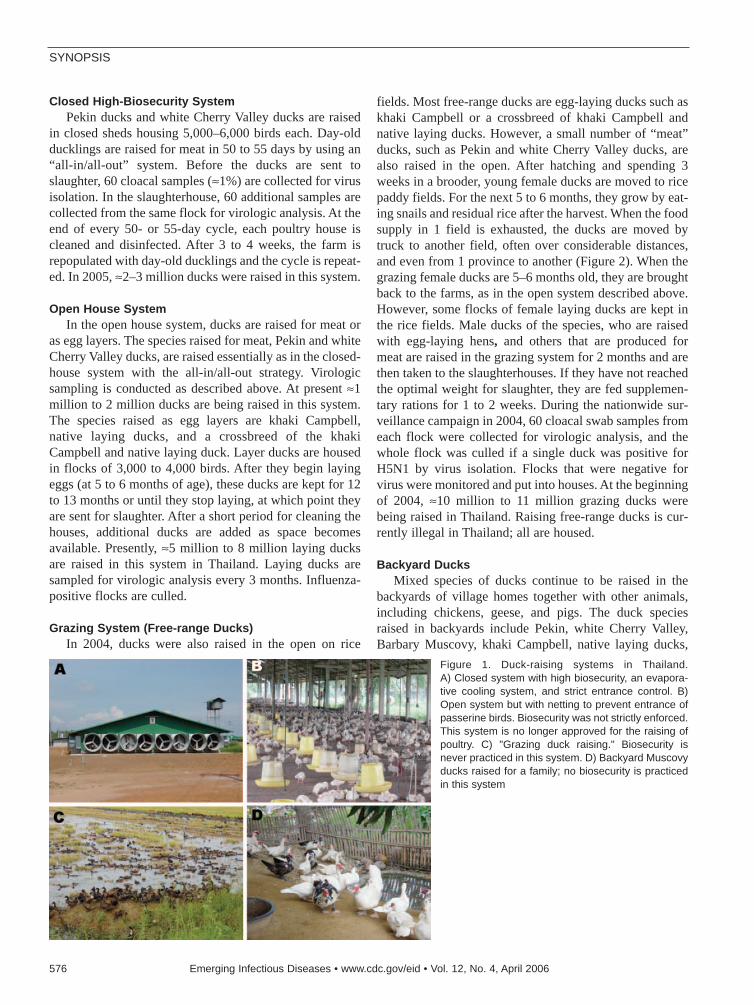
Closed High-Biosecurity System Pekin ducks and white Cherry Valley ducks are raised
in closed sheds housing 5,000–6,000 birds each. Day-oldducklings are raised for meat in 50 to 55 days by using an“all-in/all-out” system. Before the ducks are sent toslaughter, 60 cloacal samples (≈1%) are collected for virusisolation. In the slaughterhouse, 60 additional samples arecollected from the same flock for virologic analysis. At theend of every 50- or 55-day cycle, each poultry house iscleaned and disinfected. After 3 to 4 weeks, the farm isrepopulated with day-old ducklings and the cycle is repeat-ed. In 2005, ≈2–3 million ducks were raised in this system.
Open House System In the open house system, ducks are raised for meat or
as egg layers. The species raised for meat, Pekin and whiteCherry Valley ducks, are raised essentially as in the closed-house system with the all-in/all-out strategy. Virologicsampling is conducted as described above. At present ≈1million to 2 million ducks are being raised in this system.The species raised as egg layers are khaki Campbell,native laying ducks, and a crossbreed of the khakiCampbell and native laying duck. Layer ducks are housedin flocks of 3,000 to 4,000 birds. After they begin layingeggs (at 5 to 6 months of age), these ducks are kept for 12to 13 months or until they stop laying, at which point theyare sent for slaughter. After a short period for cleaning thehouses, additional ducks are added as space becomesavailable. Presently, ≈5 million to 8 million laying ducksare raised in this system in Thailand. Laying ducks aresampled for virologic analysis every 3 months. Influenza-positive flocks are culled.
Grazing System (Free-range Ducks)In 2004, ducks were also raised in the open on rice
fields. Most free-range ducks are egg-laying ducks such askhaki Campbell or a crossbreed of khaki Campbell andnative laying ducks. However, a small number of “meat”ducks, such as Pekin and white Cherry Valley ducks, arealso raised in the open. After hatching and spending 3weeks in a brooder, young female ducks are moved to ricepaddy fields. For the next 5 to 6 months, they grow by eat-ing snails and residual rice after the harvest. When the foodsupply in 1 field is exhausted, the ducks are moved bytruck to another field, often over considerable distances,and even from 1 province to another (Figure 2). When thegrazing female ducks are 5–6 months old, they are broughtback to the farms, as in the open system described above.However, some flocks of female laying ducks are kept inthe rice fields. Male ducks of the species, who are raisedwith egg-laying hens, and others that are produced formeat are raised in the grazing system for 2 months and arethen taken to the slaughterhouses. If they have not reachedthe optimal weight for slaughter, they are fed supplemen-tary rations for 1 to 2 weeks. During the nationwide sur-veillance campaign in 2004, 60 cloacal swab samples fromeach flock were collected for virologic analysis, and thewhole flock was culled if a single duck was positive forH5N1 by virus isolation. Flocks that were negative forvirus were monitored and put into houses. At the beginningof 2004, ≈10 million to 11 million grazing ducks werebeing raised in Thailand. Raising free-range ducks is cur-rently illegal in Thailand; all are housed.
Backyard Ducks Mixed species of ducks continue to be raised in the
backyards of village homes together with other animals,including chickens, geese, and pigs. The duck speciesraised in backyards include Pekin, white Cherry Valley,Barbary Muscovy, khaki Campbell, native laying ducks,
SYNOPSIS
576 Emerging Infectious Diseases • www.cdc.gov/eid • Vol. 12, No. 4, April 2006
Figure 1. Duck-raising systems in Thailand. A) Closed system with high biosecurity, an evapora-tive cooling system, and strict entrance control. B)Open system but with netting to prevent entrance ofpasserine birds. Biosecurity was not strictly enforced.This system is no longer approved for the raising ofpoultry. C) "Grazing duck raising." Biosecurity isnever practiced in this system. D) Backyard Muscovyducks raised for a family; no biosecurity is practicedin this system

and mule ducks (a sterile crossbreed of Muscovy ducksand native ducks). If a single case of H5N1 infection isdetected in a village, all the poultry in the village areculled. Approximately 1.0 million to 1.5 million duckswere raised as backyard ducks at the beginning of the out-break in 2004; culling reduced that number to <1 millionby August 2005.
National Surveillance Program In response to the H5N1 influenza outbreaks in 2004,
the government of Thailand dispatched teams to villages toidentify infected birds and cull flocks in which infectionwas detected.
Sample Collection, Histopathology, Virus Isolation, and Serology
During the study period (February to September 2004),our laboratory received 450 sick, moribund, or dead ducks
from 25 flocks in the western and central provinces ofThailand. In the detailed studies (Table 1), blood was sam-pled for serologic analysis by the hemagglutination inhibi-tion (HI) test. All moribund ducks were euthanized, andtheir internal organs were collected, fixed with 10%buffered formalin, and processed for histopathologicanalysis. Additionally, parts of the brain, lung, trachea,intestine, liver, pancreas, kidney, ovary, oviduct, testes,heart, and tight muscle were collected for virus isolation.The tissues were ground and filtered through 0.2-µ filters.The filtrates of each organ were injected into 9- to 11-day-old embryonated chicken eggs and incubated at 37°C for 2days. The eggs were observed daily to determine whetherdeath occurred. The allantoic fluid was harvested and test-ed for influenza virus by HI assay. Any positive samplewas then subtyped for H5N1. A second egg passage wasperformed if the embryonated eggs were still alive 72hours after injection.
H5N1 SubtypingAvian influenza virus was subtyped by HI assay by
using antiserum specific against the H5 hemagglutinin.Reverse-transcription polymerase chain reaction (RT-PCR) analysis was used for H5 and N1 typing (14).
Immunohistochemical TestingTo evaluate histologic changes, we used immunohisto-
chemical testing by indirect immunoperoxidase staining asdescribed (15). Tissue was fixed in formalin before beingembedded in paraffin, then cut in 5-µ-thick sections andmounted onto silanized slides.
Criteria for Culling DucksThe criteria for culling duck flocks were based on
H5N1 virus isolation and identification by serologic andRT-PCR analysis (12). During the screening of villagepoultry in 2004, a single positive virus isolation resulted inthe culling of all poultry (e.g., chicken, ducks, geese, quail)in the entire village. If serologic evidence of infection wasdetected, cloacal swabs of 60 ducks in that flock were col-lected and processed for virus isolation in embryonatedchicken eggs.
Results
Detection of Influenza Viruses in Different Duck-raising Systems
Closed High-Biosecurity SystemAs mentioned earlier, ≈1% of every duck flock was
sampled for H5N1 detection before being sent to slaughter.More than 10,000 ducks were tested during the study peri-od. No virologic or serologic evidence of H5N1 virus
Domestic Ducks and H5N1 Influenza Epidemic, Thailand
Emerging Infectious Diseases • www.cdc.gov/eid • Vol. 12, No. 4, April 2006 577
Figure 2. Example of grazing-duck movement. A single flock ofducks was moved 3 times by truck in 1 season in 2004. The sizeof the flock is 3,000–10,000. The time spent at each site dependson the availability of rice fields at the site: an acre of rice could sup-port 3,000 ducks for 1 to 2 days. The duck owners have agree-ments with the landowners regarding the time of harvest and theacreage available. One flock could spend as long as 1 month at asingle site before being moved to the next.

infection was detected in the birds raised in this closed sys-tem in western Thailand, including Nakornpathom andKanchanaburi provinces, despite cocirculation of H5N1influenza viruses in other duck-raising systems in theregion.
Open House SystemMost farms that raised ducks with the open house sys-
tem are in western Thailand, including the 4 provinces ofNakornpathom, Kanchanaburi, Suphanburi, and Racha-buri. Birds from 17 farms were tested for infection withvirus; in birds from 4 (23.5%), infection with the H5N1virus was detected.
Grazing SystemIn 28 (45.9%) of the 61 free-range duck flocks tested,
infection with H5N1 influenza virus was detected.Investigators studied H5N1 infection in 10 flocks of graz-ing ducks in Ayuthdhaya, Nakornpathom, and Suphanburiprovinces between February and July 2004 to determine thebiologic and pathologic features of H5N1 infection in thefield (Table 1). No virologic or serologic evidence of H5N1infection was detected in any of the flocks while they werelocated in the brooding houses. However, after they weremoved outdoors to the rice fields, infection with H5N1influenza was detected in all 10 flocks; the earliest infectionwas detected 12 days after the ducks left the brooding hous-es (flock 3, at 42 days of age). The interval between leav-ing the brooding houses and detection of H5N1 infectionwas 12–63 days. Of the 10 flocks, 3 (flocks 2, 8, and 9)showed disease signs; only a few birds (<1%) in each flockwere clinically affected. However, the interval between ini-tial detection of H5N1 viruses in the flock and culling was5–10 days, which supports the contention that most ducksin the flocks showed no disease signs.
Serologic evaluation of the flocks showed that low
titers of HI antibody were detected before culling, whichindicates that an immune response had already begun with-out disease signs in most birds. Cloacal virus titers in indi-vidual ducks showing disease signs before culling were2.0–3.8 log10 50% egg infectious dose (EID)50/mL whichshows that virus was being shed in feces (Table 1). Similarvirus titers were detected in asymptomatic ducks.
Signs of disease in flocks, 2, 8, and 9 were depression,lethargy, cloudy cornea, and blindness. However, nodeaths were observed in the 10 days before culling.
Backyard Ducks Of the backyard poultry, chickens were the most fre-
quently infected; 56% of the chicken flocks tested werepositive for H5N1 influenza (12). Ducks were the secondmost frequently infected; 27% of backyard duck flockswere positive for H5N1. During the second wave of H5N1infection of poultry and humans in Thailand (August–November 2004), 47% of backyard duck flocks wereH5N1 positive. During this time, scientists realized thatmost ducks infected with H5N1 were asymptomatic.
Pathologic Features As previously mentioned, our laboratory received 450
sick, moribund, or dead ducks, which were studied forpathologic features of H5N1 infection. These birds hadbeen raised in the open house system or were from back-yard flocks. They exhibited signs of disease such as highfever, dyspnea, depression, and diarrhea, and nervoussigns such as ataxia, incoordination, and convulsions(Figure 3A). Most had ocular and nasal discharge accom-panied by conjunctivitis; 20%–100% of the birds in eachflock from which these ducks originated were dead. Allcloacal and tracheal swabs and tissue samples were posi-tive for H5N1 by HI and RT-PCR (results not shown).
At necropsy, gross lesions were detected, including
SYNOPSIS
578 Emerging Infectious Diseases • www.cdc.gov/eid • Vol. 12, No. 4, April 2006

ecchymotic or petechial hemorrhage of leg and footpad;serous fluid surrounding the heart, pancreas, liver, andabdomen; cyanosis of the oral cavity; and mild pleural effu-sion. On histopathologic examination, the most strikinglesions were found in the lung, with extensive pneumoniaand severe pulmonary edema with hyaline material in thealveolar space and slight mononuclear infiltration in thearea surrounding congested vessels (Figure 3B).Nonsuppurative encephalitis with perivascular cuffing ofmononuclear cells and gliosis were detected in the brains ofducks that displayed nervous signs. Hyaline degenerationand necrosis of myocardium with mononuclear infiltrationwere detected predominantly in dead ducks from fast-growing breeds such as the Pekin and white Cherry Valleyducks. Necrotizing pancreatis with mononuclear infiltrationwas detected in all affected ducks. Most affected ducksexhibited focal hepatitis, tubulonephritis, splenic lymphoid
depletion or necrosis, and enteritis. Virus antigen wasdetected by immunohistochemical tests in all organs tested,including trachea, lung, liver, pancreas, rectum, bursa ofFabricius, spleen, brain, heart, and kidney (Figure 4).
Experimental Infection of Khaki Campbell Ducks Because culling of all H5N1-positive ducks was man-
dated in Thailand, we could not determine the natural out-come of infection in birds raised in the open on rice fields.Therefore, khaki Campbell ducks were experimentallyinfected with 4 representative H5N1 viruses isolated inThailand in 2004 and 2005. All animal experiments wereperformed in biosafety level 3+ facilities. All 4 virusescaused the deaths of infected ducks; however, their degreeof lethality varied (Table 2). The most lethal virus testedwas A/duck/Thailand/71.1/2004, which caused death in10/10 of the infected khaki Campbell ducks, a death ratecomparable to that previously reported for Mallard ducks
Domestic Ducks and H5N1 Influenza Epidemic, Thailand
Emerging Infectious Diseases • www.cdc.gov/eid • Vol. 12, No. 4, April 2006 579
Figure 3. A) A White Cherry Valley duck (Anas platyrhynchos),infected with HPAI H5N1 displays nervous signs, convulsions. B)Histopathologic features of the lung of an HPAI H5N1–infectedwhite Cherry Valley duck; infiltration of inflammatory cells in thelung parenchyma (magnification ×100).
Figure 4. Immunohistochemistry of an HPAI H5N1–infected whiteCherry Valley duck (Anas platyrhynchos). The viral antigen isdetected in myocardial cells and lymphoid cells (arrow) (A) andrenal tubular cells (B) (magnification ×100). The primary antibodyused for immunohistochemistry in this study was a mouseanti–avian influenza H5 antibody (Magellan Biotechnology,Chunan, Taiwan).
A
B

(16). Also tested was a human virus isolated in 2004,A/Thailand/MK2/2004, which resulted in the death of 2/10khaki Campbell ducks. Of the two 2005 viruses tested, 1caused very slight disease and resulted in only 1/10 deaths(A/quail/Thailand/551/2005) whereas the other(A/duck/Thailand/144/2005) resulted in 5/10 deaths.Ducks inoculated with A/Thailand/MK2/04 shed virus forthe longest period of time (day 10 postinfection), whereasthe 2005 virus isolates were shed only until day 8 postin-fection. These results indicate that the H5N1 avian virusesrecently isolated in Thailand can cause death in khakiCampbell ducks; however, several infected ducksremained completely healthy with no signs of diseasethroughout the study.
Current Status of Duck Raising in ThailandAs of October 2005, the government of Thailand for-
bids the practice of raising ducks in open fields and mov-ing grazing ducks from 1 region to another. Farmers whodo so are subject to fines and other punishments.Additionally, they receive no compensation if they raiseducks in the open free-range system, and the ducksbecome infected with H5N1. Farmers were initially com-pensated for the culling of their ducks. Duck raising is nowconfined to the high-biosecurity system.
After a lull of almost 1 year, a case of human H5N1infection was reported in Thailand in October 2005. Thereport was preceded by the illegal grazing of 3 flocks of3,000 to 5,000 free-range ducks in rice fields in the area(Kanchanaburi Province). Although no direct contactbetween the grazing ducks and backyard chickens wasknown, within 2 weeks of the arrival of the ducks, chick-ens in the area began dying, and a person who had directcontact with the diseased chickens died of H5N1 infection.Approximately 500 backyard chickens were culled in thevillage. Sequence analysis of the human isolate and avianisolates (duck and chicken) from this area would be essen-tial to confirm the epidemiologic link between these casesand, coupled with the chronology of events, to assesswhether free-grazing ducks were indeed the source ofinfection for this outbreak.
DiscussionThe 4 duck-raising systems in wide use at the beginning
of the 2004 Thai epidemic differed markedly in cases ofinfluenza detected. No infections with H5N1 influenzavirus were detected in ducks raised in the closed system,attesting to the effectiveness of the biosecurity employed.In contrast, H5N1 infection was detected in ducks raised inall 3 open systems. Notably, infection in the hatchery orduring the 3 weeks of brooding was detected only after theducks were released into the rice fields. The source of theH5N1 viruses infecting domestic ducks in the rice fieldsremains controversial. Because H5N1 viruses were detect-ed in herons, storks, egrets, and other dead waterfowl inEastern Asia, the initial spread of the highly pathogenicviruses in this region of the world has been attributed towild migrating birds. What role wild migrating birds hadin the spread of H5N1 influenza virus is now a moot ques-tion. The widespread outbreaks and massive die-off of bar-headed geese and other species in western China (17,18),and the spread of H5N1 to central Asia (Kazakhstan,southern Russia, and Turkey) and more recently toRomania and Croatia in eastern Europe, are likely causedby wild migratory birds.
Detailed studies of 10 flocks of grazing ducks inThailand in the present study showed infection with H5N1influenza virus in all flocks. Although the ducks shed virusfor 5 to 10 days, few ducks showed disease signs, and insome flocks, no ducks were symptomatic. Prolonged shed-ding of H5N1 viruses in experimentally infected ducks hasbeen previously described (16,19), but prolonged sheddingin free-range ducks has not. Therefore, free-range (graz-ing) ducks that are moved long distances by truck and thatdo not necessarily show disease signs are an optimal vehi-cle for the spread of H5N1 viruses throughout the country.These findings support the need for regulations that forbidthe practice of raising ducks on the free range, a needunderscored by the association of the recent human infec-tion with illegal free-range duck grazing.
This study also points out the dangers of raising ducksin the open systems without complete biosecurity.Although stopping the commercial raising of ducks inopen system may be impossible, the more problematic
SYNOPSIS
580 Emerging Infectious Diseases • www.cdc.gov/eid • Vol. 12, No. 4, April 2006
issue is that of backyard ducks, which are part of tradition-al village livestock. Highly pathogenic H5N1 influenzavirus is now likely endemic in poultry in Vietnam,Cambodia, China, and Indonesia. The vaccine optionshould be considered if backyard duck raising is to contin-ue in Southeast Asia.
Although no human cases of H5N1 have been attrib-uted to direct contact with ducks in Thailand, free-grazingducks have been identified as a risk factor for the occur-rence of H5N1 outbreaks among chickens (13). InVietnam, however, reported human cases of H5N1 influen-za have potentially been linked to the consumption of rawduck blood dishes (http://www.who.int/csr/don/2005_01_21/ en/index.html). Therefore, H5N1-infected ducks are arisk factor for both commercial and backyard poultry andpotentially for humans as well. Since the introduction ofthe nationwide comprehensive surveillance program (“x-ray surveys”) in Thailand (12) and the culling of all infect-ed poultry, human cases of H5N1 infection have beenmarkedly reduced. Traditional methods of duck raising inThailand and in the rest of Southeast Asia must be modi-fied if we are to control highly pathogenic H5N1 avianinfluenza.
AcknowledgmentsWe thank Carol Walsh and Amanda Ball for manuscript
preparation and Margaret Carbaugh for editing the manuscript.
Support for Thaweesak Songerm, Rungroj Jun-on, NamdeeSae-Heng, and Noppadol Meemak was provided by KasetsartUniversity Research and Development Institute, Thailand.Support for Robert G. Webster, Diane Hulse-Post, and KatharineM. Sturm-Ramirez was provided by US Public Health Servicegrant AI95357 and by the American Lebanese Syrian AssociatedCharities.
Dr Songserm is a veterinary pathologist in the Faculty ofVeterinary Medicine, Kasetsart University, KamphaengsaenCampus, Nakornpathom, Thailand. His research interests includeavian pathology, diseases of ducks and geese, and emerging dis-eases in animals.
References
1. Tang X, Tian G, Zhao C, Zhou J. Isolation and characterization ofprevalent strains of avian influenza viruses in China [in Chinese].Chinese J Anim Poultry Infect Dis. 1998;20:1:5.
2. Chen H, Deng G, Li Z, Tian G, Li Y, Jiao P, et al. The evolution ofH5N1 influenza viruses in ducks in southern China. Proc Natl AcadSci U S A. 2004;101:10452–7.
3. Sims LD, Domenech J, Benigno C, Kahn S, Kamata A, Lubroth J, etal. Origin and evolution of highly pathogenic H5N1 avian influenzain Asia. Vet Rec. 2005;157:159–64.
4. Bridges CB, Lim W, Hu-Primmer J, Sims L, Fukuda K, Mak KH, etal. Risk of influenza A (H5N1) infection among poultry workers,Hong Kong, 1997–1998. J Infect Dis. 2002;185:1005–10.
5. Guan Y, Peiris JSM, Lipatov AS, Ellis TM, Dyrting KC, Krauss S, etal. Emergence of multiple genotypes of H5N1 avian influenza virus-es in Hong Kong SAR. Proc Natl Acad Sci U S A. 2002;99:8950–5.
6. Ellis TM, Bousfield RB, Bissett LA, Dyrting KC, Luk GSM, TsimST, et al. Investigation of outbreaks of highly pathogenic H5N1 avianinfluenza in waterfowl and wild birds in Hong Kong in late 2002.Avian Pathol. 2004;33:492–505.
7. Sturm-Ramirez KM, Ellis T, Bousfield B, Bissett L, Dyrting K, RehgJE, et al. Reemerging H5N1 influenza viruses in Hong Kong in 2002are highly pathogenic to ducks. J Virol. 2004;78:4892–901.
8. Li KS, Guan Y, Wang J, Smith GJ, Xu KM, Duan L, et al. Genesis ofa highly pathogenic and potentially pandemic H5N1 influenza virusin eastern Asia. Nature. 2004;430:209–13.
9. Chotpitayasunondh T, Ungchusak K, Hanshaowarakul W,Chunsuthiwat S, Sawanpanyalert P, Kijphati R, et al. Human diseasefrom influenza A (H5N1), Thailand, 2004. Emerg Infect Dis.2005;11:201–9.
10. Ungchusak K, Auewarakul P, Dowell SF, Kitphati R, Auwanit W,Puthavathana P, et al. Probable person-to-person transmission ofavian influenza A (H5N1). N Engl J Med. 2005;352:333–40.
11. Hien TT, Liem NT, Dung NT, Luong TS, Mai PP, van Vinh Chau, N.Avian influenza A (H5N1) in 10 patients in Vietnam. N Engl J Med.2004;350:1179–88.
12. Tiensin T, Chaitaweesub P, Songserm T, Chaisingh A, Hoonsuwan W,Buranathai C, et al. Highly pathogenic avian influenza H5N1,Thailand, 2004. Emerg Infect Dis. 2005;11:1664–72.
13. Gilbert M, Chaitaweesub P, Parakamawongsa T, Premashthira S,Tiensin T, Kalpravidh W, et al. Free-grazing ducks and highly patho-genic avian influenza, Thailand. Emerg Infect Dis. 2006;12:227–34.
14. Poddar, SK. Influenza virus types and subtypes detection by singlestep single tube multiplex reverse transcription-polymerase chainreaction (RT-PCR) and agarose gel electrophoresis. J Virol Methods.2002;99:63–70.
15. Songserm T, Sae-Heng N, Jam-on R, Witoonsatient K, Meemak N.Clinical, gross-histopathologic and immunohistochemical finding ofgrazing ducks affected with HPAI H5N1 in Thailand [abstract 74]. In:Abstracts of the OIE/FAO International Conference on AvianInfluenza. Paris; 2005 Apr 7–8.
16. Sturm-Ramirez KM, Hulse-Post DJ, Govorkova EA, Humberd J,Seiler P, Puthavathana P, et al. Are ducks contributing to the endemic-ity of highly pathogenic H5N1 influenza virus in Asia? J Virol.2005;79:11269–79.
17. Liu J, Xiao H, Lei F, Zhu Q, Qin K, Zhang X-W, et al. Highly patho-genic H5N1 influenza virus infection in migratory birds. Science.2005;309:1206.
18. Chen H, Smith GJ, Zhang SY, Qin J, Wong J, Lee KS, et al. Avian flu:H5N1 virus outbreak in migratory waterfowl. Nature. 2005;436:191–2.
19. Hulse-Post DJ, Sturm-Ramirez KM, Humberd J, Seiler P, GovorkovaEA, Krauss S, et al. Role of domestic ducks in the propagation andbiological evolution of highly pathogenic H5N1 influenza viruses inAsia. Proc Natl Acad Sci U S A. 2005;102:10682–7.
Address for correspondence: Robert G. Webster, St. Jude Children’sResearch Hospital, Department of Infectious Diseases, Division ofVirology, 332 N Lauderdale, Mailstop 330, Memphis, TN 38105, USA;fax: 901-523-2622; email: [email protected]
Domestic Ducks and H5N1 Influenza Epidemic, Thailand
Emerging Infectious Diseases • www.cdc.gov/eid • Vol. 12, No. 4, April 2006 581

The city of Erechim, Brazil, has a 17% prevalence ofocular toxoplasmosis, and type 1 Toxoplasma gondii pre-dominates. To examine risk factors for acute T. gondii infec-tion in this area, we administered a questionnaire torecently infected persons (n = 131) and seronegative con-trols (n = 110). Eating undercooked meat; having a garden;working in the garden or yard more than once per week;eating rare meat; eating cured, dried, or smoked meat; eat-ing frozen lamb; and being male increased risk for T. gondiiinfection in univariate analysis. Risk factors independentlyassociated with acute T. gondii infection in multivariateanalysis were working in the garden (odds ratio [OR] 2.35,95% confidence interval [CI] 1.27–4.33) and eating frozenlamb (OR 2.06, 95% CI 1.15–3.67). Among women (n =86), having had children markedly increased the risk for T.gondii infection (OR 14.94, 95% CI 3.68–60.73).
In recent years, because of the large number of postnatalToxoplasma gondii infections, researchers estimate that
most ocular disease from T. gondii is caused by infectionafter birth (1–3). In Erechim, a city in southern Brazil, arepresentative population-based household survey showedthat 17.7% of >1,000 persons examined had ocular toxo-plasmosis (4). This high rate is believed to be due to acuteinfection after birth because the rate of infection in youngchildren in this area is low (4–6). Erechim has a populationof 96,310 and is located in northern Rio Grande do Sul(population 9,619,416), the southernmost state of Brazil,which borders Uruguay and Argentina. The area was most-ly settled by Italian, German, and Polish immigrants in theearly 20th century. Of the 3 primary restriction fragment
polymorphism (SAG2) genetic types of T. gondii, investi-gators have primarily found the type 1 organism in south-ern Brazil (7) and have identified it from chickens (8,9).Type 1 T. gondii is uncommon in the United States andmay be more virulent for ocular disease (10,11). However,with worldwide food and animal distribution it couldbecome more common in the United States and othercountries.
Although ocular toxoplasmosis has been a problem insouthern Brazil for many years, no controlled studies havedetermined the sources of infection. Therefore, in 2003and 2004, we conducted a case-control study at the princi-pal ophthalmology clinic in Erechim to evaluate the riskfactors responsible for T. gondii infection so that preven-tion messages could be tailored to the factors identified.
MethodsThe study was conducted by ophthalmologists at Clinica
Silveira and the Federal University at São Paulo, Brazil, incollaboration with researchers from the University ofCalifornia, Los Angeles, California, and the Centers forDisease Control and Prevention (CDC), Atlanta, Georgia.The questionnaire was adapted from those used in previousT. gondii case-control studies (12,13), with modificationand input from the study researchers. The questionnairetook ≈25 minutes to complete and inquired about demo-graphic variables and a comprehensive set of risk factorsrelated to meat, vegetables, food preparation, soil contact,drinking water, and animal exposure (especially cat and catfeces exposure). Information about the number of pregnan-cies and children was collected from women participatingin the survey. Questions involving habitual behaviorfocused on the most recent 12 months. The questionnairewas self-administered at Clinica Silveira, the principal oph-thalmology clinic in Erechim, with parental assistance forchildren <16 years of age.
Recently Acquired Toxoplasmagondii Infection, Brazil
Jeffrey L. Jones,* Cristina Muccioli,† Rubens Belfort, Jr,† Gary N. Holland,‡ Jacquelin M. Roberts,* and Claudio Silveira§
RESEARCH
582 Emerging Infectious Diseases • www.cdc.gov/eid • Vol. 12, No. 4, April 2006
*Centers for Disease Control and Prevention, Atlanta, Georgia,USA; †Federal University of São Paulo Paulista School ofMedicine, São Paulo, São Paulo, Brazil; ‡David Geffen School ofMedicine at UCLA, Los Angeles, California, USA; and §ClinicaSilveira, Erechim, Rio Grande do Sul, Brazil
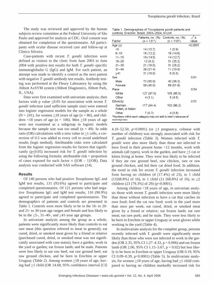
The study was reviewed and approved by the humansubjects review committee at the Federal University of SãoPaulo and approved for analysis at CDC. Oral consent wasobtained for completion of the questionnaire. All partici-pants with ocular disease received care and follow-up atClinica Silveira.
Case-patients with recent T. gondii infection weredefined as visitors to the clinic from June 2003 to June2004 with positive test results for both T. gondii–specificimmunoglobulin G (IgG) and IgM. For each patient, anattempt was made to identify a control as the next patientwith negative T. gondii antibody test results. Antibody test-ing was performed at the Fleury Laboratory by using theAbbott AxSYM system (Abbott Diagnostics, Abbott Park,IL, USA).
Data were first examined with univariate analysis; thenfactors with p value <0.05 for association with recent T.gondii infection (and sufficient sample size) were enteredinto logistic regression models for the sample as a whole(N = 241), for women >18 years of age (n = 86), and chil-dren <18 years of age (n = 106). Men >18 years of agewere not examined as a subgroup in logistic modelsbecause the sample size was too small (n = 49). In oddsratio (OR) calculations with a zero value in >1 cells, a cor-rection of 0.5 was added to every cell to avoid undefinedresults (logit method). Attributable risks were calculatedfrom the logistic regression results for factors that signifi-cantly (p<0.05) increased risk for T. gondii infection byusing the following formula: attributable risk = proportionof cases exposed for each factor × ([OR – 1]/OR). Dataanalysis was conducted with SAS software (14).
ResultsOf 140 persons who had positive Toxoplasma IgG and
IgM test results, 131 (93.6%) agreed to participate andcompleted questionnaires. Of 121 persons who had nega-tive Toxoplasma IgG and IgM test results, 110 (90.9%)agreed to participate and completed questionnaires. Thedemographics of patients and controls are presented inTable 1. Controls were more likely to be in the 16- to 20-and 21- to 30-year age ranges and female and less likely tobe in the <5-, 31–40-, and >41-year age groups.
In univariate analysis among the group as a whole,patients were significantly more likely than controls to eatrare meat (this question referred to meat in general); eatcured, dried, or smoked meat given by a friend or relative(purchased cured, dried, or smoked meat was not signifi-cantly associated with case status); have a garden; work inthe yard or garden; eat frozen lamb; and be male. Patientswere less likely to eat raw beef, eat raw ground beef, eatraw ground chicken, and be born in Erechim or upperUruguay (Table 2). Among women >18 years of age, hav-ing had >1 child (OR 14.68, 95% confidence interval [CI]
4.10–52.50, p<0.0001) (or >1 pregnancy, colinear withnumber of children) was strongly associated with risk forT. gondii infection (Table 3). Women infected with T.gondii were also more likely than those not infected tohave lived in their present home >12 months, work withanimals (all types), work in the garden, and have >1 cat orkitten living at home. They were less likely to be infectedif they ate raw ground beef, raw chicken, rare or rawground chicken, and fed their cat dried food. In addition,the trend in risk for recent T. gondii infection increasedfrom having no children (4 [17.4%] of 23), to 1 child(11[68.8%] of 16), to 1 child (11 [68.8%] of 16), to >2children (23 [79.3%] of 29) (p<0.0001).
Among children <18 years of age, in univariate analy-sis those with recent T. gondii infection were more likelythan those without infection to have a cat that catches itsown food; feed the cat raw food; work in the yard morethan once per week; eat cured, dried, or smoked meatgiven by a friend or relative; eat frozen lamb; eat raremeat; eat rare pork; and be male. They were less likely tobe born in Erechim or upper Uruguay or wear gloves whileworking in the yard (Table 4).
In multivariate analysis for the complete group, personsrecently infected with T. gondii were significantly morelikely than those who were not infected to work in the gar-den (OR 2.35, 95% CI 1.27–4.33, p = 0.006) and eat frozenlamb (OR 2.06, 95% CI 1.15–3.67, p = 0.02) but less like-ly to be born in Erechim or upper Uruguay (OR 0.19, 95%CI 0.09–0.39, p<0.0001) (Table 5). In multivariate analy-sis, for women >18 years of age, having had >1 child com-pared to having no children markedly increased risk for
Toxoplasma gondii Infection, Brazil
Emerging Infectious Diseases • www.cdc.gov/eid • Vol. 12, No. 4, April 2006 583

recent T. gondii infection (OR 14.94, 95% CI 3.68–60.73,p<0.0002) (Table 5). For children <18 years of age, beingborn in Erechim or upper Uruguay was associated with areduced risk for recent T. gondii infection compared tothose born elsewhere (OR 0.04, 95% CI 0.01–0.22, p =0.0002), and male sex (OR 5.70, 95% CI 1.95–16.66, p =0.002) was associated with an increased risk for T. gondiiinfection (Table 5).
The attributable risks for the factors that significantlyincreased the risk for recent T. gondii infection in logisticregression for the group as a whole (from Table 5) wereworking in the garden (0.25) and eating frozen lamb(0.15). Among women >18 years of age, having had >1child was associated with a high attributable risk (0.84).Among children <18 years of age, male sex was associat-ed with an attributable risk of 0.64.
DiscussionIn the study group overall (Table 2), we found that a
number of meat- and soil-related factors were associated
with recent T. gondii infection, which included eatingcured, dried, or smoked meat given by a friend or relative;eating rare meat; eating frozen lamb; and having a gardenand working in the yard or garden (soil contact). The pres-ence of meat-related factors emphasizes the importance ofcooking meat well (>67°C) and not assuming that all homefreezing methods will kill T. gondii cysts. Having a gardenor working in the yard increases the chances of oocystexposure through soil contact. Men were at increased riskfor recent T. gondii infection, which could also be relatedto increased soil contact. To minimize T. gondii exposure,gloves should be worn while gardening, and hands shouldbe washed thoroughly afterward with soap and water.Although sample size limitations precluded us fromincluding all the variables in multivariate analysis, work-ing in the garden remained significant in multivariateanalysis.
Although the number of persons who ate raw meat wassmall, eating raw ground chicken was associated with adecreased risk for T. gondii infection. Chickens grown
RESEARCH
584 Emerging Infectious Diseases • www.cdc.gov/eid • Vol. 12, No. 4, April 2006

commercially are probably not involved in the transmis-sion of T. gondii because modern methods reduce soilexposure, chickens are usually frozen for storage beforepurchase, and they are thoroughly cooked (15). Eatingchicken could be associated with a decrease in risk becausethose who eat chicken may be less likely to eat other meatsthat are associated with a greater risk. However, free-rangechickens in Brazil can be infected with T. gondii (9), andeating fresh, undercooked meat from free-range chickenscould pose a risk for infection. In addition, eating raw beefor raw ground beef decreased the risk for T. gondii infec-tion in univariate analysis, although the sample size wassmall for this variable as well. Beef is rarely contaminatedwith T. gondii and is not believed to be a consequentialsource of infection (15), but the role of beef in transmis-
sion to humans has not been completely determined.Eating raw ground beef that is contaminated with porkbecause the grinding machine was not cleaned after grind-ing pork could increase the risk for T. gondii infection.Eating rare meat (all types combined) was a risk factor forT. gondii infection. Pork and lamb are the most likelymeats to be contaminated with T. gondii (15). Althoughthese data are not specifically from Erechim, researchers insouthern Brazil have recently reported that 8.7% of 149fresh pork sausage samples were positive for T. gondii bybioassay in mice in the state of Parana (16), and 17% of286 finishing pigs tested positive for T. gondii antibodieswith the modified agglutination test in the state of SãoPaulo (17). Dubey et al. determined that tissue cysts sur-vive in pork for <3 minutes if heated to 64°C (18), so
Toxoplasma gondii Infection, Brazil
Emerging Infectious Diseases • www.cdc.gov/eid • Vol. 12, No. 4, April 2006 585

cooking meat (especially pork or lamb) below this temper-ature could lead to infection.
The strongest factor in analysis of the group overall wasthe reduced risk for T. gondii infection among persons bornin Erechim or upper Uruguay compared to persons bornelsewhere. This reduction in risk was also significant inmultivariate analysis and in children and is likely due toreferral of patients with ocular disease from outside thearea. Risk for T. gondii infection was also reduced amongwomen born in Erechim or upper Uruguay, but the reduc-tion was not significant (data not shown).
For women >18 years of age, having had >1 child wasstrongly associated with recent T. gondii infection in bothunivariate and multivariate analysis (Tables 3 and 5).Having had >1 pregnancy also showed a strong associationwith recent T. gondii infection in univariate analysis, butwe did not examine it in multivariate analysis because it ishighly correlated with the number of children. Pregnancyand number of children were risk factors for T. gondiiinfection in other studies, including several from Brazil(19–22). Avelino et al. (20) suggest that the greater vulner-ability of pregnant women to T. gondii infection is proba-bly due to alterations in immune mechanisms associatedwith gestation. In some settings, pregnancy could affectculinary habits. In our study, the number of children andpast pregnancies increased risk for recent T. gondii infec-tion. Another possible explanation for this finding is thatchildren bring, or track, contaminated soil into the house,increasing risk for infection.
Among children <18 years of age, in univariate analysismany of the meat- and soil-related risk factors were associ-ated with an increased risk for infection that was seen in thegroup as a whole (Table 4). In addition, feeding cats rawfood and having cats that catch their own food were associ-ated with increased risk. To prevent cats from becominginfected with T. gondii, they should be fed only dry, canned,or well-cooked food and kept indoors when possible to dis-courage hunting. In multivariate analysis of the risk factorsfor children, male sex was the principal factor that signifi-cantly increased risk for infection, although eating raremeat and frozen lamb approached significance.
One of the strengths of our study is that serologicallydefined acute T. gondii infection was required for the casedefinition. Most T. gondii IgM- and IgG-positive personsare infected for <1 year, so their more recent behavior cor-relates with the time of their infection. Our study also hasa number of limitations. The Clinica Silveira is known forexpertise in ocular toxoplasmosis and draws patients withtoxoplasmosis-related eye disease from the surroundingarea. This fact is the likely reason why patients with acuteT. gondii infection were more likely to come from outsidethe Erechim area than from Erechim. Although the samplesize was adequate to identify numerous risk factors for
T. gondii infection, it was not large enough to allow analy-sis for all subgroups (for example, multivariate analysis ofadult men). Some associations in our study may have beendue to confounders that we did not consider in the analysis(especially in univariate analysis); however, we inquiredabout a comprehensive set of variables, including thosepreviously known to be associated with T. gondii infection,and performed multivariate analysis. In addition, our studywas limited to 1 area of Brazil, and the results cannot nec-essarily be applied to other areas of the country.
In conclusion, our study identified a number of risk fac-tors for T. gondii infection among persons with ocular dis-ease. Because T. gondii infection often leads to oculardisease in this region of Brazil, avoiding infection canimprove ocular health. Proper meat and soil- and water-related hygiene could reduce the risk for infection with T.gondii, especially with preventive education efforts basedon risk factors identified in this case-control study. Theassociation of T. gondii infection in women with the num-ber of children they have had (and their pregnancies)requires further study to fully elucidate the factors thatcontribute to this increase in risk. In the future, type 1 T.gondii could spread to other areas of the world andincrease the risk for ocular disease in those regions.
Dr Jones has worked at CDC for 15 years, first in the area ofHIV/AIDS and most recently in parasitic diseases. His currentresearch focuses on toxoplasmosis.
References
1. Holland GN. Reconsidering the pathogenesis of ocular toxoplasmo-sis. Am J Ophthalmol. 1999;128:502–5.
2. Gilbert RE, Stanford MR. Is ocular toxoplasmosis caused by prenatalor postnatal infection? Br J Ophthalmol. 2000;84:244–6.
3. Holland GN. LX Edward Jackson memorial lecture. Ocular toxoplas-mosis: a global reassessment. Part 1: epidemiology and course of dis-ease. Am J Ophthalmol. 2003;136:973–88.
4. Glasner PD, Silveira C, Kruszon-Moran D, Martins MC, Burnier MJr, Silveira S, et al. An unusually high prevalence of ocular toxoplas-mosis in southern Brazil. Am J Ophthalmol. 1992;114:136–44.
5. Nussenblatt RB, Belfort R Jr. Ocular toxoplasmosis. An old diseaserevisited. JAMA. 1994;271:304–7.
6. Silveira C, Belfort R Jr, Muccioli C, Abreu MT, Martins MC, VictoraC, et al. A follow-up study of Toxoplasma gondii infection in south-ern Brazil. Am J Ophthalmol. 2001;131:351–4.
7. Vallochi AL, Muccioli C, Martins MC, Silveira C, Belfort R, RizzoLV. The genotype of Toxoplasma gondii strains causing ocular toxo-plasmosis in humans in Brazil. Am J Ophthalmol. 2005;139:350–1.
8. Dubey JP, Graham DH, Blackston CR, Lehmann T, Gennari SM,Ragozo AM, et al. Biological and genetic characterization ofToxoplasma gondii isolates from chickens (Gallus domesticus) fromSão Paulo, Brazil: unexpected findings. Int J Parasitol.2002;32:99–105.
9. Dubey JP, Navarro IT, Graham DH, Dahl E, Freire RL, PrudencioLB, et al. Characterization of Toxoplasma gondii isolates from free-range chickens from Parana, Brazil. Vet Parasitol. 2003;117:229–34.
RESEARCH
586 Emerging Infectious Diseases • www.cdc.gov/eid • Vol. 12, No. 4, April 2006

10. Grigg ME, Ganatra J, Boothroyd JC, Margolis TP. Unusual abun-dance of atypical strains associated with human ocular toxoplasmo-sis. J Infect Dis. 2001;184:633–9.
11. Darde ML. Genetic analysis of the diversity in Toxoplasma gondii.Ann Ist Super Sanita. 2004;40:57–63.
12. Cook AJ, Gilbert RE, Buffolano W. Sources of Toxoplasma infectionin pregnant women: European multicentre case-control study.European Research Network on Congenital Toxoplasmosis. BMJ.2000;321:142–7.
13. Bowie WR, King AS, Werker DH, Isaac-Renton JL, Bell A, Eng SB,et al. Outbreak of toxoplasmosis associated with municipal drinkingwater. Lancet. 1997;350:173–7.
14. SAS version 8.02. Cary (NC): SAS Institute Inc.; 2001.15. Dubey JP. Toxoplasmosis. J Am Vet Med Assoc. 1994;205:1593–8.16. Dias RAF, Navarro IT, Ruffolo BB, Bugni FM, de Castro MV, Freire
RL. Toxoplasma gondii in fresh pork sausage and seroprevalence inbutchers from factories in Londrina, Parana State, Brazil. Rev InstMed Trop Sao Paulo. 2005;47:185–9.
17. de A Dos Santos CB, de Carvalho AC, Ragozo AM, Soares RM,Amaku M, Yai LE, et al. First isolation and characterization ofToxoplasma gondii from finishing pigs from São Paulo State, Brazil.Vet Parasitol. 2005;131:207–11.
18. Dubey JP, Kotula AW, Sharar A, Andrews CD, Lindsay DS. Effect ofhigh temperature on infectivity of Toxoplasma gondii tissue cysts inpork. J Parasitol. 1990;76:201–4.
19. Avelino MM, Junior DC, de Parada JB, de Castro AM. Risk factorsfor Toxoplasma gondii infection in women of childbearing age. BrazJ Infect Dis. 2004;8:164–74.
20. Avelino MM, Campos D Jr, de Parada JCB, Castro AM. Pregnancy asa risk factor for acute toxoplasmosis seroconversion. Eur J ObstetGyn Reprod Biol. 2003;108:19–24.
21. Nissapathorn V, Noor Azmi MA, Cho SM, Fong MY, Init I, RohelaM, et al. Toxoplasmosis: prevalence and risk factors. J ObstetGynaecol. 2003;23:618–24.
22. Jara M, Hsu HW, Eaton RB, Demaria A Jr. Epidemiology of congen-ital toxoplasmosis identified by population-based newborn screeningin Massachusetts. Pediatr Infect Dis J. 2001;20:1132–5.
Address for correspondence: Jeffrey L. Jones, Centers for DiseaseControl and Prevention, 4770 Buford Hwy NE, Mailstop F22, Atlanta,GA 30341-3724, USA; fax: 770-488-7761; email: [email protected]
Toxoplasma gondii Infection, Brazil
Emerging Infectious Diseases • www.cdc.gov/eid • Vol. 12, No. 4, April 2006 587
Searchpast issues
Monochloramine disinfection of municipal water sup-plies is associated with decreased risk for Legionnaires’disease. We conducted a 2-year, prospective, environmen-tal study to evaluate whether converting from chlorine tomonochloramine for water disinfection would decreaseLegionella colonization of hot water systems. Water andbiofilm samples from 53 buildings were collected forLegionella culture during 6 intervals. Prevalence ratios(PRs) comparing Legionella colonization before and aftermonochloramine disinfection were adjusted for water sys-tem characteristics. Legionella colonized 60% of the hotwater systems before monochloramine versus 4% afterconversion (PR 0.07, 95% confidence interval 0.03–0.16).The median number of colonized sites per buildingdecreased with monochloramine disinfection. Increasedprevalence of Legionella colonization was associated withwater heater temperatures <50°C, buildings taller than 10stories, and interruptions in water service. Increasing use ofmonochloramine in water supplies throughout the UnitedStates may reduce Legionella transmission and incidenceof Legionnaires’ disease.
Legionnaires’ disease, named after an outbreak ofsevere pneumonia at a legionnaires’ convention in
1976, is a form of community-acquired and nosocomialpneumonia. It is caused by inhalation of aerosols ormicroaspiration of water containing Legionella bacteria.Legionella spp. are ubiquitous in fresh water and occurnaturally as intracellular parasites of amebae (1). Potable
hot water systems provide environments for amplificationof Legionella pneumophila, the most common species iso-lated from patients with Legionnaires’ disease. L. pneu-mophila grows optimally at 35°C and multiplies between25°C and 42°C. Investigations of outbreaks ofLegionnaires’ disease in hospitals and other communitysettings have implicated potable hot water systems assources of transmission (2–5).
No strategies have been proven to prevent community-acquired Legionnaires’ disease. Prevention of transmissionwithin healthcare facilities focuses primarily on preventingor limiting Legionella colonization of plumbing systemsthrough temperature control or use of biocides (6).Healthcare facilities are of special concern because ofincreased susceptibility to and a high case-fatality ratio ofLegionnaires’ disease among immunocompromisedpatients and those with underlying illnesses (5,7). Becausecolonized water distribution systems are often implicatedin Legionella transmission (2,5,8,9), effective water disin-fection strategies could provide the best measure to pre-vent Legionnaires’ disease.
Chloramination is a method of drinking water disinfec-tion that provides a lasting residual disinfectant in the dis-tribution system. The process involves adding ammonia tochlorinated water; aqueous chlorine reacts with ammoniato form inorganic chloramines (10). Monochloramine isthe most active compound and forms preferentially at cer-tain ratios of ammonia to chlorine. Approximately 55% of11.8 million people living in the 25 largest cities inCalifornia currently receive water disinfected with mono-chloramine (unpub. data). A survey in 2004 of municipalwater utilities in the United States found that 30% usedmonochloramine for residual disinfection (11). TheEnvironmental Protection Agency estimates that municipal
Reducing Legionella Colonization of Water Systems with
MonochloramineBrendan Flannery,* Lisa B. Gelling,† Duc J. Vugia,‡ June M. Weintraub,§ James J. Salerno,¶
Michael J. Conroy,¶ Valerie A. Stevens,* Charles E. Rose,* Matthew R. Moore,* Barry S. Fields,* and Richard E. Besser*
RESEARCH
588 Emerging Infectious Diseases • www.cdc.gov/eid • Vol. 12, No. 4, April 2006
*Centers for Disease Control and Prevention, Atlanta, Georgia,USA; †California Emerging Infections Program, Oakland,California, USA; ‡California Department of Health Services,Richmond, California, USA; §City and County of San FranciscoDepartment of Public Health, San Francisco, California, USA; and¶San Francisco Public Utilities Commission, Burlingame,California, USA

water utilities using surface water sources will increasing-ly convert to monochloramine to meet federal regulationsthat limit disinfection byproducts in drinking water (12).
Use of monochloramine for residual disinfection com-pared with chlorine was associated with a lower preva-lence of Legionella colonization in plumbing systems (13)and decreased risk of nosocomial outbreaks ofLegionnaires’ disease in cross-sectional and retrospectivecase-control studies (14,15). The planned conversion tomonochloramine for municipal drinking water disinfectionin San Francisco, California, provided an opportunity toprospectively investigate the effect of chloramination onLegionella colonization in potable hot water systems. Wereport here on the results of a 2-year environmental study.
Methods
Study SiteThe San Francisco Public Utilities Commission pro-
vides an average of 250 million gallons (950 million liters)of water per day to ≈2.4 million residents in northernCalifornia, including 750,000 in the city and county of SanFrancisco. Surface water makes up >99% of the water sup-ply. Chlorine was added to kill microorganisms present insource water (primary disinfection) throughout the studyperiod. Chlorine concentrations are monitored at severallocations throughout the distribution system. Chlorineused for residual (or secondary) disinfection was replacedwith monochloramine on February 2, 2004.
Buildings with >3 stories in San Francisco were identi-fied from lists of commercial customers of the SanFrancisco Public Utilities Commission and real propertyowned by the city and county of San Francisco. Buildingmanagers and owners gave permission for sample collec-tion inside the buildings for the duration of the study.Results of Legionella cultures were provided only at thecompletion of the study. Standardized questionnaires wereadministered to building engineers and facilities managersto obtain information on the age of the building, capacityof water heaters and hot water storage tanks, type of waterheating system (boiler, heat exchanger, or instantaneousheaters), and type of pipe material used throughout most ofthe building. At the completion of the study, building engi-neers were surveyed about routine maintenance plans forthe potable hot water system, standard procedures forflushing outlets after a disruption of water service, andknowledge of industry guidelines for controllingLegionella growth in building water systems (16).
Environmental SamplingSamples from each building were collected 6 times dur-
ing the 2-year period, 3 times before and 3 times after con-version to monochloramine disinfection. Each round of
sampling lasted 8–10 weeks. Preconversion and postcon-version rounds of sampling were conducted at correspon-ding seasonal intervals.
Nine samples were collected from each building duringsampling rounds, including a 1-L water sample from awater heater or heat exchanger, four 1-L samples of hotwater, and 4 swabs of biofilm at point-of-use outlets(faucets or shower heads). Water samples were collected insterile, 1-L plastic bottles (Nalge Nunc International,Rochester, NY, USA) containing 0.5 mL 0.1 N sodium thio-sulfate solution to neutralize free chlorine and chloramines.Water heater samples were drawn from the drain valve,pressure relief valve, or from the closest outlet to heatexchangers. Point-of-use outlets were selected at farthestpoints from water heaters when possible. Biofilm sampleswere collected from shower outlets and faucets by insertinga sterile, polyester-tipped applicator swab (Falcon, BectonDickinson and Company, Sparks, MD, USA) and rotating itfirmly against the interior surface. Biofilm swabs wereplaced in sterile, screw-capped test tubes containing 0.1 mLsodium thiosulfate solution in 5 mL of water from the samesite. Hot water taps were run until the temperature reacheda maximum for collection of water samples. The same loca-tions were sampled in each round. When sampling couldnot be performed at the selected site, the nearest substitutesite was sampled; however, only samples collected from thesame site before and after monochloramine conversionwere included in analyses.
Water temperature, pH, and free (disassociated) andtotal chlorine concentrations were measured in a separatesample bottle. Total chlorine includes free chlorine plusmonochloramine. Temperature was measured with a hand-held thermometer. pH was measured with a digital meter(pHep 3, Hanna Instruments, Leighton Buzzard, UK).Free and total chlorine residuals were measured by usingthe N, N-diethyl-p-phenylenediamine method with a col-orimeter and test kit (Model DR/890, Hach Chemical Co.,Loveland, CO, USA). Building engineers were askedabout any interruptions in water service affecting thebuilding or specific sites in the 3 months preceding thesampling date.
Laboratory ProceduresAll culturing for Legionella species and amebae was
performed in the Legionella Laboratory at the Centers forDisease Control and Prevention in Atlanta, Georgia, fol-lowing standard procedures (17). Legionella organismswere speciated or serogrouped by macroscopic slideagglutination with a panel of polyclonal rabbit antiseraagainst Legionella species and L. pneumophila serogroups(18). Laboratorians were blinded to the identity of build-ings from which samples were obtained, and buildingswere assigned different identification numbers in each
Reducing Legionella Colonization with Monochloramine
Emerging Infectious Diseases • www.cdc.gov/eid • Vol. 12, No. 4, April 2006 589
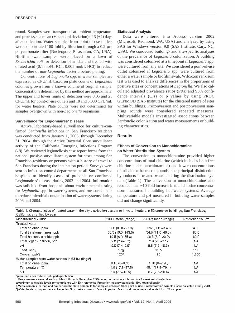
round. Samples were transported at ambient temperatureand processed a mean (± standard deviation) of 3 (±2) daysafter collection. Water samples from point-of-use outletswere concentrated 100-fold by filtration through a 0.2-µmpolycarbonate filter (Nucleopore, Pleasanton, CA, USA).Biofilm swab samples were placed on a lawn ofEscherichia coli for detection of ameba and treated withdiluted acid (0.1 mol/L KCl, 0.005 mol/L HCl) to reducethe number of non-Legionella bacteria before plating.
Concentrations of Legionella spp. in water samples areexpressed as CFU/mL based on plate counts of Legionellacolonies grown from a known volume of original sample.Concentrations determined by this method are approximate.The upper and lower limits of detection were 0.05 and 25CFU/mL for point-of-use outlets and 10 and 5,000 CFU/mLfor water heaters. Plate counts were not determined forsamples overgrown with non-Legionella organisms.
Surveillance for Legionnaires’ DiseaseActive, laboratory-based surveillance for culture-con-
firmed Legionella infections in San Francisco residentswas conducted from January 1, 2003, through December31, 2004, through the Active Bacterial Core surveillanceactivity of the California Emerging Infections Program(19). We reviewed legionellosis case report forms from thenational passive surveillance system for cases among SanFrancisco residents or persons with a history of travel toSan Francisco during the incubation period. Surveys weresent to infection control departments at all San Franciscohospitals to identify cases of probable or confirmedLegionnaires’ disease during 2003 and 2004. Informationwas solicited from hospitals about environmental testingfor Legionella spp. in water systems, and measures takento reduce microbial contamination of water systems during2003 and 2004.
Statistical AnalysisData were entered into Access version 2002
(Microsoft, Redmond, WA, USA) and analyzed by usingSAS for Windows version 9.0 (SAS Institute, Cary, NC,USA). We conducted building- and site-specific analysesof the prevalence of Legionella colonization. A buildingwas considered colonized at a timepoint if Legionella spp.were cultured from any site. We considered a point-of-useoutlet colonized if Legionella spp. were cultured fromeither a water sample or biofilm swab. Wilcoxon rank sumtest was used to analyze differences in the proportions ofpositive sites or concentrations of Legionella. We also cal-culated adjusted prevalence ratios (PRs) and 95% confi-dence intervals (CIs) or p values by using PROCGENMOD (SAS Institute) for the clustered nature of siteswithin buildings. Preconversion and postconversion sam-pling rounds were considered repeated measures.Multivariable models investigated associations betweenLegionella colonization and water measurements or build-ing characteristics.
Results
Effects of Conversion to Monochloramine on Water Distribution System
The conversion to monochloramine provided higherconcentrations of total chlorine (which includes both freechlorine and monochloramine) and lower concentrationsof trihalomethane compounds, the principal disinfectionbyproducts in treated water entering the distribution sys-tem (Table 1). The conversion to monochloramine alsoresulted in an ≈10-fold increase in total chlorine concentra-tions measured in building hot water systems. Averagetemperature and pH measured in building water samplesdid not change significantly.
RESEARCH
590 Emerging Infectious Diseases • www.cdc.gov/eid • Vol. 12, No. 4, April 2006
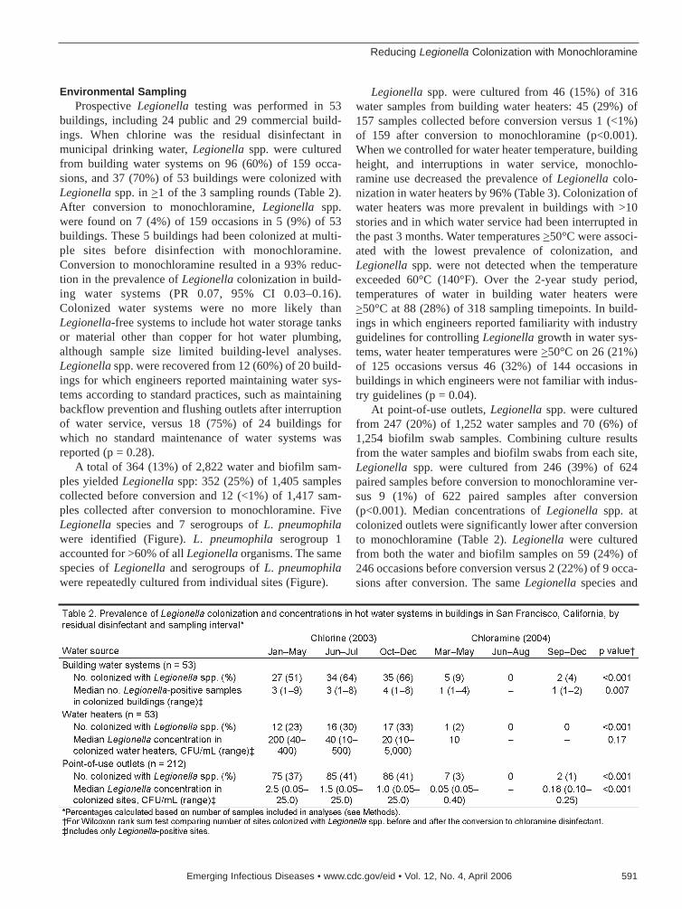
Environmental SamplingProspective Legionella testing was performed in 53
buildings, including 24 public and 29 commercial build-ings. When chlorine was the residual disinfectant inmunicipal drinking water, Legionella spp. were culturedfrom building water systems on 96 (60%) of 159 occa-sions, and 37 (70%) of 53 buildings were colonized withLegionella spp. in >1 of the 3 sampling rounds (Table 2).After conversion to monochloramine, Legionella spp.were found on 7 (4%) of 159 occasions in 5 (9%) of 53buildings. These 5 buildings had been colonized at multi-ple sites before disinfection with monochloramine.Conversion to monochloramine resulted in a 93% reduc-tion in the prevalence of Legionella colonization in build-ing water systems (PR 0.07, 95% CI 0.03–0.16).Colonized water systems were no more likely thanLegionella-free systems to include hot water storage tanksor material other than copper for hot water plumbing,although sample size limited building-level analyses.Legionella spp. were recovered from 12 (60%) of 20 build-ings for which engineers reported maintaining water sys-tems according to standard practices, such as maintainingbackflow prevention and flushing outlets after interruptionof water service, versus 18 (75%) of 24 buildings forwhich no standard maintenance of water systems wasreported (p = 0.28).
A total of 364 (13%) of 2,822 water and biofilm sam-ples yielded Legionella spp: 352 (25%) of 1,405 samplescollected before conversion and 12 (<1%) of 1,417 sam-ples collected after conversion to monochloramine. FiveLegionella species and 7 serogroups of L. pneumophilawere identified (Figure). L. pneumophila serogroup 1accounted for >60% of all Legionella organisms. The samespecies of Legionella and serogroups of L. pneumophilawere repeatedly cultured from individual sites (Figure).
Legionella spp. were cultured from 46 (15%) of 316water samples from building water heaters: 45 (29%) of157 samples collected before conversion versus 1 (<1%)of 159 after conversion to monochloramine (p<0.001).When we controlled for water heater temperature, buildingheight, and interruptions in water service, monochlo-ramine use decreased the prevalence of Legionella colo-nization in water heaters by 96% (Table 3). Colonization ofwater heaters was more prevalent in buildings with >10stories and in which water service had been interrupted inthe past 3 months. Water temperatures >50°C were associ-ated with the lowest prevalence of colonization, andLegionella spp. were not detected when the temperatureexceeded 60°C (140°F). Over the 2-year study period,temperatures of water in building water heaters were>50°C at 88 (28%) of 318 sampling timepoints. In build-ings in which engineers reported familiarity with industryguidelines for controlling Legionella growth in water sys-tems, water heater temperatures were >50°C on 26 (21%)of 125 occasions versus 46 (32%) of 144 occasions inbuildings in which engineers were not familiar with indus-try guidelines (p = 0.04).
At point-of-use outlets, Legionella spp. were culturedfrom 247 (20%) of 1,252 water samples and 70 (6%) of1,254 biofilm swab samples. Combining culture resultsfrom the water samples and biofilm swabs from each site,Legionella spp. were cultured from 246 (39%) of 624paired samples before conversion to monochloramine ver-sus 9 (1%) of 622 paired samples after conversion(p<0.001). Median concentrations of Legionella spp. atcolonized outlets were significantly lower after conversionto monochloramine (Table 2). Legionella were culturedfrom both the water and biofilm samples on 59 (24%) of246 occasions before conversion versus 2 (22%) of 9 occa-sions after conversion. The same Legionella species and
Reducing Legionella Colonization with Monochloramine
Emerging Infectious Diseases • www.cdc.gov/eid • Vol. 12, No. 4, April 2006 591
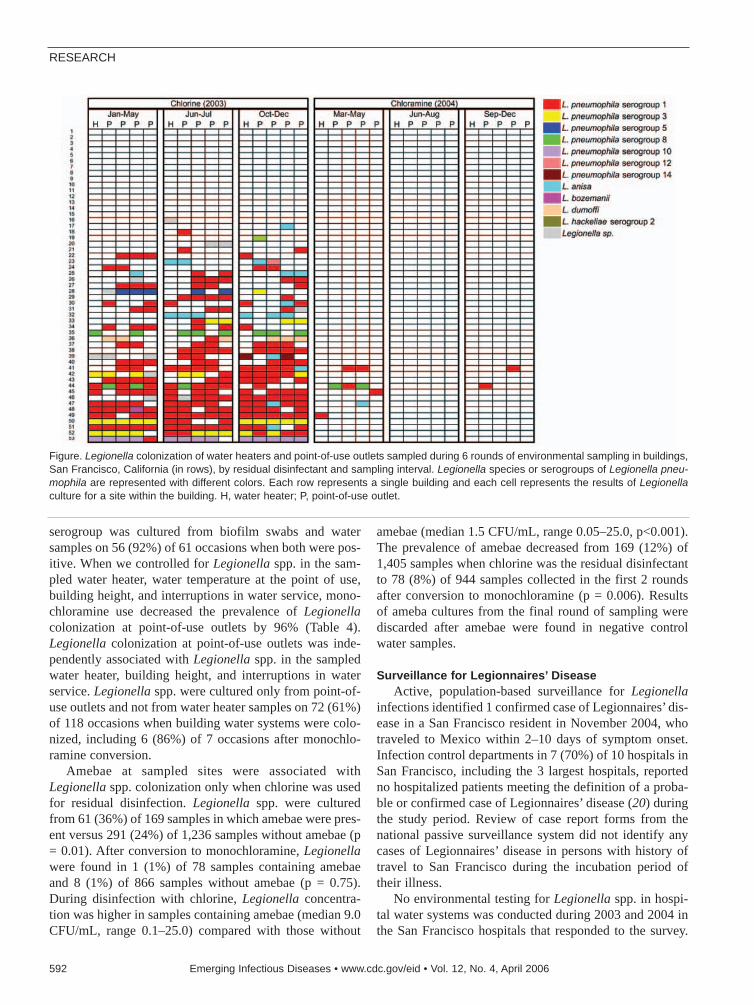
serogroup was cultured from biofilm swabs and watersamples on 56 (92%) of 61 occasions when both were pos-itive. When we controlled for Legionella spp. in the sam-pled water heater, water temperature at the point of use,building height, and interruptions in water service, mono-chloramine use decreased the prevalence of Legionellacolonization at point-of-use outlets by 96% (Table 4).Legionella colonization at point-of-use outlets was inde-pendently associated with Legionella spp. in the sampledwater heater, building height, and interruptions in waterservice. Legionella spp. were cultured only from point-of-use outlets and not from water heater samples on 72 (61%)of 118 occasions when building water systems were colo-nized, including 6 (86%) of 7 occasions after monochlo-ramine conversion.
Amebae at sampled sites were associated withLegionella spp. colonization only when chlorine was usedfor residual disinfection. Legionella spp. were culturedfrom 61 (36%) of 169 samples in which amebae were pres-ent versus 291 (24%) of 1,236 samples without amebae (p= 0.01). After conversion to monochloramine, Legionellawere found in 1 (1%) of 78 samples containing amebaeand 8 (1%) of 866 samples without amebae (p = 0.75).During disinfection with chlorine, Legionella concentra-tion was higher in samples containing amebae (median 9.0CFU/mL, range 0.1–25.0) compared with those without
amebae (median 1.5 CFU/mL, range 0.05–25.0, p<0.001).The prevalence of amebae decreased from 169 (12%) of1,405 samples when chlorine was the residual disinfectantto 78 (8%) of 944 samples collected in the first 2 roundsafter conversion to monochloramine (p = 0.006). Resultsof ameba cultures from the final round of sampling werediscarded after amebae were found in negative controlwater samples.
Surveillance for Legionnaires’ Disease Active, population-based surveillance for Legionella
infections identified 1 confirmed case of Legionnaires’ dis-ease in a San Francisco resident in November 2004, whotraveled to Mexico within 2–10 days of symptom onset.Infection control departments in 7 (70%) of 10 hospitals inSan Francisco, including the 3 largest hospitals, reportedno hospitalized patients meeting the definition of a proba-ble or confirmed case of Legionnaires’ disease (20) duringthe study period. Review of case report forms from thenational passive surveillance system did not identify anycases of Legionnaires’ disease in persons with history oftravel to San Francisco during the incubation period oftheir illness.
No environmental testing for Legionella spp. in hospi-tal water systems was conducted during 2003 and 2004 inthe San Francisco hospitals that responded to the survey.
RESEARCH
592 Emerging Infectious Diseases • www.cdc.gov/eid • Vol. 12, No. 4, April 2006
Figure. Legionella colonization of water heaters and point-of-use outlets sampled during 6 rounds of environmental sampling in buildings,San Francisco, California (in rows), by residual disinfectant and sampling interval. Legionella species or serogroups of Legionella pneu-mophila are represented with different colors. Each row represents a single building and each cell represents the results of Legionellaculture for a site within the building. H, water heater; P, point-of-use outlet.

Two hospitals added supplemental chlorine to their watersystem to prevent microbial contamination before mono-chloramine conversion; supplemental chlorination wasdiscontinued after monochloramine was added to munici-pal drinking water.
DiscussionThis is the largest study to prospectively evaluate the
effect of monochloramine disinfection on Legionella colo-nization in a water distribution system. Legionella spp.were prevalent and stable in building water systems over 3rounds of sampling when chlorine was used for residualdisinfection of drinking water. Monochloramine disinfec-tion of the water supply reduced Legionella colonization inhot water systems. Our findings suggest that monochlo-ramine in drinking water provides better control ofLegionella growth in building plumbing systems than
chlorine. This study supports the biologic plausibility ofdecreased risk of nosocomial outbreaks of Legionnaires’disease associated with chloraminated water comparedwith chlorinated water (14,15).
The conversion from chlorine to monochloramine forresidual disinfection resulted in lower concentrations of tri-halomethane compounds in drinking water, which met theobjectives of the municipal water supplier. Increased stabil-ity of monochloramine resulted in higher disinfectant con-centrations in potable hot water systems because chlorinedissipates rapidly at higher temperatures. Higher concentra-tions of disinfectant and the ability of monochloramine topenetrate biofilms were likely responsible for the effect onLegionella spp. In model systems, monochloramine elimi-nates 99.9% of biofilm-associated Legionella spp. (21), andLegionella spp. are cleared rapidly after addition of mono-chloramine (22). Although amebae in model systems
Reducing Legionella Colonization with Monochloramine
Emerging Infectious Diseases • www.cdc.gov/eid • Vol. 12, No. 4, April 2006 593

protect Legionella spp. from the short-term effects ofmonochloramine (21), we found no evidence of this protec-tive effect in the buildings we sampled.
The results of this study are relevant for strategies tocontrol Legionnaires’ disease in hospitals. Several strate-gies are currently used by hospitals to control Legionellagrowth in water systems and prevent nosocomial transmis-sion of Legionnaires’ disease (23). Thermal eradication(superheating water followed by flushing point-of-use out-lets) and hyperchlorination were among the earliest meth-ods effective at controlling Legionella growth (23,24).However, superheating increases the risk of scaldinginjuries and hyperchlorination is associated with increasedcorrosion of plumbing. Copper-silver ionization has alsobeen used with mixed success (25–27). Monochloramineuse for drinking water disinfection has been associated withlower prevalence of Legionella spp. in plumbing systems ofhospitals (13). Our study demonstrated that Legionella col-onization in a plumbing system was effectively eliminatedby monochloramine. Hospitals or other facilities colonizedwith Legionella spp. might control Legionella growth andprevent disease transmission by adding monochloramine totheir potable water system. The potential use of supplemen-tal monochloramine in hospitals to prevent nosocomialLegionnaires’ disease needs to be evaluated.
The results of our study are striking considering that weobserved few cases of Legionnaires’ disease despite evi-dence that Legionella spp. colonized most of the SanFrancisco buildings tested before use of monochloramine.Some cases of Legionnaires’ disease may have gone unde-tected because patients with community-acquired pneumo-nia are increasingly treated empirically with antimicrobialdrugs without microbiologic confirmation (28). Althoughwe sampled 4 point-of-use outlets in each building, expo-sures to aerosols produced by these outlets may have beenminimal. Persons exposed to any Legionella-containingaerosols may have been at low risk for Legionnaires’ dis-ease. Alternatively, the Legionella organisms present, eventhough some were L. pneumophila serogroup 1, might lackvirulence factors needed to cause human disease (29).
Routine maintenance programs for plumbing systemswere not effective in preventing colonization withLegionella spp., which is consistent with a previous studyof hospital water systems (30). However, our findings sug-gest that existing guidelines were not fully implemented inthe buildings sampled. Although nearly half of buildingengineers reported knowledge of industry guidelines forpreventing Legionella colonization of potable water sys-tems, only 13% of sampled water heaters were set at therecommended temperature of >60°C (140°F) (16).Legionella spp. were not found in water heaters set at therecommended temperature. Maintaining the recommendedtemperatures in water heaters could help prevent
Legionella growth in hot water systems. Investigations oflegionellosis outbreaks have consistently demonstratedthat temperatures of 25°C to 42°C facilitate the growth andamplification of Legionella spp. to high concentrations (1).
The repeated measurement of Legionella colonizationat the same sites over time represents a strength of thisstudy. Colonization was stable during the first 3 samplingrounds and no seasonal effect on the prevalence of colo-nization was observed before conversion to monochlo-ramine. Collection of samples at multiple point-of-useoutlets in each building, in addition to water heater sam-ples, increased detection of colonization within buildings.In an outbreak setting, widespread sampling, includingsampling of sites that served as likely exposures for cases,is an important step in identifying possible sources oftransmission. Filter concentration of water samples frompoint-of-use outlets increased the yield of positive culturesand provided additional information about the distributionof Legionella spp.
This study was not designed to analyze effects of con-version from chlorine to monochloramine on outcomesother than Legionella colonization in building water sys-tems. Few data exist on the health effects of ingestion ofmonochloramine despite a long history of its use in waterdisinfection (31). Since monochloramine eliminatesLegionella spp., other organisms may colonize water dis-tribution systems (32). Our findings may be specific tocharacteristics of the water or distribution system in SanFrancisco, although they are consistent with results of asimilar study in Pinellas County, Florida (33). Becausemonochloramine was added continuously to the municipalwater supply after conversion and concentrations weremaintained within specified ranges, effects on Legionellaspp. at different monochloramine concentrations may vary.
Monochloramine disinfection of municipal water sup-plies is the only community-based intervention associatedwith reduced risk of Legionnaire’s disease (14,15). Controlof Legionnaires’ disease is unlikely to be a major factor ina water utility’s decision to convert to monochloramine forresidual disinfection. However, if water suppliers increas-ingly convert to monochloramine to reduce concentrationsof disinfection byproducts, control of the growth ofLegionella spp. in potable water systems may be an addi-tional health benefit.
AcknowledgmentsWe thank Jon Rosenberg for his assistance with
Legionnaires’ disease surveillance; Gretchen Rothrock, JoséBeltrán, and Paul Gladden for study coordination, interviews ofbuilding managers, and sample collection; Ronald Jetke, RoselleFerrer, Steve Francies, and Fernando Jimenez for technicalexpertise and sample collection; Ben Christmann, ClaressaLucas, and Ellen Brown for culturing Legionella spp. and
RESEARCH
594 Emerging Infectious Diseases • www.cdc.gov/eid • Vol. 12, No. 4, April 2006

amebae; and Carolyn Wright for review of legionellosis casereport forms. We also thank the owners and managers of the SanFrancisco buildings for permission to collect water samples forthe duration of the study, building engineers for providing infor-mation about building water systems and facilitating sampling,and hospital infection control practitioners who provided infor-mation about Legionnaires’ disease and environmental testing.
This research was supported by the Centers for DiseaseControl and Prevention’s Emerging Infections Program, theCalifornia Department of Health Services, and the USEnvironmental Protection Agency.
Dr Flannery is an epidemiologist at the Centers for DiseaseControl and Prevention. His work focuses on surveillance, pre-vention, and control of bacterial pneumonia.
References
1. Fields BS, Benson RF, Besser RE. Legionella and Legionnaires’ dis-ease: 25 years of investigation. Clin Microbiol Rev. 2002;15:506–26.
2. Stout JE, Yu VL, Muraca P, Joly J, Troup N, Tompkins LS. Potablewater as a cause of sporadic cases of community-acquiredLegionnaires’ disease. N Engl J Med. 1992;326:151–5.
3. Breiman R. Modes of transmission in epidemic and nonepidemicLegionella infection: directions for further study. In: Barbaree J,Breiman R, Dufour A, eds. Legionella: current status and emergingperspectives. Washington: American Society for Microbiology; 1993.p. 30–5.
4. Joseph C, Morgan D, Birtles R, Pelaz C, Martin-Bourgon C, BlackM, et al. An international investigation of an outbreak ofLegionnaires’ disease among UK and French tourists. Eur JEpidemiol. 1996;12:215–9.
5. Kool J, Fiore A, Kioski C, Brown E, Benson R, Pruckler J, et al. Morethan 10 years of unrecognized nosocomial transmission ofLegionnaires’ disease among transplant patients. Infect Control HospEpidemiol. 1998;19:898–904.
6. Sehulster L, Chinn RY. Guidelines for environmental infection con-trol in health-care facilities. Recommendations of CDC and theHealthcare Infection Control Practices Advisory Committee (HIC-PAC). MMWR Morb Mortal Wkly Rep. 2003;52:1–42.
7. Benin A, Benson R, Besser RE. Trends in legionnaires’ disease,1980–1998: declining mortality and new patterns of diagnosis. ClinInfect Dis. 2002;35:1039–46.
8. Straus WL, Plouffe JF, File TM Jr, Lipman HB, Hackman BH,Salstrom SJ, et al. Risk factors for domestic acquisition ofLegionnaires’ disease. Arch Intern Med. 1996;156:1685–92.
9. Sabria M, Modol JM, Garcia-Nunez M, Reynaga E, Pedro-Botet ML,Sopena N, et al. Environmental cultures and hospital-acquiredLegionnaires’ disease: a 5-year prospective study in 20 hospitals inCatalonia, Spain. Infect Control Hosp Epidemiol. 2004;25:1072–6.
10. Environmental Protection Agency. Alternative disinfectants and oxi-dants guidance manual. Washington: US Environmental ProtectionAgency Office of Water. EPA 815-R-99-014; 1999.
11. Seidel CJ, McGuire MJ, Summers RS, Via S. Have utilities switchedto chloramines? Journal of the American Water Works Association.2005;97:87–97.
12. Environmental Protection Agency. National primary drinking waterregulations. Stage 2 disinfectants and disinfection byproducts rule;national primary and secondary drinking water regulations: approvalof analytical methods for chemical contaminants. Federal Register.68;2003:49547–96.
13. Kool J, Bergmire-Sweat D, Butler J, Brown E, Peabody D, Massi D,et al. Hospital characteristics associated with colonization of watersystems by Legionella and risk of nosocomial Legionnaires’ disease:a cohort study of 15 hospitals. Infect Control Hosp Epidemiol.1999;20:798–805.
14. Kool J, Carpenter J, Fields B. Effect of monochloramine disinfectionof municipal drinking water on risk of nosocomial Legionnaires’ dis-ease. Lancet. 1999;353:272–7.
15. Heffelfinger JD, Kool JL, Fridkin S, Fraser VJ, Hageman J, CarpenterJ, et al. Risk of hospital-acquired Legionnaires’ disease in cities usingmonochloramine versus other water disinfectants. Infect ControlHosp Epidemiol. 2003;24:569–74.
16. Minimizing the risk of legionellosis associated with building watersystems. Atlanta (GA): American Society of Heating, Refrigeratingand Air-Conditioning Engineers, Inc.; 2000. p 16.
17. Centers for Disease Control and Prevention. Procedures for therecovery of Legionella from the environment. Atlanta: USDepartment of Health and Human Services; 1994.
18. Fields BS. Legionellae and Legionnaire’s disease. In: Hurst CJ,Crawford RL, Knudsen GR, McInerney MJ, Stetzenbach LD, editors.Manual of environmental microbiology. 2nd ed. Washington:American Society for Microbiology; 2002. p. 860–70.
19. Schuchat A, Hilger T, Zell E, Farley MM, Reingold A, Harrison L, etal. Active bacterial core surveillance of the emerging infections pro-gram network. Emerg Infect Dis. 2001;7:92–9.
20. Centers for Disease Control and Prevention. Case definitions forinfectious conditions under public health surveillance. MMWRRecomm Rep. 1997;46:1–55.
21. Donlan RM, Forster T, Murga R, Brown E, Lucas C, Carpenter J, etal. Legionella pneumophila associated with the protozoanHartmannella vermiformis in a model multi-species biofilm hasreduced susceptibility to disinfectants. Biofouling. 2005;21:1–7.
22. Cunliffe DA. Inactivation of Legionella pneumophila by monochlo-ramine. J Appl Bacteriol. 1990;68:453–9.
23. Lin YS, Stout JE, Yu VL, Vidic RD. Disinfection of water distributionsystems for Legionella. Semin Respir Infect. 1998;13:147–59.
24. Helms CM, Massanari RM, Wenzel RP, Pfaller MA, Moyer NP, HallN. Legionnaires’ disease associated with a hospital water system. A5-year progress report on continuous hyperchlorination. JAMA.1988;259:2423–7.
25. Rohr U, Senger M, Selenka F, Turley R, Wilhelm M. Four years ofexperience with silver-copper ionization for control of Legionella ina German university hospital hot water plumbing system. Clin InfectDis. 1999;29:1507–11.
26. Lin YS, Vidic RD, Stout JE, Yu VL. Negative effect of high pH onbiocidal efficacy of copper and silver ions in controlling Legionellapneumophila. Appl Environ Microbiol. 2002;68:2711–5.
27. Stout JE, Yu VL. Experiences of the first 16 hospitals using copper-silver ionization for Legionella control: implications for the evalua-tion of other disinfection modalities. Infect Control Hosp Epidemiol.2003;24:563–8.
28. Bartlett JG. Decline in microbial studies for patients with pulmonaryinfections. Clin Infect Dis. 2004;39:170–2.
29. Helbig JH, Bernander S, Castellani Pastoris M, Etienne J, Gaia V,Lauwers S, et al. Pan-European study on culture-provenLegionnaires’ disease: distribution of Legionella pneumophilaserogroups and monoclonal subgroups. Eur J Clin Microbiol InfectDis. 2002;21:710–6.
30. Vickers RM, Yu VL, Hanna SS, Muraca P, Diven W, Carmen N, et al.Determinants of Legionella pneumophila contamination of water dis-tribution systems: 15-hospital prospective study. Infect Control.1987;8:357–63.
31. Moore GS, Calabrese EJ. The health effects of chloramines in potablewater supplies: a literature review. J Environ Pathol Toxicol.1980;4:257–63.
Reducing Legionella Colonization with Monochloramine
Emerging Infectious Diseases • www.cdc.gov/eid • Vol. 12, No. 4, April 2006 595
32. Pryor M, Springthorpe S, Riffard S, Brooks T, Huo Y, Davis G, et al.Investigation of opportunistic pathogens in municipal drinking waterunder different supply and treatment regimes. Water Sci Technol.2004;50:83–90.
33. Moore MR, Pryor M, Fields B, Lucas C, Phelan M, Besser RE,Introduction of monochloramine into a municipal water system:impact on colonization of buildings by Legionella spp. Appl EnvironMicrobiol. 2006;72:378–83.
Address for correspondence: Brendan Flannery, Division of Bacterial andMycotic Diseases, National Center for Infectious Diseases, Centers forDisease Control and Prevention, 1600 Clifton Rd, Mailstop C23, Atlanta,GA 30333, USA; fax: 404-639-3970; email: [email protected]
RESEARCH
596 Emerging Infectious Diseases • www.cdc.gov/eid • Vol. 12, No. 4, April 2006
Use of trade names is for identification only and does not implyendorsement by the Public Health Service or by the U.S.Department of Health and Human Services.
Searchpast issues

Some clinical isolates of enteropathogenic Escherichiacoli (EPEC) lack bundle-forming pili and are termed atypicalEPEC. The aim of this study was to determine if atypicalEPEC are pathogens by comparing the clinical features ofpatients infected with atypical EPEC with those of childreninfected with other causative agents of diarrhea. Fecal sam-ples obtained from children attending the Royal Children’sHospital in Melbourne for investigation of diarrhea wereexamined for adenovirus, rotavirus, Campylobacter spp.,Salmonella spp., protozoa, and pathogenic E. coli. Clinicaldata were obtained by using a standardized pro forma andanalyzed separately. Patients infected with atypical EPECexperienced mild, nondehydrating, and noninflammatorydiarrhea that was not particularly associated with fever,vomiting, or abdominal pain. However, the duration of diar-rhea in patients infected with atypical EPEC was significant-ly longer than that caused by the other species or where nopathogens were identified. Infection with atypical EPEC isassociated with prolonged diarrhea.
The varieties of Escherichia coli that cause diarrhea areclassified into pathogenic groups (pathotypes) accord-
ing to their virulence determinants (1,2). The specificnature of these virulence determinants imbues each patho-type with the capacity to cause clinical syndromes with dis-tinctive epidemiologic and pathologic characteristics (2).For example, enterotoxigenic E. coli causes watery diarrheain children in developing countries and in travelers to thosecountries, whereas enterohemorrhagic E. coli (EHEC) maycause hemorrhagic colitis and the hemolytic uremic syn-drome because of the production of Shiga toxins.Enteropathogenic E. coli (EPEC) shares several key viru-lence determinants with the most common varieties ofEHEC but does not produce Shiga toxins nor cause hemor-
rhagic colitis or hemolytic uremic syndrome. Instead, itcauses nonspecific gastroenteritis, especially in children indeveloping countries (3,4). EPEC also differs from otherpathotypes of E. coli in that it typically carries an EPECadherence factor plasmid (pEAF). This plasmid encodes 1)bundle-forming pili (Bfp), which promote bacterial adher-ence to epithelial cells and are an essential virulence deter-minant (5), and 2) a transcriptional activator, Per, thatupregulates genes within a chromosomal pathogenicityisland, termed the locus for enterocyte effacement (6,7).This pathogenicity island encodes a number of essentialvirulence proteins, including the surface protein intimin(the product of the eae gene), which is required to producethe attaching-effacing lesions that are a key feature ofEPEC-induced pathology. A subset of EPEC, known asatypical EPEC, do not carry pEAF and hence do not pro-duce Bfp or Per (4). Accordingly, their role in disease iscontroversial. Recently, we and others investigated thecauses of community-acquired gastroenteritis in Melbourne(8,9). Among the infectious agents that were sought in thesestudies was atypical EPEC, which emerged as the singlemost frequent pathogen in the study population (9).
To determine if atypical EPEC are also responsible fordiarrhea in hospitalized children, we undertook a compre-hensive microbiologic study of patients with diarrhea atthe Royal Children’s Hospital in Melbourne.
Patients and Methods
PatientsPatients were children with diarrhea attending the
Royal Children’s Hospital, Melbourne, between March 1and August 31, 2003. They were considered for inclusion
Atypical EnteropathogenicEscherichia coli Infection andProlonged Diarrhea in Children
Rang N. Nguyen,*1 Louise S. Taylor,*† Marija Tauschek,*† and Roy M. Robins-Browne*†
Emerging Infectious Diseases • www.cdc.gov/eid • Vol. 12, No. 4, April 2006 597
*University of Melbourne, Parkville, Australia; and †MurdochChildren’s Research Institute, Parkville, Victoria, Australia
1Current affiliation: An Giang General Hospital, Long Xuyen City,An Giang, Vietnam
in the study when an obviously loose stool sample from achild <14 years of age was received at the DiagnosticMicrobiology Laboratory for investigation. After theircaregivers, attending physicians, and medical records hadbeen consulted, patients were considered eligible for inclu-sion in the study if, during the current illness, they hadpassed >3 loose stools within a day or had experiencedloose stools plus vomiting, abdominal pain, or rectal bleed-ing. Patients with chronic gastrointestinal disorders, suchas inflammatory bowel or celiac disease, were excluded, aswere those with cystic fibrosis, leukemia, and otherimmunosuppressive disorders. Repeat samples and sam-ples from children who had received antimicrobial agentswithin the preceding week were also excluded.
Clinical data were obtained in accordance with a stan-dardized pro forma and were analyzed before the results ofthe laboratory findings were known. Data collected includ-ed age; gender; date of onset of illness; symptoms and clin-ical signs, including characteristics of stools, abdominalpain, vomiting (number per day and duration), fever,abdominal tenderness, largest number of bowel move-ments in a 24-hour period preceding the sample collection,and extent of dehydration. Duration of diarrhea was esti-mated from the passage of the first loose stool to thepatient’s last appearance in the ward or 1 day after dis-charge. Patients with temperatures of >38°C, taken bytympanic thermometer, were considered febrile. Severityof illness was estimated by using the 20-point scale devel-oped by Ruuska and Vesikari (10).
Laboratory MethodsAll stool specimens were the first specimen obtained
from a patient on a hospital visit, and specimens wereinvestigated within 4 hours of collection. Specimens wereexamined macroscopically for color and consistency andby light microscopy for leukocytes, erythrocytes, and par-asitic forms (amebas, cysts, and ova) by using a saline-and-iodine wet preparation and a modified Ziehl-Neelsenstain for oocysts of Cryptosporidium spp. (11). Sampleswere tested by enzyme immunoassay for enteric aden-oviruses and rotaviruses and cultured for E. coli,Salmonella, Shigella, Yersinia, and Campylobacter spp.(12).
To reduce the cost of the investigation, diarrheogenicstrains of E. coli were sought only during the first 11weeks of the study, from March 1 to May 15. Bacteriawere isolated from fecal samples by direct plating onMacConkey agar (Oxoid Ltd., Basingstoke, UK). Afterovernight incubation at 37°C, a sterile cotton swab wasused to transfer the entire growth from each plate intoLuria broth containing 30% (vol/vol) glycerol, which wasthen frozen at –70°C until required. E. coli pathotypeswere identified by polymerase chain reaction (PCR) and
confirmed by Southern hybridization (9). Briefly, templateDNA for use in PCR was prepared from bacteria isolatedfrom MacConkey agar plates and grown in 2.5 mLMacConkey broth with shaking at 37°C overnight.Bacteria from this culture were washed in phosphate-buffered saline, resuspended in sterile distilled water, andheated for 10 min at 100°C. Samples were then placed onice for 5 min and recentrifuged for 5 min at 16,000 × g.Aliquots of the supernatant were pipetted into sterile tubes,stored at –20°C for <1 week, and then diluted 1 in 10 indistilled water before being added to the PCR mix. PCRamplifications were performed in a GeneAmp PCRSystem 9700 thermal cycler (Applied Biosystems, FosterCity, CA, USA) with the PCR primers and conditionsdescribed previously (9). Genes identified by these primersand their association with each pathotype of diarrheogenicE. coli are listed in Table 1. PCR for the lacZ gene, whichis present in almost all wildtype strains of E. coli, wasincluded as a control to ensure that negative PCR resultswere not due to the absence of viable bacteria in the sam-ple or the presence of inhibitors in the reaction mixture.Samples that were PCR negative for lacZ were excludedfrom further analysis. At the conclusion of the PCR, 10 µLof the reaction mixture underwent electrophoresis on 2.5%96-well format agarose gels (Electro-fast; Abgene, Epsom,UK). Gels were stained with ethidium bromide, visualizedon a UV transilluminator, and photographed. A portion ofthe PCR product was retained for Southern blotting, whichwas performed by using capillary transfer of separatedDNA fragments onto positively charged nylon membranes(Roche Diagnostics Ltd., Lewes, UK). Digoxigenin-labeled DNA probes were prepared from the control strainsof diarrheogenic E. coli listed in Table 1 and used asdescribed (9). PCR- and probe-positive bacteria wereassigned to a pathotype according to the criteria in Table 1.Equivocal or ambiguous assays were repeated, and if stillunclear, were excluded from further analysis. AtypicalEPEC strains were isolated in pure culture from the origi-nal sample and then serotyped by using hyperimmune rab-bit antisera to O-antigens O1 through O181 (18). Thesestrains were also subjected to PCR to determine the intiminsubtype and to investigate the presence of selected viru-lence-associated genes by using the PCR primers and con-ditions described previously (9).
Statistical Analysis Statistical analysis of quantitative and qualitative data
was performed by using InStat, Version 3.05 (GraphPadSoftware Inc., San Diego, CA, USA). A 2-tailed p value of<0.05 indicated statistical significance. For the analysis ofclinical features associated with infection, patients whosestools yielded >1 pathogen were excluded.
RESEARCH
598 Emerging Infectious Diseases • www.cdc.gov/eid • Vol. 12, No. 4, April 2006

Results
Frequency of Viral, Parasitic, and Bacterial PathogensAfter exclusion of repeat samples and samples from
patients >14 years of age or with cystic fibrosis, chronicinflammatory bowel disease, leukemia, or a history ofrecent antimicrobial drug usage, 303 of 972 consecutivefecal samples remained for analysis. Of these, 134 werefrom the first period of the study, March 1–May 15, whendiarrheogenic E. coli were sought together with otherenteropathogens, and 169 were from the period May16–August 31, when E. coli were not sought.
The frequency of bacterial, viral, and parasiticpathogens identified during the 2 phases of the study areshown in Table 2. During the first period, a putative etio-logic agent was identified in 88 (66%) of 134 children.Diarrheogenic E. coli were found in 42 (31%) of thesechildren, followed by enteric adenovirus (10%),Salmonella sp. (10%), Campylobacter spp. (9%), Giardiasp. (6%), rotavirus (4%), and Cryptosporidium sp. (2%).Of the 42 E. coli isolates, 30 (71%) were EPEC; 6 (14%)were Shiga toxin–producing E. coli (STEC), of which 3were EHEC; 4 (10%) were enteroaggregative E. coli(EAEC), 1 (2%) was enterotoxigenic E. coli; and 1 (2%)was enteroinvasive E. coli. Nine children (7%) wereinfected with >1 pathogen, including 2 concurrently infect-ed with EPEC and adenovirus or EPEC and rotavirus, and
1 each with EPEC and Giardia sp.; STEC andCampylobacter sp.; STEC and Giardia sp.; EAEC andCampylobacter sp., and EAEC and rotavirus.
All EPEC isolates were atypical EPEC (i.e., PCR neg-ative for bfpA). Determination of the O:H serotype andintimin subtype of 29 of the 30 EPEC strains (1 was notviable) indicated that they were highly heterogeneous(Table 3). Although 3 strains (R41, R151, and R446) wereO-nontypable:H34, intimin-α2; and 2 (R89 and R104)were O153:H7, intimin β, these isolates were neither tem-porally nor geographically related to each other andshowed some differences in their carriage of accessory vir-ulence-related factors (data not shown). Two other isolates(R250 and R436) were O33:H6 but had different intimintypes. Ten isolates were O-serogroups that were classifiedas nontypable because they did not react with any of theavailable O-typing sera (O1–O181), and 2 isolates couldnot be serotyped because they were rough. Only 1 isolate(R404) belonged to an E. coli serotype, O128:H2, that iscommonly associated with EPEC (4).
During the second period of the study, when E. coli wasnot sought, putative pathogens were identified in 99(58.6%) of 169 children; rotavirus was the most frequent(33.7%), followed by Campylobacter (11.8%), adenovirus(7.7%), Salmonella (5.3%), Giardia (1.8%), andCryptosporidium (0.6%) spp. Four patients were infectedwith >1 pathogen: 3 concurrently infected with rotavirus
Atypical Enteropathogenic Escherichia coli Infection
Emerging Infectious Diseases • www.cdc.gov/eid • Vol. 12, No. 4, April 2006 599
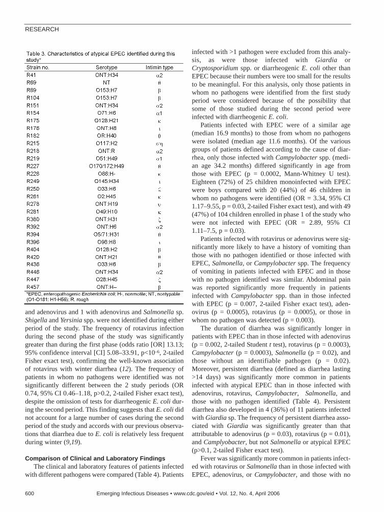
and adenovirus and 1 with adenovirus and Salmonella sp.Shigella and Yersinia spp. were not identified during eitherperiod of the study. The frequency of rotavirus infectionduring the second phase of the study was significantlygreater than during the first phase (odds ratio [OR] 13.13;95% confidence interval [CI] 5.08–33.91, p<10–6, 2-tailedFisher exact test), confirming the well-known associationof rotavirus with winter diarrhea (12). The frequency ofpatients in whom no pathogens were identified was notsignificantly different between the 2 study periods (OR0.74, 95% CI 0.46–1.18, p>0.2, 2-tailed Fisher exact test),despite the omission of tests for diarrheogenic E. coli dur-ing the second period. This finding suggests that E. coli didnot account for a large number of cases during the secondperiod of the study and accords with our previous observa-tions that diarrhea due to E. coli is relatively less frequentduring winter (9,19).
Comparison of Clinical and Laboratory FindingsThe clinical and laboratory features of patients infected
with different pathogens were compared (Table 4). Patients
infected with >1 pathogen were excluded from this analy-sis, as were those infected with Giardia orCryptosporidium spp. or diarrheogenic E. coli other thanEPEC because their numbers were too small for the resultsto be meaningful. For this analysis, only those patients inwhom no pathogens were identified from the first studyperiod were considered because of the possibility thatsome of those studied during the second period wereinfected with diarrheogenic E. coli.
Patients infected with EPEC were of a similar age(median 16.9 months) to those from whom no pathogenswere isolated (median age 11.6 months). Of the variousgroups of patients defined according to the cause of diar-rhea, only those infected with Campylobacter spp. (medi-an age 34.2 months) differed significantly in age fromthose with EPEC (p = 0.0002, Mann-Whitney U test).Eighteen (72%) of 25 children monoinfected with EPECwere boys compared with 20 (44%) of 46 children inwhom no pathogens were identified (OR = 3.34, 95% CI1.17–9.55, p = 0.03, 2-tailed Fisher exact test), and with 49(47%) of 104 children enrolled in phase 1 of the study whowere not infected with EPEC (OR = 2.89, 95% CI1.11–7.5, p = 0.03).
Patients infected with rotavirus or adenovirus were sig-nificantly more likely to have a history of vomiting thanthose with no pathogen identified or those infected withEPEC, Salmonella, or Campylobacter spp. The frequencyof vomiting in patients infected with EPEC and in thosewith no pathogen identified was similar. Abdominal painwas reported significantly more frequently in patientsinfected with Campylobacter spp. than in those infectedwith EPEC (p = 0.007, 2-tailed Fisher exact test), aden-ovirus (p = 0.0005), rotavirus (p = 0.0005), or those inwhom no pathogen was detected (p = 0.003).
The duration of diarrhea was significantly longer inpatients with EPEC than in those infected with adenovirus(p = 0.002, 2-tailed Student t test), rotavirus (p = 0.0003),Campylobacter (p = 0.0003), Salmonella (p = 0.02), andthose without an identifiable pathogen (p = 0.02).Moreover, persistent diarrhea (defined as diarrhea lasting>14 days) was significantly more common in patientsinfected with atypical EPEC than in those infected withadenovirus, rotavirus, Campylobacter, Salmonella, andthose with no pathogen identified (Table 4). Persistentdiarrhea also developed in 4 (36%) of 11 patients infectedwith Giardia sp. The frequency of persistent diarrhea asso-ciated with Giardia was significantly greater than thatattributable to adenovirus (p = 0.03), rotavirus (p = 0.01),and Camplyobacter, but not Salmonella or atypical EPEC(p>0.1, 2-tailed Fisher exact test).
Fever was significantly more common in patients infect-ed with rotavirus or Salmonella than in those infected withEPEC, adenovirus, or Campylobacter, and those with no
RESEARCH
600 Emerging Infectious Diseases • www.cdc.gov/eid • Vol. 12, No. 4, April 2006
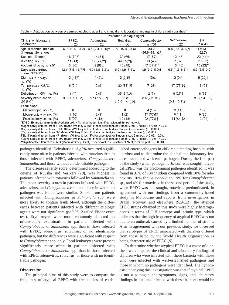
pathogen identified. Dehydration of >5% occurred signifi-cantly more often in patients infected with rotavirus than inthose infected with EPEC, adenovirus, Campylobacter,Salmonella, and those without an identifiable pathogen.
The disease severity score, determined according to thecriteria of Ruuska and Vesikari (10), was highest inpatients infected with rotavirus followed by Salmonella sp.The mean severity scores in patients infected with EPEC,adenovirus, and Campylobacter sp. and those in whom nopathogen was found were similar. Stools from patientsinfected with Campylobacter or Salmonella spp. weremore likely to contain frank blood, although the differ-ences between patients infected with different etiologicagents were not significant (p>0.05, 2-tailed Fisher exacttest). Erythrocytes were more commonly detected onmicroscopic examination in patients infected withCampylobacter or Salmonella spp. than in those infectedwith EPEC, adenovirus, rotavirus, or no identifiablepathogen, but the differences were significant with respectto Campylobacter spp. only. Fecal leukocytes were presentsignificantly more often in patients infected withCampylobacter or Salmonella spp. than in those infectedwith EPEC, adenovirus, rotavirus, or those with no identi-fiable pathogen.
DiscussionThe principal aims of this study were to compare the
frequency of atypical EPEC with frequencies of estab-
lished enteropathogens in children attending hospital withdiarrhea and to determine the clinical and laboratory fea-tures associated with each pathogen. During the first partof the study (when pathogenic E. coli was sought), atypi-cal EPEC was the predominant pathogen identified; it wasfound in 31% of 134 children compared with 10% for ade-novirus, 10% for Salmonella sp., 9% for Campylobactersp., and 4% for rotavirus. In the second period of the study,when EPEC was not sought, rotavirus predominated. Inagreement with our findings from a community-basedstudy in Melbourne and reports from investigators inBrazil, Norway, and elsewhere (9,20,21), the atypicalEPEC strains obtained in this study were highly heteroge-neous in terms of O:H serotype and intimin type, whichindicates that the high frequency of atypical EPEC was notdue to an outbreak caused by a limited number of strains.Also in agreement with our previous study, we observedthat serotypes of EPEC associated with diarrhea differedfrom those listed by the World Health Organization asbeing characteristic of EPEC (9).
To determine whether atypical EPEC is a cause of diar-rhea, we compared the clinical and laboratory findings ofchildren who were infected with these bacteria with thosewho were infected with well-established pathogens andthose in whom no pathogens were identified. The hypoth-esis underlying this investigation was that if atypical EPECis not a pathogen, the symptoms, signs, and laboratoryfindings in patients infected with these bacteria would be
Atypical Enteropathogenic Escherichia coli Infection
Emerging Infectious Diseases • www.cdc.gov/eid • Vol. 12, No. 4, April 2006 601

the same as those in patients in whom no pathogens werefound. The results showed that diarrhea attributable toatypical EPEC was significantly more common in boysand that it persisted significantly longer than diarrhea inpatients without an identifiable pathogen or in those infect-ed with adenovirus, rotavirus, Campylobacter spp., orSalmonella sp. This study also showed that infection withatypical EPEC generally occurred in children <2 years ofage, with 72% <24 months of age compared with 55% forthe first study group as a whole (OR 3.0, 95% CI1.17–7.85, p = 0.03, 2-tailed Fisher exact test). Infectionwith EPEC was associated with vomiting in ≈50% ofpatients, was generally not accompanied by fever, abdom-inal pain, or dehydration, and was not characterized byfecal blood or leukocytes, indicating that it was not inflam-matory in nature. The reason for the higher frequency ofatypical EPEC in boys is not known but confirms ourunpublished observations from a community-based study,in which 55 isolates were obtained from 338 male patients,and 34 were obtained from 358 female patients (OR 1.85,95% CI 1.17–2.92, p = 0.009, 2-tailed Fisher exact test).
The validity of the clinical and laboratory assessmentsperformed in this study was indicated by the confirmationof the well-known associations of specific pathogens withparticular parameters: younger age of children infectedwith EPEC and viruses than those infected withCampylobacter or Salmonella spp.; rotavirus andSalmonella infections with fever; rotavirus with dehydra-tion and an overall greater severity of disease;Campylobacter sp. with fecal blood; and Campylobacterand Salmonella spp. with fecal leukocytes (22).
Persistent diarrhea (lasting more then 14 days) eventu-ally develops in a substantial proportion of children withacute infectious gastroenteritis and may become chronic,leading to malabsorption, failure to thrive, and malnutri-tion (23). A wide range of infectious agents has been impli-cated in the cause of persistent diarrhea, including viruses,in particular rotavirus; protozoa, such as Giardia andCryptosporidium spp., and bacteria, including E. coli(23,24). In most cases, however, laboratory investigationof children with persistent diarrhea fails to yield an identi-fiable cause. The findings of this study suggest that a num-ber of these cases may be caused by infection with atypicalEPEC, which is seldom sought in these patients.
Despite the persuasive evidence of a volunteer studyand reports of outbreaks of diarrhea attributed to atypicalEPEC (25,26), the role of atypical EPEC in disease is con-troversial. In several reports, however, from countries asdiverse as Iran, Norway, Peru, Poland, South Africa, theUnited States, and the United Kingdom (20,27–32), aswell as Australia (9,33), atypical EPEC strains have beenidentified in children with acute diarrheal disease. AtypicalEPEC has also previously been reported in association
with prolonged diarrhea (34). For example, Hill et al. (35)reported that of 26 children infected with EPEC requiringhospital admission for acute diarrhea, life-threatening,chronic symptoms developed in 6 (23%). Five of these 6children were infected with EPEC of serogroups O114 orO128, which frequently do not produce Bfp (14,36).Notwithstanding these previous reports, however, the cur-rent study is the first to characterize the illness caused byatypical EPEC in a systematic way and to compare the fea-tures of atypical EPEC infection with those of other etio-logic agents of diarrhea.
The reasons why persistent diarrhea develops more fre-quently in children infected with atypical EPEC than inthose infected with adenovirus, rotavirus, Campylobacteror Salmonella spp. are not known. In a recent study,Mellmann et al. (37) found that only 12 (<9%) of 137patients who were infected with eae-positive EHEC strainswhen investigated within 14 days of the onset of diarrhearemained culture-positive when retested 3–16 days later,compared with all 5 patients who were initially infectedwith eae-positive, stx-negative E. coli (i.e., atypical EPEC)(OR 110.4, 95% CI 5.8–2117.6, p<0.0001, 2-tailed Fisherexact test). These findings indicate that atypical EPECmay have an innate propensity to persist longer in theintestine than varieties of E. coli which cause diarrhea thatis more transient in nature. EPEC adheres tightly to epithe-lial cells and disrupts normal cellular processes (38), andevidence suggests that atypical EPEC may retard apopto-sis of intestinal epithelial cells (39), possibly because ofthe lack of Bfp (40). These features may favor prolongedintestinal colonization by atypical EPEC compared withother intestinal pathogens. Although disease due to atypi-cal EPEC was mild and generally not associated withdehydration, its importance lies in its association with pro-longed diarrhea, a major contributor to childhood illness,especially in developing countries. Our findings also sug-gest that interventions targeted towards atypical EPECmay be beneficial in managing children with prolongeddiarrhea.
AcknowledgmentsWe are grateful to to K.A. Bettelheim and the staff of the
diagnostic microbiology and virology laboratories at the RoyalChildren’s Hospital for their assistance.
This study was supported by grants to R.R.B. from theAustralian National Health and Medical Research Council andthe Murdoch Children’s Research Institute.
Dr Nguyen is head of pediatric infectious diseases at AnGiang General Hospital in Long Xuyen City, An Giang, Vietnam.His major research interests are viral infections and the develop-ment of methods for the rapid diagnosis of infectious diseases.
RESEARCH
602 Emerging Infectious Diseases • www.cdc.gov/eid • Vol. 12, No. 4, April 2006

References
1. Nataro JP, Kaper JB. Diarrheagenic Escherichia coli. Clin MicrobiolRev. 1998;11:142–201.
2. Robins-Browne RM, Hartland EL. Escherichia coli as a cause ofdiarrhea. J Gastroenterol Hepatol. 2002;17:467–75.
3. Robins-Browne RM. Traditional enteropathogenic Escherichia coliof infantile diarrhea. Rev Infect Dis. 1987;9:28–53.
4. Trabulsi LR, Keller R, Gomes TAT. Typical and atypical enteropath-ogenic Escherichia coli. Emerg Infect Dis. 2002;8:508–3.
5. Bieber D, Ramer SW, Wu CY, Murray WJ, Tobe T, Fernandez R, etal. Type IV pili, transient bacterial aggregates, and virulence ofenteropathogenic Escherichia coli. Science. 1998;280:2114–8.
6. Frankel G, Phillips AD, Rosenshine I, Dougan G, Kaper JB, KnuttonS. Enteropathogenic and enterohaemorrhagic Escherichia coli: moresubversive elements. Mol Microbiol. 1998;30:911–21.
7. Gomez-Duarte OG, Kaper JB. A plasmid-encoded regulatory regionactivates chromosomal eaeA expression in enteropathogenicEscherichia coli. Infect Immun. 1995;63:1767–76.
8. Hellard ME, Sinclair MI, Forbes AB, Fairley CK. A randomized,blinded, controlled trial investigating the gastrointestinal healtheffects of drinking water quality. Environ Health Perspect.2001;109:773–8.
9. Robins-Browne RM, Bordun A-M, Tauschek M, Bennett-Wood V,Russell J, Oppedisano F, et al. Atypical enteropathogenic Escherichiacoli: a leading cause of community-acquired gastroenteritis inMelbourne, Australia. Emerg Infect Dis. 2004;10:1797–805.
10. Ruuska T, Vesikari T. Rotavirus disease in Finnish children: use ofnumerical scores for clinical severity of diarrhoeal episodes. Scand JInfect Dis. 1990;22:259–67.
11. Garcia LS. Diagnostic medical parasitology, 4th ed. Washington:ASM Press; 2001.
12. Barnes GL, Uren E, Stevens KB, Bishop RF. Etiology of acute gas-troenteritis in hospitalized children in Melbourne, Australia, fromApril 1980 to March 1993. J Clin Microbiol. 1998;36:133–8.
13. Vial P, Robins-Browne R, Lior H, Prado V, Kaper JB, Nataro JP, et al.Characterization of enteroadherent-aggregative Escherichia coli, aputative agent of diarrheal disease. J Infect Dis. 1988;158:70–9.
14. Levine MM, Nataro JP, Karch H, Baldini MM, Kaper JB, Black RE,et al. The diarrheal response of humans to some classic serotypes ofenteropathogenic Escherichia coli is dependent on a plasmid encod-ing an enteroadhesiveness factor. J Infect Dis. 1985;152:550–9.
15. Wood PK, Morris JG Jr, Small PLC, Sethabutr O, Toledo MRF,Trabulsi L, et al. Comparison of DNA probes and the Sereny test foridentification of invasive Shigella and Escherichia coli strains. J ClinMicrobiol. 1986;24:498–500.
16. Satterwhite TK, Evans DG, DuPont HL, Evans DJ Jr. Role ofEscherichia coli colonization factor antigen in acute diarrhoea.Lancet. 1978;ii(8082):181–4.
17. Perna NT, Plunkett G, Burland V, Mau B, Glasner JD, Rose DJ, et al.Genome sequence of enterohaemorrhagic Escherichia coli O157:H7.Nature. 2001;409:529–33.
18. Bettelheim KA, Thompson CJ. New method of serotypingEscherichia coli: implementation and verification. J Clin Microbiol.1987;25:781–6.
19. Robins-Browne RM. Seasonal and racial incidence of infantile gas-troenteritis in South Africa. Am J Epidemiol. 1984;119:350–5.
20. Afset JE, Bergh K, Bevanger L. High prevalence of atypicalenteropathogenic Escherichia coli (EPEC) in Norwegian childrenwith diarrhoea. J Med Microbiol. 2003;52:1015–9.
21. Vieira MA, Andrade JR, Trabulsi LR, Rosa AC, Dias AM, Ramos SR,et al. Phenotypic and genotypic characteristics of Escherichia colistrains of non-enteropathogenic E. coli (EPEC) serogroups that carryEAE and lack the EPEC adherence factor and Shiga toxin DNA probesequences. J Infect Dis. 2001;183:762–72.
22. Blaser MJ, Smith PD, Ravdin JI, Greenberg HB, Guerrant RL.Infections of the gastrointestinal tract. 2nd ed. New York: RavenPress; 2002.
23. Walker-Smith JA. Post-infective diarrhoea. Curr Opin Infect Dis.2001;14:567–71.
24. Nataro JP, Sears CL. Infectious causes of persistent diarrhea. PediatrInfect Dis J. 2001;20:195–6.
25. Viljanen MK, Peltola T, Junnila SY, Olkkonen L, Jarvinen H, KuistilaM, et al. Outbreak of diarrhoea due to Escherichia coli O111:B4 inschoolchildren and adults: association of Vi antigen-like reactivity.Lancet. 1990;336:831–4.
26. Yatsuyanagi J, Saito S, Miyajima Y, Amano K, Enomoto K.Characterization of atypical enteropathogenic Escherichia coli strainsharboring the astA gene that were associated with a waterborne out-break of diarrhea in Japan. J Clin Microbiol. 2003;41:2033–9.
27. Bokete TN, Whittam TS, Wilson RA, Clausen CR, O’Callahan CM,Moseley SL, et al. Genetic and phenotypic analysis of Escherichiacoli with enteropathogenic characteristics isolated from Seattle chil-dren. J Infect Dis. 1997;175:1382–9.
28. Bouzari S, Jafari MN, Shokouhi F, Parsi M, Jafari A. Virulence-relat-ed DNA sequences and adherence patterns in strains of enteropatho-genic Escherichia coli. FEMS Microbiol Lett. 2000;185:89–93.
29. Galane PM, Le Roux M. Molecular epidemiology of Escherichia coliisolated from young South African children with diarrhoeal diseases.J Health Popul Nutr. 2001;19:31–8.
30. Knutton S, Shaw R, Phillips AD, Smith HR, Willshaw GA, Watson P,et al. Phenotypic and genetic analysis of diarrhea-associatedEscherichia coli isolated from children in the United Kingdom. JPediatr Gastroenterol Nutr. 2001;33:32–40.
31. Nataro JP, Baldini MM, Kaper JB, Black RE, Bravo N, Levine MM.Detection of an adherence factor of enteropathogenic Escherichiacoli with a DNA probe. J Infect Dis. 1985;152:560–5.
32. Paciorek J. Virulence properties of Escherichia coli faecal strains iso-lated in Poland from healthy children and strains belonging toserogroups O18, O26, O44, O86, O126 and O127 isolated from chil-dren with diarrhoea. J Med Microbiol. 2002;51:548–56.
33. Kukuruzovic R, Robins-Browne RM, Anstey NM, Brewster DR.Enteric pathogens, intestinal permeability and nitric oxide productionin acute gastroenteritis. Pediatr Infect Dis J. 2002;21:730–9.
34. Afset JE, Bevanger L, Romundstad P, Bergh K. Association of atyp-ical enteropathogenic Escherichia coli (EPEC) with prolonged diar-rhoea. J Med Microbiol. 2004;53:1137–44.
35. Hill SM, Phillips AD, Walker-Smith JA. EnteropathogenicEscherichia coli and life threatening chronic diarrhoea. Gut.1991;32:154–8.
36. Scotland SM, Smith HR, Rowe B. Escherichia coli O128 strains frominfants with diarrhea commonly show localized adhesion and positiv-ity in the fluorescent-actin staining test but do not hybridize with anenteropathogenic E. coli probe. Infect Immun. 1991;59:1569–71.
37. Mellmann A, Bielaszewska M, Zimmerhackl LB, Prager R, HarmsenD, Tschäpe H, et al. Enterohemorrhagic Escherichia coli in humaninfection: in vivo evolution of a bacterial pathogen. Clin Infect Dis.2005;41:785–92.
38. Chen HD, Frankel G. Enteropathogenic Escherichia coli: unravellingpathogenesis. FEMS Microbiol Rev. 2005;29:83–98.
39. Heczko U, Carthy CM, O’Brien BA, Finlay BB. Decreased apoptosisin the ileum and ileal Peyer’s patches: a feature after infection withrabbit enteropathogenic Escherichia coli O103. Infect Immun.2001;69:4580–9.
40. Melo AR, Lasunskaia EB, de Almeida CM, Schriefer A, Kipnis TL,as da SW. Expression of the virulence factor, BfpA, by enteropatho-genic Escherichia coli is essential for apoptosis signalling but not forNF-kappaB activation in host cells. Scand J Immunol.2005;61:511–9.
Address for correspondence: R.M. Robins-Browne, Department ofMicrobiology and Immunology, University of Melbourne, Melbourne,Victoria 3010, Australia; fax: 61-3-8344-8276; email: [email protected]
Atypical Enteropathogenic Escherichia coli Infection
Emerging Infectious Diseases • www.cdc.gov/eid • Vol. 12, No. 4, April 2006 603
We examined the degree of host specialization of dif-ferent strains of Borrelia burgdorferi, the tickbornepathogen that causes Lyme borreliosis in the northeasternUnited States. We first assessed the genetic populationstructures of B. burgdorferi in ticks obtained from differentmammalian host species and in questing ticks sampled ina woodland ecosystem in Connecticut. By comparing thepatterns found in our study with data from another cross-sectional study, we demonstrate that B. burgdorferi is ageneralist microparasite and conclude that efficient cross-species transmission of B. burgdorferi is a key feature thathas allowed the rapid spread of Lyme borreliosis across thenortheastern United States.
The evolution of specialization remains a major problemin ecology and evolutionary biology; why some species
are generalists and others are specialists is not resolved(1,2). Like all organisms, parasites have evolved to differ-ent levels of ecologic specialization (3–5). The level of hostspecialization of parasites is a key issue in infectious dis-ease research because patterns of cross-species transmis-sion affect parasite dispersal and can facilitate epidemics.West Nile virus is a recent example illustrating that the uti-lization of many highly mobile host species can enable apathogen to disperse across an entire continent within a fewyears (6). Multihost parasites are usually considered to begeneralists; however, this is not universally true, and sever-al examples exist in which generalist parasites are struc-tured into subpopulations that are host specialized (7).Theory predicts that natural selection favors host special-ization if hosts are abundant and predictable, whereas gen-eralist strategies evolve if hosts are erratic (8).
Borrelia burgdorferi, the spirochetal agent of Lymeborreliosis (LB) in the United States, is a tickborne zoonot-ic pathogen that infects an expansive range of vertebrate
species, involving diverse mammalian and avian hosts(9–14). For this reason, it has been suggested B. burgdor-feri is likely less specialized than the other genospeciesthat cause LB in Eurasia (15–18). Several loci of B.burgdorferi are polymorphic (19), and balancing selectionseems to maintain the bacterium’s diversity (20). Given thepronounced strain structure of this bacterial species, natu-ral selection possibly has driven B. burgdorferi towardshost specialization, and different spirochete strains exploitdifferent sets of vertebrate hosts (4,13).
The issue of vertebrate host specialization of B.burgdorferi is of substantial public health importance.Since the reemergence of LB 3 decades ago, the diseasehas been spreading across the entire northeastern UnitedStates and beyond (21,22). A condition necessary for thisdispersal has been the geographic expansion of its princi-pal and generalist tick vector, Ixodes scapularis. Thisexpansion is believed to be driven by large-scale reforesta-tion and an explosive growth of deer populations (21).Deer, however, do not contribute directly to the dispersalof B. burgdorferi (23). Only hosts that can infect ticksaffect spirochete migration. If B. burgdorferi were hostspecialized, the strains of this microparasite would migratedifferentially, resulting in geographic structuring of thispathogen. Unrestricted cross-species transmission, in con-trast, would generate a spatially uniform population struc-ture of B. burgdorferi and substantially facilitate itsdispersal. Information on the level of host specialization ofthis multihost pathogen is required to understand the pat-terns and mechanisms of the current spread of LB.
We examined the level of host specialization of B.burgdorferi in the northeastern United States by using a
Epidemic Spread of LymeBorreliosis, Northeastern
United StatesKlára Hanincová,*1 Klaus Kurtenbach,*2 Maria Diuk-Wasser,* Brandon Brei,* and Durland Fish*
RESEARCH
604 Emerging Infectious Diseases • www.cdc.gov/eid • Vol. 12, No. 4, April 2006
*Yale University, New Haven, Connecticut, USA
1Current affiliation: New York Medical College, Valhalla, New York,USA2Current affiliation: University of Bath, Bath, United Kingdom
comparative approach. We first assessed the genetic popu-lation structures of B. burgdorferi in ticks obtained fromdifferent mammalian host species and in questing tickssampled in a woodland ecosystem at Lake Gaillard,Branford, Connecticut. By comparing the patterns in ourstudy with data from another cross-sectional study carriedout in a similiar ecosystem in Millbrook, New York (13),we aimed to capture patterns of cross-species transmissionand to identify the niche breadth of the various genotypesof B. burgdorferi.
Materials and Methods
Mammal SamplingThe fieldwork was carried out at Lake Gaillard
(41°34′N, 72°77′W), Connecticut, as described previously(24). Mammals were captured alive at 2-week intervalsfrom early June until late August in 2002 and until mid-September in 2003. All trapping and handling procedureswere approved by the Yale University Institutional AnimalCare and Utilization Committee (Study Protocol 07596).Small mammals were trapped for 23 days/nights (432 trapnights) using Sherman (Tallahassee, FL, USA) traps. Inaddition, Pitfall traps were set up for 14 days/nights (98trap nights) in 2003. Medium-sized mammals were cap-tured for 27 days/nights (820 trap nights) and 25days/nights (724 trap nights) using Tomahawk(Tomahawk, WI, USA) traps no. 205 and no. 207, respec-tively. All captured mammals were housed over pans ofwater for 72 hours to recover engorged ticks. Ticks wereallowed to molt to the next developmental stage, deter-mined to species, and stored in 70% ethanol. Mammalswere marked, sexed, and measured. Before handling,mammals were anesthetized with ketamine hydrochlorideor a combination of ketamine hydrochloride and xylazine.After captivity, mammals were released at their originallocation.
Host-seeking TicksQuesting I. scapularis nymphs were collected over the
same period and in the same area where the mammals werecaptured by dragging the vegetation with 1–m2 drag cloths.Collected ticks were preserved in 70% ethanol.
DNA Extraction and PCRDNA was extracted from ticks according to a DNeasy
Tissue Kit protocol (Qiagen, Valencia, CA, USA) asdescribed previously (25). Ticks were screened for B.burgdorferi DNA by real-time Taqman polymerase chainreaction (PCR) targeting the 16S rDNA of B. burgdorferi(24). Positive samples were then subjected to a nested PCRamplifying a fragment of the rrs (16S)–rrl (23S) intergenicspacer of B. burgdorferi and sequenced (19).
Data AnalysisInfectivity of hosts to ticks was determined by identify-
ing B. burgdorferi in molted nymphs derived from mam-mals. Since transovarial transmission of B. burgdorferi tolarval I. scapularis has not been demonstrated, infectionsfound in molted nymphs were assumed to be acquiredfrom a host through feeding. A mammal, therefore, wasconsidered infectious to ticks if >1 nymphs that had fed, aslarva, on that mammal tested positive.
To evaluate the exposure of animals to infected nymphsfor each host species, the attachment rate of nymphs peranimal per day (RDS) was computed for each capture timepoint as RDS = A/F; A is the mean number of feeding ticksper host, and F is the average feeding time of I. scapularisnymphs which was conservatively assumed to be 5 days(26). The minimum attachment rate of nymphs per animalper season (RSS) was computed as RSS = Σ (RDS × C); C isthe number of days between capture points. The number ofnymphs infected with a genotype encountered by a hostper season (RSIS) was calculated as RSIS = IP/N × RSS; IPis the infection prevalence of a genotype in field-collectedquesting nymphs, and N is the number of nymphs tested.Since no data for May were obtained empirically, weextrapolated the data on nymphal infestation obtained atthe end of the nymphal peak activity (i.e., end of June) andapplied it to May. This provided a conservative estimate ofthe total number of infected nymphs a host encounteredover the nymphal activity season.
Statistical AnalysisDifferences in mean numbers of ticks per host were
examined by using the nonparametric Kruskal-Wallis test.Logistic regression was used to estimate the infectionprevalences in ticks or hosts and to compare them amonghost species. Presence of B. burgdorferi infection in a tickor host was the response variable in the model, and adummy variable for host species was used as the predictor.The advantage of using logistic regression models for pro-portional data is that different coding systems can beapplied to compare infection prevalence among variousgroupings of host species (e.g., mice versus other hosts).Additionally, logistic models can control for the fact thatseveral ticks were collected from the same mammal andwere not independent samples. In this analysis, a cross-sectional procedure (Stata xtlogit) was applied to controlfor the correlation among ticks collected from the samemammal (27). To test for a sample size effect on the num-ber of genotypes found in a host species, a Spearman rankcorrelation was performed between the number of geno-types and the number of mammals sampled for each hostspecies. The differences in genotype frequency distribu-tions were estimated through exact nonparametric infer-ence by the Fisher-Freeman-Halton test (Monte Carlo
Epidemic Spread of Lyme Borreliosis
Emerging Infectious Diseases • www.cdc.gov/eid • Vol. 12, No. 4, April 2006 605

testing). Pearson’s χ2 test was used to compare the propor-tions of ticks infected with different genotypes within andamong host species. Data were analyzed with Stata, ver-sion 8, (Stata Corporation, College Station, TX, USA) andStataXact, version 6, (Cytel, San Diego, CA, USA).
Results
Mammal TrappingSampling over 2 years yielded 403 captures that includ-
ed 222 individual mammals, representing 9 mammalianspecies of 6 families (Muridae, Soricidae, Sciuridae,Mustelidae, Procyonidae, and Didelphiidae) belonging to 4orders (Rodentia, Insectivora, Carnivora, and Marsupialia).Six species (white-footed mouse, pine vole, eastern chip-munk, gray squirrel, Virginia opossum, and raccoon)accounted for 98% of all mammals caught (Table 1).
Tick InfestationAltogether, 9,032 immature ticks were collected from
399 captured hosts. The most abundant tick species, I.scapularis, represented 99% (7,611 larvae and 1,373nymphs) of all ticks examined. The additional 3 species, I.texanus, Dermacentor variabilis, and Amblyomma macu-latum, comprised the remaining 1% and were omitted fromfurther analysis. The mean numbers of I. scapularis ticksper host varied significantly among mammalian speciesfor both larvae and nymphs (Table 1).
B. burgdorferi Prevalence in Host-derived TicksOf the nymphs sampled from 62 mammals as engorged
larvae, 1,117 specimens were screened for presence ofB. burgdorferi. The number of tested nymphs per host var-ied from 1 to 51, depending mainly on the number ofengorged larvae recovered. B. burgdorferi–positive tickswere obtained from all 6 mammalian species.
Infection prevalence of B. burgdorferi in animals variedsignificantly among host species (logistic regression, χ2
=14.15, p<0.01) (Table 2). Each of the 3 tested chipmunks
produced >1 infected nymphs and, therefore, this specieswas excluded from the logistic regression model, since thepresence of a zero category (noninfectious chipmunks)produced infinite odds ratios (OR), which precluded theestimation of the model. No significant differences werefound between voles, squirrels, raccoons, and opossums.Hence, these species were pooled and compared withmice. The proportion of infectious mice was significantlyhigher than that of the pooled group of other host species(logistic regression, OR 13.42, 95% confidence interval(CI) 1.63–110.41, p<0.001).
Infection prevalences of B. burgdorferi in host-derivedticks also varied significantly among host species (logisticregression, χ2 = 42.38, p<0.001) (Table 3). A considerablyhigher infection prevalence in ticks was observed for micethan for voles (logistic regression, OR 16.37, 95% CI4.73–56.69, p<0.001). On the other hand, no significantdifferences in tick infection prevalence were found amongraccoons, opossums, squirrels, and chipmunks. Therefore,data for these host species were pooled into 1 group.Infection prevalence in ticks from mice was significantlyhigher than in ticks from the pooled group (logistic regres-sion, OR 47.89, 95% CI 14.97–153.23, p<0.001), as wasinfection prevalence in ticks from voles compared to thepooled group (logistic regression, OR 2.92, 95% CI1.16–7.34, p<0.001).
Population Structure of B. burgdorferiin Host-derived Ticks
A total of 205 B. burgdorferi infections in nymphsobtained from mammals as engorged larvae could besequenced successfully. The IGS alleles were assigned topreviously identified multilocus genotypes (19), designat-ed here as genotypes 1 to 9. A total of 8 genotypes wasshown (Tables 2–4). The white-footed mouse was the onlyhost species that transmitted all 8 genotypes to ticks. Noneof the genotypes was transmitted by all host species.However, genotypes 1–5 and 7 were found in ticks collect-ed from as many as 5 host species belonging to 3 different
RESEARCH
606 Emerging Infectious Diseases • www.cdc.gov/eid • Vol. 12, No. 4, April 2006

orders (Rodentia, Carnivora, Marsupialia). Only micewere found to be infectious for genotype 6. No significantrelationship was found between the number of genotypesand the number of sampled individuals of a host species(Spearman rank correlation, rs = 0.5, p>0.05). The frequen-cy distribution of transmitted genotypes differed signifi-cantly among host species (6 × 8 Fisher-Freeman-Haltontest, Fisher statistic = 41.93; Monte Carlo p 0.05) (Table2). The average number of genotypes per infectious hostwas 2.4 (standard error [SE] = 0.3) for mice, 1.7 (SE = 0.3)for opossums, 1.7 (SE = 0.7) for raccoons, 1.5 (SE = 0.5)for squirrels, 1.3 (SE = 0.2) for voles, and 1.3 (SE = 0.3)for chipmunks.
The frequency distributions of genotypes in host-derived ticks are shown in Table 3. In most of the ticksobtained from mice, genotype 2 was identified, followedby genotype 3. Most of the ticks that had fed on voles werefound to carry genotype 1. On the other hand, genotypes 8and 4 were the most frequently detected variants in ticksobtained from chipmunks and squirrels, respectively.Genotype 5 was the most common genotype found in ticksderived from raccoons, and genotype 3 was the most fre-quent in ticks obtained from opossums.
Transmissibility of each genotype from infectiousmammals to ticks can be regarded as a fitness index ofstrains infecting hosts. The values varied significantlywithin and among host species as shown in Table 4.
B. burgdorferi in Field-collected Questing Nymphs and Exposure of Animals
A total of 178 field-collected questing nymphs werescreened for B. burgdorferi. The overall infection preva-lence was 39%. In this tick population, the same 8 geno-types as in nymphs derived from the animal pool werefound. However, significant differences in the genotypefrequency distribution between these 2 tick populationswere observed (2 × 8 Fisher-Freeman-Halton, Fisher sta-tistic = 19.93, Monte Carlo p<0.01) (Table 3). Questingnymphs were chosen to estimate the exposure of hosts toB. burgdorferi genotypes. The calculated values of expo-sure show that animals with higher nymphal burdens werefrequently exposed to >1 infected nymph. This value wasoccasionally <1, reflecting the conservative estimation ofexposure (Table 5).
Comparative Analysis of B. burgdorferiPopulation Structures
Only 1 other study has analyzed the population struc-tures of B. burgdorferi in different vertebrate host speciesin the northeastern United States (13). As in our study,transmissible infections in hosts were determined throughhost-derived ticks. In contrast, the population structures ofB. burgdorferi were measured at the outer surface proteinC (ospC) locus. However, because the ospC locus and theIGS used in our study are linked (19), the population struc-
Epidemic Spread of Lyme Borreliosis
Emerging Infectious Diseases • www.cdc.gov/eid • Vol. 12, No. 4, April 2006 607
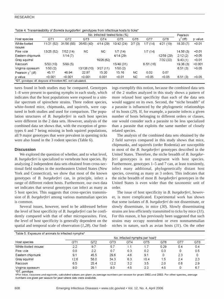
tures found in both studies may be compared. Genotypes1–8 were present in questing nymphs in each study, whichindicates that the host populations were exposed to a sim-ilar spectrum of spirochete strains. Three rodent species,white-footed mice, chipmunks, and squirrels, were cap-tured in both studies and used for comparison. The popu-lation structures of B. burgdorferi in each host specieswere different in the 2 data sets. However, analysis of thecombined data set shows that, with the exception of geno-types 6 and 7 being missing in both squirrel populations,all 9 major genotypes that were prevalent in questing tickswere also found in the 3 rodent species (Table 6).
DiscussionWe explored the question of whether, and to what level,
B. burgdorferi is specialized to vertebrate host species. Byanalyzing 2 independent data sets obtained from cross-sec-tional field studies in the northeastern United States (NewYork and Connecticut), we show that most of the knowngenotypes of B. burgdorferi can, in principle, infect arange of different rodent hosts. Furthermore, our own dataset indicates that several genotypes can infect as many as5 host species. This suggests that cross-species transmis-sion of B. burgdorferi among various mammalian speciesis common.
Several issues, however, need to be addressed beforethe level of host specificity of B. burgdorferi can be confi-dently compared with that of other microparasites. First,the level of host specificity is generally dependent on thespatial and temporal scale of observation (1,28). Our find-
ings exemplify this notion, because the combined data setsof the 2 studies analyzed in this study shows a pattern ofmore relaxed host specificity than each of the data setswould suggest on its own. Second, the “niche breadth” ofa parasite is influenced by the phylogenetic relationshipsof its hosts (29). If, for example, a parasite infects a givennumber of hosts belonging to different orders or classes,one would consider such a parasite to be less specializedthan a parasite that exploits the same number of closelyrelated species.
The analysis of the combined data sets obtained by the2 field surveys compared in this study shows that mice,chipmunks, and squirrels (order Rodentia) are susceptibleto most of the B. burgdorferi genotypes described in theUnited States. Therefore, the niche breadth of B. burgdor-feri genotypes is not congruent with host species.Furthermore, genotypes 1–5 and 7 can, at least transiently,infect many additional, phylogenetically distant hostspecies, covering as many as 3 orders. This indicates thatthe niche breadth of most B. burgdorferi genotypes in theUnited States is even wider than the taxonomic unit oforder.
The issue of host specificity in B. burgdorferi, howev-er, is more complicated. Experimental work has shownthat some isolates of B. burgdorferi do not disseminate, orslowly disseminate, in mice (30). Slowly disseminatingstrains are less efficiently transmitted to ticks by mice (31).For this reason, it has previously been suggested that suchstrains may occupy nonrodent or even nonmammalianniches in nature, such as avian hosts (31). On the other
RESEARCH
608 Emerging Infectious Diseases • www.cdc.gov/eid • Vol. 12, No. 4, April 2006
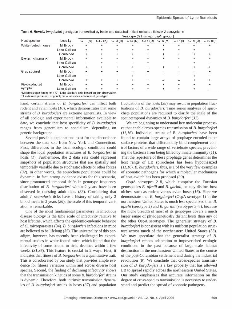
hand, certain strains of B. burgdorferi can infect bothrodent and avian hosts (10), which demonstrates that somestrains of B. burgdorferi are extreme generalists. In viewof all ecologic and experimental information available todate, we conclude that host specificity of B. burgdorferiranges from generalism to specialism, depending ongenetic background.
Several possible explanations exist for the discordancebetween the data sets from New York and Connecticut.First, differences in the local ecologic conditions couldshape the local population structures of B. burgdorferi inhosts (1). Furthermore, the 2 data sets could representsnapshots of population structures that are spatially andtemporally variable due to stochastic effects or other forces(32). In other words, the spirochete populations could bedynamic. In fact, strong evidence exists for this scenario,since pronounced temporal shifts in genotype frequencydistribution of B. burgdorferi within 2 years have beenobserved in questing adult ticks (33). Considering thatadult I. scapularis ticks have a history of taking only 2blood meals in 2 years (26), the scale of this temporal vari-ation is remarkable.
One of the most fundamental parameters in infectiousdisease biology is the time scale of infectivity relative tohost lifetime, which affects the epidemic/endemic behaviorof all microparasites (34). B. burgdorferi infections in miceare believed to be lifelong (35). The universality of this par-adigm, however, has recently been challenged by experi-mental studies in white-footed mice, which found that theinfectivity of some strains to ticks declines within a fewweeks (31,36). This feature is crucial in 2 ways. First, itindicates that fitness of B. burgdorferi is a quantitative trait.This is corroborated by our study that provides ample evi-dence for fitness variation within and across diverse hostspecies. Second, the finding of declining infectivity showsthat the transmission kinetics of some B. burgdorferi strainsis dynamic. Therefore, both intrinsic transmission dynam-ics of B. burgdorferi strains in hosts (37) and population
fluctuations of the hosts (38) may result in population fluc-tuations of B. burgdorferi. Time series analyses of spiro-chete populations are required to clarify the scale of thespatiotemporal dynamics of B. burgdorferi (32).
We are beginning to understand key molecular process-es that enable cross-species transmission of B. burgdorferi(11,16). Individual strains of B. burgdorferi have beenfound to contain large arrays of prophage-encoded outersurface proteins that differentially bind complement con-trol factors of a wide range of vertebrate species, prevent-ing the bacteria from being killed by innate immunity (11).That the repertoire of these prophage genes determines thehost range of LB spirochetes has been hypothesized(11,16). B. burgdorferi, thus, is 1 of the very few examplesof zoonotic pathogens for which a molecular mechanismof host-switch has been proposed (39).
OspA serotypes 2–8, which comprise the Eurasiangenospecies B. afzelii and B. garinii, occupy distinct hostniches, such as rodent versus avian hosts (16). Here wedemonstrate that B. burgdorferi (OspA serotype 1) in thenortheastern United States is much less specialized than B.afzelii (serotype 2) and B. garinii (serotypes 3–8), becausethe niche breadth of most of its genotypes covers a muchlarger range of phylogenetically distant hosts than any ofthe other OspA serotypes. The generalist strategy of B.burgdorferi is consistent with its uniform population struc-ture across much of the northeastern United States (33).We may speculate that the generalist strategy of B.burgdorferi echoes adaptation to impoverished ecologicconditions in the past because of large-scale habitatdestruction in the northeastern United States in the courseof the post-Columbian settlement and during the industrialrevolution (8). We conclude that cross-species transmis-sion of B. burgdorferi is a key property that has allowedLB to spread rapidly across the northeastern United States.Our study emphasizes that accurate information on thedegree of cross-species transmission is necessary to under-stand and predict the spread of zoonotic pathogens.
Epidemic Spread of Lyme Borreliosis
Emerging Infectious Diseases • www.cdc.gov/eid • Vol. 12, No. 4, April 2006 609
AcknowledgementsWe thank A.G. Barbour and J. Bunikis for providing us with
the opportunity to carry out the molecular biology in their labo-ratory at University of California at Irvine; B. Gray, A. Conway,M. O’Connell, M. Papero, H. Mattaous and L. Scheibani for tech-nical assistance; A. Gatewood, H. Brown, G. Margos and J. Tsaofor helpful comments; and South Central Connecticut RegionalWater Authority for use of its property.
This research was supported by grants from the NationalInstitutes of Health R01-AR41511 and R01-AI37248, USDepartment of Agriculture/Agriculture Research ServiceCooperative Agreements 58-0790-7-073 and 58-079-5-068, and agrant from the G. Harold and Leila Y. Mathers CharitableFoundation.
Dr Hanincová is a postdoctoral fellow at New York MedicalCollege. Her interests include microbial pathogenesis andecology of vectorborne diseases.
References
1. Fox LR, Morrow PA. Specialization—species property or local phe-nomenon. Science. 1981;211:887–93.
2. Timms R, Read AF. What makes a specialist special? Trends EcolEvol. 1999;14:333–4.
3. Kawecki TJ. Red queen meets Santa Rosalia: arms races and the evo-lution of host specialization in organisms with parasitic lifestyles. AmNat. 1998;52:635–51.
4. McCoy KD, Boulinier T, Tirard C, Michalakis Y. Host specificity ofa generalist parasite: genetic evidence of sympatric host races in theseabird tick Ixodes uriae. J Evol Biol. 2001;4:395–405.
5. Krasnov BR, Poulin R, Shenbrot GI, Mouillot D, Khokhlova IS.Geographical variation in host specificity of fleas (Siphonaptera) par-asitic on small mammals: the influence of phylogeny and local envi-ronmental conditions. Ecography. 2004;27:787–97.
6. Granwehr BP, Lillibridge KM, Higgs S, Mason PW, Aronson JF,Campbell GA, et al. West Nile virus: where are we now? LancetInfect Dis. 2004;4:547–56.
7. McCoy KD. Sympatric speciation in parasites—what is sympatry?Trends Parasitol. 2003;19:400–4.
8. Combes C. Fitness of parasites: pathology and selection. Int JParasitol. 1997;27:1–10.
9. Fish D, Daniels TJ. The role of medium-sized mammals as reservoirsof Borrelia burgdorferi in southern New York. J Wildl Dis.1990;26:339–45.
10. Richter D, Spielman A, Komar N, Matuschka FR. Competence ofAmerican robins as reservoir hosts for Lyme disease spirochetes.Emerg Infect Dis. 2000;6:133–8.
11. Stevenson B, El-Hage N, Hines MA, Miller JC, Babb K. Differentialbinding of host complement inhibitor factor H by Borrelia burgdor-feri erp surface proteins: a possible mechanism underlying the expan-sive host range of Lyme disease spirochetes. Infect Immun.2002;70:491–7.
12. LoGiudice K, Ostfeld RS, Schmidt KA, Keesing F. The ecology ofinfectious disease: effects of host diversity and community composi-tion on Lyme disease risk. Proc Natl Acad Sci U S A. 2003;100:567–71.
13. Brisson D, Dykhuizen DE. ospC diversity in Borrelia burgdorferi:different hosts are different niches. Genetics. 2004;168:713–22.
14. Ginsberg HS, Buckley PA, Balmforth MG, Zhioua E, Mitra S,Buckley FG. Reservoir competence of native North American birdsfor the Lyme disease spirochete, Borrelia burgdorferi. J MedEntomol. 2005;42:445–9.
15. Kurtenbach K, Sewell HS, Ogden NH, Randolph SE, Nuttall PA.Serum complement sensitivity as a key factor in Lyme disease ecolo-gy. Infect Immun. 1998;66:1248–51.
16. Kurtenbach K, De Michelis S, Etti S, Schäfer SM, Sewell HS, BradeV, Kraiczy P. Host association of Borrelia burgdorferi sensu lato—the key role of host complement. Trends Microbiol. 2002;10:74–9.
17. Hanincová K, Schäfer SM, Etti S, Sewell HS, Taragelová V, Ziak D,et al. Association of Borrelia afzelii with rodents in Europe.Parasitology. 2003;126:11–20.
18. Hanincová K, Taragelová V, Koci J, Schäfer SM, Hails R, UllmannAJ. Association of Borrelia garinii and B. valaisiana with songbirdsin Slovakia. Appl Environ Microbiol. 2003;69:2825–30.
19. Bunikis J, Garpmo U, Tsao J, Berglund J, Fish D, Barbour AG.Sequence typing reveals extensive strain diversity of the Lyme borre-liosis agents Borrelia burgdorferi in North America and Borreliaafzelii in Europe. Microbiology. 2004;150:1741–55.
20. Dykhuizen DE, Baranton G. The implications of a low rate of hori-zontal transfer in Borrelia. Trends Microbiol. 2001;9:344–50.
21. Spielman A. The emergence of Lyme disease and human babesiosisin a changing environment. Ann N Y Acad Sci. 1994;740:146–56.
22. Centers for Disease Control and Prevention. Lyme disease—UnitedStates, 2001–2002. MMWR Morb Mortal Wkly Rep. 2004;53:365–9.
23. Telford SR 3rd, Mather TN, Moore SI, Wilson ML, Spielman A.Incompetence of deer as reservoirs of the Lyme disease spirochete.Am J Trop Med Hyg. 1988;39:105–9.
24. Tsao JI, Wootton JT, Bunikis J, Luna MG, Fish D, Barbour AG. Anecological approach to preventing human infection: vaccinating wildmouse reservoirs intervenes in the Lyme disease cycle. Proc NatlAcad Sci U S A. 2004;101:18159–64.
25. Beati L, Keirans JE. Analysis of the systematic relationships amongticks of the genera Rhipicephalus and Boophilus (Acari:Ixodidae)based on mitochondrial 12S ribosomal DNA gene sequences andmorphological characters. J Parasitol. 2001;87:32–48.
26. Fish D. Population ecology of Ixodes dammini. In: Ginsberg HS, edi-tor. Ecology and environmental management of Lyme disease. NewBrunswick (NJ): Rutgers University Press; 1993. p. 25–41.
27. Hardin JW, Hilbe JM. Generalized estimating equations. Boca Raton(FL): Chapman and Hall/CRC; 2003. p. 240.
28. Gaston KJ, Blackburn TM, Lawton JH. Interspecific abundancerange size relationships: An appraisal of mechanisms. J Anim Ecol.1997;66:579–601.
29. Poulin R, Mouillot D. Parasite specialization from a phylogeneticperspective: a new index of host specificity. Parasitology.2003;126:473–80.
30. Wang G, Ojaimi C, Wu H, Saksenberg V, Iyer R, Liveris D, et al.Disease severity in a murine model of Lyme borreliosis is associatedwith the genotype of the infecting Borrelia burgdorferi sensu strictostrain. J Infect Dis. 2002;186:782–91.
31. Derdaková M, Dudioak V, Brei B, Brownstein JS, Schwartz I, Fish D.Interaction and transmission of two Borrelia burgdorferi sensu stric-to strains in a tick-rodent maintenance system. Appl EnvironMicrobiol. 2004;70:6783–8.
32. Bjørnstad ON, Grenfell BT. Noisy clockwork: time series analysis ofpopulation fluctuations in animals. Science. 2001;293:638–43.
33. Qiu WG, Dykhuizen DE, Acosta MS, Luf BJ. Geographic uniformityof the Lyme disease spirochete (Borrelia burgdorferi) and its sharedhistory with tick vector (Ixodes scapularis) in the northeastern UnitedStates. Genetics. 2002;160:833–49.
34. Gog JR, Grenfell BT. Dynamics and selection of many-strainpathogens. Proc Natl Acad Sci U S A. 2002;99:17209–14.
RESEARCH
610 Emerging Infectious Diseases • www.cdc.gov/eid • Vol. 12, No. 4, April 2006
35. Donahue JG, Piesman J, Spielman A. Reservoir competence of white-footed mice for Lyme disease spirochetes. Am J Trop Med Hyg.1987; 36:92–6.
36. Lindsay LR, Barker IK, Surgeoner GA, McEwen SA, Campbell GD.Duration of Borrelia burgdorferi infectivity in white-footed mice forthe tick vector Ixodes scapularis under laboratory and field condi-tions in Ontario. J Wildl Dis. 1997;33:766–75.
37. Hofmeister EK, Glass GE, Childs JE, Persing DH. Populationdynamics of a naturally occurring heterogeneous mixture of Borreliaburgdorferi clones. Infect Immun. 1999;67:5709–16.
38. Jones CG, Ostfeld RS, Richard MP, Schauber EM, Wolff JO. Chainreactions linking acorns to gypsy moth outbreaks and Lyme diseaserisk. Science. 1998;279:1023–6.
39. Woolhouse ME, Taylor LH, Haydon DT. Population biology of mul-tihost pathogens. Science. 2001;292:1109–12.
Address for correspondence: Klára Hanincová, Department ofMicrobiology and Immunology, New York Medical College, Valhalla,NY, 10595, USA; email: [email protected]
Epidemic Spread of Lyme Borreliosis
Emerging Infectious Diseases • www.cdc.gov/eid • Vol. 12, No. 4, April 2006 611
Searchpast issues

In 2002, Canada introduced routine, mandatory HIVantibody screening for all residency applicants, includingselected children. We report screening results fromJanuary 2002 to February 2005. Thirty-six pediatric HIVcases were detected (14/100,000 applicants); 94% ofinfected children were eligible to arrive in Canada. Thirty-two of the affected children were from Africa, and maternalinfection was the main risk factor. Only 4 (11%) of the chil-dren had received antiretroviral therapy. In countries of lowHIV incidence, migration-related imported infection in chil-dren may be an emerging epidemic. The early identificationof HIV-infected immigrant women permits intervention toprevent mother-to-child HIV transmission. Routine HIVtesting as a component of the medical examination of immi-grants has national and international health policy and pro-grammatic implications.
In 2004, the Joint United Nations Programme onHIV/AIDS reported that the number of persons living
with HIV and AIDS in the world was 39.4 million, 4.9 mil-lion were newly infected with HIV, and 3.1 million haddied because of HIV/AIDS (1). Within these numbers arean estimated 2.2 million children <16 years of age who areliving with HIV/AIDS. Globally in 2003, ≈640,000 newHIV pediatric infections and 510,000 pediatric deathsoccurred. Most pediatric HIV/AIDS occurs in the develop-ing world because of mother-to-child transmission, butpediatric HIV/AIDS is also a concern in Western nations,where several strategies have been developed and imple-mented to prevent pediatric HIV infection and subsequentAIDS-related disease.
A link between HIV infection in hyperendemic zones ofthe developing world and pediatric HIV infections inWestern countries is largely undocumented (2). Irregularmigrants (those who arrive by smuggling or trafficking)and those seeking asylum in Europe represent a separate
epidemiologic pattern of HIV/AIDS introduction (3). Forregions without extensive immigration assessment pro-grams, or where most international arrivals for permanentresidency are seeking asylum or are arriving through otherirregular means, migration-associated HIV/AIDS preva-lence and the pediatric HIV/AIDS epidemic are emergingpolicy and programmatic issues. Injection drug use andsexual exploitation, particularly for women, are 2 potentialrisks associated with illegal immigration status thatincrease HIV exposure, with the potential consequence ofmother-to-child viral transmission (4).
Prearrival immigration medical screening has beenused to identify conditions such as tuberculosis, syphilis,and HIV/AIDS that could affect admission to receivingnations. Many immigrant-receiving nations in the industri-alized world now have national policies designed to betteraddress the needs of vulnerable, foreign-born migrants orto facilitate the immigration process for preferred appli-cants. Nations who either have existing medical screeningprograms or who are planning such programs are likely toidentify persons with HIV/AIDS. Decisions on screeningimmigrants for HIV infection not only have direct implica-tions for admissibility programs but also affect the need forculturally and linguistically appropriate clinical and publichealth services.
The 2002–2003 annual report of the MinisterialCouncil on HIV/AIDS in Canada “estimated that 70% ofall maternal HIV transmissions to children in Canada haveoccurred among women of African and Caribbean origin”(5). From November 1985 to June 2004, the Public HealthAgency of Canada reported notification of 56,523 positiveHIV test results (6). From 1984 to 2002, it also reported420 HIV infections in 1,584 children born to HIV-positivemothers (7).
Apart from the potential for perinatal HIV exposure,other pediatric risk factors for nonmaternal HIV acquisi-tion in the industrialized world, such as blood transfusion,tattooing, or illicit drug use, are rarely encountered or doc-umented. Strategies designed to reduce mother-to-child
Emerging Pediatric HIV EpidemicRelated to Migration
Douglas W. MacPherson,*† Militza Zencovich,‡ and Brian D. Gushulak*
RESEARCH
612 Emerging Infectious Diseases • www.cdc.gov/eid • Vol. 12, No. 4, April 2006
*Migration Health Consultants Inc., Cheltenham, Ontario, Canada;†McMaster University, Hamilton, Ontario, Canada; and‡Citizenship and Immigration Canada, Ottawa, Ontario, Canada

HIV transmission and pediatric HIV infection include HIVscreening programs for pregnant women (8), risk behaviorcounseling, recommendations for antiretroviral treatmentto prevent mother-to-child-transmission or to treat a new-born exposed to HIV at birth, conception control, andbirthing methods. These strategies have been relativelysuccessful in reducing pediatric HIV infections in mostindustrialized nations but have been less effective in devel-oping nations (9–11).
The role of population mobility between hyperendemicHIV transmission zones and countries of lower prevalenceis emerging as a contributing factor in risk for pediatricHIV infection between HIV high-prevalence and low-prevalence regions. This article describes the results of thefirst 3 years of a medical screening program for HIV anti-bodies in selected children who were applicants for resi-dency in Canada. The results of this study confirm thatmobile population dynamics between countries andregions and demographic changes in pediatric HIV/AIDSepidemiology are directly related and have consequencesfor immigration and health policy at domestic and interna-tional levels.
Methods
PopulationMandatory, routine serologic HIV testing of immi-
grants in Canada began in January 2002 as a component ofthe Canadian immigration medical examination (12,13).While routine testing was not conducted on applicants <15years of age, those identified as being at risk in the pedi-atric migrant populations were evaluated for HIV infec-tion. An enzyme-linked immunoassay (EIA) HIVscreening test for HIV-1 and HIV-2 antibodies wasrequired for all children <15 years of age who had receivedblood or blood products, had an HIV-positive mother, or inthe judgment of the assessing medical practitioner, wasnoted to have any other identified HIV infection risk fac-tor. Potential adoptees were also screened for HIV antibod-ies until a policy decision changed this requirement in late2004. Reflecting national and international legal processesof adoption, international adoptees brought to Canada byCanadian citizens or permanent residents of Canada areconsidered part of the existing family, and although theyare required to apply for Canadian citizenship, they areexempted from most immigration processes (including themedical examination to determine inadmissibility).
The HIV-tested migrant groups included children ofapplicants for permanent residence (immigrants andrefugees) and those who filed refugee or asylum claims inCanada. Immigration medical screening, including routineHIV testing for those >15 years of age is also required forsome other persons arriving in Canada, including visitors
staying >6 months from certain locations (e.g., tourists,students, and seasonal workers). Children of persons inthis group could also be referred for HIV testing if risk fac-tors were noted during the process (14).
Medical Assessment and HIV Testing ProtocolsThe guardians of all pediatric applicants were coun-
seled and consented to HIV testing. Venous blood was col-lected and transported to an approved local testing facility.HIV antibody was tested by EIA. Immunofluorescenceantibody testing and Western blot (or other approved man-ufacturer’s EIA kit) testing on another blood sample wereused as confirmatory tests on positive or indeterminateHIV antibody test results.
Data Management and Protection of Personal Information
Immigration medical screening data were provided byCitizenship and Immigration Canada. Personal data wereprotected according to national guidelines on informationprivacy (15,16). Age was calculated from the date of birthand application date on the immigration file. The 5 immi-gration categories used in the analysis, reflecting currentCanadian classifications, were economic, family, refugee(abroad), refugee claimant (in Canada), and other (17).The “other” category includes temporary resident appli-cants, such as visitors, workers, and students.
Currently, Canadian immigration policy reflects 3major goals. New immigrants are selected on the basis of1 of these 3 basic principles: reunification of families, eco-nomic ability to become successfully established inCanada, or humanitarian reasons based on displacement orpersecution. Specific components of each of these majorimmigration selection groups are further described by leg-islation and regulation (18).
Refugee claimants, known as asylum seekers inEurope, differ from refugees whose convention-definedstatus has been determined before arrival (19–21). Refugeeclaimants arrive through uncontrolled and irregular meansand complete the immigration medical assessmentprocesses entirely within Canada as part of the proceduresfor determining their refugee status. During the pastdecade, the number of refugee claimants arriving inCanada has varied from 23,000 to 43,000 claimants peryear (22). Since the implementation of the Safe ThirdCountry Agreement between Canada and the United Stateson December 29, 2004, the number of refugee claimantshas declined by ≈25% (23).
ResultsFrom January 2002 to February 2005, a total of
1,307,718 persons underwent a Canadian immigrationmedical assessment (24), including 256,970 applicants
Emerging Pediatric HIV Epidemic Related to Migration
Emerging Infectious Diseases • www.cdc.gov/eid • Vol. 12, No. 4, April 2006 613

(124,195 female, 132,775 male) <15 years of age. Thirty-six new HIV-positive diagnoses were made in the pediatricapplicant population; 18 were boys and 18 were girls.Twelve HIV-positive applicants were <1 year of age atdiagnosis, 7 were 2–4 years, 7 were 5–7 years, 2 were 8–10years, and 8 were 11–14 years. Median age for girls was 6years (range from infancy to 14 years) and for boys, 4years (range from infancy to 12 years). All HIV-infectedchildren except 4 (2 from Europe, 2 from Asia/PacificIslands) identified a country in Africa as either their par-ents’ home or their country of birth. Twenty-seven (75%)of the 36 HIV-infected children were tested because ofknown maternal HIV positivity, 6 children (17%) weretested because of adoption, 2 (6%) were tested because ofan HIV-positive sibling, and 1 (3%) was detected becauseof concurrent treatment for tuberculosis. Two girls (ages 6and 12 years) and 2 boys (ages 6 and 11 years) (11%) haddocumentation of having received highly active antiretro-viral therapy (HAART). Two of the 4 received HAART inNorth America, 1 in the United Kingdom, and 1 inEthiopia. By applicant category, 2 of the children receivingHAART were family class, 1 was a refugee claimant, and1 was an economic applicant.
HIV-positive children by immigration category andyear of testing are shown in the Table. Refugees represent-ed 26 (72%) of the 36 HIV diagnoses, family class repre-sented 7 (19%), and economic, refugee claimant, andtemporary resident applicants represented 1 case each(3%). Only 2 HIV-infected children were processed in theimmigration medical office responsible for North America,which indicates their application originated in Canada orthe United States.
Thirty-three (92%) of the 36 HIV pediatric casesoccurred in populations deemed to be eligible for admis-sion to Canada, despite medical status, on the basis ofthese application categories (26 in the refugee class and 7in the family class). Of those children found to be HIVpositive in the study period, 24 (66%) had arrived inCanada.
DiscussionIn the context of all immigration applicants, 36 HIV-
infected children were identified. During the study,256,970 applicants <15 years of age underwent medicalexaminations. This number represents a crude ratio of14/100,000 pediatric applicants from January 2002through February 2005. A ratio per tested pediatric appli-cant cannot be determined because negative serologic testresults are not recorded for this age group, and no estimateof the tested population size is possible because of varia-tions in medical examiner and adjudicator practices duringthe study. All but 4 of the pediatric HIV -positive appli-cants were originally from Africa (89%), which reflects the
relationship between population flow and the global epi-demiologic features of HIV infection and disease (25). Incontrast, domestically reported pediatric HIV cases arerare in Canada (0.02/100,000 general population).
Applicants in the refugee (26 cases) and family (7cases) categories accounted for 33 (91%) of the 36 HIVdiagnoses in this study period. The refugee and family cat-egories are exempted from provisions that can renderapplicants inadmissible because of medical reasons.Consequently, the 33 children in these groups were allmedically eligible to be admitted to Canada. When thosechildren are combined with the HIV-positive pediatricrefugee claimant who was in Canada when tested, 34(94%) of the 36 HIV-infected children detected during thefirst 3 years of the mandatory immigration screening pro-gram were medically admissible or had already arrived inCanada.
In the 2004 World Health Organization report,Canadian public health officials estimated an overall HIVprevalence ratio in Canada of 3–5 cases per 10,000 preg-nant women on the basis of pregnancy HIV screeningresults (26). Fecundity data in Canada from 1986 to 2001show that 1,197,300 children were born to Canadian-bornwomen and 337,700 children were born to immigrantwomen (1,535,000 total newborns) (27). If the estimatedHIV prevalence rate for pregnant women in Canada isapplied to the average of 102,333 newborn children peryear, an estimated 92–154 singleton births occur to at-riskpregnant women.
The actual number of documented mother-to-child HIVexposures reported in Canada has been less than that fig-ure. From 1984 to 2002, national statistics in Canada showthat 1,584 infants (an average of 88 per year) were perina-tally exposed to HIV. Of these infants, 420 have beenreported as HIV infected. An additional 120 children hadunconfirmed HIV status, including those with indetermi-nate serologic status, those who died, or those who werelost to follow-up. As a possible reflection of the effective-ness of the national perinatal HIV screening, mother-to-child transmission–prevention programs, and other factors,<4% of at-risk pregnancies in HIV-positive mothers result-ed in viral transmission to their newborns in 2001 and2002, with only 12 pediatric HIV cases reported in those 2years (0.02 cases/100,000 population per year).
As indicated in this study of immigrant applicants,more HIV-infected children are detected through selectiveimmigration medical screening in migrant pediatric popu-lations arriving or arrived in Canada. Growing global pop-ulation mobility and immigration could more than doublethe annual domestic pediatric HIV/AIDS caseload inCanada.
Despite the success of industrialized nations’ domesticmother-to-child transmission prevention programs, the
RESEARCH
614 Emerging Infectious Diseases • www.cdc.gov/eid • Vol. 12, No. 4, April 2006
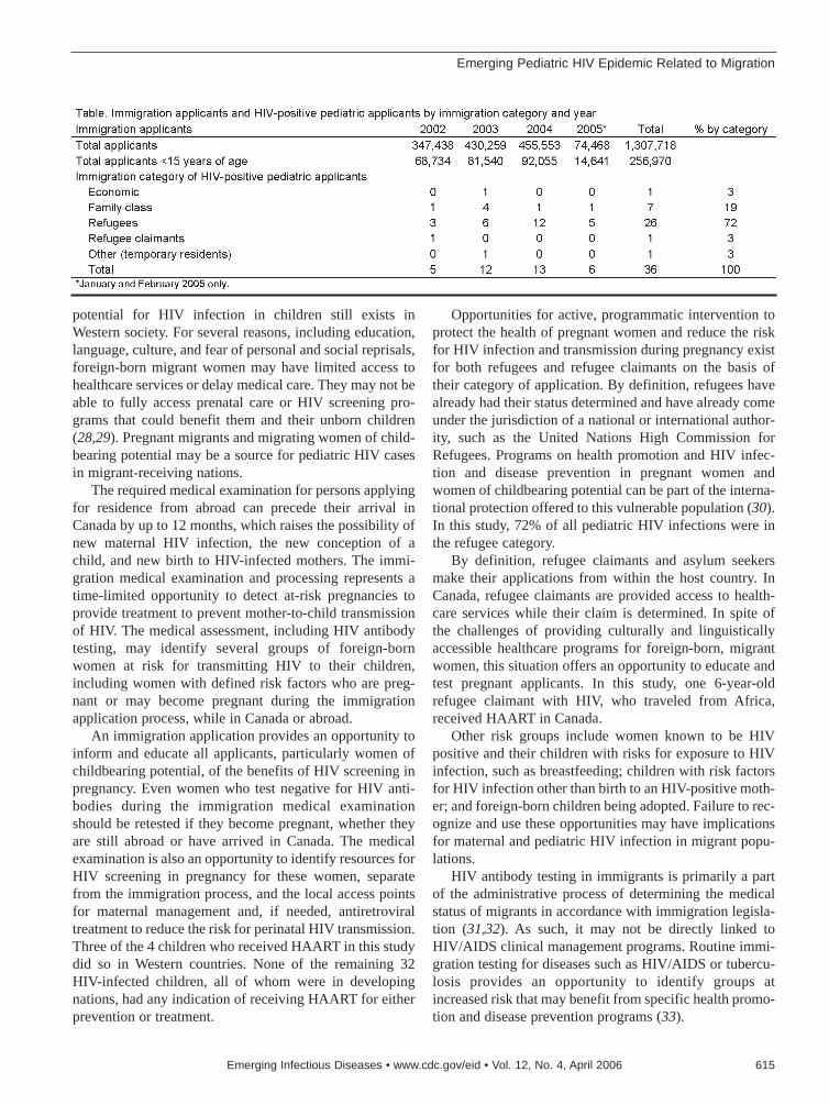
potential for HIV infection in children still exists inWestern society. For several reasons, including education,language, culture, and fear of personal and social reprisals,foreign-born migrant women may have limited access tohealthcare services or delay medical care. They may not beable to fully access prenatal care or HIV screening pro-grams that could benefit them and their unborn children(28,29). Pregnant migrants and migrating women of child-bearing potential may be a source for pediatric HIV casesin migrant-receiving nations.
The required medical examination for persons applyingfor residence from abroad can precede their arrival inCanada by up to 12 months, which raises the possibility ofnew maternal HIV infection, the new conception of achild, and new birth to HIV-infected mothers. The immi-gration medical examination and processing represents atime-limited opportunity to detect at-risk pregnancies toprovide treatment to prevent mother-to-child transmissionof HIV. The medical assessment, including HIV antibodytesting, may identify several groups of foreign-bornwomen at risk for transmitting HIV to their children,including women with defined risk factors who are preg-nant or may become pregnant during the immigrationapplication process, while in Canada or abroad.
An immigration application provides an opportunity toinform and educate all applicants, particularly women ofchildbearing potential, of the benefits of HIV screening inpregnancy. Even women who test negative for HIV anti-bodies during the immigration medical examinationshould be retested if they become pregnant, whether theyare still abroad or have arrived in Canada. The medicalexamination is also an opportunity to identify resources forHIV screening in pregnancy for these women, separatefrom the immigration process, and the local access pointsfor maternal management and, if needed, antiretroviraltreatment to reduce the risk for perinatal HIV transmission.Three of the 4 children who received HAART in this studydid so in Western countries. None of the remaining 32HIV-infected children, all of whom were in developingnations, had any indication of receiving HAART for eitherprevention or treatment.
Opportunities for active, programmatic intervention toprotect the health of pregnant women and reduce the riskfor HIV infection and transmission during pregnancy existfor both refugees and refugee claimants on the basis oftheir category of application. By definition, refugees havealready had their status determined and have already comeunder the jurisdiction of a national or international author-ity, such as the United Nations High Commission forRefugees. Programs on health promotion and HIV infec-tion and disease prevention in pregnant women andwomen of childbearing potential can be part of the interna-tional protection offered to this vulnerable population (30).In this study, 72% of all pediatric HIV infections were inthe refugee category.
By definition, refugee claimants and asylum seekersmake their applications from within the host country. InCanada, refugee claimants are provided access to health-care services while their claim is determined. In spite ofthe challenges of providing culturally and linguisticallyaccessible healthcare programs for foreign-born, migrantwomen, this situation offers an opportunity to educate andtest pregnant applicants. In this study, one 6-year-oldrefugee claimant with HIV, who traveled from Africa,received HAART in Canada.
Other risk groups include women known to be HIVpositive and their children with risks for exposure to HIVinfection, such as breastfeeding; children with risk factorsfor HIV infection other than birth to an HIV-positive moth-er; and foreign-born children being adopted. Failure to rec-ognize and use these opportunities may have implicationsfor maternal and pediatric HIV infection in migrant popu-lations.
HIV antibody testing in immigrants is primarily a partof the administrative process of determining the medicalstatus of migrants in accordance with immigration legisla-tion (31,32). As such, it may not be directly linked toHIV/AIDS clinical management programs. Routine immi-gration testing for diseases such as HIV/AIDS or tubercu-losis provides an opportunity to identify groups atincreased risk that may benefit from specific health promo-tion and disease prevention programs (33).
Emerging Pediatric HIV Epidemic Related to Migration
Emerging Infectious Diseases • www.cdc.gov/eid • Vol. 12, No. 4, April 2006 615
Given the emerging patterns of global HIV/AIDS epi-demiology and current immigration patterns, similar situa-tions could be observed over time in other locations. Aproportional shift in total and pediatric HIV cases relatedto foreign-born migrant arrivals in immigrant-receivingnations, where cases in children are less common, can beanticipated. Migrants, and the communities of newlyarrived persons that they tend to gravitate to, often havehealth needs that may differ substantially from those of thehost population. The shifting demographics of pediatricHIV infection in Canada can thus be expected to influencelocal aspects of healthcare planning and delivery. The mul-ticultural aspects of HIV infection in immigrants willaffect case management by local public health authorities,social services, clinical pediatric HIV/AIDS services, andother health and social service providers. Existing pro-grams, designed in the context of domestically acquiredinfections, may not have considered either the size of thepopulation or the culturally and linguistically diverse char-acteristics of immigrant, HIV-infected children. Otherunanticipated program effects, including policy expecta-tions from the international community on the medical,cultural, and social aspects of pediatric HIV/AIDS andimmigration should receive greater attention (34,35).
One of the potential consequences of this shared knowl-edge related to pediatric HIV and AIDS for domestic pro-gram development could be the integration of immigrationmedical programs and public health programs to preventmother-to-child transmission of HIV overseas. Only 4(11%) of 36 of HIV-infected children received specific anti-retroviral therapy, and none of the 12 HIV-positive childrenwho were <1 year of age, nor their mothers, were known tohave received perinatal HAART to prevent HIV transmis-sion (36,37). Programs and strategies designed to mitigatesome infectious disease risks in migrant populations beforearrival have already been used in some situations (38); sim-ilar rationales could be evaluated for HIV infection in thosealready involved in immigration formalities.
The analysis and opinions expressed in this article are thoseof the authors and are not to be attributed to Citizenship andImmigration Canada or the government of Canada, and they donot necessarily reflect or represent the position of any govern-ment department, agency, university, or professional society towhich the authors may belong or have belonged.
Dr MacPherson is qualified in internal medicine, medicalmicrobiology, and clinical tropical medicine. In addition to hisclinical, laboratory, and academic career in Hamilton, Canada,and McMaster University, he has consulted extensively withgovernments and international agencies on health, health emer-gencies, and migration.
References
1. United Nations Joint Programme on HIV/AIDS and World HealthOrganization. AIDS epidemic update [ISBN 92 9173390 3]. Geneva:the Organization; 2004.
2. Centers for Disease Control and Prevention. Unexplained immunod-eficiency and opportunistic infections in infants—New York, NewJersey, California. MMWR Morb Mortal Wkly Rep. 1982;31:665–7.
3. European Centre for the Epidemiological Monitoring of AIDS.HIV/AIDS surveillance in Europe. End-year report 2003–2004. 2004Nov [cited 2005 Jul 10]. Available from http://www.eurohiv.org/reports/report_70/pdf/report_eurohiv_70.pdf
4. Department of State. The link between HIV/AIDS and trafficking inpersons. Trafficking in persons report. 2005 Jun [cited 2005 Oct 10].Available from http://www.state.gov/g/tip/rls/tiprpt/2005/46611.htm
5. Public Health Agency of Canada. The federal initiative to addressHIV/AIDS in Canada. Ministerial Council on HIV/AIDS. Annualreport 2002–2003. [cited 2005 Jul 10]. Available fromhttp://www.phac-aspc.gc.ca/aids-sida/hiv_aids/federal_initiative/ministerial/annual_02.html
6. Public Health Agency of Canada. At a glance: HIV/AIDS surveil-lance to 30 June 2004. 2004 Dec 1 [cited 2005 Jul 10]. Available fromhttp: / /www.phac-aspc.gc.ca/publicat /ccdr-rmtc/04vol30/dr3023ea.html
7. Public Health Agency of Canada. HIV/AIDS epi update–May 2004.Perinatal transmission of HIV. [cited 2005 Jul 10]. Available at:http://www.phac-aspc.gc.ca/publicat/epiu-aepi/epi_update_may_04/7_e.html
8. Public Health Agency of Canada. HIV/AIDS epi update. Perinataltransmission of HIV. 2003 Apr [cited 2005 Oct 2]. Available fromhttp://www.phac-aspc.gc.ca/publicat/epiu-aepi/hiv-vih/peri_e.html
9. Mayaux MJ, Teglas JP, Blanche S, French Pediatric HIV InfectionStudy Group. Characteristics of HIV-infected women who do notreceive preventive antiretroviral therapy in the French PerinatalCohort. J Acquir Immune Defic Syndr. 2003;34:338–43.
10. Centers for Disease Control and Prevention. Prenatal HIV testing andantiretroviral prophylaxis at an urban hospital–Atlanta, Georgia,1997–2000. MMWR Morb Mortal Wkly Rep. 2004;52:1245–8.
11. MTCT programs work in a variety of ways. UNAIDS, others work-ing to help pregnant women. AIDS Alert. 2001;16:144–5.
12. Citizenship and Immigration Canada. Immigration health policy andstandards (HMP)–OP 02-04, IP 02-01, PE 02-03. Routine HIV test-ing, medical services (HMD). 2002 Jan 15.
13. Citizenship and Immigration Canada. Designated medical practition-er handbook. Section 6: conducting an immigration medical exami-nation. [cited 2006 Feb 8] Available from http://www.cic.gc.ca/english/pub/dmp%2Dhandbook/index.html
14. Citizenship and Immigration Canada. Designated countries/territorieslist. [cited 2005 Jul 10]. Available from http://www.cic.gc.ca/eng-lish/visit/dcl.html
15. Department of Justice Canada. Consolidated statutes and regulations.Privacy Act, chapter P-21. [cited 2006 Feb 8]. Available fromhttp://laws.justice.gc.ca/en/p-21/text.html
16. Department of Justice Canada. Consolidated statutes and regulations.Personal Information Protection and Electronic Documents Act.[cited 2005 Jul 10]. Available from http://laws.justice.gc.ca/en/p-8.6/91355.html
17. Citizenship and Immigration Canada. Immigrants and RefugeeProtection Act. [cited 2005 Oct 2]. Available from http://www.cic.gc.ca/english/irpa/
18. Citizenship and Immigration Canada. Policy and program manuals.OP 1 procedures. [cited 2005 Oct 10]. Available fromhttp://www.cic.gc.ca/manuals-guides/english/op/op01e.pdf
RESEARCH
616 Emerging Infectious Diseases • www.cdc.gov/eid • Vol. 12, No. 4, April 2006
19. United Nations High Commission for Refugees. Convention and pro-tocol relating to the status of refugees. 1951 [cited 2005 Jul 10].Available at: http://www.unhcr.ch/cgi-bin/texis/vtx/home?page=PROTECT&id=3c0762ea4&ID=3c0762ea4&PUBLISHER=TWO
20. Gushulak BD, MacPherson DW. Health issues associated with thesmuggling and trafficking of migrants. J Immigr Health.2000;2:67–78.
21. MacPherson DW, Gushulak BD. Global migration perspectives.Irregular migration and health. [cited 2005 Jul 10]. Available fromhttp://www.gcim.org/gmp/Global%20Migration%20Perspectives%20No%207.pdf
22. Citizenship and Immigration Canada. First statistics underCanada–U.S. Safe Third Country agreement show decline in refugeeclaimants. [cited 2005 Oct 2]. Available from http://www.cic.gc.ca/english/policy/safe%2Dthird%2Dstats.html
23. Citizenship and Immigration Canada. Facts and figures 2004.Immigration overview: temporary residents. [cited 2005 Oct 10].Available from http://www.cic.gc.ca/english/pub/facts2004/tempo-rary/1.html
24. Zencovich M, Kennedy K, MacPherson DW, Gushulak BD.Immigration medical screening and human immuno-deficiency virusinfection in Canada. Int J STD/AIDS. In press 2006.
25. World Health Organization. Epidemiological fact sheets by country.[cited 2006 Feb 8]. Available from http://www.who.int/hiv/pub/epi-demiology/pubfacts/en/
26. World Health Organization. Epidemiological fact sheets onHIV/AIDS and sexually transmitted infections. Canada. [cited 2006Feb 8]. Available from http://www.who.int/GlobalAtlas/predefinedReports/EFS2004/EFS_PDFs/EFS2004_CA.pdf
27. Statistics Canada. Canada’s demographic situation: fertility of immi-grant women. 2003 Dec 22 [cited 2005 Jul 10]. Available fromhttp://www.statcan.ca/Daily/English/031222/d031222c.htm
28. Aynalem G, Mendoza P, Frederick T, Mascola L. Who and why? HIV-testing refusal during pregnancy: implication for pediatric HIV epi-demic disparity. AIDS Behav. 2004;8:25–31.
29. Kang E, Rapkin BD, Springer C, Kim JH. The “demon plague” andaccess to care among Asian undocumented immigrants living withHIV disease in New York City. J Immigr Health. 2003;5:49–58.
30. Citizenship and Immigration Canada. Immigrant and RefugeeProtection Act. Statutes of Canada 2001. Chapter 27. [cited 2005 Jul10]. Available from http://www.cic.gc.ca/english/pdf/pub/C-11_4.pdf
31. UNHCR. Refugees, HIV and AIDS: UNHCR’s strategic plan2005–2007. 2005 [cited 2006 Feb 8]. Available from http://www.unhcr.org/cgi-bin/texis/vtx/publ/opendoc.pdf?tbl=PUBL&id=42f31d492
32. Canada Gazette. Immigration and refugee protection regulations.2002 Jun 14 [cited 2005 Jul 10]. Available from http://canadagazette.gc.ca/partII/2002/20020614-x/html/sor227-e.html
33. Centers for Disease Control and Prevention. Recommendations forprevention and control of tuberculosis among foreign-born persons.Report of the Working Group on Tuberculosis Among Foreign-BornPersons. MMWR Recomm Rep. 1998;47(RR-16):1–26.
34. UNAIDS/IOM statement on HIV/AIDS-related travel restrictions.2004 Jun 1 [cited 2008 Feb 8]. Available from http://www.iom.int/en/PDF_Files/HIVAIDS/UNAIDS_IOM_statement_travel_restrictions.pdf
35. Cocker R. Compulsory screening of immigrants for tuberculosis andHIV. BMJ. 2004;328:298–300.
36. World Health Organization and US Department of Health and HumanServices–Centers for Disease Control and Prevention. Prevention ofmother-to-child transmission of HIV (PMTCT) generic training pack-age. [cited 2006 Feb 8]. Available from http://www.cdc.gov/nchstp/od/gap/PMTCT/
37. World Health Organization. Antiretroviral drugs for treating pregnantwomen and preventing HIV infection in infants. Guidelines on care,treatment and support for women living with HIV/AIDS and theirchildren in resource-constrained settings [monograph on theInternet]. 2004 [cited 2006 Feb 8]. Available from http://www.who.int/hiv/pub/mtct/en/arvdrugswomenguidelinesfinal.pdf
38. Miller JM, Boyd HA, Ostrowski SR, Cookson ST, Parise ME,Gonzaga PS, et al. Malaria, intestinal parasites, and schistosomiasisamong Barawan Somali refugees resettling to the United States: astrategy to reduce morbidity and decrease the risk of imported infec-tions. Am J Trop Med Hyg. 2000;62:115–21.
Address for correspondence: Douglas W. MacPherson, Migration HealthConsultants Inc., 14130 Creditview Rd, Cheltenham, Ontario L7C 1Y4,Canada; email: [email protected]
Emerging Pediatric HIV Epidemic Related to Migration
Emerging Infectious Diseases • www.cdc.gov/eid • Vol. 12, No. 4, April 2006 617
Use of trade names is for identification only and does not implyendorsement by the Public Health Service or by the U.S.Department of Health and Human Services.
The Public Health Image Library (PHIL), Centers for Disease Control andPrevention, contains thousands of public health-related images, including high-resolution (print quality) photographs, illustrations, and videos.
PHIL collections illustrate current events and articles, supply visual content forhealth promotion brochures, document the effects of disease, and enhanceinstructional media.
PHIL Images, accessible to PC and Macintosh users, are in the public domainand available without charge.
Visit PHIL at http://phil.cdc.gov/phil.
The Public Health Image Library (PHIL)
Two different West Nile virus (WNV) strains causedlethal encephalitis in a flock of geese and a goshawk insoutheastern Hungary in 2003 and 2004, respectively.During the outbreak in geese, 14 confirmed human casesof WNV encephalitis and meningitis were reported in thesame area. Sequencing of complete genomes of both WNVstrains and phylogenetic analyses showed that the goose-derived strain exhibits closest genetic relationship to strainsisolated in 1998 in Israel and to the strain that emerged in1999 in the United States. WNV derived from the goshawkshowed the highest identity to WNV strains of lineage 2 iso-lated in central Africa. The same strain reemerged in 2005in the same location, which suggests that the virus mayhave overwintered in Europe. The emergence of an exoticWNV strain in Hungary emphasizes the role of migratingbirds in introducing new viruses to Europe.
Geographically, West Nile virus (WNV) is the mostwidespread member of the Japanese encephalitis
virus (JEV) complex within the genus Flavivirus and thefamily Flaviviridae. The first strain (B 956) was isolatedfrom a human patient in the West Nile district of Ugandain 1937; later the virus was also detected in several mos-quito species, horses, humans, and other hosts in Africa,Europe, Asia, and Australia (where it has been namedKunjin virus) (1–3). WNV was introduced into the UnitedStates in 1999, and it spread quickly over large parts ofNorth America and reached Mexico (4–7). The clinicalimpact of WNV varies in different regions. In the OldWorld, WNV causes relatively mild infections with
influenzalike symptoms or no apparent disease (2);encephalitis and fatalities in the human population, horses,or poultry are spasmodic (3,8,9). In the New World, WNVexhibits increased virulence among the local wild birdpopulations and causes more frequent severe central nerv-ous system symptoms and deaths in humans and horses(6,10). Although exactly how WNV was introduced intoNew York is unclear, phylogenetic comparison of the viralnucleic acid sequences has shown a close relationshipbetween the American WNV isolates and strains isolatedfrom encephalitic geese and storks in Israel in 1998(11–13). Experimental infections of rodents indicated thatthe neurovirulence of WNV correlates with its genotype,and the North American strains are highly neurovirulentfor mice (14).
WNV shows relatively high levels of sequence diversi-ty. Comprehensive studies on the phylogenetic relatednessof WNV strains show that they form at least 2 main line-ages (15–17). Lineage 1 is composed of WNV strains fromdifferent geographic regions, and it is subdivided into atleast 3 clades. Clade A contains strains from Europe,Africa, the Middle East, and America; clade B representsthe Australian (Kunjin) strains; and clade C containsIndian WNV isolates. Lineage 2 contains the B 956 proto-type strain and other strains isolated so far exclusively insub-Saharan Africa and Madagascar. In addition to the 2major WNV lineages, we recently proposed 2 lineages forviruses that exhibited considerable genetic differences tothe known WNV lineages: lineage 3 consists of a virusstrain isolated from Culex pipiens mosquitoes at the CzechRepublic/Austria border (named Rabensburg virus), andlineage 4 consists of a unique virus isolated in theCaucasus. These 2 viruses, however, may also be consid-ered independent flaviviruses within the JEV complex(18).
Lineage 1 and 2 Strains ofEncephalitic West Nile Virus,
Central Europe Tamás Bakonyi,*† Éva Ivanics,‡ Károly Erdélyi,‡ Krisztina Ursu,‡ Emöke Ferenczi,§
Herbert Weissenböck,* and Norbert Nowotny*¶
RESEARCH
618 Emerging Infectious Diseases • www.cdc.gov/eid • Vol. 12, No. 4, April 2006
*University of Veterinary Medicine, Vienna, Austria; †Szent IstvánUniversity, Budapest, Hungary; ‡Central Veterinary Institute,Budapest, Hungary; §“Béla Johan” National Center forEpidemiology, Budapest, Hungary; and ¶United Arab EmiratesUniversity, Al Ain, United Arab Emirates

WNV has been known to be present in central Europefor a long time. Seroprevalence in humans was reported inseveral countries, including Hungary, and WNV strainswere isolated from mosquitoes, humans, migrating birds,and rodents during the last 30 years (3). Until 2003, how-ever, WNV infections in Hungary have never been associ-ated with clinical symptoms, although a severe outbreak ofWest Nile encephalitis in humans was reported in 1996 and1997 in neighboring Romania.
In late summer 2003, an outbreak of encephalitisemerged in a Hungarian goose flock, resulting in a 14%death rate among 6-week-old geese (Anser anser domesti-cus). Based on histopathologic alterations, serologic inves-tigations, and nucleic acid detection by reversetranscription–polymerase chain reaction (RT-PCR), WNVwas diagnosed as the cause of the disease (19).Chronologically and geographically related to the outbreakin geese, a serologically confirmed WNV outbreak wasalso observed in humans, which involved 14 cases of mildencephalitis and meningitis (20).
One year later, in August 2004, a goshawk (Accipitergentilis) fledgling showed central nervous system symp-toms and died in a national park in southeastern Hungary.When histopathologic methods and RT-PCR were used,WNV antigen and nucleic acid were detected in the organsof the bird. Furthermore, the virus was isolated after injec-tion of suckling mice. Here we report the sequencing andphylogenetic results of these 2 encephalitic WNV strainsthat emerged recently in central Europe.
Materials and MethodsBrain specimens from one 6-week-old goose, which
died during the encephalitis outbreak in a Hungarian gooseflock, and brain samples from a goshawk, which also diedfrom encephalitis, were used for WNV nucleic acid deter-mination. The brain samples were homogenized in ceram-ic mortars by using sterile quartz sand, and thehomogenates were suspended in RNase-free distilledwater. Samples were stored at –80°C until nucleic acidextraction was performed.
Viral RNA was extracted from 140 µL of brainhomogenates by using the QIAamp viral RNA Mini Kit(Qiagen, Hilden, Germany) according to the manufactur-er’s instructions. First, a universal JEV-group specificoligonucleotide primer pair designed on the nonstructuralprotein 5 (NS5) and 3′-untranslated regions (UTR) ofWNV (forward primer: 5′-GARTGGATGACVACRGAA-GACATGCT-3′ and reverse primer: 5′-GGGGTCTCCTC-TAACCTCTAGTCCTT-3′; [21]) was applied on the RNAextracts in a continuous RT-PCR system employing theQIAGEN OneStep RT-PCR Kit (Qiagen). Each 25-µLreaction mixture contained 5 µL of 5× buffer (final MgCl2concentration 2.5 mmol/L), 0.4 mmol/L of each deoxynu-
cleoside triphosphate, 10 U RNasin RNase Inhibitor(Promega, Madison, WI, USA), 20 pmol of the genomicand reverse primers, 1 µL enzyme mix (containingOmniscript and Sensiscript Reverse Transcriptases andHotStarTaq DNA polymerase) and 2.5 µL template RNA.Reverse transcription was carried out at 50°C for 30 min,followed by a denaturation step at 95°C for 15 min.Thereafter, the cDNA was amplified in 40 cycles of heatdenaturation at 94°C for 40 s, primer annealing at 57°C for50 s, and DNA extension at 72°C for 1 min, and the reac-tion was completed by a final extension for 7 min at 72°C.Reactions were performed in a Perkin-Elmer GeneAmpPCR System 2400 thermocycler (Wellesley, MA, USA)and in a Hybaid PCR Sprint thermocycler (ThermoElectron Corporation, Waltham, MA, USA).
After RT-PCR, 10 µL of the amplicons was subjected toelectrophoresis in a 1.2% Tris acetate-EDTA-agarose gelat 5 V/cm for 80 min. The gel was stained with ethidiumbromide; bands were visualized under UV light and pho-tographed with a Kodak DS ElectrophoresisDocumentation and Analysis System using the KodakDigital Science 1D software program (Eastman KodakCompany, Rochester, NY, USA). Product sizes were deter-mined with reference to a 100-bp DNA ladder (Promega).
Where clear PCR products of the previously calculatedsizes were observed, the fragments were excised from thegel, and DNA was extracted by using the QIAquick GelExtraction Kit (Qiagen). Fluorescence-based directsequencing was performed in both directions on PCRproducts. Sequencing of PCR products was carried outwith the ABI Prism Big Dye Terminator cycle sequencingready reaction kit (Perkin-Elmer), according to the manu-facturer’s instructions, and an ABI Prism 310 genetic ana-lyzer (Perkin-Elmer) automated sequencing system.Nucleotide sequences were identified by Basic LocalAlignment Search Tool (BLAST, www.ncbi.nlm.nih.gov/blast) search against gene bank databases. Based onthe sequence information obtained from the amplificationproducts, complete WNV sequences that exhibited thehighest nucleotide identities with the Hungarian genotypeswere selected from the GenBank database to designprimers that amplify overlapping RT-PCR products cover-ing the entire genome of the strains. Oligonucleotideprimers were designed with the help of the PrimerDesigner 4 for Windows 95 (Scientific and EducationalSoftware, Version 4.10; Microsoft, Redmond, WA, USA)and were synthesized by GibcoBRL Life Technologies,Ltd. (Paisley, Scotland, UK). Detailed information on allprimers is available as an online appendix (http://www.cdc.gov/ncidod/EID/vol12no04/05-1379_app.htm). PCRamplification products were directly sequenced in bothdirections; the sequences were compiled and aligned tocomplete genome sequences of selected representatives of
Encephalitic West Nile Virus, Central Europe
Emerging Infectious Diseases • www.cdc.gov/eid • Vol. 12, No. 4, April 2006 619

WNV lineages 1a, 1b, 2, and putative lineages 3 and 4(listed in Table). Phylogenetic analysis was performed byusing the modified neighbor-joining method (ClustalX;[22]), and trees were constructed to demonstrate the rela-tionship between the Hungarian WNVs and other WNVstrains (Figure).
The nucleotide sequences of the Hungarian WNVstrains goose-Hungary/03 (Hu03) and goshawk-Hungary/04 (Hu04) were submitted to the GenBank database. Theyare available under accession numbers DQ118127 andDQ116961, respectively.
ResultsIn this study, the complete genome sequences of WNV
strains derived from a 6-week-old goose, which died in2003 during an outbreak of encephalitis in a Hungariangoose flock (strain goose-Hungary/03), and from agoshawk, which also died from encephalitis in the sameregion 1 year later (strain goshawk-Hungary/04), weredetermined, aligned, and phylogenetically analyzed. Thegenome of the goose-Hungary/03 strain is composed of10,969 nucleotides (nt) and contains 1 open reading framebetween nucleotide positions 97 and 10,398, coding for a3,433 amino acid (aa)–long putative polyprotein precursor.The complete genomic sequence of the virus was subject-ed to a BLAST search against gene bank databases. Thehighest identity rates (98% at the nucleotide and 99% atthe amino acid level) were found with WNV strains isolat-ed in 1998 in Israel and in 1999 in the United States. In
addition, phylogenetic analysis was performed to indicatethe relationships between the Hungarian goose–derivedWNV strain and selected representatives of WNV cladesand clusters. The resulting phylogenetic tree (Figure) con-firmed the results of the BLAST search, i.e., the Hungariangoose–derived WNV strain is clustering close to the previ-ously mentioned WNV strains isolated in the United Statesand Israel, which belong to lineage 1a of WNV. OtherEuropean WNV strains (isolated in Italy, France, andRomania) are more distant to the Hungarian strain; theyform a separate cluster consisting of a Romanian/Russianand a French/Italian subcluster.
The complete nucleotide sequence of the goshawk-Hungary/04 WNV strain is composed of 11,028 nt andcontains 1 open reading frame between nucleotide posi-tions 97 and 10,401, coding for a 3,434-aa putativepolyprotein precursor. In BLAST search, the strain showedthe highest (96% nt and 99% aa) identity to the WNV pro-totype strain B 956. Consequently, as the phylogram alsoindicates (Figure), this virus belongs to lineage 2 of WNV.Alignments of the available partial sequences from the Eprotein coding regions of other representatives of this clus-ter showed even higher identities (97%–98% nt and 100%aa) with WNV strains isolated in central Africa in 1972(AnB3507, AF001563) and in 1983 (HB83P55,AF001557), respectively (15).
More recently (in early August 2005), additional lethalcases of encephalitis occurred in birds of prey in the sameplace in which the goshawk died of West Nile encephalitis
RESEARCH
620 Emerging Infectious Diseases • www.cdc.gov/eid • Vol. 12, No. 4, April 2006

in 2004, involving up to a total of 3 goshawks and 2 spar-row hawks (A. nisus); 2 of the goshawks and 1 sparrowhawk died. Preliminary investigations detected WNV-spe-cific nucleic acid in the brains of the birds. The partialnucleotide sequence of the 2005 virus (1,000 bp at theNS5′–3′-UTR regions) showed 99.9% identity with thegoshawk-Hungary/04 strain (only 1 substitution atnucleotide position 9,376 [g→a] has been observed, whichdid not influence the putative amino acid sequence).Additional observation of the outbreak and investigationsof the cases are in progress.
DiscussionThe primary aim of our investigations was to show the
genetic relatedness of the WNV strains detected inHungary in the last 2 years and to estimate their clinicaland epidemiologic impact. The phylogenetic analysisemphasizes the close genetic relationship of the goose-Hungary/03 strain with a WNV strain isolated in Israel in1998 and the WNV strain introduced in New York in 1999,since the 3 WNVs form 1 single cluster within clade 1a oflineage 1. These strains caused outbreaks in birds, humans,and horses. Previous European WNV isolates exhibitedlower identity values, e.g., the strain that was responsiblefor the Romanian outbreak(s) in 1996 and 1997 showedonly 96% nt identity with the Hungarian goose-2003strain, and in the phylogenetic tree the other European iso-lates form a separate cluster consisting of 2 subclusters(Figure). The earliest representatives of the Israel/USA/goose-Hungary/03 cluster were reported by Malkinson etal. (23) from ill and dead white storks (Ciconia ciconia) inIsrael in 1998. These storks, however, had hatched in cen-tral Europe, and during their autumn migration south-wards, strong winds had blown them off course, from theirusual route to Africa, to southern Israel. Malkinson et al.suspected that these birds introduced the neurovirulentgenotype of WNV to Israel from their hatching place. Thewetlands of southeastern Hungary are foraging and nestinghabitats for storks and many other wild bird species, andthe goose farm, where the WNV outbreak occurred in2003, is located in this region. These facts, together withthe close phylogenetic relatedness of the Israeli/US/Hungarian WNV strains, strongly support the theory thatstorks carried the neurovirulent WNV strain from centralEurope (that is, from Hungary) to Israel, which sheds newlight on the introduction of WNV to New York. This viruscould have originated in Israel (which is the generallyaccepted although not proven theory) or central Europe. Inboth cases, however, the virus seems to have its true originin Europe. In a recent publication, Lvov et al. suggestedthat WNV could have been introduced into New York byships traveling from Black Sea ports (24).
When a WNV infection was detected in 2004 in agoshawk fledgling, which died from encephalitis in thesame region of Hungary in which the outbreak in geeseand humans occurred during the previous year, we antici-pated a WNV strain more or less identical to the genotypedetected there in 2003. The genomic sequence of this strainwas not closely related to the sequence of the WNV straindetected in geese in the year before, however, but belongedto the group of central African lineage 2 WNV strains. Aclosely related strain from this cluster (ArB3573,AF001565, and AF458349) was identified as a neuroinva-sive strain of WNV in a mouse model (14). To our knowl-edge, this report is the first on the emergence of a lineage2 WNV strain outside Africa. Migratory birds that hadoverwintered in central Africa probably introduced thisexotic strain to the wetlands of Hungary. On the otherhand, as the goshawk is not a migratory species, and infec-tion occurred in August, the African WNV strain musthave already successfully adapted to local mosquito vec-tors. Consequently, this neurotropic, exotic WNV strainmay become a resident pathogen in Europe with all thepossible public health consequences.
Encephalitic West Nile Virus, Central Europe
Emerging Infectious Diseases • www.cdc.gov/eid • Vol. 12, No. 4, April 2006 621
Figure. Phylogenetic tree based on the complete nucleotidesequences of selected West Nile virus strains demonstrating thegenetic relatedness of these strains (abbreviations are listed inTable.) Boxes indicate different lineages and clades. TheHungarian strains reported in this article are highlighted with graybackground). RabV, Rabensburg virus; JEV, Japanese encephali-tis virus. Scale bar depicts degree of relatedness.
Our results indicate that the WNV strains that emergedin 2 consecutive years and caused avian deaths in Hungaryare epidemiologically unrelated. Genetically distinct WNVstrains are circulating simultaneously yet independently inlocal birds and thus most likely also in local mosquito pop-ulations within the same region. They cause sporadic casesof encephalitis and also raise the possibility of spreading toother European countries or even to other continents, ashappened in 1999 with another WNV strain, which result-ed in a public health catastrophe in America.
In addition to the above 2 novel WNVs, we recentlycharacterized another novel flavivirus of so far unknownhuman pathogenicity named Rabensburg virus, which hasbeen isolated from Culex pipiens mosquitoes in 1997 and1999 at the Czech Republic–Austria border, only a fewhundred kilometers from the region where the HungarianWNVs emerged. After the entire genome was sequenced,Rabensburg virus turned out to represent either a new(third) lineage of WNV or a novel flavivirus of the JEVgroup (18). Thus, several distinct WNV strains seem to cir-culate in central Europe. In 2001 another flavivirus of theJEV group, Usutu virus, which has never previously beenobserved outside Africa, emerged in Austria and resultedin deaths in several species of birds, especially Eurasianblackbirds (Turdus merula) (21). This virus became a resi-dent pathogen in Austria and continues to disperse andcause deaths in blackbirds and other species of birds(25,26).
The snowy winter and rainy spring of 2005 resulted inserious floods in the area in which the Hungarian WNVstrains were identified. Since the floodplains and polderswere under water, the conditions for mosquito develop-ment were ideal. The summer was also very rainy, whichresulted in more floods in the region and continuous mos-quito gradation. The most recent data imply that the line-age 2 WNV strain may have overwintered in Hungary,causing several clinical cases of encephalitis in Accipiterspecies in 2005 as well.
The routine diagnostic techniques in most of theEuropean public health and veterinary laboratories aredesigned to detect lineage 1 WNV strains. In a recent PCRexternal quality assurance multicenter test, <40% of theinvolved laboratories could detect lineage 2 WNV strains(Matthias Niedrig, pers. comm.). Therefore, a major goalof this article is to increase the scientific and public aware-ness of this potential public health threat for Europe and,perhaps, America. Furthermore, comprehensive investiga-tions on the occurrence, ecology, and epidemiology of thedifferent WNV strains circulating in central Europe, aswell as the development of monitoring and surveillanceprograms, must be of highest priority. One may also spec-ulate on environmental factors, such as climate change or
global warming, that may have enhanced the recent emer-gence of viruses, which had previously been restricted toAfrica, in new habitats and continents. Improved observa-tion, reporting, and detection methods have also con-tributed to the apparent increasing emergence of theseviruses.
AcknowledgmentsWe thank Róbert Glávits, Csaba Drén, and Vilmos Palya for
their help in detecting and identifying the goose WNV strain.
This study was partially supported by grant OTKA D048647.
Dr Bakonyi is lecturer of virology at the Faculty ofVeterinary Science, Budapest, and also works as a guestresearcher at the University of Veterinary Medicine, Vienna. Heis interested in the molecular diagnosis and epidemiology of ani-mal and human viruses.
References
1. Smithburn KC, Hughes TP, Burke AW, Paul JH. A neurotropic virusisolated from the blood of a native of Uganda. Am J Trop Med Hyg.1940;20:471.
2. Peiris JSM, Amerasinghe FP. West Nile fever. In: Beran GW, SteeleJH, editors. Handbook of zoonoses. Section B: Viral. 2nd ed. BocaRaton (FL): CRC Press; 1994. p. 139–48.
3. Hubálek Z, Halouzka J. West Nile fever—a reemerging mosquito-borne viral disease in Europe. Emerg Infect Dis. 1999;5:643–50.
4. Anderson JF, Andreadis TG, Vossbrink CR, Tirrell S, Wakem EM,French RA, et al. Isolation of West Nile virus from mosquitoes,crows, and a Cooper’s hawk in Connecticut. Science.1999;286:2331–3.
5. Blitvich BJ, Fernandez-Salas I, Contreras-Cordero JF, Marlenee NL,Gonzalez-Rojas JI, Komar N, et al. Serologic evidence of West Nilevirus infection in horses, Coahuila State, Mexico. Emerg Infect Dis.2003;9:853–6.
6. Glaser A. West Nile virus and North America: an unfolding story. RevSci Tech. 2004;23:557–68.
7. Spielman A, Andreadis TG, Apperson CS, Cornel AJ, Day JF, EdmanJD, et al. Outbreak of West Nile virus in North America. Science.2004;306:1473–5.
8. Zeller HG, Schuffenecker I. West Nile virus: an overview of its spreadin Europe and the Mediterranean basin in contrast to its spread in theAmericas. Eur J Clin Microbiol Infect Dis. 2004;23:147–56.
9. Van der Meulen KM, Pensaert MB, Nauwynck HJ. West Nile virus inthe vertebrate world. Arch Virol. 2005;150:637–57.
10. Campbell GL, Marfin AA, Lanciotti RS, Gubler DJ. West Nile virus.Lancet Infect Dis. 2002;2:519–29.
11. Lanciotti RS, Roehrig JT, Deubel V, Smith J, Parker M, Steele K, etal. Origin of the West Nile virus responsible for an outbreak ofencephalitis in the northeastern United States. Science.1999;286:2333–7.
12. Giladi M, Metzkor-Cotter E, Martin DA, Siegman-Igra Y, KorczynAD, Rosso R, et al. West Nile encephalitis in Israel, 1999: the NewYork connection. Emerg Infect Dis. 2001;7:654–8.
13. Banet-Noach C, Malkinson M, Brill A, Samina I, Yadin H, WeismanY, et al. Phylogenetic relationships of West Nile viruses isolated frombirds and horses in Israel from 1997 to 2001. Virus Genes.2003;26:135–41.
RESEARCH
622 Emerging Infectious Diseases • www.cdc.gov/eid • Vol. 12, No. 4, April 2006

14. Beasley DW, Li L, Suderman MT, Barrett AD. Mouse neuroinvasivephenotype of West Nile virus strains varies depending upon virusgenotype. Virology. 2002;296:17–23.
15. Berthet FX, Zeller HG, Drouet MT, Rauzier J, Digoutte JP, Deubel V.Extensive nucleotide changes and deletions within the envelope gly-coprotein gene of Euro-African West Nile viruses. J Gen Virol.1997;78:2293–7.
16. Lanciotti RS, Ebel GD, Deubel V, Kerst AJ, Murri S, Meyer R, et al.Complete genome sequences and phylogenetic analysis of West Nilevirus strains isolated from the United States, Europe, and the MiddleEast. Virology. 2002;298:96–105.
17. Charrel RN, Brault AC, Gallian P, Lemasson JJ, Murgue B, Murri S,et al. Evolutionary relationship between Old World West Nile virusstrains. Evidence for viral gene flow between Africa, the Middle East,and Europe. Virology. 2003;315:381–8.
18. Bakonyi T, Hubalek Z, Rudolf I, Nowotny N. Novel flavivirus or newlineage of West Nile virus, central Europe. Emerg Infect Dis.2005;11:225–31.
19. Glávits R, Ferenczi E, Ivanics É, Bakonyi T, Mató T, Zarka P, et al.Occurrence of West Nile Fever in a circovirus infected goose flock inHungary. Avian Pathol. 2005;34:408–14.
20. Ferenczi E, Rácz G, Faludi G, Czeglédi A, Mezey I, Berencsi G.Natural foci of classical and emerging viral zoonoses in Hungary. In:Berencsi G, Khan AS, Halouzka J, editors. Emerging biologicalthreat. Amsterdam: IOS Press; 2005. p. 43–9.
21. Weissenböck H, Kolodziejek J, Url A, Lussy H, Rebel-Bauder B,Nowotny N. Emergence of Usutu virus, an African mosquito-borneflavivirus of the Japanese encephalitis virus group, central Europe.Emerg Infect Dis. 2002;8:652–6.
22. Thompson JD, Gibson TJ, Plewniak F, Jeanmougin F, Higgins DG.The CLUSTAL_X windows interface: flexible strategies for multiplesequence alignment aided by quality analysis tools. Nucleic AcidsRes. 1997;25:4876–82.
23. Malkinson M, Banet C, Weisman Y, Pokamunski S, King R, DrouetMT, et al. Introduction of West Nile virus in the Middle East bymigrating white storks. Emerg Infect Dis. 2002;8:392–7.
24. Lvov DK, Butenko AM, Gromashevsky VL, Kovtunov AI, PrilipovAG, Kinney R, et al. West Nile virus and other zoonotic viruses inRussia: examples of emerging-reemerging situations. Arch VirolSuppl. 2004;18:85–96.
25. Bakonyi T, Gould EA, Kolodziejek J, Weissenböck H, Nowotny N.Complete genome analysis and molecular characterization of Usutuvirus that emerged in Austria in 2001: comparison with the SouthAfrican strain SAAR-1776 and other flaviviruses. Virology.2004;328:301–10.
26. Weissenböck H, Kolodziejek J, Fragner K, Kuhn R, Pfeffer M,Nowotny N. Usutu virus activity in Austria, 2001-2002. MicrobesInfect. 2003;5:1132–6.
Address for correspondence: Norbert Nowotny, Zoonoses and EmergingInfections Group, Clinical Virology, Clinical Department of DiagnosticImaging, Infectious Diseases and Clinical Pathology, University ofVeterinary Medicine, Vienna, Veterinärplatz 1, A-1210 Vienna, Austria;fax: 43-1-25077-2790; email: [email protected]
Encephalitic West Nile Virus, Central Europe
Emerging Infectious Diseases • www.cdc.gov/eid • Vol. 12, No. 4, April 2006 623
How little the dying seem to need–A drink perhaps, a little food,A smile, a hand to hold, medication,A change of clothes, an unspokenUnderstanding about what's happening.You think it would be more, much more,Something more difficult for usTo help with in this great disruption,But perhaps it's because as the huge shapeRears up higher and darker each hourThey are anxious that we should see it tooAnd try to show us with a hand-squeeze.
We panic to do more for them,And especially when it's your father,And his eyes are far away, and your tearsAre all down your face and clothes,And he doesn't see them now, but smilesPerhaps, just perhaps because you're there.How little he needs. Just love. More Love.
ANOTHER DIMENSION
Bedside MannersChristopher Wiseman
From In John Updike's Room: New and Selected Poems, by Christopher Wiseman.Copyright 2005 The Porcupine's Quill. Available from www.amazon.com Reprinted with author's permission.
We used 5 diagnostic tests in a cross-sectional inves-tigation of the prevalence of Trypanosoma cruzi in Tejupilcomunicipality, State of Mexico, Mexico. Our findings showeda substantial prevalence of immunoglobulin G (IgG) andIgM antibodies to T. cruzi in human (n = 293, IgG 2.05%,IgM 5.5%, both 7.1%) and dog (n = 114, IgG 15.8%, IgM11.4%, both 21%) populations. We also found antibodies toT. cruzi (n = 80, IgG 10%, IgM 15%, both 17.5%) in dogsfrom Toluca, an area previously considered free of T. cruzi.Our data demonstrate the need for active epidemiologicsurveillance programs in these regions. A direct correlation(r2 = 0.955) of seropositivity between humans and dogssuggests that seroanalysis in dogs may help identify thehuman prevalence of T. cruzi infection in these areas.
Trypanosoma cruzi, which causes Chagas disease,affects ≈17.4 million people in the Western
Hemisphere (1). The first case of human infection with T.cruzi in Mexico was reported in 1936 (2). A national sero-survey from 1987 to 1989 reported a seroprevalence of1.6% (»1.6 million people) and widespread T. cruzi infec-tion in the inhabitants of 23 of the 32 provinces of Mexico(3,4). A similar prevalence of T. cruzi–specific antibodies(1.5%) was observed in national blood bank repositories(5). Rural Mexican villages were confirmed as endemiczones for T. cruzi. Other investigators reported <20%seropositivity in inhabitants of rural areas south of theTropic of Cancer (4,6,7).
In 1992, the State of Mexico was documented to be freeof T. cruzi (n = 2,800 seropositive, <0.2%) (3). Another
survey of 3,300 blood donors in Mexico City identified aseropositive rate of 0.3% (8), and many of these donorshad no history of traveling to disease-endemic areas. Otherstudies from 1998 to 2000 reported acute cases of T. cruziinfection and seropositivity among inhabitants of the Stateof Mexico (9 and C. Guzman-Bracho, unpub. data).
Circulation of T. cruzi is maintained by the interactionof bloodsucking triatomines with humans and reservoiranimal hosts (1). Of the 31 triatomine species identified inMexico, Triatoma barberi, Triatoma dimidiata, andTriatoma pallidipennis have the highest vectorial activityin central and southern Mexico (4,10,11). An entomologicsurvey in the spring of 2001 documented widespread dis-tribution of T. pallidipennis in the southern part of theState of Mexico (infestation index 9.9%, density index2.7%–3.0%) and suggested that active transmission of T.cruzi may occur (12).
Dogs are considered important in the dynamics of T.cruzi infection of triatomines and transmission withinhuman dwellings (1,13,14). Seropositive domestic andstray dogs have been found in some states of Mexico(15–17). However, the prevalence of T. cruzi in dogs andthe role of these reservoir animals in parasite transmissionin the State of Mexico have not been determined.
In this study, we report the seroprevalence of T. cruziamong persons and dogs in the villages in the southern partof the State of Mexico and discuss the potential diagnosticmeaning of seropositivity in dogs for identifying sero-prevalence in humans. We also present data suggesting thelikelihood of T. cruzi transmission in Toluca. Our observa-tions emphasize that relevant health agencies need toconduct active epidemiologic surveillance programs andimplement vector control strategies in the State of Mexico.
Human Trypanosoma cruzi Infectionand Seropositivity in Dogs, Mexico
Jose G. Estrada-Franco,*1 Vandanajay Bhatia,*1 Hector Diaz-Albiter,† Laucel Ochoa-Garcia,† Alberto Barbabosa,‡ Juan C. Vazquez-Chagoyan,‡ Miguel A. Martinez-Perez,‡
Carmen Guzman-Bracho,§ and Nisha Garg*
RESEARCH
624 Emerging Infectious Diseases • www.cdc.gov/eid • Vol. 12, No. 4, April 2006
*University of Texas Medical Branch, Galveston, Texas, USA;†Instituto de Salud del Estado de Mexico, Toluca, Mexico;‡Universidad Autonoma del Estado de Mexico, Toluca, Mexico;and §Instituto de Diagnostico y Referencia EpidemiologicosSecretaría de Salud, Mexico City, Mexico 1These authors contributed equally to this article.

Materials and Methods
ParasitesT. cruzi epimastigotes were cultivated as previously
described (18). Epimastigote (Mexican isolates) antigenextract was used for the serologic tests conducted at theInstituto de Diagnostico y Referencia EpidemiologicosSecretaría de Salud (InDRE) Mexico City. Trypoma-stigotes (SylvioX10/4) were propagated in monolayers ofC2C12 cells (19) and used in the studies at the Universityof Texas Medical Branch (UTMB) in Galveston.
Study Area, Population, and Sample Collection This study was conducted in southern villages of the
Tejupilco municipality, State of Mexico (Figure 1). Thearea has seasonal climate variations (dry season fromNovember through May and rainy season from Junethrough October). The population is primarily indigenous,and the main occupations are agriculture and livestock pro-duction. Migration occurs among the men to cities inMexico and to northern border regions near the UnitedStates.
For the human serosurvey (N = 356), we selected 5 vil-lages (altitude range 1,090–1,730 m) where triatomineinfestation was reported by the Instituto de Salud del Estadode Mexico (ISEM) in >50% of the households. For compar-ison, we also selected La Comunidad village (altitude 2,500m) in the same area. Since this study focused on evaluatingactive T. cruzi transmission, most test samples (>94%) werefrom children (age range 2–15 years) with a sex distributionconsistent with the regional and national census. Samplerandomization was controlled by using EpiInfo version3.3.2 (Centers for Disease Control and Prevention, Atlanta,GA, USA). Oral informed consent was obtained fromadults and parents of minors enrolled in the study. TrainedISEM personnel performed venipuncture to obtain bloodsamples. The study was reviewed and approved by thehuman subjects committees at ISEM and UTMB.
Dog serum samples were collected in Toluca and thevillages selected for human screening. Toluca, the capitalof the State of Mexico (altitude 2,680 m, average temper-ature 15°C, range 5°C–24°C) is considered free of vectori-al T. cruzi transmission because triatomines (with orwithout T. cruzi) have never been documented in the area,and triatomines are believed not to proliferate at altitudes>2,500 m (3,4). Serum samples from 20 healthy dogs froman animal clinic in Hamburg, Germany, were used as neg-ative controls. All animal experiments were reviewed andapproved by the animal welfare committee at ISEM.
Serologic Analysis At UTMB, human and dog serum samples were
screened for antibodies to T. cruzi by enzyme-linked
immunosorbent assay (ELISA) as previously described(20,21). All samples and controls were assayed in triplicatein at least 2 independent experiments. Seropositive sam-ples were confirmed by immunofluorescence flow cytom-etry (IFC) (21), and data were expressed as the relativepercentage of positively fluorescent parasites.
At InDRE, serum samples were analyzed forimmunoglobulin G (IgG) antibodies to T. cruzi by ELISA,an indirect hemagglutination (IHA) test, and an indirectimmunofluorescence (IIF) assay. For the ELISA, 96-well,flat-bottomed plates were UV irradiated, incubated for 1 hat 37°C with epimastigote antigen extract, and blockedwith 50 µL Tris-buffered saline, 0.1% Tween 20, and 5%nonfat dry milk. Plates were incubated at 37°C with 50 µLof each test serum sample (1:50 dilution) for 2 h, horserad-ish peroxidase–conjugated IgG (1:50 dilution) for 1 h, andsubstrate (o-phenylenediamine) for 20 min. The reactionwas stopped by adding 2 N H2SO4, and the optical density(OD) was read at 490 nm (22). The IHA and IIF assayswere performed with 4-fold serial dilutions of serum sam-ples (range 1:8–1:128) (5), and samples were consideredseropositive when a strong signal was obtained at a dilu-tion >1:16. Epimastigote antigen extract was not used todetermine IgM seropositivity for T. cruzi in this studybecause it has shown limited sensitivity (23,24).
Trypanosoma cruzi in Mexico
Emerging Infectious Diseases • www.cdc.gov/eid • Vol. 12, No. 4, April 2006 625
Figure 1. Study site in Mexico. A) Country of Mexico. B) State ofMexico. C) Southern part of the State of Mexico. Shown are themunicipalities and villages in the State of Mexico where epidemio-logic serosurveys were conducted.

Statistical AnalysisSignificance (p<0.05) was determined with the Student
t test and validated with the Fisher exact test. The level ofagreement for serologic data from 5 tests conducted atUTMB and InDRE was assessed as previously described(25).
Results
Standardization of Serologic Assays Results of the trypomastigote-based ELISAs for IgG
and IgM antibodies in positive and negative samples areshown in Figure 2. Variations in reactivity of negative andpositive sera in different experiments and the same exper-iment ranged from 3% to 12%. The highest signal-to-noiseratios between positive and negative controls from humans(IgG 4.0, 6.5, 9.1 and IgM 3.6, 5.5, 11.3) were obtained atdilutions of 1:80, 1:160, and 1:320, respectively (Figure2A). Thirty-five serum samples and pooled negative sam-ples (1:100 dilution) were analyzed by ELISA (Figure 2A).Mean cutoff OD values were 0.194 for IgG and 0.270 forIgM.
The highest signal-to-noise ratios by ELISA betweenpositive and negative controls from dogs (IgG 4.3, 7.9, 8.8and IgM 2.4, 3.0, 3.5) were obtained at dilutions of 1:80,1:160, and 1:320, respectively (Figure 2B). Cutoff OD val-ues of 0.288 for IgG and 0.219 for IgM were obtained withserum samples from uninfected dogs in Mexico andGermany.
All serum samples were analyzed at a 1:100 dilution byELISA and IFC. The highest signal-to-noise ratio fordetection of antibody to trypomastigote surface antigensby IFC was obtained with 5 × 10⁵ parasites/reaction, whichwas also reported in other studies (21,26). Positive andnegative control peaks were distinguishable: >90% of try-pomastigotes incubated with negative sera (1:100 dilution)had a fluorescence intensity (LFI) <10 and 50%–98% oftrypomastigotes incubated with positive sera (1:100 dilu-tion) had an LFI of 102–103 (Figure 3).
Seroprevalence of T. cruzi–specific Antibodies in Humans
At UTMB, we identified 9 seropositive persons (meanseroprevalence 3.1%, range 0%–21%) from the villages ofTejupilco municipality. The mean OD value, after subtract-ing for background, for the seropositive population was0.213, and the highest value was 0.419. IgG seropositivitywas similarly distributed among men and women (55% vs.45%). All seropositive samples identified by ELISA werepositive for T. cruzi–specific IgG by IFC. Approximately73% of the trypomastigotes (range 62%–91%) incubatedwith positive serum samples had an LFI of 10 to 500.Trypomastigotes (>98%) incubated with negative serum
samples had an LFI <10 (Figure 3). The results of ELISAand IFC for detection of T. cruzi–specific antibodiesshowed 100% agreement. T. cruzi–specific IgG was alsodetected by ELISA, IIF, and IHA in a blind study at InDRE.This study identified 7 seropositive human patients, ofwhom 6 were positive by UTMB tests (Table 1). The max-imal percentage seropositivity was identified in El CarmenIxtapan and El Puerto del Salitre, which are located at lowaltitudes (Table 2). All 63 persons tested in La Comunidadwere seronegative (Table 2).
Our data showed that 16 (5.5%) of 293 persons inTejupilco were seropositive for IgM antibodies to T. cruzi(Table 2). The prevalence of IgM antibodies was higher infemale than in male patients (64% vs. 36%). All serum
RESEARCH
626 Emerging Infectious Diseases • www.cdc.gov/eid • Vol. 12, No. 4, April 2006
Figure 2. Serum titration curves of enzyme-linked immunosorbentassays comparing absorbance values for serial dilutions of pooledpositive (solid lines) and negative (dashed lines) control sera fromhumans (A) and dogs (B). Absorbance values for immunoglobulinG (IgG) and IgM antibodies to Trypanosoma cruzi are representedby triangles and diamonds, respectively.

samples positive by ELISA for IgM antibodies were alsopositive by IFC (50%–93% of the parasites with an LFI of102–103) (Figure 3). The overall prevalence of T.cruzi–specific antibodies (IgG and IgM) in persons inTejupilco was 7.1% (21/293) (Table 2).
Seroprevalence of T. cruzi–specific Antibodies in Dogs
We used dog serum samples from Chiapas, where T.cruzi infection and transmission were reportedly endemic(7), as positive controls. These dogs had a seropositivity of39.3% for IgG and 14.3% for IgM (Table 3 and Figure 4B).Dogs in Tejupilco had antibodies to T. cruzi (IgG 15.8%,IgM 11.4%, IgG and IgM 21.0%) (Table 3). A total of6.1% of the dogs from Tejupilco were positive for bothIgG and IgM (Figure 4C), and no sex-related differences inprevalence of parasite-specific antibodies were observed.IgG seropositivity increased with age, with the highestseroprevalence in dogs 3–6 years of age. All samplesseropositive by ELISA were seropositive by IFC. A total of57% to 94% of the parasites showed IgG-specific staining
(LFI 102–104), and 86%–98% showed IgM-specific stain-ing (LFI 100 to 4 × 10³) (Figure 3). Samples seropositivefor IgG were confirmed by IHA (data not shown). None ofthe serum samples from dogs in northern villages (Apaxco,Hueypoxtla, Jaltenco, and Nextlalpan) in the State ofMexico or the German veterinary clinic (Figure 4A) had T.cruzi–specific antibodies. This result demonstrated thespecificity and sensitivity of the assays used. Pairwise lin-ear analysis showed a positive correlation of IgG seropos-itivity in dogs and humans in study area (r2 = 0.955).Parasite-specific antibodies (IgG 10%, IgM 15%, IgG andIgM 17.5%) were detected in dogs from Toluca (Table 3and Figure 4D), a region previously considered free of T.cruzi infection.
DiscussionWe detected T. cruzi–specific IgG and IgM in 7.1% of
persons and 21.0% of dogs in Tejupilco. In addition, weobserved an IgG and IgM seroprevalence of 17.5% in dogsin Toluca, which was previously reported to be free of T.cruzi infection. Epimastigote and trypomastigote antigensand 5 different tests were used to determine IgG seroposi-tivity in selected areas. Nine of 293 IgG-positive patientswere identified by trypomastigote-based tests, of whom 6were also identified by >2 of the epimastigote-based testsat InDRE. Low IgG seropositivity in the InDRE surveymight have occurred because epitopes shared by epi-mastigote and trypomastigote forms are intracellular anti-gens, whereas the IgG antibodies are specific fortrypomastigote surface antigens (27). Alternatively, lowseropositivity may be attributed to different parasite strainsused for serologic tests at the 2 institutes. A positive corre-lation between IgG seropositivity in humans and dogs (r2
= 0.955) implies that dogs help identify or monitor sero-prevalence in these populations.
Serologic analysis has been reported to be limited inacute infections with T. cruzi (23,24). With trypomastigoteantigens, we detected an IgM seroprevalence of 5.5% inhumans, thus demonstrating that a robust T. cruzi–specificIgM response is mounted by humans. In addition, most ofthe IgM-positive persons were IgG negative. These data,
Trypanosoma cruzi in Mexico
Emerging Infectious Diseases • www.cdc.gov/eid • Vol. 12, No. 4, April 2006 627
Figure 3. Detection of antibodies to Trypanosoma cruzi byimmunofluorescence flow cytometry. Fluorescein isothiocyanatefluorescence intensities for T. cruzi–specific immunoglobulin G(IgG) and IgM antibodies in human and dog serum samples areshown. Background staining with normal serum, positive stainingwith chronic serum, and representative staining with 2 of the testserum samples are shown.
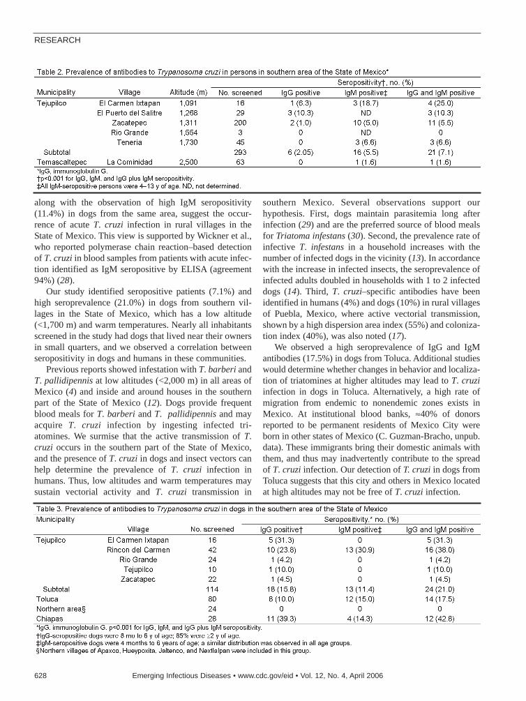
along with the observation of high IgM seropositivity(11.4%) in dogs from the same area, suggest the occur-rence of acute T. cruzi infection in rural villages in theState of Mexico. This view is supported by Wickner et al.,who reported polymerase chain reaction–based detectionof T. cruzi in blood samples from patients with acute infec-tion identified as IgM seropositive by ELISA (agreement94%) (28).
Our study identified seropositive patients (7.1%) andhigh seroprevalence (21.0%) in dogs from southern vil-lages in the State of Mexico, which has a low altitude(<1,700 m) and warm temperatures. Nearly all inhabitantsscreened in the study had dogs that lived near their ownersin small quarters, and we observed a correlation betweenseropositivity in dogs and humans in these communities.
Previous reports showed infestation with T. barberi andT. pallidipennis at low altitudes (<2,000 m) in all areas ofMexico (4) and inside and around houses in the southernpart of the State of Mexico (12). Dogs provide frequentblood meals for T. barberi and T. pallidipennis and mayacquire T. cruzi infection by ingesting infected tri-atomines. We surmise that the active transmission of T.cruzi occurs in the southern part of the State of Mexico,and the presence of T. cruzi in dogs and insect vectors canhelp determine the prevalence of T. cruzi infection inhumans. Thus, low altitudes and warm temperatures maysustain vectorial activity and T. cruzi transmission in
southern Mexico. Several observations support ourhypothesis. First, dogs maintain parasitemia long afterinfection (29) and are the preferred source of blood mealsfor Triatoma infestans (30). Second, the prevalence rate ofinfective T. infestans in a household increases with thenumber of infected dogs in the vicinity (13). In accordancewith the increase in infected insects, the seroprevalence ofinfected adults doubled in households with 1 to 2 infecteddogs (14). Third, T. cruzi–specific antibodies have beenidentified in humans (4%) and dogs (10%) in rural villagesof Puebla, Mexico, where active vectorial transmission,shown by a high dispersion area index (55%) and coloniza-tion index (40%), was also noted (17).
We observed a high seroprevalence of IgG and IgMantibodies (17.5%) in dogs from Toluca. Additional studieswould determine whether changes in behavior and localiza-tion of triatomines at higher altitudes may lead to T. cruziinfection in dogs in Toluca. Alternatively, a high rate ofmigration from endemic to nonendemic zones exists inMexico. At institutional blood banks, ≈40% of donorsreported to be permanent residents of Mexico City wereborn in other states of Mexico (C. Guzman-Bracho, unpub.data). These immigrants bring their domestic animals withthem, and thus may inadvertently contribute to the spreadof T. cruzi infection. Our detection of T. cruzi in dogs fromToluca suggests that this city and others in Mexico locatedat high altitudes may not be free of T. cruzi infection.
RESEARCH
628 Emerging Infectious Diseases • www.cdc.gov/eid • Vol. 12, No. 4, April 2006
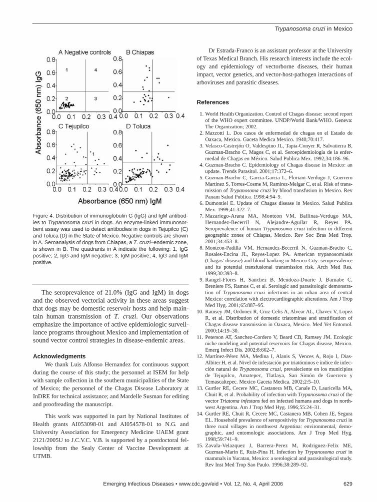
The seroprevalence of 21.0% (IgG and IgM) in dogsand the observed vectorial activity in these areas suggestthat dogs may be domestic reservoir hosts and help main-tain human transmission of T. cruzi. Our observationsemphasize the importance of active epidemiologic surveil-lance programs throughout Mexico and implementation ofsound vector control strategies in disease-endemic areas.
AcknowledgmentsWe thank Luis Alfonso Hernandez for continuous support
during the course of this study; the personnel at ISEM for helpwith sample collection in the southern municipalities of the Stateof Mexico; the personnel of the Chagas Disease Laboratory atInDRE for technical assistance; and Mardelle Susman for editingand proofreading the manuscript.
This work was supported in part by National Institutes ofHealth grants AI053098-01 and AI054578-01 to N.G. andUniversity Association for Emergency Medicine UAEM grant2121/2005U to J.C.V.C. V.B. is supported by a postdoctoral fel-lowship from the Sealy Center of Vaccine Development atUTMB.
Dr Estrada-Franco is an assistant professor at the Universityof Texas Medical Branch. His research interests include the ecol-ogy and epidemiology of vectorborne diseases, their humanimpact, vector genetics, and vector-host-pathogen interactions ofarboviruses and parasitic diseases.
References
1. World Health Organization. Control of Chagas disease: second reportof the WHO expert committee. UNDP/World Bank/WHO. Geneva:The Organization; 2002.
2. Mazzotti L. Dos casos de enfermedad de chagas en el Estado deOaxaca, Mexico. Gaceta Medica Mexico. 1940;70:417.
3. Velasco-Castrejón O, Valdespino JL, Tapia-Conyer R, Salvatierra B,Guzman-Bracho C, Magos C, et al. Seroepidemiología de la enfer-medad de Chagas en México. Salud Publica Mex. 1992;34:186–96.
4. Guzman-Bracho C. Epidemiology of Chagas disease in Mexico: anupdate. Trends Parasitol. 2001;17:372–6.
5. Guzman-Bracho C, Garcia-Garcia L, Floriani-Verdugo J, GuerreroMartinez S, Torres-Cosme M, Ramirez-Melgar C, et al. Risk of trans-mission of Trypanosoma cruzi by blood transfusion in Mexico. RevPanam Salud Publica. 1998;4:94–9.
6. Dumontiel E. Update of Chagas disease in Mexico. Salud PublicaMex. 1999;41:322–7.
7. Mazariego-Arana MA, Monteon VM, Ballinas-Verdugo MA,Hernandez-Becerril N, Alejandre-Aguilar R, Reyes PA.Seroprevalence of human Trypanosoma cruzi infection in differentgeographic zones of Chiapas, Mexico. Rev Soc Bras Med Trop.2001;34:453–8.
8. Monteon-Padilla VM, Hernandez-Becerril N, Guzman-Bracho C,Rosales-Encina JL, Reyes-Lopez PA. American trypanosomiasis(Chagas’ disease) and blood banking in Mexico City: seroprevalenceand its potential transfusional transmission risk. Arch Med Res.1999;30:393–8.
9. Rangel-Flores H, Sanchez B, Mendoza-Duarte J, Barnabe C,Breniere FS, Ramos C, et al. Serologic and parasitologic demonstra-tion of Trypanosoma cruzi infections in an urban area of centralMexico: correlation with electrocardiographic alterations. Am J TropMed Hyg. 2001;65:887–95.
10. Ramsey JM, Ordonez R, Cruz-Celis A, Alvear AL, Chavez V, LopezR, et al. Distribution of domestic triatominae and stratification ofChagas disease transmission in Oaxaca, Mexico. Med Vet Entomol.2000;14:19–30.
11. Peterson AT, Sanchez-Cordero V, Beard CB, Ramsey JM. Ecologicniche modeling and potential reservoirs for Chagas disease, Mexico.Emerg Infect Dis. 2002;8:662–7.
12. Martínez-Pérez MA, Medina I, Alanis S, Vences A, Rojo I, Dias-Albiter H, et al. Nivel de infestación por triatóminos e índice de infec-ción natural de Trypanosoma cruzi, prevaleciente en los municipiosde Tejupilco, Amatepec, Tlatlaya, San Simón de Guerrero yTemascaltepec. Mexico Gaceta Medica. 2002;2:5–10.
13. Gurtler RE, Cecere MC, Castanera MB, Canale D, Lauricella MA,Chuit R, et al. Probability of infection with Trypanosoma cruzi of thevector Triatoma infestans fed on infected humans and dogs in north-west Argentina. Am J Trop Med Hyg. 1996;55:24–31.
14. Gurtler RE, Chuit R, Cecere MC, Castanera MB, Cohen JE, SeguraEL. Household prevalence of seropositivity for Trypanosoma cruzi inthree rural villages in northwest Argentina: environmental, demo-graphic, and entomologic associations. Am J Trop Med Hyg.1998;59:741–9.
15. Zavala-Velazquez J, Barrera-Perez M, Rodriguez-Felix ME,Guzman-Marin E, Ruiz-Pina H. Infection by Trypanosoma cruzi inmammals in Yucatan, Mexico: a serological and parasitological study.Rev Inst Med Trop Sao Paulo. 1996;38:289–92.
Trypanosoma cruzi in Mexico
Emerging Infectious Diseases • www.cdc.gov/eid • Vol. 12, No. 4, April 2006 629
Figure 4. Distribution of immunoglobulin G (IgG) and IgM antibod-ies to Trypanosoma cruzi in dogs. An enzyme-linked immunosor-bent assay was used to detect antibodies in dogs in Tejupilco (C)and Toluca (D) in the State of Mexico. Negative controls are shownin A. Seroanalysis of dogs from Chiapas, a T. cruzi–endemic zone,is shown in B. The quadrants in A indicate the following: 1, IgGpositive; 2, IgG and IgM negative; 3, IgM positive; 4, IgG and IgMpositive.
16. Salazar-Schettino PM, Bucio MI, Cabrera M, Bautista J. First case ofnatural infection in pigs. Review of Trypanosoma cruzi reservoirs inMexico. Mem Inst Oswaldo Cruz. 1997;92:499–502.
17. Sosa-Jurado F, Zumaquero-Ríos JL, Reyes PA, Cruz-García A,Guzmán-Bracho C, Monteon VM. Factores bióticos y abióticos quedeterminan la seroprevalencia de anticuerpos contra Trypanosomacruzi en el municipio de Palmar de Bravo, Puebla, Mexico. SaludPublica Mex. 2004;46:39–48.
18. Castellani O, Ribeiro LV, Fernandes JF. Differentiation ofTrypanosoma cruzi in culture. J Protozool. 1967;14:447–51.
19. Plata F, Garcia Pons F, Eisen H. Antigenic polymorphism ofTrypanosoma cruzi: clonal analysis of trypomastigote surface anti-gens. Eur J Immunol. 1984;14:392–9.
20. Antas PR, Azevedo EN, Luz MR, Medrano-Mercado N, Chaves AC,Vidigal PG, et al. A reliable and specific enzyme-linked immunosor-bent assay for the capture of IgM from human chagasic sera usingfixed epimastigotes of Trypanosoma cruzi. Parasitol Res.2000;86:813–20.
21. Bhatia V, Sinha M, Luxon B, Garg N. Utility of Trypanosoma cruzisequence database for the identification of potential vaccine candi-dates: in silico and in vitro screening. Infect Immun.2004;72:6245–54.
22. Voller A, Draper C, Bidwell DE, Bartlett A. Microplate enzyme-linked immunosorbent assay for Chagas’ disease. Lancet.1975;1:426–8.
23. Umezawa ES, Shikanai-Yasuda MA, Gruber A, Pereira-ChioccolaVL, Zingales B. Trypanosoma cruzi defined antigens in the serologi-cal evaluation of an outbreak of acute Chagas disease in Brazil(Catole do Rocha, Paraiba). Mem Inst Oswaldo Cruz.1996;91:87–93.
24. Umezawa ES, Nascimento MS, Kesper N Jr, Coura JR, Borges-Pereira J, Junqueira AC, et al. Immunoblot assay using excreted-secreted antigens of Trypanosoma cruzi in serodiagnosis ofcongenital, acute, and chronic Chagas’ disease. J Clin Microbiol.1996;34:2143–7.
25. Latour J, Abraira V, Cabello JB, Lopez Sanchez J. Investigationmethods in clinical cardiology. IV. Clinical measurements in cardiol-ogy: validity and errors of measurements. Rev Esp Cardiol.1997;50:117–28.
26. Shadomy SV, Waring SC, Chappell CL. Combined use of enzyme-linked immunosorbent assay and flow cytometry to detect antibodiesto Trypanosoma cruzi in domestic canines in Texas. Clin Diagn LabImmunol. 2004;11:313–9.
27. Umezawa ES, Shikanai-Yasuda MA, Stolf AM. Changes in isotypecomposition and antigen recognition of anti-Trypanosoma cruzi anti-bodies from acute to chronic Chagas disease. J Clin Lab Anal.1996;10:407–13.
28. Wincker P, Telleria J, Bosseno MF, Cardoso MA, Marques P, YaksicN, et al. PCR-based diagnosis for Chagas’ disease in Bolivian chil-dren living in an active transmission area: comparison with conven-tional serological and parasitological diagnosis. Parasitology.1997;114:367–73.
29. Gurtler RE, Solard ND, Lauricela MA, Haedo AS, Pietrokovski SM,Alberti AA, et al. Dynamics of transmission of Trypanosoma cruzi ina rural area of Argentina. III. Persistence of T. cruzi parasitemiaamong canine reservoirs in a two-year follow-up. Rev Inst Med TropSao Paulo. 1986;28:213–9.
30. Gurtler RE, Cohen JE, Cecere MC, Chuit R. Shifting host choices ofthe vector of chagas disease Triatoma infestans and the availability ofhosts in houses in north-west Argentina. Journal of Applied Ecology.1997;34:699–715.
Address for correspondence: Nisha Garg, Department of Microbiologyand Immunology, University of Texas Medical Branch, 3.142 MedicalResearch Bldg, 301 University Blvd, Galveston, TX 77555, USA; fax:409-747-6869; email: [email protected]
RESEARCH
630 Emerging Infectious Diseases • www.cdc.gov/eid • Vol. 12, No. 4, April 2006
measles
Highly contagious disease caused by a virus of the genus Morbillivirus, marked by an eruption ofdistinct, red, circular spots. From the Middle Dutch masel, “blemish.” References to the disease dateback to at least 700 AD, but the first recorded scientific description of measles was in the 10thcentury AD by the Persian physician Ibn Razi, who described it as “more dreaded than smallpox.”Prior to 1963, when the first measles vaccine was licensed, 3–4 million cases and 450 deathsoccurred in the United States every year. Measles remains a primary cause of death in developingnations, where vitamin A deficiency is common. According to the World Health Organization,measles is the leading cause of vaccine-preventable death in children; it is responsible for ≈850,000deaths each year.
Sources: Merriam-Webster's collegiate dictionary. 11th ed. Springfield (MA): Merriam-Webster Incorporated; 2003; Centersfor Disease Control and Prevention. Measles history. Available from http://www.cdc.gov/nip/diseases/measles/history.htm;and World Health Organization. Measles mortality reduction and regional elimination. Available from http://www.who.int/vac-cines-documents/DocsPDF01/www573.pdf
eettyymmoollooggiiaa

We performed a prospective study of all inpatient andoutpatient methicillin-resistant Staphylococcus aureus(MRSA) isolates identified at the University of ChicagoHospitals from November 2003 through November 2004.Differences in resistance to non–β-lactam antimicrobialdrugs were determined after stratification of the 578 MRSAisolates into 4 groups by patient age (pediatric vs. adult)and onset location (community vs. hospital). Non–β-lactamresistance was significantly greater among the 288 adultthan the 177 pediatric community-associated isolates forerythromycin (93.2 vs. 87.0%, p = 0.03), clindamycin (51.8vs. 7.3%, p<0.001), ciprofloxacin (62.1 vs. 10.7%,p<0.001), gentamicin (11.1 vs. 1.1%, p<0.001), and tetra-cycline (19.9 vs. 6.4%, p<0.001). In contrast, hospital-asso-ciated MRSA isolates from children and adults had similarrates of non–β-lactam antimicrobial drug resistance. In ourregion, clindamycin is an appropriate empiric therapy ofcommunity-associated MRSA infection in children butshould be used with caution in adults.
Colonization by and infection with methicillin-resistantStaphylococcus aureus (MRSA) in children and adults
who have little or no contact with the healthcare system,phenomena almost unknown before the mid-1990s, havebeen reported with increasing frequency (1–34). In somecities and rural areas, rates of these community-associatedMRSA (CA-MRSA) infections, particularly in skin andsoft tissue, are increasing rapidly (1–4). Many CA-MRSAinfections have been attributed to isolates that are distinctfrom hospital-associated MRSA (HA-MRSA) strains.
The definition of CA-MRSA has not been standardized.No single definition would likely suffice for all purposes.Attempts to define CA-MRSA have focused upon a variety
of criteria: temporal (i.e., the MRSA isolate from a speci-men is submitted <72 hours after hospital admission),host-risk-factor profile (i.e., the patient with the MRSAisolate lacked certain exposure parameters relevant tohealthcare facilities, variously defined), antimicrobial drugsusceptibility of the MRSA isolates (e.g., susceptibility toclindamycin), and certain molecular aspects of the isolates(e.g., SCCmec type or pulsed-field gel electrophoresistype). Which definition is most important in the choice ofempiric antimicrobial drug treatment is not clear.However, a clinician confronted with a putative S. aureusinfection needs guidance.
The epidemiology of the emerging CA-MRSA strains,including their origins, reservoirs, modes of dissemination,and effective approaches to their control, is not complete-ly understood. We previously noted that CA-MRSA iso-lates often lacked resistance to multiple non–β-lactamantimicrobial drugs when compared with HA-MRSAstrains (5,6). This phenomenon has been documented inadults (4,7) and children (8–10). More recently, we anec-dotally observed that children had MRSA infectionscaused by strains susceptible to non–β-lactam antimicro-bial drugs more often than adults. Few data exist that com-pare antimicrobial drug resistance profiles of adult andpediatric MRSA infection isolates stratified by site of onset(hospital vs. community). Accordingly, using the temporaldefinition of CA-MRSA, we examined the hypothesis thatthese 2 groups may be infected by MRSA strains with dif-ferent rates of resistance to non–β-lactam antimicrobialdrugs when the isolates had onset in the community.Therefore, we compared several criteria for the temporaldefinition of CA-MRSA to assess the impact of changingthese criteria on our findings. This article summarizes ourstudy.
Contrasting Pediatric and AdultMethicillin-resistant
Staphylococcus aureus Isolates Michael Z. David,* Susan E. Crawford,* Susan Boyle-Vavra,* Mark A. Hostetler,*
Daniel C. Kim,* and Robert S. Daum*
Emerging Infectious Diseases • www.cdc.gov/eid • Vol. 12, No. 4, April 2006 631
*University of Chicago, Chicago, Illinois, USA

Materials and MethodsThe University of Chicago Hospitals (UCH) is a terti-
ary-care medical center on the south side of Chicago with577 inpatient beds and 29,500 annual admissions. Itincludes an outpatient care facility with 379,000 annualvisits. UCH serves an inner city population and draws ter-tiary referrals from the surrounding region.
The UCH Clinical Microbiology Laboratories prospec-tively identified all MRSA isolates collected at the medicalcenter from November 7, 2003, to November 7, 2004,from inpatients and outpatients. S. aureus isolates wereidentified by gram stain, growth on BBL mannitol salt fer-mentation agar (Beckton, Dickinson and Company,Sparks, MD, USA), positive catalase test results, and apositive Staphaurex Plus test (Remel Europe Ltd.,Dartford, UK) result.
The Clinical Microbiology Laboratories determined thesusceptibility profile of each isolate by using the Vitek 2system (bioMérieux Vitek, Inc., Durham, NC, USA) formethicillin, erythromycin, clindamycin, ciprofloxacin,rifampin, gentamicin, tetracycline, and vancomycin. FromNovember 7, 2003, to July 2004, any isolate with an MICof vancomycin >2 µg/mL reported by the Vitek systemwas retested for vancomycin susceptibility by the EtestStrip (AB Biodisk, Solna, Sweden). After July 2004, theEtest was performed for these isolates only if growthoccurred on a vancomycin agar screen plate. For isolatesthat tested resistant to erythromycin but susceptible to clin-damycin, a D-test to detect inducible resistance to clin-damycin was performed, as was a Kirby-Bauerdisk-diffusion test for susceptibility to trimethoprim-sul-famethoxazole (TMP-SMX), both according to Clinicaland Laboratory Standards Institute (CLSI) guidelines (35).
For each MRSA specimen, patient information was col-lected from the laboratory information system and theelectronic medical record, including the date of specimenprocurement, age and sex of the patient, location of speci-men procurement (inpatient, outpatient, emergency depart-ment), and antimicrobial drug susceptibility profile. Forinpatient isolates, the number of days from admission toculture procurement was determined by subtracting theadmission date from the procurement date.
We defined CA-MRSA to include all isolates culturedfrom outpatients and isolates from hospitalized patientsobtained within 72 hours of admission. We also examinedthe impact of changing the 72-hour criterion in the defini-tion to 24 and 48 hours. With each change in definition, theeffect on the percentage of MRSA infections considered tobe community associated was assessed.
The bacteriologic and patient information was com-piled in an electronic database designed for the study byusing Access software (Microsoft, Redmond, WA, USA).Only the first isolate from each patient collected during the
surveillance was included in the database. The study wasapproved by the institutional review board of theBiological Sciences Division of the University of Chicago.Data were analyzed with Stata, version 8.0 (Stata Corp,College Station, TX, USA). Comparisons between groupswere performed by the χ2 test or the Fisher exact test. Allhypotheses were 2-tailed and were considered significantif p<0.05.
ResultsThe UCH Clinical Microbiology Laboratories identi-
fied 1,149 MRSA-positive cultures from 578 patients. Ofthese, 201 (34.7%) were from children <18 years of ageand 377 (65.3%) were from adults >19 years of age. Of theadult MRSA isolates, 27.9% were cultured from outpa-tients; of the pediatric MRSA isolates, 57.2% were cul-tured from outpatients. The median age of pediatricpatients was 3 years (16 days to 18 years), with a medianof 6 years among outpatients and 2 years among inpatients.For adults, the median age was 56 years (range 19–100),with a median of 51 years among outpatients and 60 yearsamong inpatients.
Using the 72-hour definition, 64.3% of adult and 72.1%of pediatric inpatient isolates were CA-MRSA (p = 0.2)(Table 1). Restricting the definition of CA-MRSA to the 48-hour definition, 58.5% of adult and 69.8% of pediatric inpa-tient isolates were CA-MRSA (p = 0.06). Fifty-four percentand 65.1%, respectively, of adult and pediatric inpatient iso-lates met the criteria for the 24-hour definition of CA-MRSA (p = 0.07). Thus, by using the 24-, 48-, or 72-hourcriteria for CA-MRSA, most of the pediatric and adult inpa-tient isolates would be considered community-associated.
Combining the isolates cultured in the emergencydepartment with those cultured in clinics, 105 outpatientisolates were obtained from adults and 115 from children.Adult isolates were substantially more likely than pediatricisolates to be resistant to ciprofloxacin, clindamycin, gen-tamicin, and tetracycline. No difference was seen in resist-ance rates for erythromycin or rifampin (Table 2).
A small percentage of the temporally defined CA-MRSA isolates (i.e., those either from outpatients or cul-tured <72 hours after admission from inpatients) wasresistant only to β-lactam antimicrobial drugs; this resist-ance pattern was more common among pediatric (9.6%)than adult (3.6%) isolates (p = 0.001) (Table 3). Among thetemporally defined CA-MRSA isolates, those from adultswere more likely than those from children to be resistant tomost of the non–β-lactam antimicrobial drugs. The rate ofresistance to erythromycin was high among both the pedi-atric and adult isolates, although significantly higheramong the adult isolates. The rate of resistance to clin-damycin, ciprofloxacin, gentamicin, and tetracycline wasmuch higher for adult than for pediatric CA-MRSA
RESEARCH
632 Emerging Infectious Diseases • www.cdc.gov/eid • Vol. 12, No. 4, April 2006

isolates (Table 3). There was no significant difference forrifampin, TMP-SMX, or vancomycin.
The D-test for inducible clindamycin resistance wasperformed on 97% (112/116) of adult and 89% (125/142)of pediatric CA-MRSA isolates that were resistant toerythromycin and susceptible to clindamycin. Amongthose tested, adult (20.5%, 23/112) isolates were morelikely than pediatric (15.2%, 19/125) isolates to have apositive D-test result (Table 3).
In contrast to the differences we found among CA-MRSA isolates from adults and children, however, the hos-pital-associated MRSA isolates (>72 hours afteradmission) had similar rates of antimicrobial drug resist-ance (Table 3). For example, clindamycin resistanceoccurred in 74.2% of adult and 75.0% of pediatric HA-MRSA isolates (p = 0.9). Only the rates of ciprofloxacin (p= 0.004) and gentamicin (p = 0.01) resistance were sub-stantially different when the HA-MRSA isolates werecompared (Table 3).
We defined a multidrug-resistant (MDR) MRSA isolateas being resistant to >3 of the non–β-lactam antimicrobialdrugs tested. When the temporal definition (<72 hoursafter admission) was used, adult CA-MRSA isolates weremore likely than pediatric CA-MRSA isolates (52.3% vs.
6.4%, p<0.0001) to be MDR (Table 3). HA-MRSA isolatesfrom both children and adults were more often MDR, butthe rates did not differ substantially (76% vs. 66.7%, p =0.4) (Table 3).
DiscussionWhen community association or onset was defined by
using the temporal criterion of procuring isolates <72hours after admission, most adult and most pediatricMRSA isolates in our study would be considered to have acommunity origin. A narrower procurement definition,e.g., <24 hours or <48 hours, would have led to the sameconclusion.
MRSA isolates from children’s specimens comparedwith isolates from adults’ specimens obtained <72 hoursafter admission were more likely to be resistant to only β-lactams and more likely to be susceptible to clindamycin,ciprofloxacin, and gentamicin. These differences suggestthat there may be distinct CA-MRSA isolates colonizingand infecting children and adults in the population servedby our medical center or, less likely, that there may be areservoir of CA-MRSA that affects children and adults dif-ferently.
Children may have unique risk factors for MRSA colo-nization in the community related to their environment,such as daycare centers (11,12), schools (13), or recre-ational facilities, and may have different behavioral habitsthan most adults. Alternatively, children may be differentfrom adults as hosts, and they may encounter novel MRSAstrains either by different colonization of the skin or bysome undefined difference in host defense. Antimicrobialdrugs used to treat infections among children may differfrom those used among adults; perhaps distinct MRSAstrains colonizing children result from differential antimi-crobial drug selection pressures in the community.
Contrasting Pediatric and Adult MRSA Isolates
Emerging Infectious Diseases • www.cdc.gov/eid • Vol. 12, No. 4, April 2006 633

Our findings have obvious implications for empirictherapy of CA-MRSA infections and MRSA control meas-ures. In the population served by UCH, for children withsuspected community-onset MRSA infections, clin-damycin is an appropriate first-line empiric antimicrobialdrug, at least for those who are not critically ill (8,10). Inadults, by contrast, clindamycin would be much less suit-able in this role, as most temporally defined CA-MRSAisolates are clindamycin-resistant. Therefore, amongadults at our institution, clindamycin can be consideredonly after susceptibility testing results have been obtainedfrom a culture specimen.
CA-MRSA isolates from children have a low rate ofclindamycin resistance. Studies of children with CA-MRSA infections or colonization in many cities, includingHouston, Corpus Christi, Memphis, Nashville, Louisville,Providence, and Chicago, have demonstrated high rates ofclindamycin susceptibility among these isolates, rangingfrom 67%–100% (1,8,10,14–16). The 2 studies with thelowest susceptibility rates were conducted in Providencefrom 1997 to 2001 with a 74% rate (8), and Louisville from1999 to 2001 with a 67% rate (16). These lower rates mayhave been due to geographic variation or were a windowon an earlier phase in the CA-MRSA epidemic.
A possible confounder in calculating clindamycinresistance rates is the 2004 CLSI guideline stating thateach isolate that initially tests susceptible to clindamycinand resistant to erythromycin should be tested for
inducible clindamycin resistance by the D-test; if the testresult is positive, that isolate should be considered resist-ant (35). Published data from before 2004 may not haveincluded D-test results in reporting clindamycin resistancerates among MRSA isolates. However, the impact of thenew CLSI guideline does not change the conclusionsderived from our data, although the rate of clindamycinresistance among all pediatric MRSA isolates in our sam-ple would increase from 15.4% to 24.9% when the newguideline was considered.
We have continued to use clindamycin for initial empir-ic therapy of mild or moderately ill pediatric patients like-ly to have a CA-MRSA infection. We take this approachbecause better alternative therapy is not available. Theresults of TMP-SMX therapy for CA-MRSA have beenreported for relatively few patients (36); therapy was fre-quently unsuccessful despite in vitro susceptibility of theinfecting MRSA strains. Tetracyclines are not suitable foryoung children, and linezolid is prohibitively expensive.Our policy is to abandon clindamycin when susceptibilitytesting or D-test results suggest the possibility of treatmentfailure, usually 2–4 days into the treatment course or, asmentioned, for severe illness when CA-MRSA is suspect-ed. To date, this approach has been suitable.
The pattern of clindamycin resistance among adult CA-MRSA isolates has been more complex. At UCH, morethan half of the CA-MRSA isolates from adults wereresistant to clindamycin. Similarly, at Northwestern
RESEARCH
634 Emerging Infectious Diseases • www.cdc.gov/eid • Vol. 12, No. 4, April 2006

Memorial Hospital, also in Chicago, from 1998 to 1999,few MRSA isolates were clindamycin susceptible amongthose that were collected <72 hours after admission fromadults with no known hospitalizations in the previous 2years (37).
In contrast, more recent studies of CA-MRSA infec-tions and MRSA colonization isolates have demonstratedlow rates of clindamycin and other non–β-lactam resist-ance among MRSA isolates from young and urban pooradults. For example, 9 of 67 patients with MRSA infec-tions in a military-beneficiary population in Texas from1999 to 2001 had no risk factors for HA-MRSA. Thesepatients all had onset of infection in the community andhad an isolate that was susceptible to clindamycin.Moreover, they were younger than the patients reported tohave HA-MRSA infections (4). In San Francisco in1999–2000, among asymptomatic, homeless and margin-ally housed adults in the community, 23 MRSA isolateswere identified from nasal swabs of which only 2 (8.7%)were resistant to clindamycin (17). In a sample of 45 colo-nizing and infecting MRSA isolates collected from healthyyoung adults (18–44 years of age) training at a militaryfacility in Texas, 22% were resistant to clindamycin (7).
Why these disparities exist in the rates of clindamycinsusceptibility among CA-MRSA isolates obtained fromadults is unclear. Possible explanations include regionalvariation or characteristics of the patient populations sam-pled. The 2 studies with high clindamycin resistance rateswere both conducted in Chicago at tertiary medical cen-ters. Community-based surveys among adults, in contrast,have demonstrated a high rate of clindamycin susceptibil-ity, perhaps reflecting an exposure to a community MRSAisolate pool similar to the pool affecting pediatric popula-tions in many urban centers.
Unlike clindamycin, erythromycin resistance has beencommon among both pediatric and adult CA-MRSA iso-lates. In hospitalized children with MRSA infections atUCH during 1988–1990 and 1993–1995, 74.3% carriedisolates resistant to erythromycin (5). Our current rate,88.1% in pediatric isolates, is slightly higher.Erythromycin resistance has also been common amongCA-MRSA isolates colonizing and infecting children atmany urban centers from 1990 to 2002, with rates from52% to 100% (8,10,14,16). In adults, recent studies haveshown similarly high rates of erythromycin resistance,60.9%–80%, among CA-MRSA strains in selected popula-tions, including soldiers (7), inmates in jail (38), andhomeless and impoverished urban adults (17).
Analysis of antimicrobial resistance patterns amongMRSA isolates obtained >72 hours after admission pro-duced a sharp contrast with the CA isolates. Children andadults in this HA group were infected by MRSA strainswith similar rates of resistance to non–β-lactam antimicro-
bial drugs. This finding suggests that adults and childrenface a common source of antimicrobial drug selectionpressure and a common reservoir of MRSA isolates in ourhospital, but not in the community.
Before this study, few data have been available to com-pare the resistance patterns of MRSA isolates collected in1 medical center or region stratified by both age group(children vs. adults) and venue of onset (community vs.hospital). One previous analysis conducted from 1988 to1997 at another medical center in Chicago compared adultand pediatric MRSA isolates and also found that MRSAstrains susceptible to clindamycin were more commonamong pediatric than adult isolates; the isolates were notstratified by hospital or community origin (18). In contrast,among CA-MRSA isolates collected during 2000 from 12Minnesota clinical microbiology laboratories serving bothinpatients and outpatients, no differences in the rate ofclindamycin susceptibility were found in isolates fromchildren and adults (19). These investigators used arisk–factor-based definition for CA-MRSA, while we useda temporal definition. Other possible explanations for thediscrepancy include a secular change in the 4 years sepa-rating the studies or demographic differences in the studypopulations.
Our study has certain limitations. It was conducted in 1center and in 1 city, and it is unknown whether our data arerepresentative of the CA-MRSA epidemic in other areas.Our study also assumes that adults and children seekingcare in all areas of our medical center had an approximate-ly equal chance of having a culture performed. We did notevaluate whether an assessment of risk factors for HA-MRSA would have determined the likelihood of resistanceto non–β-lactam antimicrobial drugs.
With the recognition of CA-MRSA as a distinct epi-demiologic phenomenon and the accompanying upsurge inboth the S. aureus disease burden and methicillin resist-ance rates at many centers, considerable controversy hassurrounded the definition most appropriate for CA-MRSA.Whether the best definition should be based on temporal,molecular, antimicrobial drug susceptibility, or host risk-factor criteria will not be resolved by this study and willcontinue to vary according to the issue being addressed.Despite these uncertainties, our data, using the temporaldefinition, have practical value in the initial antimicrobialdrug management of patients with suspected CA-MRSAinfections.
Understanding the complex epidemiology of CA-MRSA infection is critical to the development of controlpolicies and treatment guidelines in areas with a high col-onization and disease prevalence. Our data demonstratethe complexity of the contrasting definitions of CA-MRSA. While neither a time criterion alone nor a timecriterion in addition to an antimicrobial drug susceptibility
Contrasting Pediatric and Adult MRSA Isolates
Emerging Infectious Diseases • www.cdc.gov/eid • Vol. 12, No. 4, April 2006 635

profile is adequate to define CA-MRSA, our data providevaluable guidance for clinical practice.
AcknowledgmentsWe thank Diane Lauderdale for assistance with the data
analysis.
Dr Daum is the recipient of R01 CCR523379 from theCenters for Disease Control and Prevention, R01 AI40481-01A1from the National Institute of Allergy and Infectious Diseases, aswell as support from the Grant Health Care Foundation. DrBoyle-Vavra is supported by the Grant Health Care Foundation.Dr David is supported by the Robert Wood Johnson ClinicalScholars Program.
Dr David is a Robert Wood Johnson Clinical Scholar and acandidate for an MS in health studies at the University ofChicago. His research interests are antimicrobial drug resistance,the history of infectious diseases, and international health.
References
1. Purcell K, Fergie JE. Exponential increase in community-acquiredmethicillin-resistant Staphylococcus aureus infections in south Texaschildren. Pediatr Infect Dis J. 2002;21:988–9.
2. Baum SE, Morris JT, Dooley DP, Watson R. Methicillin-resistantStaphylococcus aureus in an adult military beneficiary populationlacking risk factors: susceptibility to orally available agents. MilMed. 2003;168:126–30.
3. Young DM, Harris HW, Charlebois ED, Chambers H, Campbell A,Perdreau-Remington F, et al. An epidemic of methicillin-resistantStaphylococcus aureus soft tissue infections among medically under-served patients. Arch Surg. 2004;139:947–53.
4. Kallen AJ, Driscoll TJ, Thornton S, Olson PE, Wallace MR. Increasein community-acquired methicillin-resistant Staphylococcus aureusat a naval medical center. Infect Control Hosp Epidemiol.2000;21:223–6.
5. Herold BC, Immergluck LC, Maranan MC, Lauderdale DS, GaskinRE, Boyle-Vavra S, et al. Community-acquired methicillin-resistantStaphylococcus aureus in children with no identified predisposingrisk. JAMA. 1998;279:593–8.
6. Hussain FM, Boyle-Vavra S, Bethel CD, Daum RS. Current trends incommunity-acquired methicillin-resistant Staphylococcus aureus at atertiary care pediatric facility. Pediatr Infect Dis J. 2000;19:1163–6.
7. Ellis MW, Hospenthal DR, Dooley DP, Gray PJ, Murray CK. Naturalhistory of community-acquired methicillin-resistant Staphylococcusaureus colonization and infection in soldiers. Clin Infect Dis.2004;39:971–9.
8. Dietrich DW, Auld DB, Mermel LA. Community-acquired methi-cillin-resistant Staphylococcus aureus in southern New England chil-dren. Pediatrics. 2004;113:e347–52.
9. Buckingham SC, McDougal LK, Cathey LD, Comeaux K, Craig AS,Fridkin SK, et al. Emergence of community-associated methicillin-resistant Staphylococcus aureus at a Memphis, Tennessee Children’sHospital. Pediatr Infect Dis J. 2004;23:619–24.
10. Frank AL, Marcinak JF, Mangat PD, Tjhio JT, Kelkar S,Schreckenberger PC, et al. Clindamycin treatment of methicillin-resistant Staphylococcus aureus infections in children. Pediatr InfectDis J. 2002;21:530–4.
11. Adcock PM, Pastor P, Medley F, Patterson JE, Murphy TV.Methicillin-resistant Staphylococcus aureus in two child care centers.J Infect Dis. 1998;178:577–80.
12. Shahin R, Johnson IL, Jamieson F, McGreer A, Tolkin J, Ford-JonesEL. Methicillin-resistant Staphylococcus aureus carriage in a childcare center following a case of the disease. Arch Pediatr AdolescMed. 1999;153:864–8.
13. Huang Y, Su L, Lin T. Nasal carriage of methicillin-resistantStaphylococcus aureus in contacts of an adolescent with community-acquired disseminated disease. Pediatr Infect Dis J. 2004;23:919–22.
14. Kaplan SL, Hulten KG, Gonzalez BE, Hammerman WA, Lamberth L,Versalovic J, et al. Three-year surveillance of community-acquiredStaphylococcus aureus infections in children. Clin Infect Dis.2005;40:1785–91.
15. Nakamura MM, Rohling KL, Shashaty M, Lu H, Tang Y, EdwardsKM. Prevalence of methicillin-resistant Staphylococcus aureus nasalcarriage in the community pediatric population. Pediatr Infect Dis J.2002;21:917–21.
16. Campbell AL, Bryant KA, Stover B, Marshall GS. Epidemiology ofmethicillin-resistant Staphylococcus aureus at a children’s hospital.Infect Control Hosp Epidemiol. 2003;24:427–30.
17. Charlebois ED, Bangsberg DR, Moss NJ, Moore MR, Moss AR,Chambers HF, et al. Population-based community prevalence ofmethicillin-resistant Staphylococcus aureus in the urban poor of SanFrancisco. Clin Infect Dis. 2002;34:425–33.
18. Frank AL, Marcinak JF, Mangat PD, Schreckenberger PC. Increase incommunity-acquired methicillin-resistant Staphylococcus aureus inchildren. Clin Infect Dis. 1999;29:935–6.
19. Naimi TS, LeDell KH, Como-Sabetti K, Borchardt SM, Boxrud DJ,Etienne J, et al. Comparison of community- and health care-associat-ed methicillin-resistant Staphylococcus aureus infection. JAMA.2003;290:2976–84.
20. Centers for Disease Control and Prevention. Four pediatric deathsfrom community-acquired methicillin-resistant Staphylococcusaureus—Minnesota and North Dakota, 1997–1999. MMWR MorbMortal Wkly Rep. 1999;48:707–10.
21. Hussain FM, Boyle-Vavra S, Daum RS. Community-acquired methi-cillin-resistant Staphylococcus aureus colonization in healthy chil-dren attending an outpatient pediatric clinic. Pediatr Infect Dis J.2001;20:763–7.
22. Zinderman CE, Conner B, Malakooti MA, LaMar JE, Armstrong A,Bohnker BK. Community-acquired methicillin-resistantStaphylococcus aureus among military recruits. Emerg Infect Dis.2004;10:941–4.
23. Groom AV, Wolsey DH, Naimi TS, Smith K, Johnson S, Boxrud D,et al. Community-acquired methicillin-resistant Staphylococcusaureus in a rural American Indian community. JAMA. 2001;286:1201–5.
24. Kazakova SV, Hageman JC, Matava M, Srinivasan A, Phelan L,Garfinkel B, et al. A clone of methicillin-resistant Staphylococcusaureus among professional football players. N Engl J Med.2005;352:468–75.
25. Stacey AR, Endersby KE, Chan PC, Marples RR. An outbreak ofmethicillin-resistant Staphylococcus aureus infection in a rugby foot-ball team. Br J Sports Med. 1998;32:153–4.
26. Naimi TS, LeDell KH, Boxrud DJ, Groom AV, Steward CD, JohnsonSK, et al. Epidemiology and clonality of community-acquired methi-cillin-resistant Staphylococcus aureus in Minnesota, 1996–1998. ClinInfect Dis. 2001;33:990–6.
27. Suggs AH, Maranan MC, Boyle-Vavra S, Daum RS. Methicillin-resistant and borderline methicillin-resistant asymptomaticStaphylococcus aureus colonization in children without identifiablerisk factors. Pediatr Infect Dis J. 1999;18:410–4.
28. Fridkin SK, Hageman JC, Morrison M, Sanza LT, Como-Sabetti K,Jernigan JA, et al. Methicillin-resistant Staphylococcus aureus dis-ease in three communities. N Engl J Med. 2005;352:1436–44.
29. Gorak EJ, Yamada SM, Brown JD. Community-acquired methicillin-resistant Staphylococcus aureus in hospitalized adults and childrenwithout known risk factors. Clin Infect Dis. 1999;29:797–800.
RESEARCH
636 Emerging Infectious Diseases • www.cdc.gov/eid • Vol. 12, No. 4, April 2006

30. Centers for Disease Control and Prevention. Community-associatedmethicillin-resistant Staphylococcus aureus infections in PacificIslanders—Hawaii, 2001–2003. MMWR Morb Mortal Wkly Rep.2004;53:767–70.
31. Baggett HC, Hennessy TW, Leman R, Hamlin C, Bruden D,Reasonover A, et al. An outbreak of community-onset methicillin-resistant Staphylococcus aureus skin infections in southwesternAlaska. Infect Control Hosp Epidemiol. 2003;24:397–402.
32. Fang YH, Hsueh PR, Hu JJ, Lee PI, Chen JM, Lee CY, et al.Community-acquired methicillin-resistant Staphylococcus aureus inchildren in northern Taiwan. J Microbiol Immunol Infect.2004;37:29–34.
33. Charlebois ED, Perdreau-Remington F, Kreiswirth B, Bangsberg DR,Ciccarone D, Diep BA, et al. Origins of community strains of methi-cillin-resistant Staphylococcus aureus. Clin Infect Dis.2004;39:47–54.
34. Hidron AI, Kourbatova EV, Halvosa JS, Terrell BJ, McDougal LK,Tenover FC, et al. Risk factors for colonization with methicillin-resistant Staphylococcus aureus (MRSA) in patients admitted to anurban hospital: emergence of community-associated MRSA nasalcarriage. Clin Infect Dis. 2005;41:159–66.
35. National Committee for Clinical Laboratory Standards. Performancestandards for antimicrobial disk susceptibility testing: 14thInformational Supplement, M100-S14. Villanova (PA): TheCommittee; 2004.
36. Iyer S, Jones DH. Community-acquired methicillin-resistantStaphylococcus aureus skin infection: a retrospective analysis of clin-ical presentation and treatment of a local outbreak. J Am AcadDermatol. 2004;50:854–8.
37. Suntharam N, Hacek D, Peterson LR. Low prevalence of communi-ty-acquired methicillin-resistant Staphylococcus aureus in adults at auniversity hospital in the central United States. J Clin Microbiol.2001;39:1669–71.
38. Pan ES, Diep BA, Carleton HA, Charlebois ED, Sensabaugh GF,Haller BL, et al. Increasing prevalence of methicillin-resistantStaphylococcus aureus infection in California jails. Clin Infect Dis.2003;37:1384–8.
Address for correspondence: Michael Z. David, Robert Wood JohnsonClinical Scholars Program, University of Chicago, 5841 S. MarylandAve, MC2007, Chicago, IL 60637, USA; fax: 773-702-1295; email:[email protected]
Contrasting Pediatric and Adult MRSA Isolates
Emerging Infectious Diseases • www.cdc.gov/eid • Vol. 12, No. 4, April 2006 637
Searchpast issues

Identification of genetic variations of influenza virusesis essential for epidemic and pandemic outbreak surveil-lance and determination of vaccine strain selection. In thisstudy, we combined a random amplification strategy withhigh-density resequencing microarray technology todemonstrate simultaneous detection and sequence-basedtyping of 25 geographically distributed human influenzavirus strains collected in 2004 and 2005. In addition to iden-tification, this method provided primary sequence informa-tion, which suggested that distinct lineages of influenzaviruses co-circulated during the 2004–2005 season, andsimultaneously identified and typed all component strainsof the trivalent FluMist intranasal vaccine. The resultsdemonstrate a novel, timely, and unbiased method for themolecular epidemiologic surveillance of influenza viruses.
Influenza viruses are a major cause of respiratory infec-tions in humans and result in substantial illness, death,
and economic problems throughout the world. Along withregular seasonal epidemic outbreaks caused by commoncirculating strains, novel strains emerge sporadicallybecause of reassortment in the segmented influenza RNAgenome and have resulted in devastating influenza pan-demics (1–3). Since mutations and reassortments are oftendeterminants for infectious potential, antiviral drug sus-ceptibility, and viral escape from vaccine-elicited immu-nity, continually surveying the genetic composition (i.e.,primary sequence) of circulating and emerging variants isnecessary. These needs have become increasingly relevantrecently because the World Health Organization (WHO)has reported 85 human deaths caused by avian A/H5N1
influenza viruses throughout Asia since 2003 and raisedconcerns about the potential for another influenza pan-demic (4).
Automated Sanger/electrophoresis-based sequencingtechnology has been used as the standard platform forDNA and genome sequencing. Although conventionalsequencing produces accurate data, the requirement forknowledge of template sequences and the inability toquickly process multiple targets hinder its practical appli-cation in epidemiologic and diagnostic investigations. Asan alterative, high-density oligonucleotide resequencingmicroarrays represent a promising new technology that hasbeen used to rapidly and accurately identify nucleotidesequence variants (5–7) from viral, bacterial, and eukary-otic genomes (8–13). Use of resequencing microarrays todetect single nucleotide polymorphisms and generate pri-mary sequences enables identification of genetic variantsand provides valuable epidemiologic information that iscritical for outbreak surveillance. In most cases, however,this technology has relied on specific amplification of alimited number of target sequences before hybridization,thus restricting throughput and limiting final identificationto strains that retain primer-targeted sequences.
In an attempt to adapt resequencing microarray technol-ogy to surveillance and diagnostics, we developed the res-piratory pathogen microarray (RPM) version 1 fordetection and sequence typing of 20 common respiratoryand 6 category A biothreat pathogens known to causefebrile respiratory illness (14). A large portion of RPM ver-sion 1 is focused on a subset of tiled sequences correspon-ding to partial fragments from the hemagglutinin (HA),neuraminidase (NA), and matrix (M) genes for detectionof influenza A and B viruses. In this study, we demonstrateunbiased determination of viral subtype and lineage bygeneration of primary sequence using random nucleic acidamplification and resequencing microarray technology.
Identifying Influenza Viruses withResequencing Microarrays
Zheng Wang,*† Luke T. Daum,‡ Gary J. Vora,* David Metzgar,§ Elizabeth A. Walter,¶ Linda C. Canas,‡ Anthony P. Malanoski,* Baochuan Lin,* and David A. Stenger*
RESEARCH
638 Emerging Infectious Diseases • www.cdc.gov/eid • Vol. 12, No. 4, April 2006
*Naval Research Laboratory, Washington, DC, USA; †NOVAResearch Inc., Alexandria, Virginia, USA; ‡Air Force Institute forOperational Health, Brooks City Base, San Antonio, Texas, USA;§Naval Health Research Center, San Diego, California, USA; and¶Lackland Air Force Base, San Antonio, Texas, USA

Methods
RPM Version 1 DesignEach tiled prototype sequence was selected to have an
intermediate level of sequence homology across a group ofmicrobial or viral strains, which allowed for efficienthybridization and unique identification of most or all sub-types of targeted pathogenic species. For each relevantbase of a given prototype sequence, the array containseight 25mer probes (4 sense and 4 antisense). Two of 8probes represent perfect matches, while the others corre-spond to possible mismatches at the central (13th) positionof the 25mers. The prototype regions targeting influenzaviruses were composed of partial sequences from HAgenes of influenza A virus subtypes (H1, H3, and H5) andinfluenza B virus, NA genes of influenza A virus subtypes(N1 and N2) and influenza B virus, and the M genes ofinfluenza A virus (full-length M1 and partial M2) andinfluenza B virus (Table 1). Both HA and NA regionsencompassed a sufficient number of polymorphic sites todefine subtypes. These regions were combined with proto-type sequences for 22 other pathogens and tiled on 12.8-µm chips (Affymetrix Inc., Santa Clara, CA, USA), whichcontain ≈240 K 25mer probes and have the capacity toresolve 30,000 nucleotides. The design and content ofRPM version 1 array have been previously described (14).
Sample Collection and Nucleic Acid IsolationThe influenza clinical specimens used in this study
were collected through the Department of Defense GlobalEmerging Infections System during the 2004–2005influenza season. Influenza throat swab specimens werecollected in accordance with the case criteria previouslydescribed (15). Throat swabs were obtained within the first72 h of the onset of symptoms, placed in viral transportmedium (MicroTest M4, Remel Inc., Lenexa, KS, USA),and delivered by commercial carrier to the Air ForceInstitute for Operational Health in Brooks City Base, SanAntonio, Texas, for culturing and molecular characteriza-tion. Specimens were passaged once through primary rhe-sus monkey kidney tissue culture (BioWhittaker,Walkersville, MD, USA). Cultures were tested for influen-
za A or B viruses by using the centrifugation-enhancedshell-vial technique with monoclonal antibody detection aspreviously described (16). Cultures testing positive forinfluenza A or B viruses were confirmed by using reversetranscription–polymerase chain reaction (RT-PCR) analy-sis with previously reported protocols (16,17). Total nucle-ic acids were extracted from 90-µL cultured samples oraliquots of live trivalent nasally administered influenzavaccine (FluMist 2004/05, MedImmune, Inc., Gaithersburg,MD, USA) by using the MasterPure DNA purification kit(Epicentre Technologies, Madison, WI, USA) and dis-solved in 30 µL of nuclease-free water.
Random RT-PCR Total RNA amplification from cultured samples using a
random (RT-PCR) protocol was performed as previouslydescribed (18) with minor modifications. Briefly, 4 µL oftotal nucleic acids were reverse transcribed with 40 pmolof primer D (5′-GTTTCCCAGTAGGTCTCNNNNNNNNN-3′) and Superscript III reverse transcriptase(Invitrogen, Carlsbad, CA, USA) in a 20-µL volume at42°C for 1 h, followed by heat denaturation at 70°C for 15min. Aliquots (10 µL) of the RT reaction products werethen amplified with the TaqPlus Long PCR System(Stratagene, La Jolla, CA, USA) for 35 cycles in a 50-µLvolume consisting of 5 µL of 10× low salt buffer, 5 µL (25mmol/L) MgCl2, 2 µL (10 mmol/L) dNTP mix, 1 µLprimer E (100 µmol/L) (5′-GTTTCCCAGTAGGTCTC-3′), and 0.5 µL TaqPlus Long polymerase (5 U/µL). Eachcycle consisted of 94°C for 30 s, 40°C for 30 s, 50°C for30 s, and 72°C for 2 min. This was followed by a finalextension at 72°C for 7 min. After amplification, the PCRproducts were purified with the QIAquick PCRPurification Kit (Qiagen, Valencia, CA, USA) and elutedin 45 µL of EB buffer (10 mmol/L Tris, pH 8.5).
RPM Version 1 Hybridization and Processing Purified DNA amplicons were adjusted to 2 µg in 35
µL of EB buffer, mixed with 15.1 µL of fragmentationcocktail buffer (5 µL NEB buffer 4, 5 µL 10 mmol/L Tris,pH 7.8, and 0.1 µL GeneChip fragmentation reagent [3U/µL], Affymetrix Inc.), and incubated for 10 min at 37°C
Microarray Identification of Influenza Viruses
Emerging Infectious Diseases • www.cdc.gov/eid • Vol. 12, No. 4, April 2006 639
and 15 min at 95°C. The fragmented products were thenbiotin labeled with 1.5 µL of Biotin-N6-ddATP(PerkinElmer Life and Analytical Sciences, Boston, MA,USA) and 1 µL of terminal transferase (20 U/µL) (NewEngland Biolabs, Beverly, MA, USA) for 45 min at 37°Cand 15 min at 95°C. RPM version 1 arrays were prehy-bridized with 200 µL of prehybridization buffer (10mmol/L Tris, pH 7.8, and 0.01% Tween 20) for 15 min at45°C. After the prehybridization step, 167.5 µL ofhybridization cocktail master mix (3 mol/L tetramethylam-monium chloride, 10 mmol/L Tris, pH 7.8, 0.01% Tween20, 0.5 mg/mL bovine serum albumin, 0.1 mg/mL herringsperm DNA [Promega, Madison, WI, USA], 50 pmol/LOligo B2 [Affymetrix Inc.]) and biotin-labeled DNA frag-ments were heated for 5 min at 95°C, equilibrated for 5min at 45°C, and added to RPM version 1. All hybridiza-tions were incubated for 16 h at 45°C in the GeneChiphybridization oven 640 at 60 revolutions per minute. Themicroarrays were then washed and stained with theGeneChip Fluidics Station 450 and scanned with theGeneChip Scanner 300 according to the GeneChipCustomSeq array protocol.
The hybridization intensities were analyzed with theGeneChip operating software to generate raw image files(.DAT) and simplified image files (.CEL) with intensitiesassigned to each of the corresponding probe positions.GeneChip DNA analysis software version 3.0 (GDAS),which implements the ABACUS algorithm (7), was usedto produce an estimate of corrected base calls file (.CHP).Base calls generated from each tiled region of the arraywere then exported from GDAS as Federal AcquisitionStreamlining Act (FASTA)–formatted sequences.
DNA SequencingAutomated DNA sequencing was performed as previ-
ously described (17). HA nucleotide sequences for influen-za strains used in this study are available at GenBank(accession nos. DQ265706–DG265730). The nucleotidesequences of primers used for amplification and sequenc-ing are available upon request.
Sequence Analysis DNA sequences generated from RPM version 1 were
searched against the Influenza Sequence Database(http://www.flu.lanl.gov/) (19) by using the BLAST algo-rithm (20). Advanced options for blastn search were set asfollows: –W (word size) 7, –r (reward for a nucleotidematch) 1, –q (penalty for a nucleotide mismatch) –1. Theseparameters were chosen to maximize sensitivity and allowsequences with as many as 50% ambiguous calls to stillproduce full-length searches. Sequence alignments wereperformed with the ClustalX program (ftp://ftp-igbmc.u-strasbg.fr/pub/ClustalX/).
Results
Microarray HybridizationTo assess the performance of RPM version 1 with a
real-world clinical isolate set, we tested 25 cultured strainscollected from 4 continents during the 2004–2005 influen-za season and previously diagnosed by culture and RT-PCR as influenza. One influenza subtype was identified ineach tested sample based on the RPM version 1 hybridiza-tion profiles and sequence reads shown in Figure 1A–Cand E. DNA fragments of HA1, NA1, and M genes ran-domly amplified from an H1N1 isolate specificallyhybridized to their corresponding prototype regions onRPM version 1 (Figure 1A). Prototype regions of 1influenza subtype exhibited no interference from othersubtypes (Figure 1A–C), and prototype regions of otherpathogens on RPM version 1 showed no cross-hybridiza-tion with any influenza virus segments (Figure 1D). Of the25 isolates tested, we identified 12 A/H3N2, 12 influenzaB, and 1 A/H1N1 (Table 2). The A/H1N1 and A/H3N2subtypes effectively hybridized to the same prototype Msequence (derived from the A/NWS/33 H1N1 strain), con-firming that M genes are conserved among different H/Nsubtypes of influenza A to allow the universal identifica-tion of influenza A subtypes with a single tiled prototyperegion. A computational hybridization simulation modelwe developed confirms this suggestion (A. Malanoski,unpub. data). Aside from the highly conserved matrixregion, no cross-hybridization was observed between sub-types, which suggests that the more variable HA and NAtiles are subtype specific. The GDAS generated DNAsequences from 3 genes (HA, NA, and M) from each sam-ple with 42%–92% of the prototype tiled sequences, result-ing in unambiguous calls by the microarray (Table 2). Todemonstrate the accuracy of microarray resequencingreads, the HA genes from all 25 samples were amplified bya specifically primed RT-PCR and subjected to conven-tional sequencing. The sequences produced by randomamplification and RPM version 1 were identical to thoseidentified by the conventional sequencing method with theexception of ambiguous base calls (Ns). That is, in caseswhere both methods assigned a base identity at a particu-lar sequence position, those assignments were alwaysidentical (data not shown).
Sequence Analysis and Strain IdentificationMicroarray resequencing data and conventional
sequencing data were searched by using the InfluenzaSequence Database with the BLAST algorithm. Results forthe highest bit scores were taken as strain identificationsand are shown in Table 2.
RESEARCH
640 Emerging Infectious Diseases • www.cdc.gov/eid • Vol. 12, No. 4, April 2006

Influenza ABased on sequences of HA genes, which are routinely
used for genetic and antigenic characterization, microarray
strain identifications of all 13 influenza A isolates correlat-ed with identifications from the conventional sequencingmethod. Although A/H3N2 isolates were sometimesmatched with different specific strain sequences from theInfluenza Sequence Database based on the top BLAST hitsfor each isolate, all were redundant representatives of thesame A/Fujian/411/02 lineage identified by conventionalsequencing. These results indicate that ambiguous calls(Ns) did not affect the accuracy of BLAST identification.At most, only 6 mismatches occurred between the actualsequence of each isolate and sequence of its top BLASTsearch hit (Table 2, column M1).
Alignment of the HA peptide sequences translated fromRPM version 1–obtained DNA sequences for 12 A/H3N2isolates (Figure 2) showed that they all shared signatureFujian-like lineage amino acid substitutions (threonine andhistidine) at positions 155 and 156 (17). Serine (position227), which is located within antibody binding site D, wasalso conserved in these isolates, distinguishing them fromthe A/California/7/04 strain, which has proline at this posi-tion (17). In addition, isolate A/Ecuador/1968/04 sharedsimilar amino acids with those observed in theA/Fujian/411/02 strain at antigenic sites A (lysine, position145) and B (serine, position 189). Because of a more lim-ited collection of NA and M gene sequences in theInfluenza Sequence Database, strain identifications basedon these 2 genes could only place them into clade A strainsof H3N2 influenza A viruses sampled from New YorkState, which caused the A/Fujian/411/2002-like epidemicof the 2003–2004 influenza season (data not shown) (21).Although the only tiled M sequence was adopted from anA/H1N1 strain (A/NW/33), M results generated from theH3N2 isolates were still clearly identifiable as belongingto the A/H3N2 subtype and more specifically to the Fujian-like strain. The A/England/400/05 isolate was the only iso-late appropriately identified as A/H1N1, and all 3sequences (HA, NA, and M) generated from RPM version1 and conventional sequencing for this isolate matchedA/New York/227/2003 (H1N1). This is an A/NewCaledonia/20/99-like strain that has been consistently cir-culating globally since 1999 (16).
Influenza BThe 12 influenza B isolates were classified as belong-
ing to 2 distinct subgroups based on BLAST searches of 3genes generated from RPM version 1 analysis. The topBLAST hits for the RPM-obtained sequences of the HAgene identified subgroup 1 isolates as eitherB/Milano/66/04 or B/Texas/3/2002, both of which areB/Shanghai/361/2002-like strains and belong to theB/Yamagata/16/88 lineage. BLAST queries of convention-al sequencing data yielded similar identifications for theseisolates. Subgroup 2 isolates were identified as
Microarray Identification of Influenza Viruses
Emerging Infectious Diseases • www.cdc.gov/eid • Vol. 12, No. 4, April 2006 641
Figure 1. Hybridization images of the respiratory pathogenmicroarray (RPM) version 1 prototype regions for 3 influenza virusisolates and trivalent FluMist vaccine. A) A/H1N1, B) A/H3N2, C)influenza B, and D) trivalent FluMist vaccine. In A, B, and C, onlythe influenza-specific tiled prototype regions of RPM version 1 areshown. Hybridization-positive identifications are shown on theright. In D, the image of the entire RPM version when hybridizedwith FluMist vaccine is shown. The single influenza prototyperegion that was hybridization negative is denoted on the right. E)Magnification of a portion of profile B showing an example of theprimary sequence data generated by the hybridization of random-ly amplified targets to the RPM version 1 HA3 probe set. The pri-mary sequence generated can be read from left to right. HA,hemagglutinin; NA, neuraminidase; IQEX, internal positivehybridization control (Affymetrix); M, matrix.

B/Tehran/80/02 by microarray and as B/New York/1/2002by conventional sequencing. The query results of bothmethods were similar (different identification can beattributed to ambiguous base calls), and all isolates weremembers of the B/Victoria/2/87 lineage. This lineage is notcovered by the 2004–2005 influenza B vaccine (L. Daum,pers. comm.). These results correspond to a Centers forDisease Control and Prevention (Atlanta, GA, USA)influenza activity report documenting that both of the iden-tified influenza B lineages were reported worldwide andthat the Yamagata lineage viruses predominated in the2004–2005 influenza season (22).
GenotypingRPM version 1 can differentiate a broad number of
variants based on a single-tiled “prototype” probe regionwithout relying on predetermined hybridization patterns(9). A number of nucleotide mismatches that distinguishedtested isolates from tiled prototype probe sequences wereidentified in each sample (Table 2, column M2). Somewere unique with respect to existing influenza database-
recorded sequences. All of these polymorphisms were ver-ified by conventional sequencing (Table 3). Analysis ofHA sequences generated from 12 A/H3N2 isolates byRPM version 1 showed that 4 of these nucleotide varia-tions are common to 11 of the samples, excluding the out-lying A/Ecuador/1968/04 isolate. Two of these commonbase substitutions, 313 G→A and 352 A→C, are at thethird nucleotide of their respective codons and representsynonymous mutations. Such mutations do not code foramino acid changes and are usually selectively neutral andmuch more likely to be shared by common ancestry thanby parallel evolution. These facts strongly support phylo-genetic grouping of these 11 strains (Figure 3). In contrast,393 A→T and 483 G→A are nonsynonymous mutationsand code for critical amino acid changes. Analysis of con-ventional sequencing data confirmed that these 2 positionsare in the antigenic site B and that the affected amino acidswere changed from tyrosine to phenylalanine and from ser-ine to asparagine, respectively. These 2 substitutions areboth characteristic features of the A/California/7/04 strainthat distinguish this group at both sequence and antigenic
RESEARCH
642 Emerging Infectious Diseases • www.cdc.gov/eid • Vol. 12, No. 4, April 2006
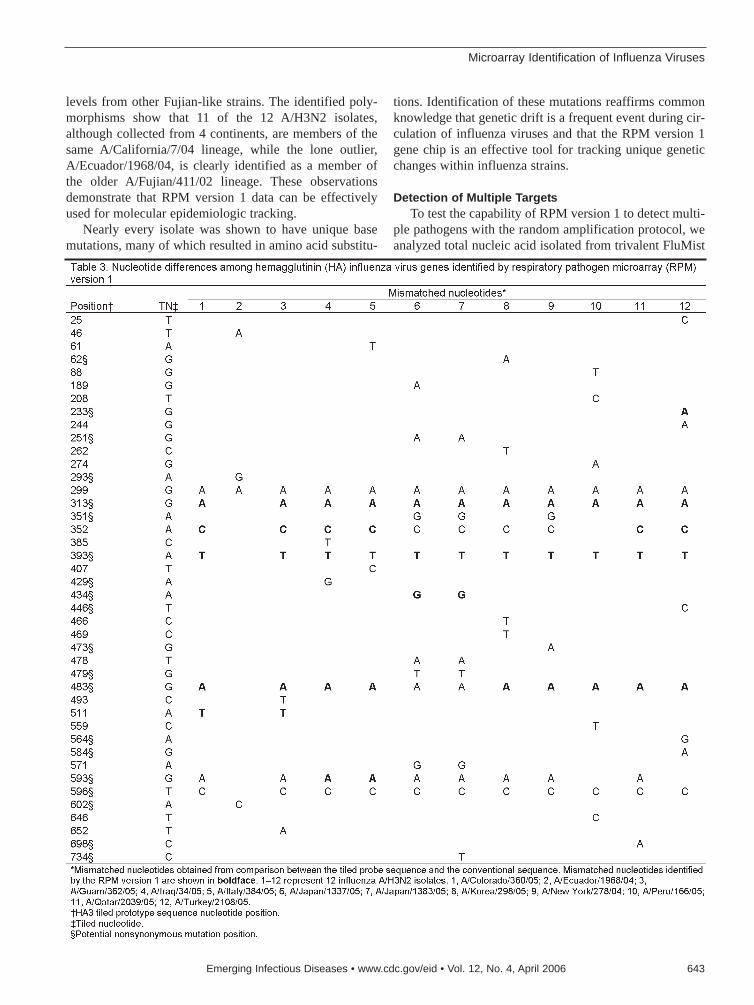
levels from other Fujian-like strains. The identified poly-morphisms show that 11 of the 12 A/H3N2 isolates,although collected from 4 continents, are members of thesame A/California/7/04 lineage, while the lone outlier,A/Ecuador/1968/04, is clearly identified as a member ofthe older A/Fujian/411/02 lineage. These observationsdemonstrate that RPM version 1 data can be effectivelyused for molecular epidemiologic tracking.
Nearly every isolate was shown to have unique basemutations, many of which resulted in amino acid substitu-
tions. Identification of these mutations reaffirms commonknowledge that genetic drift is a frequent event during cir-culation of influenza viruses and that the RPM version 1gene chip is an effective tool for tracking unique geneticchanges within influenza strains.
Detection of Multiple TargetsTo test the capability of RPM version 1 to detect multi-
ple pathogens with the random amplification protocol, weanalyzed total nucleic acid isolated from trivalent FluMist
Microarray Identification of Influenza Viruses
Emerging Infectious Diseases • www.cdc.gov/eid • Vol. 12, No. 4, April 2006 643
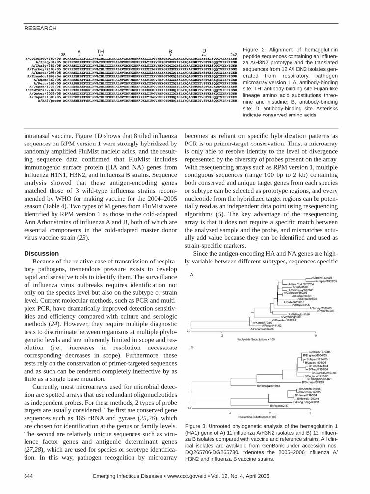
intranasal vaccine. Figure 1D shows that 8 tiled influenzasequences on RPM version 1 were strongly hybridized byrandomly amplified FluMist nucleic acids, and the result-ing sequence data confirmed that FluMist includesimmunogenic surface protein (HA and NA) genes frominfluenza H1N1, H3N2, and influenza B strains. Sequenceanalysis showed that these antigen-encoding genesmatched those of 3 wild-type influenza strains recom-mended by WHO for making vaccine for the 2004–2005season (Table 4). Two types of M genes from FluMist wereidentified by RPM version 1 as those in the cold-adaptedAnn Arbor strains of influenza A and B, both of which areessential components in the cold-adapted master donorvirus vaccine strain (23).
DiscussionBecause of the relative ease of transmission of respira-
tory pathogens, tremendous pressure exists to developrapid and sensitive tools to identify them. The surveillanceof influenza virus outbreaks requires identification notonly on the species level but also on the subtype or strainlevel. Current molecular methods, such as PCR and multi-plex PCR, have dramatically improved detection sensitiv-ities and efficiency compared with culture and serologicmethods (24). However, they require multiple diagnostictests to discriminate between organisms at multiple phylo-genetic levels and are inherently limited in scope and res-olution (i.e., increases in resolution necessitatecorresponding decreases in scope). Furthermore, thesetests rely on the conservation of primer-targeted sequencesand as such can be rendered completely ineffective by aslittle as a single base mutation.
Currently, most microarrays used for microbial detec-tion are spotted arrays that use redundant oligonucleotidesas independent probes. For these methods, 2 types of probetargets are usually considered. The first are conserved genesequences such as 16S rRNA and gyrase (25,26), whichare chosen for identification at the genus or family levels.The second are relatively unique sequences such as viru-lence factor genes and antigenic determinant genes(27,28), which are used for species or serotype identifica-tion. In this way, pathogen recognition by microarray
becomes as reliant on specific hybridization patterns asPCR is on primer-target conservation. Thus, a microarrayis only able to resolve identity to the level of divergencerepresented by the diversity of probes present on the array.With resequencing arrays such as RPM version 1, multiplecontiguous sequences (range 100 bp to 2 kb) containingboth conserved and unique target genes from each speciesor subtype can be selected as prototype regions, and everynucleotide from the hybridized target regions can be poten-tially read as an independent data point using resequencingalgorithms (5). The key advantage of the resequencingarray is that it does not require a specific match betweenthe analyzed sample and the probe, and mismatches actu-ally add value because they can be identified and used asstrain-specific markers.
Since the antigen-encoding HA and NA genes are high-ly variable between different subtypes, sequences specific
RESEARCH
644 Emerging Infectious Diseases • www.cdc.gov/eid • Vol. 12, No. 4, April 2006
Figure 2. Alignment of hemagglutininpeptide sequences containing an influen-za A/H3N2 prototype and the translatedsequences from 12 A/H3N2 isolates gen-erated from respiratory pathogenmicroarray version 1. A, antibody-bindingsite; TH, antibody-binding site Fujian-likelineage amino acid substitutions threo-nine and histidine; B, antibody-bindingsite; D, antibody-binding site. Asterisksindicate conserved amino acids.
Figure 3. Unrooted phylogenetic analysis of the hemagglutinin 1(HA1) gene of A) 11 influenza A/H3N2 isolates and B) 12 influen-za B isolates compared with vaccine and reference strains. All clin-ical isolates are available from GenBank under accession nos.DQ265706-DG265730. *denotes the 2005–2006 influenza A/H3N2 and influenza B vaccine strains.
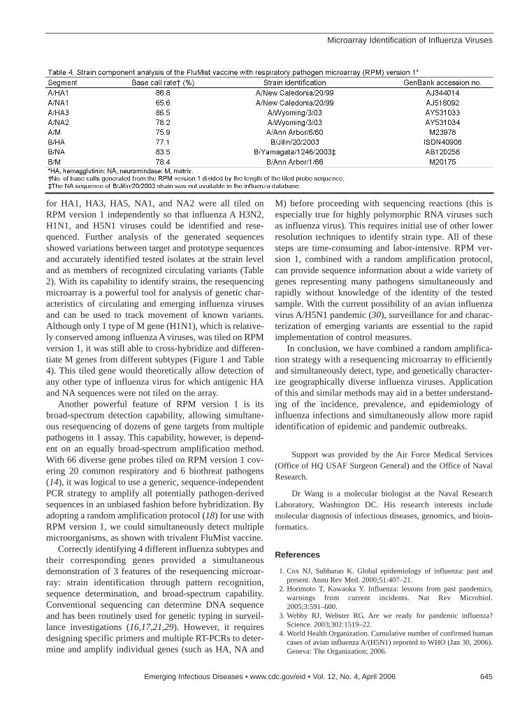
for HA1, HA3, HA5, NA1, and NA2 were all tiled onRPM version 1 independently so that influenza A H3N2,H1N1, and H5N1 viruses could be identified and rese-quenced. Further analysis of the generated sequencesshowed variations between target and prototype sequencesand accurately identified tested isolates at the strain leveland as members of recognized circulating variants (Table2). With its capability to identify strains, the resequencingmicroarray is a powerful tool for analysis of genetic char-acteristics of circulating and emerging influenza virusesand can be used to track movement of known variants.Although only 1 type of M gene (H1N1), which is relative-ly conserved among influenza A viruses, was tiled on RPMversion 1, it was still able to cross-hybridize and differen-tiate M genes from different subtypes (Figure 1 and Table4). This tiled gene would theoretically allow detection ofany other type of influenza virus for which antigenic HAand NA sequences were not tiled on the array.
Another powerful feature of RPM version 1 is itsbroad-spectrum detection capability, allowing simultane-ous resequencing of dozens of gene targets from multiplepathogens in 1 assay. This capability, however, is depend-ent on an equally broad-spectrum amplification method.With 66 diverse gene probes tiled on RPM version 1 cov-ering 20 common respiratory and 6 biothreat pathogens(14), it was logical to use a generic, sequence-independentPCR strategy to amplify all potentially pathogen-derivedsequences in an unbiased fashion before hybridization. Byadopting a random amplification protocol (18) for use withRPM version 1, we could simultaneously detect multiplemicroorganisms, as shown with trivalent FluMist vaccine.
Correctly identifying 4 different influenza subtypes andtheir corresponding genes provided a simultaneousdemonstration of 3 features of the resequencing microar-ray: strain identification through pattern recognition,sequence determination, and broad-spectrum capability.Conventional sequencing can determine DNA sequenceand has been routinely used for genetic typing in surveil-lance investigations (16,17,21,29). However, it requiresdesigning specific primers and multiple RT-PCRs to deter-mine and amplify individual genes (such as HA, NA and
M) before proceeding with sequencing reactions (this isespecially true for highly polymorphic RNA viruses suchas influenza virus). This requires initial use of other lowerresolution techniques to identify strain type. All of thesesteps are time-consuming and labor-intensive. RPM ver-sion 1, combined with a random amplification protocol,can provide sequence information about a wide variety ofgenes representing many pathogens simultaneously andrapidly without knowledge of the identity of the testedsample. With the current possibility of an avian influenzavirus A/H5N1 pandemic (30), surveillance for and charac-terization of emerging variants are essential to the rapidimplementation of control measures.
In conclusion, we have combined a random amplifica-tion strategy with a resequencing microarray to efficientlyand simultaneously detect, type, and genetically character-ize geographically diverse influenza viruses. Applicationof this and similar methods may aid in a better understand-ing of the incidence, prevalence, and epidemiology ofinfluenza infections and simultaneously allow more rapididentification of epidemic and pandemic outbreaks.
Support was provided by the Air Force Medical Services(Office of HQ USAF Surgeon General) and the Office of NavalResearch.
Dr Wang is a molecular biologist at the Naval ResearchLaboratory, Washington DC. His research interests includemolecular diagnosis of infectious diseases, genomics, and bioin-formatics.
References
1. Cox NJ, Subbarao K. Global epidemiology of influenza: past andpresent. Annu Rev Med. 2000;51:407–21.
2. Horimoto T, Kawaoka Y. Influenza: lessons from past pandemics,warnings from current incidents. Nat Rev Microbiol.2005;3:591–600.
3. Webby RJ, Webster RG. Are we ready for pandemic influenza?Science. 2003;302:1519–22.
4. World Health Organization. Cumulative number of confirmed humancases of avian influenza A/(H5N1) reported to WHO (Jan 30, 2006).Geneva: The Organization; 2006.
Microarray Identification of Influenza Viruses
Emerging Infectious Diseases • www.cdc.gov/eid • Vol. 12, No. 4, April 2006 645

5. Hacia JG. Resequencing and mutational analysis using oligonu-cleotide microarrays. Nat Genet. 1999;21:42–7.
6. Warrington JA, Shah NA, Chen X, Janis M, Liu C, Kondapalli S, etal. New developments in high-throughput resequencing and variationdetection using high density microarrays. Hum Mutat.2002;19:402–9.
7. Cutler DJ, Zwick ME, Carrasquillo MM, Yohn CT, Tobin KP, KashukC, et al. High-throughput variation detection and genotyping usingmicroarrays. Genome Res. 2001;11:1913–25.
8. Vahey M, Nau ME, Barrick S, Cooley JD, Sawyer R, Sleeker AA, etal. Performance of the Affymetrix GeneChip HIV PRT 440 platformfor antiretroviral drug resistance genotyping of human immunodefi-ciency virus type 1 clades and viral isolates with length polymor-phisms. J Clin Microbiol. 1999;37:2533–7.
9. Gingeras TR, Ghandour G, Wang E, Berno A, Small PM,Drobniewski F, et al. Simultaneous genotyping and species identifi-cation using hybridization pattern recognition analysis of genericMycobacterium DNA arrays. Genome Res. 1998;8:435–48.
10. Wilson KH, Wilson WJ, Radosevich JL, DeSantis TZ, ViswanathanVS, Kuczmarski TA, et al. High-density microarray of small-subunitribosomal DNA probes. Appl Environ Microbiol. 2002;68:2535–41.
11. Zwick ME, McAfee F, Cutler DJ, Read TD, Ravel J, Bowman GR, etal. Microarray-based resequencing of multiple Bacillus anthracis iso-lates. Genome Biol. 2005;6:R10.
12. Winzeler EA, Richards DR, Conway AR, Goldstein AL, Kalman S,McCullough MJ, et al. Direct allelic variation scanning of the yeastgenome. Science. 1998;281:1194–7.
13. Wang DG, Fan JB, Siao CJ, Breno A, Young P, Sapolsky R, et al.Large-scale identification, mapping, and genotyping of single-nucleotide polymorphisms in the human genome. Science.1998;280:1077–82.
14. Lin B, Wang Z, Vora GJ, Thornton JA, Schnur JM, Thach DC, et al.Broad spectrum respiratory tract pathogen identification using rese-quencing DNA microarray. Genome Res. 2006 Feb 15; [Epub aheadof print].
15. Canas LC, Lohman K, Pavlin JA, Endy T, Singh DL, Pandey P, et al.The Department of Defense laboratory-based global influenza sur-veillance system. Mil Med. 2000;165 (Suppl 2):52–6.
16. Daum LT, Canas LC, Smith CB, Klimov A, Huff W, Barnes W, et al.Genetic and antigenic analysis of the first A/New Caledonia/20/99-like H1N1 influenza isolates reported in the Americas. Emerg InfectDis. 2002;8:408–12.
17. Daum LT, Shaw MW, Klimov AI, Canas LC, Macias EA, NiemeyerD, et al. Influenza A (H3N2) outbreak, Nepal. Emerg Infect Dis.2005;11:1186–91.
18. Wang D, Coscoy L, Zylberberg M, Avila PC, Boushey HA, GanemD, et al. Microarray-based detection and genotyping of viralpathogens. Proc Natl Acad Sci U S A. 2002;99:15687–92.
19. Macken C, Lu H, Goodman J, Boykin L. Options for the control ofinfluenza. In: Osterhaus AD, Cox N, Hampson AW, editors. The valueof a database in surveillance and vaccine selection. Amsterdam:Elsevier Science; 2001. p. 103–6.
20. Altschul SF, Madden TL, Schaffer AA, Zhang J, Zhang Z, Miller W,et al. Gapped BLAST and PSI-BLAST: a new generation of proteindatabase search programs. Nucleic Acids Res. 1997;25:3389–402.
21. Holmes EC, Ghedin E, Miller N, Taylor J, Bao Y, St George K, et al.Whole-genome analysis of human influenza A virus reveals multiplepersistent lineages and reassortment among recent H3N2 viruses.PLoS Biol. 2005;3:e300.
22. Centers for Disease Control and Prevention. 2004–05 U.S. influenzaseason summary. [cited 2006 Jan 30]. Available fromhttp://www.cdc.gov/flu/weekly/weeklyarchives2004-2005/04-05summary.htm
23. Wareing MD, Tannock GA. Live attenuated vaccines against influen-za: a historical review. Vaccine. 2001;19:3320–30.
24. Ellis JS, Zambon MC. Molecular diagnosis of influenza. Rev MedVirol. 2002;12:375–89.
25. Warsen AE, Krug MJ, LaFrentz S, Stanek DR, Loge FJ, Call DR.Simultaneous discrimination between 15 fish pathogens by using 16Sribosomal DNA PCR and DNA microarrays. Appl EnvironMicrobiol. 2004;70:4216–21.
26. Roth SB, Jalava J, Ruuskanen O, Ruohola A, Nikkari S. Use of anoligonucleotide array for laboratory diagnosis of bacteria responsiblefor acute upper respiratory infections. J Clin Microbiol.2004;42:4268–74.
27. Chizhikov V, Rasooly A, Chumakov K, Levy DD. Microarray analy-sis of microbial virulence factors. Appl Environ Microbiol.2001;67:3258–63.
28. Wang Z, Vora GJ, Stenger DA. Detection and genotyping ofEntamoeba histolytica, Entamoeba dispar, Giardia lamblia, andCryptosporidium parvum by oligonucleotide microarray. J ClinMicrobiol. 2004;42:3262–71.
29. Ghedin E, Sengamalay NA, Shumway M, Zaborsky J, Feldblyum T,Subbu V, et al. Large-scale sequencing of human influenza reveals thedynamic nature of viral genome evolution. Nature. 2005;437:1162-6.
30. The World Health Organization Global Influenza ProgramSurveillance Network. Evolution of H5N1 avian influenza viruses inAsia. Emerg Infect Dis. 2005;11:1515–21.
Address for correspondence: David A. Stenger, Center for Bio/MolecularScience and Engineering, Code 6900, Naval Research Laboratory,Washington, DC 20375, USA; fax: 202-767-9598; email:[email protected]
RESEARCH
646 Emerging Infectious Diseases • www.cdc.gov/eid • Vol. 12, No. 4, April 2006

We conducted a systematic review of the scientific lit-erature from 1966 to 2005 to determine whether animalscould provide early warning of a bioterrorism attack, serveas markers for ongoing exposure risk, and amplify or prop-agate a bioterrorism outbreak. We found evidence that, forcertain bioterrorism agents, pets, wildlife, or livestock couldprovide early warning and that for other agents, humanswould likely manifest symptoms before illness could bedetected in animals. After an acute attack, active surveil-lance of wild or domestic animal populations could helpidentify many ongoing exposure risks. If certain bioterror-ism agents found their way into animal populations, theycould spread widely through animal-to-animal transmissionand prove difficult to control. The public health infrastruc-ture must look beyond passive surveillance of acute animaldisease events to build capacity for active surveillance andintervention efforts to detect and control ongoing outbreaksof disease in domestic and wild animal populations.
Most priority bioterrorism agents are zoonotic in ori-gin. As a result, an attack on human populations with
a bioterrorism agent would likely pose a health risk to ani-mal populations in the target area; therefore, integratingveterinary and human public health surveillance efforts isessential. The Centers for Disease Control and Prevention(CDC), in planning for the early detection and manage-ment of a biological terrorism attack, has recommendedthe “prompt diagnosis of unusual or suspicious healthproblems in animals,” as well as establishing “criteria forinvestigating and evaluating suspicious clusters of humanand animal disease or injury and triggers for notifying lawenforcement of suspected acts of biological or chemical
terrorism” (1). Similarly, an indicator of a biological ter-rorism attack would be “increased numbers of sick or deadanimals, often of different species. Some BW (biologicalwarfare) agents are capable of infecting/intoxicating awide range of hosts” (2). In part because of such recom-mendations, calls have been made for enhanced veterinarysurveillance for outbreaks of animal disease caused bybioterrorism agents and better communication betweenanimal health and human health professionals. For suchefforts to succeed, the relevance to human health of diseaseevents in animals must be established. The potential use ofanimals as “sentinels” of a human bioterrorism attack canbe differentiated from the possibility of a direct attack onanimals of agricultural importance (agroterrorism) and isthe subject of this review.
First, animals could provide an early warning tohumans if clinical signs could be detected before humanillness emerged or soon enough to allow preventive meas-ures to be initiated. This early detection could occurbecause an animal species had increased susceptibility to aparticular agent, because the disease caused by the agenthad a shorter incubation period, or because animals wereexposed sooner (or at more intense and continuous levels)than the human population (2). The simultaneous appear-ance of disease signs and symptoms in animals may con-tribute to the more rapid identification of a biologicalwarfare agent that was producing nonspecific effects innearby persons.
Second, if a released biological agent persists in theenvironment (such as soil, water, or air), active surveil-lance for sporadic illness in animals could help detectongoing exposure risks. Additionally, the geographic pat-tern of sick or dead animals could indicate the persistenceof a biological threat (2).
Finally, animal populations such as wild birds, com-mercially shipped livestock, and animals involved in the
Animals as Sentinels ofBioterrorism Agents
Peter Rabinowitz,* Zimra Gordon,*† Daniel Chudnov,* Matthew Wilcox,* Lynda Odofin,* Ann Liu,* and Joshua Dein‡
Emerging Infectious Diseases • www.cdc.gov/eid • Vol. 12, No. 4, April 2006 647
*Yale University School of Medicine, New Haven, Connecticut,USA; †Rippowam Animal Hospital, Stamford, Connecticut, USA;and ‡US Geological Survey National Wildlife Health Center,Madison, Wisconsin, USA

local or international pet trade, could play a role in themaintenance and spread of an epidemic attributable to anintentional release of a biological agent. Detecting theagent in such mobile populations could therefore signal theongoing spread of the agent and provide an opportunity forinterventions to prevent further spread.
Previous reviews have discussed the implications ofbioterrorism attacks on human and animal health (3). Yetthese reviews did not examine the strength of evidence orattempt to determine whether animals could be effectivesentinels for particular agents.
We therefore reviewed the biomedical literature for evi-dence that animals could fulfill the above criteria for sen-tinel potential. We also hypothesized that large gaps inknowledge exist in this area, including different levels ofevidence regarding specific agents and types of animals.
MethodsWe systematically searched the biomedical literature
from 1966 to 2005 for reports of adverse health events inanimals that were attributed to potential bioterrorismagents. The CDC publication Biological and ChemicalTerrorism: Strategic Plan for Preparedness and Response(1) contains a list of biological and chemical agents thatcould be used in a terrorist attack. Infectious agents arecategorized as A, B, or C, depending on their risk to pub-lic health. We searched the Medline database for reports ofanimal exposure to these biological agents. As a furthercheck, we performed focused searches for individualagents in the CAB Abstracts and Agricultural OnlineAccess (AGRICOLA) databases and also reviewed thebibliographies of recent bioterrorism reviews to locateadditional sources.
Our search method used both the name of the agent andthe terms “animals, wild” “animals, domestic” and “ani-mals, zoo.” For each agent, we searched for peer-reviewedstudies of infection in animal populations caused by a spe-cific agent, as well as authoritative subject reviews. Theepisodes of infection included both actual bioterrorismevents as well as naturally occurring epizootics. Thissearch process identified ≈6,000 potential citations,including original journal articles, textbook chapters, andreviews, which were manually culled for relevance to ani-mal sentinel issues; this process resulted in ≈200 citationsavailable for final analysis.
Studies that included data about relative incubationperiods and susceptibilities in animals were compared tohuman data to determine whether evidence supported theuse of animals as early warning of bioterrorism agents. Wealso included in this category reports of animals displayingevidence of infection before nearby human populationsdid. Data on human incubation periods and infective dosesfor individual agents were obtained from standard refer-
ences on biological warfare and terrorism (4). Studies thatdetected symptomatic infection or biomarkers of infectionfor agents that persist in the environment were reviewed todetermine whether they supported the utility of animals forongoing exposure monitoring. Studies that demonstrated asubstantial degree of animal-to-animal or animal-to-human transmission (with or without a vector) were con-sidered to provide evidence that animals could propagatean outbreak of infectious disease caused by a deliberatelyreleased pathogen.
Analysis of EvidenceStudies located in the above search were classified
according to agent, disease, species, and study method, andthese data were then entered into an online database of ani-mals as sentinels of human environmental health hazards(available from http://canarydatabase.org/) for furtheranalysis. For the purposes of this review, we created a tax-onomy for evidence regarding animals of sentinels basedon existing evidence-based medicine taxonomies that pro-vide guidelines for assigning levels of evidence based onquality and consistency of scientific studies (5). We con-sidered level 1 evidence studies to include experimentalstudies, cohort studies, and systematic reviews of high-quality studies with consistent findings. We classifiedcase-control studies and cross-sectional surveys of animalsas level 2 evidence. Evidence from professional consensusstatements, textbooks, and descriptive case reports wasclassified as level 3 evidence. To arrive at an overallstrength of recommendation based on a body of evidence,we used these levels of evidence to determine the overallstrength of the recommendation that animals could serve inthe 3 sentinel capacities of early warning signal, ongoingexposure indicator, and potential propagator and spreaderof a bioterrorism agent.
ResultsThe Table displays the evidence found for animals serv-
ing in a sentinel capacity for specific agents or classes ofagents.
Evidence That Animals Provide Early Warning of an Acute Bioterrorism Attack
For a number of agents, this review found evidence thatanimals might be affected before human populations. ForBacillus anthracis, whether animals would have a shorterincubation period in the event of an aerosol release was notclear, since the incubation period in the 2001 mail attackswas <4 days, while during the 1979 release of B. anthracisfrom a Soviet military laboratory, human symptoms beganin 2 days, with death in as few as 6 days (6). At the sametime, while human cases in Sverdlovsk were concentratedalong the path of the prevailing wind <4 km from the lab-
RESEARCH
648 Emerging Infectious Diseases • www.cdc.gov/eid • Vol. 12, No. 4, April 2006
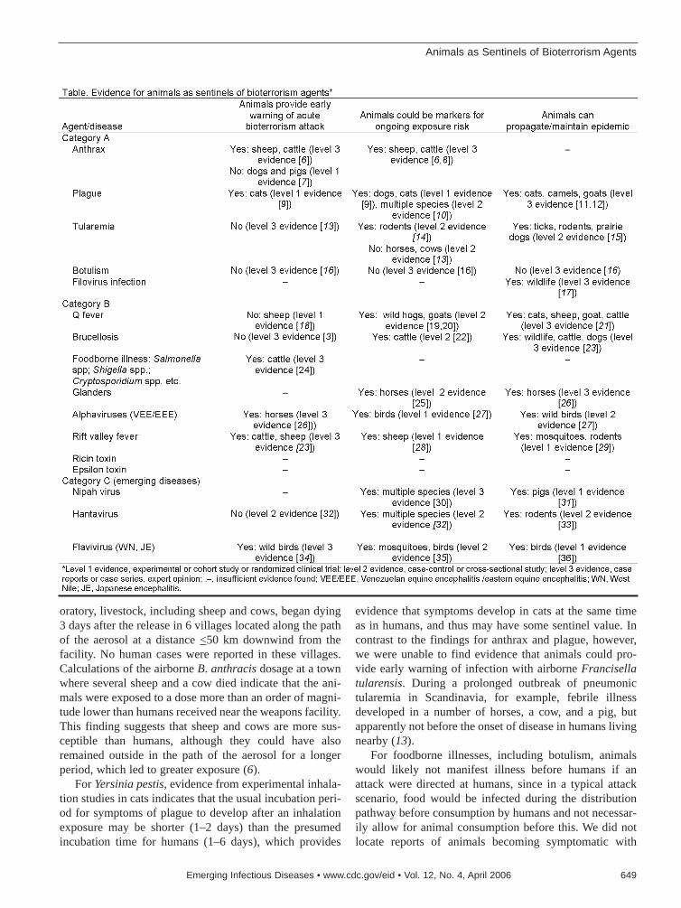
oratory, livestock, including sheep and cows, began dying3 days after the release in 6 villages located along the pathof the aerosol at a distance <50 km downwind from thefacility. No human cases were reported in these villages.Calculations of the airborne B. anthracis dosage at a townwhere several sheep and a cow died indicate that the ani-mals were exposed to a dose more than an order of magni-tude lower than humans received near the weapons facility.This finding suggests that sheep and cows are more sus-ceptible than humans, although they could have alsoremained outside in the path of the aerosol for a longerperiod, which led to greater exposure (6).
For Yersinia pestis, evidence from experimental inhala-tion studies in cats indicates that the usual incubation peri-od for symptoms of plague to develop after an inhalationexposure may be shorter (1–2 days) than the presumedincubation time for humans (1–6 days), which provides
evidence that symptoms develop in cats at the same timeas in humans, and thus may have some sentinel value. Incontrast to the findings for anthrax and plague, however,we were unable to find evidence that animals could pro-vide early warning of infection with airborne Francisellatularensis. During a prolonged outbreak of pneumonictularemia in Scandinavia, for example, febrile illnessdeveloped in a number of horses, a cow, and a pig, butapparently not before the onset of disease in humans livingnearby (13).
For foodborne illnesses, including botulism, animalswould likely not manifest illness before humans if anattack were directed at humans, since in a typical attackscenario, food would be infected during the distributionpathway before consumption by humans and not necessar-ily allow for animal consumption before this. We did notlocate reports of animals becoming symptomatic with
Animals as Sentinels of Bioterrorism Agents
Emerging Infectious Diseases • www.cdc.gov/eid • Vol. 12, No. 4, April 2006 649

foodborne illness before the onset of human cases.However, if the attack on the food supply were directed atthe animals themselves, they could potentially manifestsymptoms before humans who would consume the meat,eggs, or dairy products (24). Attacks on water supplieswith agents such as Clostridium botulinum could puthumans at risk as well, although dilution and water treat-ment would reduce the risk (37). An exception may beCryptosporidium spp., which have caused widespread out-breaks through the water supply. Waterfowl die-offs fromtype C and type E botulism have been well documented,although these types are not well recognized as causes ofclinical C. botulinum poisoning in humans, but the fact thatprimates are susceptible to type C makes C. botulinum poi-soning a possibility. On the whole, however, an attack onhuman populations with C. botulinum would probably notbe first detected in animals; the illness would have such ashort incubation period in humans that they would becomesymptomatic at the same time as or before the animals.
For alphaviruses, natural outbreaks have often appearedin animal populations before they affected humans, forexample, eastern equine encephalitis virus often appears inequines 2 weeks before humans become symptomatic (28).Whether the same pattern would hold true during an attackwith an aerosol is not clear. For certain newer agents, suchas filoviruses and Nipah virus, current evidence is insuffi-cient to state whether after a generalized release of anaerosolized agent, animal infection would precede that inhumans. Studies in Africa have demonstrated that Ebolavirus outbreaks can be preceded by deaths in primates aswell as in other animal species such as duikers (type ofantelope) (17), but whether a generalized attack that usedEbola virus in the United States would affect certain ani-mal species first is unknown.
For a number of agents, including Brucella spp.,Coxiella burnetii, and hantavirus, infection in animals iseither asymptomatic or develops so slowly that recogniza-ble human cases seem certain to precede animal cases ifthe agents are released as an aerosol. Finally, the illnessescaused by some agents appear to have shorter incubationtimes in animals, for example, the 12-hour incubation peri-od for Rift Valley fever in calves and lambs (23) comparedto the incubation period of several days in humans.
Evidence That Animals Could Be Markers for Ongoing Exposure Risk
After the acute release of a bioterrorist agent, publichealth officials could be faced with the possibility of anagent persisting in the environment. Anthrax spores cansurvive for years in soil. Therefore, monitoring for spo-radic cases in animal populations such as livestock couldindicate so-called exposure hot spots. Although dogs andcats are less susceptible to B. anthracis than ruminants,
their proximity to humans and their contact with soil couldmake them sentinels; for example, anthrax developed in aLabrador retriever after the dog hunted in a freshly plowedfield (8). In the case of ongoing exposures to an agent thathas become established in an animal population, casedetection could be useful; this situation has been seen forplague in cats (9). Similarly, agents like Brucella spp. andC. burnetii, although they do not cause severe acute illnessin animals, could be detected by recognizing increasedrates of abortion among a variety of species.
Aside from case detection, active surveillance with sur-veys of animals may be useful; this surveillance mayrequire testing wildlife as well. Such testing could involveantibody seroprevalence or use of polymerase chain reac-tion techniques to detect antigen. Evidence about the use-fulness of such an approach was inconsistent. During anepidemic of pneumonic tularemia, attributable to contami-nated hay, the etiologic agent, F. tularensis, can persist inthe environment; a serosurvey of asymptomatic livestock(horses and cows) did not show evidence of exposure (13).By contrast, in a more recent tularemia outbreak, serosur-veys in the wildlife population did show antibodies in askunk and a rat that lived near persons who had becomeinfected after mowing fields (14).
Evidence That Animals Could Propagate an Epidemic Caused by a Bioterrorism Agent
A number of biological terrorism agents have littlepotential for secondary spread in either animal or humanpopulations, including B. anthracis and C. botulinum. Forother agents, however, we found evidence that their intro-duction into an animal population could cause an epizoot-ic that would then place additional human populations atrisk. For example, studies of mosquitoes native to theUnited States have demonstrated their potential to spread adisease such as Rift Valley fever through livestock andother animal populations (29), even though person-to-per-son transmission does not occur. The results of animal sur-veillance for Ebola virus in Africa found that ongoingoutbreaks in both primates and duikers suggest that thevirus may be able to propagate in a wildlife population(17), however, this characteristic has not been demonstrat-ed in US wildlife species.
Agents such as C. burnetii and Brucella spp. spreadeasily in animal populations through direct contact and canthen pose a wider risk to humans, even though human-to-human transmission does not occur. Agents such asalphaviruses that are prevalent in wild bird populations canspread over a wide area in a short time (27). Experimentalstudies have documented that viruses such as West Nilevirus can easily spread from animal to animal in bird pop-ulations (36).
RESEARCH
650 Emerging Infectious Diseases • www.cdc.gov/eid • Vol. 12, No. 4, April 2006

DiscussionFor a number of biological terrorism agents, we found
evidence that animals could provide early warning of anacute attack. For the agents for which we found evidenceof sentinel potential, a key factor was the relative exposurerisk of an animal compared to that of a nearby human pop-ulation. However, in an actual event involving bothhumans and animals, the fact that disease was detectedsooner in animals could be due to an interplay of a numberof factors, including local infrastructure of animal andhuman health services, public awareness, and laboratorycapacity. For other agents, however, humans woulddemonstrate symptoms at the same time as nearby animalsor before. Therefore, the strength of evidence regardinganimals serving as early indicators of an attack dependsstrongly on the agent and species involved. For someagents for which animals would not provide early warning,however, animals could help detect pockets of ongoingexposure risk. For the remainder of agents, evidenceregarding the value of animals as sentinels is insufficient atthis time.
Overall, according to our classification taxonomy, thestrength of the recommendation that animals could provideearly warning of an acute bioterrorism attack seems to be,at best, “fair” because of the inconsistency of the evidence.A somewhat more consistent level of evidence appeared tosupport the recommendation that animals could be mark-ers for ongoing exposure risk and also that animals couldplay a strong role in propagating outbreaks caused by par-ticular agents. At the same time, our ability to assess theoverall strength of evidence for such recommendationswas hampered by large gaps in current knowledge.
These findings suggest the need for certain steps relat-ed to preparedness for biological agent attacks. First,improved communication is needed between animal healthand human health professionals, so that sentinel eventscould be rapidly detected. Such improvement would meanovercoming existing barriers to communication; a recentsurvey found that physicians and veterinarians communi-cate little about zoonotic issues (38). Also, an adequatesurveillance network should be developed to detect unusu-al health events in animal populations. Data on usualtrends is missing for most animal species that could bepotential sentinels. Whether public health resources can becommitted to gathering such baseline data remains an openquestion.
Second, the results of this review indicate that activesurveillance of animal populations, including wildlife andcompanion animals, could fill a critical need in the after-math of an attack involving certain bioterrorism agents byhelping identify persistent sources of infection in the envi-ronment. Third, better approaches for intervention areneeded to be able to stem the propagation and amplifica-
tion of an introduced biological warfare agent into a wildor domestic animal population. The US experience withWest Nile virus reflects the difficulties of controlling anemerging zoonotic threat as it spreads through animal pop-ulations (39).
Finally, the results of this review point out the need foradditional research to fill knowledge gaps about animals assentinels of human disease threats, including data on rela-tive susceptibilities and exposure pathways for animalspecies living near human populations. Concrete stepscould include establishment of surveillance veterinaryclinics in strategic areas with incentives for practitioners toreport unusual events. Another approach would be to makegreater use of electronic databases of animal diseases suchas those used by the Banfield Clinics, a nationwide chainof veterinary practices. Similar efforts could be useful withwildlife populations.
Such steps would foster ongoing communicationbetween community practitioners and regional public andprivate veterinary diagnostic laboratories to establish base-line disease incidence trends and algorithms to identifyoutbreaks. Common links or web-based interfaces shouldbe developed to integrate human and animal disease sur-veillance information. Reporting systems for wildlife pro-fessionals and the public should be created, and their useshould be encouraged to document unusual disease eventsand die-offs. Another constructive step would be toimprove the capacity of existing veterinary rapid-responseteams, which exist in many states, to carry out active sur-veillance with animal populations as well as to improve thecoordination of veterinary diagnostic laboratories. Again,barriers to funding and cooperation between animal andhuman health agencies need to be addressed. In the past,these have hampered efforts to have a coordinatedapproach to collection of animal surveillance data). Inaddition, state-based efforts would need to be coordinatedon a regional and national scale. The growing awarenessthat animal health and human health are inextricablylinked, however, makes cooperation between human andanimal health professionals imperative to strengthen theevidence base that will allow for rational use of animaldata in public health decision-making.
AcknowledgmentWe thank Ron Romero for assistance with document
retrieval.
Funding for this project was provided by National Library ofMedicine Grant #1-G08-LM07881-01. Some of this material waspresented as a poster at the International Conference on EmergingInfectious Diseases, March 2004, Atlanta, Georgia, USA.
Dr Rabinowitz is associate professor of medicine at YaleUniversity School of Medicine and director of clinical services
Animals as Sentinels of Bioterrorism Agents
Emerging Infectious Diseases • www.cdc.gov/eid • Vol. 12, No. 4, April 2006 651
for the Yale Occupational and Environmental Medicine Program.His research focuses on the use of animals as sentinels of humanenvironmental health hazards.
References
1. Biological and chemical terrorism: strategic plan for preparednessand response. Recommendations of the CDC Strategic PlanningWorkgroup. MMWR Morb Mortal Wkly Rep. 2000;49:1–14.
2. Cottrell TS, Morgan ER. Animal surveillance in NBC defensive oper-ations. J R Army Med Corps. 2003;149:225–30.
3. Davis RG. The ABCs of bioterrorism for veterinarians, focusing onCategory A agents. J Am Vet Med Assoc. 2004;224:1084–95.
4. Kortepeter M, Eitzen E Jr, Mckee K Jr, editors. USAMRIID’s med-ical management of biological casualties handbook. 4th ed. McLean(VA): International Medical Publishing Inc; 2001.
5. Phillips B, Ball C, Sackett D, Badenoch D, Straus S, Haynes B, et al.Levels of evidence and grades of recommendation. Oxford Centre forEvidence Based Medicine; 2001 [cited 2005 Nov 28]. Available fromhttp://www.cebm.net/levels_of_evidence.asp#levels.
6. Meselson M, Guillemin J, Hugh-Jones M, Langmuir A, Popova I,Shelokov A, et al. The Sverdlovsk anthrax outbreak of 1979.Science.1994;266:1202–8.
7. Gleiser CA, Gochenour WS Jr, Ward MK. Pulmonary lesions in dogsand pigs exposed to a cloud of anthrax spores. J Comp Pathol.1968;78:445–8.
8. McGee ED, Fritz DL, Ezzell JW, Newcomb HL, Brown RJ, Jaax NK.Anthrax in a dog. Vet Pathol. 1994;31:471–3.
9. Rust JH Jr, Cavanaugh DC, O’Shita R, Marshall JD Jr. The role ofdomestic animals in the epidemiology of plague. I. Experimentalinfection of dogs and cats. J Infect Dis. 1971;124:522–6.
10. Kilonzo BS, Mbise TJ, Makundi RH. Plague in Lushoto district,Tanzania, 1980-1988. Trans R Soc Trop Med Hyg. 1992;86:444-5.
11. Eidson M, Thilsted JP, Rollag OJ. Clinical, clinicopathologic, andpathologic features of plague in cats: 119 cases (1977–1988). J AmVet Med Assoc.1991;199:1191–7.
12. Christie AB, Chen TH, Elberg SS. Plague in camels and goats: theirrole in human epidemics. J Infect Dis. 1980;141:724–6.
13. Dahlstrand S, Ringertz O, Zetterberg B. Airborne tularemia inSweden. Scandinavian J Infect Dis. 1971;3:7–16.
14. Feldman, KA, Enscore RE, Lathrop SR, Matyas BT, McGuill M,Schriefer ME, et al. An outbreak of primary pneumonic tularemia onMartha’s Vineyard. N Engl J Med. 2001;345:1601–6.
15. Avashia SB, Peterson JM, Lindley CM, Schriefer ME, Gage KL,Cetron M, et al. First reported prairie dog-to-human tularemia trans-mission, Texas, 2002. Emerg Infect Dis. 2004;10:483–6.
16. Arnon SS, Schechter R, Inglesby TV, Henderson DA, Bartlett JG,Ascher MS, et al. Botulinum toxin as a biological weapon: medicaland public health management. JAMA. 2001;285:1059–70. Erratumin: JAMA 2001;285:2081
17. Leroy EM, Rouquet P, Formenty P, Souquiere S, Kilbourne A,Forment J, et al. Multiple Ebola virus transmission events and rapiddecline of central African wildlife. Science. 2004;303:387–90.
18. Martinov SP, Neikov P, Popov GV. Experimental Q fever in sheep.Eur J Epidemiol. 1989;5:428–31.
19. Clark RK, Jessup DA, Hird DW, Ruppanner R, Meyer ME. Serologicsurvey of California wild hogs for antibodies against selected zoonot-ic disease agents. J Am Vet Med Assoc. 1983;183:1248–51.
20. Scrimgeour EM, Al-Ismaily SI, Rolain JM, Al-Dhahry SH, El-Khatim HS, Raoult D. Q fever in human and livestock populations inOman. Ann N Y Acad Sci. 2003;990:221–5.
21. McQuiston JH, Childs JE, Thompson HA. Q fever. J Am Vet MedAssoc. 2002;221:796–9.
22. Kubuafor DK, Awumbila B, Akanmori BD. Seroprevalence of bru-cellosis in cattle and humans in the Akwapim-South district of Ghana:public health implications. Acta Trop. 2000;76:45-8.
23. Radostits OMG, Gay CC, Blood DC, Hinchcliff KW, editors.Veterinary medicine: a textbook of the diseases of cattle, sheep, pigs,goats and horses. 9th ed. London: Harcourt Publishers Ltd; 2000.
24. Cupp OS, Walker DE 2nd, Hillison J. Agroterrorism in the U.S.: keysecurity challenge for the 21st century. Biosecurity and Bioterrorism.2004;2:97–105.
25. Arun S, Neubauer H, Gurel A, Ayyildiz G, Kuscu B, Yesidere T, et al.Equine glanders in Turkey. Institute for Animal Pathology.1999;144:255–8.
26. Greene CE. Infectious diseases of the dog and cat. 2nd ed.Philadelphia: W.B. Saunders; 1998.
27. Howard JJ, Grayson MA, White DJ, Oliver J. Evidence for multiplefoci of eastern equine encephalitis virus (Togaviridae:Alphavirus) incentral New York State. J Med Entomol. 1996;33:421–32.
28. Olaleye OD, Tomori O, Schmitz H. Rift Valley fever in Nigeria:infections in domestic animals. Rev Sci Tech. 1996;15:937–46.
29. Gargan TP 2nd, Clark GG, Dohm DJ, Turell MJ, Bailey CL. Vectorpotential of selected North American mosquito species for Rift Valleyfever virus. Am J Trop Med Hyg. 1988;38:440–6.
30. Bunning M. Nipah virus outbreak in Malaysia, 1998–1999. Journal ofSwine Health Products. 2001;9:295–9.
31. Middleton DJ, Westbury HA, Morrissy CJ, van der Heide BM,Russell GM, Braun MA, et al. Experimental Nipah virus infection inpigs and cats. J Comp Pathol. 2002; 126:124–36.
32. Mills JN, Yates TL, Ksiazek TG, Peters CJ, Childs JE. Long-termstudies of Hantavirus reservoir populations in the southwesternUnited States: rationale, potential, and methods. Emerg Infect Dis.1999;5:95–101.
33. Traavik T, Sommer A, Mehl R, Berdal BP, Stavem K, Hunderi OH, etal., Nephropathia epidemica in Norway: antigen and antibodies inrodent reservoirs and antibodies in selected human populations. JHyg (Camb). 1984;93: 139–46.
34. Mostashari F, Kulldorff M, Hartman JJ, Miller JR, Kulasekera V.Dead bird clusters as an early warning system for West Nile virusactivity. Emerg Infect Dis. 2003;9:641–6.
35. Komar O, Robbins MB, Klenk K, Blitvich BJ, Marlenee NL,Burkhalter KL, et al. West Nile virus transmission in resident birds,Dominican Republic. Emerg Infect Dis. 2003;9:1299–302.
36. Komar N, Langevin S, Hinten S, Nemeth N, Edwards E, Hettler D, etal. Experimental infection of North American birds with the NewYork 1999 strain of West Nile virus. Emerg Infect Dis.2003;9:311–22.
37. Jernigan JA, Stephen DS, Ashford DA, Omenaca C, Topiel MS,Galbraith M, et al. Bioterrorism-related inhalational anthrax: the first10 cases reported in the United States. Emerg Infect Dis.2001;7:933–44.
38. Grant S, Olsen CW. Preventing zoonotic diseases in immunocompro-mised persons: the role of physicians and veterinarians. Emerg InfectDis. 1999;5:159–63.
39. Dauphin G, Zientara S, Zeller H, Murgue B. West Nile: worldwidecurrent situation in animals and humans. Comp Immunol MicrobiolInfect Dis. 2004;27:343–55.
Address for correspondence: Peter Rabinowitz, Yale University School ofMedicine, 135 College St, 3rd Floor, New Haven, CT, USA; fax: 203-785-7391; email: [email protected]
RESEARCH
652 Emerging Infectious Diseases • www.cdc.gov/eid • Vol. 12, No. 4, April 2006

To assess the economic impact of Lyme disease (LD),the most common vectorborne inflammatory disease in theUnited States, cost data were collected in 5 counties of theMaryland Eastern Shore from 1997 to 2000. Patients weredivided into 5 diagnosis groups, clinically defined early-stage LD, clinically defined late-stage LD, suspected LD,tick bite, and other related complaints. From 1997 to 2000,the mean per patient direct medical cost of early-stage LDdecreased from $1,609 to $464 (p<0.05), and the mean perpatient direct medical cost of late-stage LD decreased from$4,240 to $1,380 (p<0.05). The expected median of allcosts (direct medical cost, indirect medical cost, nonmed-ical cost, and productivity loss), aggregated across all diag-nosis groups of patients, was ≈$281 per patient. Thesefindings will help assess the economics of current andfuture prevention and control efforts.
Lyme disease (LD) is a multisystem, multistage, inflam-matory tickborne disorder caused by the spirochete
Borrelia burgdorferi. LD usually begins with an initialexpanding skin lesion, erythema migrans (EM), whichmay be followed by musculoskeletal, neurologic, and car-diac manifestations in later stages of the disease (1–3).Enzyme-linked immunosorbent assay and Western blottingtest are widely used to diagnose LD (4–6). LD is mostresponsive to antimicrobial drugs in the early stage, whilefurther intensive therapy may be necessary in the late stage(7,8). A variety of prevention and control procedures canbe implemented to prevent and reduce LD incidence,including, but not limited to, public education; personalprotection measures such as wearing protective clothing(gloves, long clothes), checking one’s body daily for ticks,avoiding tick-infested areas, and applying tick repellent(DEET, permethrin); host management; habitat modifica-tion; and chemical control (9,10). In 1998, the Food andDrug Administration approved a recombinant outer-sur-
face protein A (rOspA) LD vaccine (LYMErix, SmithKlineBeecham Biologicals, Rixensart, Belgium) for persons15–70 years of age (11). However, in 2002, SmithKlinewithdrew the vaccine, citing low demand. Therefore, per-sonal protection measures, early diagnosis, and early treat-ment are extremely important in preventing andcontrolling LD.
Since the first case reported in 1975 (12), LD hasbecome the most common vectorborne inflammatory dis-ease in the United States. Foci of LD are widely spread inthe northeastern, mid-Atlantic, and north-central regionsof the United States (13). Despite federal, state, and localefforts to prevent and control LD, total reported cases ofLD increased almost 3-fold from 1991 to 2002 (Figure 1).In 2002, the Centers for Disease Control and Prevention(CDC) received reports of 23,763 LD cases, 95% of whichwere from Connecticut, Delaware, Maine, Maryland,Massachusetts, Minnesota, New Hampshire, New Jersey,New York, Pennsylvania, Rhode Island, and Wisconsin(14). In Maryland, the overall incidence of LD was morethan twice as high as the overall incidence of LD in theUnited States (13.0 vs. 6.3 cases per 100,000 population)(13).
Assessing the economic impact of LD will help assessthe economics of current and future prevention and controlefforts. Although several studies of cost estimates of LDhave been published (e.g., 15), information on the econom-ic impact of LD is limited. Therefore, we conducted a 4-year study to estimate the economic impact of LD on theMaryland Eastern Shore.
Methods
Study Population and DataThis study was conducted in 5 counties (Caroline,
Dorchester, Kent, Queen Anne, and Talbot) on theMaryland Eastern Shore, an area where LD is endemic
Economic Impact of Lyme DiseaseXinzhi Zhang,* Martin I. Meltzer,* César A. Peña,†1 Annette B. Hopkins,†
Lane Wroth,‡ and Alan D. Fix†
Emerging Infectious Diseases • www.cdc.gov/eid • Vol. 12, No. 4, April 2006 653
*Centers for Disease Control and Prevention, Atlanta, Georgia,USA; †University of Maryland, Baltimore, Maryland, USA; and‡Care First-Easton Branch (previously Delmarva Health Plan),Easton, Maryland, USA
1Current affiliation: Maryland Department of Health and MentalHygiene, Baltimore, Maryland, USA
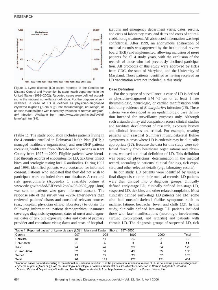
(Table 1). The study population includes patients living inthe 4 counties enrolled in Delmarva Health Plan (DHP, amanaged healthcare organization) and non-DHP patientsreceiving health care from office-based physicians in KentCounty from 1997 to 2000. Eligible patients were identi-fied through records of encounters for LD, tick bites, insectbites, and serologic testing for LD antibodies. During 1997and 1998, identified patients were contacted for informedconsent. Patients who indicated that they did not wish toparticipate were excluded from our database. A cost andrisk questionnaire (Appendix 1 available online atwww.cdc.gov/ncidod/EID/vol12no04/05-0602_app1.htm)was sent to patients who gave informed consent. Theresponse rate of the survey was ≈22%. Interviewers thenreviewed patients’ charts and consulted relevant sources(e.g., hospital, physician office, laboratory) to obtain thefollowing information: patient demographics; insurancecoverage; diagnosis; symptoms; dates of onset and diagno-sis; dates of tick bite exposure; dates and costs of primaryprovider and consultant visits; dates and costs of hospital-
izations and emergency department visits; dates, results,and costs of laboratory tests; and dates and costs of antimi-crobial drug treatment. All abstracted information was keptconfidential. After 1999, an anonymous abstraction ofmedical records was approved by the institutional reviewboard (IRB) and implemented, allowing inclusion of morepatients for all 4 study years, with the exclusion of therecords of those who had previously declined participa-tion. All protocols of this study were approved by IRBsfrom CDC, the state of Maryland, and the University ofMaryland. Those patients identified as having received anLD vaccination were not included in this study.
Case DefinitionFor the purpose of surveillance, a case of LD is defined
as physician-diagnosed EM >5 cm or at least 1 laterheumatologic, neurologic, or cardiac manifestation withlaboratory evidence of B. burgdorferi infection (16). Thesecriteria were developed as an epidemiologic case defini-tion intended for surveillance purposes only. Althoughsuch a standard may aid comparison across clinical studiesand facilitate development of research, exposure historyand clinical features are critical. For example, treatingpatients with seasonal (summer) musculoskeletal flulikesymptoms in areas where LD is endemic may be clinicallyappropriate (12). Because the data for this study were col-lected directly from healthcare organizations and physi-cians, we used a clinical definition of LD. This definitionwas based on physicians’ determination in the medicalrecord, according to patients’ clinical findings, tick expo-sure, and other relevant details (e.g., laboratory results).
In our study, LD patients were identified by using afinal diagnosis code in their medical records. LD patientswere then divided into 5 diagnosis groups: clinicallydefined early-stage LD, clinically defined late-stage LD,suspected LD, tick bite, and other related complaints. Mostclinically defined early-stage LD patients had EM; somealso had musculoskeletal flulike symptoms such asmalaise, fatigue, headache, fever, and chills (12). In thisstudy, clinically defined late-stage LD patients includedthose with later manifestations (neurologic involvement,cardiac involvement, and arthritis) and patients withchronic LD. The diagnosis groups of suspected LD, tick
RESEARCH
654 Emerging Infectious Diseases • www.cdc.gov/eid • Vol. 12, No. 4, April 2006
Figure 1. Lyme disease (LD) cases reported to the Centers forDisease Control and Prevention by state health departments in theUnited States (1991–2002). Reported cases were defined accord-ing to the national surveillance definition. For the purpose of sur-veillance, a case of LD is defined as physician-diagnosederythema migrans >5 cm or >1 late rheumatologic, neurologic, orcardiac manifestation with laboratory evidence of Borrelia burgdor-feri infection. Available from http://www.cdc.gov/ncidod/dvbid/lyme/epi.htm (14).
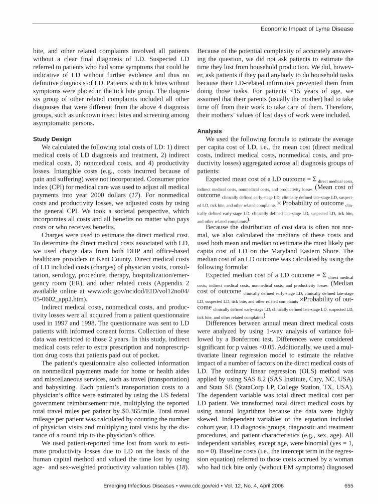
bite, and other related complaints involved all patientswithout a clear final diagnosis of LD. Suspected LDreferred to patients who had some symptoms that could beindicative of LD without further evidence and thus nodefinitive diagnosis of LD. Patients with tick bites withoutsymptoms were placed in the tick bite group. The diagno-sis group of other related complaints included all otherdiagnoses that were different from the above 4 diagnosisgroups, such as unknown insect bites and screening amongasymptomatic persons.
Study DesignWe calculated the following total costs of LD: 1) direct
medical costs of LD diagnosis and treatment, 2) indirectmedical costs, 3) nonmedical costs, and 4) productivitylosses. Intangible costs (e.g., costs incurred because ofpain and suffering) were not incorporated. Consumer priceindex (CPI) for medical care was used to adjust all medicalpayments into year 2000 dollars (17). For nonmedicalcosts and productivity losses, we adjusted costs by usingthe general CPI. We took a societal perspective, whichincorporates all costs and all benefits no matter who payscosts or who receives benefits.
Charges were used to estimate the direct medical cost.To determine the direct medical costs associated with LD,we used charge data from both DHP and office-basedhealthcare providers in Kent County. Direct medical costsof LD included costs (charges) of physician visits, consul-tation, serology, procedure, therapy, hospitalization/emer-gency room (ER), and other related costs (Appendix 2available online at www.cdc.gov/ncidod/EID/vol12no04/05-0602_app2.htm).
Indirect medical costs, nonmedical costs, and produc-tivity losses were all acquired from a patient questionnaireused in 1997 and 1998. The questionnaire was sent to LDpatients with informed consent forms. Collection of thesedata was restricted to those 2 years. In this study, indirectmedical costs refer to extra prescription and nonprescrip-tion drug costs that patients paid out of pocket.
The patient’s questionnaire also collected informationon nonmedical payments made for home or health aidesand miscellaneous services, such as travel (transportation)and babysitting. Each patient’s transportation costs to aphysician’s office were estimated by using the US federalgovernment reimbursement rate, multiplying the reportedtotal travel miles per patient by $0.365/mile. Total travelmileage per patient was calculated by counting the numberof physician visits and multiplying total visits by the dis-tance of a round trip to the physician’s office.
We used patient-reported time lost from work to esti-mate productivity losses due to LD on the basis of thehuman capital method and valued the time lost by usingage- and sex-weighted productivity valuation tables (18).
Because of the potential complexity of accurately answer-ing the question, we did not ask patients to estimate thetime they lost from household production. We did, howev-er, ask patients if they paid anybody to do household tasksbecause their LD-related infirmities prevented them fromdoing those tasks. For patients <15 years of age, weassumed that their parents (usually the mother) had to taketime off from their work to take care of them. Therefore,their mothers’ values of lost days of work were included.
AnalysisWe used the following formula to estimate the average
per capita cost of LD, i.e., the mean cost (direct medicalcosts, indirect medical costs, nonmedical costs, and pro-ductivity losses) aggregated across all diagnosis groups ofpatients:
Expected mean cost of a LD outcome = Σ direct medical costs,
indirect medical costs, nonmedical costs, and productivity losses (Mean cost ofoutcome clinically defined early-stage LD, clinically defined late-stage LD, suspect-
ed LD, tick bite, and other related complaints × Probability of outcome clin-
ically defined early-stage LD, clinically defined late-stage LD, suspected LD, tick bite,
and other related complaints).Because the distribution of cost data is often not nor-
mal, we also calculated the medians of these costs andused both mean and median to estimate the most likely percapita cost of LD on the Maryland Eastern Shore. Themedian cost of an LD outcome was calculated by using thefollowing formula:
Expected median cost of a LD outcome = Σ direct medical
costs, indirect medical costs, nonmedical costs, and productivity losses (Mediancost of outcome clinically defined early-stage LD, clinically defined late-stage
LD, suspected LD, tick bite, and other related complaints ×Probability of out-come clinically defined early-stage LD, clinically defined late-stage LD, suspected LD,
tick bite, and other related complaints)Differences between annual mean direct medical costs
were analyzed by using 1-way analysis of variance fol-lowed by a Bonferroni test. Differences were consideredsignificant for p values <0.05. Additionally, we used a mul-tivariate linear regression model to estimate the relativeimpact of a number of factors on the direct medical costs ofLD. The ordinary linear regression (OLS) method wasapplied by using SAS 8.2 (SAS Institute, Cary, NC, USA)and Stata SE (StataCorp LP, College Station, TX, USA).The dependent variable was total direct medical cost perLD patient. We transformed total direct medical costs byusing natural logarithms because the data were highlyskewed. Independent variables of the equation includedcohort year, LD diagnosis groups, diagnostic and treatmentprocedures, and patient characteristics (e.g., sex, age). Allindependent variables, except age, were binomial (yes = 1,no = 0). Baseline costs (i.e., the intercept term in the regres-sion equation) referred to those costs accrued by a womanwho had tick bite only (without EM symptoms) diagnosed
Economic Impact of Lyme Disease
Emerging Infectious Diseases • www.cdc.gov/eid • Vol. 12, No. 4, April 2006 655

in 1997 during an office visit. Such a patient had no hospi-tal or ER stay, no serologic tests, no consultation from otherphysicians, no antimicrobial drug therapy, and no other pro-cedures outside a physician office and hospital/ER.Additional direct medical costs were added or subtracted tothe baseline costs for each independent variable of interestif significant (Appendix 3, available online athttp://www.cdc.gov/ ncidod/EID/vol12no04/05-0602_app3.htm). We tested heteroscedasticity in Stata andcorrected mild heteroscedasticity by using “robust” and“hc3” procedures. We also tested both linearity and multi-collinearity in SAS and Stata.
ResultsFrom 1997 to 2000, we identified 3,415 LD-relevant
patients in the 5 counties studied on Maryland EasternShore (Table 2). Among them, 10% had clinically definedearly-stage LD while almost 5% of all patients had clini-cally defined late-stage LD. Of 284 patients who returneda completed patient questionnaire, 59 patients had clinical-ly defined early-stage LD; 25 patients had clinicallydefined late-stage LD.
Table 3 provides cohort years, medians, means, andstandard deviations of direct medical costs comparing thedifferent diagnosis groups. During the study’s time frame,the mean (range) direct medical cost of clinically definedearly-stage LD decreased from $1,609 ($95–$11,286) in
1997 to $464 ($5–$5,338) in 2000 (p<0.05). The meandirect medical cost of clinically defined late-stage LDdecreased from $4,240 ($275–$24,985) in 1997 to $1,380($45–$6,918) in 2000 (p<0.05).
From 1997 to 2000, the mean cost of therapy of alldiagnosis groups decreased 75%, from $189 to $47, andthe mean cost of hospitalization/ER decreased 61%, from$41 to $16 (Figure 2). During the same period, the meancost of an office visit, consultation, and serologic tests alsodecreased 20%, 15%, and 4%, respectively. Additionally,
RESEARCH
656 Emerging Infectious Diseases • www.cdc.gov/eid • Vol. 12, No. 4, April 2006

the proportion of patients within the highest percentile(95th percentile for all 4 years) of therapy cost graduallydecreased from 8% in 1997 to 7% in 1998, to 4% in 1999,and 3% in 2000 (data available upon request).
A patient with clinically defined early-stage LD paid anaverage of $164 in 1997 and $307 in 1998 (in 2000 dol-lars) for extra prescription and nonprescription drugs(Table 4). Those with clinically defined late-stage LD paid,for similar items, an average of $579 in 1997 and $389 in1998. The mean nonmedical cost for clinically definedearly-stage LD was $109 in 1997 and $23 in 1998. Forpatients with clinically defined late-stage LD, mean non-medical costs were $60 in 1997 and $6,703 in 1998.
During the survey period, the mean productivity loss ofclinically defined early-stage LD was $411 in 1997 and$88 in 1998, and the mean productivity loss of clinicallydefined late-stage LD was $7,762 in 1997 and $9,108 in1998. For all 3 types of costs shown in Table 4, a large dif-ference was seen between mean and median values, withthe latter often less than half of the mean value, indicatingthat a small number of LD patients account for a large por-tion of total costs.
Using multivariate linear regression analysis, we foundthat patients with clinically defined early- and late-stageLD had direct medical costs that were ≈50% and 100%,respectively, higher (p<0.001) relative to patients whoonly had tick bite, if the impact from other factors was notconsidered (Table 5). Moreover, patients who were hospi-talized or made ER visits, who underwent serologic test-ing, who needed therapy, who were referred forconsultation, and who had other procedures had substan-tially (p<0.001) higher direct medical cost than those whodid not (Table 5). No cost difference was seen betweenmen and women. After controlling for other factors, directmedical costs per LD patient in 2000 were lower thanthose in 1997 (Table 5).
In year 2000 dollars, the expected mean total costattributable to LD was $1,965 per patient, and the expect-ed median total cost attributable to LD was estimated at$281 per patient (Figure 3). For LD patients at the clinical-ly defined early stage, the median total cost was ≈$397(mean $1,310), whereas for patients at the clinicallydefined late stage, the median cost rose to $923 (mean$16,199). Suspected LD cases, tick bite cases, and otherLD-related complaints had median costs of $238 (mean$461), $108 (mean $316), and $256 (mean $714), respec-tively.
Economic Impact of Lyme Disease
Emerging Infectious Diseases • www.cdc.gov/eid • Vol. 12, No. 4, April 2006 657
Figure 2. Distribution of elements of direct medical cost (US$) perLyme disease (LD) patient in Maryland Eastern Shore(1997–2000). Mean is based on direct medical costs of LDpatients. Direct medical costs were collected from medical recordabstraction (1997–2000). Direct medical costs of LD includedcosts of physician visits, consultation, serologic tests, procedure,therapy, hospitalization/emergency room, and other relevantcosts. All costs were converted to 2000 equivalent.
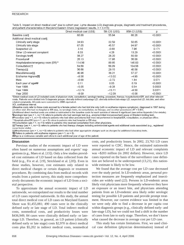
DiscussionPrevious studies of the economic impact of LD were
often based on numerous assumptions and experts’ sug-gestions (e.g., Maes et al. [15]). Only a few studies provid-ed cost estimates of LD based on data collected from thefield (e.g., Fix et al. [19], Strickland et al. [20]). Even inthose studies, however, cost estimates only related todirect medical charges or certain diagnosis or treatmentprocedures. By combining data from medical records withresults from a patient survey, this study more comprehen-sively documents the economic impact of LD from a soci-etal perspective.
To approximate the annual economic impact of LDnationwide, we extrapolated our results to the total numberof LD cases reported nationwide. In this study, the annualtotal direct medical cost of LD cases on Maryland EasternShore was $1,455,081; 490 cases were in the clinicallydefined early or late stage of LD. Total indirect medicalcosts, nonmedical costs, and productivity losses were$436,949; 84 cases were clinically defined early- or late-stage LD. Therefore, in general, an LD patient (clinicallydefined early or late stage) costs $2,970 in direct medicalcosts plus $5,202 in indirect medical costs, nonmedical
costs, and productivity losses. In 2002, 23,763 LD caseswere reported to CDC. Hence, the estimated nationwideannual economic impact of LD and relevant complaintswas ≈$203 million (in 2002 dollars). However, since LDcases reported on the basis of the surveillance case defini-tion are believed to be underreported (13,21), this nation-wide estimate is likely to be low.
We found that the average cost per LD case decreasedover the study period. In LD-endemic areas, personal pro-tection measures are frequently emphasized and insecti-cides are widely used (22). Persons in LD-endemic areaslikely visit physicians more frequently whenever they havean exposure or an insect bite, and physicians attendingpatients from an LD-endemic area likely order serologictesting for possible LD patients and provide prompt treat-ment. However, our current evidence was limited in thatwe were only able to find a decrease in per capita costwithin diagnosis groups (e.g., clinically defined early- andlate-stage LD), but we could not find a shift in the numberof cases from late to early stage. Therefore, we don’t knowwhat caused the decrease in average cost per LD case.
This study has certain limitations. First, we used clini-cal case definition (physician determination) instead of
RESEARCH
658 Emerging Infectious Diseases • www.cdc.gov/eid • Vol. 12, No. 4, April 2006
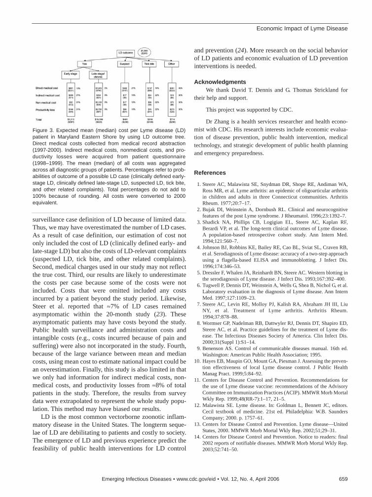
surveillance case definition of LD because of limited data.Thus, we may have overestimated the number of LD cases.As a result of case definition, our estimation of cost notonly included the cost of LD (clinically defined early- andlate-stage LD) but also the costs of LD-relevant complaints(suspected LD, tick bite, and other related complaints).Second, medical charges used in our study may not reflectthe true cost. Third, our results are likely to underestimatethe costs per case because some of the costs were notincluded. Costs that were omitted included any costsincurred by a patient beyond the study period. Likewise,Steer et al. reported that ≈7% of LD cases remainedasymptomatic within the 20-month study (23). Theseasymptomatic patients may have costs beyond the study.Public health surveillance and administration costs andintangible costs (e.g., costs incurred because of pain andsuffering) were also not incorporated in the study. Fourth,because of the large variance between mean and mediancosts, using mean cost to estimate national impact could bean overestimation. Finally, this study is also limited in thatwe only had information for indirect medical costs, non-medical costs, and productivity losses from ≈8% of totalpatients in the study. Therefore, the results from surveydata were extrapolated to represent the whole study popu-lation. This method may have biased our results.
LD is the most common vectorborne zoonotic inflam-matory disease in the United States. The longterm seque-lae of LD are debilitating to patients and costly to society.The emergence of LD and previous experience predict thefeasibility of public health interventions for LD control
and prevention (24). More research on the social behaviorof LD patients and economic evaluation of LD preventioninterventions is needed.
AcknowledgmentsWe thank David T. Dennis and G. Thomas Strickland for
their help and support.
This project was supported by CDC.
Dr Zhang is a health services researcher and health econo-mist with CDC. His research interests include economic evalua-tion of disease prevention, public health intervention, medicaltechnology, and strategic development of public health planningand emergency preparedness.
References
1. Steere AC, Malawista SE, Snydman DR, Shope RE, Andiman WA,Ross MR, et al. Lyme arthritis: an epidemic of oligoarticular arthritisin children and adults in three Connecticut communities. ArthritisRheum. 1977;20:7–17.
2. Bujak DI, Weinstein A, Dornbush RL. Clinical and neurocognitivefeatures of the post Lyme syndrome. J Rheumatol. 1996;23:1392–7.
3. Shadick NA, Phillips CB, Logigian EL, Steere AC, Kaplan RF,Berardi VP, et al. The long-term clinical outcomes of Lyme disease.A population-based retrospective cohort study. Ann Intern Med.1994;121:560–7.
4. Johnson BJ, Robbins KE, Bailey RE, Cao BL, Sviat SL, Craven RB,et al. Serodiagnosis of Lyme disease: accuracy of a two-step approachusing a flagella-based ELISA and immunoblotting. J Infect Dis.1996;174:346–53.
5. Dressler F, Whalen JA, Reinhardt BN, Steere AC. Western blotting inthe serodiagnosis of Lyme disease. J Infect Dis. 1993;167:392–400.
6. Tugwell P, Dennis DT, Weinstein A, Wells G, Shea B, Nichol G, et al.Laboratory evaluation in the diagnosis of Lyme disease. Ann InternMed. 1997;127:1109–23.
7. Steere AC, Levin RE, Molloy PJ, Kalish RA, Abraham JH III, LiuNY, et al. Treatment of Lyme arthritis. Arthritis Rheum.1994;37:878–88.
8. Wormser GP, Nadelman RB, Dattwyler RJ, Dennis DT, Shapiro ED,Steere AC, et al. Practice guidelines for the treatment of Lyme dis-ease. The Infectious Diseases Society of America. Clin Infect Dis.2000;31(Suppl 1):S1–14.
9. Benenson AS. Control of communicable diseases manual. 16th ed.Washington: American Public Health Association; 1995.
10. Hayes EB, Maupin GO, Mount GA, Piesman J. Assessing the preven-tion effectiveness of local Lyme disease control. J Public HealthManag Pract. 1999;5:84–92.
11. Centers for Disease Control and Prevention. Recommendations forthe use of Lyme disease vaccine: recommendations of the AdvisoryCommittee on Immunization Practices (ACIP). MMWR Morb MortalWkly Rep. 1999;48(RR-7):1–17, 21–5.
12. Malawista SE. Lyme disease. In: Goldman L, Bennett JC, editors.Cecil textbook of medicine. 21st ed. Philadelphia: W.B. SaundersCompany; 2000. p. 1757–61.
13. Centers for Disease Control and Prevention. Lyme disease—UnitedStates, 2000. MMWR Morb Mortal Wkly Rep. 2002;51;29–31.
14. Centers for Disease Control and Prevention. Notice to readers: final2002 reports of notifiable diseases. MMWR Morb Mortal Wkly Rep.2003;52:741–50.
Economic Impact of Lyme Disease
Emerging Infectious Diseases • www.cdc.gov/eid • Vol. 12, No. 4, April 2006 659
Figure 3. Expected mean (median) cost per Lyme disease (LD)patient in Maryland Eastern Shore by using LD outcome tree.Direct medical costs collected from medical record abstraction(1997-2000). Indirect medical costs, nonmedical costs, and pro-ductivity losses were acquired from patient questionnaire(1998–1999). The mean (median) of all costs was aggregatedacross all diagnostic groups of patients. Percentages refer to prob-abilities of outcome of a possible LD case (clinically defined early-stage LD, clinically defined late-stage LD, suspected LD, tick bite,and other related complaints). Total percentages do not add to100% because of rounding. All costs were converted to 2000equivalent.
15. Maes E, Lecomte P, Ray N. A cost-of-illness study of Lyme diseasein the United States. Clin Ther. 1998;20:993–1008.
16. Centers for Disease Control and Prevention. Case definitions forinfectious conditions under public health surveillance. MMWR MorbMortal Wkly Rep. 1997;46(RR-10):20–1.
17. US Department of Labor. Consumer price index. [cited 2004 June16]. Available from http://www.bls.gov/cpi/home.htm
18. Haddix AC, Teutsch SM, Corso PS, editors. Prevention effectiveness:a guide to decision analysis and economic evaluation. 2nd ed. NewYork: Oxford University Press; 2003. p. 70–1.
19. Fix AD, Strickland GT, Grant J. Tick bites and Lyme disease in anendemic setting: problematic use of serologic testing and prophylac-tic antibiotic therapy. JAMA. 1998;279:206–10.
20. Strickland GT, Karp AC, Mathews A, Peña CA. Utilization and costof serologic tests for Lyme disease in Maryland. J Infect Dis.1997;176:819–21.
21. Meek JI, Roberts CL, Smith EV Jr, Cartter ML. Underreporting ofLyme disease by Connecticut physicians, 1992. J Public HealthManag Pract. 1996;2:61–5.
22. Barbour AG, Fish D. The biological and social phenomenon of Lymedisease. Science. 1993;260:1610–6.
23. Steer AC, Sikand VK, Schoen RT, Nowakowski J. Asymptomaticinfection with Borrelia burgdorferi. Clin Infect Dis. 2003;37:528–32.
24. Steere AC, Coburn J, Glickstein L. The emergence of Lyme disease[review]. J Clin Invest. 2004;113:1093–101.
Address for correspondence: Xinzhi Zhang, Centers for Disease Controland Prevention, 1600 Clifton Rd NE, Mailstop D59, Atlanta, GA 30333,USA; fax: 404-371-5445; email: [email protected]
RESEARCH
660 Emerging Infectious Diseases • www.cdc.gov/eid • Vol. 12, No. 4, April 2006
Search

Influenza poses a continuing public health threat inepidemic and pandemic seasons. The 1951 influenza epi-demic (A/H1N1) caused an unusually high death toll inEngland; in particular, weekly deaths in Liverpool even sur-passed those of the 1918 pandemic. We further quantifiedthe death rate of the 1951 epidemic in 3 countries. InEngland and Canada, we found that excess death ratesfrom pneumonia and influenza and all causes were sub-stantially higher for the 1951 epidemic than for the 1957and 1968 pandemics (by >50%). The age-specific patternof deaths in 1951 was consistent with that of other interpan-demic seasons; no age shift to younger age groups, remi-niscent of pandemics, occurred in the death rate. Incontrast to England and Canada, the 1951 epidemic wasnot particularly severe in the United States. Why this epi-demic was so severe in some areas but not others remainsunknown and highlights major gaps in our understanding ofinterpandemic influenza.
Influenza is responsible for large increases in deaths inpandemic seasons when emerging viral subtypes with
novel surface antigens become predominant, and also insome interpandemic seasons, when established subtypesexhibit antigenic drift (1). The circulating viral subtype isassociated with varying severity of influenza epidemics(2): in the last 2 decades in the United States, estimatedexcess death rates were on average 2.8-fold higher inA/H3N2-dominated seasons than in A/H1N1 and B sea-sons (3). Within a given subtype, however, the strain-spe-cific determinants of epidemic severity are still poorlyunderstood. For instance in the United States in the sameperiod, excess death rates varied nearly 4-fold among
A/H3N2 seasons, even after adjustments for populationaging (3). Better characterizations of past severe influenzaepidemics can help understand and perhaps help predictthe occurrence of severe epidemics.
Anecdotal accounts exist in the literature of historicalinfluenza epidemics associated with unusual numbers ofdeaths, such as occurred in the 1951 epidemic in Englandin the midst of the first era of A/H1N1 viruses(1918–1957) (4). In Liverpool, where the epidemic wassaid to originate, it was “the cause of the highest weeklydeath toll, apart from aerial bombardment, in the city’svital statistics records, since the great cholera epidemic of1849” (5). This weekly death toll even surpassed that ofthe 1918 influenza pandemic (Figure 1).
The international pattern of influenza-related deaths in1951 has not been adequately quantified in the pastbecause of lack of methodologic tools and historical deathrecords. However, this historical epidemic is a good exam-ple to illustrate major gaps in our current understanding ofinfluenza virus epidemiology. We revisited the 1951 epi-demic by quantifying its death rate in 3 countries (Englandand Wales, Canada, the United States) and comparing itsage-specific mortality pattern with that of surrounding epi-demic and pandemic seasons (1).
Methods
DataWe obtained monthly pneumonia and influenza (P&I)
and all-cause numbers of deaths for 1950 to 1999 fromHealth Canada (6), by 5-year age groups (details on theInternational Classification of Diseases codes used aregiven in Table 1). Canada was the only country withdetailed age-specific mortality data for the 1950s readilyavailable in electronic format.
1951 Influenza Epidemic,England and Wales, Canada,
and the United StatesCécile Viboud,* Theresa Tam,† Douglas Fleming,‡ Mark A Miller,* and Lone Simonsen*
Emerging Infectious Diseases • www.cdc.gov/eid • Vol. 12, No. 4, April 2006 661
*National Institutes of Health, Bethesda, Maryland, USA; †PublicHealth Agency of Canada, Ottawa, Ontario, Canada; and ‡RoyalCollege of General Practitioners, Harborne, Birmingham, UnitedKingdom
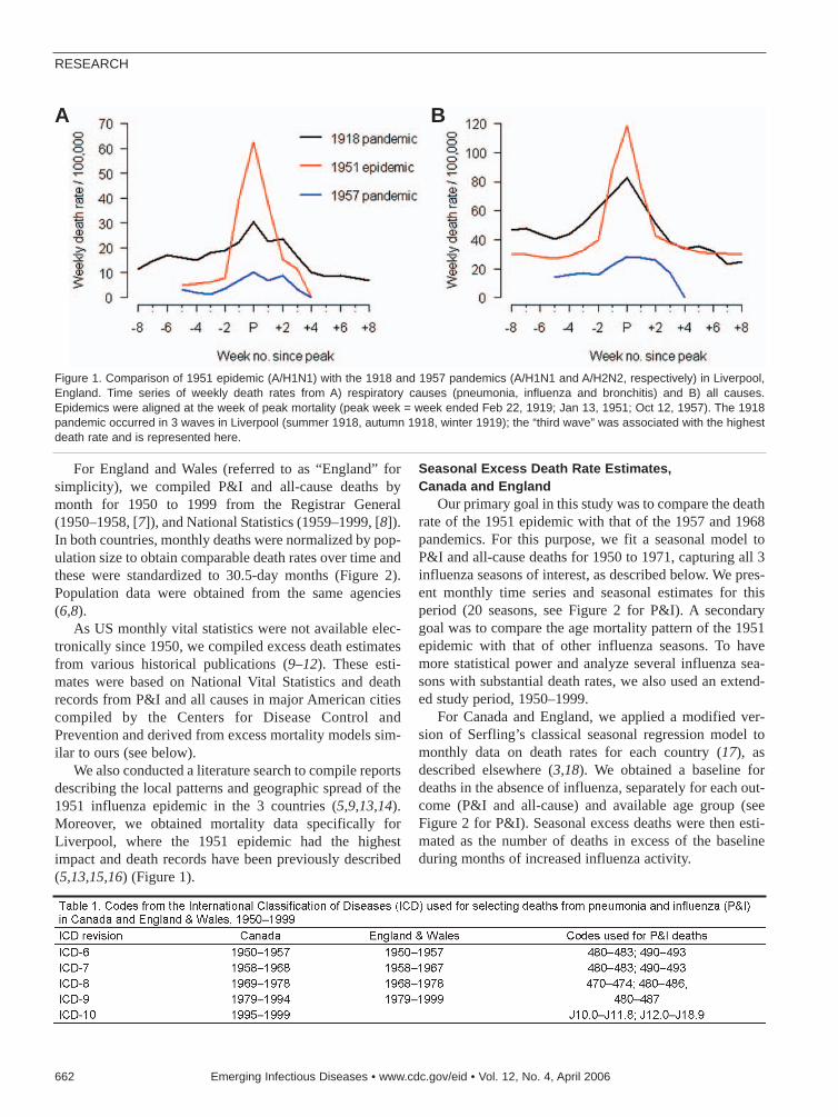
For England and Wales (referred to as “England” forsimplicity), we compiled P&I and all-cause deaths bymonth for 1950 to 1999 from the Registrar General(1950–1958, [7]), and National Statistics (1959–1999, [8]).In both countries, monthly deaths were normalized by pop-ulation size to obtain comparable death rates over time andthese were standardized to 30.5-day months (Figure 2).Population data were obtained from the same agencies(6,8).
As US monthly vital statistics were not available elec-tronically since 1950, we compiled excess death estimatesfrom various historical publications (9–12). These esti-mates were based on National Vital Statistics and deathrecords from P&I and all causes in major American citiescompiled by the Centers for Disease Control andPrevention and derived from excess mortality models sim-ilar to ours (see below).
We also conducted a literature search to compile reportsdescribing the local patterns and geographic spread of the1951 influenza epidemic in the 3 countries (5,9,13,14).Moreover, we obtained mortality data specifically forLiverpool, where the 1951 epidemic had the highestimpact and death records have been previously described(5,13,15,16) (Figure 1).
Seasonal Excess Death Rate Estimates, Canada and England
Our primary goal in this study was to compare the deathrate of the 1951 epidemic with that of the 1957 and 1968pandemics. For this purpose, we fit a seasonal model toP&I and all-cause deaths for 1950 to 1971, capturing all 3influenza seasons of interest, as described below. We pres-ent monthly time series and seasonal estimates for thisperiod (20 seasons, see Figure 2 for P&I). A secondarygoal was to compare the age mortality pattern of the 1951epidemic with that of other influenza seasons. To havemore statistical power and analyze several influenza sea-sons with substantial death rates, we also used an extend-ed study period, 1950–1999.
For Canada and England, we applied a modified ver-sion of Serfling’s classical seasonal regression model tomonthly data on death rates for each country (17), asdescribed elsewhere (3,18). We obtained a baseline fordeaths in the absence of influenza, separately for each out-come (P&I and all-cause) and available age group (seeFigure 2 for P&I). Seasonal excess deaths were then esti-mated as the number of deaths in excess of the baselineduring months of increased influenza activity.
RESEARCH
662 Emerging Infectious Diseases • www.cdc.gov/eid • Vol. 12, No. 4, April 2006
Figure 1. Comparison of 1951 epidemic (A/H1N1) with the 1918 and 1957 pandemics (A/H1N1 and A/H2N2, respectively) in Liverpool,England. Time series of weekly death rates from A) respiratory causes (pneumonia, influenza and bronchitis) and B) all causes.Epidemics were aligned at the week of peak mortality (peak week = week ended Feb 22, 1919; Jan 13, 1951; Oct 12, 1957). The 1918pandemic occurred in 3 waves in Liverpool (summer 1918, autumn 1918, winter 1919); the “third wave” was associated with the highestdeath rate and is represented here.
A B
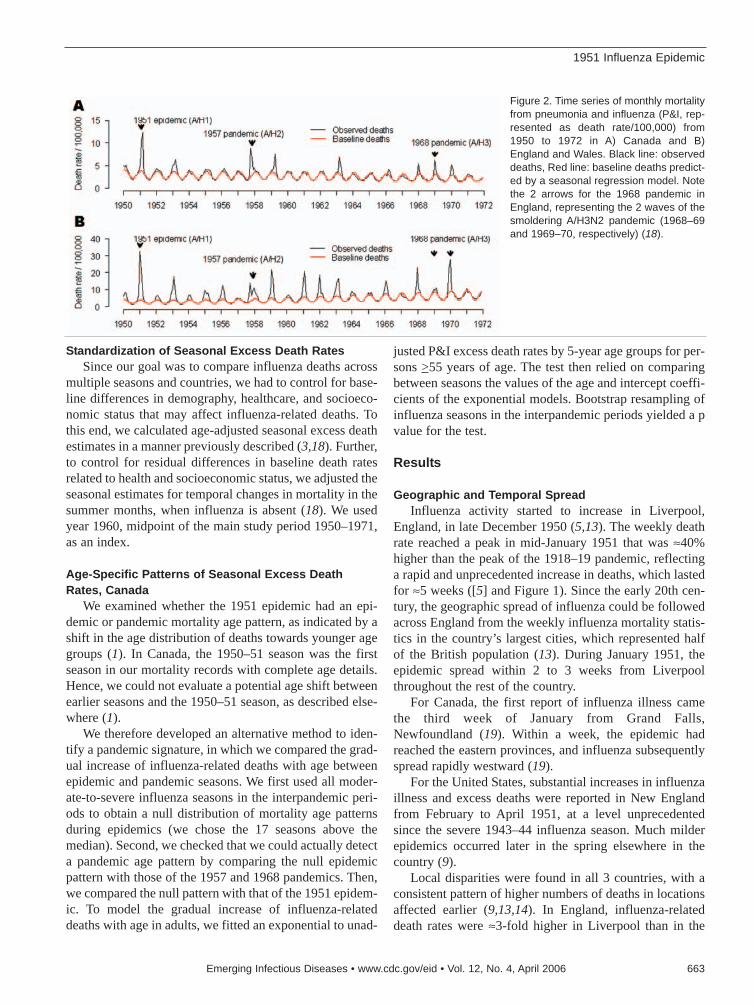
Standardization of Seasonal Excess Death RatesSince our goal was to compare influenza deaths across
multiple seasons and countries, we had to control for base-line differences in demography, healthcare, and socioeco-nomic status that may affect influenza-related deaths. Tothis end, we calculated age-adjusted seasonal excess deathestimates in a manner previously described (3,18). Further,to control for residual differences in baseline death ratesrelated to health and socioeconomic status, we adjusted theseasonal estimates for temporal changes in mortality in thesummer months, when influenza is absent (18). We usedyear 1960, midpoint of the main study period 1950–1971,as an index.
Age-Specific Patterns of Seasonal Excess DeathRates, Canada
We examined whether the 1951 epidemic had an epi-demic or pandemic mortality age pattern, as indicated by ashift in the age distribution of deaths towards younger agegroups (1). In Canada, the 1950–51 season was the firstseason in our mortality records with complete age details.Hence, we could not evaluate a potential age shift betweenearlier seasons and the 1950–51 season, as described else-where (1).
We therefore developed an alternative method to iden-tify a pandemic signature, in which we compared the grad-ual increase of influenza-related deaths with age betweenepidemic and pandemic seasons. We first used all moder-ate-to-severe influenza seasons in the interpandemic peri-ods to obtain a null distribution of mortality age patternsduring epidemics (we chose the 17 seasons above themedian). Second, we checked that we could actually detecta pandemic age pattern by comparing the null epidemicpattern with those of the 1957 and 1968 pandemics. Then,we compared the null pattern with that of the 1951 epidem-ic. To model the gradual increase of influenza-relateddeaths with age in adults, we fitted an exponential to unad-
justed P&I excess death rates by 5-year age groups for per-sons >55 years of age. The test then relied on comparingbetween seasons the values of the age and intercept coeffi-cients of the exponential models. Bootstrap resampling ofinfluenza seasons in the interpandemic periods yielded a pvalue for the test.
Results
Geographic and Temporal SpreadInfluenza activity started to increase in Liverpool,
England, in late December 1950 (5,13). The weekly deathrate reached a peak in mid-January 1951 that was ≈40%higher than the peak of the 1918–19 pandemic, reflectinga rapid and unprecedented increase in deaths, which lastedfor ≈5 weeks ([5] and Figure 1). Since the early 20th cen-tury, the geographic spread of influenza could be followedacross England from the weekly influenza mortality statis-tics in the country’s largest cities, which represented halfof the British population (13). During January 1951, theepidemic spread within 2 to 3 weeks from Liverpoolthroughout the rest of the country.
For Canada, the first report of influenza illness camethe third week of January from Grand Falls,Newfoundland (19). Within a week, the epidemic hadreached the eastern provinces, and influenza subsequentlyspread rapidly westward (19).
For the United States, substantial increases in influenzaillness and excess deaths were reported in New Englandfrom February to April 1951, at a level unprecedentedsince the severe 1943–44 influenza season. Much milderepidemics occurred later in the spring elsewhere in thecountry (9).
Local disparities were found in all 3 countries, with aconsistent pattern of higher numbers of deaths in locationsaffected earlier (9,13,14). In England, influenza-relateddeath rates were ≈3-fold higher in Liverpool than in the
1951 Influenza Epidemic
Emerging Infectious Diseases • www.cdc.gov/eid • Vol. 12, No. 4, April 2006 663
Figure 2. Time series of monthly mortalityfrom pneumonia and influenza (P&I, rep-resented as death rate/100,000) from1950 to 1972 in A) Canada and B)England and Wales. Black line: observeddeaths, Red line: baseline deaths predict-ed by a seasonal regression model. Notethe 2 arrows for the 1968 pandemic inEngland, representing the 2 waves of thesmoldering A/H3N2 pandemic (1968–69and 1969–70, respectively) (18).

rest of the country (13). In Canada, death rates were ≈2.4-fold higher in the eastern seaboard provinces than in therest of the country (14). Similarly, in the United States,rates were ≈2.3-fold higher in New England than in the restof the country (9).
Patterns of Seasonal Excess Death Rates, All Ages, 3 Countries
Crude and adjusted seasonal excess death estimates inthe 3 countries are presented for the period 1950–1971 inFigure 3 (P&I). A specific comparison of the 1951 epidem-ic and 1957 pandemic is provided in Table 2 (P&I and all-cause).
For Canada, the 1951 epidemic was the most severeinfluenza season in the period 1950–1999, as indicated bycrude seasonal excess death rates from P&I and all causes(data not shown). On the basis of both outcomes, the 1951epidemic caused a 1.5-fold higher death rate than the 1957pandemic; the rate was 3- to 4-fold higher than that seen inthe 1968 pandemic. Adjusting for factors unrelated toinfluenza, such as demographics, health, and socioeco-nomic status only marginally modified our estimates (by<11%).
In England, the 1951 epidemic had similar death pat-terns. It was responsible for the largest increase in winterdeaths from P&I and all causes in the period 1950–1999(data not shown), with 1.3- to 1.4-fold higher crude excessdeath rates than those seen in the 1957 and 1968 pan-
demics. In contrast to Canada, adjusting for trends in demo-graphics and health care substantially changed our excessdeath estimates, exacerbating the impact of the 1951 epi-demic. Baseline P&I summer death rates doubled from1950 to 1970 (Figure 2), probably because of rapid aging ofthe British population. During these 2 decades, the propor-tion of persons >65 years of age increased by 2.3% inEngland, which explains the trend in British death rates; incomparison, it increased by only 0.3% in Canada (6,8).
In the United States, by contrast, the 1951 epidemicwas not particularly severe, except possibly in the NewEngland region, adjacent to Canada. In the United States,this epidemic ranked with low-to-moderate influenza sea-sons, with only half the impact of the 1957 pandemic forP&I deaths and even less for all-cause deaths.
Estimation of crude and adjusted excess deaths sug-gests that the 1951 epidemic was unusually severe inEngland and Canada but not in the United States. Theabsolute rates of excess deaths were very different betweencountries, with systematically higher rates in England (by3- to 5-fold, Table 2). The difference remained even afteradjusting for international differences in demographics andhealthcare and was also found for deaths from all causes,which controls for potential differences in the coding ofdeath certificates (Table 2). Such international discrepan-cies in influenza-related death rates have been highlightedon several occasions in the past, although not elucidated(18,20–22). Because of these unresolved differences, this
RESEARCH
664 Emerging Infectious Diseases • www.cdc.gov/eid • Vol. 12, No. 4, April 2006
Figure 3. Seasonal pneumonia andinfluenza (P&I) excess death rates,all ages, 1950–51 to 1970–71.Crude death rates (blue bars),death rates adjusted for summertrends in mortality unrelated toinfluenza (pink bars) and adjustedfor demographics (green bars). A)Canada. B) England and Wales. C)USA. Note that for England, com-parisons used the second A/H3N2pandemic wave of major impact(18). Note the 2 arrows for the 1968pandemic in England, representingthe 2 A/H3N2 pandemic waves(1968–69 and 1969–70, respec-tively); the second wave of majorimpact was used in comparisons(18).

analysis focused on the relative impact of the 1951 epi-demic as compared with surrounding influenza seasons.
Age-specific Patterns of Seasonal Excess Death Rates, Canada
Given the unusual death rate of the 1951 influenza epi-demic in England and Canada, we hypothesized that anemerging virus subtype may have circulated there, perhapswith pandemic potential. We investigated the age-specificmortality pattern of the 1951 epidemic in Canada in rela-tion to other seasons to address this aspect.
Inspection of P&I excess death rates by age shows thatthe 1951 epidemic had the typical pattern of death ratesfound in other epidemics, steeply increasing with age afterinfancy (Figure 4). Statistical analysis showed that the agepattern in 1951 was well within the range of the null distri-bution of reference epidemic seasons in influenza inter-pandemic periods (Table 3). Conversely, we found lowerage coefficients for the 1957 and 1968 pandemics as com-pared with reference epidemic seasons, illustrating thatdeaths increased less with age in pandemics than in epi-demics and that our statistical approach could detect a pan-demic signature (p<0.001) (1).
DiscussionWe have shown that the 1951 influenza epidemic had
greater death rate than all subsequent influenza epidemicsor pandemics in England and Canada. In Canada, whereage-detailed data were available, deaths in persons <65years of age attributable to the 1951 epidemic were nearlyequivalent to those of the 1957 pandemic. But what setsthe 1951 epidemic apart from pandemics is that the olderpopulation was also severely affected, with twice thedeaths as occurred in the 1957 pandemic. By contrast, the
1951 epidemic had minor impact in the United States,except possibly in New England.
To study influenza death patterns, we used P&I deaths,a reliable proxy for the timing and relative impact ofinfluenza epidemics, as well as all-cause deaths, an indica-tor of their overall impact (23). Cardiovascular deaths arealso widely used to quantify the impact of influenza(24,25); however, they were not available to us for thisstudy. But since winter increases in P&I, cardiovascular,and all-cause deaths are synchronized and correlated inamplitude during influenza epidemics (25), we capturehere the timing and death rate of these epidemics.
Influenza-related death rates reflect the combination of2 underlying epidemiologic parameters: the attack rate, ameasure of a pathogen’s transmissibility, and the case-fatality rate, a measure of a pathogen’s virulence. Theunusual severity of the 1951 epidemic in England andCanada may stem from higher attack rates, higher case-fatality rates, or both. To isolate these factors, we exam-ined Liverpool, England, where comparable data on illnessand death exist for the 1951 and 1957 pandemics (5). InLiverpool, influenza attack rates in schoolchildren were 3-fold lower in 1951 than in 1957, which suggests lowertransmissibility in this age group in 1951. By contrast, anequal number of influenza-related deaths occurred in the 2seasons in children in Liverpool, which suggests a highercase-fatality rate in 1951 than in 1957. Similar findingswere observed in the working adult population (5), an indi-cation of unusual virulence in the influenza virus circulat-ing in 1951 in Liverpool. This argument must be takenwith caution, however, since most influenza-related deathsoccur in the elderly (3), and attack rates are not availablefor this age group. Estimation of the transmissibility of the1951 virus using a mathematical model for influenza
1951 Influenza Epidemic
Emerging Infectious Diseases • www.cdc.gov/eid • Vol. 12, No. 4, April 2006 665

transmission fitted to mortality data (26) might answer thisquestion, not only in Liverpool but also in the rest ofEngland and Canada.
Laboratory surveillance data from the World HealthOrganization (WHO) indicate that influenza A viruses cir-culating at the time were characterized as H1N1 (27), asubtype circulating since the 1918 pandemic (28).Although an unusually large drift event in the hemagglu-tinin of A/H1N1 viruses was reported in 1947 (29), subse-quent changes in this protein remained minor until after1951 (27). Hence, no virologic evidence of a shift orunusual drift in the hemagglutinin antigen exists for 1951viruses. In support of the virologic evidence, we haveshown that no epidemiologic pandemic signature occurredin 1951, as indicated by an age shift of deaths towardsyounger age groups (1).
The 1951 epidemic exhibited geographic disparities ininfluenza-related deaths, as illustrated by the contrastbetween England and Canada (countries with high deathrate) and the United States (low death rate). These dispar-ities are in part explained by laboratory surveillancereports by WHO (27,30), indicating that 2 antigenicallydistinct influenza A/H1N1 strains cocirculated in theNorthern Hemisphere during the 1951 epidemic (27,30).The so-called “Scandinavian strain” was isolated in north-ern Europe and associated with mild illnesses. By contrast,the “Liverpool strain” was associated with severe illnessesand high deaths in Great Britain, Canada, southern Europe,and Mediterranean countries (27). As both strains cocircu-lated in some countries (27), intrasubtypic cross-immunitymight have existed, with these 2 strains competing for sus-ceptible hosts.
RESEARCH
666 Emerging Infectious Diseases • www.cdc.gov/eid • Vol. 12, No. 4, April 2006
Figure 4. Age-specific pneumonia and influenza (P&I)death rates in the 1951 influenza epidemic, 1957, and1968 pandemics, Canada. A) Observed. B) Exponentialmodels using 5-year age groups starting at age 55 yearsand ending at >90 years (R2 >0.85 for all seasons).Black curve: “null distribution” of expected pattern in epi-demic seasons, based on major epidemics in the inter-pandemic periods, 1950–1999 (N = 17). The agecoefficient was set at the mean of the “null” distribution(see Table 3 for values). The intercept was set at theminimum of the distribution for legibility.

The precise reasons for the unusually high death rateassociated with the Liverpool strain remain elusive. Thegenetic markers of influenza virulence are still uncleartoday, but a multibasic cleavage site in the hemagglutinin,as well as minor changes in internal genes, are believed toenhance viral pathogenicity (31,32). Only hemagglutinininhibition tests could be performed in 1951, and to ourknowledge, no influenza virus isolate or genetic sequencefrom 1951 is available in the public domain. Furthermolecular analysis of 1951 influenza specimens could helpexplain the extreme local pathogenicity in that season.
We have described an influenza season that was unex-pectedly severe in some countries and mild in others. Thisgeographic disparity in influenza-related deaths is not com-mon; influenza mortality is generally correlated betweenthe United States and Europe and within the United States(33,34). Occasional disparities have been reported, howev-er. For instance, the impact of the 2 waves of the 1968 pan-demic differed markedly between North American andEurasian countries, perhaps because of differences in pre-existing immunity and evolving viruses (3,18). In this con-text, the 1951 epidemic appears as another striking exampleof geographic disparities in influenza impact, perhapsexplained in this case by cocirculation of 2 influenzaA/H1N1 strains. Other competing hypotheses include dif-ferences in preexisting population immunity or socioeco-nomic factors, but these are less parsimonious explanations.
Many countries are actively preparing for the nextinfluenza pandemic (35–37). Previous pandemics in the20th century have been responsible for large numbers ofdeaths in all age groups (1); however, the age pattern ofdeaths in the 1918 and 1968 pandemics suggest that theelderly may actually be relatively protected against anemerging pandemic virus (35,38,39). By contrast, we haveshown that the 1951 epidemic was not associated with theemergence of a new influenza subtype, yet had a higherdeath rate than 2 of the 3 pandemics of the past century inEngland and Canada, especially among the elderly, andhigher death rate than all 3 pandemics in Liverpool. Weconclude that pandemics are not always more severe interms of deaths than epidemics, for reasons still unclear. Athorough investigation of the full genome of the influenzaviruses involved in the unusually severe 1951 epidemiccould shed light on the virulence and transmissibility fac-tors at play and fill key gaps in our current understandingof interpandemic influenza (40).
AcknowledgmentsWe thank June Leach for kindly providing historical mortal-
ity data for England and Wales, Edward Ng and Russell Wilkinsfor help with the Canadian mortality and population data, andBryan T. Grenfell for helpful discussions at an earlier stage of thisstudy.
Dr Viboud is a mathematical epidemiologist with the USNational Institutes of Health, with an interest in the patterns ofillness and death for infectious diseases. Her research focuses onthe spatial and temporal patterns of influenza and rotavirus epi-demics and how the genetic make-up of the pathogen relates toits epidemiologic impact.
References
1. Simonsen L, Clarke MJ, Schonberger LB, Arden NH, Cox NJ,Fukuda K. Pandemic versus epidemic influenza mortality: a patternof changing age distribution. J Infect Dis. 1998;178:53–60.
2. Simonsen L, Clarke MJ, Williamson GD, Stroup DF, Arden NH,Schonberger LB. The impact of influenza epidemics on mortality:introducing a severity index. Am J Public Health. 1997;87:1944–50.
3. Simonsen L, Reichert TA, Viboud C, Blackwelder WC, Taylor RJ,Miller MA. Impact of influenza vaccination on seasonal mortality inthe US elderly population. Arch Intern Med. 2005;165:265–72.
4. Burnet M. The influenza virus. Scientific American. 1953;188:28–31.5. Semple AB, Davies JB, Disley PJ. Influenza in Liverpool. Lancet.
1958;1:95–76. Statistics Canada. [accessed 2004 Nov]. Available from
ht tp : / /www.s ta tcan .ca /cg i -b in / imdb/p2SV.pl?Funct ion=getSurvey&SDDS=3233&lang=en&db=IMDB&dbg=f&adm=8&dis=2
7. Great Britain, General Register Office. Annual summary of marriages,births, and deaths, in England and Wales. London: the StationeryOffice (HSMO); 1958.
8. National Statistics, UK. [cited 2004 Nov]. Available fromhttp://www.statistics.gov.uk/StatBase/Product.asp?vlnk=1253&More=Y
9. Collins S, Lehmann J. Trends and epidemics of influenza and pneu-monia, 1918–1951. Public Health Rep. 1951;46:1487–517.
10. Serfling RE, Sherman IL, Houseworth WJ. Excess pneumonia-influenza mortality by age and sex in three major influenza A2 epi-demics, United States, 1957-58, 1960 and 1963. Am J Epidemiol.1967;86:433–41.
11. Housworth WJ, Spoon MM. The age distribution of excess mortalityduring A2 Hong Kong influenza epidemics compared with earlier A2outbreaks. Am J Epidemiol. 1971;94:348–50.
12. Noble G. Epidemiological and clinical aspects of influenza. In: BearA, editor. Basic and applied influenza research. Boca Raton (FL):CRC Press; 1982.
13. Bradley WH, Massey A, Logan WP, Semple AB, Benjamin B, GristNR, et al. Discussion: influenza 1951. Proc R Soc Med.1951;44:789–801.
14. Canada, Dominion Bureau of Statistics. Influenza in Canada: somestatistics on its characteristics and trends. Ottawa: The Bureau; 1958.p. 1–23.
15. Freyche M, Klimt C. World incidence of influenza, 1950–1951.World Health Org, Epidemiological and Vital Statistics Report.1951;5:141–61.
16. Jordan EO. Epidemic influenza. Chicago: American MedicalAssociation; 1927.
17. Serfling R. Methods for current statistical analysis of excess pneumo-nia-influenza deaths. Public Health Rep. 1963;78:494–506.
18. Viboud C, Grais RF, Lafont BA, Miller MA, Simonsen L.Multinational impact of the 1968 Hong Kong influenza pandemic:evidence for a smoldering pandemic. J Infect Dis. 2005;192:233–48.
19. Nagler FP, Burr MM, Gillen AL. The influenza virus epidemic inCanada during January-February, 1951. Can J Public Health.1951;42:367–74.
20. Langmuir AD, Housworth J. A critical evaluation of influenza sur-veillance. Bull World Health Organ. 1969;41:393–8.
1951 Influenza Epidemic
Emerging Infectious Diseases • www.cdc.gov/eid • Vol. 12, No. 4, April 2006 667

21. Assaad F, Cockburn WC, Sundaresan TK. Use of excess mortalityfrom respiratory diseases in the study of influenza. Bull World HealthOrgan. 1973;49:219–33.
22. World Health Organization. Mortality at ages 65 and older—UnitedStates, Canada, and Western Europe. Stat Bull Metropol Life InsurCo. 1971;52:8–11.
23. Simonsen L. The global impact of influenza on morbidity and mor-tality. Vaccine. 1999;17(Suppl 1):S3–10.
24. Fleming DM, Cross KW, Pannell RS. Influenza and its relationship tocirculatory disorders. Epidemiol Infect. 2005;133:255–62.
25. Reichert TA, Simonsen L, Sharma A, Pardo SA, Fedson DS, MillerMA. Influenza and the winter increase in mortality in the UnitedStates, 1959-1999. Am J Epidemiol. 2004;160:492–502.
26. Mills CE, Robins JM, Lipsitch M. Transmissibility of 1918 pandem-ic influenza. Nature. 2004;432:904–6.
27. Isaacs A, Gledhill AW, Andrewes CH. Influenza A viruses; laborato-ry studies, with special reference to European outbreak of 1950–1.Bull World Health Organ. 1952;6:287–315.
28. Reid AH, Taubenberger JK, Fanning TG. Evidence of an absence: thegenetic origins of the 1918 pandemic influenza virus. Nat RevMicrobiol. 2004;2:909–14.
29. Kilbourne ED, Smith C, Brett I, Pokorny BA, Johansson B, Cox N.The total influenza vaccine failure of 1947 revisited: major intrasub-typic antigenic change can explain failure of vaccine in a post-WorldWar II epidemic. Proc Natl Acad Sci U S A. 2002;99:10748–52.
30. Isaacs A, Andrewes CH. The spread of influenza; evidence from1950-1951. Br Med J. 1951;4737:921–7.
31. Palese P. Influenza: old and new threats. Nat Med. 2004;10:S82–7.32. Nicholson K, Hay A. Textbook of influenza. Oxford: Blackwell;
1998.
33. Lui KJ, Kendal AP. Impact of influenza epidemics on mortality in theUnited States from October 1972 to May 1985. Am J Public Health.1987;77:712–6.
34. Viboud C, Boelle PY, Pakdaman K, Carrat F, Valleron AJ, Flahault A.Influenza epidemics in the United States, France, and Australia,1972–1997. Emerg Infect Dis. 2004;10:32–9.
35. Simonsen L, Olson D, Viboud C, Miller M. Pandemic influenza andmortality: past evidence and projections for the future. In: Knobler S,Oberholtzer K, eds. Forum on microbial threats. Pandemic influenza:assessing capabilities for prevention and response. Washington:Institute of Medicine; 2004. p. 109–14.
36. Holmes EC, Taubenberger JK, Grenfell BT. Heading off an influenzapandemic. Science. 2005;309:989.
37. US Department of Health and Human Services. Pandemic influenzaresponse and preparedness plan. [Accessed 2004 Mar 22]. Availablefrom http://www.hhs.gov/nvpo/pandemicplan
38. Simonsen L, Reichert TA, Miller M. The virtues of antigenic sin: con-sequences of pandemic recycling on influenza-associated mortality.In: Options for the control of influenza V, International CongressSeries 1263. Okinawa, Japan: Elsevier; 2003. p. 791–4.
39. Olson DR, Simonsen L, Edelson PJ, Morse SS. Epidemiological evi-dence of an early wave of the 1918 influenza pandemic in New YorkCity. Proc Natl Acad Sci U S A. 2005;102:11059–63.
40. Influenza virus resource. [cited 2005 May 25]. Available fromhttp://www.ncbi.nlm.nih.gov/genomes/FLU/FLU.html
Address for correspondence: Cécile Viboud, National Institutes of Heath,Fogarty International Center, 16 Center Dr, Bethesda, MD, 20892, USA;fax: 301-496-8496; email: [email protected]
RESEARCH
668 Emerging Infectious Diseases • www.cdc.gov/eid • Vol. 12, No. 4, April 2006
Searchpast issues
HIV Transmissionin Correctional
FacilityAbe Macher,* Deborah Kibble,†
and David Wheeler‡
Acute retroviral syndrome developed in an inmate in adetention center after he had intercourse with 2 HIV-infected inmates. Correctional facilities house a dispropor-tionate number of HIV-infected persons, and most do notprovide inmates with condoms. Correctional healthcareproviders should be familiar with primary HIV infection andacute retroviral syndrome.
Correctional facilities house a disproportionate numberof HIV-infected inmates (1) and are a setting for
unprotected sexual intercourse (2). Although symptoms ofacute retroviral syndrome develop in up to 89% of personsnewly infected with HIV (3), the timely recognition anddiagnosis of primary HIV infection and initiation of anti-retroviral treatment before HIV seroconversion have rarelybeen reported from a correctional facility.
The CaseIn October 2003, a man with a history of noninjection
multiple-drug abuse (including methamphetamine) wasadmitted to a detention center (regional jail). His pre-incar-ceration history included unprotected sex with men in thecommunity, most recently in April 2003; however, resultsof multiple serologic assays for HIV performed in thecommunity had been negative, most recently in June 2003and December 2002. He had an unremarkable past medicalhistory.
On December 31, 2003, this patient came to the correc-tional facility’s medical clinic with perianal and rectal dis-comfort; perianal condylomata were present. He reportedthat during December he had consensual, unprotected,receptive anal intercourse with 2 male inmates at the cor-rectional facility; both of these inmates had chronic HIVinfection. One, who was not receiving antiretroviral treat-ment, had a plasma HIV RNA level of 53,000 copies/mL;the second, who was receiving antiretroviral treatment,had a plasma HIV RNA level of 92 copies/mL. On January6, enzyme immunoassay (EIA) testing of the index patientfor HIV was negative.
On January 9, the patient came to the medical clinicwith fever, sore throat, myalgia, headache, vertigo, nausea,and vomiting. His oral temperature was 40.7°C, and hewas profusely diaphoretic. Posterior pharyngeal erythemawas present, as well as tender, minimally enlarged, anteri-or cervical lymphadenopathy. Laboratory testing showedplasma HIV RNA of 436,000 copies/mL; CD4+ T-lympho-cyte count of 616 cells/µL (28%); serum alkaline phos-phatase 183 IU/L; and negative serologic test results forhepatitis A, B, and C viruses. He denied participating intattooing or injection drug use.
On January 13, he reported vertigo and urinary reten-tion; his temperature was 39.4°C, and he was ataxic. Acuteurinary retention required urethral catheterization. OnJanuary 14, physical examination showed bilateral hori-zontal nystagmus and perianal ulcerations; plasma HIVRNA was >750,000 copies/mL. On January 15, a secondEIA for HIV was negative; however, a Western blot of thatserum sample showed an equivocal p24 band.
On January 16, he reported difficulty defecating andurinating. Physical examination showed a scattered macu-lar exanthem of discrete erythematous macules on thetrunk and extremities with involvement of the palms; oralmucositis; a tender prostate; and a friable, mildly inflamedanal mucosa with some ulcerations and excoriations. Hispersistent urinary retention required short-term urethralcatheterization. Swab cultures of the rectum for her-pesvirus, Chlamydia, and Neisseria gonorrhoeae werenegative. Serologic testing for syphilis was negative.
On January 20, a CD4+ T-lymphocyte count drawn onJanuary 15 showed 338 cells/µL (26%); plasma HIV RNAlevel was 234,000 copies/mL. His exanthem had becomemaculopapular, diffuse, and pruritic. Antiretroviral therapywith efavirenz, zidovudine, and lamivudine was initiatedat the correctional facility to treat his primary HIV infec-tion. A genotype of his pretreatment HIV isolate collectedon January 14 showed sensitivity to all antiretroviralagents.
On January 21, he complained of paresthesias involv-ing the tips of his fingers. On January 22, he was able tourinate spontaneously. On January 30, he reported a poorappetite; his weight had decreased 1.4 kg since January 16;physical examination showed a resolving, salmon-coloredmacular exanthem across the trunk and upper and lowerextremities and resolution of his oral and anal lesions.
On February 4, his plasma HIV RNA was 2,463copies/mL, and his CD4+ T-lymphocyte count was 1,575cells/µL. An EIA for HIV was positive, and a Western blotwas positive with bands present for p24, gp40, p55, andgpl60. On February 9, he reported that his appetite hadrecovered and his paresthesias had resolved; his weighthad increased 0.5 kg since January 30. On March 19, hisplasma HIV RNA was <50 copies/mL, and his CD4+
Emerging Infectious Diseases • www.cdc.gov/eid • Vol. 12, No. 4, April 2006 669
*US Public Health Service (retired), Bethesda, Maryland, USA;†Metropolitan Washington Council of Governments, Washington,DC, USA; and ‡Infectious Diseases Physicians, Annandale,Virginia, USA

T-lymphocyte count was 1,056 cells/µL. On April 1, thecourt ordered that he be released, and he moved to anotherstate.
ConclusionsIn April 2005, Lambert et al. (4) reported concerns that
a resurgence of HIV/AIDS may be imminent, fueled inpart by increasing indicators of high-risk behavior in thegay and bisexual population. The March 2005 report byMarkowitz et al. (5) regarding men who have sex withmen, use of methamphetamine, and transmission of HIVunderscores these concerns. The high prevalence of HIVinfection in overcrowded and understaffed correctionalfacilities further accentuates these concerns and poses apublic health challenge.
On December 31, 2002, 2.0% of state prison inmateswere positive for HIV (1); among interviewed jail inmates,1.3% disclosed they were HIV positive. Estimates of theproportion of inmates who indulge in homosexual inter-course while in prison range from 2% to 65%, and most ofthis sexual contact is likely unsafe because few correction-al facilities address the issue of intraprison sex or distrib-ute condoms (2). Nevertheless, inmate-to-inmatetransmission of HIV has rarely been documented. Taylor etal. (6) proposed that the paucity of evidence for transmis-sion of HIV infection within correctional facilities is prob-ably accounted for by the difficulties in determining thetime of HIV seroconversion in relation to the period ofincarceration, rather than by the rarity of the event.
Krebs and Simmons (2) used surveillance data from a22-year period (January 1, 1978–January 1, 2000) to iden-tify inmates who contracted HIV while incarcerated in theFlorida state prison system. They reported that a minimumof 33 inmates contracted HIV while in prison, compared to238 who contracted HIV after leaving prison; inmateswere more likely to have contracted HIV in prison by hav-ing sex with other men than through injection drug use.
Additional reports of HIV transmission in correctionalfacilities have been published from Illinois (8 HIV sero-conversions) (7), Nevada (2 seroconversions) (8),Maryland (2 seroconversions) (9), Australia (1 seroconver-sion) (10), and Scotland (11). Yirrell et al. (11) determinedthat 13 inmates had acquired HIV infection by sharing nee-dles during their incarceration.
Acute retroviral syndrome and primary HIV infectionmay be frequently unsuspected by the evaluating clinicianbecause the signs and symptoms are relatively nonspecif-ic. However, within correctional facilities, the diagnosis ofprimary HIV infection should be considered in the differ-ential diagnosis of any inmate with an acute febrile illnessassociated with pharyngitis and mucocutaneous lesions.
Our report is limited in that virus was not sequenced todocument transmission between inmates.
Early diagnosis of primary HIV infection can lead tosuccessful antiretroviral intervention (12) and preventionof secondary transmission. Whether antiretroviral treat-ment of acute HIV infection results in long-term virologic,immunologic, or clinical benefit is unknown. In October2005, the US Department of Health and Human ServicesClinical Practices Panel noted that antiretroviral treatmentof acute HIV infection is optional. If the clinician andpatient elect to treat acute HIV infection with antiretrovi-ral therapy, treatment should be implemented with the goalof suppressing plasma HIV RNA to below detectable lev-els; resistance testing at baseline will likely optimize viro-logic response (13).
We urge correctional facilities to address the issue ofunprotected sex among inmates and the associated trans-mission of sexually transmitted diseases within institutions(14). In 2001, Wolfe et al. (14) reported that from 1991 to1999, >5 outbreaks of syphilis occurred in Alabama pris-ons; multiple concurrent sex networks involving 4, 7, and10 inmates were identified in the 1999 outbreak. Wolfe etal. recommended that condom distribution should be usedto control sexually transmitted disease in correctionalfacilities. Nevertheless, in 2006, <1% of US correctionalfacilities provide inmates with condoms. Reasons for notproviding condoms include the conflict with policies for-bidding sexual intercourse (or sodomy) and the potentialfor condoms to be used as weapons or to smuggle contra-band (15). In contrast, condoms are available to inmates inall Canadian federal prisons and some provincial prisons;few problems related to condom distribution have beenreported from those systems (15). Wolfe et al. proposedthat providing condoms to prisoners may yield additionalpublic health advantages beyond the prison walls if expo-sure to and experience with condoms in this setting trans-late into increased use after release.
Correctional staff and inmates should be educatedabout the consequences of unprotected sex and the signsand symptoms of acute retroviral syndrome. Becausemany correctional systems contract for medical care, andbecause staff turnover rates are high, annual educationshould be implemented. Education for staff who screensick inmates is critical (14), and all inmates should haveaccess to HIV counseling and testing.
Dr Macher is a 30-year veteran of the US Public HealthService. He retired in the summer of 2005 and currently advo-cates for indigent inmates’ access to the standard of care. Hisresearch interests include the effects of privatization on correc-tional health care and postrelease access to continuity of care.
DISPATCHES
670 Emerging Infectious Diseases • www.cdc.gov/eid • Vol. 12, No. 4, April 2006
References
1. Bureau of Justice Statistics. HIV in prisons and jails, 2002. Bureau ofJustice Statistics Bulletin. NCJ 205333. Washington: US Departmentof Justice; 2004.
2. Krebs CP, Simmons M. Intraprison HIV transmission: an assessmentof whether it occurs, how it occurs, and who is at risk. AIDS EducPrev. 2002;14(Suppl B):53–64.
3. Schacker T, Collier AC, Hughes J, Shea T, Corey L. Clinical and epi-demiologic features of primary HIV infection. Ann Intern Med.1996;125:257–64.
4. Lambert E, Normand J, Stall R, Aral S, Vlahov D. Introduction: newdynamics of HIV risk among drug-using men who have sex withmen. J Urban Health. 2005;82(Suppl 1):il–8.
5. Markowitz M, Mohri H, Mehandru S, Shet A, Berry L,Kalyanaramen R, et al. Infection with multidrug resistant, dual-trop-ic HIV-1 and rapid progression to AIDS: a case report. Lancet.2005;365:1031–8.
6. Taylor A, Goldberg D, Emslie J, Wrench J, Gruer L, Cameron S, etal. Outbreak of HIV infection in a Scottish prison. BMJ. 1995;310:289–92.
7. Castro K, Shansky R, Scardino V, Narkunaj J, Coe J, Hammett T.HIV transmission in correctional facilities [abstract #MC3067]. In:Program and abstracts of the VII International Conference on AIDS;Florence, Italy; 1991 Jun.
8. Horsburgh CR, Jarvis JQ, MacArthur T, Ignacio T, Stock P.Seroconversion to HIV in prison inmates. Am J Public Health.1990;80:209–10.
9. Brewer TF, Vlahov D, Taylor E, Hall D, Munoz A, Polk BF.Transmission of HIV-1 within a statewide prison system. AIDS.1988;2:363–7.
10. Dolan K, Hall W, Wodak A, Gaughwin M. Evidence of HIV transmis-sion in an Australian prison. Med J Aust. 1994;160:734.
11. Yirrell DL, Robertson P, Goldberg DJ, McMenamin J, Cameron S,Leigh Brown AJ. Molecular investigation into outbreak of HIV in aScottish prison. BMJ. 1997;314:1446–50.
12. Hoen B, Dumon B, Harzic M, Venet A, Dubeaux B, Lascoux C, et al.Highly active antiretroviral treatment initiated early in the course ofsymptomatic primary HIV-1 infection: results of the ANRS 053 trial.J Infect Dis. 1999;180:1342–6.
13. Panel on Clinical Practices for Treatment of HIV Infection.Guidelines for the use of antiretroviral agents in HIV-1 infectedadults and adolescents. US Department of Health and HumanServices. 2005 Oct 6 [cited 2006 Feb 10]. Available fromhttp://www.aidsinfo.nih.gov/ContentFiles/AdultandAdolescentGL.pdf
14. Wolfe MI, Xu F, Patel P, O’Cain M, Schillinger JA, St Louis ME, etal. An outbreak of syphilis in Alabama prisons: correctional healthpolicy and communicable disease control. Am J Public Health.2001;91:1220–5.
15. Hammett TM, Widom R, Epstein J, Gross M, Sifre S, Enos T, et al.1994 Update: HIV/AIDS and STDs in correctional facilities.Washington: National Institute of Justice, US Department of Justice;1995.
Address for correspondence: Abe Macher, PO Box 34032, Bethesda, MD20827, USA; fax: 301-571-9578; email: [email protected]
HIV Transmission in Correctional Facility
Emerging Infectious Diseases • www.cdc.gov/eid • Vol. 12, No. 4, April 2006 671
Use of trade names is for identification only and does not implyendorsement by the Public Health Service or by the U.S.Department of Health and Human Services.
Searchpast issues

Fatal HumanInfection with
Rickettsia rickettsii,
Yucatán, MexicoJorge E. Zavala-Castro,*
Jorge E. Zavala-Velázquez,* David H. Walker,†Edgar E. Ruiz Arcila,‡ Hugo Laviada-Molina,*
Juan P. Olano,† José A. Ruiz-Sosa,* Melissa A. Small,† and Karla R. Dzul-Rosado*
The first fatal Rickettsia rickettsii infection was diag-nosed in the southwest of Mexico. The patient had fever,erythematous rash, abdominal pain, and severe centralnervous system involvement with convulsive crisis. Thediagnosis of R. rickettsii infection was established byimmunohistochemistry and specific polymerase chainreaction.
Five spotted fever group (SFG) rickettsioses have beendocumented in the Western Hemisphere: Rocky
Mountain spotted fever (RMSF) (Rickettsia rickettsii),fleaborne spotted fever (R. felis), rickettsialpox (R. akari),African tick-bite fever (R. africae), and infection with R.parkeri. R. rickettsii infections have been identified insouthern Canada, the United States, northern Mexico,Costa Rica, Panama, Brazil, and Argentina (1–5). R. felishas been detected in humans, Ctenocephalides fleas, andopossums in the United States, Mexico, Brazil, Uruguay,and Peru (6–9); R. parkeri in the United States, Uruguay,and Brazil (10); R. africae on islands of the Caribbean Sea;and R. akari in the United States. Among these agents,only R. rickettsii is known to cause fatal infections. Theonly SFG rickettsial agent previously documented to causehuman infections in the Yucatán Peninsula of Mexico,where >5% of the population have antibodies to SFG rick-ettsiae, is R. felis, which is present in 20% of cat fleas(Ctenocephalides felis) (7,11). Previously, RockyMountain spotted fever had been recognized mainly innorthern Mexico, beginning in the 1940s (Figure 1). Wereport a case of fatal RMSF in a previously healthy childin southwestern Mexico, where this infection had not pre-viously been recognized. The case may herald the reemer-
gence of RMSF throughout the Americas or be evidence ofa misdiagnosed disease in Latin America.
The CaseIn August 2004, a previously healthy girl, age 4 years
and 9 months, was found with 2 ticks attached to her leftear lobe 3 days before the onset of fever and headache. Shewas treated with amoxicillin and had a progressivelysevere illness with fever, abdominal pain, headache,fatigue, diarrhea, nausea, vomiting, cutaneous paresthe-sias, myalgia, rigidity of the left arm and both legs, and anerythematous rash involving the extremities and thorax. Atthe site of tick attachment on the left ear, an eschar wasobserved in association with tender regional lym-phadenopathy at the time of admission. Clinical laboratoryevaluation showed elevated serum urea and hepatictransaminase concentrations and neutrophil leukocytosis.Thrombocytopenia was not reported. On day 7 of illness,seizures developed, and the patient died.
All research was approved by the ethics committee ofthe Faculty of Medicine, Universidad Autonoma deYucatán. Necropsy showed cerebral edema and hemor-rhages in the pleura, lungs, pericardium, endocardium, andgastric mucosa. Histopathologic examination demonstrat-ed many lesions of lymphohistiocytic vasculitis, character-istic of rickettsial infection. Immunohistochemical stainingperformed with specific monoclonal antibodies againstSFG lipopolysaccharide as described previously (4) iden-tified SFG rickettsiae in vascular endothelial cells in mul-tiple foci in the brain, lung, spleen, and liver (Figure 2).
The patient lived in an urban area where many dogs andsheep also lived. Two months before the onset of thepatient’s illness, seizures developed in a dog and a sheepbelonging to the family; both died.
DNA was extracted from paraffin-embedded spleen andformaldehyde-fixed liver, lung, and brain tissue byDNeasy Tissue kit (Qiagen, Valencia, CA, USA) as previ-ously described. Polymerase chain reaction (PCR) ampli-
DISPATCHES
672 Emerging Infectious Diseases • www.cdc.gov/eid • Vol. 12, No. 4, April 2006
*Universidad Autónoma de Yucatán, Mérida Yucatán, México;†University of Texas Medical Branch at Galveston, Galveston,Texas, USA; and ‡Hospital General Agustín O’Horán, Merida,Mexico
Figure 1. Map of Mexico showing the period and regions wherehuman cases caused by Rickettsia rickettsii were detected.

fication of the extracted DNA used genus-specific primersfor the rickettsial 17-kDA protein gene, 5′-TGTCTAT-CAATTCACAACTTGCC-3′ and 5′-GCTTACAAAATTCTAAAAACCATATA-3′. The fragment was cloned intothe TPO TA pCR 2·1-TOPO vector (Invitrogen, Frederick,MD, USA), and selected clones from the same cloningreaction were sequenced 3 times with a ABI Prism 377automated sequencer (Perkin Elmer, Foster City, CA,USA), and the sequences were compared to those in theGenBank database by using the Basic Local AlignmentSearch Tool at the National Center for BiotechnologyInformation (12). Two clones (GenBank accession no.DQ176856) identified the DNA sequence of the 434-bp
product as R. rickettsii (GenBank accession no.AY281069), which differed by only 1 nucleotide.
ConclusionsThe first documentation of RMSF in southwestern
Mexico reflects, in part, the development of a regionalresearch laboratory with knowledge and interest in rick-ettsiology, a situation that is lacking in most parts of LatinAmerica. However, this finding may also represent anearly warning of widespread reemergence of RMSF. In theUnited States, 2 large waves of emergence of RMSF havebeen documented during the last century; peaks were seenin the mid-1940s and early 1980s. In 2004, a total of 1,514cases of RMSF were reported, the highest number ever ina single year, including an outbreak in Arizona, where veryfew cases had been diagnosed previously (13).
The recent diagnosis of the first cases of RMSF inArgentina (5), reemergence of RMSF in large clusters witha case-fatality ratio of 50% in Brazil (4), and reemergenceof isolation-documented fatal RMSF in Colombia suggestthat the factors responsible for the increased incidence arewidespread. This phenomenon was noted for the parallelreemergence of RMSF and Mediterranean spotted feverduring the 1970s and 1980s (14). The ecologic and epi-demiologic factors responsible for the periodicallyincreased transmission of R. rickettsii from ticks tohumans have not been determined.
Most aspects of this fatal case are typical of RMSF: tickbite, illness in a dog at the residence, disseminated lym-phohistiocytic vasculitis, acute renal failure, and fatalseizures associated with cerebral rickettsial endothelialinfection, increased vascular permeability, and cerebraledema. However, other features are unusual for RMSF.Although eschars are common in most SFG rickettsioses,they have seldom been documented in RMSF (15). Despitethe hypothetical spread of SFG rickettsiae from the site oftick feeding through lymphatic vessels to regional lymphnodes, regional lymphadenopathy is not a typical featureof RMSF. Moreover, hemorrhages are not a prominent fea-ture in most cases of RMSF in North America, comparedwith reports of severe hemorrhages in cases from Brazil.Whether such clinical and pathologic differences are realor not remains to be determined as well as their potentialassociation with genetic differences in rickettsial virulencefactors or host factors, including deleterious effects ofmedications taken early in the course of illness.
This case illustrates the major deficiency in controllingRMSF, the lack of a diagnostic test that is effective early inthe course and widely available. Patients seldom have anti-bodies to R. rickettsii when they are first seen by a clini-cian. PCR detection of rickettsial DNA in blood isinsensitive, particularly early in the course. Diagnosticimmunohistochemistry and PCR are available in only a
Fatal Human Infection with Rickettsia rickettsii
Emerging Infectious Diseases • www.cdc.gov/eid • Vol. 12, No. 4, April 2006 673
Figure 2. Immunohistochemical stain shows spotted fever grouprickettsia in endothelial cells of a blood vessel in brain (top panel)and lung (bottom panel).

few reference laboratories. Timely consideration of thediagnosis and empiric treatment with doxycycline are thebest that can be achieved in most settings in Mexico, theUnited States, or elsewhere.
This work was supported by grant 44064-M from theCONACyT to J.E.Z.-V.
Dr Zavala-Castro investigates rickettsial diseases at theFaculty of Medicine of the University of Yucatán, Mexico. Hisresearch interests focus on host-bacteria-vector relationships andmechanisms, rickettsial evolution, and new diagnostic methodsand vaccines for rickettsial diseases.
References
1. Bustamante ME, Varela G. Distribucion de las rickettsiasis enMexico. Rev Inst Salubr Enferm Trop. 1947;8:3–14.
2. Fuentes L. Ecological study of Rocky Mountain spotted fever inCosta Rica. Am J Trop Med Hyg. 1986;35:192–6.
3. Peacock MG, Ormsbee RA, Johnson KM. Rickettsioses of CentralAmerica. Am J Trop Med Hyg. 1971;20:941–9.
4. Galvao MAM, Dumler JS, Mafra CL, Calic SB, Chamone CB, FilhoGC, et al. Fatal spotted fever rickettsiosis, Minas Gerais, Brazil.Emerg Infect Dis. 2003;9:1402–5.
5. Ripoll CM, Remondegui CEOG, Arazamendi R. Evidence of rick-ettsial spotted fever and ehrlichial infections in a subtropical territoryof Jujuy, Argentina. Am J Trop Med Hyg. 1999;61:350–4.
6. Schriefer ME, Sacci JB Jr, Taylor JP, Higgins JA, Azad AF. Murinetyphus: updated roles of multiple urban components and a secondtyphuslike rickettsia. J Med Entomol. 1994;31:681–5.
7. Zavala-Velazquez JE, Ruiz-Sosa JA, Sanchez-Elias RA, Becerra-Carmona G, Walker DH. Rickettsia felis rickettsiosis in Yucatán.Lancet. 2000;356:1079–80.
8. Galvao MAM, Mafra CL, Chamone CB, Calic SB, Zavala-VelazquezJE, Walker DH. Clinical and laboratorial evidence of Rickettsia felisinfections in Latin America. Rev Soc Bras Med Trop.2004;37:238–40.
9. Blair PJ, Jiang J, Schoeler GB, Moron C, Anaya E, Cespedes M, et al.Characterization of spotted fever group rickettsiae in flea and tickspecimens from northern Peru. J Clin Microbiol. 2004;42:4961–7.
10. Paddock CD, Sumner JW, Comer JA, Zaki SR, Goldsmith CS,Goddard J, et al. Rickettsia parkeri: a newly recognized cause of spot-ted fever rickettsiosis in the United States. Clin Infect Dis.2004;38:805–11.
11. Zavala-Velazquez JE, Ruiz-Sosa J, Vado-Solis I, Billings A, WalkerDH. Serologic study of the prevalence of rickettsiosis in Yucatán: evi-dence for a prevalent spotted fever group rickettsiosis. Am J TropMed Hyg. 1999;61:405–8.
12. Altschul SF, Madden TL, Schäffer AA, Zhang J, Zhang Z, Miller W,et al. Gapped BLAST and PSI-BLAST: a new generation of proteindatabase search programs. Nucleic Acids Res. 1997;25:3389–402.
13. Demma LJ, Traeger MS, Nicholson WL, Paddock CD, Blau DM,Eremeeva ME, et al. Rocky Mountain spotted fever from an unex-pected tick vector in Arizona. N Engl J Med. 2005;353:587–94.
14. Mansueto S, Tringali G, Walker DH. Widespread, simultaneousincrease in the incidence of spotted fever group rickettsioses. J InfectDis. 1986;154:539–40.
15. Walker DH, Gay RM, Valdes-Dapena M. The occurrence of escharsin Rocky Mountain spotted fever. J Am Acad Dermatol.1981;4:571–6.
Address for correspondence: Jorge E. Zavala-Velázquez, Facultad deMedicina, Departamento de Patología Tropical, Av Itzaes No 498 x 59 y59ª Centro, CP 97000, Mérida Yucatán, Mexico; fax: 52-999-923-3297;email [email protected]
DISPATCHES
674 Emerging Infectious Diseases • www.cdc.gov/eid • Vol. 12, No. 4, April 2006
Searchpast issues

Shrews asReservoir Hosts of
Borna DiseaseVirus
Monika Hilbe,* Romana Herrsche,* Jolanta Kolodziejek,† Norbert Nowotny,†‡
Kati Zlinszky,* and Felix Ehrensperger*
Borna disease virus (BDV) is the causative agent ofsevere T-cell–mediated meningoencephalitis in horses,sheep, and other animal species in central Europe. Herewe report the first unequivocal detection of a BDV reser-voir species, the bicolored white-toothed shrew, Crociduraleucodon, in an area in Switzerland with endemic Bornadisease.
Borna disease (BD) is a severe immunopathologic dis-order of the central nervous system induced by infec-
tion with Borna disease virus (BDV), the prototype of anew virus family, Bornaviridae, within the orderMononegavirales. With 1 notable exception (strain No/98[1,2]), all BDV isolates exhibit a highly conserved genome(3). BD primarily affects horses and sheep, although manyanimals can be infected experimentally (4–6). BD isrestricted to well-defined disease-endemic regions in cen-tral Europe (7). The precise pathogenesis and epidemiolo-gy of natural BDV infections are unknown; however,several unique epidemiologic features point towards theexistence of BDV reservoir populations other than the finalhosts (4,8,9).
Laboratory diagnosis of BD relies on postmortemexamination of the brain by histologic techniques,immunohistologic (IHC) testing, reverse transcription–polymerase chain reaction (RT-PCR), and recentlyTaqMan real-time RT-PCR (5,7). Histologically, BD man-ifests as mononuclear inflammation (meningoencephali-tis), especially in the hippocampal area, where frequentlyintranuclear, eosinophilic (so-called Joest-Degen) inclu-sion bodies can be observed (7,10). The nucleoprotein(p38/40, open reading frame [ORF] I) and the phosphopro-tein (p24, ORF II) are the 2 most important BDV targetproteins and genes for IHC, RT-PCR, and TaqMan real-time RT-PCR, respectively (4,5,7,8,10–12).
The StudyThe objective of this study was to search for the puta-
tive natural reservoir hosts or vectors of BDV in an envi-ronment in which BD is endemic in horses and sheep.Eight moles, 3 shrews, and 87 mice of different specieswere trapped between 1999 and 2003 in a small villagenear Chur, Switzerland (Malix, located 1130 m above sealevel), an area in which BD is endemic in horses andsheep. The animals were euthanized and stored at –20°Cfor later examination. Their brains were divided into 2equal parts, one half was fixed in 4% formaldehyde, cuttransversally into several equal parts and embedded inparaffin for microscopic evaluation; the other half wasstored in tubes at –20°C. IHC was performed as describedpreviously (5,7,10). A recently established TaqMan real-time RT-PCR system (Applied Biosystems, Rotkreuz,Switzerland) (5) was used to detect and quantify BDVnucleic acid in all brain samples and selected heart samplesfrom the mice, shrews, and moles.
All samples that were positive by TaqMan real-timeRT-PCR in the Zurich laboratory as well as selected nega-tive samples were reevaluated blind in the Vienna labora-tory by conventional RT-PCR, beginning with frozenparallel samples that had been stored. RT-PCR andsequencing were carried out as described by Kolodziejeket al. (3). The resulting amplicons of 1 shrew (no. 144)were sequenced by employing the ABI PRISM BigDyeTerminator Cycle Sequencing Ready Reaction Kit (PEApplied Biosystems, Foster City, CA, USA) and an ABIPrism 310 genetic analyzer (PE Applied Biosystems).
Histologic examination showed no inflammation ordegenerative processes in any of the 98 brains. Three of the98 brains, however, were positive for BDV antigen by IHCwith both monoclonal antibodies. Labeling of inclusionbodies and some intracytoplasmic staining was notablefrom the forebrain (prosencephalon) to the mesencephalon(Figure, panel A). The 3 BDV-antigen–positive brainsoriginated from the 3 shrews investigated, while all sam-ples from moles and different species of mice proved neg-ative. Identical results were obtained by TaqMan real-timeRT-PCR; all 95 mouse and mole brains were negative,while the brain samples of all 3 shrews were positive(Table).
In addition to brain tissues, the hearts of the 3 BDV-positive shrews and of 2 BDV-negative mice were exam-ined. In the heart of shrew 144, BDV-positive labeling wasfound in a multifocal pattern (Figure, panel B). Themyocardiocytes showed intracytoplasmic and intranuclearlabeling, but no inclusion bodies could be recognized. Thehearts of the other 2 shrews and the 2 mice were negativeby IHC. By TaqMan real-time RT-PCR, however, thehearts of all 3 shrews proved positive (Table), while thehearts of the mice were negative. The observed differences
Emerging Infectious Diseases • www.cdc.gov/eid • Vol. 12, No. 4, April 2006 675
*University of Zurich, Zurich, Switzerland; †University of VeterinaryMedicine, Vienna, Austria; and ‡United Arab Emirates University,Al Ain, United Arab Emirates

in the results obtained by the 2 methods can be explainedby the higher sensitivity of the TaqMan method comparedto IHC.
When the brains of the shrews were analyzed by con-ventional RT-PCR, all 6 assays yielded amplicons of theexpected sizes in all 3 shrews, whereas mouse samplesnegative by TaqMan real-time RT-PCR were also negativeby conventional RT-PCR. The compiled sequences ofshrew 144 resulted in a stretch of 2,041 nucleotides (nt),composing the complete N, X, and P protein-encoding
regions, as well as the 5′-untranslated region of the X/Ptranscript of the BDV genome. The shrew-derivedsequence (GenBank accession no. DQ251041) was veri-fied as the expected BDV sequence by BLAST (availablefrom http://www.ncbi.nlm.nih.gov/blast/) search. Thesequence showed 99.9% identity to a BDV sequencederived from a horse (GenBank accession no. DQ251042),which died of BD near the location where the shrews weretrapped, and to a BDV sequence from another horse fromthis region (GenBank accession no. AY374547), whichdiffered by only 1 nt and 2 nt, respectively), confirming theidentity of BDVs in this area.
ConclusionsBicolored white-toothed shrews (Crocidura leucodon)
are insectivores that are distributed from central Europeeastward to the Caspian Sea. In Switzerland, these shrewsare found in the same areas where BD is endemic in hors-es, sheep, and other animal species. Bicolored white-toothed shrews are 93–125 mm long and weigh 7–13 g. Innorthern regions, they often live in gardens, outhouses, andfarm buildings, and in the Alps, they can be found<1,600 m above sea level. Shrews are carnivores, and theirdiet consists of insects and snails, but they may also eatforage (13). They live on the ground and do not climb,which could explain why mainly horses (during the graz-ing season) or animals located in old-fashioned farmhous-es without feeding troughs are affected by BD. BD showsan increased incidence in spring and (early) summer, whenmost shrews are found in pastures and in close contact withgrazing animals. In winter, some species of the genusCrocidura show social acceptance and group together,which may be a possible source of infection betweenshrews (13). In an experimental study by Sauder andStaeheli (14), rats persistently infected with BDV andnaive rats were put together, which led to infection of thenaive rats after cohabitation. Most animals showed signsof disease 5–6 weeks after first contact with carrier rats,and high virus titers were found in their urine.
In the case of the shrews, no other organs, urine, orbody fluids were available. Nevertheless, the experimentalresults in rats led to the conclusion that persistently infect-ed rodents, in this case, shrews, most likely transmit thevirus by excretions such as saliva or urine deposited onforage for horses and sheep. This suggestion is supportedby the observation that the bulbus olfactorius near the eth-moid is heavily inflamed in cases of naturally occurringBD. Levels of viral antigen or viral RNA are also high inthis region of the brain.
In conclusion, we postulate that shrews are reservoirhosts of BDV. Shrews also meet the criteria established forpotential BDV reservoir species in a recent review article(9). Our finding, however, does not exclude the possibility
DISPATCHES
676 Emerging Infectious Diseases • www.cdc.gov/eid • Vol. 12, No. 4, April 2006
Figure. A) Distribution of Borna disease virus (BDV) p24 antigen inthe hippocampus of shrew 144. Note the so-called Joest-Degenintranuclear inclusion bodies in multiple neurons in the gyrus den-tatus; in some neurons, a homogenous intracytoplasmic stainingcan be seen (immunohistochemistry, ChemMate method [DAKO,Cygomation, Zug, Switzerland], ×40 [bar, 100 µm]). Inset: Joest-Degen intranuclear inclusion bodies are visible in multiple neuronsin the gyrus dentatus (ChemMate method, ×100). B) Focal distri-bution of BDV p38/40 antigen in the heart of shrew 144: granularintracytoplasmic and homogenous intranuclear labeling(ChemMate method, ×40 [bar, 100 µm]).

that other animal species living in this environment couldalso harbor BDV. In further studies, we will examine moreshrews and additional organs, excretions, and secretions aswell as other possible reservoir and vector species.
AcknowledgmentsWe thank Mr and Mrs Othmar Allenspach, Malix, for their
help and cooperation in trapping mice, shrews, and moles, J.P.Müller for species identification of the mice and shrews, and L.Vaughan for critical revision of the manuscript.
Dr Hilbe is veterinary pathologist at the Institute ofVeterinary Pathology, Vetsuisse Faculty, University of Zurich.Her primary research interests are Borna disease, bovine viraldiarrhea, and immunohistochemical techniques for diagnosticand research purposes.
References
1. Nowotny N, Kolodziejek J, Jehle CO, Suchy A, Staeheli P,Schwemmle M. Isolation and characterization of a new subtype ofBorna disease virus. J Virol. 2000;74:5655–8.
2. Pleschka S, Staeheli P, Kolodziejek J, Richt JA, Nowotny N,Schwemmle M. Conservation of coding potential and terminalsequences in four different isolates of Borna disease virus. J GenVirol. 2001;82:2681–90.
3. Kolodziejek J, Dürrwald R, Herzog S, Ehrensperger F, Lussy H,Nowotny N. Genetic clustering of Borna disease virus natural animalisolates, laboratory and vaccine strains strongly reflects their region-al geographical origin. J Gen Virol. 2005;86:385–98.
4. Jordan I, Lipkin WI. Borna disease virus. Rev Med Virol.2001;11:37–8.
5. Schindler AR. Real Time RT-PCR for tracing and quantification ofBorna disease virus RNA in diseased hosts compared to experimen-tally inoculated ticks. Doctoral thesis, University of Zürich. 2004.Available from http://www.dissertationen.unizh.ch/namenalphabet.html
6. Narayan O, Herzog S, Frese K, Scheefers H, Rott R. Behavioral dis-ease in rats caused by immunopathological responses to persistentborna virus in the brain. Science. 1983;220:1401–3.
7. Caplazi P, Melzer K, Goetzmann R, Rohner-Cotti A, Bracher V,Zlinszky K, et al. Borna disease in Switzerland and in the principali-ty of Liechtenstein. Schw Arch Tierheilkd. 1999;141:521–7.
8. Hornig M, Briese T, Lipkin WI. Borna disease virus. J Neurovirol.2003;9:259–73.
9. Dürrwald R, Kolodziejek J, Herzog S, Muluneh A, Nowotny N.Epidemiological pattern of classical Borna disease and regionalgenetic clustering of Borna disease viruses point towards existence ofto-date unknown endemic reservoir populations. Microbes Infect.Epub 2006 Jan 6.
10. Caplazi P, Ehrensperger F. Spontaneous Borna disease in sheep andhorses: immunophenotyping of inflammatory cells and detection ofMHC-I and MHC-II antigen expression in Borna encephalitis lesions.Vet Immunol Immunopathol. 1998;61:203–20.
11. Stitz L, Bilzer T, Planz O. The immunopathogenesis of Borna diseasevirus infection. Front Biosci. 2002;7:d541–d555.
12. De la Torre JC. Bornavirus and the brain. J Infect Dis.2002;186:S241–7.
13. Raese J. “Crocidura leucodon” animal diversity web, University ofMichigan Museum of Zoology. 2004 [cited 2006 Feb 27]. Availablefrom http://animaldiversity.ummz.umich.edu/site/accounts/informa-tion/Crocidura_leucodon.html
14. Sauder Ch, Staeheli P. Rat model of Borna disease virus transmission:epidemiological implications. J Virol. 2003;23:12886–90.
Address for correspondence: Monika Hilbe, Institute of VeterinaryPathology, Winterthurerstrasse 268, CH-8057 Zurich, Switzerland; fax:41-1-635-8934; email: [email protected].
Borna Disease Virus Hosts
Emerging Infectious Diseases • www.cdc.gov/eid • Vol. 12, No. 4, April 2006 677
Search past issues of EID at www.cdc.gov/eid

TularemiaOutbreak, Bulgaria,
1997–2005Todor Kantardjiev,* Ivan Ivanov,*
Tzvetan Velinov,* Plamen Padeshki,* Boris Popov,* Roumiana Nenova,*
and Milcho Mincheff†
The 1997–2005 tularemia outbreak in Bulgaria affected285 people. Ten strains were isolated from humans, a tick,a hare, and water. Amplified fragment length polymorphismtyping of the present isolates and of the strain isolatedin 1962 suggests that a new genetic variant caused theoutbreak.
Tularemia is a zoonotic disease caused by the gram-neg-ative bacterium Francisella tularensis (1). During the
last 10 years, several outbreaks occurred in different coun-tries, causing tularemia to become a major problem on theBalkan Peninsula (2–5).
The first Bulgarian F. tularensis strain, isolated in 1962from a muskrat (Ondatra zibethica) found in the lake ofSrebarna reserve near the Danube River, was designatedSrebarna19 (6). The first 4 tularemia cases in Bulgariawere reported in 1963 (6,7) after a small epidemic involv-ing mostly employees in the Srebarna reserve. After 35years of tularemia surveillance with no cases reported, afocal epidemic was detected near the end of 1997 (2,8).New cases appeared, and strains were isolated and charac-terized. A total of 285 cases of tularemia were reported andregistered at the Bulgarian Ministry of Health in the peri-od 1997–2004 and the first quarter of 2005. The outbreakareas in 1962 and 1997–2005 in Bulgaria are shown onFigure 1. The first case of tularemia was reported inNovember 1997 in a patient from a small town in theSlivnitsa region. From 1998 to 2000, 171 cases werereported (8). The outbreak seemed to abate during 2001and 2002, when only 16 cases were documented. The inci-dence increased again in 2003 when 76 new cases werereported. An area ≈4,000 km2 near the western border withSerbia and Montenegro was the epidemic focus of the out-break.
All the patients exhibited the typical clinical picture oforopharyngeal, oculoglandular, or ulceroglandulartularemia. Four (1.4%) of the 285 patients had the oculog-landular form, 6 (2.1%) had the ulceroglandular form, and
275 (96.5%) had the oropharyngeal form. No deaths, com-plications, or relapses were observed.
Except for 1 seronegative patient, tularemia cases werediagnosed according to the confirmed case definition ofthe Centers for Disease Control and Prevention (9).Clinically relevant information was gathered by inter-views, referral to hospitals, and questionnaires sent to gen-eral practitioners in the region and submitted to thereference centers for epidemiologic analysis. Three serumsamples (acute phase, convalescent phrase, and l collected3 months ± 15 days after the end of therapy) were collect-ed from all patients (online Appendix; available fromht tp : / /www.cdc.gov/ncidod/EID/vol12no04/05-0709_app.htm). All samples were tested with hemaggluti-nation and tube-agglutination assays (BulBio-NCIPD,Sofia, Bulgaria) for anti-Francisella antibodies.
Fine needle biopsy specimens from enlarged lymphnodes were processed from 20 patients. Half of the volumefrom each specimen was cultured on modified Thayer-Martin agar (10), and the other half was processed forpolymerase chain reaction (PCR). Water samples, collect-ed from 41 wells, were also cultured through passage inguinea pigs (Appendix). Ten strains were isolated, 4 frompatients, 4 from water, 1 from a hare, and 1 from a tick(Appendix). One of the human isolates (isolate Las) wasfrom a seronegative patient. Identification of the strainswas performed according to their microbiologic and anti-genic properties by using standard methods (10). Directimmunofluorescence assay (IFA) with fluorescein isothio-cyanate–conjugated anti-Francisella serum (BulBio-NCIPD) was used to detect F. tularensis antigens. DNAfrom biopsy specimens and strains was subjected to PCRwith tul4 and RD1 primers (11). All investigated biopsyspecimens and strains were PCR and IFA positive.
16S-PCR restriction fragment length polymorphisms(RFLP) and amplified fragment length polymorphism(AFLP) methods were used for molecular typing. For 16S-PCR RFLP, the genomic DNA was amplified by 16SrRNA universal primers (12). The 948-bp PCR productwas digested with MboI, RsaI, and HaeIII enzymes. Allstrains exhibited a characteristic F. tularensis fingerprint-ing pattern, and no variations were found. For AFLP, DNAwas digested with HindIII and MboI enzymes, adaptorswere ligated, and selective PCR was carried out withHind+0 and Mbo+C primers. Pearson correlation andunweighted pair group method with arithmetic averages(UPGMA) algorithms were applied for generating dendro-gram (Figure 2). Three of the water isolates showed 100%similarity, and only 1 (Aqua D) was included in the den-drogram (Figure 2). A set of DNA samples from 27F. tularensis strains originating from Asia, Europe,America, and Bulgaria were also typed. The AFLP methodclearly discriminated the representatives of different
DISPATCHES
678 Emerging Infectious Diseases • www.cdc.gov/eid • Vol. 12, No. 4, April 2006
*National Center for Infectious and Parasitic Diseases, Sofia,Bulgaria; and †The George Washington University MedicalCenter, Washington, DC, USA
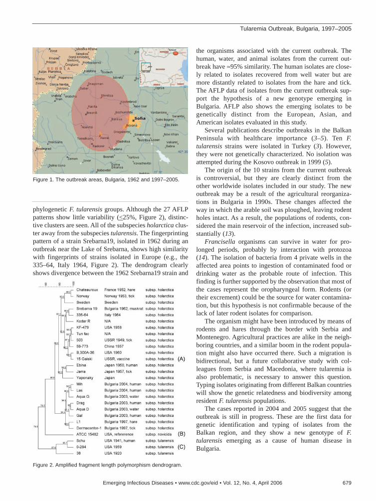
phylogenetic F. tularensis groups. Although the 27 AFLPpatterns show little variability (<25%, Figure 2), distinc-tive clusters are seen. All of the subspecies holarctica clus-ter away from the subspecies tularensis. The fingerprintingpattern of a strain Srebarna19, isolated in 1962 during anoutbreak near the Lake of Srebarna, shows high similaritywith fingerprints of strains isolated in Europe (e.g., the335–64, Italy 1964, Figure 2). The dendrogram clearlyshows divergence between the 1962 Srebarna19 strain and
the organisms associated with the current outbreak. Thehuman, water, and animal isolates from the current out-break have ≈95% similarity. The human isolates are close-ly related to isolates recovered from well water but aremore distantly related to isolates from the hare and tick.The AFLP data of isolates from the current outbreak sup-port the hypothesis of a new genotype emerging inBulgaria. AFLP also shows the emerging isolates to begenetically distinct from the European, Asian, andAmerican isolates evaluated in this study.
Several publications describe outbreaks in the BalkanPeninsula with healthcare importance (3–5). Ten F.tularensis strains were isolated in Turkey (3). However,they were not genetically characterized. No isolation wasattempted during the Kosovo outbreak in 1999 (5).
The origin of the 10 strains from the current outbreakis controversial, but they are clearly distinct from theother worldwide isolates included in our study. The newoutbreak may be a result of the agricultural reorganiza-tions in Bulgaria in 1990s. These changes affected theway in which the arable soil was ploughed, leaving rodentholes intact. As a result, the populations of rodents, con-sidered the main reservoir of the infection, increased sub-stantially (13).
Francisella organisms can survive in water for pro-longed periods, probably by interaction with protozoa(14). The isolation of bacteria from 4 private wells in theaffected area points to ingestion of contaminated food ordrinking water as the probable route of infection. Thisfinding is further supported by the observation that most ofthe cases represent the oropharyngeal form. Rodents (ortheir excrement) could be the source for water contamina-tion, but this hypothesis is not confirmable because of thelack of later rodent isolates for comparison.
The organism might have been introduced by means ofrodents and hares through the border with Serbia andMontenegro. Agricultural practices are alike in the neigh-boring countries, and a similar boom in the rodent popula-tion might also have occurred there. Such a migration isbidirectional, but a future collaborative study with col-leagues from Serbia and Macedonia, where tularemia isalso problematic, is necessary to answer this question.Typing isolates originating from different Balkan countrieswill show the genetic relatedness and biodiversity amongresident F. tularensis populations.
The cases reported in 2004 and 2005 suggest that theoutbreak is still in progress. These are the first data forgenetic identification and typing of isolates from theBalkan region, and they show a new genotype of F.tularensis emerging as a cause of human disease inBulgaria.
Tularemia Outbreak, Bulgaria, 1997–2005
Emerging Infectious Diseases • www.cdc.gov/eid • Vol. 12, No. 4, April 2006 679
Figure 1. The outbreak areas, Bulgaria, 1962 and 1997–2005.
Figure 2. Amplified fragment length polymorphism dendrogram.

Dr Kantardjiev is professor of medicine, microbiology, andepidemiology at the National Center of Infectious and ParasiticDiseases, Sofia, Bulgaria. His research interests are in the field ofclinical microbiology, high-risk infections, and management ofimmunocompromised patients.
References
1. Sjöstedt A. Family XVII. Francisellaceae, genus I. Francisella. In:Brenner DJ, editor. Bergey’s manual of systematic bacteriology.Springer: New York; 2003. p. 111–35.
2. Christova I., Velinov T, Kantardjiev T, Galev A. Tularaemia outbreakin Bulgaria. Scand J Infect Dis. 2004;36:785–9.
3. Helvac S, Gediko?lu S, Akalin H, Oral HB. Tularemia in Bursa,Turkey: 205 cases in ten years. Eur J Epidemiol. 2000;16:271–6.
4. Gurcan S, Otkun MT, Otkun M, Arikan OK, Ozer B. An outbreak oftularemia in Western Black Sea region of Turkey. Yonsei Med J.2004;45:17–22.
5. Reintjes R, Dedushaj I, Gjini A, Jorgensen TR, Cotter B, Lieftucht A,et al. Tularemia outbreak investigation in Kosovo: case control andenvironmental studies. Emerg Infect Dis. 2002;8:69–73.
6. Dinev T, Zlatanov Z. Bacteriological study of a natural focus oftularemia in the Lake Reserve of Srebrna, the Silistra region,Bulgaria. J Hyg Epidemiol Microbiol Immunol. 1972;16:341–5.
7. Gotev N, Sumnaliev M, Zhecheva M. A case of the pulmonary formof tularemia. [Article in Bulgarian]. Suvr Med (Sofiia).1963;14:42–5.
8. Velinov T, Kantardjiev T, Kouzmanov A. An outbreak of tularemia inBulgaria—January, 1998–2000. Problems of Infectious Diseases.2001;29:13–4.
9. Centers for Disease Control and Prevention, Division of PublicHealth Surveillance and Informatics. Tularemia (Francisella tularen-sis). 1999 case definition. [cited 20 Feb 2000]. Available from:http://www.cdc.gov/epo/dphsi/casedef/tularemia_current.htm
10. Wong J, Shapiro D. Francisella. In: Murray P, editor. Manual of clin-ical microbiology, 7th ed. Washington: American Society forMicrobiology Press; 1999. p. 647–51.
11. Johansson A, Forsman M, Sjöstedt A. The development of tools fordiagnosis of tularemia and typing of Francisella tularensis. APMIS.2004;112:898–907.
12. Forsman M, Nyrén A, Sjöstedt A, Sjökvist L, Sandström G.Identification of Francisella tularensis in natural water samples byPCR. FEMS Microbiol Ecol. 1995;16:83–92.
13. Christova I, Gladnishka T. Prevalence of infection with Francisellatularensis, Borrelia burgdorferi sensu lato and Anaplasma phagocy-tophilum in rodents from an endemic focus of tularemia in Bulgaria.Ann Agric Environ Med. 2005;12:149–52.
14. Kantardjiev T, Velinov T. Interaction between protozoa and microor-ganisms of the genus Francisella. Problems of Infectious Diseases.1995;22:34–5.
Address for correspondence: Todor Kantardjiev, National Center forInfectious and Parasitic Diseases, Microbiology Department, 26 YankoSakazov, 1504 Sofia, Bulgaria; fax: 359-2-846-5520; email:[email protected]
DISPATCHES
680 Emerging Infectious Diseases • www.cdc.gov/eid • Vol. 12, No. 4, April 2006
Searchpast issues

Avian InfluenzaH5N1 in NaturallyInfected Domestic
Cat Thaweesak Songserm,* Alongkorn Amonsin,†
Rungroj Jam-on,* Namdee Sae-Heng,* Noppadol Meemak,‡ Nuananong Pariyothorn,†
Sunchai Payungporn,† Apiradee Theamboonlers,†
and Yong Poovorawan†
We report H5N1 virus infection in a domestic catinfected by eating a pigeon carcass. The virus isolated fromthe pigeon and the cat showed the same cluster as theviruses obtained during the outbreak in Thailand. Sincecats are common house pets, concern regarding diseasetransmission to humans exists.
Highly pathogenic avian influenza (HPAI) H5N1 caus-es death in many avian species and mammals, includ-
ing humans (1–5). In Thailand, infection by HPAI H5N1has been reported in mammalian species such as tigers(1,3) and cats (6). Most infected mammals had high fever,panted, and showed symptoms of depression, myalgia, andnervousness (4). This article reports H5N1 infection in acat during the early H5N1 outbreaks in Thailand and char-acterizes the genome of H5N1 virus isolated from theinfected domestic cat.
The StudyIn early February 2004, during the outbreak of HPAI
(H5N1) in Thailand, a carcass of a 2-year-old male cat(Felis catus) was taken in an icebox 6 hours postmortem tothe Faculty of Veterinary Medicine at Kasetsart University,Nakornpathom, Thailand. The cat’s owner volunteered theinformation that the cat had eaten a pigeon (Columbalevia) carcass 5 days before illness onset. The ownerreported that the cat had a temperature of 41°C, was pant-ing, and appeared to be depressed. Furthermore, the cathad convulsions and ataxia and died 2 days after onset ofillness. The cat was given a single dose of 75 mg aspirin1 day before it died; however, its body temperatureremained elevated. Many dead pigeons were found in thearea where the cat lived. Necropsy of the cat showed cere-
bral congestion, conjunctivitis, pulmonary edema, severepneumonia, renal congestion, and hemorrhage in the intes-tinal serosa. Tissues from brain, trachea, lungs, mesentericlymph nodes, intestines (duodenum, jejunum, and ileum),kidneys, liver, pancreas, spleen, and heart were collected,fixed with 10% buffered formalin, and processed forhistopathologic examination. Histopathologic examinationresults showed nonsuppurative encephalitis, gliosis,mononuclear infiltration into the Virchow-Robin space,vasculitis, and congestion in both cerebrum and cerebel-lum. A microscopic lesion in the lung was caused bysevere pulmonary edema, interstitial pneumonia, and con-gestion (Figure 1A). Multifocal necrosis in the liver(Figure 1B), tubulonephritis, and lymphoid depletion inthe spleen were found. No abnormalities were detected inany other organs.
The paraffin-embedded tissues, including brain, lung,kidney, heart, spleen, pancreas, liver, and intestine tissue,were examined immunohistochemically.A polyclonal goatanti-HPAI H5N1 (Kasetsart University, Nakornpathom,Thailand) diluted 1:400 in phosphate-buffered saline wasused as the primary antibody. The secondary antibody waspolyclonal mouse anti-goat immunoglobulin G (ZymedLaboratories, Inc., San Francisco, CA, USA) diluted 1:200in phosphate-buffered saline. Diamino benzidine was thesubstrate developed as a chromogen. Tissue from a cat thathad been hit and killed by a car was used as the negativecontrol. Sites displaying a positive H5N1 antigen reactionwere in cerebral neurons (Figure 1C), heart (myocardialcells) (Figure 1D), pneumocytes, renal tubular epithelialcells, hepatic cells, and white pulp of the spleen
Emerging Infectious Diseases • www.cdc.gov/eid • Vol. 12, No. 4, April 2006 681
*Kasetsart University, Nakorn Pathom, Thailand; †ChulalongkornUniversity, Pathumwan, Thailand; and ‡Western VeterinaryResearch and Development Center, Chombueng, Ratchaburi,Thailand
Figure 1. Microscopic lesions of the infected cat, lung edema withhomogeneous pink material and congestion (A) and multifocalnecrosis in the liver (B). Positive sites are shown by immunohisto-chemical examination of the infected cat in neurons (C) and car-diac muscle cells (D) (magnification ×100).

(macrophages). The pancreas and intestine were negativefor H5N1 antigen.
Parts of frozen brain, lung, liver, kidney, spleen, andduodenum content were ground separately, and virus isola-tion testing was conducted by using embryonated egginjection. Virus isolation testing was also conducted onpleural fluid and urine. Virus isolation testing was con-ducted by injecting pleural fluid, urine, and filtratesobtained from the ground tissues into the allantoic sac of10-day-old embryonated chicken eggs. Embryonic deathoccurred 18 hours after injection. The allantoic fluids ofthe dead embryos were subjected to hemagglutination(HA) and hemagglutination inhibition tests. All fluids fromthe dead embryos were positive for avian influenza A (H5).The virus could be isolated from all injected specimens. Toidentify the subtype, reverse transcription–polymerasechain reaction was conducted, and the virus was confirmedto be influenza A H5N1 (7,8). The HPAI H5N1 isolaterecovered from the infected cat’s lung was labeledA/Cat/Thailand/KU-02/04. In addition, an isolate of HPAIH5N1 from an infected pigeon in the area where the catlived was included in the study and labeledA/Pigeon/Thailand/KU-03/04.
H5N1 viruses isolated from the cat’s (KU-02) and thepigeon’s (KU-03) lung tissue were characterized in thisstudy. The entire genome sequence was determined in theH5N1 isolate from the cat, while the H5N1 isolate fromthe pigeon was sequenced to specifically obtain the HA,neuraminidase, and PB2 genes. The sequences obtainedfrom the cat (H5N1) (A/Cat/Thailand/KU-02/04) weresubmitted to the GenBank database under accession num-bers PB2 (DQ236079), PB1 (DQ236080), PA (DQ236081), HA (DQ236077), NP (DQ236082), NA (DQ236078), M (DQ236084), and NS (DQ236083). Thesequences obtained from the pigeon (H5N1) (A/Pigeon/Thailand/KU-03/04) were submitted to GenBank underaccession numbers HA (DQ236085 ), NA (DQ236086 ),and PB2 (DQ236087). Sequencing and phylogeneticanalysis of the HA (Figure 2A) and NA (Figure 2B) genesof HPAI isolates (cat and pigeon) showed that the HA andNA genes of the viruses were similar to each other as wellas to those of the viruses isolated from tigers, chickens,and humans in Thailand. Genetic comparisons of eachgene of the cat isolate (KU-02) to those of the viruses iso-lated from chickens (January and July 2004) and tigers(January and October 2004) are shown in the Table. Theanalyses showed that the cat isolate (KU-02) was closelyrelated to other H5N1 isolates collected from the region in2004. This finding indicated that the H5N1 infection in thecat resulted from the virus circulating during the H5N1outbreaks in early 2004. The HA gene of KU-02 and KU-03 contained multiple basic amino acid insertions at the
HA cleavage site (SPQRERRRKKRR) as well as gluta-mine and glycine (Q222–G224) at the receptor bindingsite. The NA genes of KU-02 and KU-03 also had 20amino acid deletions at positions 49–68 and contained his-tidine at position 274, indicating absence of antiviral drugresistant residues. The NS gene of the KU-02 isolate con-tained a 5–amino acid deletion (79–83), and the M2 geneof the KU-02 isolate contained an amino acid (asparagine)at position 31, conveying amantadine resistance. In sum-mary, the viruses from the cat and the pigeon were similarto the H5N1 viruses isolated in Thailand and Vietnam in2004, which had then been identified as genotype Z (9). Asingle amino acid substitution at position 627 of the PB2gene (glutamic acid to lysine) was observed in the cat iso-late (KU-02), as had previously been shown in the tigerisolates (1). In contrast, the PB2-627 amino acid residue ofthe pigeon isolate (KU-03) remained unchanged (glutamicacid).
DISPATCHES
682 Emerging Infectious Diseases • www.cdc.gov/eid • Vol. 12, No. 4, April 2006
Figure 2. Phylogenetic analysis of the hemagglutinin (A) and neu-raminidase (B) gene sequences of highly pathogenic avianinfluenza H5N1 from the cat in this study, compared with othersequences from GenBank database.
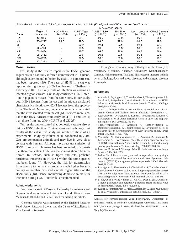
ConclusionsThis study is the first to report entire H5N1 genome
sequences in a naturally infected domestic cat in Thailand,although experimental infection by H5N1 in domestic catshas been reported (10). The case of H5N1 in a cat wasreported during the early H5N1 outbreaks in Thailand inFebruary 2004. The likely route of infection was eating aninfected pigeon carcass. Our study confirmed H5N1 infec-tion in pigeon carcasses from the same area. In our study,both H5N1 isolates from the cat and the pigeon displayedcharacteristics identical to H5N1 isolates from the epidem-ic in Thailand. Moreover, genetic comparison indicatedthat the virus isolated from the cat (KU-02) was more sim-ilar to the H5N1 viruses from early 2004 (Ti-1 and Leo-1)than those from late 2004 (CU-T3 and CU-23).
Our results demonstrated that domestic cats are also atrisk for H5N1 infection. Clinical signs and pathologic testresults of the cat in this study are similar to those of anexperimental study by Kuiken et al. conducted in 2004.Cats are companion animals and may live in very closecontact with humans. Although no direct transmission ofH5N1 from cats to humans has been reported, it is possi-ble; therefore, cats in H5N1-endemic areas should be scru-tinized. In Felidae, such as tigers and cats, probablehorizontal transmission of H5N1 within the same specieshas been found (4). However, the risk for transmissionfrom poultry to humans is probably much higher becausepoultry outnumber cats and excrete higher titers of theH5N1 virus (10). Hence, monitoring domestic animals forinfection during H5N1 outbreak is recommended.
AcknowledgmentsWe thank the staff of Kasetsart University for assistance and
Orawan Booddee for immunohistochemical work. We also thankMettanando Bhikkhu and Petra Hirsch for editing the article.
Genomic research was supported by the Thailand ResearchFund, Senior Research Scholar, and the Center of Excellence inViral Hepatitis Research.
Dr Songserm is a veterinary pathologist at the Faculty ofVeterinary Medicine, Kasetsart University, KamphaengsaenCampus, Nakornpathom, Thailand. His research interests includeavian pathology, duck and goose diseases, and emerging diseasesin animals.
References
1. Amonsin A, Payungporn S, Theamboonlers A, Thanawongnuwech R,Suradhat S, Pariyothorn N, et al. Genetic characterization of H5N1influenza A viruses isolated from zoo tigers in Thailand. Virology.2006;344:480–91.
2. Grose C, Chokephaibulkit K. Avian influenza virus infection of chil-dren in Vietnam and Thailand. Pediatr Infect Dis J. 2004;23:793–4.
3. Keawcharoen J, Oraveerakul K, Kuiken T, Fouchier RA, Amonsin A,Payungporn S, et al. Avian influenza H5N1 in tigers and leopards.Emerg Infect Dis. 2004;10:2189–91.
4. Thanawongnuwech R, Amonsin A, Tantilertcharoen R,Damrongwatanapokin S, Theamboonlers A, Payungporn S, et al.Probable tiger-to-tiger transmission of avian influenza H5N1. EmergInfect Dis. 2005;11:699–701.
5. Viseshakul N, Thanawongnuwech R, Amonsin A, Suradhat S,Payungporn S, Keawchareon J, et al. The genome sequence analysisof H5N1 avian influenza A virus isolated from the outbreak amongpoultry populations in Thailand. Virology. 2004;328:169–76.
6. Enserink M, Kaiser J. Virology. Avian flu finds new mammal hosts.Science. 2004;305:1385.
7. Poddar SK. Influenza virus types and subtypes detection by singlestep single tube multiplex reverse transcription-polymerase chainreaction (RT-PCR) and agarose gel electrophoresis. J Virol Methods.2002;99:63–70.
8. Payungporn S, Phakdeewirot P, Chutinimitkul S, Theamboonlers A,Keawcharoen J, Oraveerakul K, et al. Single step multiplex reversetranscription-polymerase chain reaction (RT-PCR) for influenza Avirus subtype H5N1 detection. Viral Immunol. 2004;17:588–93.
9. Li KS, Guan Y, Wang J, Smith GJ, Xu KM, Duan L, et al. Genesis ofa highly pathogenic and potentially pandemic H5N1 influenza virusin eastern Asia. Nature. 2004;430:209–13.
10. Kuiken T, Rimmelzwaan G, Riel D, Amerongen G, Baars M, FouchierR, et al. Avian H5N1 influenza in cats. Science. 2004;306:241.
Address for correspondence: Yong Poovorawan, Department ofPediatrics, Faculty of Medicine, Chulalongkorn University, 1873 RamaIV Rd, Patumwan, Bangkok 10330, Thailand; fax: 66-2-256-4911; email:[email protected]
Avian Influenza H5N1 in Domestic Cat
Emerging Infectious Diseases • www.cdc.gov/eid • Vol. 12, No. 4, April 2006 683

CryptosporidiosisAssociated withOzonated Apple
CiderBrian G. Blackburn,*1 Jacek M. Mazurek,*†2
Michele Hlavsa,*‡ Jean Park,* Matt Tillapaw,§MaryKay Parrish,† Ellen Salehi,†
William Franks,§ Elizabeth Koch,† Forrest Smith,† Lihua Xiao,* Michael Arrowood,*Vince Hill,* Alex da Silva,* Stephanie Johnston,*
and Jeffrey L. Jones*
We linked an outbreak of cryptosporidiosis to ozonatedapple cider by using molecular and epidemiologic methods.Because ozonation was insufficient in preventing this out-break, its use in rendering apple cider safe for drinking isquestioned.
Cryptosporidium spp. are protozoan parasites transmit-ted by the fecal-oral route that cause prolonged diar-
rhea. Only 2 reports describe outbreaks associated withapple cider (1,2), and none have been associated withozonated cider. In October 2003, a northeast Ohio healthdepartment identified 12 local residents with laboratory-confirmed cryptosporidiosis; 11 had drunk a locally pro-duced, ozonated apple cider (cider A) in the 2 weeksbefore illness. The cider was embargoed on October 24,and we initiated an investigation by using epidemiologicand molecular techniques to determine the cause andextent of the outbreak and the role played by the cider andozonation.
The StudyWe defined a probable case as a northeast Ohio resident
with otherwise unexplained diarrhea for >3 days fromSeptember 1 to November 30, 2003, and a laboratory-con-firmed case as a person with diarrhea and a positiveCryptosporidium laboratory result. Case finding encom-passed interviewing persons with diarrhea who came tolocal health departments and emergency rooms and partic-ipants of school outings at which cider A was served. Wethen conducted 2 epidemiologic studies in which question-naires showed exposures classically associated withCryptosporidium transmission such as food, drinking and
recreational water, person-to-person contact, animals, andtravel.
Study 1 compared laboratory-confirmed case-patientsand 2 controls (persons without diarrhea, abdominal pain,or vomiting) per case matched on age and county of resi-dence and identified through random-digit dialing.Additionally, we conducted a retrospective cohort study ofschool children (study 2) who attended field trips at whichcider A was served.
Stool samples from case-patients were screened by wetpreparation and tested for Cryptosporidium by using animmunofluorescent assay (Meridian Merifluor Crypto-sporidium/Giardia DFA kit, Meridian Bioscience,Cincinnati, OH, USA). Identification of Cryptosporidiumin these samples was attempted by using 2 methods:1) genotyping isolates by polymerase chain reaction–restriction fragment length polymorphism (PCR-RFLP)analysis of the small subunit (SSU) rRNA gene (3,4) withsubtyping by DNA sequence analysis of the GP60 gene (5),and 2) amplification of the SSU rRNA gene with genotypedifferentiation by a microsatellite marker (ML-1) (6,7).
Cider samples were concentrated by centrifugation, andwater samples were concentrated by EnvironmentalProtection Agency method 1623 (8). Cryptosporidiumgenotyping was performed by using the same methodsdescribed for stool samples (4). Newly designed GP60primers were used to subtype cider samples (9).
We identified 23 laboratory-confirmed and 121 proba-ble case-patients with onset dates from September 3 toNovember 19, 2003 (Figure); the first cider-related caseoccurred September 22. The median patient age was 20years (range 1–80). The median incubation period was 7days (range 1–21), and median period of diarrhea was 7days (range 3–52). Two patients were hospitalized andnone died.
In study 1, we enrolled 19 laboratory-confirmed case-patients and 38 age-matched, community-based controls.Twelve of 19 case-patients, but 0 of 38 controls, had drunkcider A. Although the matched odds ratio (OR) for thisassociation was incalculably high, the lower limit of the95% confidence interval (CI) was 5.6. Although 3 otherexposures were also associated with illness by univariateanalysis (Table), only drinking cider A was associated withillness in a conditional logistic regression model thatincluded all of these exposures (estimated OR 14.0, 95%CI 1.8–167).
In study 2, we enrolled 402 persons who participated inoutings at which cider A was served. Thirty-three (10%) of
DISPATCHES
684 Emerging Infectious Diseases • www.cdc.gov/eid • Vol. 12, No. 4, April 2006
*Centers for Disease Control and Prevention, Atlanta, Georgia,USA; †Ohio Department of Health, Columbus, Ohio, USA;‡Atlanta Research and Education Foundation, Atlanta, Georgia,USA; and §Stark County Health Department, Canton, Ohio, USA
1Current affiliation: Stanford University School of Medicine,Stanford, California, USA2Current affiliation: Centers for Disease Control and Prevention,Morgantown, West Virginia, USA

the 329 persons who drank cider A became ill, while only2 (3%) of 73 who did not drink cider A became ill (adjust-ed relative risk 4.7, 95% CI 1.2–18.1). Only drinking ciderA remained significantly associated with illness in a multi-variate logistic regression model that included 4 otherexposures that increased risk in univariate analysis (esti-mated OR 5.7, 95% CI 1.2–26.6).
No employees from the orchard or the separate ciderpressing facility reported diarrhea from September 1 toNovember 7, 2003 (date of interview). The water supplyfor both was negative for Cryptosporidium; employeesused “few” dropped apples for cider production. Duringproduction, an ozonating apparatus (Golden BuffaloCompany, Orange, CA, USA) was used to treat the cider,which was then stored in refrigerated tanks. Most ciderwas ozonated a second time, then sold in plastic jugs on-site and at nearby grocery stores. Remaining cider wassold through a tap in the orchard’s store. In contrast tojugged cider, this cider was not reozonated.
We performed a Cryptosporidium PCR on all 14 avail-able samples from the laboratory-confirmed case-patients.
Twelve (85.7%) of these were PCR positive; 11 of these 12samples were identified as Cryptosporidium parvum and 1as the cervine Cryptosporidium genotype (W4). Subtypeidentification of stool samples yielded 2 closely related C.parvum subtypes (IIaA15G2R1 and IIaA17G2R1).
The remaining contents of a jug of cider A that a labo-ratory-confirmed case-patient had partially drunk werealso positive by PCR for C. parvum subtype IIaA17G2R1.This case-patient’s stool sample yielded the same subtypeof C. parvum, as did 4 other case-patients’ stool samples;all of these persons drank cider A in the 2 weeks before ill-ness onset.
ConclusionsOur investigation strongly implicates cider A as the
cause of this outbreak. The timing of cider A productionclosely paralleled the outbreak, and drinking cider A wasthe only predictor significantly associated with illness inboth univariate and multivariate analyses of both epidemi-ologic studies. Furthermore, detection of C. parvum sub-type IIaA17G2R1 from the sample of partially drunk ciderA from a laboratory-confirmed case-patient provided fur-ther evidence of this link.
The 2 C. parvum subtypes found in this outbreak likelyrepresent a common contamination source; both are com-mon in cattle, and multiple subtypes are commonly foundon farms (10). This outbreak highlights the need for con-tinued development of molecular biologic methodsbecause these techniques will be useful to identify anddefine future Cryptosporidium outbreaks and supplementepidemiologic associations.
An issue raised by this outbreak is the role of ozonationin the treatment of apple cider. New regulations (HazardAnalysis and Critical Control Point [HACCP] standards)enacted by the US Food and Drug Administration (FDA)in 2001 (11) require juice manufacturers to demonstrate a
Cryptosporidiosis and Apple Cider
Emerging Infectious Diseases • www.cdc.gov/eid • Vol. 12, No. 4, April 2006 685
Figure. Laboratory-confirmed (n = 23) and probable (n = 121)cases of cryptosporidiosis from drinking ozonated apple cider,Ohio, 2003.
5-log reduction of “the most resistant microorganism ofpublic health significance” in their production process.
Because this is the third reported Cryptosporidium out-break related to unpasteurized apple cider (1,2), whateversterilization procedure is used must be effective againstCryptosporidium. Although pasteurization kills Crypto-sporidium oocysts (12), no data exist on the use of ozona-tion against Cryptosporidium in food or juice products,where turbidity and low temperature render ozonation lesseffective (13,14). Furthermore, ozonation is difficult tostandardize because effectiveness depends on contact timeand concentration.
The consideration of ozonation is important becauseeffective disinfection would have prevented the outbreakunless contamination occurred at the final step before dis-tribution. Furthermore, of the 12 ill persons in the case-control study who drank cider A, 6 drank once-ozonatedcider and 6 drank twice-ozonated cider, suggesting thateven repeated ozonation was inadequate to killCryptosporidium. The failure of ozone could have beendue to an inherent inadequacy for killing Cryptosporidiumin apple cider or improper use; either possibility empha-sizes the problems with ozonation in this setting and theneed for further testing before its use is accepted.
Given the paucity of evidence supporting ozonation forapple cider disinfection or for killing Cryptosporidium inthis product, and its apparent failure in this outbreak, theFDA issued an addendum to its HACCP rule (15). Thisaddendum advises that juice makers should not use ozonein their manufacturing process unless they can prove a 5-log pathogen reduction through ozonation. To our knowl-edge, no studies have established this reduction to date.
AcknowledgmentsWe thank Ling Zhou and Jianlin Jiang for performing much
of the molecular biological laboratory work and PaulDePasquale, Jane Dancy, Susan Kovach, Cheryl Long, BettyMatecheck, and Christina R. Henning for assisting with the epi-demiologic and environmental investigation.
Dr Blackburn is currently a clinical assistant professor ofinfectious diseases at Stanford University. He performed thiswork while an Epidemic Intelligence Service officer in theDivision of Parasitic Diseases at the Centers for Disease Controland Prevention. His research interests include cryptosporidiosis,schistosomiasis, and strongyloidiasis in refugee populations;angiostrongyliasis; and use of insecticide-treated bed nets to con-trol malaria and lymphatic filariasis.
References
1. Millard PS, Gensheimer KF, Addiss DG, Sosin DM, Beckett GA,Houck-Jankoski A, et al. An outbreak of cryptosporidiosis fromfresh-pressed apple cider. JAMA. 1994;272:1592–6.
2. Centers for Disease Control and Prevention. Outbreaks ofEscherichia coli O157:H7 infection and cryptosporidiosis associatedwith drinking unpasteurized apple cider–Connecticut and New York,October 1996. JAMA. 1997;277:781–3.
3. Xiao L, Bern C, Limor J, Sulaiman I, Roberts J, Checkley W, et al.Identification of 5 types of Cryptosporidium parasites in children inLima, Peru. J Infect Dis. 2001;183:492–7.
4. Xiao L, Singh A, Limor J, Graczyk TK, Gradus S, Lal A. 2001.Molecular characterization of Cryptosporidium oocysts in samples ofraw surface water and wastewater. Appl Environ Microbiol.2001;67:1097–101.
5. Glaberman S, Moore JE, Lowery CJ, Chalmers RM, Sulaiman I,Elwin K, et al. Three drinking-water-associated cryptosporidiosisoutbreaks, Northern Ireland. Emerg Infect Dis. 2002;8:631–3.
6. Da Silva AJ, Bornay-Llinares FJ, Moura IN, Slemenda SB, Tuttle JL,Pieniazek NJ. Fast and reliable extraction of protozoan parasite DNAfrom fecal specimens. Mol Diagn. 1999;4:57–64.
7. Caccio S, Homan W, Camilli R, Traldi G, Kortbeek T, Pozio E. Amicrosatellite marker reveals population heterogeneity within humanand animal genotypes of Cryptosporidium parvum. Parasitology.2000;120:237–44.
8. US Environmental Protection Agency. Method 1623:Cryptosporidium and Giardia in water by filtration/IMS/FA.Publication no. EPA-821-R-99-006. Office of Water. Washington:The Agency; 1999.
9. Sulaiman IM, Hira PR, Zhou L, Al-Ali FM, Al-Shelahi FA, ShweikiHM, et al. Unique endemicity of cryptosporidiosis in children inKuwait. J Clin Microbiol. 2005;43:2805–9.
10. Peng MM, Wilson ML, Holland RE, Meshnick SR, Lal AA, Xiao L.Genetic diversity of Cryptosporidium spp. in cattle in Michigan:implications for understanding the transmission dynamics. ParasitolRes. 2003;90:175–80.
11. US Food and Drug Administration. Hazard analysis and critical con-trol point (HAACP); procedures for the safe and sanitary processingand importing of juice; final rule. Federal Register.2001;66:6137–202.
12. Deng MQ, Cliver DO. Inactivation of Cryptosporidium parvumoocysts in cider by flash pasteurization. J Food Prot. 2001;64:523–7.
13. Korich DG, Mead JR, Madore MS, Sinclair NA, Sterling CR. Effectsof ozone, chlorine dioxide, chlorine, and monochloramine onCryptosporidium parvum oocyst viability. Appl Environ Microbiol.1990;56:1423–8.
14. Driedger AM, Rennecker JL, Marinas BJ. Inactivation ofCryptosporidium parvum oocysts with ozone and monochloramine atlow temperature. Water Res. 2001;35:41–8.
15. US Food and Drug Administration/CFSAN. Guidance for industry:recommendations to processors of apple juice or cider on the use ofozone for pathogen reduction purposes, 2004 Aug. [cited 2005 May24]. Available from http://www.cfsan.fda.gov/~dms/juicgu12.html
Address for correspondence: Brian G. Blackburn, Division of InfectiousDiseases and Geographic Medicine, Stanford University School ofMedicine, 300 Pasteur Dr, Grant Building, Room S-169, Stanford, CA94305-5107, USA; fax: 650-723-3474; email: [email protected]
DISPATCHES
686 Emerging Infectious Diseases • www.cdc.gov/eid • Vol. 12, No. 4, April 2006

Emerging Infectious Diseases • www.cdc.gov/eid • Vol. 12, No. 4, April 2006 687
Long-term Follow-up for
Multidrug-resistantTuberculosis
Sonya S. Shin,* Jennifer J. Furin,* Felix Alcántara,† Jaime Bayona,†
Epifanio Sánchez,‡ and Carole D. Mitnick*§
Patients treated in Peru for multidrug-resistant tuber-culosis (MDR-TB) were followed-up for a median of 67months. Among 86 patients considered cured after comple-tion of treatment, 97% remain healthy; 1 patient relapsed.Employment increased from 34% before treatment to 71%.We observed favorable long-term outcomes among MDR-TB patients.
Increasing awareness of the rising global rates of mul-tidrug-resistant tuberculosis (MDR-TB) has led to a con-
certed international effort to confront this disease,particularly in countries with a high incidence of TB (1–3).Nonetheless, despite cure rates >80% in some programs,MDR-TB patients tend to have chronic disease and requireprolonged therapy.
Little is known about the long-term follow-up ofpatients treated for MDR-TB, including rates of relapse andchronic disability among cured persons. Among patientstreated for pansusceptible TB, chronic disability caused bypulmonary sequelae and malnutrition can be substantial (4).Given the prolonged nature of MDR-TB, one might expecthigher rates of chronic disability among patients with drug-resistant TB compared with those with pansusceptible TB.To explore these questions, we conducted long-term fol-low-up, defined as follow-up for a minimum of 4 yearsafter treatment was initiated, of MDR-TB patients whoreceived individualized therapy in Lima, Peru (1).
The StudyWe performed a retrospective study among all patients
who initiated individualized, community-based MDR-TBtherapy from August 1, 1996, to March 1, 2000. Thedetails of patient identification, enrollment, and treatmentare described elsewhere (5). Patients were resistant to amedian of 5 drugs (range 2–9). Regimens generally includ-ed at least 5 drugs to which the infecting isolate was sus-ceptible, and treatment duration was 18–24 months.Routine follow-up after completion of MDR-TB therapy
included 1) routine smear microscopy and culture on spu-tum samples 1 month after completion of treatment andthen every 6 months for 1 year; 2) subsequent smearmicroscopy, culture, and clinical evaluation by a TB physi-cian for any episode of potential TB symptoms, e.g., a res-piratory illness lasting >14 days, hemoptysis, or weightloss of unclear cause; and 3) continued contact with Sociosen Salud (the community-based organization working withthe Ministry of Health on this MDR-TB treatment project)through the network of health promoters, patient grouptherapy sessions, and a social assistance program. Thus,loss to follow-up or undocumented medical attention forrespiratory illness is rare.
When reporting cohort outcomes, the MDR-TB work-ing group recommends follow-up for 2 years from the timeof treatment initiation when reporting cohort outcomes (6);however no international definition of long-term follow-upfor MDR-TB cohorts exists. Therefore, we defined long-term follow-up as twice the duration set forth by the MDR-TB working group.
We conducted a chart review to determine TB-relatedsymptoms and employment status of persons recorded atbaseline by the intake physician before they receivedMDR-TB therapy. In addition to data obtained throughroutine patient follow-up as per program norms describedabove, Socios en Salud staff involved in the social assis-tance program, members of group therapy sessions, andhealth promoters were interviewed to obtain additional fol-low-up information about the patients, including income,employment status, and household information. A commu-nity health worker conducted home visits to interview allpatients; patients were questioned about current symptomsas well as their socioeconomic status. The study wasreviewed and approved by the institutional review board atHarvard Medical School; local institutional review was notrequired.
Among 120 persons enrolled in this study, 23 patientsdied during treatment, and 1 person remained in culture-negative treatment at the time of analysis. Two patients(both of whom had defaulted from treatment) were lost tofollow-up.
Data are reported on the remaining 96 (80%) patientswho were alive at the time of stopping MDR-TB therapy.Patients were followed for a median (95% confidenceinterval) of 67 (47–88) months after initiation of treatmentand a median of 46 (3–84) months after completion oftreatment. As summarized in the Table, 86 (72%) patientswere considered cured, 9 (8%) defaulted from treatment,and 1 (1%) had failed treatment.
Among those who were considered cured at the time oftreatment completion, 83 (97%) are currently healthy. Onepatient relapsed 1 month after completion of treatment; thispatient refused retreatment and subsequently died of TB.
*Brigham and Women’s Hospital, Boston, Massachusetts, USA;†Socios en Salud, Lima, Peru; ‡Peruvian Ministry of Health, Lima,Peru; and §Harvard Medical School, Boston, Massachusetts, USA
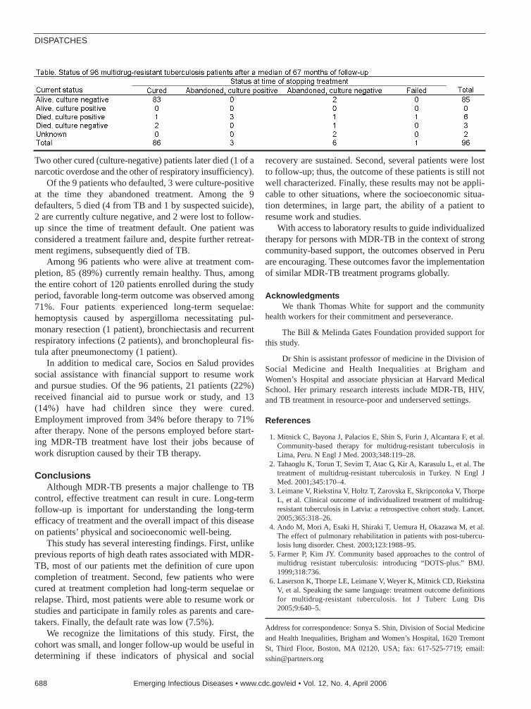
Two other cured (culture-negative) patients later died (1 of anarcotic overdose and the other of respiratory insufficiency).
Of the 9 patients who defaulted, 3 were culture-positiveat the time they abandoned treatment. Among the 9defaulters, 5 died (4 from TB and 1 by suspected suicide),2 are currently culture negative, and 2 were lost to follow-up since the time of treatment default. One patient wasconsidered a treatment failure and, despite further retreat-ment regimens, subsequently died of TB.
Among 96 patients who were alive at treatment com-pletion, 85 (89%) currently remain healthy. Thus, amongthe entire cohort of 120 patients enrolled during the studyperiod, favorable long-term outcome was observed among71%. Four patients experienced long-term sequelae:hemoptysis caused by aspergilloma necessitating pul-monary resection (1 patient), bronchiectasis and recurrentrespiratory infections (2 patients), and bronchopleural fis-tula after pneumonectomy (1 patient).
In addition to medical care, Socios en Salud providessocial assistance with financial support to resume workand pursue studies. Of the 96 patients, 21 patients (22%)received financial aid to pursue work or study, and 13(14%) have had children since they were cured.Employment improved from 34% before therapy to 71%after therapy. None of the persons employed before start-ing MDR-TB treatment have lost their jobs because ofwork disruption caused by their TB therapy.
ConclusionsAlthough MDR-TB presents a major challenge to TB
control, effective treatment can result in cure. Long-termfollow-up is important for understanding the long-termefficacy of treatment and the overall impact of this diseaseon patients’ physical and socioeconomic well-being.
This study has several interesting findings. First, unlikeprevious reports of high death rates associated with MDR-TB, most of our patients met the definition of cure uponcompletion of treatment. Second, few patients who werecured at treatment completion had long-term sequelae orrelapse. Third, most patients were able to resume work orstudies and participate in family roles as parents and care-takers. Finally, the default rate was low (7.5%).
We recognize the limitations of this study. First, thecohort was small, and longer follow-up would be useful indetermining if these indicators of physical and social
recovery are sustained. Second, several patients were lostto follow-up; thus, the outcome of these patients is still notwell characterized. Finally, these results may not be appli-cable to other situations, where the socioeconomic situa-tion determines, in large part, the ability of a patient toresume work and studies.
With access to laboratory results to guide individualizedtherapy for persons with MDR-TB in the context of strongcommunity-based support, the outcomes observed in Peruare encouraging. These outcomes favor the implementationof similar MDR-TB treatment programs globally.
AcknowledgmentsWe thank Thomas White for support and the community
health workers for their commitment and perseverance.
The Bill & Melinda Gates Foundation provided support forthis study.
Dr Shin is assistant professor of medicine in the Division ofSocial Medicine and Health Inequalities at Brigham andWomen’s Hospital and associate physician at Harvard MedicalSchool. Her primary research interests include MDR-TB, HIV,and TB treatment in resource-poor and underserved settings.
References
1. Mitnick C, Bayona J, Palacios E, Shin S, Furin J, Alcantara F, et al.Community-based therapy for multidrug-resistant tuberculosis inLima, Peru. N Engl J Med. 2003;348:119–28.
2. Tahaoglu K, Torun T, Sevim T, Atac G, Kir A, Karasulu L, et al. Thetreatment of multidrug-resistant tuberculosis in Turkey. N Engl JMed. 2001;345:170–4.
3. Leimane V, Riekstina V, Holtz T, Zarovska E, Skripconoka V, ThorpeL, et al. Clinical outcome of individualized treatment of multidrug-resistant tuberculosis in Latvia: a retrospective cohort study. Lancet.2005;365:318–26.
4. Ando M, Mori A, Esaki H, Shiraki T, Uemura H, Okazawa M, et al.The effect of pulmonary rehabilitation in patients with post-tubercu-losis lung disorder. Chest. 2003;123:1988–95.
5. Farmer P, Kim JY. Community based approaches to the control ofmultidrug resistant tuberculosis: introducing “DOTS-plus.” BMJ.1999;318:736.
6. Laserson K, Thorpe LE, Leimane V, Weyer K, Mitnick CD, RiekstinaV, et al. Speaking the same language: treatment outcome definitionsfor multidrug-resistant tuberculosis. Int J Tuberc Lung Dis2005;9:640–5.
Address for correspondence: Sonya S. Shin, Division of Social Medicineand Health Inequalities, Brigham and Women’s Hospital, 1620 TremontSt, Third Floor, Boston, MA 02120, USA; fax: 617-525-7719; email:[email protected]
DISPATCHES
688 Emerging Infectious Diseases • www.cdc.gov/eid • Vol. 12, No. 4, April 2006

ImmuneRestoration
Disease in HIVPatient
Neil E. Jenkins,* Mike B.J. Beadsworth,* James J. Anson,* Fred J. Nye,*
Vanessa J. Martlew,* and Nick J. Beeching*
We describe a severely immunosuppressed HIV-1–positive man in whom immune restoration disease asso-ciated with pulmonary infection caused by Mycobacteriummicroti developed after antiretroviral treatment. The diagno-sis was made by using convenient spoligotyping tech-niques, but invasive investigations were required toexclude a tumor.
During the first few months of highly active antiretro-viral treatment, immune restoration may be compli-
cated by clinical events in which either previouslysubclinical infections are found or preexisting partiallytreated opportunistic infections deteriorate. This condition,termed immune restoration disease (IRD), is thought to becaused by the improvement in the host’s immune responseto pathogens. We report what we believe is the first record-ed case of IRD with confirmed Mycobacterium microtiinfection. M. microti is an unusual infection associatedwith small rodents. Novel genetic techniques have con-firmed it as a human pathogen, but its true incidenceremains unclear.
Case ReportA 33-year-old man was admitted to the hospital in 2002
with a 3-month history of intermittent hemoptysis. He hadpreviously visited hematology outpatient clinics for manyyears for hemophilia A (3%–5% factor VIII) and HIV-1had been diagnosed in 1996 (a stored sample from 1989retrospectively tested HIV-1–antibody positive). CD4 cellcounts had been recorded as 434 × 106/L in 1996 and 260× 106/L in 1998. After this time, he failed to attend any fur-ther clinic appointments.
In addition to worsening hemoptysis, he had minorweight loss but no night sweats. He appeared moderatelyunhealthy, with oral thrush and mild splenomegaly. A chestradiograph showed left upper lobe shadowing (Figure 1,panel A), sputum smears contained numerous acid-fastbacilli, and sputum was sent for culture, speciation, and
sensitivity testing. Blood tests showed a CD4 cell count of6 ×106/L and HIV viral load of 272,000 (log 5.4)copies/mL. His hemoglobin was 12.1 g/dL, leukocytecount 4.9 × 10⁹/L, and platelets 367 × 109/L. Renal andliver function test results were normal, apart from a γ-glu-tamyltransferase level of 200 U/L (normal <35 U/L). Anultrasound scan of his abdomen showed a diffuse increasein liver echogenicity and an enlarged (13.6-cm) spleen. Hewas naturally immune to hepatitis B, and tests for hepatitisC antibody and viremia were negative.
He was given broad-spectrum antimycobacterial thera-py (rifampin, isoniazid, pyrazinamide, ethambutol, andazithromycin) as treatment for both M. tuberculosis com-plex (MTB complex) and opportunistic mycobacteria. Heimproved throughout a 2-week hospital stay and highlyactive antiretroviral therapy (HAART) was begun. It con-sisted of zidovudine and lamivudine and an increased doseof efavirenz (800 mg daily) because of concomitantrifampicin therapy, together with cotrimoxazole for pro-phylaxis against Pneumocystis jirovecii.
At a 3-week follow-up appointment, the patient hadcontinued to improve. No further hemoptysis had occurred,and he had gained 2 kg body weight. After that, by tele-phone, he complained of occasional fevers and malaise butdeclined to be seen. Nine weeks after his initial visit, hecame to the ward and with symptoms of breathlessness,nausea, and diarrhea. He had fevers spiking to >38°C, andnewly palpable lymph nodes were evident in the right axil-la and over the left parotid gland. A chest radiographshowed increased shadowing (Figure 1, panel B) and acomputed tomographic (CT) scan of the chest showed a6×3-cm solid mass in the upper left chest with visible airbronchograms and minor volume loss, reported as “almostcertainly solid neoplasm” (Figure 2). A bronchoscopy,before which the patient received factor VIII, showed anarea of irregular nodular tissue in the posterior segment ofthe left upper lobe. His CD4 count was 26 × 106/L.
While we awaited histologic examination results, thepatient’s medication was withdrawn, and he improvedwithin days. The lymph nodes reduced in size, and thespiking temperature resolved. Eight weeks after admis-sion, the results of IS6110 polymerase chain reaction(PCR) testing on the slow-growing, positive mycobacteri-al culture confirmed the presence of MTB complex. Thebronchial biopsy specimen showed necrotizing granulo-matous bronchitis; acid-fast bacilli were not visible, andthis specimen did not grow any mycobacteria.
The Mycobacterium species was not growing well inthe laboratory, but M. microti was identified in the culturewith spoligotyping methods (1,2). On extension of culture,the isolate was confirmed as M. microti phenotypically andfound to be sensitive in vitro to rifampin, isoniazid, etham-butol, and clarithromycin but resistant to pyrazinamide.
Emerging Infectious Diseases • www.cdc.gov/eid • Vol. 12, No. 4, April 2006 689
*Liverpool School of Tropical Medicine, Liverpool, United Kingdom
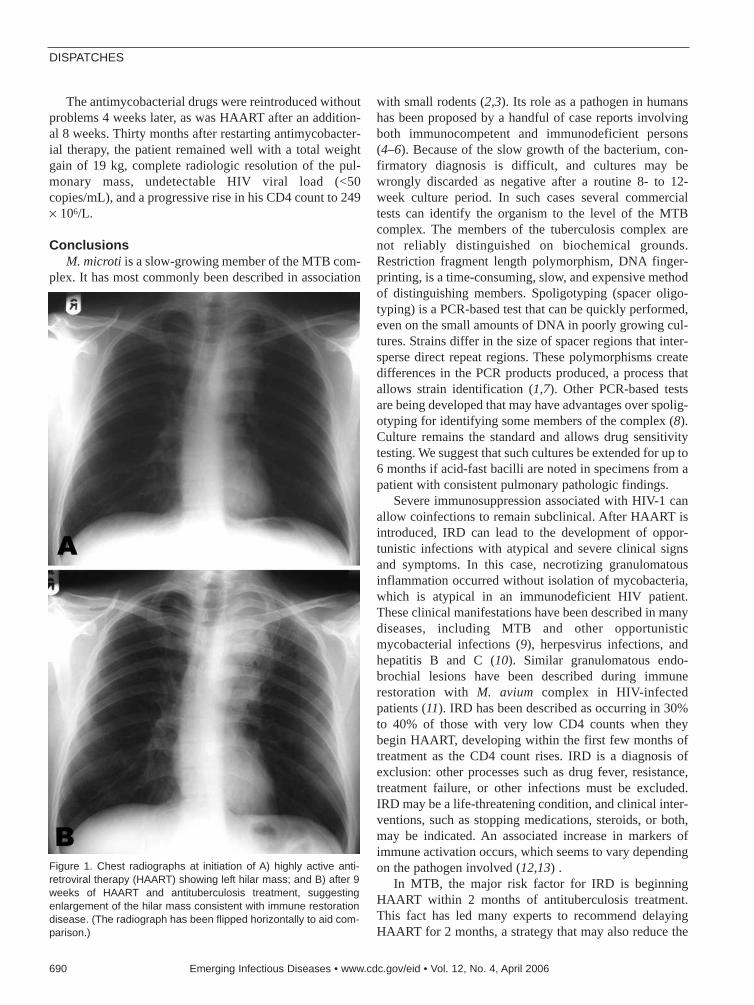
The antimycobacterial drugs were reintroduced withoutproblems 4 weeks later, as was HAART after an addition-al 8 weeks. Thirty months after restarting antimycobacter-ial therapy, the patient remained well with a total weightgain of 19 kg, complete radiologic resolution of the pul-monary mass, undetectable HIV viral load (<50copies/mL), and a progressive rise in his CD4 count to 249× 106/L.
ConclusionsM. microti is a slow-growing member of the MTB com-
plex. It has most commonly been described in association
with small rodents (2,3). Its role as a pathogen in humanshas been proposed by a handful of case reports involvingboth immunocompetent and immunodeficient persons(4–6). Because of the slow growth of the bacterium, con-firmatory diagnosis is difficult, and cultures may bewrongly discarded as negative after a routine 8- to 12-week culture period. In such cases several commercialtests can identify the organism to the level of the MTBcomplex. The members of the tuberculosis complex arenot reliably distinguished on biochemical grounds.Restriction fragment length polymorphism, DNA finger-printing, is a time-consuming, slow, and expensive methodof distinguishing members. Spoligotyping (spacer oligo-typing) is a PCR-based test that can be quickly performed,even on the small amounts of DNA in poorly growing cul-tures. Strains differ in the size of spacer regions that inter-sperse direct repeat regions. These polymorphisms createdifferences in the PCR products produced, a process thatallows strain identification (1,7). Other PCR-based testsare being developed that may have advantages over spolig-otyping for identifying some members of the complex (8).Culture remains the standard and allows drug sensitivitytesting. We suggest that such cultures be extended for up to6 months if acid-fast bacilli are noted in specimens from apatient with consistent pulmonary pathologic findings.
Severe immunosuppression associated with HIV-1 canallow coinfections to remain subclinical. After HAART isintroduced, IRD can lead to the development of oppor-tunistic infections with atypical and severe clinical signsand symptoms. In this case, necrotizing granulomatousinflammation occurred without isolation of mycobacteria,which is atypical in an immunodeficient HIV patient.These clinical manifestations have been described in manydiseases, including MTB and other opportunisticmycobacterial infections (9), herpesvirus infections, andhepatitis B and C (10). Similar granulomatous endo-brochial lesions have been described during immunerestoration with M. avium complex in HIV-infectedpatients (11). IRD has been described as occurring in 30%to 40% of those with very low CD4 counts when theybegin HAART, developing within the first few months oftreatment as the CD4 count rises. IRD is a diagnosis ofexclusion: other processes such as drug fever, resistance,treatment failure, or other infections must be excluded.IRD may be a life-threatening condition, and clinical inter-ventions, such as stopping medications, steroids, or both,may be indicated. An associated increase in markers ofimmune activation occurs, which seems to vary dependingon the pathogen involved (12,13) .
In MTB, the major risk factor for IRD is beginningHAART within 2 months of antituberculosis treatment.This fact has led many experts to recommend delayingHAART for 2 months, a strategy that may also reduce the
DISPATCHES
690 Emerging Infectious Diseases • www.cdc.gov/eid • Vol. 12, No. 4, April 2006
Figure 1. Chest radiographs at initiation of A) highly active anti-retroviral therapy (HAART) showing left hilar mass; and B) after 9weeks of HAART and antituberculosis treatment, suggestingenlargement of the hilar mass consistent with immune restorationdisease. (The radiograph has been flipped horizontally to aid com-parison.)

incidence of adverse events. However, a recent retrospec-tive review suggested that delaying HAART would, partic-ularly in those with CD4 cell counts<100 cells × 106/L,lead to significant increase in risk for death and newopportunistic infections (14). IRD should always be con-sidered along with treatment failure if a patient’s clinicalcondition deteriorates after HAART is introduced.
AcknowledgmentsWe thank Tony Hart, Malcolm Bennett, and Rachel
Cavanagh for advice on M. microti and Ann Barrett and her col-leagues in the Health Protection Agency Reference Laboratory,Newcastle, for confirming the identity and antimicrobial sensitiv-ities of the isolate.
Dr Jenkins is a specialist registrar training in infectious dis-eases at Liverpool and Oxford, United Kingdom. He is also aWellcome Clinical Training Fellow at the Liverpool School ofTropical Medicine, United Kingdom.
References
1. Goyal M, Saunders NA, van Embden JD, Young DB, Shaw RJ.Differentiation of Mycobacterium tuberculosis isolates by spoligo-typing and IS6110 restriction fragment length polymorphism. J ClinMicrobiol. 1997;35:647–51.
2. Cavanagh R, Begon M, Bennett M, Ergon T, Graham IM, de HaasPEW, et al. Mycobacterium microti infection (vole tuberculosis) inwild rodent populations. J Clin Microbiol. 2002;40:3281–5.
3. Wells AQ, Oxon DM. Tuberculosis in wild voles. Lancet.1937;1:1221.
4. Horstkotte MA, Sobottka I, Schewe CK, Schafer P, Laufs R, Rusch-Gerged S, et al. Mycobacterium microti llama-type infection present-ing as pulmonary tuberculosis in a human immunodeficiencyvirus-positive patient. J Clin Microbiol. 2001;39:406–7.
5. Niemann S, Richter E, Dalügge-Tamm H, Schlesinger H, GraupnerD, Königstein B, et al. Two cases of Mycobacterium microti–derivedtuberculosis in HIV-negative immunocompetent patients. EmergInfect Dis. 2000;6:539–42.
6. Foudraine NA, van Soolingen D, Noordhoek GT, Reiss P. Pulmonarytuberculosis due to Mycobacterium microti in a human immunodefi-ciency virus–infected patient. Clin Infect Dis. 1998;27:1543–4.
7. Van Soolingen D, van der Zanden AGM, de Haas PEW, NoordhoekGT, Kiers A, Foudraine NA, et al. Diagnosis of Mycobacteriummicroti infections among humans by using novel genetic markers. JClin Microbiol. 1998;36:1840–5.
8. Huard RC, Oliveira Lazzarini LC, Butler WR, van Soolingen D, HoJL. PCR-based method to differentiate the subspecies of theMycobacterium tuberculosis complex on the basis of genomic dele-tions. J Clin Microbiol. 2003;41:1637–50.
9. Lawn SD, Bekker LG, Miller RF. Immune reconstitution diseaseassociated with mycobacterial infections in HIV-infected individualsreceiving antiretrovirals. Lancet Infect Dis. 2005;5:361–73.
10. DeSimone JA, Pomerantz RJ, Babichak TJ. Inflammatory reactionsin HIV-1 infected patients after initiation of highly active antiretrovi-ral therapy. Ann Intern Med. 2000;133:447–54.
11. Bartley PB, Allworth AM, Eisen DP. Mycobacterium avium complexcausing endobronchial disease in AIDS patients after partial immunerestoration. Int J Tuberc Lung Dis. 1999;3:1132–6.
12. Price P, Mathiot N, Krueger R, Stone S, Keane NM, French M.Immune dysfunction and immune restoration disease in HIV patientsgiven highly active antiretroviral therapy. J Clin Virol.2001;22:279–87.
13. French MA, Price P, Stone SF. Immune restoration disease after retro-viral therapy. AIDS. 2004;18:1615–27.
14. Dheda K, Lampe FC, Johnson MA, Lipman MC. Outcome of HIV-associated tuberculosis in the era of highly active antiretroviral ther-apy. J Infect Dis. 2004;190:1670–6.
Address for correspondence: Neil E Jenkins, Tropical and InfectiousDiseases Unit, Royal Liverpool University Hospital, Liverpool,Merseyside L7 8XP, UK; fax: 44-151-706-5944; email: [email protected]
Immune Restoration Disease in HIV Patient
Emerging Infectious Diseases • www.cdc.gov/eid • Vol. 12, No. 4, April 2006 691
Figure 2. Computed tomographic scan of thorax at patient’s initialvisit. Results are highly suggestive of tumor.
All material published in Emerging Infectious Diseases is in thepublic domain and may be used and reprinted without special per-mission; proper citation, however, is required.
Search past issues of EID at www.cdc.gov/eid
MassTagPolymerase Chain
Reaction forDifferential
Diagnosis of ViralHemorrhagic Fevers
Gustavo Palacios,*1 Thomas Briese,*1
Vishal Kapoor,* Omar Jabado,* Zhiqiang Liu,*Marietjie Venter,† Junhui Zhai,* Neil Renwick,*
Allen Grolla,‡ Thomas W. Geisbert,§ Christian Drosten,¶ Jonathan Towner,#
Jingyue Ju,* Janusz Paweska,** Stuart T. Nichol,# Robert Swanepoel,**
Heinz Feldmann,‡†† Peter B. Jahrling,‡‡ and W. Ian Lipkin*
Viral hemorrhagic fevers are associated with highrates of illness and death. Although therapeutic options arelimited, early differential diagnosis has implications for con-tainment and may aid in clinical management. We describea diagnostic system for rapid, multiplex polymerase chainreaction identification of 10 different causes of viral hemor-rhagic fevers.
Increasing international travel, trafficking in wildlife,political instability, and terrorism have made emerging
infectious diseases a global concern. Viral hemorrhagicfevers (VHF) warrant specific emphasis because of theirhigh rates of illness and death, and the potential for rapiddissemination by human-to-human transmission. The term“viral hemorrhagic fever” characterizes a severe multisys-tem syndrome associated with fever, shock, and bleedingdiathesis caused by infection with any of several RNAviruses, including Ebola virus and Marburg virus (MARV)(family Filoviridae); Lassa virus (LASV) and the SouthAmerican hemorrhagic fever viruses Guanarito virus, Junín
virus, Machupo virus, and Sabiá virus (Arenaviridae); RiftValley fever virus (RVFV), Crimean-Congo hemorrhagicfever virus (CCHFV), and hantaviruses (Bunyaviridae);and Kyasanur Forest disease virus (KFDV), Omsk hemor-rhagic fever virus, yellow fever virus (YFV), and dengueviruses (Flaviviridae) (1,2). Although clinical managementof VHF is primarily supportive, early diagnosis is needed tocontain the contagion and implement public health meas-ures, especially if agents are encountered out of their natu-ral geographic context.
Vaccines have been developed for YFV, RVFV, Junínvirus, KFDV, and hantaviruses (3–7), but only YFV vac-cine is widely available. Early treatment with immune plas-ma was effective in Junín virus infection (8). Thenucleoside analog ribavirin may be helpful if given early inthe course of Lassa fever (9), Crimean-Congo hemorrhagicfever (10), or hemorrhagic fever with renal syndrome (11)and is recommended in postexposure prophylaxis and earlytreatment of arenavirus and bunyavirus infections (12).
Methods for direct detection of nucleic acids of micro-bial pathogens in clinical specimens are rapid, sensitive,and obviate the need for high-level biocontainment.Numerous systems are described for nucleic acid detectionof VHF agents; however, none are multiplex (13).Although geographic location or travel history of suspect-ed patients usually restricts the number of agents to be con-sidered, diagnosis of VHF may be difficult in case of anintentional release (12). Symptoms of VHF are initiallynonspecific and may include fever, headache, myalgia, andgastrointestinal or upper respiratory tract complaints (1);thus, assays that allow simultaneous consideration of mul-tiple agents are needed.
We recently described the application of MassTag poly-merase chain reaction (PCR) in the context of differentialdiagnosis of respiratory disease (14). MassTag PCR is amultiplex assay in which microbial gene targets are codedby a library of 64 distinct mass tags. Nucleic acids (RNAor DNA) are amplified by multiplex (reverse transcrip-tion–) PCR using up to 64 primers, each labeled by aphoto-cleavable link with a different molecular weight tag.After separation of the amplification products from unin-corporated primers and release of the mass tags from theamplicons by UV irradiation, tag identity is analyzed bymass spectrometry. The identity of the microbe in the clin-ical sample is determined by the presence of its 2 cognatetags, 1 from each primer.
The StudyTo facilitate rapid differential diagnosis of VHF agents,
we established the Greene MassTag Panel VHF version 1.0,which comprises the following targets: Ebola Zaire virus
DISPATCHES
692 Emerging Infectious Diseases • www.cdc.gov/eid • Vol. 12, No. 4, April 2006
*Columbia University, New York, New York, USA; †University ofPretoria and National Health Laboratory Services, Pretoria, SouthAfrica; ‡Public Health Agency of Canada, Winnipeg, Manitoba,Canada; §United States Army Medical Research Institute ofInfectious Diseases, Fort Detrick, Frederick, Maryland, USA;¶Bernhard-Nocht-Institute of Tropical Medicine, Hamburg,Germany; #Centers for Disease Control and Prevention, Atlanta,Georgia, USA; **National Institute for Communicable Diseases,Sandringham, South Africa; ††University of Manitoba, Winnipeg,Manitoba, Canada; and ‡‡National Institutes of Allergy andInfectious Diseases Integrated Research Facility, Fort Detrick,Frederick, Maryland, USA 1These authors contributed equally to this article.

(ZEBOV), Ebola Sudan virus (SEBOV), MARV, LASV,RVFV, CCHFV, Hantaan virus (HNTV), Seoul virus(SEOV), YFV, and KFDV. Oligonucleotide primers weredesigned in conserved genomic regions to detect the broad-est number of members for a given pathogen species. Wedeveloped a software program that culls sequence informa-tion from GenBank, performs multiple alignments withClustalW, and designs primers optimized for multiplexPCR. The program uses a greedy algorithm to identify con-served sequences and create the minimum set of primers foramplification of all sequences in the alignment. Primers areselected within standard design constraints whenever possi-ble (melting temperature 55°C–65°C, guanine-cytosinecontent 40%–60%, no hairpins); degenerate positions areintroduced in cases where template divergence requiresmore flexibility. Although degeneracy is not tolerated in thefive 3′ nucleotides, MassTag PCR allows up to 4 nonneigh-boring variable positions per primer. Primers are checkedby the basic local alignment search tool for potentialhybridization to sequenced vertebrate genomes (Table 1).
Because only released mass tags are analyzed, stagger-ing the size of amplification products created in multiplexreactions is unnecessary; thus, primers are selected forefficient and consistent performance irrespective of ampli-con size (typically 80–200 bp). Before committing to syn-thesis of tagged primers, the functionality of candidatemultiplex primer panels is examined in a series of amplifi-cation reactions that use prototype templates representingindividual microbial targets. Primers that fail to yield a sin-gle, specific product band in agarose gel analysis are
replaced. Target sequence standards for evaluation arecloned into pCR2.1-TOPO (Invitrogen, Carlsbad, CA,USA) by using PCR amplification of cDNA templatesobtained by reverse transcription (RT) of extracts frominfected, cultured cells or by assembly of overlapping syn-thetic polynucleotides.
The agents assayed in the VHF panel have RNAgenomes; thus, assay sensitivity was determined by usingsynthetic RNA standards. Synthetic RNA standards weregenerated from linearized target sequence plasmids byusing T7 polymerase (mMessage mMachine, Invitrogen).After quantitation by UV spectrometry, RNA was seriallydiluted in 2.5 µg/mL yeast tRNA (Sigma, St. Louis, MO,USA), reverse transcribed with random hexamers by usingSuperscript II (Invitrogen), and analyzed by MassTag PCRas previously described (14). QIAquick 96 PCR purifica-tion cartridges (Qiagen, Hilden, Germany, with modifiedbinding and wash buffers) were used to remove unincorpo-rated primers before tags were decoupled from amplifica-tion products by UV photolysis in a flow cell and analyzedin a quadrapole mass spectrometer by using positive-modeatmospheric pressure chemical ionization (APCI-MS,Agilent Technologies, Palo Alto, CA, USA). The sensitivi-ty of the 10-plex VHF panel with synthetic RNA standardswas <50 RNA copies per assay (Table 2). Sensitivity andspecificity of multiplex primer panels is assessed empirical-ly by using calibrated synthetic standards as well as tissueculture–derived viral nucleic acid for each assembled panel.
Tissue culture extracts were used to examine assayspecificity. Random primed cDNA obtained from cultures
Differential Diagnosis of Viral Hemorrhagic Fevers
Emerging Infectious Diseases • www.cdc.gov/eid • Vol. 12, No. 4, April 2006 693

of ZEBOV, SEBOV, MARV, YFV isolates from theGambia and Côte d’Ivoire, RVFV, CCHFV, HTNV, SEOV,and LASV strains Josiah, NL, and AV were subjected tomass tag analysis. In all instances, only the appropriatecognate mass tags were detected (data not shown). No spu-rious signal was identified in assays with water or RNAcontrols.
Performance with clinical materials was tested by usingblood, sera, or oral swabs from 24 human patients of VHF
previously diagnosed through virus isolation, RT-PCR, orantigen detection enzyme-linked immunosorbent assay.Differential diagnosis by blinded MassTag PCR analysiswas accurate in all cases (Table 3). For the samples fromthe 2005 Angola Marburg outbreak the result of MassTagPCR was similar to that of diagnostic single-plex PCR.ZEBOV sample 5004, obtained on day 17 of illness whenserologic test results were positive for immunoglobulin M(IgM) and IgG, was negative by viral culture but positivein MassTag PCR.
ConclusionsThese results confirm earlier work in respiratory dis-
eases that show that MassTag PCR offers a rapid, sensi-tive, specific, and economic approach to differentialdiagnosis of infectious diseases. Small, low-cost, ormobile APCI-MS units extend the applicability of thistechnique beyond selected reference laboratories. Giventhe capacity of the method to code for up to 32 genetic tar-gets, we are expanding the hemorrhagic fever panel toinclude additional viruses (dengue and South Americanhemorrhagic fever viruses) and are exploring the inclusionof bacterial and parasitic agents that may result in similarclinical signs and symptoms and, thus, have to be consid-ered in differential diagnosis.
DISPATCHES
694 Emerging Infectious Diseases • www.cdc.gov/eid • Vol. 12, No. 4, April 2006

This work was supported by National Institutes of Healthawards AI51292, AI056118, AI55466, and U54AI57158(Northeast Biodefense Center-Lipkin) and the Ellison MedicalFoundation.
Dr Palacios is an associate research scientist in the JeromeL. and Dawn Greene Infectious Disease Laboratory at theColumbia University Mailman School of Public Health. Hisresearch focuses on the molecular epidemiology of viruses, virusinteractions with their hosts, and innovative pathogen detectionmethods.
References
1. Peters CJ, Zaki SR. Role of the endothelium in viral hemorrhagicfevers. Crit Care Med. 2002;30(5 Suppl):S268–73.
2. Geisbert TW, Jahrling PB. Exotic emerging viral diseases: progressand challenges. Nat Med. 2004;10(12 Suppl):S110–21.
3. Pugachev KV, Guirakhoo F, Monath TP. New developments in fla-vivirus vaccines with special attention to yellow fever. Curr OpinInfect Dis. 2005;18:387–94.
4. Pittman PR, Liu CT, Cannon TL, Makuch RS, Mangiafico JA, GibbsPH, et al. Immunogenicity of an inactivated Rift Valley fever vaccinein humans: a 12-year experience. Vaccine. 1999;18:181–9.
5. Enria DA, Barrera Oro JG. Junin virus vaccines. Curr Top MicrobiolImmunol. 2002;263:239–61.
6. Hooper JW, Li D. Vaccines against hantaviruses. Curr Top MicrobiolImmunol. 2001;256:171–91.
7. Dandawate CN, Desai GB, Achar TR, Banerjee K. Field evaluationof formalin inactivated Kyasanur forest disease virus tissue culturevaccine in three districts of Karnataka state. Indian J Med Res.1994;99:152–8.
8. Enria DA, Maiztegui JI. Antiviral treatment of Argentine hemorrhag-ic fever. Antiviral Res. 1994;23:23–31.
9. McCormick JB, King IJ, Webb PA, Scribner CL, Craven RB, JohnsonKM, et al. Lassa fever. Effective therapy with ribavirin. N Engl JMed. 1986;314:20–6.
10. Ozkurt Z, Kiki I, Erol S, Erdem F, Yilmaz N, Parlak M, et al.Crimean-Congo hemorrhagic fever in eastern Turkey: clinical fea-tures, risk factors and efficacy of ribavirin therapy. J Infect. Epub2005 Jun 13.
11. Huggins JW, Hsiang CM, Cosgriff TM, Guang MY, Smith JI, Wu ZO,et al. Prospective, double-blind, concurrent, placebo-controlled clini-cal trial of intravenous ribavirin therapy of hemorrhagic fever withrenal syndrome. J Infect Dis. 1991;164:1119–27.
12. Borio L, Inglesby T, Peters CJ, Schmaljohn AL, Hughes JM, JahrlingPB, et al. Hemorrhagic fever viruses as biological weapons: medicaland public health management. JAMA. 2002;287:2391–405.
13. Drosten C, Kummerer BM, Schmitz H, Gunther S. Molecular diag-nostics of viral hemorrhagic fevers. Antiviral Res. 2003;57:61–87.
14. Briese T, Palacios G, Kokoris M, Jabado O, Liu Z, Renwick N, et al.Diagnostic system for rapid and sensitive differential detection ofpathogens. Emerg Infect Dis. 2005;11:310–3.
15. Bowen MD, Rollin PE, Ksiazek TG, Hustad HL, Bausch DG, DembyAH, et al. Genetic diversity among Lassa virus strains. J Virol.2000;74:6992–7004.
Address for correspondence: Thomas Briese, Jerome L. and DawnGreene Infectious Disease Laboratory, Mailman School of Public Health,Columbia University, 722 W 168th St, Rm 1801, New York, NY 10032,USA; fax: 212-342-9044; email: [email protected]
Differential Diagnosis of Viral Hemorrhagic Fevers
Emerging Infectious Diseases • www.cdc.gov/eid • Vol. 12, No. 4, April 2006 695
The print journal is available at no charge to public health professionals
YES, I would like to receive Emerging Infectious Diseases.
Please print your name and businessaddress in the box and return by fax to404-639-1954 or mail to
EID EditorCDC/NCID/MS D611600 Clifton Road, NEAtlanta, GA 30333
Moving? Please give us your new address (in the box) and print the number of your oldmailing label here_________________________________________
�������������������� ���������������
Use of trade names is for identification only and does not implyendorsement by the Public Health Service or by the U.S.Department of Health and Human Services.
Escherichia coli is both the most abundant facultativecommensal of the human gastrointestinal tract and the
most common bacterial cause of human diarrhea (1).However, precise recognition of E. coli pathotypesremains problematic. Enteropathogenic E. coli (EPEC),classically associated with outbreaks of infant diarrhea,harbors distinctive chromosomal (the locus of enterocyteeffacement, or LEE island) and plasmidborne (residing onthe EPEC adherence factor, or EAF, plasmid) virulencefactors, which are linked by common gene regulators (1).At the Second International Conference on EPEC, held inSão Paulo, Brazil, in 1995, the foremost authorities in thefield proclaimed the global importance of such “typical”EPEC (tEPEC) but pondered the clinical relevance ofstrains carrying only the LEE island (dubbed at that con-ference “atypical EPEC,” or aEPEC) (2). Had aEPEC lostthe EAF plasmid? Had it incidentally acquired only frag-ments or incomplete packages of virulence-associatedgenes? Or were some aEPEC true pathogens of humans oranimals?
In this issue of Emerging Infectious Diseases, Nguyenet al. propose a distinct role for aEPEC in human infection(3). Previously, these investigators reported a high preva-lence of aEPEC among pediatric diarrhea patients inMelbourne, including both infants and older children (incontrast to the strong tendency for infants to be infectedwith tEPEC) (4). Now these authors show that, in contrastto patients infected with other pathogens, patients infectedwith aEPEC are far more likely to experience diarrhea past14 days, the point long recognized as a clinical watershedthat heralds increased risk for illness and death. aEPEC’sprevalence among diarrhea patients, the pathogen’s strongassociation with diarrheal symptoms, and the infection’sdistinctively persistent nature argue for a high disease bur-den in Melbourne. Although the authors define aEPECstrictly on the basis of positivity for the LEE eae gene andfailure to amplify a bfpA pilin gene (not assessing addition-al plasmid loci), the absence of tEPEC serotypes and the
occurrence of disease in children older than infants suggestthat these are indeed aEPEC.
This communication also illustrates 2 principles thatshould be recognized more generally with regard to diar-rheogenic E. coli: 1) one can authoritatively implicate aparticular strain as a pathogen by (only) outbreak implica-tion or by volunteer studies, but one cannot definitivelyprove by any data that a putative pathotype is not a humanpathogen; and 2) the implication of pathogenicity for 1strain is not sufficient to implicate any similar strain as apathogen. Thus, once definitive evidence is presented thata particular strain is pathogenic, the challenge is to deter-mine the genotypic or phenotypic fingerprints that permitextrapolation to other strains. This obstacle has longproved daunting for the enteroaggregative pathotype(shown to be pathogenic in both volunteer studies and out-breaks), and it may prove similarly frustrating for aEPEC.
Diarrheal epidemiology studies today are able to identi-fy a likely etiologic agent for most patients, in contrast tostudies of 20 years ago, because of improved diagnostictests and the identification of new pathogens, but the task isnot finished. Additional studies are needed in many placesto refine our understanding of putative and emergingpathogens and to determine their full epidemiologic roles.
References
1. Nataro JP, Kaper JB. Diarrheagenic Escherichia coli. Clin MicrobiolRev. 1998;11:142–201.
2. Kaper JB. Defining EPEC. Rev Microbiol Sao Paulo.1996;27(Suppl1):130–3.
3. Nguyen RN, Taylor LS, Tauschek M, Robins-Browne RM. Atypicalenteropathogenic Escherichia coli infection and prolonged diarrhea.Emerg Infect Dis. 2006;12:597–603.
4. Robins-Browne RM, Bordun A-M, Tauschek M, Bennett-Wood V,Russell J, Oppedisano F, et al. Escherichia coli and community-acquired gastroenteritis, Melbourne, Australia. Emerg Infect Dis.2004;10:1797–805.
Address for correspondence: James P. Nataro, Center for VaccineDevelopment, Departments of Pediatrics, Medicine, and Microbiologyand Immunology, University of Maryland School of Medicine,Baltimore, MD 21201, USA; fax: 410-706-6205; email: [email protected]
COMMENTARY
696 Emerging Infectious Diseases • www.cdc.gov/eid • Vol. 12, No. 4, April 2006
Atypical Enteropathogenic Escherichia coli: Typical Pathogens?
James P. Nataro*
*University of Maryland School of Medicine, Baltimore, Maryland,USA

LETTERS
Computer-assistedTelephoneInterview
Techniques To the Editor: Fox et al. used
computer-assisted telephone inter-view (CATI) techniques in an out-break of cryptosporidiosis (1).Australian health agencies have usedCATI for several years. A case-controlstudy during an outbreak ofSalmonella Mbandaka in 1996employed CATI to interview 15 case-patients and 45 controls; contaminat-ed peanut butter was implicated (2).Foodborne disease outbreaks areoften geographically widespread andsuited to using CATI.
Australian health authorities inves-tigate ≈100 outbreaks of foodbornedisease each year, with 3–4 usingCATI-based case-control studies.Some jurisdictions investigate out-breaks by using CATI interviews ofcontrols sampled from a bank ofpotential study participants (3).Potential study participants arerecruited at the conclusion of rollingrisk factor survey interviews, similarto the Behavioral Risk FactorSurveillance System.
A “control bank” allows investiga-tors to rapidly obtain contact detailsfor appropriately matched controlsbecause age and sex of all householdmembers are recorded in a database.Using control banks with CATI allowscompletion of studies quicker thanCATI or traditional methods alone (4).South Australia has used CATI during11 case-control studies of salmonel-losis, legionellosis, Q fever, campy-lobacteriosis, Shiga toxin–producingEscherichia coli, and cryptosporidio-sis (http://www.dh.sa.gov.au/pehs/notifiable-diseases-summary/current-outbreak-table.htm).
During an Australian CATI surveyabout gastroenteritis, 5,123 (84%) of6,087 households agreed to be in acontrol bank (5). This bank of 14,024
potential controls was used in 4 case-control studies of sporadic salmonel-losis and campylobacteriosis. Thissystem avoided randomly dialingthousands of households to enrollcontrols in young age groups. Thecontrol bank was used for 3 yearsafter initial collection, although manyjurisdictions update banks annually.
Investigators may find CATI use-ful, although it can be costly and intro-duce biases (4). Programming ques-tionnaires can delay investigations,which makes paper-based collectionbetter in small outbreaks (4). CATIcannot be used in areas where a smallproportion of the population has tele-phones. Despite limitations, CATI,when combined with control banks,may improve outbreak investigations.
Martyn Kirk,* Ingrid Tribe,† Rod Givney,† Jane Raupach,†
and Russell Stafford‡*OzFoodNet, Canberra, Australian CapitalTerritory, Australia; †CommunicableDisease Control Branch, Adelaide, SouthAustralia, Australia; and ‡OzFoodNet,Brisbane, Queensland, Australia
References
1. Fox LM, Ocfemia MCB, Hunt DC,Blackburn BG, Neises D, Kent WK, et al.Emergency survey methods in acute cryp-tosporidiosis outbreak. Emerg Infect Dis.2005;12:729–31.
2. Scheil W, Cameron S, Dalton C, Murray C,Wilson D. A South Australian SalmonellaMbandaka outbreak investigation using adatabase to select controls. Aust N Z JPublic Health. 1998;22:536–9.
3. Kenny B, Hall R, Cameron S. Consumerattitudes and behaviours—key risk factorsin an outbreak of Salmonella typhimuriumphage type 12 infection sourced to chickennuggets. Aust N Z J Public Health.1999;23:164–7.
4. Hope K, Dalton C, Beers Deeble M,Unicomb L. Biases and efficiencies associ-ated with two different control sources in acase-control study of Salmonellatyphimurium phage type 12 infection.Australas Epidemiol. 2005;12:2–6.
5. Hall G, Kirk M, Ashbolt R, Stafford RJ,Lalor K OzFoodNet Working Group.Frequency of infectious gastrointestinal ill-ness in Australia, 2002: regional, seasonaland demographic variation. EpidemiolInfect. 2006;134:111–8.
Address for correspondence: Martyn Kirk,OzFoodNet , Department of Health and Ageing,GPO Box 9848, MDP 15, Canberra 2601,Australian Capital Territory, Australia; fax: 61-2-6289-5100; email: [email protected]
In Response: We appreciate thecomments of Martyn Kirk and col-leagues, who describe their experi-ence using computer-assisted tele-phone interview (CATI) techniques inAustralia with geographically wide-spread foodborne outbreaks (1). Theintent of our article was to illustrate 1example of the use of the CATI infra-structure in investigating a large com-munitywide cryptosporidiosis out-break (2); yet we recognize the appli-cability of this infrastructure to multi-ple acute infectious disease outbreakscenarios.
In our article, we comment that theuse of existing CATI systems, like theBehavioral Risk Factor SurveillanceSystem (BRFSS), can provide a prac-tical means for obtaining controls incase-control studies, and the letter byKirk and colleagues describes theiruse of the CATI infrastructure to cre-ate a “control bank” for acute infec-tious disease outbreak investigations.This control bank includes partici-pants of longitudinal risk factor sur-veys, like BRFSS, who are subse-quently recruited as controls for out-break investigations. We acknowl-edge that a “bank” of these readilyaccessible controls could permit morerapid recruitment of participants innumerous age strata and obviate theneed for extensive random digit dial-ing to recruit an adequate age-matched control population in manyinvestigations. Nevertheless, in mostepidemiologic investigations, controlsneed to be selected from the samegeographic area as the case-patients,and even in large telephone surveys,the number of respondents in any
Emerging Infectious Diseases • www.cdc.gov/eid • Vol. 12, No. 4, April 2006 697
LETTERS
given region can be small. This wouldmake it difficult to recruit enoughcontrols within the small areas affect-ed by most outbreaks, particularlywithin specific age strata. A controlbank may therefore be more practicalfor use in large communitywide out-breaks, outbreaks that occur overlarge regions (i.e., an entire state), orin densely populated areas.Additionally, the lengthy start-up timerequired for questionnaire program-ming with a CATI system also sup-ports the view that the CATI methodmay be maximally applicable in large-scale investigations.
LeAnne M. Fox,* D. Charles Hunt,†‡
and Michael J. Beach**Centers for Disease Control andPrevention, Atlanta, Georgia, USA;†Kansas Department of Health andEnvironment, Topeka, Kansas, USA, and‡University of Kansas Medical Center,Kansas City, Kansas, USA
References
1. Kirk M, Tribe I, Givney R, Raupach J,Stafford R. Computer-assisted telephoneinterview techniques. Emerg Infect Dis.2006;12:697.
2. Fox LM, Ocfemia MCB, Hunt DC,Blackburn BG, Neises D, Kent WK, et al.Emergency survey methods in acute cryp-tosporidiosis outbreak. Emerg Infect Dis.2005;11:729–31.
Address for correspondence: LeAnne Fox,Division of Infectious Diseases, Children’sHospital, 300 Longwood Avenue, Boston, MA02115, USA; fax: 617-730-0254; email:[email protected]
Lack ofTransmission ofVaccinia Virus
To the Editor: Recently, the USgovernment completed a targeted vac-cination strategy limited to healthcareworkers, first responders, and the mil-itary because of concern that variolavirus, the etiologic agent of smallpox,might be used as a biowarfare agent(1). A concern in such programs is thepotential for unintended spread of thevaccine virus (vaccinia) from the pri-mary vaccinee to contacts who maybe at the greatest risk of havingadverse reactions resulting from sec-ondary transmission (2,3).
Contact spread of the live attenuat-ed vaccinia virus is considered thepredominant method of secondarytransmission. The conventional meth-ods of preventing a secondary trans-mission event in the household of asmallpox vaccine recipient includethe use of bandages and long sleevesto limit direct contact with the lesionand immediate hand-washing whencontact occurs (4).
Several recent reports have meas-ured the presence of vaccinia virus onthe dressings or hands of vaccinatedpersons; however, the recovery ofvaccinia virus in the environment hasnot been evaluated after vaccinationin a controlled setting (5–7). We pres-ent the first reported attempt to recov-er live vaccinia virus from the homesof recently vaccinated persons. Thisstudy was approved by the St. LouisUniversity Institutional ReviewBoard. We hypothesized that live vac-cinia virus shed from the skin reactioncould not be recovered in the naturalenvironment, and as a result, consti-tutes a limited risk for contact trans-mission.
Three hundred eighty-seven envi-ronmental swab samples were collect-ed on 3 different study days from 43persons (mean age 24 years) withmajor cutaneous reactions. Persons
who participated in this study wereselected from a randomized, double-blind, single-center study that com-pared the safety, tolerability, andimmunogenicity of 3 smallpox vac-cines (8,9). Following vaccinationand after each study visit, the vaccina-tion site was covered with an OpSitePost-Op dressing (Smith and Nephew,Massilon, OH, USA). On postvaccina-tion days 7, 10, and 15, a sterileCalgiswab type 2 applicator (HarwoodProducts Co., Guilford, ME, USA),moistened in sterile water, was rotatedover the linen from the study partici-pant’s bed (approximate location ofsleeping area), the middle of his or herbath towel, and the inside area of ashirt sleeve adjacent to the vaccina-tion bandage (before laundering).These sampling areas were chosen onthe basis of the likelihood of exposureto the semipermeable bandage and thepotential for another person to comein contact with the vaccinia virus inthese areas. An additional 129 sam-ples from the palm of the study partic-ipant’s hand used to take the environ-mental samples were taken to serve asa control mechanism.
After sampling, the tip of the swabwas stored in a 15-mL conical tubecontaining 3 mL multimicrobe trans-port media (Remel, Lenexa, KS,USA). The 15-mL conical tubes werereturned to the clinic in a cooler oncold packs the same day. Recovery ofvaccinia virus was determined byinfectivity assay. Samples were testedfor infectious vaccinia virus by inocu-lation of fluid cultures of Vero cellsgrown in 12-well plates. A samplewas defined as positive if cytopathiceffects were observed (10).
Concurrent with the environmentalsampling, the lesion and the outside ofthe bandage covering the inoculationsite for each study participant wereswabbed with a Calgiswab Type 2sterile applicator, and the sampleswere analyzed by infectivity assay.These samples served as a positivecontrol, indicating that the method
698 Emerging Infectious Diseases • www.cdc.gov/eid • Vol. 12, No. 4, April 2006
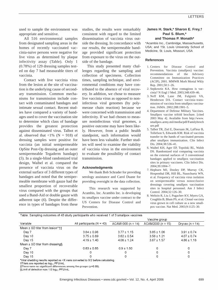
LETTERS
used to sample the environment wasappropriate and sensitive.
All 516 environmental samplesfrom designated sampling areas in thehomes of recently vaccinated vac-cinia-naive persons were negative forlive virus as determined by plaqueinfectivity assay (Table). Only 1(0.78%) of 129 dressing samples test-ed on day 7 had measurable titers ofvaccinia.
Contact with live vaccinia virusfrom the lesion at the site of vaccina-tion is the underlying cause of second-ary transmission. Common mecha-nisms for transmission include con-tact with contaminated bandages andintimate sexual contact. Recent stud-ies have compared a variety of band-ages used to cover the vaccination siteto determine which class of bandageprovides the greatest protectionagainst disseminated virus. Talbot etal. observed that <1% (N = 918) ofdressing samples were positive forvaccinia (an initial semipermeableOpSite Post-Op dressing and an outersemipermeable Tegaderm bandage)(5). In a single-blind randomized trialdesign, Waibel et al. compared thepresence of vaccinia virus on theexternal surface of 3 different types ofbandages and noted that the semiper-meable membrane with gauze had thesmallest proportion of recoverablevirus compared with the groups thatused a Band-Aid or double gauze withadherent tape (6). Despite the differ-ence in types of bandages from these
studies, the results were remarkablyconsistent with regard to the limiteddissemination of vaccinia virus out-side the dressing. In concordance withour results, the semipermeable band-age provided significant protectionfrom exposure to the virus on the out-side of the bandage.
This study presented many chal-lenges regarding the sampling andcollection of specimens. Collectiontimes, sampling technique, and envi-ronmental conditions may have con-tributed to the absence of viral recov-ery. In addition, we chose to measurelive vaccinia virus as opposed to non-infectious viral genomes (by poly-merase chain reaction) because wewere concerned with transmission andinfectivity. If we had chosen to meas-ure noninfectious viral genomes, apositive outcome may have been like-ly. However, from a public healthstandpoint, such information wouldhave been less valuable. Further stud-ies will need to examine the viabilityof vaccinia virus in the environmentto evaluate the possibility of contacttransmission.
AcknowledgmentsWe thank Bob Schrader for providing
serology assistance and Carol Duane forproviding oversight in the data collection.
This research was supported byAcambis, Inc. Acambis Inc. is developingits smallpox vaccine under contract to theUS Centers for Disease Control andPrevention.
James H. Stark,* Sharon E. Frey,†Paul S. Blum,*
and Thomas P. Monath**Acambis Inc., Cambridge, Massachusetts,USA; and †St. Louis University School ofMedicine, St. Louis, Missouri, USA
References
1. Centers for Disease Control andPrevention. Vaccinia (smallpox) vaccine:recommendations of the AdvisoryCommittee on Immunization Practices(ACIP), 2001. MMWR Morb Mortal WklyRep. 2001;50:1–26.
2. Sepkowtiz KA. How contagious is vac-cinia? N Engl J Med. 2003;348:439–46.
3. Neff KM, Lane JM, Fulginiti VA,Henderson DA. Contact vaccinia–trans-mission of vaccinia from smallpox vaccina-tion. JAMA. 2002;288:1901–5.
4. Department of Defense Military Vaccines.Smallpox vaccine trifold brochure. [cited2003 May 4]. Available from http://www.smallpox.army.mil/media/pdf/Familybrochure.pdf
5. Talbot TR, Ziel E, Doersam JK, LaFleur B,Tollefson S, Edwards KM. Risk of vacciniatransfer to the hands of vaccinated personsafter smallpox immunization. Clin InfectDis. 2004;38:536–41.
6. Waibel KH, Ager EP, Topolski RL, WalshDS. Randomized trial comparing vacciniaon the external surfaces of 3 conventionalbandages applied to smallpox vaccinationsites in primary vaccinees. Clin Infect Dis.2004;39:1004–7.
7. Hepburn MJ, Dooley DP, Murray CK,Hospenthal DR, Hill BL, Nauschuetz WN,et al. Frequency of vaccinia virus isolationon semipermeable versus nonocclusivedressings covering smallpox vaccinationsites in hospital personnel. Am J InfectControl. 2004;32:126–30.
8. Weltzin R, Liu J, Pugachev KV, Myers GA,Coughlin B, Blum PS, et al. Clonal vacciniavirus grown in cell culture as a new small-pox vaccine. Nat Med. 2003;9:1125–30.
Emerging Infectious Diseases • www.cdc.gov/eid • Vol. 12, No. 4, April 2006 699

LETTERS
700 Emerging Infectious Diseases • www.cdc.gov/eid • Vol. 12, No. 4, April 2006
9. Monath TP, Caldwell JR, Mundt W, FuscoJ, Johnson CS, Buller M, et al. ACAM2000clonal Vero cell culture vaccinia virus (NewYork City Board of Health strain): a sec-ond-generation smallpox vaccine for bio-logical defense. Int J Infect Dis. 2004;8S2:S31–44.
10. Graham BS, Belshe RB, Clements ML,Dolin R, Corey L, Wright PF, et al.Vaccination of vaccinia-naive adults withhuman immunodeficiency virus type 1gp160 recombinant vaccinia virus in ablinded, controlled, randomized clinicaltrial. The AIDS Vaccine Clinical TrialNetwork. J Infect Dis. 1992;166:244–52.
Address for correspondence: Thomas P.Monath, Acambis Inc., 38 Sidney St,Cambridge, MA 02139, USA; fax: 617-494-0924; email: [email protected]
Discriminationbetween HighlyPathogenic and
Low Pathogenic H5Avian Influenza A
VirusesTo the Editor: To thoroughly
investigate avian influenza outbreaks,identifying highly pathogenic avianinfluenza (HPAI) and low pathogenicavian influenza (LPAI) is essential.Currently, determination of insertedbasic amino acids within the hemag-glutinin cleavage site of HPAI relieson nucleotide sequencing (1–3).Direct sequencing is relatively time-consuming and laborious and thus isnot suitable for local and regionaldiagnostic laboratories that receivelarge numbers of samples that maycontain HPAI or LPAI subtype H5N1.Therefore, a rapid diagnostic assaywas developed to discriminatebetween HPAI and LPAI subtype H5viruses by 1-step real-time reversetranscription–polymerase chain reac-
tion (RT-PCR) with melting curveanalysis.
H5 primers flanking the cleavagesite were designed from conservedregions among HPAI and LPAI strainsby using nucleotide sequencesobtained from GenBank and our pre-vious studies (4–6). The primers con-sisted of a forward primer H5F3+(nucleotides 1001–1021: 5′-AACA-GATTAGTCCTTGCGACTG-3′) anda reverse primer H5R2+ (nucleotides1124–1103: 5′-CATCTACCATTCC-CTGCCATCC-3′), which yieldedproducts of ≈124 bp and ≈112 bp, cor-responding to HPAI and LPAI,respectively. Consequently, the sizesof the amplicons and percentage gua-nine-cytosine content were different,allowing discrimination betweenHPAI and LPAI by melting curveanalysis (7).
One-step, real-time RT-PCR withmelting curve analysis was performedin an ABI 7500 system (AppliedBiosystems, Foster City, CA, USA).In each reaction, 3.0 µL of RNA sam-ple was combined with a reactionmixture containing 10 µL 2× SYBRGreen PCR Master Mix (AppliedBiosystems), 0.5 µL 40× MultiScribe(Applied Biosystems) and RNaseinhibitor, each primer (at final con-centration of 0.5 µmol/L), 1.5mmol/L MgCl2, and RNase-free waterin a final volume of 20 µL. The ther-mal profile began with incubation at48°C for 45 min (reverse transcrip-tion), then incubation at 95°C for 10min (predenaturation), followed by 40cycles of amplification alternatingbetween 94°C for 15 s (denaturation)and 68°C for 40 s (annealing/exten-sion). The SYBR Green I fluorescentsignal was obtained once per cycle atthe end of the extension step. Afteramplification, melting curve analysiswas performed by heating the sampleto 95°C for 15 s, then cooling it to70°C for 1 min, followed by a lineartemperature increase to 95°C at a rateof 0.5°C/s, while continuously moni-toring the fluorescent signal. Data
were analyzed by the 7500 SystemSDS Software version 1.2 (AppliedBiosystems).
To develop the assay, samples pre-viously identified as influenzaA/chicken/Nakorn-Patom/Thailand/CU-K2/04 (H5N1) served as a controlfor HPAI, and A/duck/Hong KongSpecial Administrative Region,People’s Republic of China/308/78(H5N3) served as a control for LPAI.The H5 genes (nucleotides 914–1728)of each strain were inserted intopGEM-T Easy Vector and then tran-scribed in vitro by using RiboMAXLarge Scale RNA Production System-T7 (Promega, Madison, WI, USA).Serial 10-fold dilutions of the stan-dard H5 RNA were subjected to a sen-sitivity test (8). The fluorescent signalcan be detected at RNA dilutions aslow as 102 copies/µL. To assess thespecificity, viral RNA extracted fromother subtypes of influenza A viruses(H1–H4 and H6–H15) was tested.The assay was specific for the H5 sub-type, since no amplification wasdetected from other subtypes.
Three preliminary melting curveanalyses showed that this assay waseffective in discriminating betweenthe melting peaks of HPAI and LPAI(Figure). The variations of meltingtemperature (Tm) between runs wereexperimentally determined. The mean(standard deviation) of Tm values forHPAI and LPAI were 77.43°C(0.21°C) and 79.57°C (0.23°C),respectively. This assay provided highreliability and reproducibility, sincethe coefficients of variation were<0.30.
Seventy-eight specimens ofinfluenza A virus were used to validatethe assay. The 75 HPAI samples wereisolated during the 2004 outbreak inThailand; 3 LPAI samples, includingA/avian/NY/01 (H5N2), A/Chicken/Mexico/31381–3/94 (H5N2), andA/shoveler/Egypt/03 (H5N2), wereprovided by the Centers for DiseaseControl and Prevention. The viruseswere isolated in embryonated chicken

LETTERS
eggs as described previously (9).RNA was extracted from 140 µL ofallantoic fluid, and RT-PCR was per-formed as described above. Meltingcurve analysis showed that all H5N1isolates from the 2004 outbreak inThailand were interpreted as HPAI,whereas the 3 samples of H5N2 sub-type were classified as LPAI. The Tmvaried from 77.2°C to 78.1°C forHPAI and 78.75°C to 79.5°C forLPAI. The melting curve analysisresults were completely concordantwith the results of direct sequenceanalysis of the H5 gene for all sam-ples tested.
The melting curve analysisdescribed here provides a rapid, accu-rate, and high-throughput approach todiscriminate between HPAI and LPAI.This assay is particularly attractive forlarge-scale screening of suspectedsubtype H5 influenza A virus duringoutbreaks to identify candidate LPAIthat could be used as vaccine strains.
AcknowledgmentsWe thank K.F. Shortridge, S. Yingst,
E. Dubovi, and A. Klimov for providingspecimens.
This research was supported byThailand Research Fund.
Sunchai Payungporn,* Salin Chutinimitkul,* Arunee Chaisingh,†
Sudarat Damrongwantanapokin,†Bandit Nuansrichay,† Wasana Pinyochon,† Alongkorn Amonsin,*
Ruben O. Donis,‡ Apiradee Theamboonlers,*
and Yong Poovorawan**Chulalongkorn University, Bangkok,Thailand; †National Institute of AnimalHealth, Bangkok, Thailand; and ‡Centersfor Disease Control and Prevention,Atlanta, Georgia, USA
References
1. Lee CW, Senne DA, Linares JA, WoolcockPR, Stallknecht DE, Spackman E, et al.Characterization of recent H5 subtypeavian influenza viruses from US poultry.Avian Pathol. 2004;33:288–97.
2. Lee CW, Suarez DL, Tumpey TM, SungHW, Kwon YK, Lee YJ, et al.Characterization of highly pathogenicH5N1 avian influenza A viruses isolatedfrom South Korea. J Virol. 2005;79:3692–702.
3. Nguyen DC, Uyeki TM, Jadhao S, MainesT, Shaw M, Matsuoka Y, et al. Isolation andcharacterization of avian influenza viruses,including highly pathogenic H5N1, frompoultry in live bird markets in Hanoi,Vietnam, in 2001. J Virol. 2005;79:4201–12.
4. Viseshakul N, Thanawongnuwech R,Amonsin A, Suradhat S, Payungporn S,Keawchareon J, et al. The genomesequence analysis of H5N1 avian influenzaA virus isolated from the outbreak amongpoultry populations in Thailand. Virology.2004;328:169–76.
5. Keawcharoen J, Oraveerakul K, Kuiken T,Fouchier RA, Amonsin A, Payungporn S, etal. Avian influenza H5N1 in tigers andleopards. Emerg Infect Dis. 2004;10:2189–91.
6. Amonsin A, Payungporn S, TheamboonlersA, Thanawongnuwech R, Suradhat S,Pariyothorn N, et al. Genetic characteriza-tion of H5N1 influenza A viruses isolatedfrom zoo tigers in Thailand. Virology.2006;344:480–91.
7. Ririe KM, Rasmussen RP, Wittwer CT.Product differentiation by analysis of DNAmelting curves during the polymerase chainreaction. Anal Biochem. 1997;245:154–60.
8. Payungporn S, Chutinimitkul S, ChaisinghA, Damrongwantanapokin S, Buranathai C,Amonsin A, et al. Single step multiplexreal-time RT-PCR for H5N1 influenza Avirus detection. J Virol Methods. 2006;131:143–7.
9. World Organization for Animal Health.Manual of diagnostic tests and vaccines forterrestrial animals. Avian influenza. 2005Aug 7 [cited 2005 Sep 1]. Available fromhttp://www.oie.int/eng/normes/MMANU-AL/A_00037.htm
Address for correspondence: Yong Poovorawan,Center of Excellence in Viral HepatitisResearch, Faculty of Medicine, ChulalongkornUniversity, Bangkok 10330, Thailand; fax: 66-2-256-4929; email: [email protected]
Emerging Infectious Diseases • www.cdc.gov/eid • Vol. 12, No. 4, April 2006 701
Figure. Discrimination between highly pathogenic avian influenza (HPAI) and low patho-genic avian influenza (LPAI) by melting curve analysis based on real-time reverse tran-scription–polymerase chain reaction of the H5 HA gene with SYBR Green I fluorescentdye. The melting peaks of HPAI and LPAI were clearly separated. The cutoff value wasset at 78.50°C (midpoint between HPAI and LPAI) and used to interpret the pathogenicityof unknown samples.

LETTERS
Rift Valley Fever inGoats, Cameroon To the Editor: Rift Valley fever
(RVF) is caused by an RNA virus(Phlebovirus, Bunyaviridae), which iscarried by mosquito vectors (1). Innature, it is only known in Africa andthe Arabian Peninsula. In the centralAfrican region, RVF has been report-ed in humans and livestock in thesavanna of northern Cameroon andChad (2,3) and from forests in theCentral African Republic (4). Humanepidemics are sometimes preceded byan increase in RVF virus (RVFV)prevalence in domestic ruminants,which manifests as increased abor-tions and high neonatal deaths (3).Human epidemics can have serioushealth implications, as demonstratedby the most recent outbreaks in Kenyain 1997 and 1998 (5) and SaudiArabia in 2001 (6).
In June 2003, 14 goats from anurban market in Yaoundé (3.9°N,11.5ºE), Cameroon, and 36 goats from3 adjacent villages in tropical lowlandrainforest ≈80 km south of Mvangan(2.4ºN, 11.8ºE), Cameroon, weresampled. The goats in the rural vil-lages were bred locally and allowed toroam freely throughout the villages.No vaccinations had been given togoats in the rural sites. Goats in theurban market in Yaoundé generallyoriginated in northern Cameroon andhad been transported by train toYaoundé. Owners did not report highlevels of abortion or high neonataldeaths.
Jugular blood was collected in a 5-mL dry Vacutainer tube and cen-trifuged within 48 hours of collection.The frozen sera were shipped on ice tothe Onderstepoort Veterinary Institute,the United Nations Food andAgriculture Organization referencelaboratory for RVFV. An indirectenzyme-linked immunosorbent assay(I-ELISA) was used to detect RVFVimmunoglobulin G (IgG) antibodies in26 samples. In this assay, optical den-sity readings were converted to a per-centage of high-positive control serum(PP) and positive samples were thosewith PP >10. These samples were fur-ther tested with RVFV hemagglutina-tion inhibition (HI), and samples withtiters >20 were considered seroposi-tive (7). Positive I-ELISA and HI sam-ples were confirmed by using a serumneutralization (SN) assay (7,8). A sam-ple was considered seropositive whenit had an SN titer of >4, determinedduring experiments on laboratory-injected sheep (7–9) and testing ofwild and domestic African ruminants(7). The SN assay has been shown tobe highly specific; cross-reactivitywith other viruses from the familyBunyaviridae is not likely (9).
Of the 26 goat samples tested forRVFV, 6 tested positive to RVFV(Table). Samples from 5 goats fromthe rural villages and 1 from the urbanmarket had RVFV IgG PP >10.Samples from 2 goats from the ruralvillages had high RVFV HI titers (320and 5,120). Three of the 6 samplesfrom the rural villages with high IgGPP and HI titers had elevated neutral-ization titers (>4).
The results indicate for the firsttime that RVFV is present in forests ofsouthern Cameroon. Given the ages ofthe seropositive goats (2, 3, and 4.5years), transmission of the virusoccurred recently.
In savanna goat herds in northernCameroon, RVFV IgG prevalence hasbeen reported at 9% to 20% (2). Todetermine prevalence of RVFV ingoats in southern Cameroon, moreanimals need to be sampled; the smallsample size and isolation of the fewrural villages are unrepresentative.
The presence of RVFV antibodiesin domestic animals suggests that thisvirus may also be circulating inhuman populations, despite theabsence of reports. A study of 21 per-sons in Ngoïla in southern Cameroonfound no RVFV antibodies during abloody diarrhea epidemic in 1998(10,4); however, testing facilities forRVFV are not available in Cameroon,and the general population and health-care providers have limited awarenessof the virus and associated disease.
Central African forests have a highdiversity of forest ungulates (>10species), and RVFV has been reportedfrom a number of wild African ungu-lates (7). The potential for exchange ofRVFV and other pathogens betweengoats and wild ungulates could havesubstantial effects on animal produc-tion and on the conservation of wildspecies, some of which are considerednear-threatened. Livestock diseasesurveillance can play a role not only inassessing the distribution of livestockpathogens but also in monitoring dis-ease emergence.
702 Emerging Infectious Diseases • www.cdc.gov/eid • Vol. 12, No. 4, April 2006

LETTERS
AcknowledgmentsWe thank the US Embassy of
Yaoundé, staff of Walter Reed JohnsHopkins Cameroon Program, T. Gerdes,goat owners, and the anonymous review-ers.
This research was financed by JohnsHopkins Bloomberg School of PublicHealth Center for a Livable Future(N.D.W.), the V. Kann RasmussenFoundation (D.S.B.), the NationalInstitutes of Health Fogarty InternationalCenter (grant #5 K01 TW000003-05 toN.D.W.), and an NIH Research TrainingGrant (S.U.). The Cameroon Ministry forScientific and Technical Research provid-ed research permits.
Matthew LeBreton,* Sally Umlauf,† Cyrille F. Djoko,*‡Peter Daszak,§ Donald S. Burke,¶
Paul Yemgai Kwenkam,# and Nathan D. Wolfe¶
*Walter Reed Johns Hopkins CameroonProgram, Yaoundé, Cameroon; †TuftsUniversity School of Veterinary Medicine,North Grafton, Massachusetts, USA;‡University of Yaoundé, Yaoundé,Cameroon; §Consortium for ConservationMedicine, New York, New York, USA;¶Johns Hopkins University, Baltimore,Maryland, USA; and #Ministry of Livestock,Fisheries and Animal Industries, Yaoundé,Cameroon
References
1. Fontenille D, Traore-Lamizana M, DialloM, Thonnon J, Digoutte JP, Zeller HG. Newvectors of Rift Valley fever in West Africa.Emerg Infect Dis. 1998;4:289–93.
2. Zeller HG, Bessin R, Thiongane Y, BapetelI, Teou K, Ala MG, et al. 1995 Rift Valleyfever antibody prevalence in domesticungulates in Cameroon and several WestAfrican countries (1989–1992) followingthe 1987 Mauritanian outbreak. Res Virol.1995;146:81–5.
3. Ringot D, Durand J-P, Tolou H, Boutin J-PDavoust B. Rift Valley fever in Chad.Emerg Infect Dis. 2004;10:945–7.
4. Nakounné E, Selekon B, Morvan J. Veillemicrobiologique : les fièvres hémorrag-iques virales en République Centrafricaine ;données sérologiques actualisées chezl’homme. Bull Soc Pathol Exot.2000;93:340–7.
5. Woods CW, Karpati AM, Grein T,McCarthy N, Gaturuku P, Muchiri E, et al.An outbreak of Rift Valley fever in north-eastern Kenya, 1997–98. Emerg Infect Dis.2002;8:138–44.
6. Balkhy HH, Memish ZA. Rift Valley fever:an uninvited zoonosis in the Arabian penin-sula. Int J Antimicrob Agents. 2003;21:153–7.
7. Paweska JT, Smith SJ, Wright IM, WilliamsR, Cohen AS, Van Dijk AA. Indirectenzyme-linked immunosorbent assay forthe detection of antibody against Rift Valleyfever virus in domestic and wild ruminantsera. Onderstepoort J Vet Res.2003;70:49–64.
8. Swanepoel R, Struthers JK, Erasmus MJ,Shepherd SP, McGillivry GM, Erasmus BJ,et al. Comparison of techniques for demon-strating antibodies to Rift Valley fevervirus. J Hyg (Lond). 1986;97:317–29.
9. Swanepoel R, Struther JK, Erasmus MJ,Shepherd SP, McGillivray GM, ShepherdAJ, et al. Comparative pathogenicity andantigenic cross-reactivity of Rift Valleyfever and other African phleboviruses insheep. J Hyg (Lond). 1986;97:331–46.
10. Germani Y, Cunin P, Tedjouka E, NcharreCB, Morvan J, Martin P. Entero-haemorrhagic Escherichia coli in Ngoila(Cameroon) during an outbreak of bloodydiarrhoea. Lancet. 1998;352:625–6.
Address for correspondence: MatthewLeBreton, Walter Reed Johns HopkinsCameroon Program; CRESAR; BP 7039;Yaoundé; Cameroon; fax: 237-221-3382;email: [email protected]
HIV-1 CRF07_BCInfections,
Injecting DrugUsers, Taiwan
To the Editor: To date, Taiwan’shuman immunodeficiency virus type1 (HIV-1) epidemic has primarilyspread via sexual contact. The sub-type B and circulating recombinantform (CRF) 01_AE account for >95%of all infections (1). However, since
2003 Taiwan has experienced a majoroutbreak of CRF07_BC among inject-ing drug users (IDUs).
The first wave of HIV-1 infectionsin Taiwan can be traced to the early1980s, when a group of hemophiliapatients received imported HIV-1-contaminated antihemophilia medica-tions. By the time these medicationshad been replaced by heat-treated fac-tor VIII concentrates, at least 53patients had contracted HIV-1 infec-tions (2). According to Taiwan’sCenter for Disease Control (CDC),HIV infections have been diagnosedin 9,229 persons (including 523 for-eigners) as of July 31, 2005 (3). Thenumber of persons living with HIV-1/AIDS has increased rapidly in thepast few years, with a 77% increase in2004, compared to 11% in 2003(online Table, available at http://www.cdc.gov/ncidod/EID/vol12no04/05-0762.htm#table). According tothe results of a risk factor analysis ofpeople living with HIV-1/AIDSreported to the Taiwan CDC, the pro-portion of IDUs increased from 1.7%(13/773) in 2002 to 8.1% (70/861) in2003 to 30.3% (462/1,521) in 2004(online Table). The Taiwan CDCreceived reports of 1,241 IDUs diag-nosed with HIV-1 infections fromJanuary 1 to July 31, 2005; theseaccount for >75% of all reported HIV-1 infections in 2005 (3). The evidencepoints to an explosive epidemic ofHIV-1 infections among IDUs inTaiwan since 2003, with no indicationof a slowdown.
Taiwan has ≈60,000 IDUs (1).According to the Republic of ChinaMinistry of Justice, the number ofincarcerated drug offenders increasedfrom 5,988 in 2003 to 9,303 in 2004;the rate of HIV-1 seropositive inmatesincreased from 13.3/100,000 in 2002to 56.8/100,000 in 2004 (Y-M. Wu,Ministry of Justice, pers. comm.).Since all inmates are routinely testedfor HIV-1 in detention centers, and allinfected inmates are separated fromHIV-1–seronegative inmates, the
Emerging Infectious Diseases • www.cdc.gov/eid • Vol. 12, No. 4, April 2006 703
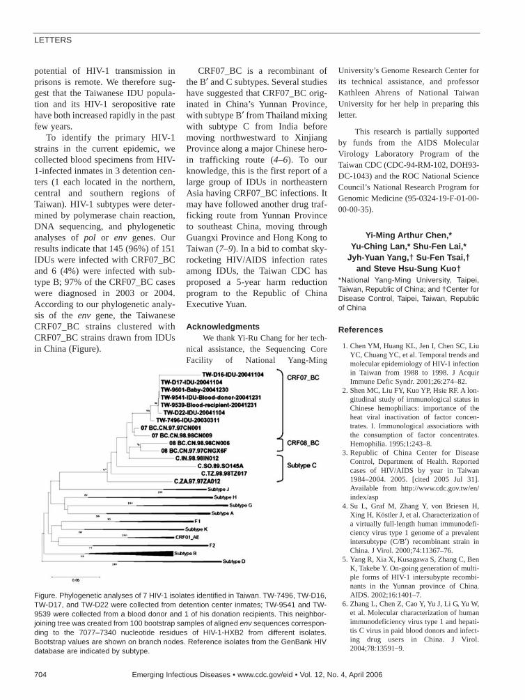
LETTERS
potential of HIV-1 transmission inprisons is remote. We therefore sug-gest that the Taiwanese IDU popula-tion and its HIV-1 seropositive ratehave both increased rapidly in the pastfew years.
To identify the primary HIV-1strains in the current epidemic, wecollected blood specimens from HIV-1-infected inmates in 3 detention cen-ters (1 each located in the northern,central and southern regions ofTaiwan). HIV-1 subtypes were deter-mined by polymerase chain reaction,DNA sequencing, and phylogeneticanalyses of pol or env genes. Ourresults indicate that 145 (96%) of 151IDUs were infected with CRF07_BCand 6 (4%) were infected with sub-type B; 97% of the CRF07_BC caseswere diagnosed in 2003 or 2004.According to our phylogenetic analy-sis of the env gene, the TaiwaneseCRF07_BC strains clustered withCRF07_BC strains drawn from IDUsin China (Figure).
CRF07_BC is a recombinant ofthe B′ and C subtypes. Several studieshave suggested that CRF07_BC orig-inated in China’s Yunnan Province,with subtype B′ from Thailand mixingwith subtype C from India beforemoving northwestward to XinjiangProvince along a major Chinese hero-in trafficking route (4–6). To ourknowledge, this is the first report of alarge group of IDUs in northeasternAsia having CRF07_BC infections. Itmay have followed another drug traf-ficking route from Yunnan Provinceto southeast China, moving throughGuangxi Province and Hong Kong toTaiwan (7–9). In a bid to combat sky-rocketing HIV/AIDS infection ratesamong IDUs, the Taiwan CDC hasproposed a 5-year harm reductionprogram to the Republic of ChinaExecutive Yuan.
AcknowledgmentsWe thank Yi-Ru Chang for her tech-
nical assistance, the Sequencing CoreFacility of National Yang-Ming
University’s Genome Research Center forits technical assistance, and professorKathleen Ahrens of National TaiwanUniversity for her help in preparing thisletter.
This research is partially supportedby funds from the AIDS MolecularVirology Laboratory Program of theTaiwan CDC (CDC-94-RM-102, DOH93-DC-1043) and the ROC National ScienceCouncil’s National Research Program forGenomic Medicine (95-0324-19-F-01-00-00-00-35).
Yi-Ming Arthur Chen,* Yu-Ching Lan,* Shu-Fen Lai,*
Jyh-Yuan Yang,† Su-Fen Tsai,† and Steve Hsu-Sung Kuo†
*National Yang-Ming University, Taipei,Taiwan, Republic of China; and †Center forDisease Control, Taipei, Taiwan, Republicof China
References
1. Chen YM, Huang KL, Jen I, Chen SC, LiuYC, Chuang YC, et al. Temporal trends andmolecular epidemiology of HIV-1 infectionin Taiwan from 1988 to 1998. J AcquirImmune Defic Syndr. 2001;26:274–82.
2. Shen MC, Liu FY, Kuo YP, Hsie RF. A lon-gitudinal study of immunological status inChinese hemophiliacs: importance of theheat viral inactivation of factor concen-trates. I. Immunological associations withthe consumption of factor concentrates.Hemophilia. 1995;1:243–8.
3. Republic of China Center for DiseaseControl, Department of Health. Reportedcases of HIV/AIDS by year in Taiwan1984–2004. 2005. [cited 2005 Jul 31].Available from http://www.cdc.gov.tw/en/index/asp
4. Su L, Graf M, Zhang Y, von Briesen H,Xing H, Köstler J, et al. Characterization ofa virtually full-length human immunodefi-ciency virus type 1 genome of a prevalentintersubtype (C/B′) recombinant strain inChina. J Virol. 2000;74:11367–76.
5. Yang R, Xia X, Kusagawa S, Zhang C, BenK, Takebe Y. On-going generation of multi-ple forms of HIV-1 intersubypte recombi-nants in the Yunnan province of China.AIDS. 2002;16:1401–7.
6. Zhang L, Chen Z, Cao Y, Yu J, Li G, Yu W,et al. Molecular characterization of humanimmunodeficiency virus type 1 and hepati-tis C virus in paid blood donors and infect-ing drug users in China. J Virol.2004;78:13591–9.
704 Emerging Infectious Diseases • www.cdc.gov/eid • Vol. 12, No. 4, April 2006
Figure. Phylogenetic analyses of 7 HIV-1 isolates identified in Taiwan. TW-7496, TW-D16,TW-D17, and TW-D22 were collected from detention center inmates; TW-9541 and TW-9539 were collected from a blood donor and 1 of his donation recipients. This neighbor-joining tree was created from 100 bootstrap samples of aligned env sequences correspon-ding to the 7077–7340 nucleotide residues of HIV-1-HXB2 from different isolates.Bootstrap values are shown on branch nodes. Reference isolates from the GenBank HIVdatabase are indicated by subtype.

LETTERS
Emerging Infectious Diseases • www.cdc.gov/eid • Vol. 12, No. 4, April 2006 705
7. Piyasirisilp S, McCutchan FE, Carr JK,Anders-Buell E, Liu W, Chen J, et al. Arecent outbreak of human immunodeficien-cy virus type 1 infection in southern Chinawas initiated by two highly homogeneousgeographically separated strains, circulat-ing recombinant form AE and a novel BCrecombinant. J Virol. 2000;74:11286–95.
8. Cohen J. Asia and Africa: on different tra-jectories? Science. 2004;304:1932–8.
9. Lim WL, Xing H, Wong KH, Wong MC,Shao YM, Ng MH, et al. The lack of an epi-demiological link between HIV type 1infections in Hong Kong and MainlandChina. AIDS Res Hum Retroviruses.2004;20:259–62.
Address for correspondence: Yi-Ming ArthurChen, AIDS Prevention and Research Center,National Yang-Ming University, 155 Li-NongSt, Sec. 2, Taipei, Taiwan 112; fax: 886-2-2827-0576; email: [email protected]
ChlamydialikeOrganisms andAtherosclerosis
To the Editor: Chlamydophilapneumoniae causes pneumonia, butits role in the pathogenesis of athero-sclerosis is controversial (1–4). Therole of C. pneumoniae in atheroscle-rosis is supported by seroepidemio-logic studies and detection in athero-sclerotic lesions by polymerase chainreaction (PCR), immunohistologicanalysis, culture, and electron micro-scopy (2,3). However, these resultswere not confirmed by other serologicor PCR-based studies (4). Meijer et al.evaluated abdominal aortic aneurysmbiopsy specimens and detected C.pneumoniae membrane antigen morefrequently than lipopolysaccharideantigens but did not detect heat shockprotein 60 (1). In addition, they couldnot amplify or detect specific C. pneu-moniae DNA by PCR and fluores-cence in situ hybridization (1). They
hypothesized that this discrepancymay result from a chlamydialikeorganism present in aortic samplesthat has surface antigens similar tothose of C. pneumoniae.
Parachlamydia acanthamoebaeand Neochlamydia hartmanellae arechlamydialike organisms that share≈86% 16S rRNA sequence similaritywith C. pneumoniae (5). Like C.pneumoniae, they have elementaryand reticulate bodies visible by elec-tron microscopy (6). Neochlamydia-related DNA (GenBank accession no.AF097191) has been amplified from 5different arterial samples, including 1aortic aneurysm (7), and a relation-ship (p = 0.009) between cerebralhemorrhage and serologic evidence ofParachlamydia infection has beenreported (8). Therefore, we investigat-ed the role of Parachlamydia inpathogenesis of atherosclerosis byusing a molecular approach.
We analyzed 78 surgical samplesfrom 27 patients undergoing aortic orcarotid surgery for atherosclerotic dis-ease at Hôpital Nord in Marseillefrom June 1, 2003, to December 31,2003. The study was approved by thelocal ethics committee, and writteninformed consent was obtained fromall participants. Demographic andclinical data were prospectivelyrecorded.
DNA was extracted from aortic orcarotid samples with atheroscleroticlesions by using the QIAamp DNAtissue kit (Qiagen, Courtaboeuf,France), according to the manufactur-er’s instructions. A nested PCR wasperformed by using external primers16SIGF (5′-CGGCGTGGATGAG-GCAT-3′) and 16SIGR (5′-TCAGTC-CCAGTGTTGGC-3′) (9) and internalprimers CHL16SFOR2 (5′-CGTG-GATGAGGCATGCAAGTCGA-3′)and CHL16SREV2 (5′-CAATCTCT-CAATCCGCCTAGACGTCTTAG-3′) (7). PCR included negative con-trols from the DNA extraction step.DNA extractions and PCR amplifica-tions were conducted in a laboratory
in which parachlamydial DNA hadnot been extracted or amplified. PCRproducts were purified by using theQIAquick PCR purification kit(Qiagen) and sequenced by using thed-rhodamine terminator cyclesequencing reaction kit (Perkin-ElmerBiosystems, Warrington, UK) and a3100 ABI Prism automated sequencer(Applied Biosystems, Courtaboeuf,France).
Sequences were analyzed withBLAST (http://www.ncbi.nlm.nih.gov/BLAST/) using gap existence andextension penalties of 5 and 2, respec-tively. Results were considered posi-tive only when the sequence of theamplified product exhibited a bestBLAST hit with a chlamydialikeorganism. Statistical analyses wereperformed with STATA software(Stata Corporation, College Station,TX, USA).
A positive PCR result wasobtained with samples from 5 (18.5%)of 27 patients (Table). Threesequences had a best BLAST hit withthe sequence of Parachlamydia sp.UV7 (GenBank accession no.AJ715410), with a sequence similari-ty ranging from 99% to 100%. Theother 2 sequences had 98% sequencesimilarity with Neochlamydia-relatedsymbiont TUME-1 (GenBank acces-sion no. AF098330). PCR positivitywas not associated with age, sex, orlocation of the atherosclerotic lesion.
All patients with positive PCRresults were >68 years of age. Patientswithout cardiovascular risk factorswere more likely than those with>1 risk factor to have positive PCRresults (p = 0.023). Despite the smallnumber of patients in this study, thisassociation was also confirmed in amultivariate logistic regression modeladjusted for sex and previous cardio-vascular disease (odds ratio 0.035,95% confidence interval 0.001–0.94).
These findings suggest thatParachlamydia and Neochlamydiaare associated with atherosclerosis. Inaddition, these obligate intracellular

LETTERS
bacteria may be present in bothcarotid and aortic atheroscleroticlesions of elderly patients.
Chlamydialike organisms in ather-osclerotic lesions may explain contro-versies about the role of C. pneumoni-ae in pathogenesis of atherosclerosis(4). Some PCRs might amplify bothChlamydia and chlamydialike organ-isms, leading to erroneous conclu-sions, especially when the specificityof the product is not confirmed bysequencing. Chlamydialike organismsin atherosclerotic lesions might alsoexplain discrepancies of serologicstudies (4). Parachlamydiaceae likelycross-react with C. pneumoniae (1).Such cross-reactivity will not be rec-ognized if patients with positive sero-logic results for C. pneumoniae arenot tested for antibodies to chlamydi-alike organisms. This cross-reactivitymay result in false-positive serologicresults for C. pneumoniae, especiallywhen low antibody titers (8–16) areconsidered positive.
Since elementary and reticulatebodies are similar in both Chlamydia-ceae and Parachlamydiaceae (6),Chlamydiales cannot be identified ina specimen solely by electronmicroscopy. Consequently, elemen-tary and reticulate bodies in athero-sclerotic lesions (10) might bechlamydialike organisms and not C.pneumoniae. If chlamydialike organ-isms are involved in the pathogenesisof atherosclerosis, this finding wouldhave public health implications, giventheir presence in free-living amebaethat are widespread in water.
AcknowledgmentsWe thank Philip Tarr for reviewing
the manuscript.
Dr Greub's research on chlamydia-like organisms is currently supported bythe Swiss National Science Foundation(SNSF) grant no. 3200BO-105885.
Gilbert Greub,*1 Olivier Hartung,†Toïdi Adekambi,* Yves S. Alimi,†
and Didier Raoult* *Université de la Méditerannée, Marseille,France; and †Hôpital Nord, Marseille,France
References
1. Meijer A, van der Vliet JA, Roholl PJ,Gielis-Proper SK, de Vries A, OssewaardeJM. Chlamydia pneumoniae in abdominalaortic aneurysms: abundance of membranecomponents in the absence of heat shockprotein 60 and DNA. Arterioscler ThrombVasc Biol. 1999;19:2680–6.
2. Kalayoglu MV, Libby P, Byrne GI.Chlamydia pneumoniae as an emerging riskfactor in cardiovascular disease. JAMA.2002;288:2724–31.
3. Belland RJ, Ouellette SP, Gieffers J, ByrneGI. Chlamydia pneumoniae and atheroscle-rosis. Cell Microbiol. 2004;6:117–27.
4. Ieven MM, Hoymans VY. Involvement ofChlamydia pneumoniae in atherosclerosis:more evidence for lack of evidence. J ClinMicrobiol. 2005;43:19–24.
5. Greub G, Raoult D. Parachlamydiaceae:potential emerging pathogens. Emerg InfectDis. 2002;8:625–30.
6. Greub G, Raoult D. Crescent bodies ofParachlamydia acanthamoeba and its lifecycle within Acanthamoeba polyphaga: anelectron micrograph study. Appl EnvironMicrobiol. 2002;68:3076–84.
7. Ossewaarde JM, Meijer A. Molecular evi-dence for the existence of additional mem-bers of the order Chlamydiales.Microbiology. 1999;145:411–7.
8. Greub G, Boyadjiev I, La Scola B, RaoultD, Martin C. Serological hint suggestingthat Parachlamydiaceae are agents ofpneumonia in polytraumatized intensivecare patients. Ann N Y Acad Sci. 2003;990:311–9.
9. Everett KD, Bush RM, Andersen AA.Emended description of the orderChlamydiales, proposal of Parachla-mydiaceae fam. nov. and Simkaniaceaefam. nov., each containing one monotypicgenus, revised taxonomy of the familyChlamydiaceae, including a new genus andfive new species, and standards for theidentification of organisms. Int J SystBacteriol. 1999;49:415–40.
10. Nystrom-Rosander C, Thelin S, Hjelm E,Lindquist O, Pahlson C, Friman G. Highincidence of Chlamydia pneumoniae insclerotic heart valves of patients undergo-ing aortic valve replacement. Scand J InfectDis 1997;29:361–5.
Address for correspondence: Gilbert Greub,Center for Research on Intracellular Bacteria,Microbiology Institute, Faculty of Biology andMedicine, University of Lausanne, 1011Lausanne, Switzerland; fax: 41-21-314-4060;email: [email protected]
706 Emerging Infectious Diseases • www.cdc.gov/eid • Vol. 12, No. 4, April 2006
1Current affiliation: University of Lausanne,Lausanne, Switzerland

LETTERS
Emerging Infectious Diseases • www.cdc.gov/eid • Vol. 12, No. 4, April 2006 707
Maculopathy inDengue Fever
To the Editor: A recent article byChlebicki et al (1) described 4 patientshospitalized for dengue fever whowere found to have retinal hemor-rhages. These patients reportedreduced visual acuity and metamor-phopsia, i.e., distorted visual imagesattributable to intrinsic retinal diseaseinvolving the macula; macular hemor-rhages and exudates were found onretinal examination. The authors con-cluded that the retinal hemorrhageswere responsible for the patients’visual symptoms.
This conclusion is misleadingbecause retinal hemorrhages alonecause scotomas. Rather, the accumu-lation of subretinal fluid in the macu-la results in metamorphopsia andblurring of vision. In previous reportsof patients in whom macular changesdeveloped from dengue fever, somewere found to have macular hemor-rhages (2–4). In addition, clinicalexamination and investigation ofthese patients showed vasculopatho-logic changes in the macular regionthat affected the retinal and choroidalblood vessels (5), although the tissuesof the periphery tended to be spared.A fluorescein angiograph of the retinashowed knobby hyperfluorescence ofthe retinal arterioles with minimalleakage, as well as some spots of leak-age at the level of the retinal pigmentepithelium. An indocyanine greenangiograph showed diffuse hyperfluo-rescence of the choroid. These patho-logic changes in the macula were themost likely cause of the blurring ofvision in such patients, which hasbeen the case in our experience.
The article by Chlebicki et al. didnot state whether these procedureshad been performed on their patientsto confirm or exclude retinal orchoroidal vasculopathy in the macula.Therefore, these authors would havehad difficulty concluding that retinalhemorrhages caused blurring of
vision and metamorphopsia inpatients with dengue maculopathy.
Daniel Hsien-Wen Su* and Soon-Phaik Chee*†‡
*Singapore National Eye Centre,Singapore; †National University ofSingapore, Singapore; and ‡SingaporeEye Research Institute, Singapore
References
1. Chlebicki MP, Ang B, Barkham T, Laude A.Retinal hemorrhages in 4 patients withdengue fever. Emerg Infect Dis.2005;11:770–2.
2. Wen KH, Sheu MM, Chung CB, Wang HZ,Chen CW. The ocular fundus findings indengue fever [article in Chinese]. GaoxiongYi Xue Ke Xue Za Zhi. 1989;5:24–30.
3. Haritoglou C, Scholz F, Bialaslewicz A,Klauss V. Ocular manifestations in denguefever [article in German]. Ophthalmologe.2000;97:433–6.
4. Haritoglou C, Dotse SD, Rudolph G,Stephan CM, Thurau SR, Klauss V. Atourist with dengue fever and visual loss.Lancet. 2002;360:1070.
5. Lim WK, Mathur R, Koh A, Yeoh R, CheeSP. Ocular manifestations of dengue fever.Ophthalmology. 2004;111:2057–64.
Address for correspondence: Daniel H-W. Su,Singapore National Eye Centre, 11 ThirdHospital Ave, Singapore 168751; fax: 65-6226-3395; email: [email protected]
PulmonaryTuberculosis and
SARS, China To the Editor: As part of a cohort
study of 83 patients with severe acuterespiratory syndrome (SARS) inBeijing, China, we conducted a fol-low-up study of all the patients byroutine medical examination. Duringthe process, 3 patients with chest radi-ographs consistent with active diseasewere identified as having pulmonarytuberculosis (TB). Here we describe
the 1-year clinical outcome andimmune response in these patients.
Demographic details and coexist-ing conditions are shown in the Table.Patient 1 was a healthcare worker whobecame infected with SARS-associat-ed coronavirus (CoV) while on dutywith SARS patients. After he wastransferred to a hospital dedicated toSARS management, pulmonary TBwas diagnosed (positive acid-fastbacilli smear on sputum samples).Patients 2 and 3 were known to havecases of pulmonary TB and becameinfected with SARS-CoV after con-tact with other patients hospitalizedfor SARS. These 2 patients were spu-tum smear–negative for acid-fastbacilli, and diagnosis was made on thebasis of previous exposure to TB, rel-evant symptoms of typical pulmonaryTB, chest radiographs consistent withactive disease, a positive tuberculinskin test result, and the finding of cav-ity regression on chest radiographsafter anti-TB treatment was initiated.No cultures were obtained for isola-tion and comparison of Myco-bacterium tuberculosis strains (1). All3 patients had confirmed SARS basedon amplification of SARS-CoV RNAby reverse transcriptase–polymerasechain reaction (RT-PCR) from sputumand stool specimens (2). Patients 2and 3 recovered without complica-tions; patient 1 had the most severedisease and required mechanical ven-tilation in an intensive care unit beforerecovering.
Both cellular and humoral immu-nity were evaluated during the follow-up of these patients. T-lymphocytesubsets were measured 6 months afterdisease onset by flow cytometry usingfluorescein isothiocyanate–labeledspecific monoclonal antibodies.Compared to other SARS patients (n= 47), the 3 patients with TB hadlower mean CD4+ T cells (368.4/µLvs. 656.6/µL, respectively; p = 0.05)and lower mean CD8+ T cells(371.0/µL vs. 490.1/µL, respectively;p = 0.39). SARS-CoV immunoglobu-

LETTERS
lin G (IgG) antibody titers were meas-ured by enzyme-linked immunosor-bent assay kit (Huada Company,Beijing, China) at months 1, 2, 3, 4, 7,10, and 16 after disease onset. (Titerswere not measured for the 3 TBpatients at month l.) Compared tomost (26 [78.8%] of 33) other SARSpatients whose antibodies remaineddetectable throughout follow-up, 2 ofthe 3 TB patients (patients 1 and 3)had undetectable antibody titers as ofmonths 7 and 16, respectively. Inpatient 1, antibody titers, whendetectable, were unusually low (40).Both patients 1 and 3 had prolongedviral excretion in stools, sputum, orboth. While the median (range) dura-tion of virus excretion in stools andsputa for the entire measurable cohort(n = 56) was 27 (16–127) and 21(14–52) days, respectively (3), it was125 and 16 days for patient 1, and 109and 52 days for patient 3 (viral excre-tion data could not be obtained frompatient 2 because sequential speci-mens for detection were unavailable).
TB in SARS patients has beenreported on rare occasions (4,5). In acohort of 236 patients in Singapore, itwas diagnosed in 2 patients afterrecovery from SARS (4). As withpatient 1 in this study, TB had devel-oped after the patient acquired SARS,most likely as the result of reactiva-tion of past infection or new infectionwith M. tuberculosis, while temporar-ily immunosuppressed because of
SARS (6) and corticoid therapy. Suchphenomena have been described withother viral infections such as measlesand HIV (7,8). By contrast, patients 2and 3 were known TB patients whoacquired SARS through exposure toSARS patients in the same hospitalwards. Both diseases are known to betransiently immunosuppressive (6,9),and their combined effect resulted inmore pronounced CD4+ cell decreas-es in coinfected SARS patients thanothers. Such immunosuppression alsoresulted in poorer IgG antibodyresponse in coinfected SARS patientsthan in others and delayed viral clear-ance, as shown by longer viral excre-tion in sputum and stools. While viralexcretion could be prolonged in coin-fected patients, no virus could be iso-lated from any RT-PCR–positivespecimen collected after 6 weeks ofillness, which suggests that excretedviruses were no longer infectious (3).
These case reports remind us of theimportance of strict isolation of SARSpatients, careful use of steroids fortheir case management, and the possi-bility of coinfection with TB in SARSpatients with incomplete recovery.
This work was partly supported bythe Programme de Recherche en RéseauxFranco-Chinois (P2R), the EC grant EPIS-ARS (SP22-CT-2004-511063, SP22-CT-2004-003824), the National Institutes ofHealth CIPRA Project (NIH U19
AI51915), and the National 863 Programof China (2003AA208406, 2003AA208412C).
Wei Liu,* Arnaud Fontanet,† Pan-He Zhang,* Lin Zhan,*
Zhong-Tao Xin,‡ Fang Tang,*Laurence Baril,†
and Wu-Chun Cao**Beijing Institute of Microbiology andEpidemiology, Beijing, People’s Republic ofChina, †Institut Pasteur, Paris, France; and‡Beijing Institute of Basic MedicalSciences, Beijing, People’s Republic ofChina
References
1. Crawford JT, Braden CR, Schable BA,Onorato ID. National TuberculosisGenotyping and Surveillance Network:design and methods. Emerg Infect Dis.2002;8:1192–6.
2. Revised U.S. surveillance case definitionfor severe acute respiratory syndrome(SARS) and update on SARS cases—United States and worldwide, December2003. MMWR Morb Mortal Wkly Rep.2003;52:1202–6.
3. Liu W, Tang F, Fontanet A, Zhan L, ZhaoQM, Zhang PH, et al. Long-term SARS-coronavirus excretion from a patient cohortin China. Emerg Infect Dis.2004;10:1841–3.
4. Low JGH, Lee CC, Leo YS. Severe acuterespiratory syndrome and pulmonary tuber-culosis. Clin Infect Dis. 2004;38:e123–5.
5. Centers for Disease Control andPrevention. Nosocomial transmission ofMycobacterium tuberculosis found throughscreening for severe acute respiratory syn-drome—Taipei, Taiwan, 2003. MMWRMorb Mortal Wkly Rep. 2004;53:321–2.
708 Emerging Infectious Diseases • www.cdc.gov/eid • Vol. 12, No. 4, April 2006
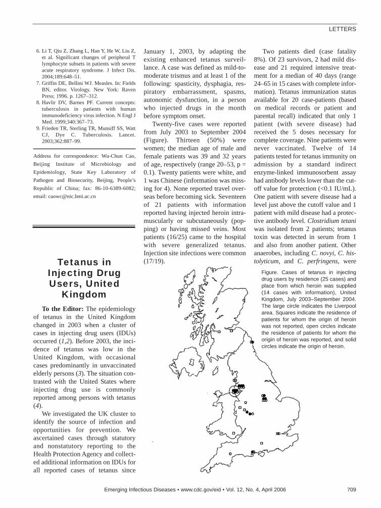
LETTERS
6. Li T, Qiu Z, Zhang L, Han Y, He W, Liu Z,et al. Significant changes of peripheral Tlymphocyte subsets in patients with severeacute respiratory syndrome. J Infect Dis.2004;189:648–51.
7. Griffin DE, Bellini WJ. Measles. In: FieldsBN, editor. Virology. New York: RavenPress; 1996. p. 1267–312.
8. Havlir DV, Barnes PF. Current concepts:tuberculosis in patients with humanimmunodeficiency virus infection. N Engl JMed. 1999;340:367–73.
9. Frieden TR, Sterling TR, Munsiff SS, WattCJ, Dye C. Tuberculosis. Lancet.2003;362:887–99.
Address for correspondence: Wu-Chun Cao,Beijing Institute of Microbiology andEpidemiology, State Key Laboratory ofPathogen and Biosecurity, Beijing, People’sRepublic of China; fax: 86-10-6389-6082;email: [email protected]
Tetanus inInjecting DrugUsers, United
KingdomTo the Editor: The epidemiology
of tetanus in the United Kingdomchanged in 2003 when a cluster ofcases in injecting drug users (IDUs)occurred (1,2). Before 2003, the inci-dence of tetanus was low in theUnited Kingdom, with occasionalcases predominantly in unvaccinatedelderly persons (3). The situation con-trasted with the United States whereinjecting drug use is commonlyreported among persons with tetanus(4).
We investigated the UK cluster toidentify the source of infection andopportunities for prevention. Weascertained cases through statutoryand nonstatutory reporting to theHealth Protection Agency and collect-ed additional information on IDUs forall reported cases of tetanus since
January 1, 2003, by adapting theexisting enhanced tetanus surveil-lance. A case was defined as mild-to-moderate trismus and at least 1 of thefollowing: spasticity, dysphagia, res-piratory embarrassment, spasms,autonomic dysfunction, in a personwho injected drugs in the monthbefore symptom onset.
Twenty-five cases were reportedfrom July 2003 to September 2004(Figure). Thirteen (50%) werewomen; the median age of male andfemale patients was 39 and 32 yearsof age, respectively (range 20–53, p =0.1). Twenty patients were white, and1 was Chinese (information was miss-ing for 4). None reported travel over-seas before becoming sick. Seventeenof 21 patients with informationreported having injected heroin intra-muscularly or subcutaneously (pop-ping) or having missed veins. Mostpatients (16/25) came to the hospitalwith severe generalized tetanus.Injection site infections were common(17/19).
Two patients died (case fatality8%). Of 23 survivors, 2 had mild dis-ease and 21 required intensive treat-ment for a median of 40 days (range24–65 in 15 cases with complete infor-mation). Tetanus immunization statusavailable for 20 case-patients (basedon medical records or patient andparental recall) indicated that only 1patient (with severe disease) hadreceived the 5 doses necessary forcomplete coverage. Nine patients werenever vaccinated. Twelve of 14patients tested for tetanus immunity onadmission by a standard indirectenzyme-linked immunosorbent assayhad antibody levels lower than the cut-off value for protection (<0.1 IU/mL).One patient with severe disease had alevel just above the cutoff value and 1patient with mild disease had a protec-tive antibody level. Clostridium tetaniwas isolated from 2 patients; tetanustoxin was detected in serum from 1and also from another patient. Otheranaerobes, including C. novyi, C. his-tolyticum, and C. perfringens, were
Emerging Infectious Diseases • www.cdc.gov/eid • Vol. 12, No. 4, April 2006 709
Figure. Cases of tetanus in injectingdrug users by residence (25 cases) andplace from which heroin was supplied(14 cases with information), UnitedKingdom, July 2003–September 2004.The large circle indicates the Liverpoolarea. Squares indicate the residence ofpatients for whom the origin of heroinwas not reported, open circles indicatethe residence of patients for whom theorigin of heroin was reported, and solidcircles indicate the origin of heroin.

LETTERS
isolated from injection site wounds of3 patients. A heroin sample from 1patient was tested by polymerase chainreaction, but no evidence of tetanuscontamination was found (J.McLauchlin, pers. comm.). One (fatal)case of tetanus was reported from theNetherlands through the EuropeanMonitoring Centre for Drugs and DrugAddiction (L. Wiessing, pers. comm.).
Tetanus, arguably the oldest infec-tion associated with IDU (5), can becaused by spore contamination duringproduction, distribution, storage, cut-ting, reconstitution, and injection ofdrugs. The widespread distributionand temporal clustering (1) of cases inthe United Kingdom suggest that itscause was contamination of heroinrather than changes in injecting prac-tices. This finding is consistent withresults of a similar investigation of acluster of C. botulinum in IDUs inCalifornia (6). Only 1 case was report-ed outside the United Kingdom, whichsuggests that contamination occurredwithin the United Kingdom. The pro-nounced clustering of the place fromwhich the heroin was supplied, com-pared with the residence of IDUs(Figure), is consistent with contami-nated heroin having been distributedfrom Liverpool.
Intramuscular or subcutaneousinjection of heroin was commonamong case-patients. This was alsofound in a large international outbreakof C. novyi in IDUs (7) and a botulismoutbreak in IDUs in California (6),and is consistent with the obligateanaerobe characteristic of Clostridiumspp. In our cluster and in other out-breaks, women and older injectorswere overrepresented compared withdemographic estimates of IDUs (8).Women and long-term IDUs mayhave difficulty accessing veins andfrequently inject intramuscularly orsubcutaneously. Furthermore, tetanusimmunity is more likely to be inade-quate or have waned with age.
The reasons for emergence ofClostridium infections in IDUs in the
United Kingdom remain speculative(9). They include an increase in con-tamination of heroin and an agingcohort of heroin users who are morelikely to use popping as the mode ofinjection.
In the United Kingdom, 5 doses oftetanus toxoid–containing vaccine atappropriate intervals are considered toprovide lifelong protection, as long astetanus-prone wounds are treated withtetanus immunoglobulin (10). Only 1case in the present cluster received therecommended 5 doses of tetanus vac-cine. This coverage is lower than whatmight be expected. During the periodin which most of the patients wereborn (1964–1984), primary immu-nization coverage increased from75% to 85% (www.hpa.org.uk). SinceIDUs are at risk for Clostridium infec-tions (9), drug action teams, needleexchange programs, prison staff, andclinicians should ensure that IDUs arevaccinated against tetanus and educat-ed about signs and symptoms of softtissue infections that require promptmedical intervention.
AcknowledgmentsWe thank V. Hope, F. Ncube, L. de
Souza-Thomas, J. McLauchlin, HealthProtection Units in England, NationalHealth Service boards in Scotland, theNational Public Health Service for Wales,and hospital microbiologists and clini-cians in the United Kingdom for help withcollection of data on the patients; and theAnaerobic Reference Unit in Cardiff forsharing results on cultures.
Susan J.M. Hahné,*1
Joanne M. White,* Natasha S. Crowcroft,*
Moira M. Brett,* Robert C. George,*Nick J Beeching,† Kirsty Roy,‡
and David Goldberg‡*Health Protection Agency, London, UnitedKingdom; †Royal Liverpool UniversityHospital, Liverpool, United Kingdom; and‡Scottish Centre for Infection andEnvironmental Health, Glasgow, UnitedKingdom
References
1. Hahné S, Crowcroft N, White J, Ncube F,Hope V, de Souza L, et al. Ongoing out-break of tetanus in injecting drug users inthe UK. Eurosurveillance Weekly. 2004;8:4.
2. Beeching NJ, Crowcroft NS. Tetanus ininjecting drug users. BMJ. 2005;330:208–9.
3. Rushdy AA, White JM, Ramsay ME,Crowcroft NS. Tetanus in England andWales, 1984–2000. Epidemiol Infect.2003;130:71–7.
4. Pascual FB, McGinley EL, Zanardi LR,Cortese MM, Murphy TV. Tetanus surveil-lance–United States 1998–2000. MMWRSurveill Summ. 2003;52:1–8.
5. Tetanus after hypodermic injection of mor-phia. Lancet. 1876;2:873.
6. Passaro DJ, Werner SB, McGee J,Mackenzie WR, Vugia DJ. Wound botulismassociated with black tar heroin amonginjecting drug users. JAMA. 1998;279:859–63.
7. McGuigan CC, Penrice GM, Gruer L,Ahmed S, Goldberg D, Black M, et al.Lethal outbreak of infection withClostridium novyi type A and other spore-forming organisms in Scottish injectingdrug users. J Med Microbiol. 2002;51:971–7.
8. Davies AG, Cormack RM, Richardson AM.Estimation of injecting drug users in theCity of Edinburgh, Scotland, and numberinfected with human immunodeficiencyvirus. Int J Epidemiol. 1999;28:117–21.
9. Brett MM, Hood J, Brazier JS, Duerden BI,Hahné SJM. Soft tissue infections causedby spore-forming bacteria in injecting drugusers in the United Kingdom. EpidemiolInfect. 2005;133:575–82.
10. Department of Health, Scottish ExecutiveHealth Department, Welsh AssemblyGovernment, DHSSPS (Northern Ireland),Immunisation against infectious disease.Nov 2005. [cited 2006 Mar 10]. Availablefrom www.dh.gov.uk/assetRoot/04/12/33/50/04123350.pdf.
Address for correspondence: Susan J.M.Hahné, Centre for Infectious DiseaseEpidemiology, National Institute for PublicHealth and the Environment, PO Box 1, 3720BA, Bilthoven, the Netherlands; fax: 31-30-274-4409; email: [email protected]
710 Emerging Infectious Diseases • www.cdc.gov/eid • Vol. 12, No. 4, April 2006
1Current affiliation: National Institute forPublic Health and the Environment,Bilthoven, the Netherlands

LETTERS
Henipavirus inPteropus vampyrus
Bats, IndonesiaTo the Editor: The emergence of
Nipah virus (NiV) in Malaysia in1999 resulted in 265 known humaninfections (105 fatal), widespreadinfection in pigs (with >1 millionculled to control the outbreak), andthe collapse of the Malaysian pigexport market (1). As with the closelyrelated Hendra virus (HeV) thatemerged in Australia in 1994 andcaused fatal disease in horses andhumans (2), bats of the genusPteropus (commonly known as flyingfoxes) were identified as the majorreservoir of Nipah virus in Malaysia(3,4). This report describes a serolog-ic survey of Pteropus vampyrus inneighboring Indonesia.
We nonrandomly sampled 106 P.vampyrus bats from market sellers onthe Indonesian islands of Java andSumatra during a 12-day period fromJuly 23 to August 3, 2002 (Figure).Bats were typically caught locally bysellers. Screening by indirect enzyme-linked immunosorbent assay withinactivated NiV antigen was done atthe Research Institute for VeterinaryScience in Bogor, Indonesia. Virusneutralization tests (VNT) with NiVand HeV were performed underbiosafety level 4 conditions at theCommonwealth Scientific andResearch Organization (CSIRO)Australian Animal Health Laboratoryin Geelong, Australia. The gold-stan-dard (6) VNT results are presentedhere; a neutralizing titer >5 was con-sidered positive.
Serum samples from 32 bats neu-tralized NiV (median titer 20, range5–160), samples from 52 bats did not,and samples from 20 bats caused toxicreactions in the cell sheet at dilutions<10 (n = 7), <20 (n = 9), or <40 (n =4), precluding a definitive test out-come. Two bats had inadequate sam-ples for NiV VNT. Samples from 19
bats neutralized HeV (median titer 10,range 5–80), samples from 60 bats didnot, and samples from 27 bats causedtoxic reactions at dilutions <10 (n =18), <20 (n = 7), or <40 (n = 2), pre-cluding a definitive test outcome. Ofthe 70 bats whose samples had adefinitive outcome in both tests, 11neutralized NiV only, 1 neutralizedHeV only, and 17 neutralized bothviruses. Of these 17 bats, 14 sampleshad a higher titer to NiV than to HeV,2 had identical titers to each virus (5and 10), and 1 had a higher titer toHeV (40) than to NiV (20). Infectionwas attributed to NiV in 25 bats (11whose samples neutralized only NiVand 14 whose sera neutralized bothviruses but had a higher titer to NiV),a prevalence of 35.7% (95% confi-dence interval [CI] 24.6%–48.1%).Infection was attributed to HeV in 2bats (1 had a HeV titer of 5 and noNiV titer, and the second had a HeVtiter of 40 and a NiV titer of 20), aprevalence of 2.9% (95% CI 0.3%–9.9%).
The detection of antibodies thatneutralized NiV at all 3 samplinglocations indicates that infection withNiV (or a cross-neutralizing virusother than HeV) is widespread in P.vampyrus in Sumatra and Java. Thesefindings, in conjunction with earlierfindings in peninsular Malaysia, sug-gest that NiV infection is likely to befound in P. vampyrus across its entirerange (Figure). Recent satellitetelemetry studies showing regular P.vampyrus movements from Malaysiato Sumatra and Thailand also supportthis contention (7). Additionally,experience with HeV in Australianflying fox populations suggests thatwhere susceptible flying fox speciesshare communal roosts, evidence ofinfection is seen in in-contact species(8). Therefore, NiV (or a Nipah-likevirus) infection probably occurs inother Pteropus species whose geo-graphic distributions overlap or abutthat of P. vampyrus. This contention issupported by the positive NiV sero-logic findings in P. lylei in Cambodia
Emerging Infectious Diseases • www.cdc.gov/eid • Vol. 12, No. 4, April 2006 711
Figure. Geographic range of Pteropus vampyrus (5) and proportion of bats whose seraneutralized Nipah virus (NiV) and Hendra virus (HeV) at each location. Numbers are givenas the ratio of the number of positive samples to the total number of positive and negativesamples (excluding bats in which a toxic reaction precluded a definitive test outcome andbats that had inadequate samples for neutralization testing).
LETTERS
in 2002 (9) and P. giganteus in India(J. Epstein et al., unpub. data) andBangladesh (10).
Infection was attributed to HeV inonly 2 bats. The finding of 2 trueHeV-positive bats in Medan andJakarta would require sporadic HeVinfection in a population in whichNiV infection predominates or, alter-natively, nomadic movement of ani-mals from a population in which HeVcirculates. Given the equivocal HeVtiters in the 2 bats, these results arelikely false positives.
The findings indicate that NiV oran unidentified Nipah-like virus isendemic in P. vampyrus in Indonesia.Further interpretation is limited by thenonrandom sample, the <100% speci-ficity of the VNT, and the inability tocorroborate serologic results by virusisolation or polymerase chain reaction(tissue collection was not permittedby Indonesian wildlife authorities).
Similar serologic findings are like-ly in overlapping P. vampyrus popula-tions and possible in overlapping pop-ulations of other Pteropus species.Further research is needed to explainthe geographic extent of NiV infec-tion in flying foxes and the nature andstability of the interface between HeVand NiV, and to investigate the possi-ble presence of other cross-neutraliz-ing henipaviruses.
AcknowledgmentsWe thank Biosecurity Australia, the
Department of Primary Industries andFisheries, Queensland; the AustralianAnimal Health Laboratory; the ResearchInstitute for Veterinary Science, Bogor,Indonesia; and the Indonesian Ministry ofAgriculture for facilitating this research.We also thank Tatty Syafriati, Setyono,
Herlin Dyah Sumaryani, Syamsul Bahri,Ir Maharadatunkamsi, and Heri Nasutionfor help in the field and laboratory; CraigSmith for equipment and logistic support;and Jonathan Lee for his valuable experi-ence-based advice.
We dedicate this article to our col-league and friend David Banks, who diedon May 7, 2005, in an aviation accidentwhile returning from Cape York in north-ern Australia after a field survey for quar-antine pests and diseases.
Financial support was provided byBiosecurity Australia and facilitated byDavid Banks.
Indrawati Sendow,* Hume ErnestField,† John Curran,‡ Darminto,§Chris Morrissy,¶ Greer Meehan,¶Tim Buick,# and Peter Daniels¶
*Research Institute for Veterinary Science,Bogor, Indonesia; †Department of PrimaryIndustries and Fisheries, Brisbane,Queensland, Australia; ‡AustralianQuarantine and Inspection Service,Broome, Western Australia, Australia;§Assessment Institute for AgriculturalTechnology, Malang, Indonesia;¶Commonwealth Scientific and ResearchOrganization Australian Animal HealthLaboratory, Geelong, Victoria, Australia;and #Biosecurity Australia, Canberra,Australian Capital Territory, Australia.
References
1. Chua K, Bellini W, Rota P, Harcourt B,Tamin A, Lam S, et al. Nipah virus: arecently emergent deadly paramyxovirus.Science. 2000;288:1432–5.
2. Murray K, Selleck P, Hooper P, Hyatt A,Gould A, Gleeson L, et al. A morbillivirusthat caused fatal disease in horses andhumans. Science. 1995;268:94-7.
3. Johara M, Field H, Rashdi A, Morrissy C,van der Heide B, Rota P, et al. Nipah virusinfection in bats (order Chiroptera) inpeninsular Malaysia. Emerg Infect Dis.2001;7:439–41.
4. Chua K, Koh C, Hooi P, Wee K, Khong J,Chua B, et al. Isolation of Nipah virus fromMalaysian Island flying foxes. MicrobInfect. 2002;4:145–51.
5. Micklesburg S, Hutson A, Racey P. OldWorld fruit bats: an action plan for theirconservation. Gland (Switzerland):Internation Union for the Conservation ofNature and Natural Resources; 1992.
6. Daniels P, Ksiazek T, Eaton B. Laboratorydiagnosis of Nipah and Hendra virus infec-tions. Microb Infect. 2001;3:289–95.
7. Smith C, Epstein J, Rahman S, Field H,Sharifah S, Daszak P. Use of satellitetelemetry to study the movement of theMalayan flying fox (Pteropus vampyrus):implications for conservation and publichealth [abstract 89]. In: Wildlife health in ashrinking world: ecology, management andconservation. From the proceedings of theInternational Wildlife Diseases Associationconference; Cairns, Australia; June 2005. p.168. Available from http://www.rainforest-crc.jcu.edu.au/events/WildlifeDiseasesAssocConf/WDA%20Book%20of%20Abstracts%20-%20WEB.pdf
8. Field H, Young P, Yob JM, Mills J, Hall L,Mackenzie J. The natural history of Hendraand Nipah viruses. Microb Infect.2001;3:315–22.
9. Olson J, Rupprecht C, Rollin P, An U,Niezgoda M, Clemins T, et al. Antibodies toNipah-like virus in bats (Pteropus lylei),Cambodia. Emerg Infect Dis. 2002;8:987-8.
10. IDDR,B Centre for Health and Populationresearch. Person-to-person transmission ofNipah virus during outbreak in FaridpurDistrict, 2004 [monograph on the Internet].Health and Science Bulletin. 2004 [cited2006 Feb 17]. Available fromhttp://202.136.7.26/pub/publication.jsp?classificationID=56&pubID=5252
Address for correspondence: Hume ErnestField, Department of Primary Industries andFisheries, LMB 4 Moorooka 4105, Queensland,Australia; fax: 61-7-3362-9457; email: [email protected]
712 Emerging Infectious Diseases • www.cdc.gov/eid • Vol. 12, No. 4, April 2006
Search past issues of EID at www.cdc.gov/eid

BOOK REVIEWS
Emerging Infectious Diseases • www.cdc.gov/eid • Vol. 12, No. 4, April 2006 713
AIDS in Asia: AContinent in Peril
Susan S. HunterPalgrave Macmillan, New York, 2005
ISBN: 1403967741Pages: 288; Price: US $30.00
Susan Hunter’s book, AIDS inAsia: A Continent in Peril, tackles animportant subject. The subheadingleaves no doubt that this volumebelongs squarely in the “next wave”school—Africa has been devastatedby the AIDS epidemic, and Asia mustbe next. Hunter’s book paints a pic-ture of a continent about to beengulfed in social, economic, andpolitical chaos, all because of 1 smallretrovirus.
Unfortunately, Hunter presents nodata that support this hypothesis. Herchapter on the epidemiology of HIVin Asia, The Looming MushroomClouds of Infection, relies heavily ona single report by Nicholas Eberstadt,a demographer and foreign policyanalyst who has little experience inthe modeling of sexually transmittedand infectious disease. She drawsoccasionally on advocacy reports byother international organizations suchas The Joint United NationsProgramme on HIV/AIDS. The bookis poorly referenced and shows no evi-dence that Hunter has attempted toexamine surveillance data (much ofwhich is in public domain) or otheroriginal data sources. For example, sheprovides no source for the claim that“HIV… typically infects 50%–90% ofa developing country’s sex workers.”Five years of systematic review of pri-mary surveillance data from thou-sands of sentinel sites throughout Asiahas shown only 1 site with rates >50%among sex workers, Mumbai in India.Virtually all the sex worker sites in thePhilippines, Indonesia, China,Bangladesh, Pakistan, the most popu-lous countries in the region other than
India, report prevalence rates <5%.The book is consistently struc-
tured. Each chapter begins with con-versations between the downtroddenvictims of the HIV epidemic in Asia,persons whose clothing, gestures, andthoughts Hunter describes in heart-rending detail, although they are not,in fact, real persons. Next comes ahomily based on the life of EmilyPankhurst, the 19th century Englishfeminist whose relevance to the cur-rent HIV epidemic in Asia is uncer-tain. Then comes a bit of Asian histo-ry, a nicely written précis of theMongol advance or the opium wars,for example, and some observationson historical and cultural injustices oflife, injustices that ultimately leadpeople to have unprotected sex orshare needles. Each chapter closeswith an apocalyptic statement, alongthe lines of “More and more countriesin Asia are teetering on the brink of aviolent explosion… and the outcomemay be an epidemic the likes of whichhas never been seen in world history.”
Hunter may perhaps be forgivenfor absorbing the alarmist tone of theadvocacy reports she has read, butmore attention to the facts wouldshow that this sort of doom monger-ing is contradicted by every reliablemeasure of HIV prevalence and sexu-al behavior in Asia. These data sug-gest that in Asia (as in the industrialworld), HIV will largely be confinedto populations with well-defined riskof exposure to the virus. Hunter isabsolutely correct in taking Asiangovernments to task for not doingenough to confront HIV. However,suggesting that HIV is an impendingcatastrophe that requires turningAsian societies upside down isunhelpful and outdated. This may betrue in Africa, where Hunter has moreexperience, but had she lookedbeyond the hype at any real data,Hunter would have found that HIVinfection does not, in fact, threaten toengulf Asia in social and economicchaos. It is a relatively well-contained
infectious disease that, for preventionand control, requires clean needles,condoms, lubricant, screening for sex-ually transmitted infections, and treat-ment for a small proportion of thepopulation. Those goals can, andshould, be achieved shortly.
Elizabeth Pisani**London School of Hygiene and TropicalMedicine, London, United Kingdom
Address for correspondence: Elizabeth Pisani,Infectious Disease Epidemiology Unit, LondonSchool of Hygiene and Tropical Medicine,Keppel Street, London WCIB 3DP, UK; fax:44-20-7323-0638; email: [email protected]
Biological WeaponsDefense: Infectious
Diseases andCounterbioterrorismLuther E. Lindler, Frank J. Lebeda,and George W. Korch, editorsHumana Press, Totowa, New Jersey, 2004
ISBN: 1588291847Pages: 597; Price: US $145.00
This insightful text, complete withan eBook version on CD-ROM, isedited by 3 scientists from theDepartment of Defense (DOD) andincludes 58 contributors and fore-words by David Franz and MathewMeselson. The 25 chapters are divid-ed into 4 sections: 1) Preparation andMilitary Support for a PossibleBioterrorism Incident, 2) MedicalCountermeasures and Decon-tamination, 3) Emerging Threats andFuture Preparation, and 4) DiagnosticDevelopment for Biowarfare Agents.
Threats discussed include plague,glanders, Q fever, filoviruses (as a
BOOK REVIEWS
specific example of “a world aswarmwith viral zoonoses”), anthrax, small-pox, brucellosis, botulism, and ricin.Information is also provided on genet-ically-engineered protein toxins, aswell as genetic fingerprinting forforensic studies and the use ofgenomics for the agents of tularemia,brucellosis, and clostridial gas gan-grene.
Notably, several chapters aredevoted to critical topics that are oftennot found in other books. For exam-ple, 2 chapters on aerosol pathogene-sis and “Biological Weapons Defense:Effect Levels” are particularly rele-vant given the US Cities ReadinessInitiative that involves planning for anaerosol attack with anthrax or anotheragent in US metropolitan areas.Similarly valuable is the chapter on
decontamination because it providesinsight on how to respond to the chal-lenge of recreating a safe environmentin which to live and work after a bio-logical attack.
The 25-page chapter on the GlobalEmerging Infections System (GEIS)of the DOD as it applies to biodefenseis well written. Surveillance systemsused by DOD-GEIS are described,from the Electronic SurveillanceSystem for the Early Notification ofCommunity-based Epidemics tonewer systems, along with ways tointegrate DOD and civilian surveil-lance systems. Other particularly use-ful chapters that encompass multiplepresent and potential future biologicalthreats include those on diagnostics.These 3 chapters focus on explainingbiological threat identification sys-
tems, DNA-based pathogen identifi-cation, and immune response–basedassays. Taken together, the 25 chap-ters of this book are a welcome addi-tion to the growing field of counter-bioterrorism and complement well themostly clinical publications already inprint.
Daniel R. Lucey**Georgetown University School ofMedicine, Washington, DC, USA
Address for correspondence: Daniel R. Lucey,Georgetown University School of Medicine,Medical-Dental Bldg, Rm 315, Washington,DC 43221, USA; fax: 614-326-2658; email:[email protected]
714 Emerging Infectious Diseases • www.cdc.gov/eid • Vol. 12, No. 4, April 2006
Correction: Vol. 12, No. 2
In “Detecting Emerging Diseases in Farm Animals throughClinical Observations” by G. Vourc'h et al., acknowledgment ofcontributions by Sandia National Laboratories, New Mexico StateUniversity, New Mexico Department of Agriculture, and KansasState University were omitted. Acknowledgments should includethe following:
Sandia National Laboratories designed and developed the orig-inal RSVP surveillance system, a system with applications in bothhuman and animal disease surveillance.
Sandia National Laboratories and New Mexico StateUniversity/New Mexico Department of Agriculture are primarycollaborators, along with Kansas State University, on the RSVP-A project that has been jointly pursued since 2003. The opinionson RSVP-A in this article do not necessary reflect all of the pro-ject's collaborating parties.
The corrected article appears online at http://www.cdc.gov/ncidod/EID/vol12no02/05-0498.htm
We regret the omission and any confusion it may have caused.
For more information visithttp://www.cdc.gov/travel/yb/index.htm
Search past issues of EID at www.cdc.gov/eid

ABOUT THE COVER
“I’m a pop artist using natural history as my iconog-raphy,” Alexis Rockman has said of himself (1).Pop art, a movement that coincided with the youth
and music phenomena of the 1950s and 1960s, draws itssubject matter from the modern urban consumer experi-ence, adopting popular culture icons and introducing themto the art world, much as American artist Andy Warholincorporated and immortalized in his work canned soupand actress Marilyn Monroe (2). A man of his times,Rockman lives and breathes the culture of his native NewYork City, drawing inspiration from it, absorbing itstrends, obsessions, conflicts, and fears, which he thenbrings to life in fantastic images created from nature.
Apart from a brief stay in a remote area of Peru, wherehis mother, anthropologist Diana Wall, did field work,Rockman grew up an urban child in an Upper East Sideapartment oddly populated with newts, cats, boa constric-tors, iguanas, tortoises, and lizards. He collected speci-mens and kept poisonous dart frogs, which he drew from avery young age, and planned to be a scientist. His play-ground was the American Museum of Natural History,where his mother worked with anthropologist MargaretMead. The museum’s extensive collections and dioramas
became an influential part of his childhood. The artistswho created the dioramas, he later said, were guided by thesame painters who inspired him, Thomas Cole, FredericChurch, Albert Bierstadt (3).
His interest in zoology and botany was rivaled by otherinterests, among them film making and animation. Hestudied at the Rhode Island School of Design and graduat-ed from New York’s School of Visual Arts with a major inillustration. He worked as columnist and illustrator forNatural History magazine, while he gradually movedtoward fine arts and started to show his work in solo andgroup exhibitions. A major influence was artist RossBleckner, whom he served as assistant for a time and whoadvised him to move toward modernism (4).
A leader among contemporary artists returning to figu-rative content, Rockman wants to paint what he sees (5).Taken with the natural world, he studies not only nature’screatures but also the puzzles surrounding them: their ori-gins, survival, adaptability, evolution. Plants and animalsare photographed and researched in libraries, their nativehabitats, or the Bronx Zoo. He delves into taxonomy andmolecular biology and has enlisted the help and gained thefollowing of paleontologists, biologists, ecologists, ichthy-ologists, and other scientists, who provide him clues to theaccuracy of his exacting images. He has traveled to therainforests of Brazil and Guyana in search of authentic
Emerging Infectious Diseases • www.cdc.gov/eid • Vol. 12, No. 4, April 2006 715
Manifesting Ecologic and Microbial ConnectionsPolyxeni Potter*
Alexis Rockman (b. 1962). Manifest Destiny (2003–2004)Oil and acrylic on 4 wood panels (2.44 m × 7.32 m). Brooklyn Museum, New York. Courtesy of the artist
*Centers for Disease Control and Prevention, Atlanta, GeorgiaUSA
ABOUT THE COVER
specimens and to the South Pacific to sketch the extinctTasmanian tiger in a local laboratory (6). He countsCharles Darwin as a mentor.
A combination of natural science and fantasy, his workexplores the predatory relationship between nature andculture. Inspired equally by scientific curiosity and artisticcompulsion, his startling images are at once literal, natura-listic, and entirely imaginary. Challenging the way we seeand categorize the world, he questions human-animal-nature interaction by creating “in your face” scenariosbased on vital popular culture dilemmas, among themgenetic engineering and global warming.
“He tweaks my cerebrum,” late professor Stephen JayGould said of Rockman (7). His snakes grow legs andchickens sport multiple sets of wings. Kangaroo-sized ratsstroll across futuristic landscapes. A pig harbors humanorgans for harvest, and grossly oversized parasites, ticks,ants, and viruses populate his large surreal scenes.Botanical compositions, swarming with nature’s lessappreciated creatures and extinct or mutant forms featureaquatic or tree-sized dandelions. Humans are rarely pres-ent, though human handiwork always is. Riddles andhumor are mixed in with actual soil, mud, sand, vegetation,and other collage materials, adding tactile interest to richlayers of color and varnish, which create a highly finished,luminous effect.
For nearly two decades, Rockman has worked from hisstudio in TriBeCa (Triangle Below Canal, between theHudson River and Broadway), transforming historical cul-ture into naturalistic images. Referring to himself as a“paleogeek,” he favors large prehistoric landscapes rein-terpreting the ecologic past and still lifes exploring theevolutionary future.
In Manifest Destiny, on this month’s cover, Rockmanimagines Brooklyn 3,000 years in the future. Fueled byexhaustive research, his artistic imagination produces apanoramic view of Brooklyn Bridge and environs. Thepolar ice caps have melted and the borough is under water.An eerie orange glow permeates layers of underwater ruinscovered with slime and inhabited by weird creatures. Inwhat the painter has referred to as “democratic space,” pre-historic beasts paddle with newfangled mutants and every-day pests.
“I’m dying to see what scientists will think,” Rockmansaid, while still working on the painting, transformingtechnical information from his research into visual lan-guage (3). In this restructured environment, geology isturned on its ear, along with the food chain. Large fish withsnake heads or oversized whiskers swim by a tirelike cellinfected with giant HIV. Jellyfish tentacles stretch halfwayacross the seascape, past a two-tailed salmon. Flocks of
wild birds hover above the waterline. Minute life forms,enlarged against the ruins, signal the survival of the unex-pected. A galleon rests near the wreckage of a nuclear sub-marine. And the grand bridge lies broken, a fossil amidstdecaying structures and vegetation.
Rockman’s haunting vision of the future is rife withcultural and evolutionary undertones. The geologic, botan-ical, and zoologic clues to the future, rooted in the past andburied in the lurid reds of rust and pollution, are wellunderstood by scientists. For ecologic disaster and diseaseemergence evolve along the same path, guided by the samefactors: human demographics and behavior, technologyand industry, economic development and land use, interna-tional travel and commerce, microbial adaptation andchange, and the breakdown of public health measures (8).
And while short-term risk for epidemics after geophys-ical disasters may be low (9), long-term effects of ecolog-ic change on disease emergence, aptly shown in the exag-gerated size of viruses (e.g., HIV), are huge. Rockman’smeticulously drawn mutants, alluding to genetic engineer-ing or environmental pollution, also articulate dilemmasinherent in disease control: because of microbes’ evolu-tionary potential, our very drugs or pesticides, may con-tribute to selection of mutations, adaptations, and migra-tions that enable pathogens to proliferate and non-pathogens to become virulent. Manifest or not, the destinyof humans, animals, and the natural environment is inextri-cably interlinked.
References
1. Rockman’s art exhibits a terrifying nature. [cited 2006 Feb].Available from http://seattlepi.nwsource.com/visualart/21252_rock-manq.shtml
2. Janson HW, Janson AF. History of art. New York: Harrry N. Abrams,Inc.; 2001.
3. Yablonsky L. New York’s watery new grave. The New York Times.2004 Apr 11;Sect. 2:28 (col. 1).
4. Alexis Rockman. [cited 2006 Feb]. Available fromhttp://www.askart.com/AskART/artist.aspx?artist=102068&redir
5. Gould SJ, Crary J, Quammen, D. Alexis Rockman. New York:Monacelli Press; 2004.
6. Mittelbach M, Crewdson M. Carnivorous nights: on the trail of theTasmanian tiger. New York: Villard Books; 2005.
7. Beast master. [cited 2006 Feb]. Available from http://www.wired.com/wired/archive/12.04/rockman_pr.html
8. Institute of Medicine. Emerging infections: microbial threats tohealth in the United States. Washington, DC: National AcademyPress; 1992.
9. Floret N, Viel J-F, Mauny F, Hoen B, Piarroux R. Negligible risk forepidemics after geophysical disasters. Emerg Infect Dis. 2006;12:543–8.
Address for correspondence: Polyxeni Potter, EID Journal, Centers forDisease Control and Prevention, 1600 Clifton Rd NE, Mailstop D61,Atlanta, GA 30333, USA; fax: 404-639-1954; email: [email protected]
716 Emerging Infectious Diseases • www.cdc.gov/eid • Vol. 12, No. 4, April 2006
NEWS & NOTES
Emerging Infectious Diseases • www.cdc.gov/eid • Vol. 12, No. 4, April 2006 717
Upcoming Issue
Look in the May issue for the following topics:
Universal Genotyping and Tuberculosis Control Program,New York City, 2001–2003
Tuberculin Skin Testing in Children
Tuberculosis Transmission and HIV, Malawi
Isoniazid Preventive Therapy and Risk for ResistantTuberculosis
Worldwide Distribution of Beijing/W GenotypeMycobacterium tuberculosis and Drug Resistance
Tuberculosis-HIV Co-infection, Ukraine
Mycobacterium tuberculosis and Rifampin Resistance,United Kingdom
Evidence of Coronavirus HKU1 Infection in the UnitedStates
The Trojan Chicken Study, Minnesota
Aedes aegypti Larval Indices and Risk for DengueEpidemics
Shiga-toxigenic Escherichia coli O157 in Agricultural FairLivestock, United States
Complete list of articles in the May issue athttp://www.cdc.gov/ncidod/eid/upcoming.htm
Upcoming InfectiousDisease Activities
May 19–23, 2006Council of Science Editors 49thAnnual MeetingHyatt Regency TampaTampa, FL, USAhttp://www.councilscienceeditors.org
June 17–22, 2006Negative Strand Viruses 2006:Thirteenth International Conferenceon Negative Strand VirusesSalamanca, SpainContact: 404-728-0564 [email protected]://www.nsv2006.org
June 25–29, 2006ISHAM 2006 (International Societyfor Human and Animal Mycology)Palais des CongrèsParis, FranceContact: 770-751-7332 [email protected]://www.imedex.com/calendars/infectiousdisease.htm
Searchpast issues


Searchpast issues

Editorial Policy and Call for Articles
Emerging Infectious Diseases is a peer-reviewed journal established expressly to promotethe recognition of new and reemerging infectiousdiseases around the world and improve the under-standing of factors involved in disease emergence,prevention, and elimination.
The journal is intended for professionals ininfectious diseases and related sciences. We wel-come contributions from infectious disease spe-cialists in academia, industry, clinical practice, andpublic health, as well as from specialists in eco-nomics, social sciences, and other disciplines.Manuscripts in all categories should explain thecontents in public health terms. For information onmanuscript categories and suitability of proposedarticles see below and visit http://www.cdc.gov/eid/ncidod/ EID/instruct.htm.
Emerging Infectious Diseases is published inEnglish. To expedite publication, we post articlesonline ahead of print. Partial translations of thejournal are available in Japanese (print only),Chinese, French, and Spanish (http://www.cdc.gov/eid/ncidod/EID/trans.htm).
Instructions to AuthorsManuscript Preparation. For word processing,use MS Word. Begin each of the following sec-tions on a new page and in this order: title page,keywords, abstract, text, acknowledgments, biog-raphical sketch, references, tables, figure legends,appendixes, and figures. Each figure should be in aseparate file.
Title Page. Give complete information about eachauthor (i.e., full name, graduate degree(s), affilia-tion, and the name of the institution in which thework was done). Clearly identify the correspon-ding author and provide that author's mailingaddress (include phone number, fax number, andemail address). Include separate word counts forabstract and text.
Keywords. Include up to 10 keywords; use termslisted in Medical Subject Headings Index Medicus.
Text. Double-space everything, including the titlepage, abstract, references, tables, and figure leg-ends. Indent paragraphs; leave no extra spacebetween paragraphs. After a period, leave only onespace before beginning the next sentence. Use 12-point Times New Roman font and format withragged right margins (left align). Italicize (ratherthan underline) scientific names when needed.
Biographical Sketch. Include a short biographi-cal sketch of the first author—both authors if onlytwo. Include affiliations and the author's primaryresearch interests.
References. Follow Uniform Requirements(www.icmje. org/index.html). Do not use endnotesfor references. Place reference numbers in paren-theses, not superscripts. Number citations in orderof appearance (including in text, figures, andtables). Cite personal communications, unpub-lished data, and manuscripts in preparation or sub-mitted for publication in parentheses in text.
Consult List of Journals Indexed in Index Medicusfor accepted journal abbreviations; if a journal isnot listed, spell out the journal title. List the firstsix authors followed by “et al.” Do not cite refer-ences in the abstract.
Tables and Figures. Create tables within MSWord’s table tool. Do not format tables as columnsor tabs. Send graphics in native, high-resolution(200 dpi minimum) .TIF (Tagged Image File), or.EPS (Encapsulated Postscript) format. Graphicsshould be in a separate electronic file from the textfile. For graphic files, use Arial font. ConvertMacintosh files into the suggested PC format.Figures, symbols, letters, and numbers should belarge enough to remain legible when reduced.Place figure keys within the figure. For more infor-mation see EID Style Guide (http://www.cdc.gov/ncidod/ EID/style_guide.htm).
Manuscript Submission. Include a cover letterindicating the proposed category of the article(e.g., Research, Dispatch) and verifying that thefinal manuscript has been seen and approved by allauthors. Complete provided Authors Checklist. Tosubmit a manuscript, access Manuscript Centralfrom the Emerging Infectious Diseases web page(www.cdc.gov/eid).
Types of ArticlesPerspectives. Articles should be under 3,500words and should include references, not to exceed40. Use of subheadings in the main body of the textis recommended. Photographs and illustrations areencouraged. Provide a short abstract (150 words),a one-sentence summary of the conclusions, and abrief biographical sketch of first author. Articles inthis section should provide insightful analysis andcommentary about new and reemerging infectiousdiseases and related issues. Perspectives may alsoaddress factors known to influence the emergenceof diseases, including microbial adaptation andchange, human demographics and behavior, tech-nology and industry, economic development andland use, international travel and commerce, andthe breakdown of public health measures. Ifdetailed methods are included, a separate sectionon experimental procedures should immediatelyfollow the body of the text.
Synopses. Articles should be under 3,500 wordsand should include references, not to exceed 40.Use of subheadings in the main body of the text isrecommended. Photographs and illustrations areencouraged. Provide a short abstract (150 words),a one-sentence summary of the conclusions, and abrief biographical sketch of first author—bothauthors if only 2. This section comprises concisereviews of infectious diseases or closely relatedtopics. Preference is given to reviews of new andemerging diseases; however, timely updates ofother diseases or topics are also welcome. Ifdetailed methods are included, a separate sectionon experimental procedures should immediatelyfollow the body of the text.
Research Studies. Articles should be under 3,500words and should include references, not to exceed40. Use of subheadings in the main body of the textis recommended. Photographs and illustrations areencouraged. Provide a short abstract (150 words),a one-sentence summary, and a brief biographicalsketch of first author—both authors if only 2.Report laboratory and epidemiologic results with-in a public health perspective. Explain the value ofthe research in public health terms and place the
findings in a larger perspective (i.e., "Here is whatwe found, and here is what the findings mean").
Policy and Historical Reviews. Articles shouldbe under 3,500 words and should include refer-ences, not to exceed 40. Use of subheadings in themain body of the text is recommended.Photographs and illustrations are encouraged.Provide a short abstract (150 words), a one-sen-tence summary of the conclusions, and brief biog-raphical sketch. Articles in this section includepublic health policy or historical reports that arebased on research and analysis of emerging diseaseissues.
Dispatches. Articles should be no more than 1,200words and need not be divided into sections. Ifsubheadings are used, they should be general, e.g.,“The Study” and “Conclusions.” Provide a briefabstract (50 words); references (not to exceed 15);figures or illustrations (not to exceed 2); tables (notto exceed 2); and a brief biographical sketch offirst author—both authors if only 2. Dispatches areupdates on infectious disease trends and research.The articles include descriptions of new methodsfor detecting, characterizing, or subtyping new orreemerging pathogens. Developments in antimi-crobial drugs, vaccines, or infectious disease pre-vention or elimination programs are appropriate.Case reports are also welcome.
Commentaries. Thoughtful discussions (500–1,000 words) of current topics. Commentaries maycontain references but no figures or tables.
Another Dimension. Thoughtful essays, shortstories, or poems on philosophical issues related toscience, medical practice, and human health.Topics may include science and the human condi-tion, the unanticipated side of epidemic investiga-tions, or how people perceive and cope with infec-tion and illness. This section is intended to evokecompassion for human suffering and to expand thescience reader's literary scope. Manuscripts areselected for publication as much for their content(the experiences they describe) as for their literarymerit.
Letters. Letters commenting on recent articles aswell as letters reporting cases, outbreaks, or origi-nal research are welcome. Letters commenting onarticles should contain no more than 300 wordsand 5 references; they are more likely to be pub-lished if submitted within 4 weeks of the originalarticle's publication. Letters reporting cases, out-breaks, or original research should contain nomore than 800 words and 10 references. They mayhave 1 figure or table and should not be dividedinto sections. All letters should contain materialnot previously published and include a word count.
Book Reviews. Short reviews (250–500 words) ofrecently published books on emerging diseaseissues are welcome. The name of the book, pub-lisher, and number of pages should be included.
Announcements. We welcome brief announce-ments (50–150 words) of timely events of interestto our readers. (Announcements may be posted onthe journal Web page only, depending on the eventdate.)
Conference Summaries. Summaries of emerginginfectious disease conference activities are pub-lished online only. Summaries, which should con-tain 500–1,000 words, should focus on contentrather than process and may provide illustrations,references, and links to full reports of conferenceactivities.