electron microscopic localization of replication origins in oenothera chloroplast dna
TRANSCRIPT
Mol Gen Genet (1992) 232:33-39
© Springer-Verlag 1992
Electron microscopic localization of replication origins in Oenothera chloroplast DNA Wan-Ling Chiu* and Barbara B. Sears
Department of Botany and Plant Pathology, Michigan State University, East Lansing, MI 48824, USA
Received July 1, 1991 September 21, 1991
Summary. The origins of chloroplast DNA (cpDNA) replication were mapped in two plastome types of Oen- othera in order to determine whether variation in the origin of cpDNA replication could account for the dif- ferent transmission abilities associated with these plas- tomes. Two pairs of displacement loop (D-loop) initia- tion sites were observed on closed circular cpDNA mole- cules by electron microscopy. Each pair of D-loops was mapped to the inverted repeats of the Oenothera cpDNA by the analysis of restriction fragments. The starting points of the two adjacent D-loops are approximately 4 kb apart, bracketing the 16S rRNA gene. Although there are small DNA length variations near one of the D-loop initiation sites, no apparent differences in the number and the location of replication origins were ob- served between plastomes with the highest (type I) and lowest (type IV) transmission efficiencies.
Key words: Plastid chromosomes - Plastid multiplication and inheritance - DNA replication - D-loop mapping
Introduction
Chloroplast DNA (cpDNA) replication in land plants is not coupled to the synthesis of nuclear DNA. Rather, the timing of cpDNA amplification is under develop- mental control (Lammpa and Bendich 1979; Lawrence and Possingham 1986). In vivo studies using inhibitors of protein synthesis (Heinhorst et al. 1985) and studies of mutant chloroplasts completely lacking ribosomes (Walbot and Coe 1979; Scott et al. 1982) suggest that all proteins essential for cpDNA replication and plastid multiplication are nuclear-encoded. Indeed, a search of sequenced land plant chloroplast genomes has not re-
* Present address: MSU-DOE Plant Research Laboratory, Michi- gan State University, East Lansing, MI 48824, USA Offprint requests to: B.B. Sears
vealed any genes similar to those known to encode pro- teins involved in DNA replication, with the possible ex- ception of the gene for single-strand DNA binding pro- tein (SSB; reviewed by Umesono and Ozeki 1987; Su- giura 1989).
Despite the apparent dispensibility of plastid-encoded proteins in cpDNA replication, the plastid genome (plas- tome) appears to be critical in determining the efficiency of plastid multiplication, as judged by genetic studies of differential plastid transmission in the evening prim- rose, Oenothera. In crosses of Oenothera, plastids can be transmitted from both parents to the progeny (re- viewed by Kirk and Tilney-Bassett 1978). However, the frequency of biparental plastid inheritance is strongly influenced by the plastome types involved in a given cross (Sch6tz 1974, 1975; Chiu etal. 1988). Sch6tz (1974) suggested that the five major plastome types of subsection Oenothera (reviewed by Stubbe 1989) have different rates of multiplication and ranked them into three categories with regard to their abilities to compete with each other in crosses: "strong" (plastome types I and III), "medium" (plastome type II) and "weak" (plastome types IV and V). The results of comparison of plastid transmission rates in a constant nuclear back- ground have lent support to this concept (Chiu et al. 1988).
Since plastid function depends on plastid gene prod- ucts, it is essential that cpDNA replication keeps pace with plastid division to ensure that both daughter plas- tids receive DNA. With this in mind, we have considered the possibility that differences at the cpDNA origin of replication could be responsible for the observed differ- ences in plastid multiplication intrinsic to the plastome type. Variation in the efficiency of mitochondrial DNA replication has been suggested as an explanation for the differential transmission of mitochondria in baker's yeast, Saccharornyces cerevisiae (Piskur 1988, 1989). The mitochondrial genome of S. cerevisiae has eight potential DNA replication origins. Mitochondria in which some of these origins and their flanking regions are deleted are transmitted less efficiently than are wild-type mito-
34
chondria. The cpDNA of Oenothera is approximately 145 kb in size, but the five plastome types differ from each other by a number of insertions or deletions (Gor- don et al. 1982). It is possible that some of these length mutations might affect the number of replication origins or the efficiency with which the origins are recognized by nuclear-encoded proteins and, consequently, plastid multiplication. As a first step towards testing this hy- pothesis, we have located the origins of cpDNA replica- tion in two Oenothera plastomes that differ greatly in their transmission efficiencies.
Materials and methods
Plant material. Oenothera hookeri strain Johansen, carry- ing its native plastome type I, or plastome type IV from O. atrovirens (Chiu et al. 1988), was used for this study.
CpDNA preparation. Young leaves (0.1-5 cm long) from actively growing plants were used for DNA isolations. CpDNA from 30-50 g of leaves was prepared according to the protocol described by Palmer (1982), with some modifications. Sucrose gradient centrifugation was omit- ted, and cpDNA was separated from nuclear DNA by CsC1 gradient centrifugation in the presence of the fluo- rescent dye bisbenzimide (Sigma) at 0.01 mg/ml. The dye was removed by two extractions with isopropanol satu- rated with 5 M NaC1, and the cpDNA was dialyzed against three changes of buffer (10raM TRIS-HC1, pH 8.0, 5 mM EDTA, 100 mM NaC1) at 4 ° C, using a dialysis apparatus and collodion membranes with an av- erage retention of 25 kDa (Schleicher and Schuell). Puri- fied cpDNA was stored at - 2 0 ° C in small aliquots until immediately before use. CpDNA was digested with restriction enzymes (Gibco-BRL) in high salt buffer (React 3) for 2 h at room temperature and was then immediately processed for electron microscopic analysis.
Electron microscopy and data analysis. Spreading of DNA for electron microscopy was carried out according to Kolodner and Tewari (1975a). ~bX174 DNA and its RF form (obtained from Gibco-BRL) were used as inter- nal standards for single-stranded and double-stranded DNA, respectively. Grids were shadowed with platinum- palladium (80:20) and reinforced with a thin layer of carbon. The samples were examined using a Philips 201 electron microscope. The electron micrographs were en- larged and the contour of each DNA molecule was traced on a digitizing table (Calcomp 91480) connected to an IBM XT PC computer. The length of each DNA fragment was calculated and converted to kb according to the internal standards, using software written by the Michigan State Universit); Computer Center and Jeff Elhai (Michigan State University).
Results
Oenothera cpDNA replication is initiated through the formation of multiple displacement loops
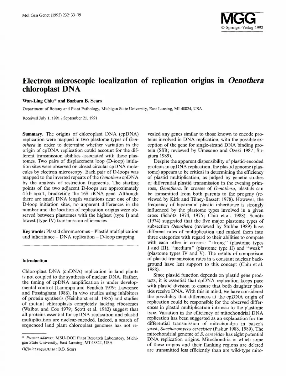
The preparation procedure yielded a relatively high fre- quency of intact Oenothera chloroplast DNA molecules. In the best preparation, nearly a quarter of the intact cpDNA molecules observed contained displacement loop (D-loop) replication intermediates, which result from unidirectional synthesis of one daughter strand (Kornberg 1980). On some molecules, more than one D-loop was observed. These D-loops lay either within several kb (Fig. 1 A) or quite far apart (estimated 30- 50 kb) from each other (Fig. 1 B, C). Very rarely, D- loops could be observed at four sites on the same mole- cule, as two distant pairs of D-loops.
D-loop initiation sites are located in both copies of the inverted repeats
In order to localize the sites of D-loop initiation, cpDNA was digested with SalI and PstI immediately prior to processing for the electron microscope. Digestion of Oenothera cpDNA with SalI or PstI yields 11 and 12 fragments, respectively (Fig. 2). Except for minor DNA fragment length differences, the restriction maps for these two enzymes are the same for plastome types I and IV (Gordon et al. 1982).
Digestion with restriction enzymes greatly reduced the frequency with which D-loops were observed. A total of 54 SalI fragments bearing D-loops (Fig. 3) were char- acterized: 29 from plastome I and 25 from plastome IV. Nine out of these 54 fragments contained two adja- cent D-loops (Fig. 3 C). The sizes and numbers of the fragments containing D-loops are presented in Fig. 4A. According to the observations with intact cpDNA, D- loops should be expected in at least two separate regions of Oenothera cpDNA (Fig. 1 B, C). As shown in Fig. 4A, D-loops were most frequently found in SalI fragments between 15 and 20 kb, but they were also present in some larger SalI fragments (30~40 kb). However, since the largest expected SalI fragment is around 30 kb (Gor- don et al. 1982), fragments much larger than 30 kb are most likely products of incomplete SalI digestion. As an initial step toward calculating the sizes of D-loop- bearing SalI fragments, those SalI fragments smaller than 24 kb were grouped into one class and those larger than 24 kb the other. However, two fragments of plas- tome I (22 and 23 kb, respectively) were later classified into the larger group because the positions of D-loops (see below) on these fragments are more similar to those on the larger fragments. For both plastome types I and IV (Table 1), the smaller class of D-loop-containing frag- ments had an average size of 18 kb (n=23), and the larger class had an average size around 32 kb (n=31). These sizes are comparable to the third largest (Sal C, 18 kb) and the largest (Sal A, 30 kb) SalI fragments, respectively. Sal A and Sal C are neighboring fragments that span most of the inverted repeats and the entire
35
Fig. 1A-C. Closed circular molecules of Oenothera chloroplast DNA cpDNA; (plastome type IV) with D-loops. Arrows indicate the displaced strand. 4~X174 DNA and its RF form (M) are used as internal standards for single-stranded and double-stranded DNA, respectively. Bar, 5 kb. A Part of an intact cpDNA molecule
that contains two adjacent D-loops. B An intact cpDNA molecule with two distant D-loops. C Part of an intact cpDNA molecule containing two D-loops approximately 30 kb apart. The D-loop on the left has a short tail resulting from strand migration
small single copy region of the Oenothera plastome (Fig. 2).
When plastome type I D N A was digested with PstI, D-loops were observed on fragments ranging from 12- 34 kb (Fig. 4B). Those fragments smaller than 20 kb have an average size of 14 kb (n=8) , which is the ex- pected size of the major PstI fragment that overlaps
Sal C (Fig. 2). The size distribution of those fragments larger than 20 kb is quite scattered. However, judging from the position of the D-loops on these fragments (see below), most of these fragments probably represent the PstI fragments (Pst B) that overlap Sal A (Fig. 2).
If D-loops are located within the regions spanned by Sal A and Sal C, double digestion of cpDNA with

36
psbC ps.bD psoAI A2 ~
psbA I ~ ~ Jrps8
....... ~ rRNA
Fig. 2. Restriction and partial gene map of Oenothera cpDNA (Gordon et al. 1982; Herrmann et al. 1983). The large inverted repeats, characteristic of DNA from most higher plant chloroplast genomes, are outlined by black arcs. SalI and PstI fragments are labeled alphabetically, with the largest fragment in each case desig- nated as A
PstI and Sall should also yield two fragments that carry D-loops (Fig. 2). For both plastome types I and IV, the double digestions of PstI and SalI resulted in two size groups of D-loop-containing fragments (Fig. 4C) with average sizes around 17 kb and 36 kb, respectively (Ta-
ble 1). The average sizes of these D-loop-containing fragments are larger than the lengths of the expected fragments, even though the same sample gave the ex- pected double digestion pattern when visualized on an agarose gel. Conceivably, complete SalI and PstI double digestions bring D-loops closer to the ends of the frag- ments and that might have rendered the D-loops less stable due to strand migration.
To orient the positions of the D-loops on the re- stricted cpDNA fragments, the distances of D-loops from the ends of each fragment were measured. A length, d, was defined as the shortest distance from an end of the fragment to a terminus of the D-loop. It is clear that D-loops can occur at two adjacent sites on the same region of the cpDNA (Fig. 1 A). The proximal terminus was chosen as the reference for measurement of D-loops closer to the end of the fragment and the distal terminus was chosen for the more distant D-loops. Each terminus of the D-loops defined in this way probably coincides with the replication origin for one daughter strand, ac- cording to the model for the initiation of cpDNA repli- cation in land plants (Kolodner and Tewari 1975b). In fact, since most of the D-loops are relatively small in size, the placement of a D-loop was not affected greatly by the choice of terminus. Two classes of d were observed and the measurements in these classes were grouped as d 1 and d 2 (Table ~). For those fragments with two D-
Fig. 3A-C. Electron micrographs of cpDNA (plastome type I) digested with Sale Arrows point to the displaced strand of each D-loop. A 18 kb fragment with one D-loop. B 30 kb fragment with one D-loop. C Two adjacent D-loops on the same fragment, Bar, 1 kb

4
co 2 i - z uJ 0
< w 4 1.1. U- O 2 re UJ m 0
z 4
A Sal
B Pst
C
5 10 15 20 25 30 35 40 45
FRAGMENT LENGTH (kb)
Fig. 4A-C. Length distribution of cpDNA fragments containing D-loops from plastome types I and IV. Fragments generated by digestion with (A) SalI (B) PstI (C) PstI +SalI. CpDNA fragments of plastome type I are represented by filled bars, while those of plastome type IV are represented by empty bars. The vertical axis indicates the number of fragments in each size class observed to have D-loops
loops, the distance between the two termini ranged from 1.5-6.6 kb, with an average of 4.1 kb (n= 17).
If the D-loops on fragments Sal A and Sal C lie within the inverted repeats, then D-loops on both PstI frag- ments should be 2 kb closer to the end of the fragments than D-loops on the SalI fragments (Fig. 2). As shown in Table 1, this prediction is fulfilled since d t and d 2 from PstI fragments are about 2-3 kb shorter than measure- ments derived from the related SalI fragments. The shortening of arm length coincides well with the position of PstI sites on Sal A and Sal C (Fig. 2). This placement of D-loops would further predict that the shorter dis-
37
tance from a D-loop to the end of a SalI fragment should be the same for both Sal A or Sal C. However, this distance is about 2 kb larger in the case of Sal A than Sal C (Table 1). This discrepancy can be explained if many of the larger SalI fragments that contain D-loops are the products of partial digestion: Sal A joined to the adjacent Sal K. Consistent with this interpretation, the average length of the larger SaII fragment is 2 kb greater than the expected length (Table 1). Furthermore, two distant D-loops are observed to be at least 30 kb apart on intact cpDNA molecules (Fig. 1 B, C). This dis- tance can be explained best if the D-loop initiation sites are located toward the distant ends of the two neighbor- ing SalI fragments within the inverted repeat.
Each pair of D-loop initiation sites is in the close vicinity of rRNA genes
In order to locate the positions of D-loop initiation sites relative to genes on the plastome, most of the SalI or PstI fragments containing D-loops were aligned to the end of their short arms (Fig. 5B-D). Fragments that are much longer than the predicted sizes due, presum- ably, to incomplete digestions, were not included in this analysis. Either Sal A or Sal C fragments that were slightly longer and may have included the neighboring Sal K were aligned in Fig. 5 to the appropriate SalI site 2 kb away. In some cases, the original alignment placed the D-loop initiation sites within the 16 S or 23 S rRNA genes. Since the rRNA genes are highly con- served, and have never been cited to function as origins of D N A replication, we realigned these fragments to alternative restriction sites. Restriction analysis of the cloned 14 kb Pst F fragment from both plastome types revealed only a 100 bp deletion in plastome type IV rela- tive to type I (W.-L. Chiu and B.B. Sears, in prepara- tion), and therefore, data from both plastome types were pooled to establish a more precise localization of D-loop initiation sites. As shown in the D-loop distribution fre- quency histogram (Fig. 5A), two regions appear to be preferential sites for D-loop initiation on both copies
Table 1. Distribution of D-loops in chlo- roplast DNA of Oenothera plastomes I and IV
Enzyme Plastome dla (n) d2 a (n) Observed Expected length (n) length b
Sall I 3.6+0.6 (6) 8.1+0.3 (5) 18.0_+0.5 (11) Sal C 17.9 IV 4.2_+0.5 (7) 9.1_+0.5 (5) 18.0_+0.8(12) SalC17.8 I 6.1___0.7(13) 11.3+_0.8(10) 32.5_+1.2(18) Sal A 30.2 IV 6.3_+0.5 (8) 10.9_+0.4 (8) 31.3+_1.6(13) SalA30.2
PstI I 2.1__.0.4 (7) 5.6_+0.3 (3) 13.6_+0.8 (8) PstF14.2 I 3.0_+0.8 (2) 8.3-t-0.8 (10) 26.8_+1.4(12) PstB28.4
PstI+SalI I 3.0-t-0.3 (9) 7.0+_0.5 (12) 17.5+_0.5 (19) Pst F 14.2 V 3.0_+0.4 (3) 7.2 (1) 16.6_+0.3 (4) PstF14.2 I 4.2_+0.4 (6) 10.9_+0.7 (6) 36.0_+1.0(10) P÷SA28.2 IV 4.3_+0.6 (5) 9.9_+0.5 (7) 35.8-+1.0(10) P+SA28.2
a Average distance +standard error (in kb) between the terminus (as defined in text) of D-loop structure and the nearer end of the fragment b Length expected from physical map of Gordon et al. (1982). Sizes expressed in megadal- tons in the original paper have been converted to kilobase pairs

38
B
c
Type +
Sal I
D
Type IV +
Sat I
oriA oriB oriB oriA
S S P 16SIA23S PSP 23SAI16S P S S
Fig. 5A-D. Proposed alignment of D-loop-containing molecules relative to the rRNA operon and the small single copy region of Oenothera cpDNA. A Frequency distribution of the D-loop structures, summing data from plastomes I and IV. Physical map of the Sal A (right) and Sat C (left) region. The positions of genes within the rRNA operon arc indicated with open boxes, with 16 S and 23 S indicating the rRNA genes, I designating trnl and A designating trnA. The most likely location for the D-loop initiation sites are indicated with oriA and oriB. B-D Scale of lines indicates length of fragments containing D-loops (indicated as black boxes). B Plastome type I DNA digested by PstI. C Plastome type I DNA digested by SaII. D Plastome type IV DNA digested by SalI
of the inverted repeat. One (oriA) is 1-2 kb upstream from the 16S rRNA gene and the other (oriB) is in the spacer of the rRNA operon near the tRNA ne gene. The distance between the two spacer regions is about 30 kb, which coincides with the distance between two distant D-loops in Fig. 1 C.
Discuss ion
Both the transmission efficiency of Oenothera plastids and their competitive multiplication rates are influenced by the plastome type (Sch6tz 1974; Chiu et al. 1988). Since plastids can multiply even in the absence of protein synthesis within the plastid (reviewed by Boffey 1985), the different multiplication efficiencies of Oenothera plastids may be attributable to differences in the non- coding components of the plastomes, such as the origins of cpDNA replication. This possibility inspired the pres- ent study.
CpDNA replication in Oenothera was found to be initiated through the formation of D-loops (Fig. 1). One pair of D-loop initiation sites was mapped to each copy of the large inverted repeat (Fig. 2) of the plastome:
one lies 1-2 kb upstream from the 16S rRNA gene, while the other lies in the spacer between the two rRNA genes (Fig. 5). In these respects, plastome type I, having the highest transmission efficiency, is indistinguishable from plastome type IV, having the lowest efficiency in trans- mission. Hence, differential plastid multiplication in Oenothera cannot be explained simply by differences in the number or placement of functional cpDNA replica- tion origins present in the plastome. However, this does not preclude the possibility that sequence differences at the replication origin could affect the efficiency of repli- cation initiation.
In order to locate the replication origins, care was taken to preserve unstable D-loop replication intermedi- ates in the purified DNA. All the restriction digestions in this study were performed at room temperature for a limited amount of time to minimize strand migration. This may have contributed to the frequent appearance of fragments that are larger than the expected size. De- spite the measures taken to minimize strand migration, D-loops were observed far less frequently in digested cpDNA than in uncut samples. It is conceivable that D-loops lying near the ends of fragments or on small fragments may have been lost. However, the relative lo- cations of D-loops on uncut DNA are consistent with the four D-loop initiation sites determined using the re- stricted fragments. It is likely that the sizes of some large fragments may have been overestimated, since larger fragments are more readily subjected to stretching forces during the spreading procedure than are the ~bX174 DNA size markers or small fragments. This factor may have contributed to the 20% overestimation of the size of pea cpDNA, when ~bX174 DNA was used as an inter- nal size marker by Kolodner and Tewari (1975 a).
In no other organism besides Oenothera has cpDNA replication been shown to proceed from two pairs of D-loops. In maize, cpDNA fragments that can initiate replication in vitro have been identified (Gold et al. 1987). Whether these fragments can initiate cpDNA rep- lication in vivo has not been confirmed by electron mi- croscopy. Pea, the only other land plant in which the D-loop initiation sites of cpDNA have been localized, has only one pair of cpDNA replication origins near the rRNA operon; one in the spacer region within the rRNA operon, as in Oenothera, and the other several kb downstream from the 23 S rRNA gene (Meeker et al. 1988). However, the plastome of pea differs from most other land plants in that it has only one copy of the rRNA operon (Palmer and Thompson 1981). Since the rRNA operon generally is found within the large in- verted repeat that is common to the cpDNA of most land plants (reviewed by Palmer 1985) including Oenoth- era, it is likely that the number and the placement of replication origins in the Oenothera plastome is typical of those in plastomes of most other land plants. The placement of replication origins near the rRNA operon may have some functional importance since several algae also have their cpDNA replication origins near the rRNA operon, although the exact position with respect to rRNA genes differs (Koller and Delius 1982; Ravel- Chapuis et al. 1982; Waddell et al. /984; preliminary re- sults in Yamada et al. /986).

39
The two adjacent D- loop initiation sites in the Oen- othera plas tome p robab ly represent separate origins for the replication o f each strand, as suggested in studies on c p D N A replication in pea and maize (Kolodner and Tewari 1975a, b) and mi tochondr ia l D N A replication in animals (reviewed by Clay ton 1982). It has been pro- posed that unidirect ional synthesis o f a daughter s t rand f rom each neighbor ing origin results in a replication eye (Cairns s tructure; Cairns 1963) with two double- s t randed arms, f rom which bidirectional D N A synthesis proceeds (Kolodner and Tewari 1975b). A l though no such structures were observed in this study, replication forks with two double-s t randed arms were observed in some fragments. In c p D N A of the algae Chlamydomonas (Waddell et al. 1984), Euglena (Koller and Delius 1982; Ravel-Chapuis et al. 1982) and Acetabularia (Santulli et al. 1983), loops with two double-s t randed arms are the ma jo r or only replication intermediates tha t have been observed. Apparent ly , in these organisms, either the same origin is used for the replication o f bo th strands or the replication origins for each strand are very close to each other. It will be necessary to analyze c p D N A replication in vitro (Wu et al. 1986; Meeker et al. 1988) to locate the exact sites o f replication initiation in plas- rids o f Oenothera and to reveal fur ther features o f the c p D N A replication origins tha t m a y be c o m m o n to land plants and algae.
Acknowledgments. The authors thank Karen L. Klomparens, John Heckman, Barry Stine, Robert Meeker, Brent Nielsen and Made- line Wu for their advice in electron microscopy. James Hancock, John Ohlrogge and Shauna Somerville are thanked for their critical evaluation of the manuscript. WLC was supported by graduate fellowships from the MSU McKnight photosynthesis training grant and MSU College of Natural Science. This research was also sup- ported by the Michigan State Agricultural Experiment Station and NSF grant DMB-9019488.
References
Boffey SA (1985) The chloroplast division cycle and its relationship to the cell division cycle. In: Bryant JA, Francis D (eds) The cell division cycle in plants, Society for Experimental Biology seminar series, vol 26. Cambridge University Press, Cambridge, pp 233-246
Cairns J (1963) The chromosome of Escherichia eoli. Cold Spring Harbor Symp Quant Biol 28:43~46
Chiu W-L, Stubbe W, Sears BB (1988) Plastid inheritance in Oen- othera: organelle genome modifies the extent of biparental plas- tid transmission. Curr Genet 13:181-189
Clayton DA (1982) Replication of animal mitochondrial DNA. Cell 28 : 693 705
Gold B, Carrillo N, Tewari KK, Bogorad L (1987) Nucleotide sequence of a preferred maize chloroplast genome template for in vitro DNA synthesis. Proc Natl Acad Sci USA 84:194-198
Gordon KHJ, Crouse E J, Bohnert H J, Herrmann RG (1982) Physi- cal mapping of differences in chloroplast DNA of the five wild- type plastomes in Oenothera subsection Euoenothera. Theor Appl Genet 61 : 373-384
Heinhorst S, Cannon GC, Weissbach A (1985) Plastid and nuclear DNA synthesis are not coupled in suspension cells of Nieotiana tabacum. Plant Mol Biol 4:3-12
Herrmann RG, Westhoff P, Alt J, Winter P, Tittgen J, Bisanz C, Sears BB, Nelson N, Hurt E, Hauska G, Viebrock A, Sebald W (1983) Identification and characterization of genes for poly- peptides of the thylakoid membrane. In: Cifferi O, Dure L III (eds) Structure and function of plant genomes. Plenum Press. New York. t~n 143-153
Kirk JTO, Tilney-Bassett RAE (1978) The Plastids. Elsevier/North Holland, Amsterdam
Koller B, Delius H (1982) Origin of replication in chloroplast DNA of Euglena gracilis located close to the region of variable size. EMBO J 1:995-998
Kolodner RD, Tewari KK (1975 a) Presence of displacement loops in the covalently closed circular chloroplast deoxyribonucleic acid from higher plants. J Biol Chem 250:8840-8847
Kolodner RD, Tewari KK (1975b) Chloroplast DNA from higher plants replicates by both the Cairns and the rolling circle mech- anism. Nature 256:708-711
Kornberg A (1980) DNA replication. WH Freeman and Company, San Francisco
Lammpa GK, Bendich AJ (1979) Changes in chloroplast DNA levels during development of pea (Pisum sativum). Plant Physiol 64:126-130
Lawrence ME, Possingham JV (1986) Microspectrofluorometric measurement of chloroplast DNA in dividing and expanding leaf cells of Spinacia oleracea. Plant Physiol 81:708-710
Meeker R, Nielsen B, Tewari KK (1988) Localization of replication origins in pea chloroplast DNA. Mol Cell Biol 8:1216-1223
Palmer JD (1982) Physical and gene mapping of chloroplast DNA from Atriplex triangularis and Cucumis sativa. Nucleic Acids Res 10:1593-1605
Palmer JD (1985) Comparative organization of chloroplast ge- nomes. Annu Rev Genet 19:325-354
Palmer JD, Thompson WF (1981) Rearrangements in the chloro- plast genome of mung bean and pea. Proc Natl Acad Sci USA 78:5533-5537
Piskur J (1988) Transmission of yeast mitochondrial loci to progeny is reduced when nearby intergenic regions containing ori/rep sequences are deleted. Mol Gen Genet 214:425-432
Piskur J (1989) Transmission of the yeast mitochondrial genome to progeny: the impact of intergenic sequences. Mol Gen Genet 218:161-168
Ravel-Chapuis P, Heizmann P, Bigon V (1982) Electron microscop- ic localization of the replication origin of Euglena gracilis chlo- roplast DNA. Nature 300:78-81
Santulli A, Casale A, Mazza A (1983) Cairns replicative intermedi- ates in Acetabularia chloroplast DNA. J Submicrosc Cytol 15 : 843-847
Sch6tz F (1974) Untersuchungen tiber die Plastidenkonkurrenz bei Oenothera. IV. Der EinfluB des Genoms auf die Durchset- zungsf~ihigkeit der Plastiden. Biol Zentralbl 93:41-64
Sch6tz F (1975) Untersuchungen fiber die Plastidenkonkurrenz bei Oenothera. V. Die Stabilitfit der Konkurrenzf/ihigkeit bei Ver- wendung verschiedenartiger mutierter Test-Plastiden. Biol Zen- tralbl 94:17-26
Scott NS, Cain P, Possingham V (1982) Plastid DNA levels in albino and green leaves of the ' albostrians' mutant of Hordeum vulgare. Z Pflanzenphysiol 108 : 187-192
Stubbe W (1989) Oenothera - An ideal system for studying the interactions of genome and plastome. Plant Mol Biol Rep 7:245-257
Sugiura M (1989) The chloroplast chromosomes in land plants. Annu Rev Cell Biol 5:51-70
Umesono K, Ozeki H (1987) Chloroplast gene organization in plants. Trends Genet 3:281 287
Waddell J, Wang X-M, Wu M (1984) Electron microscopic localiza- tion of the chloroplast DNA replicative origins in Chlamydo- monas reinhardii. Nucleic Acids Res 12:3843-3856
Walbot V, Coe EH (1979) Nuclear gene iojap conditions a pro- grammed change to ribosome-less plastids in Zea mays. Proc Natl Acad Sci USA 76:1760-1764
Wu M, Lou JK, Chang DY, Chang CH, Nie ZQ (1986) Structure and function of a chloroplast DNA replication origin of Chla- mydomonas reinhardii. Proc Natl Acad Sci USA 83 : 6761-6765
Yamada T, Shim@ M, Fukuda Y (1986) Characterization of a cpDNA sequence from Chlorella ellipsoidea that promotes au- tonomous replication in yeast. Plant Mol Biol 6: 245-252
Communica t ed by R.G. H e r r m a n n