elastin peptides activate extracellular signal-regulated kinase 1/2 via a ras-independent mechanism...
TRANSCRIPT

Elastin Peptides Activate Extracellular Signal-Regulated Kinase1/2 via a Ras-Independent Mechanism Requiring Both p110�/Raf-1 and Protein Kinase A/B-Raf Signaling in Human SkinFibroblasts
Laurent Duca, Elise Lambert, Romain Debret, Bernard Rothhut, Charlotte Blanchevoye,Frederic Delacoux, William Hornebeck, Laurent Martiny, and Laurent DebelleLaboratory of Biochemistry, Unite Mixte Recherche Centre National de la Recherche Scientifique 6198, IFR53 Biomolecules,Faculty of Sciences, University of Reims Champagne-Ardenne, Moulin de la Housse, Reims, France (L.Du., E.L., R.D., B.R.,C.B., F.D., W.H., L.M., L.De.); and EA 2070, IFR53 Biomolecules, Faculty of Pharmacy, University of Reims Champagne-Ardenne, Reims, France (R.D.)
Received May 13, 2004; accepted January 12, 2005
ABSTRACTElastin peptides (EPs) produced during cancer progression bindto the elastin binding protein (EBP) found at the surface of dermalfibroblasts, leading to the expression of collagenase-1 gene. Theproduction of this enzyme involved in stromal reaction is causedby the sustained activation of the extracellular signal-regulatedkinases 1/2 (ERK1/2) pathway via cAMP/protein kinase A (PKA)and phosphatidylinositol 3-kinase (PI3K). However, the mecha-nism of these signaling events remains unknown. We show that�-elastin (�E), a commonly used EP, induces maximum phosphor-ylation of mitogen-activated protein kinase/extracellular signal-regulated kinase (MEK)1/2 and ERK1/2 after 30 min. Thesimultaneous inhibition of PKA and PI3K, by N-(2-(p-bromocin-namylamino)ethyl)-5-isoquinolinesulfonamide (H89) and 2-(4-morpholynil)-8-phenyl-4H-1-bemzopyran-4-one (LY294002), re-spectively, blocked MEK1/2 and ERK1/2 phosphorylation, as didlactose, an EBP antagonist. �E induced Raf-1 phosphorylation
and activation in a PI3K-dependent manner. In our system, thePI3K p110� is expressed and activated by ��-derived subunitsfrom a pertussis toxin-sensitive G protein after fibroblast stimula-tion. Pertussis toxin also blocks the Raf-1/MEK1/2/ERK1/2 phos-phorylation cascade. In addition, we found that B-Raf is ex-pressed in dermal fibroblasts and activated in a PKA-dependentmanner after �E treatment, thereby integrating PKA signals toMEK1/2. It is noteworthy that Ras involvement was excludedbecause ERK1/2 activation by �E was not blocked in RasN17-transfected fibroblasts. Together, our results identify a novel Ras-independent ERK1/2 activation system in which p110�/Raf-1/MEK1/2 and PKA/B-Raf/MEK1/2 cooperate to activate ERK1/2.Thus, p110� and B-Raf seem to be important modulators ofdermal fibroblasts physiology and should now qualify as thera-peutic targets in strategies aiming at limiting elastin degradationcontribution to cancer progression.
Elastin is the extracellular matrix molecule responsible forresilience of tissues and was first thought to be restricted tothat role. It is now established that elastin degradation may
lead to the production of bioactive peptides (Hornebeck et al.,2002) influencing cell chemotaxis, cell proliferation, and pro-teases synthesis in a broad panel of normal and tumor cells(Duca et al., 2004). Their contribution to the stromal reactionis therefore envisaged (Hornebeck et al., 2002).
The receptor of these peptides comprises three subunits.The first two, a 55-kDa cathepsin A termed protective protein(EC 3.4.16.1) and a 61-kDa neuraminidase (EC 3.2.1.18), aremembrane-associated. The last subunit, which actually bindselastin-derived peptides (EPs), is a peripheral 67-kDa pro-
This work was supported by Centre National de la Recherche Scientifique,Association Regionale pour l’Enseignement Superieur et la Recherche Scien-tifique et Technologie en Champagne-Ardenne, and the Comite de l’Aisne de laLigue contre le Cancer. L.D. is the recipient of a bursary from the Frenchgovernment (Ministere de l’Education Nationale et de la Recherche).
Article, publication date, and citation information can be found athttp://molpharm.aspetjournals.org.
doi:10.1124/mol.104.002725.
ABBREVIATIONS: EP, elastin peptide; EBP, elastin binding protein; �E, �-elastin; ERK, extracellular-signal regulated kinase; PKA, protein kinaseA; PI3K, phosphatidylinositol 3-kinase; MEK, mitogen-activated protein kinase/extracellular signal-regulated kinase; EGF, epidermal growth factor;PTX, pertussis toxin; H89, N-(2-(p-bromocinnamylamino)ethyl)-5-isoquinolinesulfonamide; LY294002, 2-(4-morpholynil)-8-phenyl-4H-1-bemzo-pyran-4-one; LY303511, 2-piperazinyl-8-phenyl-4H-1-benzopyran-4-one; GST, glutathione S-transferase; DMEM, Dulbecco’s modified Eagle’smedium; �ARK1-CT, C-terminal domain of the �-adrenergic receptor kinase 1; PBS, phosphate-buffered saline; TLC, thin layer chromatography;PAGE, polyacrylamide gel electrophoresis; PI, phosphatidylinositol; SMC, smooth muscle cell; KT5720, (9S,10S,12R)-2,3,9,10,11,12-hexahydro-10-hydroxy-9-methyl-1-oxo-9,12-epoxy-1H-diindolo[1,2,3-fg:3�,2�,1�-kl]pyrrolo[3,4-i][1,6]benzodiazocine-10-carboxylic acid hexyl ester.
0026-895X/05/6704-1315–1324$20.00MOLECULAR PHARMACOLOGY Vol. 67, No. 4Copyright © 2005 The American Society for Pharmacology and Experimental Therapeutics 2725/1198121Mol Pharmacol 67:1315–1324, 2005 Printed in U.S.A.
1315

tein, termed elastin binding protein (EBP). This protein pos-sesses galactolectin properties. When galactosugars bindEBP, its affinity for EP dramatically decreases, leading totheir release and to further dissociation of EBP from thecomplex. Galactosugars, such as lactose, are therefore com-monly used as EBP antagonists (Hinek, 1996).
In human dermal fibroblasts, an elastin degradation prod-uct, �-elastin (�E), up-regulates the expression of procollage-nase-1, whose activated form is crucially involved in stromalreaction (Brassart et al., 2001). We have recently shown(Duca et al., 2002) that the extracellular-signal regulatedkinase 1/2 pathway (ERK1/2) holds a central role in thesignaling leading to this phenomenon. In our system, ERK1/2activation involved both cAMP-dependent activation of pro-tein kinase A (PKA) and phosphatidylinositol 3-kinase(PI3K). However, the details of this integrated cross-talkwere not elucidated.
ERK1/2 activation requires phosphorylation by dual-spe-cific kinases named mitogen-activated protein kinase/extra-cellular signal-regulated kinases 1 and 2 (MEK1/2), whichare themselves typically phosphorylated and activated bykinases named Raf (Houslay and Kolch, 2000). In mammals,the Raf family comprises three members: the ubiquitouslyexpressed Raf-1, A-Raf, and B-Raf, in which expressions aremore restricted. Compared with Raf-1, B-Raf is a strongeractivator of the ERK pathway, whereas A-Raf is weaker(Chong et al., 2003; Mercer and Pritchard, 2003). Raf-1 isactivated by the small GTP-binding protein Ras, but otheractivators such as protein kinase C or PI3K have also beenreported (Dhillon and Kolch, 2002). The phosphorylation ofRaf-1 on Ser338 is essential for its activation (Mason et al.,1999).
Three classes of PI3K have been characterized (Andersonand Jackson, 2003), but only those belonging to class I, com-prising subclasses IA and IB, have been shown to activateERK1/2. This activation occurs at the level of Raf-1 orMEK1/2 (King et al., 1997; Takeda et al., 1999; Mas et al.,2003).
Class IA PI3Ks present a p85 regulatory subunit associatedto a p110�, p110�, or p110� catalytic subunit. They areactivated by phosphotyrosine motifs and/or Ras. Class IB
PI3Ks possess a p101 regulatory subunit associated to ap110� catalytic subunit, which can be directly activated by Gprotein �� subunits or Ras (Anderson and Jackson, 2003).Class IA PI3Ks are expressed in fibroblasts (Anderson andJackson, 2003), but the presence of p110� has not been re-ported yet.
B-Raf is expressed as multiple alternatively spliced vari-ants but possesses two major isoforms: 68 and 95 kDa (Mer-cer and Pritchard, 2003). It is an important regulator ofERK1/2 pathway activation by cAMP-dependent signalingelements (Houslay and Kolch, 2000). cAMP activates ERK1/2cascade in cells expressing the 95-kDa isoform, whereas it isinhibited in NIH 3T3 fibroblasts lacking it (Vossler et al.,1997). cAMP-dependent activation of the 68-kDa isoform wasreported suggesting that it could participate to cAMP-depen-dent ERK1/2 pathway induction (Seidel et al., 1999). cAMP-dependent B-Raf activation could occur via two mechanismsinvolving either the cAMP receptor exchange protein directlyactivated by cAMP or the cAMP-dependent protein kinasePKA (Houslay and Kolch, 2000). Although B-Raf is expressedin mouse embryonic fibroblasts and seems to be absent in
NIH 3T3 fibroblasts (Vossler et al., 1997; Huser et al., 2001),its presence in human skin fibroblasts has not been reported.
We describe here the mechanisms leading to ERK1/2 acti-vation by elastin-peptides in dermal fibroblasts. We showthat �E activates ERK1/2 via both p110�/Raf-1/MEK1/2 andPKA/B-Raf/MEK1/2 modules. Such a Ras-independent sig-naling system is novel and provides a new regulation systemfor ERK1/2 activation. In addition, we show that p110� andB-Raf, which had never been reported in dermal fibroblasts,are important modulators of the physiology of these cells.Because their involvement could explain the strong and sus-tained activation of ERK1/2 and subsequent procollage-nase-1 production, their control in strategies aiming at lim-iting elastin peptides contribution to cancer progression isdiscussed.
Materials and MethodsMaterials. �E was prepared as described previously (Brassart
et al., 2001). Human recombinant epidermal growth factor (EGF)was from Upstate Biotechnology (Lake Placid, NY) (distributedby Euromedex, Mundelsheim, France). Lactose, pertussis toxin(PTX), phosphatidylserine, phosphatidylinositol, phosphatidylinosi-tol 3-phosphate, and proteases inhibitors cocktail (reference P8340)were from Sigma (Saint-Quentin Fallavier, France). H89, KT5720,and LY303511 were from Calbiochem (distributed by VWR Interna-tional, Strasbourg, France). LY294002 was purchased from Cell Sig-naling Technology Inc. (distributed by Ozyme, Saint-Quentin enYvelines, France). Recombinant GST-MEK1 was from Santa CruzBiotechnology, Inc. (Santa Cruz, CA) (distributed by Tebu, Le Perrayen Yvelines, France). Mouse monoclonal anti-�-actin antibody waspurchased from Sigma. Rabbit polyclonal phospho-specific antibod-ies against active forms of ERK1/2 (phosphorylated on Thr202 andTyr204), MEK1/2 (phosphorylated on Ser217 and Ser221), and phos-pho-specific antibody against phosphorylated Raf-1 Ser338 and anti-ERK1/2 antibody were from Cell Signaling Technology Inc. (Beverly,MA). Rabbit anti-p110�, anti-Raf-1, and anti-B-Raf polyclonal anti-bodies were purchased from Santa Cruz Biotechnology, Inc. Poly-clonal anti-p85 antibody was from Upstate Biotechnology (distrib-uted by Euromedex). Mouse monoclonal anti-hemagglutinin (HA)tag was from Roche Diagnostics (Meylan, France). All reagents forcell culture and transfection reagent LipofectAMINE 2000 were fromInvitrogen (Cergy Pontoise, France). Enhanced chemiluminescencesubstrate kit and [�-32P]ATP were purchased from Amersham Bio-sciences Inc. (Orsay, France). Others reagents were from Sigma.
Expression Plasmids. The plasmid pRK5-�ARK1-CT (Koch etal., 1994), which encodes the Gly495-Leu689 fragment correspondingto the C terminus of �-adrenergic receptor kinase 1 (�ARK1-CT), waskindly provided by Dr. R. J. Lefkowitz (Duke University, Durham,NC). The constructs encoding HA-tagged-ERK1 (pECE-HA-ERK1)and the dominant negative RasN17 mutant (pSV-RasN17) weredescribed previously (Guillemot et al., 2000) and were kind gifts fromDr. J. Pouyssegur (Institute of Signaling, Developmental Biology andCancer Research, Nice, France) and Dr. F. Schweighoffer (ExonHitTherapeutics, S.A., Paris, France), respectively.
Cell Culture, Treatments, and Transfection. Human skinfibroblast strains were established from explants of human adultskin biopsies obtained from informed healthy volunteers (aged 21–49years). Cells were grown as monolayer cultures in Dulbecco’s modi-fied Eagle’s medium (DMEM) supplemented with 10% fetal calfserum and 2 mM glutamine in the presence of 5% CO2. Cells atsubcultures 5 to 10 were used. For experiments, fibroblasts weregrown to subconfluence in 10% serum containing medium. Beforestimulation, cells were incubated for 18 h in DMEM containing 0.5%fetal calf serum, washed twice with PBS, and then incubated inserum-free DMEM with or without �E (50 �g/ml) or EGF (2 ng/ml)
1316 Duca et al.

for the indicated times. The pharmacological inhibitors H89 (1 �M),KT5720 (2 �M), LY294002 (25 �M), and LY303511 (25 �M) werepreincubated 1 h before stimulation, whereas lactose (1 mM) andPTX (100 ng/ml) were preincubated 3 and 18 h, respectively. Elastinpeptide stimulation was stopped by adding ice-cold PBS containing50 �M Na3VO4. For transfection experiments, cells were grown to 80to 90% of confluence in 10% serum-containing medium and incu-bated 16 h in serum-free DMEM with LipofectAMINE 2000-DNAplasmid complexes. The ratio was 2 �l of LipofectAMINE 2000 for 2�g of total plasmidic DNA for 5 � 105 cells. Growth medium was thenadded for 24 h, and cells were treated before stimulation as describedabove.
Western Blotting. Cells (106) were washed twice in ice-cold PBScontaining 50 �M Na3VO4, scrapped, and sonicated in lysis buffer(PBS, pH 7.4, 0.5% Triton X-100, 80 mM �-glycerophosphate, 50 mMEGTA, 15 mM MgCl2, 1 mM Na3VO4, and protease inhibitor cock-tail). Insoluble material was removed by centrifugation (20,000g, 20min, 4°C). Protein concentrations were determined by bicinchoninicacid protein assay (Pierce Chemical, Rockford, IL; distributed byInterchim, Montlucon, France). Equal amounts of proteins wereheated for 5 min at 100°C in Laemmli sample buffer, resolved bySDS-PAGE under reducing conditions, and transferred to nitrocel-lulose membranes. The membranes were placed in blocking buffer[5% (w/v) nonfat dry milk in Tris-buffered saline/Tween 20 (50 mMTris, pH 7.5, 150 mM NaCl, and 0.1% (v/v) Tween 20)] for 1 h at roomtemperature and incubated overnight at 4°C with anti-phospho-ERK1/2 (1:1000), anti-phospho-MEK1/2 (1:1000), anti-phospho-Ser338-Raf-1 (1:1000), anti-B-Raf (1:500), anti-p110� (1:500), anti-ERK1/2 (1:1000), or anti-�-actin (1:5000) antibodies. After fivewashings with Tris-buffered saline/Tween 20, the membranes wereincubated for 1 h at room temperature in the presence of horseradishperoxidase-coupled anti-rabbit or anti-mouse antibodies (1:4000 and1:10,000 in blocking buffer, respectively). Immunocomplexes weredetected by chemiluminescence. Blots were semiquantified by den-sitometry using PhosphorAnalyst software (Bio-Rad, Marne-la-Vallee, France).
Immunoprecipitation and PI3K Activity Assay. Cells (8 �106) were washed twice in ice-cold PBS containing 50 �M Na3VO4,scrapped in this buffer, and centrifuged (375g, 10 min, 4°C). Pelletswere resuspended and lysed for 15 min at 4°C in immunoprecipita-tion lysis buffer [10 mM Tris, pH 7.4, 150 mM NaCl, 5 mM EDTA,10% (v/v) glycerol, 1% (v/v) Brij 98, 1 mM Na3VO4, and proteasesinhibitor cocktail]. Insoluble material was removed by centrifugation(20,000g, 20 min, 4°C). Protein concentrations were determined us-ing bicinchoninic acid protein assay, and equal amounts of proteinswere incubated with 2.5 �g of anti-p85 or anti-p110� antibodies for1 h at 4°C. The antigen-antibody complexes were incubated withprotein G Plus-Sepharose (Santa Cruz Biotechnology, Inc.) for 1 h at4°C, collected by centrifugation, washed three times in immunopre-cipitation lysis buffer, and then twice with lipid kinase buffer (25mM HEPES, pH 7.4, 100 mM NaCl, 5 mM MgCl2, and 200 �Madenosine). To perform lipid kinase assay, each pellet was resus-pended in 70 �l of lipid kinase buffer supplemented with phospha-tidylinositol and phosphatidylserine (10 mg/ml each), 2.5 �M ATP,and 10 �Ci of [�-32P]ATP. The reaction was performed for 15 min at30°C and stopped by adding 100 �l of 1 M HCl. Phospholipids wereextracted with 350 �l of chloroform/methanol [1:1 (v/v)], and theorganic phase was washed twice with 200 �l of methanol/1 M HCl[1:1 (v/v)]. Organic phases (110 �l) were spotted on to oxalate-treatedthin layer chromatography (TLC) plates, and lipids were then sepa-rated using a chloroform/methanol/acetone/acetic acid/H2O [40:13:15:12:7 (v/v/v/v/v)] solvent system. Plates were revealed by autora-diography. For transfection experiments, cells were transfected witheither pRK5-�ARK1-CT or the corresponding empty vector before �Etreatment (2 �g of plasmid DNA for 0.5 � 106 cells).
Raf-1 and B-Raf Activities Assay. Samples preparation andimmunoprecipitation were performed as described above. Equalamounts of proteins were incubated with 2.5 �g of anti-Raf-1 or
anti-B-Raf antibodies for 1 h at 4°C. The antigen-antibody complexeswere incubated with protein G Plus-Sepharose for 1 h at 4°C, col-lected by centrifugation, washed three times in immunoprecipitationlysis buffer, and then twice with kinase buffer (20 mM HEPES, pH7.2, 10 mM MgCl2, and 10 mM MnCl2). For kinases activity assays,pellets were resuspended in 50 �l of kinase buffer supplementedwith 1 �g of GST-MEK1, 2.5 �M ATP, and 10 �Ci of [�-32P]ATP. Thereaction was performed for 30 min at 30°C and stopped by addingLaemmli sample buffer. After boiling at 100°C for 5 min, sampleswere resolved on a 10% SDS-PAGE, gels were dried, and bands werevisualized by autoradiography.
Assessment of HA-Tagged-ERK1 Activation. Cells (106) werecotransfected with 2 �g of pECE-HA-ERK1 and 2 �g of pSV-RasN17or with the corresponding empty vectors before stimulation. Equalamounts of proteins were incubated for 1 h at 4°C with 3 �g ofanti-HA tag antibody. The formed antigen-antibody complexes wereincubated with protein G Plus-Sepharose for 1 h at 4°C, collected bycentrifugation, washed three times in immunoprecipitation lysisbuffer, and then resuspended in Laemmli sample buffer. Proteinextracts were then resolved on 10% SDS-PAGE, and active phospho-HA-tagged-ERK1 and HA-tagged-ERK1 were visualized using anti-phospho-ERK1/2 and anti-ERK1/2 antibodies.
Statistical Analysis. All experiments were performed in tripli-cate. Results are expressed as mean � S.E.M. Comparison betweengroups were made using Student’s t test. The results were consideredsignificantly different at p � 0.05.
Results�E Induces MEK1/2 Activation via PI3K and PKA-
Dependent Signaling. In previous work, we have shownthat the treatment of human skin fibroblasts with �E wasfollowed by a rise of the intracellular cAMP level and thatPKA activation participated to ERK1/2 induction. Forskolinalone partly reproduced the effect of �E, demonstrating thatcAMP-dependent signaling was not sufficient to achievemaximum induction of the ERK1/2 pathway. Indeed, PI3Kparticipation was required to fully activate ERK1/2 (Duca etal., 2002). Nevertheless, the molecular links between ERK1/2and their upstream activators were not revealed.
MEK1/2 activates ERK1/2. We therefore analyzed its phos-phorylation pattern by Western blot by using an antibodyspecifically recognizing its active form (i.e., phospho-Ser217/Ser221-MEK1/2). In parallel, the presence of the phospho-Thr202/Tyr204-ERK1/2 active form was checked.
The treatment of fibroblasts with 50 �g of �E/ml resultedin a sustained activation of MEK1/2 up to a maximumreached at 30 min of stimulation (Fig. 1A). The time course ofthis activation closely paralleled that observed for ERK1/2(Fig. 1A). It is noteworthy that a strong activation of bothkinases still persists after 60 min of stimulation.
The importance of PKA and PI3K signaling towardMEK1/2 activation was investigated using pharmacologicalinhibitors. When cells were pretreated for 1 h with H89 (1�M) or LY294002 (25 �M), two commonly used PKA andPI3K inhibitors, respectively, MEK1/2 and ERK1/2 activa-tions were partially blocked (Fig. 1B). However, their simul-taneous use abolished MEK1/2 and ERK1/2 activation (Fig.1B). The same effect was obtained using 1 mM lactose(Fig. 1B).
To support these observations, �E-induced ERK1/2 activa-tion was assessed in the presence of LY303511 (25 �M), acompound that is structurally similar to LY294002 but doesnot inhibit PI3K. In addition, we inhibited PKA with KT5720
Mechanism of Elastin Peptide-dependent ERK1/2 Activation 1317

(2 �M), a PKA inhibitor structurally unrelated to H89. Ourresults show (Fig. 1C) that LY303511 has no effect on �E-induced ERK1/2 activation, whereas the use of KT5720 re-produced the effect of H89, supporting our previous observa-tions. PKA and PI3K thus seemed to be crucial modulators ofthe MEK1/2/ERK1/2 cascade in elastin peptide-induced sig-naling.
Inhibition of PI3K Blocks �E-Induced Raf-1 Ser338Phosphorylation and Raf-1 Activation. Raf-1 is the typ-ical activator of MEK1/2 (Houslay and Kolch, 2000), theimmediate upstream activator of ERK1/2. Its activation ismainly related to its phosphorylation on Ser338 (Mason etal., 1999). Several authors have suggested that Ser338 phos-phorylation and Raf-1 activation could be regulated by PI3K(Chaudhary et al., 2000; Sun et al., 2000). For these reasons,we examined the effect of �E on Ser338 phosphorylation andon Raf-1 activity in presence of LY294002. Using an anti-phospho-Ser338-Raf-1 antibody, we show by Western blotthat �E cell stimulation leads to Ser338 Raf-1 phosphoryla-tion (Fig. 2A) with a time course very similar to that observedfor MEK1/2 and ERK1/2 (Fig. 1A). The maximum phosphor-ylation level was observed at 30 min. The inhibition of PI3Kwith LY294002 (25 �M) totally blocked Ser338 phosphoryla-tion induced by �E treatment (Fig. 2B). However, even ifphosphorylation of Ser338 is required for Raf-1 activation, itis not a surrogate marker for Raf-1 activity (Mason et al.,1999; Chiloeches et al., 2001). We therefore analyzed Raf-1activity under the same conditions. Using GST-MEK-1 as asubstrate, we found that Raf-1 activity was very low in rest-ing skin fibroblasts (Fig. 2C, first lane). However, when cellswere treated with �E, its activity was raised 5-fold (Fig. 2C,second lane). Pretreatment of cells with LY294002 totallyblocked Raf-1 activity. PKA inhibition (1 �M H89) had noeffect on Raf-1 activity (data not shown).
These results indicated that �E induced Raf-1 Ser338phosphorylation and its activation via PI3K and that PI3Kintegrated the MEK1/2/ERK1/2 pathway at the level ofRaf-1. They also suggested that PKA signaling should in-volve a different integration system.
�E Promotes Class IB PI3K Activation in HumanSkin Fibroblasts. Both class IA and class IB PI3K wereshown to be involved in ERK1/2 activation in various celltypes (King et al., 1997; Takeda et al., 1999; Mas et al., 2003).PI3K activity of these two subfamilies was therefore ana-lyzed after �E cell stimulation.
Class IA PI3K associate with a regulatory p85 subunit.They are found in fibroblasts (Anderson and Jackson, 2003).In contrast, p110� binds a p101 regulatory subunit, and itstissue distribution is more limited (Yart et al., 2002). It ishighly expressed in hematopoietic cells, but its presence hasalso been reported in nonhematopoietic cells, notably mela-noma cells (Lee et al., 2002). To our knowledge, its expressionhas never been reported in skin fibroblasts.
First, total cells extracts were Western blotted with a spe-cific anti-p110� antibody showing that this PI3K isoform isexpressed in human skin fibroblasts (Fig. 3A). We then mea-sured PI3K activity in p85 and p110� immunoprecipitatesusing an in vitro PI3K activity assay with phosphatidylino-sitol (PI) as a substrate. We found (Fig. 3B) that �E treat-ment led to an important augmentation of PI3K activity inp110� immunoprecipitates (approximately 150% increasecompared with control). A nonsignificant increase was ob-served in p85 immunoprecipitates.
These results show that class IB PI3K, p110�, is expressedin human skin fibroblasts and that its activity is stronglystimulated by elastin peptides. In addition, Fig. 3C showsthat, in our experimental conditions, LY294002 inhibitedp110�, whereas LY303511 had no effect. These results are in
Fig. 1. Regulation of MEK1/2 and ERK1/2 activation by �E through PKA-and PI3K-dependent pathways. Western blot analysis of cellular ex-tracts. Membranes were probed with anti-phospho-ERK1/2 (Thr202/Tyr204) and anti-phospho-MEK1/2 (Ser217/Ser221) polyclonal antibod-ies. To demonstrate equal loading, blots were stripped and reprobed withan anti-�-actin antibody. Blots are representative of three independentexperiments with similar results. A, cells were incubated without (�) orwith 50 �g of �E/ml (�) 5, 15, 30, and 60 mins. B, cells were stimulatedfor 30 min. The PI3K (25 �M LY294002) and PKA (1 �M H89) inhibitorswere added 1 h before stimulation. EBP antagonist (1 mM lactose) wasadded 3 h before stimulation. The densitometric analysis obtained fromthe blots is presented. C, cells were stimulated for 30 min. LY303511 (25�M) and KT5720 (2 �M) were added 1 h before stimulation. Data aremean � S.E.M., n � 3. Significance compared with the agonist alone: ��,p � 0.01 and ���, p � 0.001.
1318 Duca et al.

agreement to those observed for the inhibition of ERK1/2activation under the same conditions (Fig. 1, B and C).
p110� Is Activated by �� Subunits of PertussisToxin-Sensitive G Protein. Several authors have sug-gested that EBP could be coupled to a PTX-sensitive G pro-tein (Brassart et al., 2001; Mochizuki et al., 2002). Becausethe ERK1/2 pathway and p110� activation can be caused bysuch a G protein (Takeda et al., 1999; Yart et al., 2002), weanalyzed the effects of PTX treatment on ERK1/2 pathwayand p110� activation after elastin peptides stimulation.
Pretreatment with PTX (100 ng/ml, 18 h) totally blockedRaf-1 Ser338 phosphorylation, whereas MEK1/2 Ser217/221
and ERK1/2 Thr202/Tyr204 phosphorylations were partiallyinhibited (Fig. 4A). Because these observations were similarto those obtained with PI3K blocking (Figs. 1B and 2B), theystrongly suggested that p110� was activated by such a Gprotein. We thus checked the effect of PTX on p110� activity.As expected, pretreatment of cells with PTX (100 ng/ml, 18 h)returned �E-induced p110� activity to control level (Fig. 4B).These data show that a PTX-sensitive G protein is requiredfor p110� activation.
At first, when p110� was cloned, it was reported that itsactivity could be regulated in vitro by either � or �� G proteinsubunits (Stoyanov et al., 1995). However, further in vivostudies pointed out the crucial role of the �� heterodimer
Fig. 2. �E stimulates phosphorylation of Ser338 Raf-1 and Raf-1 kinaseactivity in a PI3K-dependent manner. A, fibroblasts were stimulated inabsence (�) or presence (�) of �E (50 �g/ml) for 5, 15, 30, and 60 min.Membranes were Western blotted with specific anti-phospho-Ser338-Raf-1 antibody. To demonstrate equal loading, blots were stripped andreprobed with anti-�-actin. The presented Western blots are representa-tive of three independent experiments with similar results. B, same as Aexcept that LY294002 (25 �M) was preincubated 1 h before cell stimula-tion (30 min). The densitometric analysis is presented under the blot. C,cells were stimulated for 30 min with �E (50 �g/ml). LY294002 was usedas described above. Equal amounts of proteins were subjected to immu-noprecipitation using a Raf-1-specific antibody. Immunoprecipitates wereincubated in the presence of GST-MEK1 and [�-32P]ATP for 30 min andthen subjected to SDS-PAGE and autoradiographed. Results are repre-sentative of three independent experiments with similar results. Thecorresponding densitometric analysis is shown. Data are mean � S.E.M.,n � 3. Significance compared with the agonist alone: ��, p � 0.01 and ���,p � 0.001.
Fig. 3. p110� expression and PI3K activity in dermal fibroblasts. A,untreated fibroblasts lysates were Western-blotted using a p110�-specificantibody. The presented blot is representative of three independent ex-periments with similar results. B, fibroblasts were treated with �E (50�g/ml) for 30 min. Equal amounts of proteins were subjected to immu-noprecipitation using a p110�- or a p85-specific antibody. Immunopre-cipitates were incubated with a mixture of phosphatidylinositol/phospha-tidylserine and [�-32P]ATP for 15 min. Lipids were extracted, separatedusing TLC, and the plates were autoradiographed. PI-3-phosphate wasidentified by comparing its RF to that of a commercial control. Thepresented figure is representative of three independent experiments. Thecorresponding densitometric analysis is shown. C, fibroblasts weretreated with �E (50 �g/ml) for 30 min. LY294002 (25 �M) and LY303511(25 �M) were added 1 h before stimulation. Equal amounts of proteinswere subjected to immunoprecipitation using a p110�-specific antibody.The PI3K activity assay was performed as described in B. Data aremean � S.E.M., n � 3. ***, significantly different (p � 0.001) from thecorresponding control. N.S., not significantly different from the corre-sponding control.
Mechanism of Elastin Peptide-dependent ERK1/2 Activation 1319

(Brock et al., 2003). So, we investigated the possible role of ��subunits in p110� activation.
To test this hypothesis, human skin fibroblasts were tran-siently transfected with a plasmid encoding the �ARK1-CT,which acts as a ��-scavenger molecule (Koch et al., 1994).The expression of the �ARK1-CT polypeptide significantlyinhibited �E-induced p110� activation (Fig. 4C), showingthat �� subunits from PTX-sensitive G proteins are requiredfor p110� activation. It is important to underline here thatthe inhibition level reported in Fig. 4C only reflects theinhibition of this kinase in pRK5-�ARK1-CT-transfected fi-broblasts. Together, our data show that the treatment ofhuman skin fibroblasts with �E triggers a PTX-sensitive Gprotein whose �� subunits activate p110�.
B-Raf Is Expressed in Skin Fibroblasts and Is Acti-vated by Elastin Peptides in a PKA-Dependent Man-ner. B-Raf has been shown to be an upstream activator ofMEK1/2 and has the capacity to positively integrate cAMPsignals to the ERK1/2 pathway (Houslay and Kolch, 2000).Its expression pattern is restricted compared with Raf-1(Mercer and Pritchard, 2003), but, although its presence wasreported in mouse embryonic fibroblasts (Huser et al., 2001),it is absent in NIH 3T3 fibroblasts (Vossler et al., 1997). Toour knowledge, its expression has never been reported inhuman skin fibroblasts.
The Western blot analysis of B-Raf expression in thesecells (Fig. 5A) revealed that they expressed its 95-kDa iso-form, whereas the 68-kDa isoform was apparently absent.
Fig. 4. Effects of PTX on Ser338-Raf-1phosphorylation, MEK1/2, ERK1/2 (A)and p110� activation (B). Influence of�ARK1-CT expression on p110� activity(C). A, cells were stimulated for 30 min with�E (50 �g/ml). PTX (100 ng/ml) was prein-cubated for 18 h before stimulation. Cellu-lar extracts were Western blotted usinganti-phospho-Thr202/Tyr204-ERK1/2, an-ti-phospho-Ser217/Ser221-MEK1/2, andanti-phospho-Ser338-Raf-1 antibodies. Thecorresponding densitometric analysis ispresented under the blot. Blots are repre-sentative of three independent experimentswith similar results. B, cell stimulationsame as A. Cell lysates were subjected toimmunoprecipitation using a specific p110�antibody. Immunoprecipitates were incu-bated with a mixture of phosphatidylinosi-tol/phosphatidylserine and [�-32P]ATP for15 min. Lipids were extracted, separatedusing TLC and the plates were autoradio-graphed. PI-3-phosphate was identified bycomparing its RF to that of a commercialcontrol. The presented figure is representa-tive of three independent experiments. Thecorresponding densitometric analysis isshown. C, fibroblasts were transfected withpRK5-�ARK1-CT (�ARK1-CT) or with thecorresponding empty vector (V), and stimu-lated for 30 min with �E (50 �g/ml). PI3Kactivity was determined as in B. The pre-sented figure is representative of three in-dependent experiments. The correspondingdensitometric analysis is shown. Data aremean � S.E.M., n � 3. Significance com-pared with the agonist alone: ��, p � 0.01and ���, p � 0.001.
1320 Duca et al.
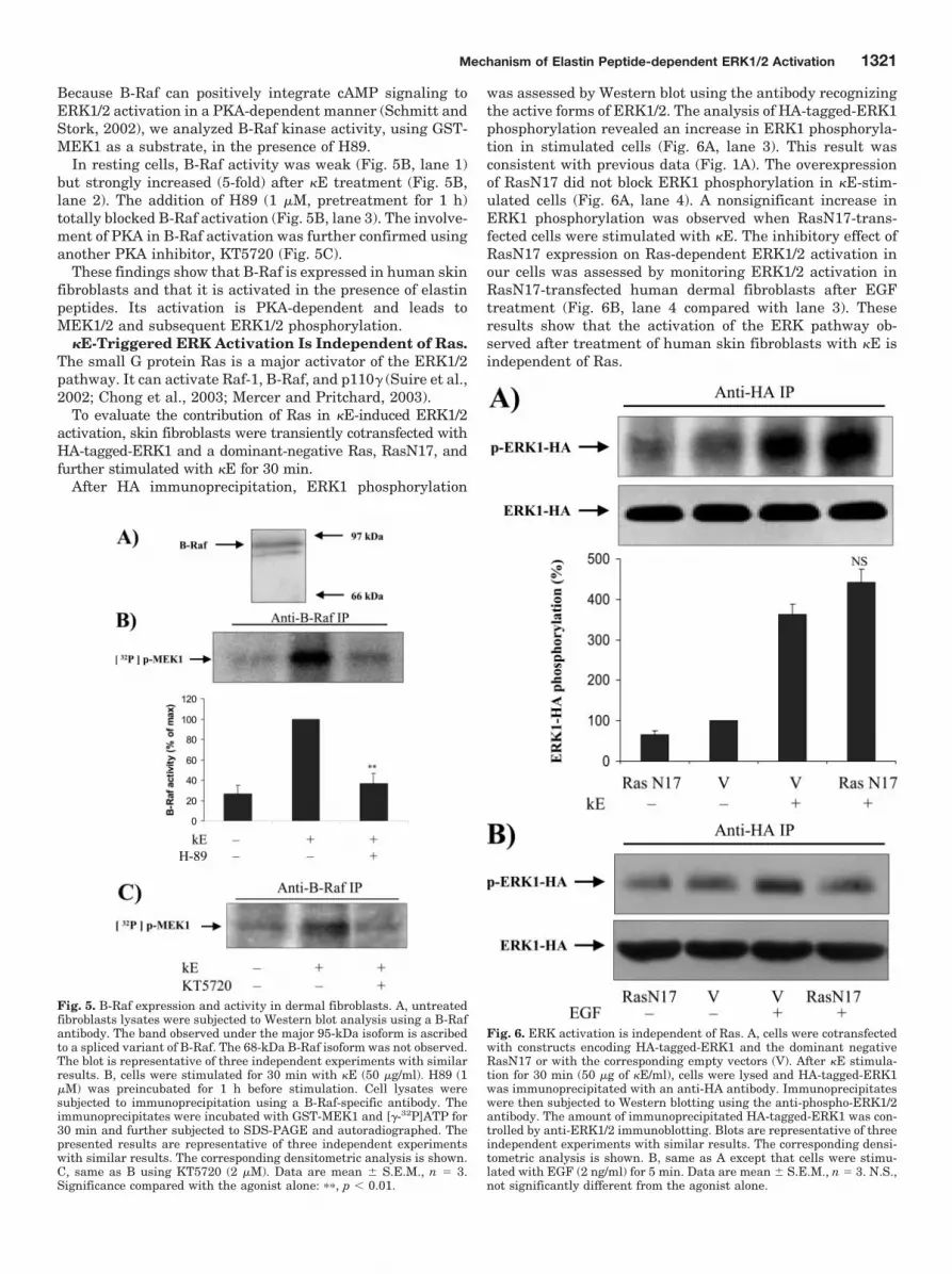
Because B-Raf can positively integrate cAMP signaling toERK1/2 activation in a PKA-dependent manner (Schmitt andStork, 2002), we analyzed B-Raf kinase activity, using GST-MEK1 as a substrate, in the presence of H89.
In resting cells, B-Raf activity was weak (Fig. 5B, lane 1)but strongly increased (5-fold) after �E treatment (Fig. 5B,lane 2). The addition of H89 (1 �M, pretreatment for 1 h)totally blocked B-Raf activation (Fig. 5B, lane 3). The involve-ment of PKA in B-Raf activation was further confirmed usinganother PKA inhibitor, KT5720 (Fig. 5C).
These findings show that B-Raf is expressed in human skinfibroblasts and that it is activated in the presence of elastinpeptides. Its activation is PKA-dependent and leads toMEK1/2 and subsequent ERK1/2 phosphorylation.
�E-Triggered ERK Activation Is Independent of Ras.The small G protein Ras is a major activator of the ERK1/2pathway. It can activate Raf-1, B-Raf, and p110� (Suire et al.,2002; Chong et al., 2003; Mercer and Pritchard, 2003).
To evaluate the contribution of Ras in �E-induced ERK1/2activation, skin fibroblasts were transiently cotransfected withHA-tagged-ERK1 and a dominant-negative Ras, RasN17, andfurther stimulated with �E for 30 min.
After HA immunoprecipitation, ERK1 phosphorylation
was assessed by Western blot using the antibody recognizingthe active forms of ERK1/2. The analysis of HA-tagged-ERK1phosphorylation revealed an increase in ERK1 phosphoryla-tion in stimulated cells (Fig. 6A, lane 3). This result wasconsistent with previous data (Fig. 1A). The overexpressionof RasN17 did not block ERK1 phosphorylation in �E-stim-ulated cells (Fig. 6A, lane 4). A nonsignificant increase inERK1 phosphorylation was observed when RasN17-trans-fected cells were stimulated with �E. The inhibitory effect ofRasN17 expression on Ras-dependent ERK1/2 activation inour cells was assessed by monitoring ERK1/2 activation inRasN17-transfected human dermal fibroblasts after EGFtreatment (Fig. 6B, lane 4 compared with lane 3). Theseresults show that the activation of the ERK pathway ob-served after treatment of human skin fibroblasts with �E isindependent of Ras.
Fig. 5. B-Raf expression and activity in dermal fibroblasts. A, untreatedfibroblasts lysates were subjected to Western blot analysis using a B-Rafantibody. The band observed under the major 95-kDa isoform is ascribedto a spliced variant of B-Raf. The 68-kDa B-Raf isoform was not observed.The blot is representative of three independent experiments with similarresults. B, cells were stimulated for 30 min with �E (50 �g/ml). H89 (1�M) was preincubated for 1 h before stimulation. Cell lysates weresubjected to immunoprecipitation using a B-Raf-specific antibody. Theimmunoprecipitates were incubated with GST-MEK1 and [�-32P]ATP for30 min and further subjected to SDS-PAGE and autoradiographed. Thepresented results are representative of three independent experimentswith similar results. The corresponding densitometric analysis is shown.C, same as B using KT5720 (2 �M). Data are mean � S.E.M., n � 3.Significance compared with the agonist alone: ��, p � 0.01.
Fig. 6. ERK activation is independent of Ras. A, cells were cotransfectedwith constructs encoding HA-tagged-ERK1 and the dominant negativeRasN17 or with the corresponding empty vectors (V). After �E stimula-tion for 30 min (50 �g of �E/ml), cells were lysed and HA-tagged-ERK1was immunoprecipitated with an anti-HA antibody. Immunoprecipitateswere then subjected to Western blotting using the anti-phospho-ERK1/2antibody. The amount of immunoprecipitated HA-tagged-ERK1 was con-trolled by anti-ERK1/2 immunoblotting. Blots are representative of threeindependent experiments with similar results. The corresponding densi-tometric analysis is shown. B, same as A except that cells were stimu-lated with EGF (2 ng/ml) for 5 min. Data are mean � S.E.M., n � 3. N.S.,not significantly different from the agonist alone.
Mechanism of Elastin Peptide-dependent ERK1/2 Activation 1321

DiscussionThe in vivo generation of EPs is thought to influence cancer
progression (Hornebeck et al., 2002). Fibroblasts play a fun-damental role during the stromal reaction (Westermarck andKahari, 1999) and because they respond to EP presence bysecreting procollagenase-1 (Brassart et al., 2001), their con-tribution to metastasis development could be accentuated insuch conditions.
In skin fibroblasts, �E up-regulates collagenase-1 expres-sion through PKA, PI3K, and ERK1/2 pathway activation(Duca et al., 2002). However, the detailed mechanisms ofthese PKA and PI3K signalings were unexplained. We showhere that �E activates ERK1/2 via a Ras-independentmechanism involving p110�/Raf-1/MEK1/2 and PKA/B-Raf/MEK1/2 cascades.
Up to now, the activation of ERK1/2 by EPs has beenreported in smooth muscle cells (SMCs) (Mochizuki et al.,2002), fibroblasts (Duca et al., 2002), and monocytes (Fulopet al., 2001). However, the activation of MEK1/2, the imme-diate upstream activator of ERK1/2, had not been reported.We show here that �E treatment leads to the phosphoryla-tion of MEK1/2 on Ser217/221, thereby generating their ac-tive forms. This activation cascade occurs in PI3K- and PKA-dependent manners (Fig. 1). This observation is consistentwith the previously described induction of ERK1/2 phosphor-ylation by these matrix peptides (Duca et al., 2002). Becausethe simultaneous inhibition of PI3K and PKA totally blockedMEK1/2 and ERK1/2 activation, reproducing the effect oflactose, we concluded that those enzymes were crucial com-ponents in EP-induced MEK1/2 and ERK1/2 activation inskin fibroblasts. In our system, the phosphorylation of Raf-1on Ser338 and its kinase activity are controlled by PI3Kactivity and lead to MEK1/2 activation (Fig. 2).
The possible involvement of Raf-1 in elastin signaling hadbeen suggested as the missing link between Ras and MEK1/2in porcine SMCs (Mochizuki et al., 2002). Indeed, these au-thors used radicicola, which acts as a depleting agent forRaf-1 (Soga et al., 1998). In contrast, our work demonstratesthe activation of Raf-1 after stimulation of cells by EPs.
It is well established that phosphorylation of Raf-1 onSer338 is essential for its activation (Mason et al., 1999). Thecontribution of PI3K to these events has been proposed pre-viously (Chaudhary et al., 2000; Sun et al., 2000). Neverthe-less, this model was challenged (Chiloeches et al., 2001). Theauthors showed that LY294002 inhibited Raf-1 Ser338 phos-phorylation only at concentrations far greater than thoserequired to inhibit PI3K. They thus suggested that Raf-1Ser338 phosphorylation inhibition could not be attributed toPI3K.
In this study, we blocked Ser338 phosphorylation andRaf-1 activity using LY294002 (Fig. 2), at a commonly usedand rather low concentration (25 �M). Our data thus suggestthat in fibroblasts, Ser338 phosphorylation could be PI3K-dependent, which supports the hypothesis that PI3K couldcontribute to Raf-1 Ser338 phosphorylation and activation(Chaudhary et al., 2000; Sun et al., 2000).
We analyzed the effects of �E stimulation on the activitiesof class I PI3K. In dermal fibroblasts, the activity of class IA
isoforms remained unchanged after �E treatment. In con-trast, p110� activity was strongly increased (Fig. 3). This
finding shows, for the first time, that human skin fibroblastsdo possess an efficient p110�.
We tested the effect of PTX on Raf-1/MEK/ERK cascadeand found that PTX blocked Raf-1 Ser338, MEK1/2, andERK1/2 phosphorylation with an efficiency comparable withthat observed using LY294002. This parallel between theresults observed with PTX and those related to PI3K inhibi-tion (Fig. 4A) prompted us to propose that a G protein-inducible PI3K closed the gap between G protein and Raf-1activation. Our data suggest that p110� could be that kinase.This proposal is strongly supported by the fact that PTXtotally blocked p110� activity (Fig. 4B).
A class IA PI3K, p110�, has been shown to be stimulated byG �� subunits (Murga et al., 2000). Nevertheless, this processrequires its preactivation by binding of its p85 regulatorysubunit to phospho-tyrosine motifs (Yart et al., 2002). In thiscase, �� act as p110� activity enhancers, and consequently,the blocking of these subunits partly inhibits the activation ofp110� downstream targets. The very weak PI3K activity weobserved in p85 immunoprecipitates (Fig. 3B) is nonsignifi-cant. So, the participation of p110� to ERK1/2 activation wasexcluded. In addition, Raf-1 Ser338 phosphorylation wasfully inhibited by PTX (Fig. 4A), indicating that the activa-tion of a G protein was sufficient to transduce the signal toRaf-1.
�� heterodimers are important regulators of p110� in vivo(Brock et al., 2003). We observed that transient transfectionof dermal fibroblasts with a construct encoding the�ARK1-CT polypeptide blocked p110� activity (Fig. 4C). Ourresults are consistent with data showing �� requirement forp110� activation (Brock et al., 2003).
Together, our observations strongly suggest that �E-medi-ated ERK1/2 activation by PI3K could be attributed to p110�.
We have shown that �E stimulation of fibroblasts in-creased the intracellular level of cAMP, thereby inducingPKA activation and its participation to ERK1/2 activation(Duca et al., 2002). However, the mechanisms involved werenot described.
PKA can participate to ERK1/2 pathway activation viaB-Raf activation, but the occurrence of this mechanism hadnever been reported in dermal fibroblasts. Our work consti-tutes the first observation of B-Raf expression in dermalfibroblasts. The 95-kDa isoform was detected, whereas its68-kDa counterpart was apparently absent (Fig. 5A). B-Rafkinase activity on MEK was strongly increased by �E andcould be totally blocked using H89 or KT5720 (Fig. 5, B andC). Thus, B-Raf activation seemed to be PKA-dependent. Toour knowledge, this is the first time that a system describingMEK induction via PKA-dependent activation of B-Raf isreported in dermal fibroblasts. B-Raf activation by cAMP viaPKA-dependent mechanism involves the small G proteinRap-1 (Houslay and Kolch, 2000). It would be interesting toexplore its role in our system.
We point out here that the mechanism leading to the cAMPincrease remains unknown and also should be explored. In-deed, PTX-sensitive G protein �� subunits can activate theadenylyl cyclase (Albert and Robillard, 2002). However, sucha mechanism cannot be involved in our system because PTXfailed to totally block EP-induced signaling.
The use of PTX has already permitted to demonstrate theinvolvement of a Gi/G0 in EP signaling (Brassart et al., 2001;Mochizuki et al., 2002). Our work supports this view but
1322 Duca et al.

raises the intriguing possibility that a Gs could also partici-pate. The dual activation of Gi and Gs by the same receptorhas been reported (Herrlich et al., 1996; Zou et al., 1999). Inour system, this point should deserve specific attention be-cause the mechanisms leading to G protein activation by theelastin receptor remain largely unknown, a fact mostly at-tributable to our ignorance of its operational mechanism.
It has been suggested that EP could lead to Ras inductionin porcine SMCs (Mochizuki et al., 2002). Moreover, Ras candirectly activate Raf-1, B-Raf, and p110� (Suire et al., 2002;Mercer and Pritchard, 2003). For these reasons, we evalu-ated Ras contribution to ERK pathway activation. Our re-sults with dermal fibroblasts transfected with the dominantnegative RasN17 mutant strongly suggest that the �E-in-duced ERK activation does not require Ras (Fig. 6A). Theslight increase in HA-tagged-ERK-1 phosphorylation we ob-served when �E-treated fibroblasts were transfected withRasN17 was nonsignificant. A comparable, but more impor-tant effect, was reported for nocodazole-treated RasN17-transfected human embryonic kidney 293T cells (Zang et al.,2001). This intriguing phenomenon is not explained.
The dual activation of Raf isoforms and their complemen-tary contribution to ERK1/2 activation have been describedin other cells (Garcia et al., 2001; Norum et al., 2003), butexamples of its occurrence remain scarce. We provide an-other example of this type of regulation and identify dermalfibroblasts as B-Raf expressing cells. Being the strongestERK1/2 activator within the Raf family (Mercer and Prit-chard, 2003), B-Raf should be considered as a crucial ERK1/2activator in these cells. We emphasize that the concertedaction of Raf-1 and B-Raf could explain the sustained ERK1/2activation observed in �E-treated fibroblasts, as shown forthrombopoietin-stimulated UT7-Mpl cells (Garcia et al.,2001).
We also establish that p110� is expressed in dermal fibro-blasts and strongly activated by �E. EPs are well known fortheir chemotactic activity and the migration they promote.p110� is a critical element in the development of the chemo-tactic response in neutrophils (Hirsch et al., 2000). We thinkthat this enzyme could be a key element of EP-induced mi-gration.
EPs are produced in normal and physiopathological condi-tions, such as wound healing and stromal reaction. Accordingto our data, Raf isoforms and p110� seem to be importantmediators of their effects and could therefore qualify as po-tential therapeutics targets in strategies aiming at limitingelastin contribution to cancer progression. But, because theyare expressed in these cells, these signaling molecules aremost certainly important modulators of fibroblast physiolog-ical functions such as their migration and their proliferation.Thus, the potential for adverse side effects on these stromalcells should be an important consideration when designingsuch therapies.
ReferencesAlbert PR and Robillard L (2002) G protein specificity: traffic direction required. Cell
Signal 14:407–418.Anderson KE and Jackson SP (2003) Class I phosphoinositide 3-kinases. Int J Bio-
chem Cell Biol 35:1028–1033.Brassart B, Fuchs P, Huet E, Alix AJ, Wallach J, Tamburro AM, Delacoux F, Haye
B, Emonard H, Hornebeck W, et al. (2001) Conformational dependence of collage-nase (matrix metalloproteinase-1) up-regulation by elastin peptides in culturedfibroblasts. J Biol Chem 276:5222–5227.
Brock C, Schaefer M, Reusch HP, Czupalla C, Michalke M, Spicher K, Schultz G, and
Nurnberg B (2003) Roles of G beta gamma in membrane recruitment and activationof p110 gamma/p101 phosphoinositide 3-kinase gamma. J Cell Biol 160:89–99.
Chaudhary A, King WG, Mattaliano MD, Frost JA, Diaz B, Morrison DK, Cobb MH,Marshall MS, and Brugge JS (2000) Phosphatidylinositol 3-kinase regulates Raf1through Pak phosphorylation of serine 338. Curr Biol 10:551–554.
Chiloeches A, Mason CS, and Marais R (2001) S338 phosphorylation of Raf-1 isindependent of phosphatidylinositol 3-kinase and Pak3. Mol Cell Biol 21:2423–2434.
Chong H, Vikis HG, and Guan KL (2003) Mechanisms of regulating the Raf kinasefamily. Cell Signal 15:463–469.
Dhillon AS and Kolch W (2002) Untying the regulation of the Raf-1 kinase. ArchBiochem Biophys 404:3–9.
Duca L, Debelle L, Debret R, Antonicelli F, Hornebeck W, and Haye B (2002) Theelastin peptides-mediated induction of pro-collagenase-1 production by humanfibroblasts involves activation of MEK/ERK pathway via PKA- and PI(3)K-dependent signaling. FEBS Lett 524:193–198.
Duca L, Floquet N, Alix AJ, Haye B, and Debelle L (2004) Elastin as a matrikine.Crit Rev Oncol Hematol 49:235–244.
Fulop T Jr, Douziech N, Jacob MP, Hauck M, Wallach J, and Robert L (2001)Age-related alterations in the signal transduction pathways of the elastin-lamininreceptor. Pathol Biol 49:339–348.
Garcia J, de Gunzburg J, Eychene A, Gisselbrecht S, and Porteu F (2001) Throm-bopoietin-mediated sustained activation of extracellular signal-regulated kinasein UT7-Mpl cells requires both Ras-Raf-1- and Rap1-B-Raf-dependent pathways.Mol Cell Biol 21:2659–2670.
Guillemot L, Levy A, Zhao ZJ, Bereziat G, and Rothhut B (2000) The protein-tyrosinephosphatase SHP-2 is required during angiotensin II-mediated activation of cyclinD1 promoter in CHO-AT1A cells. J Biol Chem 275:26349–26358.
Herrlich A, Kuhn B, Grosse R, Schmid A, Schultz G, and Gudermann T (1996)Involvement of Gs and Gi proteins in dual coupling of the luteinizing hormonereceptor to adenylyl cyclase and phospholipase C. J Biol Chem 271:16764 –16772.
Hinek A (1996) Biological roles of the non-integrin elastin/laminin receptor. BiolChem 377:471–480.
Hirsch E, Katanaev VL, Garlanda C, Azzolino O, Pirola L, Silengo L, Sozzani S,Mantovani A, Altruda F, and Wymann MP (2000) Central role for G protein-coupled phosphoinositide 3-kinase gamma in inflammation. Science (Wash DC)287:1049–1053.
Hornebeck W, Emonard H, Monboisse JC, and Bellon G (2002) Matrix-directedregulation of pericellular proteolysis and tumor progression. Semin Cancer Biol12:231–241.
Houslay MD and Kolch W (2000) Cell-type specific integration of cross-talk betweenextracellular signal-regulated kinase and cAMP signaling. Mol Pharmacol 58:659–668.
Huser M, Luckett J, Chiloeches A, Mercer K, Iwobi M, Giblett S, Sun XM, Brown J,Marais R, and Pritchard C (2001) MEK kinase activity is not necessary for Raf-1function. EMBO (Eur Mol Biol Organ) J 20:1940–1951.
King WG, Mattaliano MD, Chan TO, Tsichlis PN, and Brugge JS (1997) Phospha-tidylinositol 3-kinase is required for integrin-stimulated AKT and Raf-1/mitogen-activated protein kinase pathway activation. Mol Cell Biol 17:4406–4418.
Koch WJ, Hawes BE, Allen LF, and Lefkowitz RJ (1994) Direct evidence thatGi-coupled receptor stimulation of mitogen-activated protein kinase is medi-ated by G beta gamma activation of p21ras. Proc Natl Acad Sci USA 91:12706 –12710.
Lee HY, Bae GU, Jung ID, Lee JS, Kim YK, Noh SH, Stracke ML, Park CG, LeeHW, and Han JW (2002) Autotaxin promotes motility via G protein-coupledphosphoinositide 3-kinase gamma in human melanoma cells. FEBS Lett 515:137–140.
Mas VM, Hernandez H, Plo I, Bezombes C, Maestre N, Quillet-Mary A, Filomenko R,Demur C, Jaffrezou JP, and Laurent G (2003) Protein kinase Czeta mediatedRaf-1/extracellular-regulated kinase activation by daunorubicin. Blood 101:1543–1550.
Mason CS, Springer CJ, Cooper RG, Superti-Furga G, Marshall CJ, and Marais R(1999) Serine and tyrosine phosphorylations cooperate in Raf-1, but not B-Rafactivation. EMBO (Eur Mol Biol Organ) J 18:2137–2148.
Mercer KE and Pritchard CA (2003) Raf proteins and cancer: B-Raf is identified asa mutational target. Biochim Biophys Acta 1653:25–40.
Mochizuki S, Brassart B, and Hinek A (2002) Signaling pathways transducedthrough the elastin receptor facilitate proliferation of arterial smooth muscle cells.J Biol Chem 277:44854–44863.
Murga C, Fukuhara S, and Gutkind JS (2000) A novel role for phosphatidylinositol3-kinase � in signaling from G protein-coupled receptors to Akt. J Biol Chem275:12069–12073.
Norum JH, Hart K, and Levy FO (2003) Ras-dependent ERK activation by thehuman Gs-coupled serotonin receptors 5-HT4b and 5-HT7a. J Biol Chem 278:3098–3104.
Schmitt JM and Stork PJ (2002) G� and G� gamma require distinct Src-dependentpathways to activate Rap1 and Ras. J Biol Chem 277:43024–43032.
Seidel MG, Klinger M, Freissmuth M, and Holler C (1999) Activation of mitogen-activated protein kinase by the A2A-adenosine receptor via a rap1-dependent andvia a p21Ras-dependent pathway. J Biol Chem 274:25833–25841.
Soga S, Kozawa T, Narumi H, Akinaga S, Irie K, Matsumoto K, Sharma SV, NakanoH, Mizukami T, and Hara M (1998) Radicicol leads to selective depletion of Rafkinase and disrupts K-Ras-activated aberrant signaling pathway. J Biol Chem273:822–828.
Stoyanov B, Volinia S, Hanck T, Rubio I, Loubtchenkov M, Malek D, Stoyanova S,Vanhaesebroeck B, Dhand R, Nurnberg B, et al. (1995) Cloning and characteriza-tion of a G protein-activated human phosphoinositide-3 kinase. Science (Wash DC)269:690–693.
Mechanism of Elastin Peptide-dependent ERK1/2 Activation 1323

Suire S, Hawkins P, and Stephens L (2002) Activation of phosphoinositide 3-kinasegamma by Ras. Curr Biol 12:1068–1075.
Sun H, King AJ, Diaz HB, and Marshall MS (2000) Regulation of the protein kinaseRaf-1 by oncogenic Ras through phosphatidylinositol 3-kinase, Cdc42/Rac andPak. Curr Biol 10:281–284.
Takeda H, Matozaki T, Takada T, Noguchi T, Yamao T, Tsuda M, Ochi F, FukunagaK, Inagaki K, and Kasuga M (1999) PI 3-kinase gamma and protein kinase C-zetamediate RAS-independent activation of MAP kinase by a Gi protein-coupled re-ceptor. EMBO (Eur Mol Biol Organ) J 18:386–395.
Vossler MR, Yao H, York RD, Pan MG, Rim CS, and Stork PJ (1997) cAMP activatesMAP kinase and Elk-1 through a B-Raf- and Rap1-dependent pathway. Cell89:73–82.
Westermarck J and Kahari VM (1999) Regulation of matrix metalloproteinase ex-pression in tumor invasion. FASEB J 13:781–792.
Yart A, Chap H, and Raynal P (2002) Phosphoinositide 3-kinases in lysophosphatidic
acid signaling: regulation and cross-talk with the Ras/mitogen-activated proteinkinase pathway. Biochim Biophys Acta 1582:107–111.
Zang M, Waelde CA, Xiang X, Rana A, Wen R, and Luo Z (2001) Microtubuleintegrity regulates Pak leading to Ras-independent activation of Raf-1. Insightsinto mechanisms of Raf-1 activation. J Biol Chem 276:25157–25165.
Zou Y, Komuro I, Yamazaki T, Kudoh S, Uozumi H, Kadowaki T, and Yazaki Y(1999) Both Gs and Gi proteins are critically involved in isoproterenol-inducedcardiomyocyte hypertrophy. J Biol Chem 274:9760–9770.
Address correspondence to: Dr. Laurent Debelle, Universite de ReimsChampagne Ardenne, UFR Sciences Exactes et Naturelles, Laboratoire deBiochimie, UMR CNRS 6198, IFR53 Biomolecules, Moulin de la Housse, BP1039, 51687 REIMS Cedex 2, France. E-mail: [email protected]
1324 Duca et al.