egr2 mutations in inherited neuropathies dominant-negatively inhibit myelin gene expression
TRANSCRIPT
Neuron, Vol. 30, 355–368, May, 2001, Copyright 2001 by Cell Press
EGR2 Mutations in Inherited NeuropathiesDominant-Negatively Inhibit Myelin Gene Expression
portion of patients with inherited myelinopathies do nothave mutations in myelin genes, leading to the hypothe-sis that mutations in other genes vital for normal periph-
Rakesh Nagarajan,1 John Svaren,2 Nam Le,1
Toshiyuki Araki,1 Mark Watson,1
and Jeffrey Milbrandt1,3
eral nerve myelination were responsible for causing dis-1 Departments of Pathology and Internal Medicineease in these patients.Washington University School of Medicine
The EGR2 gene became a candidate for mutation660 South Euclid Avenue, Box 8118analysis in these patients because of results obtained inSt. Louis, Missouri 63110studies of mice containing mutations in this transcription2 Department of Comparative Biosciencesfactor. While mice heterozygous for an Egr2 (also knownSchool of Veterinary Medicineas Krox20) null allele are phenotypically normal, Egr2-University of Wisconsindeficent mice have severely defective myelination of2015 Linden Drive, Westperipheral nerves. This defect is characterized byMadison, Wisconsin 53706Schwann cells that become arrested after having formeda one-to-one relationship with axons and that expressvery low levels of MPZ and myelin basic protein (MBP)Summary(Topilko et al., 1994). Indeed, sequence analyses con-ducted by several groups revealed that some patientsThe identification of EGR2 mutations in patients withwith inherited peripheral neuropathies harbor mutationsneuropathies and the phenotype Egr2/Krox202/2 havein the EGR2 gene, predominantly in the DNA bindingdemonstrated that the Egr2 transcription factor is criti-domain (DBD) (Bellone et al., 1999; Botti et al., 1998;cal for peripheral nerve myelination. However, theTimmerman et al., 1999; Warner et al., 1998). Becausemechanism by which these mutations cause diseaseEgr2 heterozygous mice are phenotypically normal, itremains unclear, as most patients present with dis-was surprising that the disease followed a dominantease in the heterozygous state, whereas Egr21/2 micemode of inheritance in families with neuropathy causedare phenotypically normal. To understand the effectby EGR2 DBD mutations. In fact, subsequent gel shiftof aberrant Egr2 activity on Schwann cell gene expres-assays and results from transfected luciferase reportersion, we performed microarray expression profiling toconstructs demonstrated that the EGR2 DBD mutantsidentify genes regulated by Egr2 in Schwann cells.lacked DNA binding and transcriptional activity (WarnerThese include genes encoding myelin proteins andet al., 1999). Taken together, these observations led toenzymes required for synthesis of normal myelin lipids.the hypothesis that the DBD mutations in EGR2 do notUsing these newly identified targets, we have showngenerate simple loss-of-function alleles, but that thesethat neuropathy-associated EGR2 mutants dominant-mutant alleles behave dominantly to disrupt wild-typenegatively inhibit wild-type Egr2-mediated expressionEgr2 function.of essential myelin genes to levels sufficiently low to
In this study, we combine several powerful new tech-result in the abnormal myelination observed in thesenologies to prove that the DBD mutant Egr2 proteinspatients.dominantly inhibit wild-type induction of critical myelingenes. First, using microarray expression analysis onIntroductiona genome wide scale after quantitative Schwann cellinfection with Egr2-expressing adenoviruses, we showAxonal myelination by oligodendrocytes in the centralthat Egr2 is sufficient for induction of genes critical for
nervous system (CNS) and by Schwann cells in the pe-myelin formation and maintenance. These include genes
ripheral nervous system (PNS) is crucial for saltatoryencoding the major myelin proteins, MPZ, PMP22, MBP,
conduction and thus proper signal propagation. Demye- myelin associated glycoprotein (MAG), Cx32, and peri-linating diseases such as the hereditary motor and sen- axin. Furthermore, genes encoding enzymes essentialsory neuropathies (HMSN) in the PNS and multiple scle- for the synthesis of lipids and cholesterol, which arerosis in the CNS cause severe motor and sensory deficits critical components of peripheral myelin, are also in-resulting in significant patient morbidity and mortality. duced by Egr2. Second, by monitoring Schwann cellIn fact, HMSN-1, classically referred to as hypertrophic expression of these newly identified Egr2 target genesCharcot-Marie-Tooth Disease, is one of the most com- from their endogenous chromosomal loci, we demon-mon genetic disorders, affecting one person in 2500 strate that the EGR2 mutants from patients with domi-(Lupski, 1997). Mutations in several known myelin genes, nantly inherited peripheral neuropathies effectively in-myelin protein zero (MPZ), peripheral myelin protein 22 hibit wild-type Egr2-mediated Schwann cell expression(PMP22), connexin 32 (Cx32), and periaxin, have been of essential myelin genes. These results indicate that theidentified in patients with one of several inherited periph- peripheral myelinopathy observed in patients harboringeral neuropathies including Charcot-Marie-Tooth and DBD mutant EGR2 alleles can be explained by the domi-Dejerine-Sottas Syndrome (Boerkoel et al., 2001; Guilbot nant-negative inhibition of Egr2-mediated myelin geneet al., 2001; Lupski et al., 1991; De Jonghe et al., 1997; expression. Finally, we show that introduction of Egr2Raeymaekers et al., 1991). However, a significant pro- into postnatal, Egr2-deficient Schwann cells results in
expression of genes critical for myelination, suggestingthat EGR2 mutant Schwann cells found in patients with3Correspondence: [email protected]
Neuron356
inherited peripheral neuropathies may be capable ofmyelinating axons in response to reintroduction of wild-type Egr2.
Results
Neuropathy-Associated Egr2 DNA Binding DomainMutants Do Not Function as Dominant NegativeInhibitors on Synthetic Promoter-Reporter ConstructsEGR2 mutations have been found in patients with inher-ited peripheral myelinopathy, such as congenital hypo-myelinating neuropathy (CHN) or Charcot-Marie-Tooth(CMT). Most of these mutations are located within theDNA binding domain (DBD) and exhibit greatly de-creased (or absent) DNA binding and transcriptional ac-tivity (Warner et al., 1998; Warner et al., 1999). Fromthese results, they appear to be simple loss-of-functionalleles that would be expected to cause disease in arecessively inherited manner. Consistent with this hy-pothesis, mice which are heterozygous for a null Egr2allele are phenotypically normal (Murphy et al., 1996).However, all of the EGR2 DBD mutant alleles causedisease in the heterozygous state (i.e., they all displaya dominant mode of inheritance). Thus, it has been hy-pothesized that the DBD mutations in EGR2 are actingas dominant-negative alleles (Warner et al., 1999).
Mutations in the DBD of the Wilms’ tumor gene prod-uct (WT-1), associated with Denys-Drash syndrome, orin p53, associated with Li-Fraumeni syndrome, havebeen characterized. Using transfection experiments
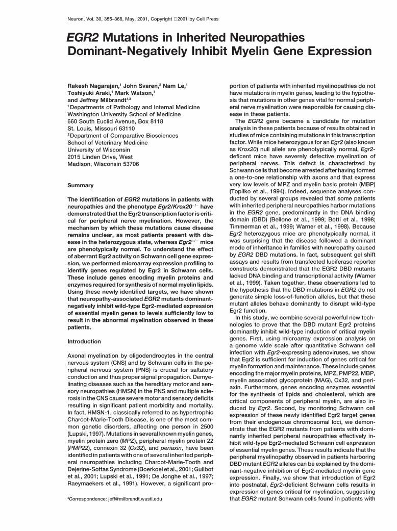
Figure 1. Synthetic Promoter-Reporter Constructs Do Not Reveal awith reporter gene constructs containing synthetic pro-Dominant-Negative Function for Egr2 DNA Binding Domain Mutants
moters composed of either WT-1 or p53 binding sites,(A) CV-1 cells were transfected with a luciferase reporter construct
it was shown that these mutant transcription factors act containing four Egr1 binding sites upstream of the prolactin minimalas dominant-negative inhibitors, as they inhibit tran- promoter and the indicated amounts of either wild-type or mutantscriptional activation by their wild-type counterpart in a (S382R,D383Y) Egr2.
(B) Rat Schwann cells were transfected with the same constructsdose-dependent fashion (Aurelio et al., 2000; Reddy etas in (A).al., 1995; Saifudeen et al., 2000). Using an analogous(C) Rat Schwann cells were transfected with a luciferase reporterapproach, we tested the Egr2(S382R, D383Y) mutant,construct containing 1.3 kb of the MPZ promoter and the indicated
which causes a severe peripheral neuropathy in the het- amounts of either wild-type or mutant (S382R,D383Y) Egr2. Theerozygous state, for dominant-negative activity. We co- scale indicates the fold activation of the reporter by Egr2.transfected expression constructs of wild-type Egr2 andthe Egr2(S382R, D383Y) mutant in varying amounts,along with a reporter plasmid containing four Egr2 bind- Identification of Egr2 Target Genesing sites upstream of the luciferase gene, into CV-1 cells. by Microarray AnalysisWe found no evidence of dominant-negative activity The dominant inheritance pattern of the neuropathy ob-using the Egr2 DBD mutant, even at ratios as high as served in families harboring EGR2 DBD mutant was in-4:1 (Figure 1A). We reasoned that the apparent lack of consistent with our inability to demonstrate dominant-dominant-negative activity might be due to an inappro- negative activity in the transfection assays. One poten-priate cell context. Thus, we conducted similar transfec- tial explanation for this discrepancy was the inability oftion experiments using wild-type Egr2 and Egr2(S382R, the promoter-reporter constructs to adequately recapit-D383Y) with the Egr2 binding site reporter plasmid ulate Egr2-mediated regulation of relevant target pro-in rat Schwann cells. Again, we were unable to ob- moters in their native chromosomal loci. Therefore, toserve any significant dominant-negative activity of elucidate the mechanism by which a single copy of theseEgr2(S382R, D383Y) (Figure 1B). To determine whether mutant EGR2 alleles results in myelinopathy, it becamethe failure to observe dominant-negative activity was apparent that the identification of genes regulated bydue to an inappropriate promoter context, we cotrans- Egr2 in Schwann cells was necessary. This would allowfected wild-type Egr2 and Egr2(S382R, D383Y) along us to test the effects of the DBD mutant Egr2 proteinswith a reporter plasmid containing 1.3 kb of the MPZ on the transcriptional regulation of specific Egr2 targetpromoter, which can be activated by Egr2 (Zorick et al., genes from their endogenous chromosomal loci.1999), into rat Schwann cells. Once again, even at ratios To identify target genes, we employed a gain-of-func-as high as 4:1, no notable dominant-negative activity tion model in which Egr2 is expressed in the absence of
stimuli that might also induce other transcription factorswas observed (Figure 1C).
EGR2-Mediated Gene Regulation in Schwann Cells357
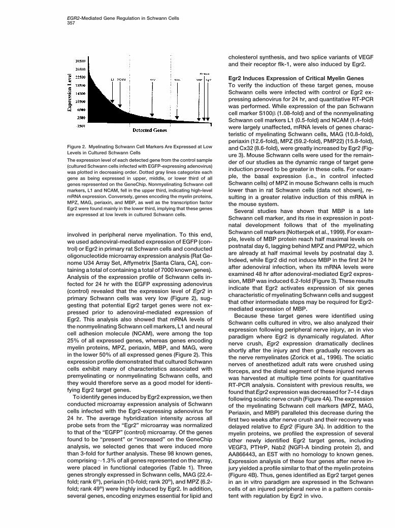
cholesterol synthesis, and two splice variants of VEGFand their receptor flk-1, were also induced by Egr2.
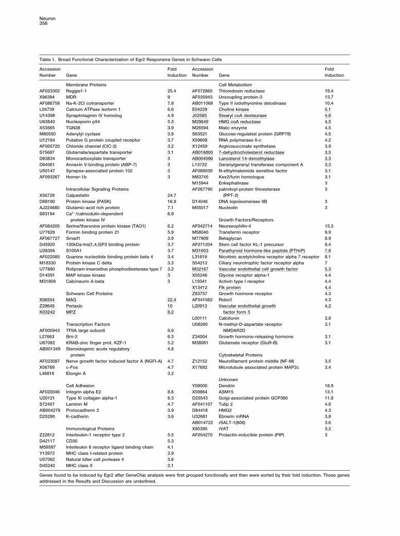
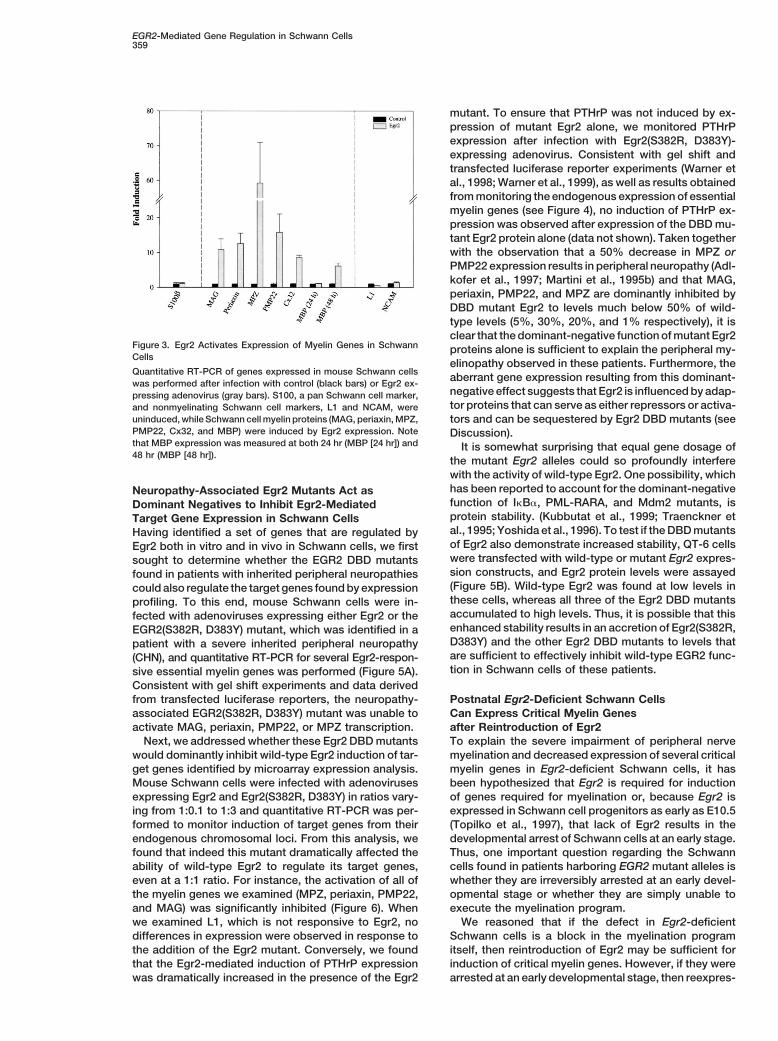
Egr2 Induces Expression of Critical Myelin GenesTo verify the induction of these target genes, mouseSchwann cells were infected with control or Egr2 ex-pressing adenovirus for 24 hr, and quantitative RT-PCRwas performed. While expression of the pan Schwanncell marker S100b (1.08-fold) and of the nonmyelinatingSchwann cell markers L1 (0.5-fold) and NCAM (1.4-fold)were largely unaffected, mRNA levels of genes charac-teristic of myelinating Schwann cells, MAG (10.8-fold),periaxin (12.6-fold), MPZ (59.2-fold), PMP22) (15.8-fold),
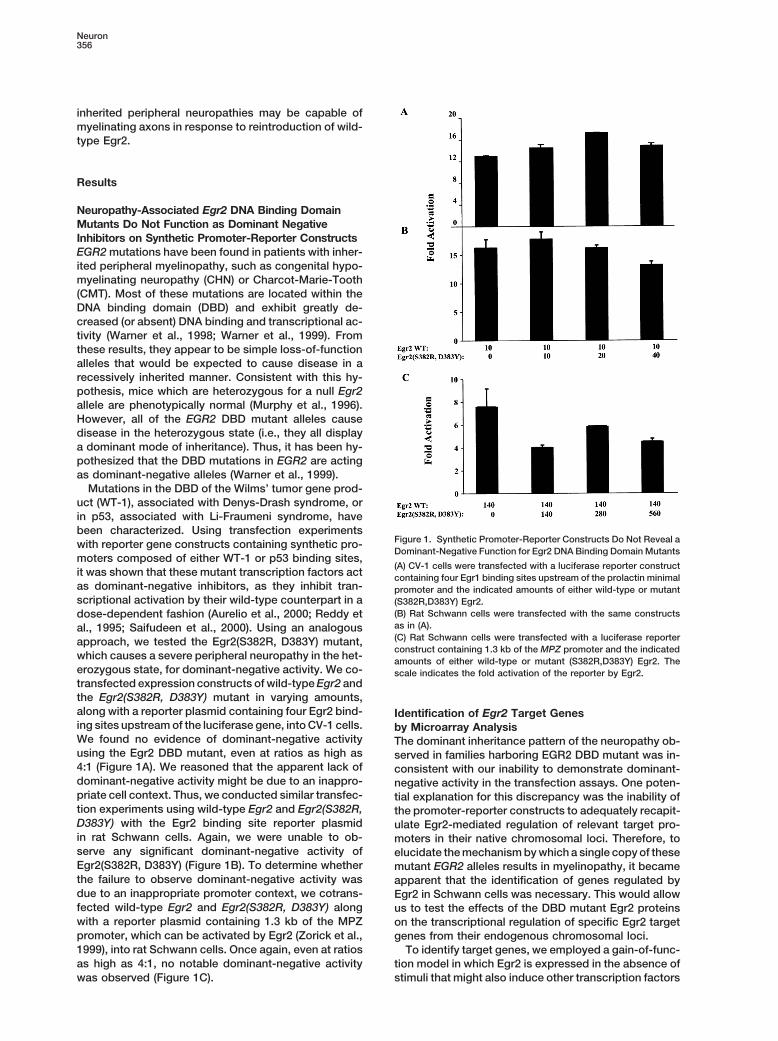
Figure 2. Myelinating Schwann Cell Markers Are Expressed at Low and Cx32 (8.6-fold), were greatly increased by Egr2 (Fig-Levels in Cultured Schwann Cells ure 3). Mouse Schwann cells were used for the remain-The expression level of each detected gene from the control sample der of our studies as the dynamic range of target gene(cultured Schwann cells infected with EGFP-expressing adenovirus) induction proved to be greater in these cells. For exam-was plotted in decreasing order. Dotted gray lines categorize each
ple, the basal expression (i.e., in control infectedgene as being expressed in upper, middle, or lower third of allSchwann cells) of MPZ in mouse Schwann cells is muchgenes represented on the GeneChip. Nonmyelinating Schwann cell
markers, L1 and NCAM, fell in the upper third, indicating high-level lower than in rat Schwann cells (data not shown), re-mRNA expression. Conversely, genes encoding the myelin proteins, sulting in a greater relative induction of this mRNA inMPZ, MAG, periaxin, and MBP, as well as the transcription factor the mouse system.Egr2 were found mainly in the lower third, implying that these genes Several studies have shown that MBP is a lateare expressed at low levels in cultured Schwann cells.
Schwann cell marker, and its rise in expression in post-natal development follows that of the myelinatingSchwann cell markers (Notterpek et al., 1999). For exam-involved in peripheral nerve myelination. To this end,ple, levels of MBP protein reach half maximal levels onwe used adenoviral-mediated expression of EGFP (con-postnatal day 6, lagging behind MPZ and PMP22, whichtrol) or Egr2 in primary rat Schwann cells and conductedare already at half maximal levels by postnatal day 3.oligonucleotide microarray expression analysis (Rat Ge-Indeed, while Egr2 did not induce MBP in the first 24 hrnome U34 Array Set, Affymetrix [Santa Clara, CA], con-after adenoviral infection, when its mRNA levels weretaining a total of containing a total of 7000 known genes).examined 48 hr after adenoviral-mediated Egr2 expres-Analysis of the expression profile of Schwann cells in-sion, MBP was induced 6.2-fold (Figure 3). These resultsfected for 24 hr with the EGFP expressing adenovirusindicate that Egr2 activates expression of six genes(control) revealed that the expression level of Egr2 incharacteristic of myelinating Schwann cells and suggestprimary Schwann cells was very low (Figure 2), sug-that other intermediate steps may be required for Egr2-gesting that potential Egr2 target genes were not ex-mediated expression of MBP.
pressed prior to adenoviral-mediated expression ofBecause these target genes were identified using
Egr2. This analysis also showed that mRNA levels ofSchwann cells cultured in vitro, we also analyzed their
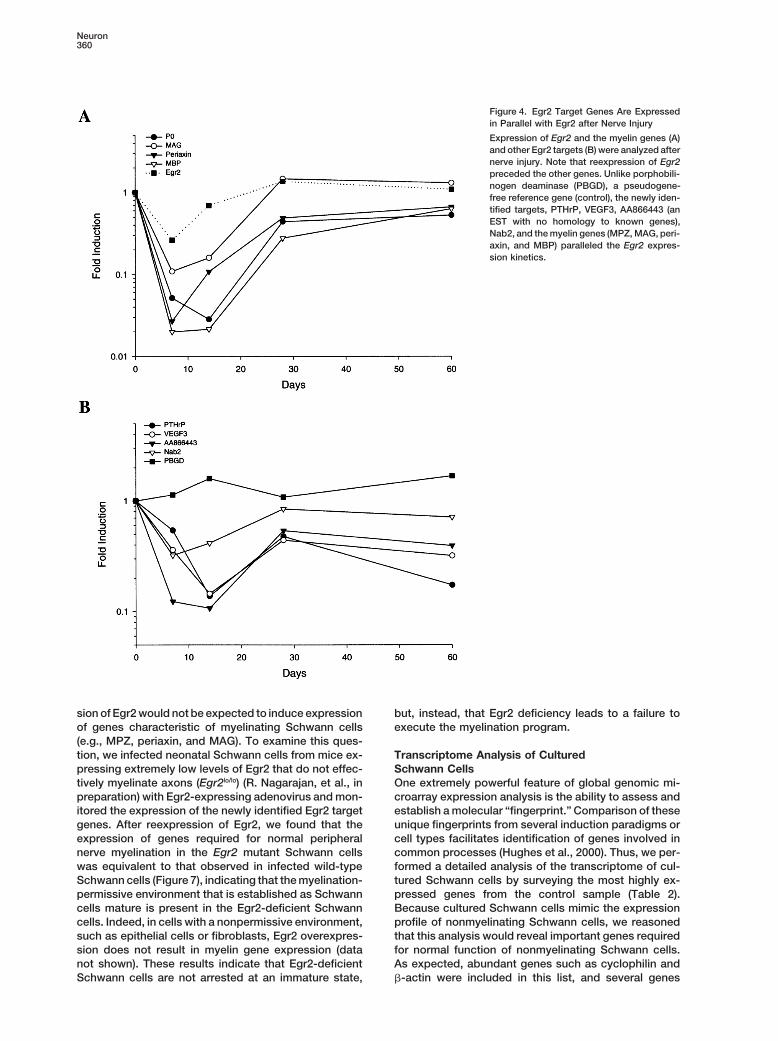
the nonmyelinating Schwann cell markers, L1 and neural expression following peripheral nerve injury, an in vivocell adhesion molecule (NCAM), were among the top paradigm where Egr2 is dynamically regulated. After25% of all expressed genes, whereas genes encoding nerve crush, Egr2 expression dramatically declinesmyelin proteins, MPZ, periaxin, MBP, and MAG, were shortly after the injury and then gradually recovers asin the lower 50% of all expressed genes (Figure 2). This the nerve remyelinates (Zorick et al., 1996). The sciaticexpression profile demonstrated that cultured Schwann nerves of anesthetized adult rats were crushed usingcells exhibit many of characteristics associated with forceps, and the distal segment of these injured nervespremyelinating or nonmyelinating Schwann cells, and was harvested at multiple time points for quantitativethey would therefore serve as a good model for identi- RT-PCR analysis. Consistent with previous results, wefying Egr2 target genes. found that Egr2 expression was decreased for 7–14 days
To identify genes induced by Egr2 expression, we then following sciatic nerve crush (Figure 4A). The expressionconducted microarray expression analysis of Schwann of the myelinating Schwann cell markers (MPZ, MAG,cells infected with the Egr2-expressing adenovirus for Periaxin, and MBP) paralleled this decrease during the24 hr. The average hybridization intensity across all first two weeks after nerve crush and their recovery wasprobe sets from the “Egr2” microarray was normalized delayed relative to Egr2 (Figure 3A). In addition to theto that of the “EGFP” (control) microarray. Of the genes myelin proteins, we profiled the expression of severalfound to be “present” or “increased” on the GeneChip other newly identified Egr2 target genes, includinganalysis, we selected genes that were induced more VEGF3, PTHrP, Nab2 (NGFI-A binding protein 2), andthan 3-fold for further analysis. These 98 known genes, AA866443, an EST with no homology to known genes.comprising z1.3% of all genes represented on the array, Expression analysis of these four genes after nerve in-were placed in functional categories (Table 1). Three jury yielded a profile similar to that of the myelin proteinsgenes strongly expressed in Schwann cells, MAG (22.4- (Figure 4B). Thus, genes identified as Egr2 target genesfold; rank 6th), periaxin (10-fold; rank 20th), and MPZ (6.2- in an in vitro paradigm are expressed in the Schwannfold; rank 49th) were highly induced by Egr2. In addition, cells of an injured peripheral nerve in a pattern consis-
tent with regulation by Egr2 in vivo.several genes, encoding enzymes essential for lipid and
Neuron358
Table 1. Broad Functional Characterization of Egr2 Responsive Genes in Schwann Cells
Accession Fold Accession FoldNumber Gene Induction Number Gene Induction
Membrane Proteins Cell MetabolismAF023302 Reggie1-1 25.4 AF072865 Thioredoxin reductase 19.4X96394 MDR 9 AF035943 Uncoupling protein-3 13.7AF086758 Na-K-2Cl cotransporter 7.8 AB011068 Type II iodothyronine deiodinase 10.4L04739 Calcium ATPase isoform 1 6.6 E04239 Choline kinase 5.1U14398 Synaptotagmin IV homolog 4.9 J02585 Stearyl coA destaurase 4.6U63840 Nucleoporin p54 5.3 M29649 HMG coA reductase 4.5X53565 TGN38 3.9 M26594 Malic enzyme 4.5M80550 Adenylyl cyclase 3.9 S63521 Glucose-regulated protein (GRP78) 4.5U12184 Putative G protein coupled receptor 3.7 X59608 RNA polymerase II-c 4.2AF005720 Chloride channel (CIC-2) 3.2 X12459 Arginosuccinate synthetase 3.9S75687 Glutamate/aspartate transporter 3.1 AB016800 7-dehydrocholesterol reductase 3.5D63834 Monocarboxylate transporter 3 AB004096 Lanosterol 14-demethylase 3.3D64061 Annexin V-binding protein (ABP-7) 3 L13722 Geranylgeranyl transferase component A 3.3U50147 Synapse-associated protein 102 3 AF089039 N-ethylmaleimide sensitive factor 3.1AF093267 Homer-1b 3 M83745 Kex2/furin homologue 3.1
M15944 Enkephalinase 3Intracellular Signaling Proteins AF067790 palmitoyl-protein thioesterase 3
X56729 Calpastatin 24.7 (PPT-2)D88190 Protein kinase (PASK) 16.9 D14046 DNA topoisomerase IIB 3AJ224680 Glutamic-acid rich protein 7.1 M55017 Nucleolin 3S83194 Ca21/calmodulin-dependent 6.9
protein kinase IV Growth Factors/ReceptorsAF084205 Serine/theronine protein kinase (TAO1) 6.2 AF042714 Neurexophilin-4 15.3U77626 Formin binding protein 21 5.9 M58040 Transferrin receptor 9.9AF067727 Smad1 3.9 M77809 Betaglycan 8.9D45920 130kDa-Ins(1,4,5)P3 binding protein 3.7 AF071204 Stem cell factor KL-1 precursor 8.4U26356 S100A1 3.7 M31603 Parathyroid hormone-like peptide (PTHrP) 7.8AF022085 Guanine nucleotide binding protein beta 4 3.4 L31619 Nicotinic acetylcholine receptor alpha 7 receptor 8.1M18330 Protein kinase C delta 3.3 S54212 Ciliary neurotrophic factor receptor alpha 7U77880 Rolipram-insensitive phosphodiesterase type 7 3.2 M32167 Vascular endothelial cell growth factor 5.3D14591 MAP kinase kinase 3 X55246 Glycine receptor alpha-1 4.4M31809 Calcineurin A-beta 3 L19341 Activin type I receptor 4.4
X13412 Flk protein 4.4Schwann Cell Proteins Z83757 Growth hormone receptor 4.3
X06554 MAG 22.4 AF041082 Robo1 4.3Z29649 Periaxin 10 L20913 Vascular endothelial growth 4.2K03242 MPZ 6.2 factor form 3
L00111 Calcitonin 3.8Transcription Factors U08260 N-methyl-D-aspartate receptor 3.1
AF000943 TFIIA large subunit 9.9 NMDAR2DL27663 Brn-2 6.3 Z34004 Growth hormone-releasing hormone 3.1U67082 KRAB-zinc finger prot. KZF-1 5.2 M38061 Glutamate receptor (GluR-B) 3.1AB001349 Steroidogenic acute regulatory 4.8
protein Cytoskeletal ProteinsAF023087 Nerve growth factor induced factor A (NGFI-A) 4.7 Z12152 Neurofilament protein middle (NF-M) 3.5X06769 c-Fos 4.7 X17682 Microtubule associated protein MAP2c 3.4L46816 Elongin A 3.2
UnknownCell Adhesion Y09000 Dendrin 18.9
AF020046 Integrin alpha E2 8.6 X59864 ASM15 13.1U20121 Type XI collagen alpha-1 8.3 D25543 Golgi-associated protein GCP360 11.8S72407 Laminin M 4.7 AF041107 Tulip 2 4.8AB004278 Protocadherin 2 3.9 D84418 HMG2 4.3D25290 K-cadherin 3.6 U32681 Ebnerin mRNA 3.8
AB014722 rSALT-1(806) 3.6Immunological Proteins X80395 rVAT 3.2
Z22812 Interleukin-1 receptor type 2 5.5 AF054270 Prolactin-inducible protein (PIP) 3D42117 CD30 5.3M58587 Interleukin 6 receptor ligand binding chain 4.1Y13972 MHC class I-related protein 3.9U57062 Natural killer cell protease 4 3.6D45240 MHC class II 3.1
Genes found to be induced by Egr2 after GeneChip analysis were first grouped functionally and then were sorted by their fold induction. Those genesaddressed in the Results and Discussion are underlined.
EGR2-Mediated Gene Regulation in Schwann Cells359
mutant. To ensure that PTHrP was not induced by ex-pression of mutant Egr2 alone, we monitored PTHrPexpression after infection with Egr2(S382R, D383Y)-expressing adenovirus. Consistent with gel shift andtransfected luciferase reporter experiments (Warner etal., 1998; Warner et al., 1999), as well as results obtainedfrom monitoring the endogenous expression of essentialmyelin genes (see Figure 4), no induction of PTHrP ex-pression was observed after expression of the DBD mu-tant Egr2 protein alone (data not shown). Taken togetherwith the observation that a 50% decrease in MPZ orPMP22 expression results in peripheral neuropathy (Adl-kofer et al., 1997; Martini et al., 1995b) and that MAG,periaxin, PMP22, and MPZ are dominantly inhibited byDBD mutant Egr2 to levels much below 50% of wild-type levels (5%, 30%, 20%, and 1% respectively), it isclear that the dominant-negative function of mutant Egr2
Figure 3. Egr2 Activates Expression of Myelin Genes in Schwann proteins alone is sufficient to explain the peripheral my-Cells
elinopathy observed in these patients. Furthermore, theQuantitative RT-PCR of genes expressed in mouse Schwann cells aberrant gene expression resulting from this dominant-was performed after infection with control (black bars) or Egr2 ex-
negative effect suggests that Egr2 is influenced by adap-pressing adenovirus (gray bars). S100, a pan Schwann cell marker,tor proteins that can serve as either repressors or activa-and nonmyelinating Schwann cell markers, L1 and NCAM, were
uninduced, while Schwann cell myelin proteins (MAG, periaxin, MPZ, tors and can be sequestered by Egr2 DBD mutants (seePMP22, Cx32, and MBP) were induced by Egr2 expression. Note Discussion).that MBP expression was measured at both 24 hr (MBP [24 hr]) and It is somewhat surprising that equal gene dosage of48 hr (MBP [48 hr]). the mutant Egr2 alleles could so profoundly interfere
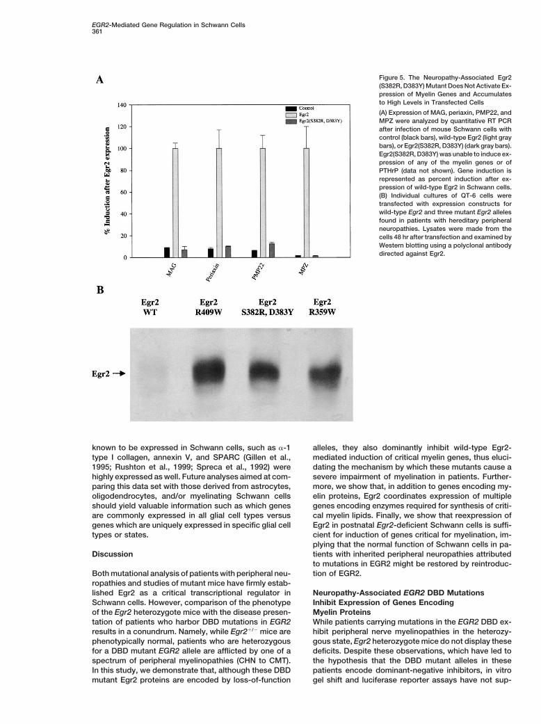
with the activity of wild-type Egr2. One possibility, whichhas been reported to account for the dominant-negativeNeuropathy-Associated Egr2 Mutants Act asfunction of IkBa, PML-RARA, and Mdm2 mutants, isDominant Negatives to Inhibit Egr2-Mediatedprotein stability. (Kubbutat et al., 1999; Traenckner etTarget Gene Expression in Schwann Cellsal., 1995; Yoshida et al., 1996). To test if the DBD mutantsHaving identified a set of genes that are regulated byof Egr2 also demonstrate increased stability, QT-6 cellsEgr2 both in vitro and in vivo in Schwann cells, we firstwere transfected with wild-type or mutant Egr2 expres-sought to determine whether the EGR2 DBD mutantssion constructs, and Egr2 protein levels were assayedfound in patients with inherited peripheral neuropathies(Figure 5B). Wild-type Egr2 was found at low levels incould also regulate the target genes found by expressionthese cells, whereas all three of the Egr2 DBD mutantsprofiling. To this end, mouse Schwann cells were in-accumulated to high levels. Thus, it is possible that thisfected with adenoviruses expressing either Egr2 or theenhanced stability results in an accretion of Egr2(S382R,EGR2(S382R, D383Y) mutant, which was identified in aD383Y) and the other Egr2 DBD mutants to levels thatpatient with a severe inherited peripheral neuropathyare sufficient to effectively inhibit wild-type EGR2 func-(CHN), and quantitative RT-PCR for several Egr2-respon-tion in Schwann cells of these patients.sive essential myelin genes was performed (Figure 5A).
Consistent with gel shift experiments and data derivedfrom transfected luciferase reporters, the neuropathy- Postnatal Egr2-Deficient Schwann Cells
Can Express Critical Myelin Genesassociated EGR2(S382R, D383Y) mutant was unable toactivate MAG, periaxin, PMP22, or MPZ transcription. after Reintroduction of Egr2
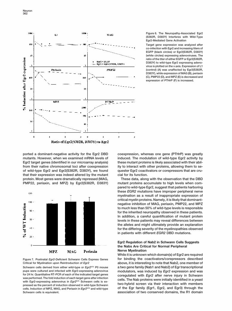
To explain the severe impairment of peripheral nerveNext, we addressed whether these Egr2 DBD mutantswould dominantly inhibit wild-type Egr2 induction of tar- myelination and decreased expression of several critical
myelin genes in Egr2-deficient Schwann cells, it hasget genes identified by microarray expression analysis.Mouse Schwann cells were infected with adenoviruses been hypothesized that Egr2 is required for induction
of genes required for myelination or, because Egr2 isexpressing Egr2 and Egr2(S382R, D383Y) in ratios vary-ing from 1:0.1 to 1:3 and quantitative RT-PCR was per- expressed in Schwann cell progenitors as early as E10.5
(Topilko et al., 1997), that lack of Egr2 results in theformed to monitor induction of target genes from theirendogenous chromosomal loci. From this analysis, we developmental arrest of Schwann cells at an early stage.
Thus, one important question regarding the Schwannfound that indeed this mutant dramatically affected theability of wild-type Egr2 to regulate its target genes, cells found in patients harboring EGR2 mutant alleles is
whether they are irreversibly arrested at an early devel-even at a 1:1 ratio. For instance, the activation of all ofthe myelin genes we examined (MPZ, periaxin, PMP22, opmental stage or whether they are simply unable to
execute the myelination program.and MAG) was significantly inhibited (Figure 6). Whenwe examined L1, which is not responsive to Egr2, no We reasoned that if the defect in Egr2-deficient
Schwann cells is a block in the myelination programdifferences in expression were observed in response tothe addition of the Egr2 mutant. Conversely, we found itself, then reintroduction of Egr2 may be sufficient for
induction of critical myelin genes. However, if they werethat the Egr2-mediated induction of PTHrP expressionwas dramatically increased in the presence of the Egr2 arrested at an early developmental stage, then reexpres-
Neuron360
Figure 4. Egr2 Target Genes Are Expressedin Parallel with Egr2 after Nerve Injury
Expression of Egr2 and the myelin genes (A)and other Egr2 targets (B) were analyzed afternerve injury. Note that reexpression of Egr2preceded the other genes. Unlike porphobili-nogen deaminase (PBGD), a pseudogene-free reference gene (control), the newly iden-tified targets, PTHrP, VEGF3, AA866443 (anEST with no homology to known genes),Nab2, and the myelin genes (MPZ, MAG, peri-axin, and MBP) paralleled the Egr2 expres-sion kinetics.
sion of Egr2 would not be expected to induce expression but, instead, that Egr2 deficiency leads to a failure toexecute the myelination program.of genes characteristic of myelinating Schwann cells
(e.g., MPZ, periaxin, and MAG). To examine this ques-tion, we infected neonatal Schwann cells from mice ex- Transcriptome Analysis of Cultured
Schwann Cellspressing extremely low levels of Egr2 that do not effec-tively myelinate axons (Egr2lo/lo) (R. Nagarajan, et al., in One extremely powerful feature of global genomic mi-
croarray expression analysis is the ability to assess andpreparation) with Egr2-expressing adenovirus and mon-itored the expression of the newly identified Egr2 target establish a molecular “fingerprint.” Comparison of these
unique fingerprints from several induction paradigms orgenes. After reexpression of Egr2, we found that theexpression of genes required for normal peripheral cell types facilitates identification of genes involved in
common processes (Hughes et al., 2000). Thus, we per-nerve myelination in the Egr2 mutant Schwann cellswas equivalent to that observed in infected wild-type formed a detailed analysis of the transcriptome of cul-
tured Schwann cells by surveying the most highly ex-Schwann cells (Figure 7), indicating that the myelination-permissive environment that is established as Schwann pressed genes from the control sample (Table 2).
Because cultured Schwann cells mimic the expressioncells mature is present in the Egr2-deficient Schwanncells. Indeed, in cells with a nonpermissive environment, profile of nonmyelinating Schwann cells, we reasoned
that this analysis would reveal important genes requiredsuch as epithelial cells or fibroblasts, Egr2 overexpres-sion does not result in myelin gene expression (data for normal function of nonmyelinating Schwann cells.
As expected, abundant genes such as cyclophilin andnot shown). These results indicate that Egr2-deficientSchwann cells are not arrested at an immature state, b-actin were included in this list, and several genes
EGR2-Mediated Gene Regulation in Schwann Cells361
Figure 5. The Neuropathy-Associated Egr2(S382R, D383Y) Mutant Does Not Activate Ex-pression of Myelin Genes and Accumulatesto High Levels in Transfected Cells
(A) Expression of MAG, periaxin, PMP22, andMPZ were analyzed by quantitative RT PCRafter infection of mouse Schwann cells withcontrol (black bars), wild-type Egr2 (light graybars), or Egr2(S382R, D383Y) (dark gray bars).Egr2(S382R, D383Y) was unable to induce ex-pression of any of the myelin genes or ofPTHrP (data not shown). Gene induction isrepresented as percent induction after ex-pression of wild-type Egr2 in Schwann cells.(B) Individual cultures of QT-6 cells weretransfected with expression constructs forwild-type Egr2 and three mutant Egr2 allelesfound in patients with hereditary peripheralneuropathies. Lysates were made from thecells 48 hr after transfection and examined byWestern blotting using a polyclonal antibodydirected against Egr2.
known to be expressed in Schwann cells, such as a-1 alleles, they also dominantly inhibit wild-type Egr2-mediated induction of critical myelin genes, thus eluci-type I collagen, annexin V, and SPARC (Gillen et al.,
1995; Rushton et al., 1999; Spreca et al., 1992) were dating the mechanism by which these mutants cause asevere impairment of myelination in patients. Further-highly expressed as well. Future analyses aimed at com-
paring this data set with those derived from astrocytes, more, we show that, in addition to genes encoding my-elin proteins, Egr2 coordinates expression of multipleoligodendrocytes, and/or myelinating Schwann cells
should yield valuable information such as which genes genes encoding enzymes required for synthesis of criti-cal myelin lipids. Finally, we show that reexpression ofare commonly expressed in all glial cell types versus
genes which are uniquely expressed in specific glial cell Egr2 in postnatal Egr2-deficient Schwann cells is suffi-cient for induction of genes critical for myelination, im-types or states.plying that the normal function of Schwann cells in pa-tients with inherited peripheral neuropathies attributedDiscussionto mutations in EGR2 might be restored by reintroduc-tion of EGR2.Both mutational analysis of patients with peripheral neu-
ropathies and studies of mutant mice have firmly estab-lished Egr2 as a critical transcriptional regulator in Neuropathy-Associated EGR2 DBD Mutations
Inhibit Expression of Genes EncodingSchwann cells. However, comparison of the phenotypeof the Egr2 heterozygote mice with the disease presen- Myelin Proteins
While patients carrying mutations in the EGR2 DBD ex-tation of patients who harbor DBD mutations in EGR2results in a conundrum. Namely, while Egr21/2 mice are hibit peripheral nerve myelinopathies in the heterozy-
gous state, Egr2 heterozygote mice do not display thesephenotypically normal, patients who are heterozygousfor a DBD mutant EGR2 allele are afflicted by one of a deficits. Despite these observations, which have led to
the hypothesis that the DBD mutant alleles in thesespectrum of peripheral myelinopathies (CHN to CMT).In this study, we demonstrate that, although these DBD patients encode dominant-negative inhibitors, in vitro
gel shift and luciferase reporter assays have not sup-mutant Egr2 proteins are encoded by loss-of-function
Neuron362
Figure 6. The Neuropathy-Associated Egr2(S382R, D383Y) Interferes with Wild-TypeEgr2-Mediated Gene Activation
Target gene expression was analyzed afterco-infection with Egr2 and increasing titers ofEGFP (black circles) or Egr2(S382R, D383Y)(white circles) expressing adenoviruses. Theratio of the titer of either EGFP or Egr2(S382R,D383Y) to wild-type Egr2 expressing adeno-virus is plotted on the x axis. Expression of L1(control) (A) was unaffected by Egr2(S382R,D383Y), while expression of MAG (B), periaxin(C), PMP22 (D), and MPZ (E) is decreased andexpression of PTHrP (F) is increased.
ported a dominant-negative activity for the Egr2 DBD coexpression, whereas one gene (PTHrP) was greatlyinduced. The modulation of wild-type Egr2 activity bymutants. However, when we examined mRNA levels of
Egr2 target genes (identified in our microarray analysis) these mutant proteins is likely associated with their abil-ity to interact with other proteins, allowing them to se-from their native chromosomal loci after coexpression
of wild-type Egr2 and Egr2(S382R, D383Y), we found quester Egr2 coactivators or corepressors that are cru-cial for its function.that their expression was indeed altered by the mutant
protein. Most genes were dramatically repressed (MAG, These data, along with the observation that the DBDmutant proteins accumulate to high levels when com-PMP22, periaxin, and MPZ) by Egr2(S382R, D383Y)pared to wild-type Egr2, suggest that patients harboringthese EGR2 mutations have improper peripheral nervemyelination as a result of inappropriate expression ofcritical myelin proteins. Namely, it is likely that dominant-negative inhibition of MAG, periaxin, PMP22, and MPZto much less than 50% of wild-type levels is responsiblefor the inherited neuropathy observed in these patients.In addition, a careful quantification of mutant proteinlevels in these patients may reveal differences betweenthe alleles and might ultimately provide an explanationfor the differing severity of the myelinopathies observedin patients with different EGR2 DBD mutations.
Egr2 Regulation of Nab2 in Schwann Cells Suggeststhe Nabs Are Critical for Normal PeripheralNerve MyelinationWhile it is unknown which domain(s) of Egr2 are requiredfor binding the coactivators/corepressors describedFigure 7. Postnatal Egr2-Deficient Schwann Cells Express Genes
Critical for Myelination upon Reintroduction of Egr2 above, it is interesting to note that Nab2, one member ofa two gene family (Nab1 and Nab2) of Egr transcriptionalSchwann cells derived from either wild-type or Egr2lo/lo P0 mouse
pups were cultured and infected with Egr2-expressing adenovirus modulators, was induced by Egr2 expression and wasfor 24 hr. Quantitative RT-PCR of each of the indicated target genes coregulated with Egr2 after nerve injury in Schwannwas performed. The fold induction of each target gene after infection cells. The Nab proteins were initially identified in a yeastwith Egr2-expressing adenovirus in Egr2lo/lo Schwann cells is ex-
two-hybrid screen via their interaction with memberspressed as the percent of induction observed in wild-type Schwannof the Egr family (Egr1, Egr2, and Egr3) through thecells. Induction of MPZ, MAG, and Periaxin in Egr2lo/lo and wild-type
Schwann cells is equivalent. association of two conserved domains, the R1 domain
EGR2-Mediated Gene Regulation in Schwann Cells363
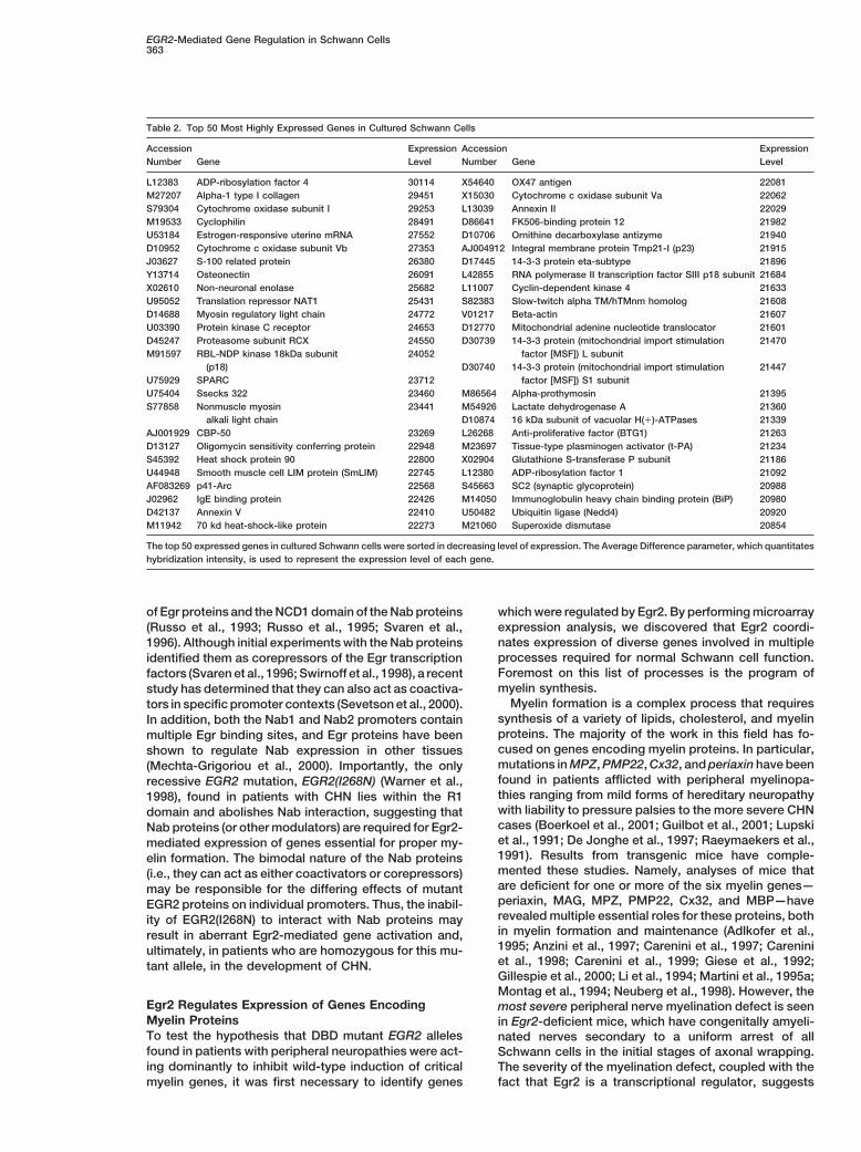
Table 2. Top 50 Most Highly Expressed Genes in Cultured Schwann Cells
Accession Expression Accession ExpressionNumber Gene Level Number Gene Level
L12383 ADP-ribosylation factor 4 30114 X54640 OX47 antigen 22081M27207 Alpha-1 type I collagen 29451 X15030 Cytochrome c oxidase subunit Va 22062S79304 Cytochrome oxidase subunit I 29253 L13039 Annexin II 22029M19533 Cyclophilin 28491 D86641 FK506-binding protein 12 21982U53184 Estrogen-responsive uterine mRNA 27552 D10706 Ornithine decarboxylase antizyme 21940D10952 Cytochrome c oxidase subunit Vb 27353 AJ004912 Integral membrane protein Tmp21-I (p23) 21915J03627 S-100 related protein 26380 D17445 14-3-3 protein eta-subtype 21896Y13714 Osteonectin 26091 L42855 RNA polymerase II transcription factor SIII p18 subunit 21684X02610 Non-neuronal enolase 25682 L11007 Cyclin-dependent kinase 4 21633U95052 Translation repressor NAT1 25431 S82383 Slow-twitch alpha TM/hTMnm homolog 21608D14688 Myosin regulatory light chain 24772 V01217 Beta-actin 21607U03390 Protein kinase C receptor 24653 D12770 Mitochondrial adenine nucleotide translocator 21601D45247 Proteasome subunit RCX 24550 D30739 14-3-3 protein (mitochondrial import stimulation 21470M91597 RBL-NDP kinase 18kDa subunit 24052 factor [MSF]) L subunit
(p18) D30740 14-3-3 protein (mitochondrial import stimulation 21447U75929 SPARC 23712 factor [MSF]) S1 subunitU75404 Ssecks 322 23460 M86564 Alpha-prothymosin 21395S77858 Nonmuscle myosin 23441 M54926 Lactate dehydrogenase A 21360
alkali light chain D10874 16 kDa subunit of vacuolar H(1)-ATPases 21339AJ001929 CBP-50 23269 L26268 Anti-proliferative factor (BTG1) 21263D13127 Oligomycin sensitivity conferring protein 22948 M23697 Tissue-type plasminogen activator (t-PA) 21234S45392 Heat shock protein 90 22800 X02904 Glutathione S-transferase P subunit 21186U44948 Smooth muscle cell LIM protein (SmLIM) 22745 L12380 ADP-ribosylation factor 1 21092AF083269 p41-Arc 22568 S45663 SC2 (synaptic glycoprotein) 20988J02962 IgE binding protein 22426 M14050 Immunoglobulin heavy chain binding protein (BiP) 20980D42137 Annexin V 22410 U50482 Ubiquitin ligase (Nedd4) 20920M11942 70 kd heat-shock-like protein 22273 M21060 Superoxide dismutase 20854
The top 50 expressed genes in cultured Schwann cells were sorted in decreasing level of expression. The Average Difference parameter, which quantitateshybridization intensity, is used to represent the expression level of each gene.
of Egr proteins and the NCD1 domain of the Nab proteins which were regulated by Egr2. By performing microarrayexpression analysis, we discovered that Egr2 coordi-(Russo et al., 1993; Russo et al., 1995; Svaren et al.,nates expression of diverse genes involved in multiple1996). Although initial experiments with the Nab proteinsprocesses required for normal Schwann cell function.identified them as corepressors of the Egr transcriptionForemost on this list of processes is the program offactors (Svaren et al., 1996; Swirnoff et al., 1998), a recentmyelin synthesis.study has determined that they can also act as coactiva-
Myelin formation is a complex process that requirestors in specific promoter contexts (Sevetson et al., 2000).synthesis of a variety of lipids, cholesterol, and myelinIn addition, both the Nab1 and Nab2 promoters containproteins. The majority of the work in this field has fo-multiple Egr binding sites, and Egr proteins have beencused on genes encoding myelin proteins. In particular,shown to regulate Nab expression in other tissuesmutations in MPZ, PMP22, Cx32, and periaxin have been(Mechta-Grigoriou et al., 2000). Importantly, the onlyfound in patients afflicted with peripheral myelinopa-recessive EGR2 mutation, EGR2(I268N) (Warner et al.,thies ranging from mild forms of hereditary neuropathy1998), found in patients with CHN lies within the R1with liability to pressure palsies to the more severe CHNdomain and abolishes Nab interaction, suggesting thatcases (Boerkoel et al., 2001; Guilbot et al., 2001; LupskiNab proteins (or other modulators) are required for Egr2-et al., 1991; De Jonghe et al., 1997; Raeymaekers et al.,mediated expression of genes essential for proper my-1991). Results from transgenic mice have comple-elin formation. The bimodal nature of the Nab proteinsmented these studies. Namely, analyses of mice that(i.e., they can act as either coactivators or corepressors)are deficient for one or more of the six myelin genes—may be responsible for the differing effects of mutantperiaxin, MAG, MPZ, PMP22, Cx32, and MBP—haveEGR2 proteins on individual promoters. Thus, the inabil-revealed multiple essential roles for these proteins, bothity of EGR2(I268N) to interact with Nab proteins mayin myelin formation and maintenance (Adlkofer et al.,result in aberrant Egr2-mediated gene activation and,1995; Anzini et al., 1997; Carenini et al., 1997; Careniniultimately, in patients who are homozygous for this mu-et al., 1998; Carenini et al., 1999; Giese et al., 1992;tant allele, in the development of CHN.Gillespie et al., 2000; Li et al., 1994; Martini et al., 1995a;Montag et al., 1994; Neuberg et al., 1998). However, the
Egr2 Regulates Expression of Genes Encoding most severe peripheral nerve myelination defect is seenMyelin Proteins in Egr2-deficient mice, which have congenitally amyeli-To test the hypothesis that DBD mutant EGR2 alleles nated nerves secondary to a uniform arrest of allfound in patients with peripheral neuropathies were act- Schwann cells in the initial stages of axonal wrapping.ing dominantly to inhibit wild-type induction of critical The severity of the myelination defect, coupled with the
fact that Egr2 is a transcriptional regulator, suggestsmyelin genes, it was first necessary to identify genes
Neuron364
that Egr2 coordinately regulates a myelination program sheathment defects (Baumgartner et al., 1996). Interest-ingly, we have found that neurexophilin-4, which inter-in Schwann cells. We have demonstrated that this is
indeed the case, as Egr2 expression alone is sufficient acts with neurexins, is induced 15.3-fold by Egr2 inSchwann cells. Thus, our data immediately suggest twofor activation of periaxin, MAG, MPZ, PMP22, Cx32, and
MBP expression in Schwann cells and that dominant- genes where a search for mutations is likely to be fruitful.After nerve injury, the expression of Egr2 and manynegative inhibition by DBD mutant Egr2 results in an
abrogation of this coordinated myelin gene activation of the myelination-associated genes decreases dramati-cally. From our microarray analysis, we identified otherprogram.Egr2 target genes in Schwann cells that mimic the nerveinjury-induced expression pattern of those encodingEgr2 Regulates Other Genes Involved in Propermyelin proteins. Namely, two different splice variants ofPeripheral Myelin FormationVEGF as well as one of their receptors (flk-1) were in-While the genes encoding the myelin proteins have beenduced by Egr2 expression in Schwann cells. VEGF isimplicated as possible target genes of Egr2, the poten-known to be a potent mitogen for endothelial cells andtial role of Egr2 in other processes, which are also criticalis critical for vasculogenesis and angiogenesis (Neufeldfor proper peripheral nerve myelination, has not beenet al., 1999). In addition, investigators have found thatdescribed. Our microarray expression analysis has re-Schwann cells express flk-1 and that VEGF promotesvealed that Egr2 may also coordinate myelin lipid as-survival and proliferation of Schwann cells (Sondell etsembly, as it regulates multiple genes encoding en-al., 1999). In a recent study, direct application of VEGFzymes required for lipid and cholesterol synthesis.intramuscularly either immediately or 10 days after aFurthermore, growth factors and their cognate recep-hypoxic insult greatly alleviated the resulting ischemictors, which have potential roles in providing trophic andperipheral neuropathy (Schratzberger et al., 2000). Inmitogenic support to Schwann cells, are also upregu-fact, the authors suggest that VEGF abrogates the motorlated by Egr2 expression.and sensory loss by promoting increased neovasculari-The principal lipid components of myelin include oleiczation of the vasa nervorum, nutrient arteries withinacid, which comprises 35%–45% of all lipid in peripheralnerves, and by enhancing Schwann cell survival, prolif-myelin (Fressinaud et al., 1986); the phospholipid, sphin-eration, and migration to repair the damaged area. Ingomyelin; and cholesterol. Our study demonstrates thatlight of these studies as well as our findings, it is possiblegenes encoding enzymes required for synthesis of eachthat Egr2 may regulate expression of both VEGF and itsof these critical myelin lipids is regulated by Egr2. First,receptors during embryonic development to promotecholine kinase, which is required for the eventual conver-vasculogenesis in the developing nerve. Thus, inductionsion of ceramide to sphingomyelin, was induced 5.1-of VEGF and flk-1 by Egr2 in Schwann cells could notfold. Another Egr2-induced gene, which is also develop-only promote autocrine survival and proliferative effects,mentally regulated in Schwann cells (Garbay et al.,but it might also facilitate angiogenesis through the tro-1998), stearyl CoA desaturase, catalyzes the D9 desatu-phic and mitogenic effects of VEGF on endothelial cells.ration of stearyl CoA to form oleic acid. Finally, the syn-
thesis and deposition of cholesterol, a major componentof myelin, is also thought to be a highly regulated pro- Elucidation of Disease Mechanisms
Using Neurogenomicscess in Schwann cells (Fu et al., 1998). Cholesterol issynthesized through a series of complex biochemical One powerful feature of the global genomic microarray
expression technology is the ability to compare datareactions including formation of mevalonic acid from3-hydroxy-3-methylglutaryl CoA via the rate-limiting en- sets derived from experiments which may compare dif-
ferent induction paradigms, cell or tissue types, orzyme of cholesterol synthesis, HMG CoA reductase. Thisenzyme (4.5-fold) along with two others in the choles- mutants. For example, Hughes et al. (2000) conducted
microarray expression profiling from 300 different muta-terol synthesis pathway, 7-dehydrocholesterol reduc-tase (3.5-fold) and lanosterol 14-demethylase (3.3-fold), tions and chemical treatments in S. cerevisiae and iden-
tified affected cellular pathways by pattern matching.were induced by Egr2 expression in Schwann cells.Thus, several enzymes responsible for formation of criti- These pattern matching techniques, which include ag-
glomerative hierarchical clustering, k-means clustering,cal lipid components of myelin are under coordinatedEgr2 regulation. and self-organizing maps, facilitate the sorting of genes
into classes and allow the rapid discernment of an un-Several genes regulated by Egr2 in Schwann cells(e.g., those encoding myelin proteins—MPZ, PMP22, known gene’s function. For example, Hughes et al. iden-
tified cellular functions of uncharacterized ORFs simplyCx32, and recently periaxin [Boerkoel et al., 2001; Guil-bot et al., 2001; Lupski et al., 1991; De Jonghe et al., by the fact that expression profile of these unknown
genes coclustered with genes with a known biological1997; Raeymaekers et al., 1991]) are mutated in patientswith inherited neuropathies. In families with inherited function. Thus, the capability to compare multiple data
sets derived from diverse expression profiling experi-neuropathies in which the mutated gene is unknown,a search for mutations in other Egr2-regulated genes ments can dramatically accelerate the determination of
gene function, an exciting prospect, particularly in lightshould be performed. For example, a recent study indi-cated that Fray, a serine-threonine protein kinase, is of the recent completion of the sequence of the human
genome.required for axon ensheathment (Leiserson et al., 2000).In this study, we show that PASK, the mammalian homo- With respect to the data generated in this study, it
will be interesting to compare this data set with thoselog of Fray, is induced 16.9-fold by Egr2 in Schwanncells. Mutations in neurexin IV also result in en- derived from myelinating Schwann cells, Schwann cells
EGR2-Mediated Gene Regulation in Schwann Cells365
ml) (Bottenstein and Sato, 1979). At this titer and time of adenoviralfrom sciatic nerve injury, and/or Schwann cells fromexposure, greater than 95% of Schwann cells were infected asanimal models of diabetic and toxin-induced neuropa-determined by GFP fluorescence and no cytopathic effects werethies. Information from such analyses will be importantobserved. Furthermore, a time course of expression after infection
in deciphering the molecular program elaborated by the with Egr2-expressing adenovirus demonstrated that Egr2 mRNASchwann cell to myelinate or to provide trophic support levels were maximal 20 hr after infection. Thus, RNA was harvested
4 hr after the peak of Egr2 expression (i.e., 24 hr after infection) tofor an axon. Furthermore, such comparisons can beallow for target gene induction. For cultures where RNA was isolatedexpanded to include other glia such as oligodendro-48 hr after infection, cultures were washed after 24 hr with PBScytes and astrocytes to identify common genes requiredfollowed by reincubation in N2 for an additional 24 hr.for normal function of glial cell types or to discover
conserved genes required for both central and periph-GeneChip Hybridization
eral nerve myelination. Hybridization probes for GeneChip analysis were synthesized asExamination of oligodendrocytes is particularly rele- described from poly(A)1 RNA prepared from cultures of Schwann
cells that had been infected with either the adenovirus expressingvant with respect to the Egr proteins as Egr1 is knownEgr2 or the control adenovirus. The poly(A)1 RNA was converted toto be expressed in oligodendrocyte progenitors and isdouble-stranded cDNA using an oligo dT primer containing the T7induced upon differentiation (Sock et al., 1997). Further-promoter, and this was used to prepare biotinylated cRNA usingmore, several groups have demonstrated that Egr1 regu-the Bioarray HighYield kit (Enzo, Farmingdale, NY) according to
lates PDGF-A chain (Silverman et al., 1997; Svaren et the manufacturer’s directions. The biotinylated cRNA probes wereal., 2000) and that PDGF-A deficient mice have a central fragmented and applied as described (Lipshutz et al., 1999; Lockhart
et al., 1996) to Rat Genome U34 GeneChip arrays (Affymetrix, Santanervous system dysmyelinating phenotype due to re-Clara, CA). The signal intensities from hybridized cRNA were quanti-duced numbers of oligodendrocytes (Fruttiger et al.,fied, and the GeneChip analysis software was used to identify differ-1999). These studies are particularly intriguing in lightentially expressed genes.of recent findings that certain forms of multiple sclerosis
are caused by a primary dysfunction in oligodendro- Sciatic Nerve Crushcytes (Lucchinetti et al., 2000). Thus, it will be interesting All surgical procedures followed the NIH guidelines for care and useto evaluate the role of the Egr proteins, particularly Egr1, of laboratory animals at Washington University. Male Sprague–
Dawley rats (200–300 g) were anesthetized, and the right sciaticin the pathogenesis of multiple sclerosis subtypes whichnerve was injured at the hip level by compressing the nerve withare caused by oligodendrocyte malfunction.forceps for 30 s. The contralateral nerve was exposed, but left unin-In conclusion, with the use of global genomic microar-jured (control). After the indicated length of time, the animals were
ray expression profiling, we demonstrate that Egr2 coor- euthanized and decapitated for immediate collection of tissues fordinates the peripheral nerve myelination program by RNA isolation.inducing genes encoding myelin proteins and enzymesrequired for myelin lipid assembly. Furthermore, we have Cell Lines and Transfection
Culture conditions for the CV-1 cell line have been described pre-revealed that DBD EGR2 mutants cause myelinopathyviously (Paulsen et al., 1992). CV-1 and rat Schwann transfectionsby dominantly inhibiting wild-type EGR2 induction ofwere performed in 12-well plates (Corning, Corning, NY) using 3.5 3these newly identified target genes. Finally, data gener-104 cells per well. All transfections were performed essentially as
ated in this study in combination with other expression described (Russo et al., 1993), using 250 ng of the luciferase reporter,profiling experiments will be invaluable in unraveling 50 ng of a CMV driven lacZ reporter, and the indicated amounts ofthe molecular program required for normal central and the expression plasmids. Bluescript plasmid (Stratagene, La Jolla,
CA) was added as required to make a total of 1 mg DNA per transfec-peripheral nerve myelination and will ultimately facilitatetion. The average luciferase activity of duplicate samples was nor-design and implementation of effective therapeuticmalized to the b-galactosidase activity from the transfected lacZtreatments for demyelinating diseases such as HMSNreporter. Means and standard deviations of two separate transfec-
and multiple sclerosis. tion experiments are shown.
Quantitative RT-PCR (TaqMan) Analysis of Gene ExpressionExperimental ProceduresTotal RNA was purified and 1 mg of RNA was used to prepare cDNAessentially as described (Lee et al., 1996). Expression levels of genesCell Culture, Adenoviral Production, and Infectionwere measured by quantitative RT-PCR analysis using the TaqManRat Schwann cells were purified from P0-P2 sciatic nerves as pre-7700 Sequence Detection System (Perkin Elmer, Wellesley, MA).viously described (Brockes et al., 1979). Briefly, sciatic nerves fromReal time detection of PCR product accumulation was monitoredP0-P2 rat pups were isolated and treated with collagenase (0.1%)using the increase in fluorescence of the SYBR-GREEN dye as de-for 1 hr at 378C. After trituration, dissociated cells were plated onscribed (Morrison et al., 1998). Relative expression levels of thesecollagen-coated plates. To eliminate fibroblasts, the cells were sub-genes in each sample were determined using a standard curve ofjected to three successive rounds (48 hr each) of cytosine arabino-serial dilutions of cDNA samples containing the highest expressionside (10 mM) (Sigma, St. Louis, MO) treatment followed by a 24 hrof these genes. Average fold induction relative to control infectedrecovery period in DMEM with 10% FCS. At this point, greater thancells was determined after normalizing to the amount of GAPDH99% of remaining cells were Schwann cells. Schwann cells werepresent in each sample. Standard deviation is obtained from tripli-expanded by adding bovine pituitary extract (20 mg/ml) (Sigma) andcate measurements of each sample. Primer sequences used forforskolin (2 mM) (Sigma). Mouse Schwann cells were isolated fromquantitative analysis of each gene are available upon request.sciatic nerves of P0-P2 pups and were used directly for experiments
5–6 days later.Adenoviruses expressing EGFP, Egr2, and Egr2(S382R, D383Y) Immunoblot Analysis
Quail fibroblast (QT6) cells (2 3 105 cells per 3.5 cm dish) werewere made essentially as described (Ehrengruber et al., 1998; Heet al., 1998). Viral titers were determined by following the Tissue transfected with 10 mg each of expression constructs for wild-type
and mutant versions of Egr2 (Warner et al., 1998). After 48 hr, cellsCulture Infectious Dose 50 (TCID50) Method from the AdEasy VectorSystem Application Manual (Version 1.2) published by Qbiogene were washed twice with phosphate-buffered saline and then lysed
in Laemmli buffer. Lysates were boiled for 10 min, electrophoresed(Carlsbad, CA). Schwann cells in culture were subjected to adenovi-ral infection in defined medium (N2) for 24 hr at 378C (1.9 3 107 pfu/ on a sodium dodecyl sulfate 10% polyacrylamide gel, and trans-
Neuron366
ferred to a nitrocellulose membrane (Midwest Scientific, St. Louis, deficient Schwann cells myelinate dorsal root ganglion neurons inculture. GLIA 22, 213–220.MO). After overnight blocking with Tris-buffered saline containing
5% milk, blots were incubated first with an anti-Egr2 polyclonal Carenini, S., Montag, D., Schachner, M., and Martini, R. (1999). Sub-antibody (Santa Cruz Biotechnology, Santa Cruz, CA) at 1 mg/ml, tle roles of neural cell adhesion molecule and myelin-associatedand then with a horseradish peroxidase-conjugated antirabbit sec- glycoprotein during schwann cell spiralling in P0-deficient mice.ondary antibody (Jackson Laboratories, Bar Harbor, ME) at a dilution GLIA 27, 203–212.of 1:10,000. Protein blots were washed five times in TBST and visual-
De Jonghe P., Timmerman V., Nelis E., Martin J.J., and Broeckhoven,ized by chemiluminescence detection (Amersham, Piscataway, NJ).
C.V. (1997). Charcot-Marie Tooth disease and related peripheralneuropathies. J. Periph. Nerv. Sys. 2, 370–387.
AcknowledgmentsEhrengruber, M.U., Lanzrein, M., Xu, Y., Jasek, M.C., Kantor, D.B.,Xu, Y., Schuman, E.M., Lester, H.A., and Davidson, N. (1998). Re-J.S. was supported by a development grant from the Muscularcombinant adenovirus-mediated expression in nervous system ofDystrophy Association. We wish to thank Betsy Apel for assistancegenes coding for ion channels and other molecules involved in syn-with transfections, and we are grateful to Markus Ehrengruber (Brainaptic function. In Methods in Enzymology, C.P.M., ed. (San Diego:Research Institute, University of Zurich, Switzerland) for generatingAcademic Press), pp. 483–503.and providing Egr2-expressing adenovirus. We thank James LupskiFressinaud, C., Rigaud, M., and Vallat, J.M. (1986). Fatty acid com-(Department of Molecular and Human Genetics, Baylor College ofposition of endoneurium and perineurium from adult rat sciaticMedicine, Houston, Texas) for stimulating discussions and valuablenerve. J. Neurochem. 46, 1549–1554.advice. GeneChip analysis was performed by the Siteman Cancer
Center GeneChip Core at Washington University School of Medicine. Fruttiger, M., Karlsson, L., Hall, A.C., Abramsson, A., Calver, A.R.,This work was supported by NIH grant 5 P01 CA49712-08 (J.M.). Bostrom, H., Willetts, K., Bertold, C.H., Heath, J.K., Betsholtz, C.,
and Richardson, W.D. (1999). Defective oligodendrocyte develop-ment and severe hypomyelination in PDGF-A knockout mice. Devel-Received July 17, 2000; revised February 25,2001.opment 126, 457–467.
Fu, Q., Goodrum, J.F., Hayes, C., Hostettler, J.D., Toews, A.D., andReferencesMorell, P. (1998). Control of cholesterol biosynthesis in Schwanncells. J. Neurochem. 71, 549–555.Adlkofer, K., Frei, R., Neuberg, D.H., Zielasek, J., Toyka, K.V., and
Suter, U. (1997). Heterozygous peripheral myelin protein 22-deficient Garbay, B., Boiron-Sargueil, F., Shy, M., Chbihi, T., Jiang, H., Kam-mice are affected by a progressive demyelinating tomaculous neu- holz, J., and Cassagne, C. (1998). Regulation of oleoyl-CoA synthesisropathy. J. Neurosci. 17, 4662–4671. in the peripheral nervous system: demonstration of a link with myelin
synthesis. J. Neurochem. 71, 1719–1726.Adlkofer, K., Martini, R., Aguzzi, A., Zielasek, J., Toyka, K.V., andSuter, U. (1995). Hypermyelination and demyelinating peripheral Giese, K.P., Martini, R., Lemke, G., Soriano, P., and Schachner, M.neuropathy in Pmp22- deficient mice. Nat. Genet. 11, 274–280. (1992). Mouse P0 gene disruption leads to hypomyelination, abnor-
mal expression of recognition molecules, and degeneration of my-Anzini, P., Neuberg, D.H., Schachner, M., Nelles, E., Willecke, K.,elin and axons. Cell 71, 565–576.Zielasek, J., Toyka, K.V., Suter, U., and Martini, R. (1997). Structural
abnormalities and deficient maintenance of peripheral nerve myelin Gillen, C., Gleichmann, M., Spreyer, P., and Muller, H.W. (1995).in mice lacking the gap junction protein connexin 32. J. Neurosci. Differentially expressed genes after peripheral nerve injury. J. Neu-17, 4545–4551. rosci. Res. 42, 159–171.
Aurelio, O.N., Kong, X.T., Gupta, S., and Stanbridge, E.J. (2000). p53 Gillespie, C.S., Sherman, D.L., Fleetwood-Walker, S.M., Cottrell,mutants have selective dominant-negative effects on apoptosis but D.F., Tait, S., Garry, E.M., Wallace, V.C., Ure, J., Griffiths, I.R., Smith,not growth arrest in human cancer cell lines. Mol. Cell. Biol. 20, A., and Brophy, P.J. (2000). Peripheral demyelination and neuro-770–778. pathic pain behavior in periaxin-deficient mice. Neuron 26, 523–531.
Baumgartner, S., Littleton, J.T., Broadie, K., Bhat, M.A., Harbecke, Guilbot, A., Williams, A., Ravise, N., Verny, C., Brice, A., Sherman,R., Lengyel, J.A., Chiquet-Ehrismann, R., Prokop, A., and Bellen, D.L., Brophy, P.J., LeGuern, E., Delague, V., Bareil, C., Megarbane,H.J. (1996). A Drosophila neurexin is required for septate junction A., Claustres, M.. (2001). A mutation in periaxin is responsible forand blood-nerve barrier formation and function. Cell 87, 1059–1068. CMT4F, an autosomal recessive form of Charcot-Marie-Tooth dis-
ease. Hum. Mol. Gene. 10, 415–421.Bellone, E., Di Maria, E., Soriani, S., Varese, A., Doria, L.L., Ajmar,F., and Mandich, P. (1999). A novel mutation (D305V) in the early He, T.C., Zhou, S., da Costa, L.T., Yu, J., Kinzler, K.W., and Vo-growth response 2 gene is associated with severe Charcot-Marie- gelstein, B. (1998). A simplified system for generating recombinantTooth type 1 disease. Hum Mutat 14, 353–354. adenoviruses. Proc. Natl. Acad. Sci. USA 95, 2509–2514.
Boerkoel, C.F., Takashima, H., Stankiewicz, P., Garcia, C.A., Leber, Hughes, T.R., Marton, M.J., Jones, A.R., Roberts, C.J., Stoughton,S.M., Rhee-Morris, L., and Lupski, J.R. (2001). Periaxin mutations R., Armour, C.D., Bennett, H.A., Coffey, E., Dai, H., He, Y.D., Kidd,cause Recessive Dejerine-Sottas neuropathy. Am. J. Hum. Genet. M.J., King A.M., Meyer, M.R., Slade D., Lum P.Y., Stepaniants S.B.,68, 325–333. Shoemaker, D.D., Gachotte D., Chakraburtty K., Simon J., Bard M.,
Friend S.H. (2000). Functional discovery via a compendium of ex-Bottenstein, J.E., and Sato, G.H. (1979). Growth of a rat neuro-pression profiles. Cell 102, 109–126.blastoma cell line in serum-free supplemented medium. Proc. Natl.
Acad. Sci. USA 76, 514–517. Kubbutat, M.H., Ludwig, R.L., Levine, A.J., and Vousden, K.H. (1999).Analysis of the degradation function of Mdm2. Cell Growth Differ.Botti, S., Pareyson, D., Sghirlanzoni, A., Nemni, R., Riva, D., and10, 87–92.Taroni, F. (1998). Mutations in the transcription factor EGR2 in pa-
tients with severe hereditary demyelinating neuropathies. Am. J. Lee, S.L., Wang, Y., and Milbrandt, J. (1996). Unimpaired macro-Hum. Genet. 63, A352. phage differentiation and activation in mice lacking the zinc finger
transplantation factor NGFI-A (EGR1). Mol. Cell. Biol. 16, 4566–4572.Brockes, J.P., Fields, K.L., and Raff, M.C. (1979). Studies on culturedrat Schwann cells. I. Establishment of purified populations from Leiserson, W.M., Harkins, E.W., and Keshishian, H. (2000). Fray,cultures of peripheral nerve. Brain Res. 165, 105–118. a Drosophila serine/threonine kinase homologous to mammalian
PASK, is required for axonal ensheathment. Neuron 28, 793–806.Carenini, S., Montag, D., Cremer, H., Schachner, M., and Martini, R.(1997). Absence of the myelin-associated glycoprotein (MAG) and Li, C., Tropak, M.B., Gerlai, R., Clapoff, S., Abramow-Newerly, W.,the neural cell adhesion molecule (N-CAM) interferes with the main- Trapp, B., Peterson, A., and Roder, J. (1994). Myelination in thetenance, but not with the formation of peripheral myelin. Cell Tissue absence of myelin-associated glycoprotein. Nature 369, 747–750.Res. 287, 3–9. Lipshutz, R.J., Fodor, S.P., Gingeras, T.R., and Lockhart, D.J. (1999).
High density synthetic oligonucleotide arrays. Nat. Genet. 21, 20–24.Carenini, S., Montag, D., Schachner, M., and Martini, R. (1998). MAG-
EGR2-Mediated Gene Regulation in Schwann Cells367
Lockhart, D.J., Dong, H., Byrne, M.C., Follettie, M.T., Gallo, M.V., of NAB1, a repressor of NGFI-A and Krox20 mediated transcription.Proc. Natl. Acad. Sci. USA 92, 6873–6877.Chee, M.S., Mittmann, M., Wang, C., Kobayashi, M., Horton, H., and
Brown, E.L. (1996). Expression monitoring by hybridization to high- Saifudeen, Z., Du H., Dipp, S., and El-Dahr, S.S. (2000). The bradyki-density oligonucleotide arrays. Nat. Biotechnol. 14, 1675–1680. nin type 2 receptor is a target for p53-mediated transcriptional acti-Lucchinetti, C., Bruck, W., Parisi, J., Scheithauer, B., Rodriguez, vation. J. Biol. Chem. 275, 15557–15562.M., and Lassmann, H. (2000). Heterogeneity of multiple sclerosis
Schratzberger, P., Schratzberger, G., Silver, M., Curry, C., Kearney,lesions: implications for the pathogenesis of demyelination. Ann.M., Magner, M., Alroy, J., Adelman, L.S., Weinberg, D.H., Ropper,Neurol. 47, 707–717.A.H., and Isner, J.M. (2000). Favorable effect of VEGF gene transfer
Lupski, J.R. (1997). Charcot-Marie-Tooth disease: a gene-dosage on ischemic peripheral neuropathy. Nat. Med. 6, 405–413.effect. Hosp. Pract. (Off Ed) 32, 83–4, 89–91, 94–5 passim.
Sevetson, B., Svaren, J., and Milbrandt, J. (2000). A novel activationLupski, J.R., de Oca-Luna, R.M., Slaugenhaupt, S., Pentao, L., Guz- function for NAB proteins in Egr-dependent transcription of thezetta, V., Trask, B.J., Saucedo-Cardenas, O., Barker, D.F., Killian, Luteinizing hormone-beta gene. J. Biol. Chem. 275, 9749–9757.J.M., Garcia, C.A, et al. (1991). DNA duplication associated with
Silverman, E.S., Khachigian, L.M., Lindner, V., Williams, A.J., andCharcot-Marie-Tooth disease type 1A. Cell 66, 219–232.Collins, T. (1997). Inducible PDGF A-chain transcription in smooth
Martini, R., Mohajeri, M.H., Kasper, S., Giese, K.P., and Schachner,muscle cells is mediated by Egr-1 displacement of Sp1 and Sp3.
M. (1995a). Mice doubly deficient in the genes for P0 and myelinAm J. Physiol. 273, H1415–H1426.
basic protein show that both proteins contribute to the formationSock, E., Leger, H., Kuhlbrodt, K., Schreiber, J., Enderich, J., Richter-of the major dense line in peripheral nerve myelin. J. Neurosci. 15,
4488–4495. Landsberg, C., and Wegner, M. (1997). Expression of Krox proteinsduring differentiation of the O-2A progenitor cell line CG-4. J. Neuro-Martini, R., Zielasek, J., Toyka, K.V., Giese, K.P., and Schachner,chem. 68, 1911–1919.M. (1995b). Protein zero (P0)-deficient mice show myelin degenera-
tion in peripheral nerves characteristic of inherited human neuropa- Sondell, M., Lundborg, G., and Kanje, M. (1999). Vascular endothelialthies. Nat. Genet. 11, 281–286. growth factor stimulates Schwann cell invasion and neovasculariza-
tion of acellular nerve grafts. Brain Res. 846, 219–228.Mechta-Grigoriou, F., Garel, S., and Charnay, P. (2000). Nab proteinsmediate a negative feedback loop controlling Krox-20 activity in the Spreca, A., Rambotti, M.G., Giambanco, I., Pula, G., Bianchi, R.,developing hindbrain. Development 127, 119–128. Ceccarelli, P., and Donato, R. (1992). Immunocytochemical localiza-Montag, D., Giese, K.P., Bartsch, U., Martini, R., Lang, Y., Bluthmann, tion of annexin V (CaBP33), a Ca(21)-dependent phospholipid- andH., Karthigasan, J., Kirschner, D.A., Wintergerst, E.S., Nave, K.A., membrane-binding protein, in the rat nervous system and skeletalet al. (1994). Mice deficient for the myelin-associated glycoprotein muscles and in the porcine heart. J. Cell. Physiol. 152, 587–598.show subtle abnormalities in myelin. Neuron 13, 229–246.
Svaren, J., Ehrig, T., Abdulkadir, S.A., Ehrengruber, M.U., Watson,Morrison, T.B., Weis, J.J., and Wittwer, C.T. (1998). Quantification M.A., and Milbrandt, J. (2000). EGR1 target genes in prostate carci-of low-copy transcripts by continuous SYBR Green I monitoring noma cells identified by microarray analysis. J. Biol. Chem. 275,during amplification. Biotechniques 24, 954–958, 960, 962. 38524–38531.Murphy, P., Topilko, P., Schneider-Maunoury, S., Seitanidou, T., Svaren, J., Sevetson, B.R., Apel, E.D., Zimonjic, D.B., Popescu, N.C.,Baron-Van Evercooren, A., and Charnay, P. (1996). The regulation and Milbrandt, J. (1996). NAB2, a corepressor of NGFI-A (Egr-1) andof Krox-20 expression reveals important steps in the control of Krox20, is nduced by proliferative and differentiative stimuli. Mol.peripheral glial cell development. Development 122, 2847–2857. Cell. Biol. 16, 3545–3553.Neuberg, D.H., Carenini, S., Schachner, M., Martini, R., and Suter,
Swirnoff, A.H., Apel, E.D., Svaren, J., Sevetson, B.R., Zimonjic, D.B.,U. (1998). Accelerated demyelination of peripheral nerves in micePopescu, N.C., and Milbrandt, J. (1998). Nab1, a corepressor ofdeficient in connexin 32 and protein zero. J. Neurosci. Res. 53,NGFI-A (Egr-1), contains an active transcriptional repression do-542–550.main. Mol. Cell. Biol. 18, 512–524.
Neufeld, G., Cohen, T., Gengrinovitch, S., and Poltorak, Z. (1999).Timmerman, V., De Jonghe, P., Ceuterick, C., De Vriendt, E., Lofgren,Vascular endothelial growth factor (VEGF) and its receptors. FASEBA., Nelis, E., Warner, L.E., Lupski, J.R., Martin, J.J., and Van Broeck-J. 13, 9–22.hoven, C. (1999). Novel missense mutation in the early growth re-
Notterpek, L., Snipes, G.J., and Shooter, E.M. (1999). Temporal ex-sponse 2 gene associated with Dejerine-Sottas syndrome pheno-
pression pattern of peripheral myelin protein 22 during in vivo andtype. Neurology 52, 1827–1832.
in vitro myelination. GLIA 25, 358–369.Topilko, P., Levi, G., Merlo, G., Mantero, S., Desmarquet, C., Man-Paulsen, R.E., Weaver, C.A., Fahrner, T.J., and Milbrandt, J. (1992).cardi, G., and Charnay, P. (1997). Differential regulation of the zincDomains regulating transcriptional activity of the inducible orphanfinger genes Krox-20 and Krox-24 (Egr-1) suggests antagonisticreceptor NGFI-B. J. Biol. Chem. 267, 16491–16496.roles in Schwann cells. J. Neurosci. Res 50, 702–712.
Raeymaekers, P., Timmerman, V., Nelis, E., De Jonghe, P., Hoogen-Topilko, P., Schneider-Maunoury, S., Levi, G., Baron-Van Ever-dijk, J.E., Baas, F., Barker, D.F., Martin, J.J., De Visser, M., Bolhuis,cooren, A., Chennoufi, A.B.Y., Seitanidou, T., Babinet, C., and Char-P.A., et al. (1991). Duplication in chromosome 17p11.2 in Charcot-nay, P. (1994). Krox-20 controls myelination in the peripheral nervousMarie-Tooth neuropathy type 1a (CMT 1a). The HMSN Collaborativesystem. Nature 371, 796–799.Research Group. Neuromuscul. Disord. 1, 93–97.
Traenckner, E.B., Pahl, H.L., Henkel, T., Schmidt, K.N., Wilk, S., andReddy, J.C., Morris, J.C., Wang, J., English, M.A., Haber, D.A., Shi,Baeuerle, P.A. (1995). Phosphorylation of human I kappa B-alphaY., and Licht, J.D. (1995). WT1-mediated transcriptional activationon serines 32 and 36 controls I kappa B-alpha proteolysis and NF-is inhibited by dominant-negative mutant proteins. J. Biol. Chem.kappa B activation in response to diverse stimuli. EMBO J. 14,270, 10878–10884.2876–2883.Rushton, J.A., Schmitz, S., Gunn-Moore, F., Sherman, D., Pappas,
C.A., Ritchie, J.M., and Haynes, L.W. (1999). Growth arrest and spon- Warner, L.E., Mancias, P., Butler, I.J., McDonald, C.M., Keppen, L.,taneous differentiation are initiated through an autocrine loop in Koob, K.G., and Lupski, J.R. (1998). Mutations in the early growthclonally derived Schwann cells by alpha1-procollagen I C-propep- response 2 (EGR2) gene are associated with hereditary myelinopa-tide. J. Neurochem. 73, 1816–1827. thies. Nat. Genet. 18, 382–384.
Russo, M.W., Matheny, C., and Milbrandt, J. (1993). Transcriptional Warner, L.E., Svaren, J., Milbrandt, J., and Lupski, J.R. (1999). Func-activity of the zinc finger protein NGFI-A is influenced by its interac- tional consequences of mutations in the early growth response 2tion with a cellular factor. Mol. Cell. Biol. 13, 6858–6865. gene (EGR2) correlate with severity of human myelinopathies. Hum.
Mol. Genet. 8, 1245–1251.Russo, M.W., Sevetson, B.R., and Milbrandt, J. (1995). Identification

Neuron368
Yoshida, H., Kitamura, K., Tanaka, K., Omura, S., Miyazaki, T., Hach-iya, T., Ohno, R., and Naoe, T. (1996). Accelerated degradation ofPML-retinoic acid receptor alpha (PML-RARA) oncoprotein by all-trans-retinoic acid in acute promyelocytic leukemia: possible roleof the proteasome pathway. Cancer Res. 56, 2945–2948.
Zorick, T.S., Syroid, D.E., Arroyo, E., Scherer, S.S., and Lemke, G.(1996). The transcription factors SCIP and Krox-20 mark dis-tinctstages and cell fates in Schwann cell differentiation. Mol. Cell.Neurosci. 8, 129–145.
Zorick, T.S., Syroid, D.E., Brown, A., Gridley, T., and Lemke, G.(1999). Krox-20 controls SCIP expression, cell cycle exit and suscep-tibility to apoptosis in developing myelinating Schwann cells. Devel-opment 126, 1397–1406.