early increases in plasminogen activator activity following partial hepatectomy in humans
TRANSCRIPT
BioMed CentralComparative Hepatology
ss
Open AcceResearchEarly increases in plasminogen activator activity following partial hepatectomy in humansDavid Mangnall*, Kirsty Smith, Nigel C Bird and Ali W MajeedAddress: Liver Research Group, Division of Clinical Sciences South, K Floor, Royal Hallamshire Hospital, Sheffield S10 2JF, UK
Email: David Mangnall* - [email protected]; Kirsty Smith - [email protected]; Nigel C Bird - [email protected]; Ali W Majeed - [email protected]
* Corresponding author
AbstractBackground: Increases in urokinase-like plasminogen activator (uPA) activity are reported to beamongst the earliest events occurring in remnant liver following partial hepatectomy in rats, andhave been proposed as a key component of the regenerative response. Remodelling of theextracellular matrix, conversion of single chain hepatocyte growth factor to the active two-chainform and a possible activation of a mitogenic signalling pathway have all been ascribed to theincreased uPA activity. The present study aimed to determine whether similar early increases inuPA activity could be detected in the remnant liver following resection of metastatic tumours insurgical patients.
Results: Eighteen patients undergoing partial hepatectomy for the removal of hepatic metastasessecondary to primary colonic tumours were studied. Increased plasminogen activator activity wasfound in the final liver samples for the group of patients in whom the resection size was at least50%. For smaller resections, the increased activity was not observed. The increased activity did notcorrelate with the age of the patient or with the time between the start of resection and the endof the operation. There was, however, a negative correlation between plasminogen activatoractivity and the time for which blood supply to the liver was clamped.
Conclusions: Our findings are in accordance with those from experimental animal models andshow, for the first time, that rapid increases in plasminogen activator activity can occur followingsimilarly large liver resection in humans. Thus, increases in plasminogen activator activity are anearly event in the remnant liver following major liver resection in man. Our observations providesupport for the contention that increases in plasminogen activators play a key role in the initiationof hepatic regeneration in man.
BackgroundUrokinase-like plasminogen activator (uPA), initially rec-ognised by its ability to convert plasminogen to plasminand to participate in the fibrinolytic cascade, is now con-sidered to have a wider role, which encompasses meta-static invasion by tumour cells and liver regeneration. In
regeneration of the liver following partial hepatectomy,uPA has a number of potential roles. These include initi-ating the remodelling of the extracellular matrix to allowcell division, activation of extra-cellular pro-metallopro-teases and the release of the bound single-chain form ofhepatocyte growth factor (HGF) from the extracellular
Published: 23 December 2004
Comparative Hepatology 2004, 3:11 doi:10.1186/1476-5926-3-11
Received: 28 September 2004Accepted: 23 December 2004
This article is available from: http://www.comparative-hepatology.com/content/3/1/11
© 2004 Mangnall et al; licensee BioMed Central Ltd. This is an Open Access article distributed under the terms of the Creative Commons Attribution License (http://creativecommons.org/licenses/by/2.0), which permits unrestricted use, distribution, and reproduction in any medium, provided the original work is properly cited.
Page 1 of 10(page number not for citation purposes)
Comparative Hepatology 2004, 3:11 http://www.comparative-hepatology.com/content/3/1/11
matrix (ECM). In vitro uPA and tissue-like plasminogenactivator (tPA) have been shown to convert single chaininactive HGF into the active two chain form [1] in culturesof hepatocytes. In normal rodent liver, both the inactiveand active forms of HGF can be detected, with the pre-dominance of the inactive form [2]. Following partialhepatectomy in the rat there is an early net decrease in thetotal amount of HGF in the liver, but the relative propor-tion of the single chain, inactive form, is decreased andthe active two-chain form increased [2]. This implies anearly proteolytic conversion, possibly mediated by theplasminogen activators. The importance of the uPA-plas-minogen system to liver repair has been further demon-strated by the inability of plasminogen deficient animalsto form regenerative nodules in response to acute liverinjury [3]. As discussed by Mangnall et al. [4], uPA mayalso activate a signalling pathway leading to mitosis of thehepatocyte.
Increases in uPA activity are amongst the earliest docu-mented changes following partial hepatectomy in rats [5].Raised uPA activity was detected in the remnant liver atone-minute post-hepatectomy and continued to increasefor at least one hour, although there were no changes inthe total amount of uPA protein detectable by Westernblotting. The binding of uPA to the uPA receptor (uPAR)is also associated with an increase in uPA enzymatic activ-ity [6]. In the rat partial hepatectomy model, the increasein uPA activity is thought to be due to an increase in thelevel of uPAR and subsequent binding and activation ofuPA. In the remnant liver, increases in the amount ofuPAR have been detected by Western blotting also as earlyas 1 min post hepatectomy and more clearly at 1 hour.This had decreased by 6 h and was back to basal levels by24 h [5]. The mechanism underlying these changesremains unclear.
Additional support for a role for uPA in the hepatic regen-erative process comes from studies of uPA-deficient (uPA-/-) mice. In these animals, uptake of [3H]-thymidine intoDNA and mitotic index were reduced by almost half at 44h post-hepatectomy (the peak time for control mice), sug-gesting a slower hepatocyte growth response [7]. In a sep-arate study uPA-/-mice were treated with anti-Fasmonoclonal antibody to induce extensive hepatocyteapoptosis. Fas (a member of the TNF-receptor super-family) is present in the inactive state as a monomer, buton binding the appropriate ligand (in this case the anti-body) the receptors aggregate and activate apoptosis lead-ing to cell destruction. In these uPA-deficient animals, theregeneration response following anti-Fas treatment wasdelayed relative to normal control animals [8]. Genera-tion of mature HGF and time of peak levels were delayedin the uPA-/-mice and peak levels of proliferating cellnuclear antigen at 96 h were also delayed relative to con-
trols, which peaked at 48 h. Treatment of the uPA-/-micewith the uPA gene by lipofection reversed these effects.The results support a role for uPA in the generation ofmature HGF and in the regeneration after Fas-mediatedliver damage.
More recently, studies with uPA or plasminogen deficientmice confirmed the requirement for plasminogen activa-tion in liver regeneration and also showed a need for plas-minogen in regeneration-associated hepatic angiogenesis[9]. Collectively, these studies strongly suggest that a veryearly increase in uPA activity is a key feature of the liverregenerative response in rodents. It is generally assumedthat regeneration in the human liver follows a similarcourse but the relative paucity of studies in humansmeans that, at present, it is unclear whether a similar rolefor uPA exists in the regeneration of human liver.
Though not necessarily identical, it is clear from the liter-ature that regeneration in humans and rodents share sim-ilar mechanisms. Many of the cytokines and growthfactors essential for regeneration in rodents [reviewed in[4]] are also found in increased amounts in the regenerat-ing human liver, implying once more similarmechanisms.
However, clear differences between species do exist; anotable example being the differences in the time atwhich DNA synthesis peaks in the remnant liver. In rats,this is at about 24 h; in mice, at about 40 h; and in man,at 180–200 h following hepatectomy. In the case of thehuman studies, this may partially reflect the relativelygreater age of the patients since the rate of regenerationslows with age. Such age related effects are less likely in therodent studies where the timing of hepatocyte entry intoDNA synthesis following partial hepatectomy has beenshown to be an intrinsic, cell-autonomous, feature [10].Thus, although the basic mechanisms may be fundamen-tally similar, there are inherent differences between spe-cies (such as the timing of the cell cycle clock) whichunderscores the need not to assume that all aspects ofregeneration operate identically in all mammals.
The unique sensitivity of the human hepatocyte to TRAIL(tumour necrosis factor-related apoptosis-inducing lig-and) [11] likewise emphasises the need for caution whenextrapolating from rodent liver to human liver.
The vast majority of the literature concerns regenerationin rats and mice and much less information is availablefrom human studies since the opportunity to study liverregeneration in humans is generally limited to units spe-cialising in liver surgery and is necessarily constrained byethical considerations. Surgical removal of liver metas-tases affords the opportunity to obtain small samples of
Page 2 of 10(page number not for citation purposes)
Comparative Hepatology 2004, 3:11 http://www.comparative-hepatology.com/content/3/1/11
liver at the start, time of resection and time of wound clo-sure approximating to the early sampling times in the ani-mal studies. In this vein, the aim of the present study wasto determine whether very early increases in uPA activityoccur in the remnant liver following resection in man.
ResultsThe basal uPA activity associated with the membranepreparations showed a wide patient to patient variationranging from 4 to 24 nmol/min/mg protein with a meanof 9.94 nmol/min/mg protein (n = 18, SD = 5.06). Thisvariation in basal activity correlated neither with the ageof the patient – linear regression analysis gave a slope of -0.03 and correlation analysis gave a Spearman coefficientof -0.097 (p = 0.7), nor there was any difference betweenthe values for male patients (mean = 9.14 nmol/min/mg,SD = 3.84, n = 7) and female patients (mean = 10.45, SD= 5.83, n = 11) with a non-significant unpaired Student'st test (p = 0.61).
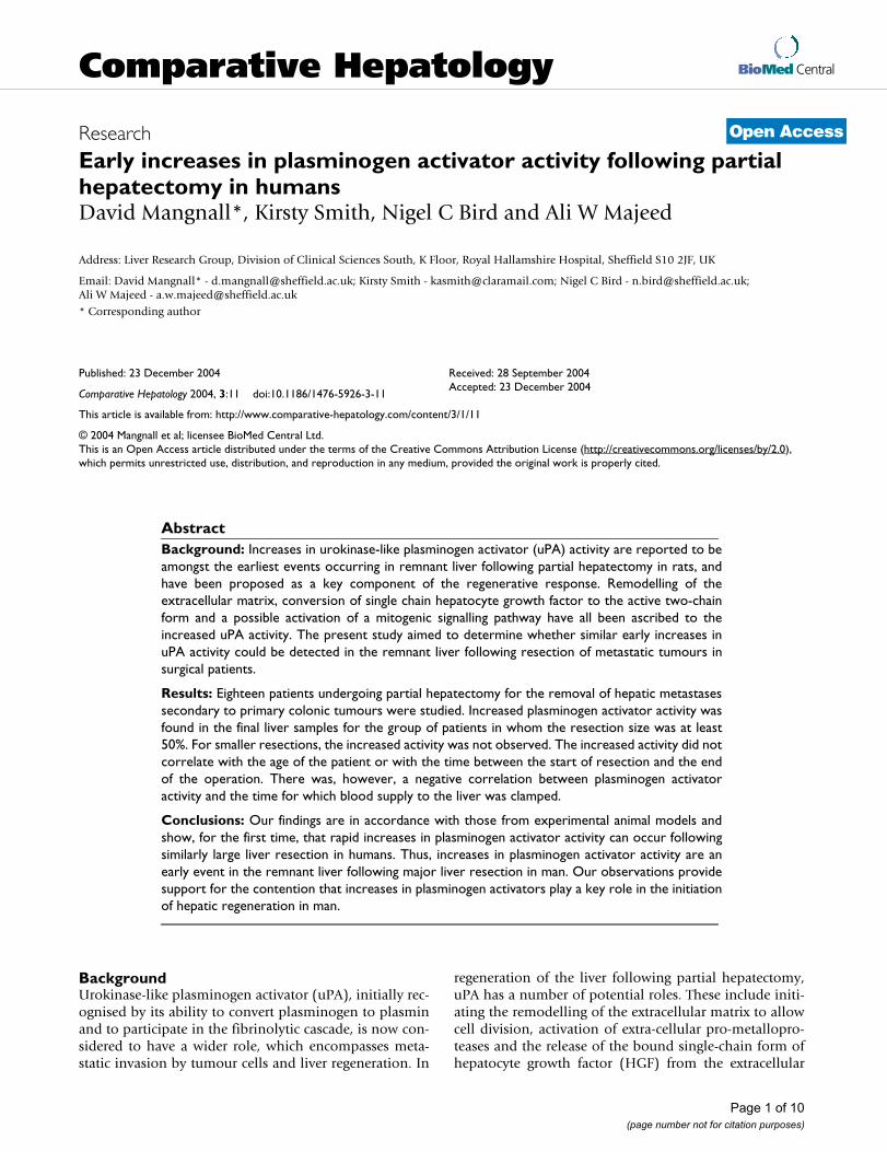
The uPA activity associated with the membrane fractionsprepared from samples taken during the operation isshown in Figure 1, for all the patients studied. The activityof the final remnant fraction taken at the end of the oper-ation was increased significantly above the activity of theother fractions. The increased activity of the final remnantfraction was, almost exclusively, confined to those
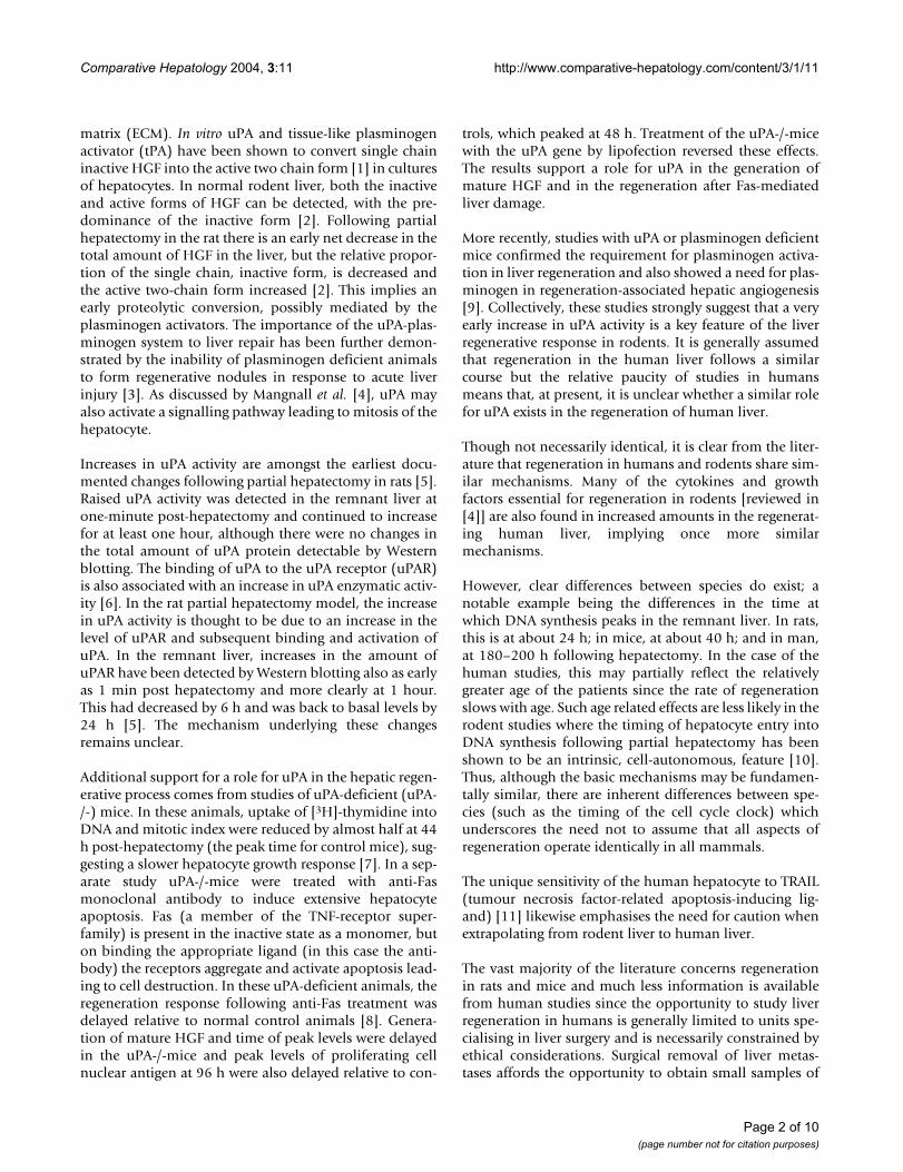
patients who had undergone a resection estimated at 50%or greater (Figure 2A) and there was no increase in thosepatients in whom the resection was less than 50% (Figure2B). The percentage change in uPA activity as a functionof the resection size for the individual patients is shown inFigure 2C. There was no correlation between uPA activityand the size of the resection below about 50% resection,but a positive correlation was observed when the resectionsize was 50% or greater.
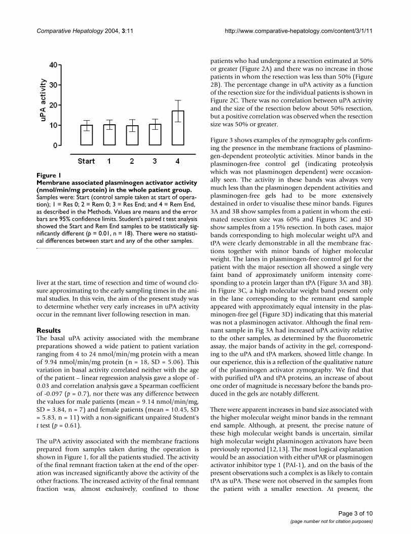
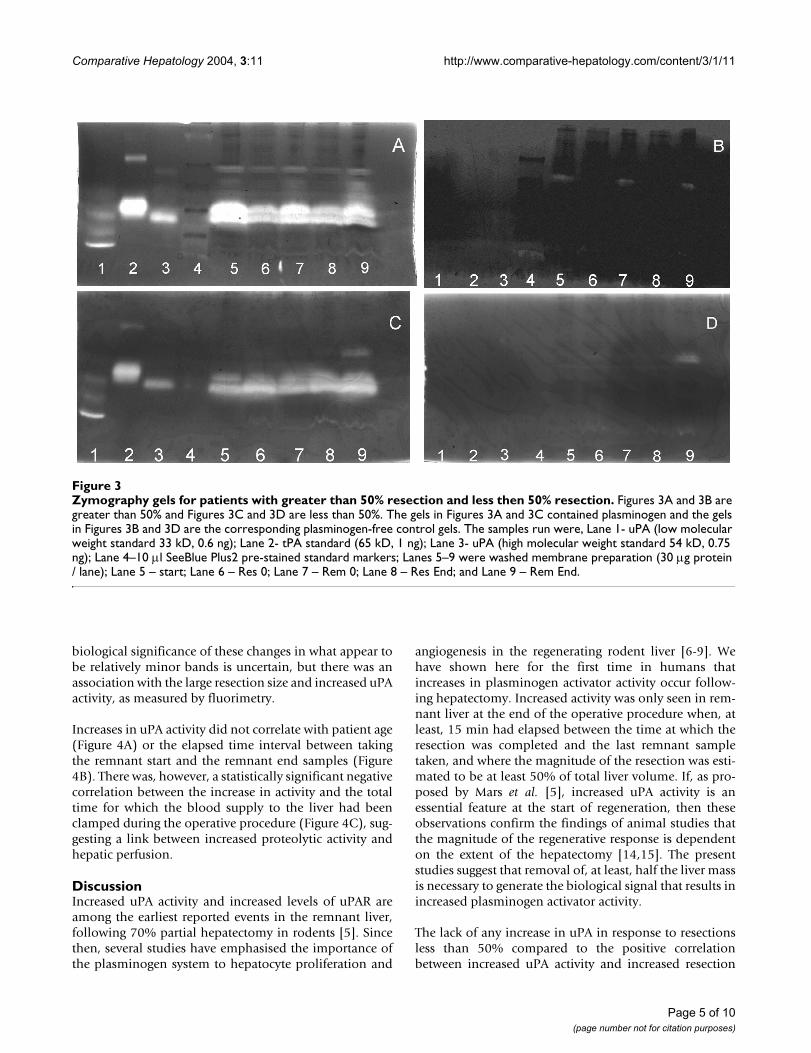
Figure 3 shows examples of the zymography gels confirm-ing the presence in the membrane fractions of plasmino-gen-dependent proteolytic activities. Minor bands in theplasminogen-free control gel (indicating proteolysiswhich was not plasminogen dependent) were occasion-ally seen. The activity in these bands was always verymuch less than the plasminogen dependent activities andplasminogen-free gels had to be more extensivelydestained in order to visualise these minor bands. Figures3A and 3B show samples from a patient in whom the esti-mated resection size was 60% and Figures 3C and 3Dshow samples from a 15% resection. In both cases, majorbands corresponding to high molecular weight uPA andtPA were clearly demonstrable in all the membrane frac-tions together with minor bands of higher molecularweight. The lanes in plasminogen-free control gel for thepatient with the major resection all showed a single veryfaint band of approximately uniform intensity corre-sponding to a protein larger than tPA (Figure 3A and 3B).In Figure 3C, a high molecular weight band present onlyin the lane corresponding to the remnant end sampleappeared with approximately equal intensity in the plas-minogen-free gel (Figure 3D) indicating that this materialwas not a plasminogen activator. Although the final rem-nant sample in Fig 3A had increased uPA activity relativeto the other samples, as determined by the fluorometricassay, the major bands of activity in the gel, correspond-ing to the uPA and tPA markers, showed little change. Inour experience, this is a reflection of the qualitative natureof the plasminogen activator zymography. We find thatwith purified uPA and tPA proteins, an increase of aboutone order of magnitude is necessary before the bands pro-duced in the gels are notably different.
There were apparent increases in band size associated withthe higher molecular weight minor bands in the remnantend sample. Although, at present, the precise nature ofthese high molecular weight bands is uncertain, similarhigh molecular weight plasminogen activators have beenpreviously reported [12,13]. The most logical explanationwould be an association with either uPAR or plasminogenactivator inhibitor type 1 (PAI-1), and on the basis of thepresent observations such a complex is as likely to containtPA as uPA. These were not observed in the samples fromthe patient with a smaller resection. At present, the
Membrane associated plasminogen activator activity (nmol/min/mg protein) in the whole patient groupFigure 1Membrane associated plasminogen activator activity (nmol/min/mg protein) in the whole patient group. Samples were: Start (control sample taken at start of opera-tion); 1 = Res 0; 2 = Rem 0; 3 = Res End; and 4 = Rem End, as described in the Methods. Values are means and the error bars are 95% confidence limits. Student's paired t test analysis showed the Start and Rem End samples to be statistically sig-nificantly different (p = 0.01, n = 18). There were no statisti-cal differences between start and any of the other samples.
Page 3 of 10(page number not for citation purposes)
Comparative Hepatology 2004, 3:11 http://www.comparative-hepatology.com/content/3/1/11
Membrane associated plasminogen activator activity (nmol/min/mg protein) in the group for whom the estimated resection size was 50% or greater and for the group where the resection size was less than 50%Figure 2Membrane associated plasminogen activator activity (nmol/min/mg protein) in the group for whom the esti-mated resection size was 50% or greater and for the group where the resection size was less than 50%. Samples were: Start (control sample taken at start of operation); 1 = Res 0; 2 = Rem 0; 3 = Res End; and 4 = Rem End; as described in the Methods. Values are means and the error bars are 95% confidence limits. For the 50% and greater group (Figure 2A), Stu-dent's paired t test analysis showed the Start and Rem End samples to be statistically significantly different (p = 0.002, n = 8). There were no statistical differences between start and any of the other samples. For the less than 50% group (Figure 2B), there were no statistical differences between any of the samples (n = 10). The relationship between increased uPA activity in the Rem End samples and extent of resection is shown in Figure 2C. There was no statistical correlation below 50% resection (Spearman correlation coefficient = -0.22, p = 0.268) but for 50% resection and higher a positive statistical correlation was observed (Spearman correlation coefficient = 0.67, p = 0.025, n = 9)
A
B
C
Page 4 of 10(page number not for citation purposes)
Comparative Hepatology 2004, 3:11 http://www.comparative-hepatology.com/content/3/1/11
biological significance of these changes in what appear tobe relatively minor bands is uncertain, but there was anassociation with the large resection size and increased uPAactivity, as measured by fluorimetry.
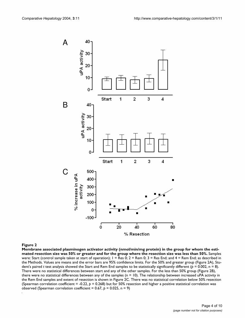
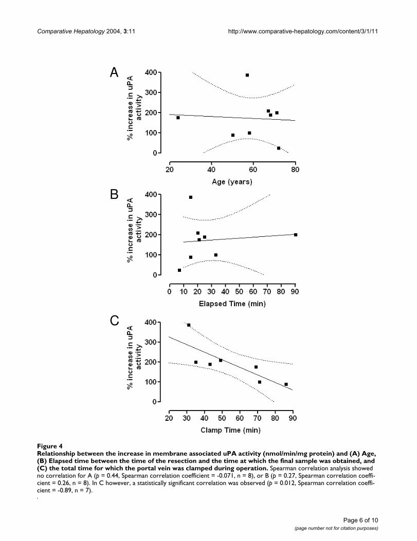
Increases in uPA activity did not correlate with patient age(Figure 4A) or the elapsed time interval between takingthe remnant start and the remnant end samples (Figure4B). There was, however, a statistically significant negativecorrelation between the increase in activity and the totaltime for which the blood supply to the liver had beenclamped during the operative procedure (Figure 4C), sug-gesting a link between increased proteolytic activity andhepatic perfusion.
DiscussionIncreased uPA activity and increased levels of uPAR areamong the earliest reported events in the remnant liver,following 70% partial hepatectomy in rodents [5]. Sincethen, several studies have emphasised the importance ofthe plasminogen system to hepatocyte proliferation and
angiogenesis in the regenerating rodent liver [6-9]. Wehave shown here for the first time in humans thatincreases in plasminogen activator activity occur follow-ing hepatectomy. Increased activity was only seen in rem-nant liver at the end of the operative procedure when, atleast, 15 min had elapsed between the time at which theresection was completed and the last remnant sampletaken, and where the magnitude of the resection was esti-mated to be at least 50% of total liver volume. If, as pro-posed by Mars et al. [5], increased uPA activity is anessential feature at the start of regeneration, then theseobservations confirm the findings of animal studies thatthe magnitude of the regenerative response is dependenton the extent of the hepatectomy [14,15]. The presentstudies suggest that removal of, at least, half the liver massis necessary to generate the biological signal that results inincreased plasminogen activator activity.
The lack of any increase in uPA in response to resectionsless than 50% compared to the positive correlationbetween increased uPA activity and increased resection
Zymography gels for patients with greater than 50% resection and less then 50% resectionFigure 3Zymography gels for patients with greater than 50% resection and less then 50% resection. Figures 3A and 3B are greater than 50% and Figures 3C and 3D are less than 50%. The gels in Figures 3A and 3C contained plasminogen and the gels in Figures 3B and 3D are the corresponding plasminogen-free control gels. The samples run were, Lane 1- uPA (low molecular weight standard 33 kD, 0.6 ng); Lane 2- tPA standard (65 kD, 1 ng); Lane 3- uPA (high molecular weight standard 54 kD, 0.75 ng); Lane 4–10 µl SeeBlue Plus2 pre-stained standard markers; Lanes 5–9 were washed membrane preparation (30 µg protein / lane); Lane 5 – start; Lane 6 – Res 0; Lane 7 – Rem 0; Lane 8 – Res End; and Lane 9 – Rem End.
Page 5 of 10(page number not for citation purposes)
Comparative Hepatology 2004, 3:11 http://www.comparative-hepatology.com/content/3/1/11
Relationship between the increase in membrane associated uPA activity (nmol/min/mg protein) and (A) Age, (B) Elapsed time between the time of the resection and the time at which the final sample was obtained, and (C) the total time for which the portal vein was clamped during operationFigure 4Relationship between the increase in membrane associated uPA activity (nmol/min/mg protein) and (A) Age, (B) Elapsed time between the time of the resection and the time at which the final sample was obtained, and (C) the total time for which the portal vein was clamped during operation. Spearman correlation analysis showed no correlation for A (p = 0.44, Spearman correlation coefficient = -0.071, n = 8), or B (p = 0.27, Spearman correlation coeffi-cient = 0.26, n = 8). In C however, a statistically significant correlation was observed (p = 0.012, Spearman correlation coeffi-cient = -0.89, n = 7).
A
B
C
Page 6 of 10(page number not for citation purposes)
Comparative Hepatology 2004, 3:11 http://www.comparative-hepatology.com/content/3/1/11
above 50% suggests a threshold event around the 40 to50% level. The plot shown in Figure 2C bears a strikingresemblance to the data in the review by Bucher [14]showing incorporation of tritiated thymidine into DNAfollowing hepatectomy in mature rats. A similar thresholdpoint at about 40% resection, with no correlation belowthis level and a positive correlation above, was clearlydemonstrated in those studies also. The mechanism bywhich resections greater than about 50% increasinglyresult in elevated uPA activity and increased DNA synthe-sis remains elusive. It is still unclear whether the same trig-ger is responsible for the increases in both systems.
The possibility that the increased uPA activity seen hererepresents a response to injury rather than an early regen-erative response cannot be totally discounted. However,in the rat partial hepatectomy model the anatomy allowsremoval of the major liver lobe without imposing surgicaltrauma on the remnant liver suggesting that increased uPAactivity is not injury related.
Zymography clearly showed several plasminogen activa-tors to be associated with the membrane fractions. Asexpected, the major bands corresponded to the highmolecular weight uPA and tPA markers. Although uPAand its receptor uPAR have been implicated in the initia-tion of the liver regeneration process [5,16], no similarrole has been ascribed to tPA. The latter binds to both liverendothelial cells (via the mannose receptor) and hepato-cytes (by the LDL receptor-related protein) as part of theprocess by which tPA is rapidly cleared from the circula-tion by the liver. To date, however, there is no evidencefrom rodent studies to suggest that binding of tPA toreceptors is, in any way, involved in the response to hepa-tectomy. However, the present study clearly shows tPAactivity associated with the liver membrane preparations,and given the ability of tPA to generate active HGF in vitro[1] the possibility of a role for tPA in the response ofhuman liver to partial hepatectomy needs to be borne inmind. We also found several minor bands of highermolecular weight, the nature of which is uncertain. Thesecould potentially represent larger forms of the plasmino-gen activator or the plasminogen activator tightly boundto some other protein. The most likely candidates for sucha complex would be uPA associating with either uPAR orPAI-1. The final remnant sample obtained after majorresection showed increased amounts of these highermolecular weight components. High molecular weightforms of uPA have been observed in the rat prostate fol-lowing castration [12] and also in cultured Kaposi sar-coma cells [13]. In the latter, it was suggested that the highmolecular weight form of uPA contributed to the charac-teristic hyperproliferative and invasive phenotype of theKaposi sarcoma lesions. Increased uPA activity associatedwith increased metastatic activity seems well accepted and
uPA and other members of the urokinase plasminogenactivator system (including uPAR and PAI-1) have beenselected as novel targets for potential tumour therapies[17]. Whether the high molecular weight forms of uPA arealso characteristic of an increased proliferative activity inthe liver remains to be fully established.
Presently, the source of the increased uPA activity is uncer-tain. The very early increase in activity at 1 minute post-hepatectomy in the rat and the lack of any associatedincrease in mRNA for uPA, precludes any de novo proteinsynthesis [5]. In the present study, the increased activity at15 minutes post-resection also seems too rapid for amechanism requiring new protein synthesis. Mars et al.[5] suggest that the increased uPA activity seen in ratsimmediately following partial hepatectomy representsbinding of uPA from the blood to the uPA receptor in theliver. The uPAR was undetectable on Western blots fromrat liver prior to hepatectomy, but was present in the rem-nant liver as early as one minute post-hepatectomy andincreased in amount during the next 60 minutes. It hasbeen proposed that this increased uPAR binds uPA fromthe circulating blood resulting in the increased uPA activ-ity within the liver. The suggestion that this is a key ele-ment in the initiation of the regeneration processhighlights the need for adequate perfusion of the liver.Though the underlying molecular mechanisms remainunclear, interruption of hepatic perfusion generally hasadverse effects on the regenerative response [4]. Thepresent study supports the hypothesis that continued liverperfusion is important in the process by which increaseduPA activity is generated. Firstly, increases in uPA activitydid not occur in the liver that had been resected andremoved from the circulation; secondly, for those patientsin whom there was an increase in uPA activity, the magni-tude of the increase was inversely related to the clamptime, i.e., the longer the liver perfusion was interruptedthe smaller the response. Thus, in the present study,increased uPA activity was negatively correlated with totalclamp time suggesting that hypoxia, which has beenshown to induce uPAR expression in cells in culture [18-20], is not a likely mediator of the uPA increase seen here.
Finally, despite the proliferative capacity of hepatocytesand the ability of the liver to regenerate declining with age[14,15], we found no correlation between age and basaluPA activity and the increase in remnant liver uPA activitywas also not age dependent.
ConclusionsIn the present paper, we show early increases in uPA activ-ity can be demonstrated in the remnant liver followingresection of metastatic tumours in patients in whom theresection was estimated to be 50% or greater. To the bestof our knowledge this is the first time this has been
Page 7 of 10(page number not for citation purposes)
Comparative Hepatology 2004, 3:11 http://www.comparative-hepatology.com/content/3/1/11
demonstrated. Such increases are amongst the earliestevents following hepatectomy in rats, where they are con-sidered to initiate changes in the extracellular matrixessential for subsequent hepatocyte division. Thus, ourresults support a similar role in the initiation of liverregeneration in man.
MethodsPatientsThe South Sheffield Research Ethics Committee approvedthe research protocol and fully informed consent wasobtained. Eighteen patients undergoing partial hepatec-tomy for the removal of hepatic metastases secondary toprimary colonic tumours were studied. There were 7males and 11 females with an age range from 24 to 78years (median 67.5).
Operative ProcedureStandard operative procedures were followed. The liverwas mobilised and the resection delineated with dia-thermy. The portal inflow was clamped while resectionwith an ultrasonic dissector was carried out. Typically, theportal inflow was released every 15 minutes for 5 minutesintervals to prevent ischaemic damage and the totalclamping time was recorded. Resection margins were sentseparately for histopathology. The magnitude of the resec-tion was estimated as percentage of the total liver volume,by the surgeon.
The following samples were taken from tumour-freeregions of the liver during the operation. A sample wasobtained before the resection was started (this waslabelled 'Start'). Immediately following resection sampleswere taken from the remnant liver (labelled 'Rem 0' forremnant liver at time 0) and from the resected liver as faraway from the tumour as possible (labelled 'Res 0' forresected liver at time 0). The samples were placed in cryo-vials and immediately frozen in liquid nitrogen in theoperating theatre. A second sample of the resected liver(labelled 'Res end') was kept at room temperature untilthe end of the operative procedure and was only trans-ferred to liquid nitrogen when the final sample from theremnant liver was taken. The final sample ('Rem end' forremnant end) was taken from the remnant liver as lateinto the operation as possible and frozen immediately.The 'Res end' sample was also frozen at this time. Theinterval between the time of sampling 'Rem 0' and 'Remend' ranged from 7 to 90 minutes. The median was 20.5minutes and 15 of the 18 intervals were between 10 and33 minutes. Samples were stored in liquid nitrogen in thelaboratory and only thawed immediately prior to analysis.
MaterialsCasein, plasminogen, uPA (high and low molecularweight forms) and tPA were purchased from Calbiochem
(CN Biosciences Ltd., UK). The fluorometric substrates 7-amino-4-methylcoumarin (AMC), Z-Gly-Gly-Arg-AMCand EGR-CMK (Glu-Gly-Arg-Chloromethylketone) werefrom Bachem Ltd. (UK). Electrophoresis reagents werefrom BioRad and Geneflow Ltd. Other reagents were fromSigma-Aldrich Co Ltd. (Poole, UK).
Sample preparationLiver samples were homogenised in a ten-fold volume ofhomogenisation buffer: 250 mM sucrose / 10 mM MOPSpH 7.4 containing the protease inhibitors E-64 (20 µM),Pepstatin A (20 µM), and EDTA (0.2 mM). Inhibitorsagainst the serine proteases, which include uPA, were notincluded.
Membrane preparationA membrane preparation was made by differential centrif-ugation of the homogenate in a TLS 55 swinging bucketrotor in a Beckman TL-100 bench top ultracentrifuge(Beckman Coulter Ltd., High Wycombe, UK). Thehomogenate was initially centrifuged at 40,000 g, for 20minutes, to pellet large cell organelles such as nuclei andmitochondria. After centrifugation the fat at the top ofeach tube was removed with a piece of tissue and thesupernatants transferred to clean tubes and recentrifugedat 105,000 g, for 1 hour. The membranous pellets werethen washed twice by resuspending in homogenisationbuffer and recentrifuged at 105,000 g, for 1 hour. All cen-trifugations were carried out at 4°C.
The protein content of the homogenates and membranepreparations was determined by the BCA (bicinchoninicacid) method [21] using a kit from Sigma-Aldrich.
uPA fluorometric assayuPA activity was determined by a fluorimetric continuousrate assay of Z-Gly-Gly-Arg-AMC hydrolysis using a PerkinElmer LS50B fluorimeter linked to an IBM compatiblecomputer running the FLUSYS software [22]. Cleavage atthe Gly-Arg bond by uPA releases the AMC from itsquenched state [23] and the rate at which fluorescence isproduced taken as a measure of uPA activity.
At the end of the assay, EGR-CMK (Glu-Gly-Arg-Chlo-romethylketone), a chemical inhibitor of uPA, was addedto check that this compound inhibited the measuredactivity. Any activity still persisting was taken as not uPA-mediated and subtracted from the rate measured in theabsence of EGR-CMK.
Since the biologically relevant fraction of uPA is generallyconsidered to be associated with its receptor uPAR andtherefore localised to the cell membrane, uPA activitymeasurements were performed with washed membranepreparations rather than with total liver homogenates.
Page 8 of 10(page number not for citation purposes)
Comparative Hepatology 2004, 3:11 http://www.comparative-hepatology.com/content/3/1/11
Preliminary experiments demonstrated the necessary lin-ear response between the measured activity and the vol-ume of membrane preparation assayed (data not shown).
All samples were assayed in triplicate at two sample vol-umes to ensure linearity of activity with amount of extract.The measured rates were then adjusted for the proteinconcentration of each sample to give a rate in nmoles/min/mg of protein.
ZymographyZymography was carried out with 7.5% SDS PAGE two-substrate gels essentially as described by Bryson et al. [24].The control, plasminogen-free, gels contained caseinalone (final concentration of 6 mg/ml gel) and the testgels contained plasminogen at a final concentration of 9.3µg (1.12 U) / ml gel in addition to the casein. Followingelectrophoresis gels were washed in 25% (v/v) Triton X-100, for 1 hour at room temperature, and then in 50 mMTris (pH 7.6) for 16–20 hours and at 37°C, prior to stain-ing with Coomassie blue. Purified uPA (high molecularweight and low molecular weight) and tPA (all from Cal-Biochem, CN Biosciences Ltd., Nottingham UK) and See-Blue Plus2 Pre-stained standards (Invitrogen LifeTechnologies, Paisley, UK) were included on each gel asmarkers.
Graph plotting and statistical analysisAll Figures were generated and analysed with the Graph-Pad Prism package (version 3.0). Statistical analyses (Stu-dent's t tests, simple linear regression, Spearmancorrelations) were performed using the software in thecited package.
Authors ContributionsDM initiated the study, carried out the zymography exper-iments, prepared tissue extracts and drafted the manu-script. KS prepared tissue extracts and carried out thefluorometric assays. AWM and NCB participated in thedesign and coordination of the study. All authors haveread and approved the final manuscript.
AcknowledgementsKAS was supported by a grant from Yorkshire Cancer Research.
References1. Mars WM, Zarnegar R, Michalopoulos GK: Activation of hepato-
cyte growth-factor by the plasminogen activators uPA andtPA. Am J Path 1993, 143:949-958.
2. Pediadakis P, Lopez-Talavera JC, Petersen B, Monga SPS, Michalopou-los GK: The processing and utilisation of hepatocyte growthfactor/scatter factor following partial hepatectomy in therat. Hepatology 2001, 34:688-693.
3. Currier AR, Sabla G, Locaputo S, Melin-Aldana H, Degen JL, BezerraJA: Plasminogen directs the pleiotropic effects of uPA in liverinjry and repair. Am J Physiol Gastrointest Liver Physiol 2003,284:G508-G515.
4. Mangnall D, Bird NC, Majeed A: The molecular physiology ofliver regeneration following partial hepatectomy. Liver Int2003, 23:124-138.
5. Mars WM, Liu ML, Kitson RP, Goldfarb RH, Gabauer MK, Michalo-poulos GK: Immediate early detection of urokinase receptorafter partial hepatectomy and its implications for initiationof liver regeneration. Hepatology 1995, 21:1695-1701.
6. Ellis V, Behrendt N, Dano K: Plasminogen activation by recep-tor-bound urokinase- a kinetic-study with both cell-associ-ated and isolated receptor. J Biol Chem 1991, 266:12752-12758.
7. Roselli HT, Su M, Washington K, Kerins DM, Russell WE: Liverregeneration is transiently impaired in urokinase-deficientmice. Am J Physiol 1998, 275:G1472-G1479.
8. Shimizu M, Hara A, Okuno M, Matsuno H, Okada K, Uesima S, Mat-suo O, Niwa M, Akita K, Yamada Y, Yoshimi N, Vematsu T, Kojima S,Freidman SL, Moriwaki H, Mori H: Mechanism of retarded liverregeneration in plasminogen activator-deficient mice:Impaired activation of hepatocyte growth factor after Fas-mediated massive hepatic apoptosis. Hepatology 2001,33:569-576.
9. Drixler TA, Vogten JM, Gebbink MFBG, Carmeliet P, Voest EE, BorelRinkes IHM: Plasminogen mediates liver regeneration andangiogenesis after experimental hepatectomy. Br J Surg 2003,90:1384-1390.
10. Werglarz TC, Sandgren EP: Timing of hepatocyte entry intoDNA synthesis after partial hepatectomy is cellautonomous. Proc Natl Acad Sci (USA) 2000, 97:12595-12600.
11. Jo M, Kim T-H, Seol D-W, Elspen JE, Dorko K, Billiar TB, Strom SC:Apoptosis induced in normal hepatocytes by tumour necro-sis factor-related apoptosis-inducing ligand. Nature Medicine2000, 6:564-567.
12. Wilson MJ, Ludowese C, Sinha AA, Estensen RD: Effects of castra-tion on plasminogen activator activities and plasminogenactivator inhibitor type 1 in the rat ventral prostate. Prostate1996, 28:239-250.
13. Meade-Tollin LC, Way D, Witte MH: Expressions of multiplematrix metalloproteinases and urokinase type plasminogenactivator in cultured Kaposi sarcoma cells. Acta Histochem1999, 101:305-316.
14. Bucher N: Regeneration of mammalian liver. Int Rev Cytol 1963,15:245-300.
15. Lewan L, Yngner T, Engelbrecht C: The biochemistry of regener-ating liver. Int J Biochem 1977, 8:477-487.
16. Stolz DB, Mars WM, Petersen BE, Kim TH, Michalopoulos GK:Growth factor signal transduction immediately after two-thirds partial hepatectomy in the rat. Cancer Research 1999,59:3954-3960.
17. Schmitt M, Wilhelm OG, Reuning U, Kruger A, Harbeck N, LengyelE, Graeff B, Gansbacher B, Kessler H, Burgle M, Sturzebecher J, SperlS, Magdolen V: The urokinase plasminogen activator system asa novel target for tumour therapy. Fibrinol Proteol 2000,14:114-132.
18. Maity A, Solomon D: Both increased stability and transcriptioncontribute to the induction of the urokinase plasminogenreceptor (uPAR) message by hypoxia. Exptl Cell Res 2000,255:250-257.
19. Rofstad EK, Rasmussen H, Galappathi K, Mathiessen B, Nilson K,Graff BA: Hypoxia promotes lymph node metastasis in humanmelanoma xenographs by up-regulating the urokinase-typeplasminogen activator receptor. Cancer Res 2002, 62:1847-1853.
20. Graham CH, Forsdike J, Fitzgerald CJ, Macdonald-Goodfellow S:Hypoxia-mediated stimulation of carcinoma cell invasive-ness via upregulation of urokinase receptor expression. Int JCancer 1999, 80:617-623.
21. Smith PK, Krohn RI, Hermanson GT, Mallia AK, Gartner FH, Proven-zano MD, Fujimoto EK, Goeke NM, Olson BJ, Klenk DC: Measure-ment of protein using bicinchoninic acid. Anal Biochem 1985,150:76-85.
22. Rawlings ND, Barrett AJ: FLUSYS-a software package for thecollection and analysis of kinetic and scanning data from Per-kin-Elmer fluorimeters. Comput Appl Biosci 1990, 6:118-119.
23. Morita T, Kato H, Iwanaga S, Takada K, Kimura T: New fluorogenicsubstrates for α-thrombin, factor Xa, kallikreins andurokinase. J Biochem 1977, 82:1495-1498.
24. Bryson H, Bunning RAD, Feltell R, Kam CM, Kerrigan J, Powers JC,Buttle DJ: A serine proteinase inactivator inhibits chondro-
Page 9 of 10(page number not for citation purposes)
Comparative Hepatology 2004, 3:11 http://www.comparative-hepatology.com/content/3/1/11
Publish with BioMed Central and every scientist can read your work free of charge
"BioMed Central will be the most significant development for disseminating the results of biomedical research in our lifetime."
Sir Paul Nurse, Cancer Research UK
Your research papers will be:
available free of charge to the entire biomedical community
peer reviewed and published immediately upon acceptance
cited in PubMed and archived on PubMed Central
yours — you keep the copyright
Submit your manuscript here:http://www.biomedcentral.com/info/publishing_adv.asp
BioMedcentral
cyte-mediated cartilage proteoglycan breakdown occurringin response to proinflammatory cytokines. Arch Biochem Biophys1998, 355:15-25.
Page 10 of 10(page number not for citation purposes)