down-regulation of transcription factors ap1, sp1, and nf-κb precedes myocyte differentiation
TRANSCRIPT
BIOCHEMICAL AND BIOPHYSICAL RESEARCH COMMUNICATIONS 229, 36–43 (1996)ARTICLE NO. 1754
Down-Regulation of Transcription Factors AP-1, Sp-1,and NF-kB Precedes Myocyte Differentiation
Sanna K. Lehtinen,* Paavo Rahkila,† Merja Helenius,*Pauliina Korhonen,‡ and Antero Salminen‡,1
*Department of Cell Biology, University of Jyvaskyla, P.O. Box 35, FIN-40351 Jyvaskyla; †Peurunka-MedicalRehabilitation and Physical Exercise Centre, FIN-41350 Laukaa; and ‡Department of Neuroscience
and Neurology, University of Kuopio, P.O. Box 1627, FIN-70211 Kuopio, Finland
Received October 21, 1996
Terminal differentiation of myocytes involves withdrawal from the cell cycle, induction of myogeninexpression, and finally formation of myotubes. To study the factors that regulate the initial phase of muscledifferentiation, we analyzed the binding activities of transcription factors AP-1, Sp-1, and NF-kB in L6,C2C12, and rhabdomyosarcoma BA-Han-1C cells. Temporal changes in transcription factor binding activi-ties were compared to the activation of myogenin promoter-driven CAT reporter gene and the expressionlevel of myogenin, a master gene of myogenic differentiation. We observed a prominent decrease in thenuclear binding activities of AP-1, Sp-1, and NF-kB already 12 to 24 h after the transfer of cells todifferentiation medium. The response was very similar in L6 and C2C12 myocytes and in BA-Han-1Crhabdomyosarcoma cells. The down-regulation clearly preceded the activation of myogenin promoter andthe induction of myogenin and retinoblastoma expression, as well as the initiation of myocyte fusion.Cholera toxin and okadaic acid, established inhibitors of myogenin expression and muscle differentiation,strongly up-regulated the binding activities of AP-1, Sp-1, and NF-kB in differentiation medium. Myogeninexpression and myocyte fusion were also inhibited. Levels of nuclear c-Fos and c-Jun proteins, componentsof the AP-1 complex, showed a prominent decrease already after 12 h in differentiation medium. Theseresults show that the down-regulation of the proliferation-promoting transcription factors is a prerequisiteto the initiation of myocyte differentiation. q 1996 Academic Press, Inc.
Determination and differentiation phases of skeletal muscle development are regulated byMyoD gene family of myogenic regulators (1, 2). Terminal differentiation of myocytes involvesa withdrawal from the cell cycle and the induction of myogenin and MEF-2 gene expression(3). Several growth factors and oncoproteins inhibit the myogenin expression and muscledifferentiation (1, 4). The proliferation and differentiation of myocytes seem to be exclusiveprocesses although the mechanisms regulating the balance are still unknown.
Recently, retinoblastoma protein Rb (5) and cell cycle inhibitor p21 (6) have been proposedto have a central role in the initiation of myogenic differentiation program. However, the slowinduction of these proteins compared to the withdrawal from the cell cycle implies their roleas a mediator in the differentiation process and that the initiation phase could be regulated bytranscription factors, such as the early response genes. Elevation of c-fos expression, forinstance, inhibits the myocyte differentiation (7). Furthermore, the activation of protein kinasecascades, e.g. by protein kinase A and growth factor tyrosine kinases (1, 3) inhibit the with-drawal of myocytes from the cell cycle even in the low-serum differentiation medium.
Here we demonstrate that the initiation phase of the myogenic differentiation of L6, C2C12,and rhabdomyosarcoma BA-Han-1C cells involves a down-regulation of proliferation promot-ing transcription factors AP-1, Sp-1, and NF-kB, prior to the activation of myogenin promoter
1 To whom correspondence should be addressed. E-mail: [email protected].
0006-291X/96 $18.00Copyright q 1996 by Academic Press, Inc.All rights of reproduction in any form reserved.
36
AID BBRC 5716 / 6912$$$181 11-14-96 19:15:04 bbrcas AP: BBRC
Vol. 229, No. 1, 1996 BIOCHEMICAL AND BIOPHYSICAL RESEARCH COMMUNICATIONS
driven CAT reporter gene and myogenin expression. Cholera toxin and okadaic acid, inhibitorsof myogenesis (4, 8), strongly up-regulated the binding activities of these transcription factorsand opposed the withdrawal from the cell cycle, myogenin expression, and the fusion ofmyocytes to myotubes. These results support the model that the down-regulation of proliferationpromoting transcription factors is a prerequisite to the myocyte differentiation.
MATERIALS AND METHODSCell culture and myocyte differentiation. Rat L6 myocytes and mouse C2C12 myocytes were from American Type
Culture Collection (Rockville, MD). The isolation and characterization of the rat rhabdomyosarcoma cell line BA-Han-1C has been described previously (9, 10). L6, C2C12, and BA-Han-1C cells were cultured in Dulbecco’smodified Eagle’s medium supplemented with 10% (v/v) fetal calf serum and antibiotics. BA-Han-1C medium wasalso supplemented with L-arginine and L-asparagine (10).
The differentiation of L6 myocytes was induced with 2% Viable AC-2 medium supplement (Valio BioproductsLtd). C2C12 myocytes were induced to differentiate with 2% horse serum, and BA-Han-1C cells with 2% horseserum supplemented with 15 mM suramin (Bayer AG) (10). Differentiation of BA-Han-1C cells was induced by 1mM retinoic acid (11) or 50 ng/ml pertussis toxin (10).
Differentiation of myocytes was inhibited with okadaic acid (8) or cholera toxin (4). Okadaic acid concentrationused was 50 nM for L6 and C2C12 myocytes and 25 nM for BA-Han-1C cells. The concentration of cholera toxinwas 2.5 mg/ml for all cells. The effects of okadaic acid and cholera toxin were followed up to 48 h. Culture mediawere not changed during treatments.
Isolation of nuclear proteins. Nuclear proteins were isolated using the modified method of Dignam et al. (12). Wehave described the modifications in detail recently (14). Protein concentrations were measured using the Protein AssayReagent of BioRad.
Electrophoretic mobility shift assays (EMSA). The double-stranded oligonucleotides containing consensus andmutated binding sites of transcription factors AP-1, Sp-1, and NF-kB were from Santa Cruz. Oligonucleotides werelabelled with T4 polynucleotide kinase according to the manufacturer’s protocol. The labelled oligonucleotides werepurified using 4% native PAGE.
EMSA method was used to assay the binding activities of AP-1, Sp-1, and NF-kB transcription factors in nuclearextracts (13), basicly as described recently (14). The binding activity of NF-kB was also analyzed in cytoplasmicextracts after the activation of inactive cytoplasmic NF-kB with deoxycholate (15). Protein-DNA binding reactionswere carried out with 10 mg of nuclear protein or with 25 mg of cytoplasmic protein. Binding assays and electrophoreticseparation with 4% native PAGE are described earlier (14).
Western blot and immunocytochemistry. The protein levels of c-Fos and c-Jun, as well as the retinoblastoma protein(Rb) were assayed using Western blots with specific primary antibodies against c-Fos (Pharmingen), c-Jun (OncogeneSci.), and Rb (C-15, Santa Cruz). Proteins were separated on 12% SDS-PAGE and Western blot assay used wasdescribed recently (14). Prestained SDS-PAGE molecular weight standard mixture (BioRad) was used in gel electropho-resis. Results were visualized using Renaissance Western Blot Chemiluminescence Reagent (Du Pont) according tothe protocoll of the manufacturer.
Differentiation level of myocytes was followed by the immunocytochemical staining of myogenin protein usingthe same antibody and protocol as described earlier (10, 11). Sp-1 protein expression was assayed immunocytochemi-cally with anti-Sp-1 (PEP 2) (Santa Cruz) using the same protocol.
Reporter gene techniques and RNA hybridization. Production and characterization of the stable C2C12 myocyteline harboring the 1.1 kb myogenin promoter linked to CAT (chloramphenicol acetyltransferase) reporter gene asdescribed earlier (4). CAT activity of stable myocyte line was measured in supernatant fraction normalized to proteinconcentration using Boehringer Mannheim’s ELISA kit for CAT activity. As a control, the effect of differentiationwas studied using a stable C2C12 myocyte line harboring b-actin-CAT vector (4).
Northern blot assays of myogenin and GAPDH (glyceraldehyde phosphate dehydrogenase) mRNA were doneas previously (4). The specific activity of muscle creatine kinase was measured in supernatant fraction using thekit from Sigma.
RESULTS
Down-regulation of AP-1, NF-kB, and Sp-1 binding activities precedes myogenic differentia-tion. The transfer of proliferating myocytes to differentiation medium activates the expressionof myogenin, which is the main regulator of the terminal differentiation in myogenic lineage(2, 4). We have earlier observed that the activation of myogenin promoter precedes the expres-sion of myogenin protein as well as the expression of muscle specific proteins, such as creatine
37
AID BBRC 5716 / 6912$$$181 11-14-96 19:15:04 bbrcas AP: BBRC
Vol. 229, No. 1, 1996 BIOCHEMICAL AND BIOPHYSICAL RESEARCH COMMUNICATIONS
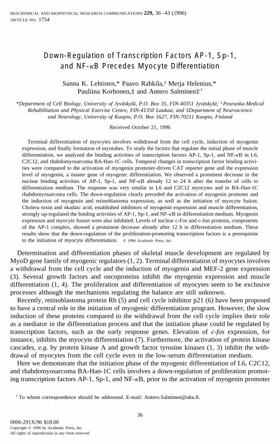
FIG. 1. Down-regulation of transcription factor AP-1, Sp-1, and NF-kB binding activities during myocyte differenti-ation. Electrophoretic mobility shift assays of AP-1 binding activities in C2C12 cells [A], Sp-1 activities in BA-Han-1C and C2C12 cells [B], and NF-kB binding activities in nuclear and cytoplasmic fractions [C] 12, 24, and 48 h afterthe transfer of myocytes to the low-serum differentiation medium [see Materials and Methods]. Cells at zero hourare the proliferating, nearly confluent myocyte cultures before the switch. Mutated oligonucleotide probe controls forAP-1, Sp-1, and NF-kB did not show any specific binding activity. The addition of 10-100 times excess of competingunlabelled probe to binding assays inhibited each of the specific bindings [data not shown].
kinase (4). In the present experiments, the immunostaining of myogenin was nearly absent inproliferating myocytes but myogenin positive nuclei were present already 24 h after transferto the differentiation medium and the fusion of myocytes appeared after 48 h differentiation.In confluent BA-Han-1C cells, the differentiation process induced e.g. by pertussis toxin waseven more vigorous than in L6 and C2C12 myocytes and fusion appeared already after 24 hin differentiation medium (10, data not shown).
Myocyte transfer to differentiation medium strongly reduced the nuclear binding activitiesof transcription factors AP-1, Sp-1, and NF-kB both in L6 and C2C12 myocytes, as well as inBA-Han-1C rhabdomyosarcoma cells (Fig. 1-4). Decrease in DNA-binding activities appearedalready 12 h after the switching cells to differentiation medium (Fig. 1). The level and timingof down-regulation slightly varied in separate experiments (Fig. 1-4), perhaps due to cultureconditions, such as the density of myocytes during the switching. After 48 h in differentiationmedium, the binding activities of AP-1, Sp-1, and NF-kB were very low in all myogenic celllines, coincidentally with the most abundant nuclear staining of myogenin. Interestingly, themost dramatic decreases in the binding activities of AP-1 and NF-kB occurred in BA-Han-1C rhabdomyosarcoma cells (Fig. 1 and 3) that also show more effective fusion than L6 andC2C12 myocytes when the differentiation is induced by, e.g., pertussis toxin (10).
Differentiation of BA-Han-1C rhabdomyosarcoma cells affected the gel retardation prop-erties of AP-1 binding complex (Fig. 3B). Differentiation induced the assembly of a faintAP-1 complex which was considerably smaller than in proliferating cells. The remodelingof AP-1 complex during differentiation appeared whether BA-Han-1C cells were inducedto differentiate with suramin, retinoic acid, or pertussis toxin (data not shown). Differentia-
38
AID BBRC 5716 / 6912$$$181 11-14-96 19:15:04 bbrcas AP: BBRC
Vol. 229, No. 1, 1996 BIOCHEMICAL AND BIOPHYSICAL RESEARCH COMMUNICATIONS
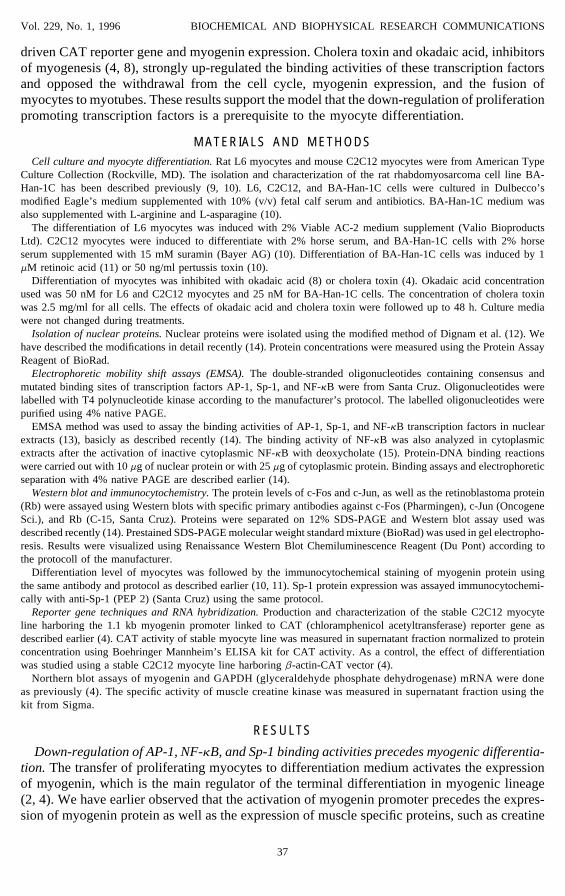
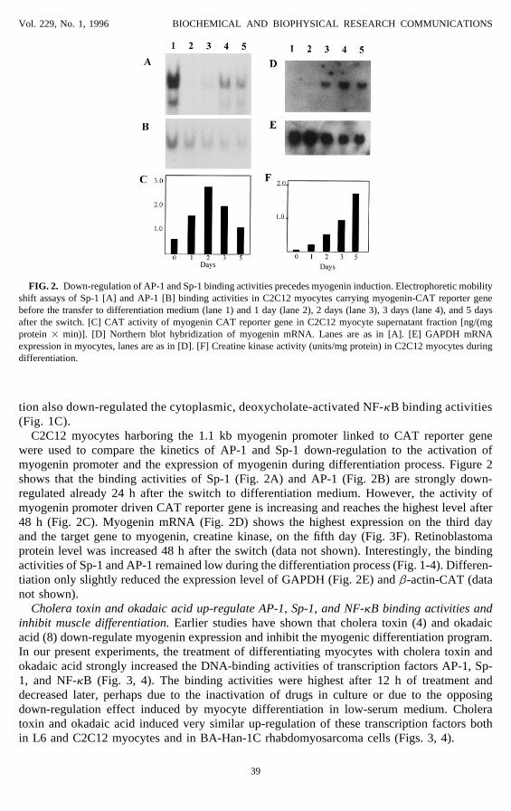
FIG. 2. Down-regulation of AP-1 and Sp-1 binding activities precedes myogenin induction. Electrophoretic mobilityshift assays of Sp-1 [A] and AP-1 [B] binding activities in C2C12 myocytes carrying myogenin-CAT reporter genebefore the transfer to differentiation medium (lane 1) and 1 day (lane 2), 2 days (lane 3), 3 days (lane 4), and 5 daysafter the switch. [C] CAT activity of myogenin CAT reporter gene in C2C12 myocyte supernatant fraction [ng/(mgprotein 1 min)]. [D] Northern blot hybridization of myogenin mRNA. Lanes are as in [A]. [E] GAPDH mRNAexpression in myocytes, lanes are as in [D]. [F] Creatine kinase activity (units/mg protein) in C2C12 myocytes duringdifferentiation.
tion also down-regulated the cytoplasmic, deoxycholate-activated NF-kB binding activities(Fig. 1C).
C2C12 myocytes harboring the 1.1 kb myogenin promoter linked to CAT reporter genewere used to compare the kinetics of AP-1 and Sp-1 down-regulation to the activation ofmyogenin promoter and the expression of myogenin during differentiation process. Figure 2shows that the binding activities of Sp-1 (Fig. 2A) and AP-1 (Fig. 2B) are strongly down-regulated already 24 h after the switch to differentiation medium. However, the activity ofmyogenin promoter driven CAT reporter gene is increasing and reaches the highest level after48 h (Fig. 2C). Myogenin mRNA (Fig. 2D) shows the highest expression on the third dayand the target gene to myogenin, creatine kinase, on the fifth day (Fig. 3F). Retinoblastomaprotein level was increased 48 h after the switch (data not shown). Interestingly, the bindingactivities of Sp-1 and AP-1 remained low during the differentiation process (Fig. 1-4). Differen-tiation only slightly reduced the expression level of GAPDH (Fig. 2E) and b-actin-CAT (datanot shown).
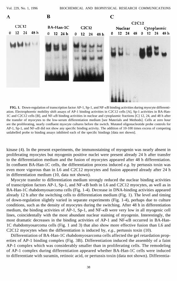
Cholera toxin and okadaic acid up-regulate AP-1, Sp-1, and NF-kB binding activities andinhibit muscle differentiation. Earlier studies have shown that cholera toxin (4) and okadaicacid (8) down-regulate myogenin expression and inhibit the myogenic differentiation program.In our present experiments, the treatment of differentiating myocytes with cholera toxin andokadaic acid strongly increased the DNA-binding activities of transcription factors AP-1, Sp-1, and NF-kB (Fig. 3, 4). The binding activities were highest after 12 h of treatment anddecreased later, perhaps due to the inactivation of drugs in culture or due to the opposingdown-regulation effect induced by myocyte differentiation in low-serum medium. Choleratoxin and okadaic acid induced very similar up-regulation of these transcription factors bothin L6 and C2C12 myocytes and in BA-Han-1C rhabdomyosarcoma cells (Figs. 3, 4).
39
AID BBRC 5716 / 6912$$$181 11-14-96 19:15:04 bbrcas AP: BBRC
Vol. 229, No. 1, 1996 BIOCHEMICAL AND BIOPHYSICAL RESEARCH COMMUNICATIONS
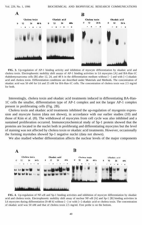
FIG. 3. Up-regulation of AP-1 binding activity and inhibition of myocyte differentiation by okadaic acid andcholera toxin. Electrophoretic mobility shift assays of AP-1 binding activities in L6 myocytes [A] and BA-Han-1Crhabdomyosarcoma cells [B] after 12, 24, and 48 h in the differentiation medium without [0] and with [/] okadaicacid and cholera toxin. Differentiation conditions are described under Materials and Methods. The concentration ofokadaic acid was 50 nM for L6 and 25 nM for BA-Han-1C cells. The concentration of cholera toxin was 2.5 mg/mlfor both.
Interestingly, cholera toxin and okadaic acid treatments induced in differentiating BA-Han-1C cells the smaller, differentiation type of AP-1 complex and not the larger AP-1 complexpresent in proliferating cells (Fig. 2B).
Cholera toxin and okadaic acid treatments inhibited the up-regulation of myogenin expres-sion and myocyte fusion (data not shown), in accordance with our earlier studies (10) andthose of Kim et al. (8). The withdrawal of myocytes from cell cycle was also inhibited and asustained proliferation occurred. Immunocytochemical study of Sp-1 protein showed that theproteins are located in the nuclei both in proliferatig and differentiating myocytes but the levelof staining was not affected by cholera toxin or okadaic acid treatments. However, occasionallythe forming myotubes showed Sp-1 negative nuclei (data not shown).
We also studied whether differentiation affects the nuclear levels of the major components
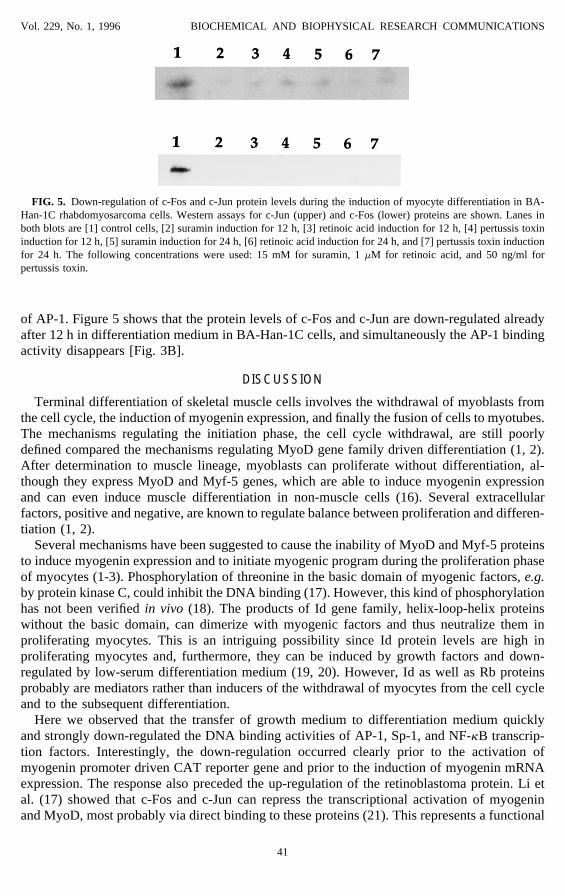
FIG. 4. Up-regulation of NF-kB and Sp-1 binding activities and inhibition of myocyte differentiation by okadaicacid and cholera toxin. Electrophoretic mobility shift assay of nuclear NF-kB [A] and Sp-1 [B] binding activities inL6 myocytes during differentiation [0-48 h] without [0] or with [/] okadaic acid or cholera toxin. The concentrationof okadaic acid was 50 nM and that of cholera toxin 2.5 mg/ml. Free probe is on the bottom.
40
AID BBRC 5716 / 6912$$$181 11-14-96 19:15:04 bbrcas AP: BBRC
Vol. 229, No. 1, 1996 BIOCHEMICAL AND BIOPHYSICAL RESEARCH COMMUNICATIONS
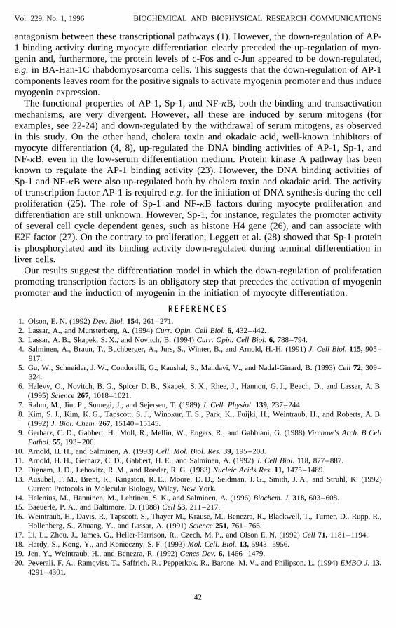
FIG. 5. Down-regulation of c-Fos and c-Jun protein levels during the induction of myocyte differentiation in BA-Han-1C rhabdomyosarcoma cells. Western assays for c-Jun (upper) and c-Fos (lower) proteins are shown. Lanes inboth blots are [1] control cells, [2] suramin induction for 12 h, [3] retinoic acid induction for 12 h, [4] pertussis toxininduction for 12 h, [5] suramin induction for 24 h, [6] retinoic acid induction for 24 h, and [7] pertussis toxin inductionfor 24 h. The following concentrations were used: 15 mM for suramin, 1 mM for retinoic acid, and 50 ng/ml forpertussis toxin.
of AP-1. Figure 5 shows that the protein levels of c-Fos and c-Jun are down-regulated alreadyafter 12 h in differentiation medium in BA-Han-1C cells, and simultaneously the AP-1 bindingactivity disappears [Fig. 3B].
DISCUSSION
Terminal differentiation of skeletal muscle cells involves the withdrawal of myoblasts fromthe cell cycle, the induction of myogenin expression, and finally the fusion of cells to myotubes.The mechanisms regulating the initiation phase, the cell cycle withdrawal, are still poorlydefined compared the mechanisms regulating MyoD gene family driven differentiation (1, 2).After determination to muscle lineage, myoblasts can proliferate without differentiation, al-though they express MyoD and Myf-5 genes, which are able to induce myogenin expressionand can even induce muscle differentiation in non-muscle cells (16). Several extracellularfactors, positive and negative, are known to regulate balance between proliferation and differen-tiation (1, 2).
Several mechanisms have been suggested to cause the inability of MyoD and Myf-5 proteinsto induce myogenin expression and to initiate myogenic program during the proliferation phaseof myocytes (1-3). Phosphorylation of threonine in the basic domain of myogenic factors, e.g.by protein kinase C, could inhibit the DNA binding (17). However, this kind of phosphorylationhas not been verified in vivo (18). The products of Id gene family, helix-loop-helix proteinswithout the basic domain, can dimerize with myogenic factors and thus neutralize them inproliferating myocytes. This is an intriguing possibility since Id protein levels are high inproliferating myocytes and, furthermore, they can be induced by growth factors and down-regulated by low-serum differentiation medium (19, 20). However, Id as well as Rb proteinsprobably are mediators rather than inducers of the withdrawal of myocytes from the cell cycleand to the subsequent differentiation.
Here we observed that the transfer of growth medium to differentiation medium quicklyand strongly down-regulated the DNA binding activities of AP-1, Sp-1, and NF-kB transcrip-tion factors. Interestingly, the down-regulation occurred clearly prior to the activation ofmyogenin promoter driven CAT reporter gene and prior to the induction of myogenin mRNAexpression. The response also preceded the up-regulation of the retinoblastoma protein. Li etal. (17) showed that c-Fos and c-Jun can repress the transcriptional activation of myogeninand MyoD, most probably via direct binding to these proteins (21). This represents a functional
41
AID BBRC 5716 / 6912$$$181 11-14-96 19:15:04 bbrcas AP: BBRC
Vol. 229, No. 1, 1996 BIOCHEMICAL AND BIOPHYSICAL RESEARCH COMMUNICATIONS
antagonism between these transcriptional pathways (1). However, the down-regulation of AP-1 binding activity during myocyte differentiation clearly preceded the up-regulation of myo-genin and, furthermore, the protein levels of c-Fos and c-Jun appeared to be down-regulated,e.g. in BA-Han-1C rhabdomyosarcoma cells. This suggests that the down-regulation of AP-1components leaves room for the positive signals to activate myogenin promoter and thus inducemyogenin expression.
The functional properties of AP-1, Sp-1, and NF-kB, both the binding and transactivationmechanisms, are very divergent. However, all these are induced by serum mitogens (forexamples, see 22-24) and down-regulated by the withdrawal of serum mitogens, as observedin this study. On the other hand, cholera toxin and okadaic acid, well-known inhibitors ofmyocyte differentiation (4, 8), up-regulated the DNA binding activities of AP-1, Sp-1, andNF-kB, even in the low-serum differentiation medium. Protein kinase A pathway has beenknown to regulate the AP-1 binding activity (23). However, the DNA binding activities ofSp-1 and NF-kB were also up-regulated both by cholera toxin and okadaic acid. The activityof transcription factor AP-1 is required e.g. for the initiation of DNA synthesis during the cellproliferation (25). The role of Sp-1 and NF-kB factors during myocyte proliferation anddifferentiation are still unknown. However, Sp-1, for instance, regulates the promoter activityof several cell cycle dependent genes, such as histone H4 gene (26), and can associate withE2F factor (27). On the contrary to proliferation, Leggett et al. (28) showed that Sp-1 proteinis phosphorylated and its binding activity down-regulated during terminal differentiation inliver cells.
Our results suggest the differentiation model in which the down-regulation of proliferationpromoting transcription factors is an obligatory step that precedes the activation of myogeninpromoter and the induction of myogenin in the initiation of myocyte differentiation.
REFERENCES1. Olson, E. N. (1992) Dev. Biol. 154, 261–271.2. Lassar, A., and Munsterberg, A. (1994) Curr. Opin. Cell Biol. 6, 432–442.3. Lassar, A. B., Skapek, S. X., and Novitch, B. (1994) Curr. Opin. Cell Biol. 6, 788–794.4. Salminen, A., Braun, T., Buchberger, A., Jurs, S., Winter, B., and Arnold, H.-H. (1991) J. Cell Biol. 115, 905–
917.5. Gu, W., Schneider, J. W., Condorelli, G., Kaushal, S., Mahdavi, V., and Nadal-Ginard, B. (1993) Cell 72, 309–
324.6. Halevy, O., Novitch, B. G., Spicer D. B., Skapek, S. X., Rhee, J., Hannon, G. J., Beach, D., and Lassar, A. B.
(1995) Science 267, 1018–1021.7. Rahm, M., Jin, P., Sumegi, J., and Sejersen, T. (1989) J. Cell. Physiol. 139, 237–244.8. Kim, S. J., Kim, K. G., Tapscott, S. J., Winokur, T. S., Park, K., Fuijki, H., Weintraub, H., and Roberts, A. B.
(1992) J. Biol. Chem. 267, 15140–15145.9. Gerharz, C. D., Gabbert, H., Moll, R., Mellin, W., Engers, R., and Gabbiani, G. (1988) Virchow’s Arch. B Cell
Pathol. 55, 193–206.10. Arnold, H. H., and Salminen, A. (1993) Cell. Mol. Biol. Res. 39, 195–208.11. Arnold, H. H., Gerharz, C. D., Gabbert, H. E., and Salminen, A. (1992) J. Cell Biol. 118, 877–887.12. Dignam, J. D., Lebovitz, R. M., and Roeder, R. G. (1983) Nucleic Acids Res. 11, 1475–1489.13. Ausubel, F. M., Brent, R., Kingston, R. E., Moore, D. D., Seidman, J. G., Smith, J. A., and Struhl, K. (1992)
Current Protocols in Molecular Biology, Wiley, New York.14. Helenius, M., Hanninen, M., Lehtinen, S. K., and Salminen, A. (1996) Biochem. J. 318, 603–608.15. Baeuerle, P. A., and Baltimore, D. (1988) Cell 53, 211–217.16. Weintraub, H., Davis, R., Tapscott, S., Thayer M., Krause, M., Benezra, R., Blackwell, T., Turner, D., Rupp, R.,
Hollenberg, S., Zhuang, Y., and Lassar, A. (1991) Science 251, 761–766.17. Li, L., Zhou, J., James, G., Heller-Harrison, R., Czech, M. P., and Olson E. N. (1992) Cell 71, 1181–1194.18. Hardy, S., Kong, Y., and Konieczny, S. F. (1993) Mol. Cell. Biol. 13, 5943–5956.19. Jen, Y., Weintraub, H., and Benezra, R. (1992) Genes Dev. 6, 1466–1479.20. Peverali, F. A., Ramqvist, T., Saffrich, R., Pepperkok, R., Barone, M. V., and Philipson, L. (1994) EMBO J. 13,
4291–4301.
42
AID BBRC 5716 / 6912$$$181 11-14-96 19:15:04 bbrcas AP: BBRC
Vol. 229, No. 1, 1996 BIOCHEMICAL AND BIOPHYSICAL RESEARCH COMMUNICATIONS
21. Bengal, E., Ransone, L., Scharfmann, R., Dwarki, V. J., Tapscott, S. J., Weintraub, H., and Verma, I. M. (1992)Cell 68, 507–519.
22. Hirsch, S., and Miskimins, W. K. (1995) Cell Growth Differ. 6, 719–726.23. Pennypacker, K. R., Hong, J.-S., and McMillian, M. K. (1994) FASEB J. 8, 475–478.24. Olashaw, N. E., Kowalik, T. F., Huang, E. S., and Pledger, W. J. (1992) Mol. Biol. Cell 3, 1131–1139.25. Riabowol, K., Schiff, J., and Gilman, M. Z. (1992) Proc. Natl. Acad. Sci. USA 89, 157–161.26. Birnbaum, M. J., Wright, K. L., van Wijnen, A. J., Ramsey-Ewing, A. L., Bourke, M. T., Last, T. J., Aziz, F.,
Frenkel, B., Rao, B. R., Aronin, N., Stein, G. S., and Stein, J. L. (1995) Biochemistry 34, 7648–7658.27. Lin, S.-Y., Black, A. R., Kostic, D., Pajovic, S., Hoover, C. N., and Azizkhan, J. C. (1996) Mol. Cell. Biol. 16,
1668–1675.28. Leggett, R. W., Armstrong, S. A., Barry, D., and Mueller, C. R. (1995) J. Biol. Chem. 270, 25879–25884.
43
AID BBRC 5716 / 6912$$$181 11-14-96 19:15:04 bbrcas AP: BBRC