double outlet right ventricle: aetiologies and associations
TRANSCRIPT
Double outlet right ventricle: aetiologies andassociations
D Obler,1 A L Juraszek,2 L B Smoot,2 M R Natowicz3
1 Children’s Hospital, Boston,Massachusetts, USA; 2 HarvardMedical School and Children’sHospital, Boston,Massachusetts, USA; 3 GenomicMedicine Institute andDepartments of Neurology,Pathology and Pediatrics,Cleveland Clinic Foundation,Cleveland, Ohio, USA
Correspondence to:D Obler, Department ofCardiology, Children’s Hospital,Boston, MA 02115, USA;[email protected]
Received 23 January 2008Revised 28 March 2008Accepted 31 March 2008Published Online First 2 May 2008
ABSTRACTBackground: Double outlet right ventricle (DORV), aclinically significant congenital heart defect, occurs in1–3% of individuals with congenital heart defects. Incontrast to other major congenital heart defects, there areno systematic or comprehensive data regarding associa-tions, aetiologies, and pathogenesis of DORV. Weanalysed reported cases in the medical literature toaddress these issues.Methods: We queried the PubMed database using keywords ‘‘double outlet right ventricle’’ and ‘‘DORV’’ for casereports, epidemiologic analyses and animal studies withthis cardiac anomaly. The anatomic subtype of DORV wasclassified according to criteria of Van Praagh.Results: Chromosomal abnormalities were present in 61of the 149 cases of DORV. Trisomies 13 and 18, and del22q11 were the most commonly associated cytogeneticlesions; different anatomic subtypes of DORV were notedin trisomies 13 and 18 versus del 22q11. DORV wasreported in many uncommon or rare non-chromosomalsyndromes. Mutations and non-synonymous sequencevariants in the CFC1 and CSX genes were the mostcommonly reported monogenic loci associated with DORVin humans; numerous genes are reported in murinemodels of DORV. Animal studies implicate maternaldiabetes and prenatal exposure to ethanol, retinoids,theophylline, and valproate in DORV teratogenesis.Conclusions: The large number of genes associated withDORV in both humans and animal models and thedifferent anatomic subtypes seen in specific aetiologiesindicate the likelihood of several distinct pathogeneticmechanisms for DORV, including impairment of neuralcrest derivative migration and impairment of normalcardiac situs and looping.
Congenital cardiovascular malformations arefound in approximately 4–8/1000 newborns andrepresent a common cause of paediatric morbidityand mortality.1–5 The incidence may be as much as10-fold greater in fetuses, due to the high frequencyof fetal demise in the setting of severe malforma-tions.6 Recent reports indicate an increasing pre-valence of congenital heart defects, butacknowledge that the increase is likely due toimprovements in ascertainment and reporting,inclusion of broader categories of defects, andadvances in pregnancy management and subse-quent repair/palliation of complex congenital mal-formations.3 7
Congenital heart defects (CHDs) represent amajor proportion of clinically significant birthdefects.1 6 While most CHDs occur as isolatedmalformations, a substantial minority occurs incombination with abnormalities of other organsystems.5 8 9 Aetiologic categories of congenital
heart disease include chromosomal abnormalities,teratogenic exposures, single gene disorders, andmultifactorial determination.5 10–15 The underlyingbasis for most cases of non-syndromic CHD iscurrently unexplained. However, there has beensubstantial recent progress in knowledge of geneticfactors involved in the development of cardiacstructural abnormalities for both isolated andsyndromic CHD.16–22
Combined cytogenetic–epidemiologic analyseshave identified discrete chromosomal regionsinvolved in the pathogenesis of many congenitalcardiac lesions.23–27 Elucidation of the moleculargenetic basis of numerous single and contiguousgene syndromes associated with cardiaclesions14 15 28–30 also adds to our current under-standing. Despite these recent advances, doubleoutlet right ventricle (DORV) remains one of theleast understood categories of CHD.
A key issue in any analysis of DORV concerns itsdefinition. In general, the term ‘‘double outlet rightventricle’’ refers to a family of anatomically relatedcomplex congenital cardiac lesions involving theoutflow tracts. During the development of theheart, the outflow tract initially connects exclu-sively with the primitive right ventricle and mustundergo extensive remodelling to divide into aseparate pulmonary artery and aorta; subse-quently, there is continued remodelling to establishdirect continuity from the left ventricle to theaorta. The endocardial cushions in the outflowtract are responsible for formation of the semilunarvalves as well as for the development of the conalseptum, the portion of the ventricular septumbetween the distal ventricular outflow tracts.
DORV anatomy was first described by Mery in1703.31 More than 200 years later, the term‘‘double-outlet ventricle’’ was employed by Braunet al32 in 1952. Shortly thereafter Witham described‘‘double outlet right ventricle’’ as a specific cardiacdiagnosis.33 In 1972, Lev et al34 used the relationshipof the VSD to the great arteries as the basis for hisclassification, which remains one of the mostwidely used clinical classification schemes appliedto DORV.
As reviewed in Walters et al,35 some authors usedthe degree of aortic override as a defining criterionfor the diagnosis of DORV such that if the aorta ismore than 50% over the right ventricle, it islabelled DORV. This ‘‘50% rule’’ becomes proble-matic in cases of tetralogy of Fallot with extremeoverride of the aorta. Alternatively, the absence orloss of normal fibrous continuity between themitral and aortic valves (that is, presence ofsubaortic conus) has been proposed as a definitionof DORV. This, too, is problematic as the presence
Review
J Med Genet 2008;45:481–497. doi:10.1136/jmg.2008.057984 481
group.bmj.com on February 19, 2013 - Published by jmg.bmj.comDownloaded from
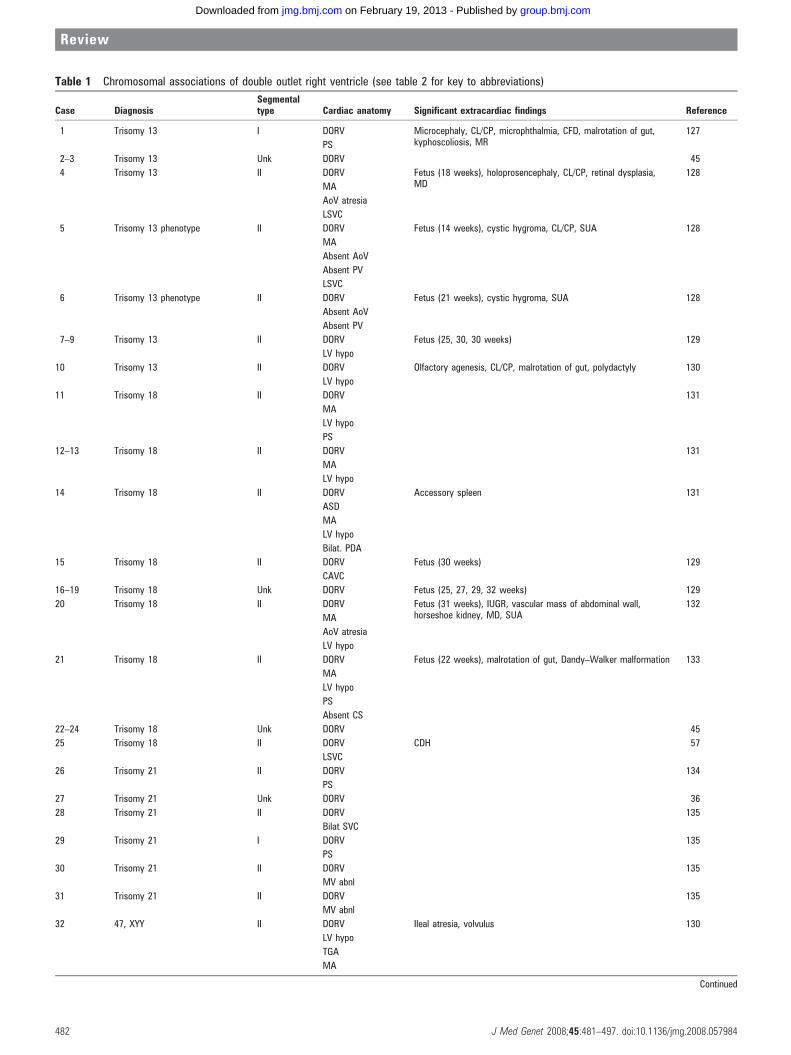
Table 1 Chromosomal associations of double outlet right ventricle (see table 2 for key to abbreviations)
Case DiagnosisSegmentaltype Cardiac anatomy Significant extracardiac findings Reference
1 Trisomy 13 I DORV Microcephaly, CL/CP, microphthalmia, CFD, malrotation of gut,kyphoscoliosis, MR
127
PS
2–3 Trisomy 13 Unk DORV 45
4 Trisomy 13 II DORV Fetus (18 weeks), holoprosencephaly, CL/CP, retinal dysplasia,MD
128
MA
AoV atresia
LSVC
5 Trisomy 13 phenotype II DORV Fetus (14 weeks), cystic hygroma, CL/CP, SUA 128
MA
Absent AoV
Absent PV
LSVC
6 Trisomy 13 phenotype II DORV Fetus (21 weeks), cystic hygroma, SUA 128
Absent AoV
Absent PV
7–9 Trisomy 13 II DORV Fetus (25, 30, 30 weeks) 129
LV hypo
10 Trisomy 13 II DORV Olfactory agenesis, CL/CP, malrotation of gut, polydactyly 130
LV hypo
11 Trisomy 18 II DORV 131
MA
LV hypo
PS
12–13 Trisomy 18 II DORV 131
MA
LV hypo
14 Trisomy 18 II DORV Accessory spleen 131
ASD
MA
LV hypo
Bilat. PDA
15 Trisomy 18 II DORV Fetus (30 weeks) 129
CAVC
16–19 Trisomy 18 Unk DORV Fetus (25, 27, 29, 32 weeks) 129
20 Trisomy 18 II DORV Fetus (31 weeks), IUGR, vascular mass of abdominal wall,horseshoe kidney, MD, SUA
132
MA
AoV atresia
LV hypo
21 Trisomy 18 II DORV Fetus (22 weeks), malrotation of gut, Dandy–Walker malformation 133
MA
LV hypo
PS
Absent CS
22–24 Trisomy 18 Unk DORV 45
25 Trisomy 18 II DORV CDH 57
LSVC
26 Trisomy 21 II DORV 134
PS
27 Trisomy 21 Unk DORV 36
28 Trisomy 21 II DORV 135
Bilat SVC
29 Trisomy 21 I DORV 135
PS
30 Trisomy 21 II DORV 135
MV abnl
31 Trisomy 21 II DORV 135
MV abnl
32 47, XYY II DORV Ileal atresia, volvulus 130
LV hypo
TGA
MA
Continued
Review
482 J Med Genet 2008;45:481–497. doi:10.1136/jmg.2008.057984
group.bmj.com on February 19, 2013 - Published by jmg.bmj.comDownloaded from
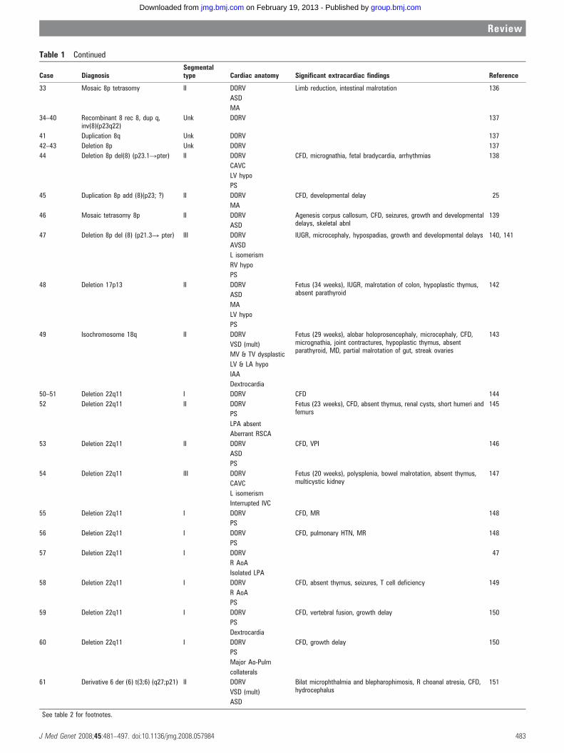
Table 1 Continued
Case DiagnosisSegmentaltype Cardiac anatomy Significant extracardiac findings Reference
33 Mosaic 8p tetrasomy II DORV Limb reduction, intestinal malrotation 136
ASD
MA
34–40 Recombinant 8 rec 8, dup q,inv(8)(p23q22)
Unk DORV 137
41 Duplication 8q Unk DORV 137
42–43 Deletion 8p Unk DORV 137
44 Deletion 8p del(8) (p23.1Rpter) II DORV CFD, micrognathia, fetal bradycardia, arrhythmias 138
CAVC
LV hypo
PS
45 Duplication 8p add (8)(p23; ?) II DORV CFD, developmental delay 25
MA
46 Mosaic tetrasomy 8p II DORV Agenesis corpus callosum, CFD, seizures, growth and developmentaldelays, skeletal abnl
139
ASD
47 Deletion 8p del (8) (p21.3R pter) III DORV IUGR, microcephaly, hypospadias, growth and developmental delays 140, 141
AVSD
L isomerism
RV hypo
PS
48 Deletion 17p13 II DORV Fetus (34 weeks), IUGR, malrotation of colon, hypoplastic thymus,absent parathyroid
142
ASD
MA
LV hypo
PS
49 Isochromosome 18q II DORV Fetus (29 weeks), alobar holoprosencephaly, microcephaly, CFD,micrognathia, joint contractures, hypoplastic thymus, absentparathyroid, MD, partial malrotation of gut, streak ovaries
143
VSD (mult)
MV & TV dysplastic
LV & LA hypo
IAA
Dextrocardia
50–51 Deletion 22q11 I DORV CFD 144
52 Deletion 22q11 II DORV Fetus (23 weeks), CFD, absent thymus, renal cysts, short humeri andfemurs
145
PS
LPA absent
Aberrant RSCA
53 Deletion 22q11 II DORV CFD, VPI 146
ASD
PS
54 Deletion 22q11 III DORV Fetus (20 weeks), polysplenia, bowel malrotation, absent thymus,multicystic kidney
147
CAVC
L isomerism
Interrupted IVC
55 Deletion 22q11 I DORV CFD, MR 148
PS
56 Deletion 22q11 I DORV CFD, pulmonary HTN, MR 148
PS
57 Deletion 22q11 I DORV 47
R AoA
Isolated LPA
58 Deletion 22q11 I DORV CFD, absent thymus, seizures, T cell deficiency 149
R AoA
PS
59 Deletion 22q11 I DORV CFD, vertebral fusion, growth delay 150
PS
Dextrocardia
60 Deletion 22q11 I DORV CFD, growth delay 150
PS
Major Ao-Pulm
collaterals
61 Derivative 6 der (6) t(3;6) (q27;p21) II DORV Bilat microphthalmia and blepharophimosis, R choanal atresia, CFD,hydrocephalus
151
VSD (mult)
ASD
See table 2 for footnotes.
Review
J Med Genet 2008;45:481–497. doi:10.1136/jmg.2008.057984 483
group.bmj.com on February 19, 2013 - Published by jmg.bmj.comDownloaded from
of subaortic conus is a continuous variable in DORV and onethat does not lend itself to a binary or dichotomous definition.35
The Congenital Heart Surgery Nomenclature and DatabaseProject was developed to provide a more unified and inclusiveframework for classification of congenital heart disease andassessment of surgical repair.35 The consensus definition ofDORV was made deliberately broad by stating ‘‘DORV is a typeof ventriculoarterial connection in which both great vessels ariseeither entirely or predominantly from the right ventricle’’.
Consistent with other complex CHDs, DORV may occur asan isolated cardiac defect, together with other cardiac lesions, orin association with extracardiac anomalies.31 36–42 It occurs inapproximately 3–9/100 000 live births,1 4 43 although at least onereport noted rates of between 15–24/100 000.3 Conservativeestimates project DORV accounting for about 1–3% of allcongenital heart defects.1 44
Unlike other major congenital heart lesions, there has beenlittle systematic study of the aetiologic bases of DORV. To date,no comprehensive investigations—retrospective or prospec-tive—have been performed to evaluate potential developmentalanomalies and genetic associations with DORV. We report herea comprehensive analysis of genetic disorders and teratogenicagents associated with DORV organised by distinct anatomicsubtypes whenever possible, in an effort to identify relevantdevelopmental processes underlying this disorder.
METHODSThe medical literature was reviewed for cases of DORV. Caseswere ascertained in the English language literature usingPubMed literature searches with ‘‘double outlet right ventricle’’and ‘‘DORV’’ as key words, as well as review of references inarticles describing cases of DORV. Both epidemiologic analysesof congenital heart disease and case reports were used.
We defined a congenital heart lesion as DORV if both greatarteries (that is, the aorta and pulmonary artery) are related tothe morphologically right ventricle either by (1) both arisingfrom the conus (infundibulum) or (2) one great artery arisesfrom the conus and the other great artery has fibrous continuitywith only the right ventricle (RV) portion of the atrioventri-cular (AV) canal (tricuspid valve, right ventricular portion of acommon AV valve or RV portion of a straddling mitral valve).
We excluded cases with preserved mitral valve to semilunarvalve fibrous continuity. And although forms of tetralogy ofFallot with extreme override sometimes have been classified ascases of DORV, this was not included in our definition ofDORV phenotypes because of arbitrariness of the ‘‘50% rule’’.
When sufficient anatomic detail was provided, cases from theliterature were further sub-categorised into three types: type IDORV as an isolated conotruncal anomaly; type II DORV withconotruncal anomalies and associated malformations of the AVvalves and ventricles; and type III DORV associated withheterotaxy (polysplenia, asplenia, atrial isomerism).31
Documentation of either cardiac isomerism or a combinationof characteristic cardiac/vascular malformations in associationwith visceral situs was necessary to be included in theheterotaxy category. This classification scheme provides adetailed anatomic framework by which to examine theheterogeneous group of DORV malformations.
Each case was reviewed for: pregnancy history and familyhistory, if available; cardiac anatomy; major physical findingsnoted on examination and/or autopsy; and results of diagnostictesting (including cytogenetic, biochemical, and moleculargenetic analyses). Only cases with a definitive genetic diagnosisor those without a definitive diagnosis but with adequate
clinical or pathologic detail were included. Cases of DORVreported in experimental animals were also reviewed.
RESULTS
Chromosomal abnormalities associated with DORVA variety of chromosomal abnormalities were noted in 61 of the149 cases of DORV included in this analysis (table 1),comprising slightly less than 41% of reported cases. DORVwas observed in conjunction with aneuploidies, as well ascytogenetic duplications, deletions and rearrangements.
DORV is a relatively rare diagnosis in the common autosomaltrisomies. Nonetheless, the common trisomies comprise asubstantial fraction of the reported chromosomal associationswith DORV (31/61 cases), with 15 and 10 cases of definite orpresumed trisomies 18 and 13, respectively. In contrast to theoverall frequency of trisomy 21 in children and fetuses, only sixcases of DORV were reported in association with thiscytogenetic abnormality. One necropsy study examiningCHDs associated with chromosomal abnormalities found a12% (15/129) prevalence of DORV, but no cases of DORVassociated with trisomy 21.45 Epidemiologic data support anincreased risk of DORV in trisomies 13 and 18 but nocomparable heightened risk in trisomy 21.46
Seven of the 10 cases (70%) of trisomy 13 had DORV withabnormal left heart development. At least six of 15 cases (40%)of trisomy 18 also demonstrated hypoplastic left heart devel-opment; insufficient anatomic detail was provided for seven ofthe other trisomy 18 cases, precluding classification of DORVsubtype in those cases. Thus, DORV can occur in individualswith trisomy 13 and trisomy 18, with the majority of thesecases occurring in conjunction with hypoplasia of left heartstructures.
Cytogenetic abnormalities involving chromosome 8 werereported in 15 cases of DORV (14/15 involving abnormal dosageof 8p), comprising 10% of cases. Most of these cases showedtype II DORV (for example, mitral atresia, ventricularhypoplasia, and complete AV canal).
Eleven cases (7%) of DORV were reported in association withdeletion of chromosome 22q11. Of these cases, eight of 11demonstrated a type I DORV cardiac phenotype (that is, withconotruncal abnormalities only); no cases of hypoplastic leftheart development were reported. In all postnatal cases wherenon-cardiac phenotypic data were reported, craniofacial dys-morphism was also noted. While cases of DORV and 22q11deletion have been reported, DORV appears to be anuncommon or rare finding within the 22q11 deletion syndromepopulations previously studied.47–49
Non-chromosomal disorders associated with DORVA variety of non-chromosomal conditions have been associatedwith DORV and comprised over 56% (84/149) of the cases inthis analysis (table 2). DORV has been reported in the followingsyndromes: Adams–Oliver, Ellis–van Creveld, Gardner–Silengo–Wachtel, Kabuki, Kalmann, Melnick–Needles, Noonan, Opitz,Ritscher–Schinzel, and Robinow syndromes.
Eight cases (,5%) of DORV (five with heterotaxy) wereassociated with a mutation or non-synonymous sequencevariant(s) in the CFC1 gene; current data support the sequencevariants in causation or predisposition to DORV in somepopulations.50–52 Although the CFC1 gene has been implicated inestablishment of left-right asymmetry in vertebrates,50 52 it hasbeen noted in cases of DORV both with and without lateralitydefects.51 52 The EGF-CFC gene family, of which CFC1 is a
Review
484 J Med Genet 2008;45:481–497. doi:10.1136/jmg.2008.057984
group.bmj.com on February 19, 2013 - Published by jmg.bmj.comDownloaded from
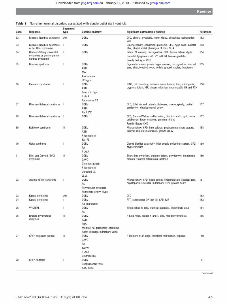
Table 2 Non-chromosomal disorders associated with double outlet right ventricle
Case DiagnosisSegmentaltype Cardiac anatomy Significant extracardiac findings Reference
62 Melnick–Needles syndrome Unk DORV CFD, skeletal dysplasia, motor delay, phosphate reabsorptionlow
152
63 Melnick–Needles syndromeor ter Haar syndrome
I DORV Brachycephaly, congenital glaucoma, CFD, hypo nails, skeletalabnl, absent distal phalanges of toes, SUA
153
64 Gardner–Silengo–Wachtelsyndrome or genito–palato-cardiac syndrome
I DORV Fetus (21 weeks), micrognathia, CFD, flexion deform digits 154
Gonadal dysgenesis: 46, XY with NL female genitalia
Family history of CHD
65 Noonan syndrome II DORV Pigmented nevus, ptosis, hypertelorism, micrognathia, low setears, short/webbed neck, widely spaced nipples, hypotonia
155
ASD
MA
AoV atresia
LV hypo
66 Kalmann syndrome II DORV IUGR, microcephaly, sensory neural hearing loss, micropenis,cryptorchidism, MR, absent olfaction, undetectable LH and FSH
156
ASD
Pulm art: hypo
R AoA
Anomalous CA
67 Ritscher–Schinzel syndrome II DORV CFD, Bilat iris and retinal colobomas, macrocephaly, partialsyndactyly, developmental delay
157
ASD
Abnl SVC
68 Ritscher–Schinzel syndrome I DORV CFD, Dandy–Walker malformation, bilat iris and L optic nervecolobomas, large fontanels, proximal thumb
157
Family history CHD
69 Robinow syndrome III DORV Microcephaly, CFD, blue sclerae, pre/postnatal short stature,delayed skeletal maturation, growth delay
158
ASD
R isomerism
TA, PS
70 Opitz syndrome I DORV Closed bladder exstrophy, bilat double collecting system, CFD,cryptorchidism
159
PA
R AoA
71 Ellis–van Creveld (EVC)syndrome
III DORV Short limb dwarfism, thoracic defect, polydactyly, ectodermaldefects, visceral heterotaxia, asplenia
160
CAVC
Common atrium
R isomerism
Unroofed CS
LSVC
72 Adams–Oliver syndrome II DORV Microcephaly, CFD, scalp defect, encephalocele, skeletal abnl,hepatoportal sclerosis, pulmonary HTN, growth delay
161
PS
Polyvalvular dysplasia
Pulmonary artery: hypo
73 Kabuki syndrome Unk DORV CFD 162
74 Kabuki syndrome II DORV FTT, submucous CP, ear pit, CFD, MR 163
Ao coarctation
75 VACTERL I DORV Single lobed R lung, tracheal agenesis, imperforate anus 164
PS
76 Rhabdo-myomatousdysplasia
III DORV R lung hypo, bilobar R and L lung, rhabdomyomatosis 165
ASD
PDA
Multiple Ao–pulmonary collaterals
Anom drainage pulmonary veins
77 CFC1 sequence variant III DORV R isomerism of lungs, intestinal malrotation, asplenia 50
CAVC
PA
TAPVR
R AoA
Dextrocardia
78 CFC1 mutation II DORV 51
Subpulmonary VSD
AoA: hypo
Continued
Review
J Med Genet 2008;45:481–497. doi:10.1136/jmg.2008.057984 485
group.bmj.com on February 19, 2013 - Published by jmg.bmj.comDownloaded from
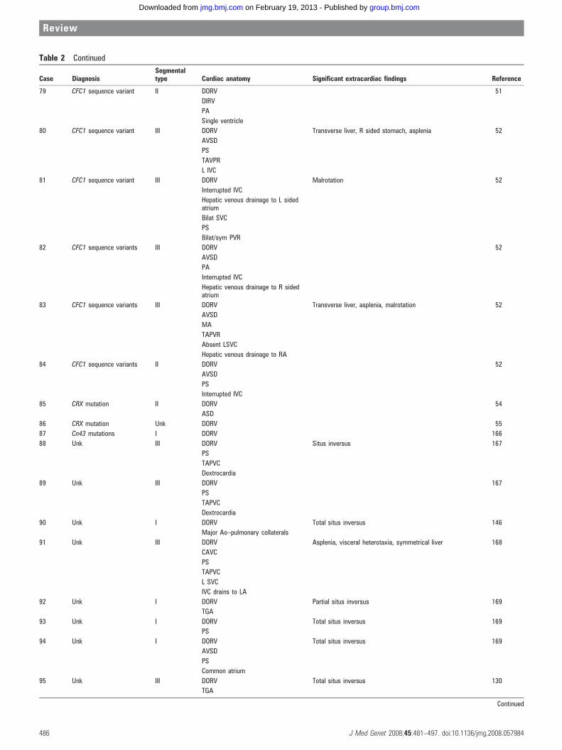
Table 2 Continued
Case DiagnosisSegmentaltype Cardiac anatomy Significant extracardiac findings Reference
79 CFC1 sequence variant II DORV 51
DIRV
PA
Single ventricle
80 CFC1 sequence variant III DORV Transverse liver, R sided stomach, asplenia 52
AVSD
PS
TAVPR
L IVC
81 CFC1 sequence variant III DORV Malrotation 52
Interrupted IVC
Hepatic venous drainage to L sidedatrium
Bilat SVC
PS
Bilat/sym PVR
82 CFC1 sequence variants III DORV 52
AVSD
PA
Interrupted IVC
Hepatic venous drainage to R sidedatrium
83 CFC1 sequence variants III DORV Transverse liver, asplenia, malrotation 52
AVSD
MA
TAPVR
Absent LSVC
Hepatic venous drainage to RA
84 CFC1 sequence variants II DORV 52
AVSD
PS
Interrupted IVC
85 CRX mutation II DORV 54
ASD
86 CRX mutation Unk DORV 55
87 Cn43 mutations I DORV 166
88 Unk III DORV Situs inversus 167
PS
TAPVC
Dextrocardia
89 Unk III DORV 167
PS
TAPVC
Dextrocardia
90 Unk I DORV Total situs inversus 146
Major Ao–pulmonary collaterals
91 Unk III DORV Asplenia, visceral heterotaxia, symmetrical liver 168
CAVC
PS
TAPVC
L SVC
IVC drains to LA
92 Unk I DORV Partial situs inversus 169
TGA
93 Unk I DORV Total situs inversus 169
PS
94 Unk I DORV Total situs inversus 169
AVSD
PS
Common atrium
95 Unk III DORV Total situs inversus 130
TGA
Continued
Review
486 J Med Genet 2008;45:481–497. doi:10.1136/jmg.2008.057984
group.bmj.com on February 19, 2013 - Published by jmg.bmj.comDownloaded from
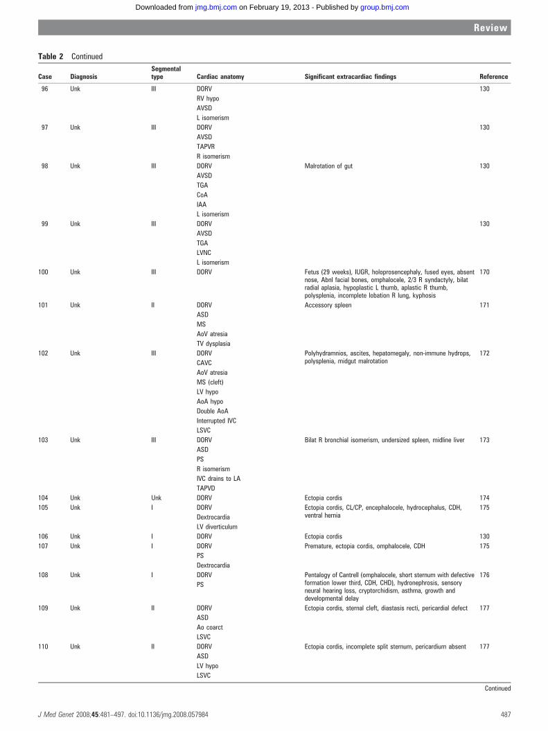
Table 2 Continued
Case DiagnosisSegmentaltype Cardiac anatomy Significant extracardiac findings Reference
96 Unk III DORV 130
RV hypo
AVSD
L isomerism
97 Unk III DORV 130
AVSD
TAPVR
R isomerism
98 Unk III DORV Malrotation of gut 130
AVSD
TGA
CoA
IAA
L isomerism
99 Unk III DORV 130
AVSD
TGA
LVNC
L isomerism
100 Unk III DORV Fetus (29 weeks), IUGR, holoprosencephaly, fused eyes, absentnose, Abnl facial bones, omphalocele, 2/3 R syndactyly, bilatradial aplasia, hypoplastic L thumb, aplastic R thumb,polysplenia, incomplete lobation R lung, kyphosis
170
101 Unk II DORV Accessory spleen 171
ASD
MS
AoV atresia
TV dysplasia
102 Unk III DORV Polyhydramnios, ascites, hepatomegaly, non-immune hydrops,polysplenia, midgut malrotation
172
CAVC
AoV atresia
MS (cleft)
LV hypo
AoA hypo
Double AoA
Interrupted IVC
LSVC
103 Unk III DORV Bilat R bronchial isomerism, undersized spleen, midline liver 173
ASD
PS
R isomerism
IVC drains to LA
TAPVD
104 Unk Unk DORV Ectopia cordis 174
105 Unk I DORV Ectopia cordis, CL/CP, encephalocele, hydrocephalus, CDH,ventral hernia
175
Dextrocardia
LV diverticulum
106 Unk I DORV Ectopia cordis 130
107 Unk I DORV Premature, ectopia cordis, omphalocele, CDH 175
PS
Dextrocardia
108 Unk I DORV Pentalogy of Cantrell (omphalocele, short sternum with defectiveformation lower third, CDH, CHD), hydronephrosis, sensoryneural hearing loss, cryptorchidism, asthma, growth anddevelopmental delay
176
PS
109 Unk II DORV Ectopia cordis, sternal cleft, diastasis recti, pericardial defect 177
ASD
Ao coarct
LSVC
110 Unk II DORV Ectopia cordis, incomplete split sternum, pericardium absent 177
ASD
LV hypo
LSVC
Continued
Review
J Med Genet 2008;45:481–497. doi:10.1136/jmg.2008.057984 487
group.bmj.com on February 19, 2013 - Published by jmg.bmj.comDownloaded from
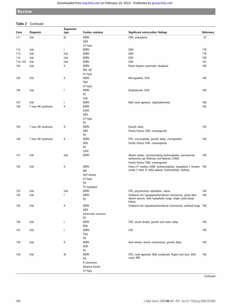
Table 2 Continued
Case DiagnosisSegmentaltype Cardiac anatomy Significant extracardiac findings Reference
111 Unk III DORV CDH, polysplenia 57
ASD
LV hypo
112 Unk I DORV CDH 178
113 Unk Unk DORV CDH 179
114 Unk Unk DORV CDH 180
115–123 Unk Unk DORV CDH 181
124 Unk II DORV Renal–hepatic–pancreatic dysplasia 169
MS, AS
LV hypo
125 Unk II DORV Micrognathia, SUA 169
TGA
LV hypo
126 Unk I DORV Omphalocele, SUA 169
PS
TGA
127 Unk I DORV Bilat renal agenesis, oligohydramnios 169
128 ? new AR syndrome II DORV 182
CAVC
ASD
LV hypo
PS
129 ? new AR syndrome II DORV Growth delay 183
ASD Family history CHD, consanguinity
PS
130 ? new AR syndrome II DORV CFD, microcephaly, growth delay, micrognathia
Family history CHD, consanguinity
183
ASD
PS
LSVC
131 Unk Unk DORV Absent testes, communicating hydrocephalus, peroxisomaldysfunction per Robinow and Beemer (1990)
184
Family history CHD, consanguinity
132 Unk II DORV Fetus (17 weeks), IUGR, hydraencephaly, hypoplastic L forearmw/abs L hand, R radial aplasia, hydrocephaly, hydrops
185
MS
AoV atresia
LV hypo
PA
TV dysplasia
133 Unk Unk DORV CFD, psychomotor retardation, ataxia 146
134 Unk I DORV Unilateral otic hypoplasia/hemifacial microsomia, spinal abnl,absent sacrum, bilat hypoplastic lungs, single cystic/dysplkidney
186
PS
135 Unk II DORV Unilateral otic hypoplasia/hemifacial microsomia, unilobed lungs 186
ASD
Ventricular inversion
PS
136 Unk I DORV CFD, sacral dimple, growth and motor delay 150
PDA
137 Unk I DORV CFD 150
TGA
PS
138 Unk II DORV Anal atresia, thumb contractures, growth delay 150
ASD
PS
139 Unk III DORV CFD, renal agenesis, Bilat syndactyly fingers and toes, bifiduvula, MR
150
PA
R isomerism
Atypical ductus
LV hypo
Continued
Review
488 J Med Genet 2008;45:481–497. doi:10.1136/jmg.2008.057984
group.bmj.com on February 19, 2013 - Published by jmg.bmj.comDownloaded from
member, codes for extracellular proteins that are thought to beessential in intercellular signalling pathways active in lateralplate mesoderm during development.53
Mutations of the CRX gene were associated with two cases ofDORV.54 55 This cardiac specific homeobox gene encodes thetranscription factor Nkx2.5, and has been implicated in bothfirst and secondary heart field development and has beenreported in atrial and ventricular septal defects as well as inelectrical conduction abnormalities.56
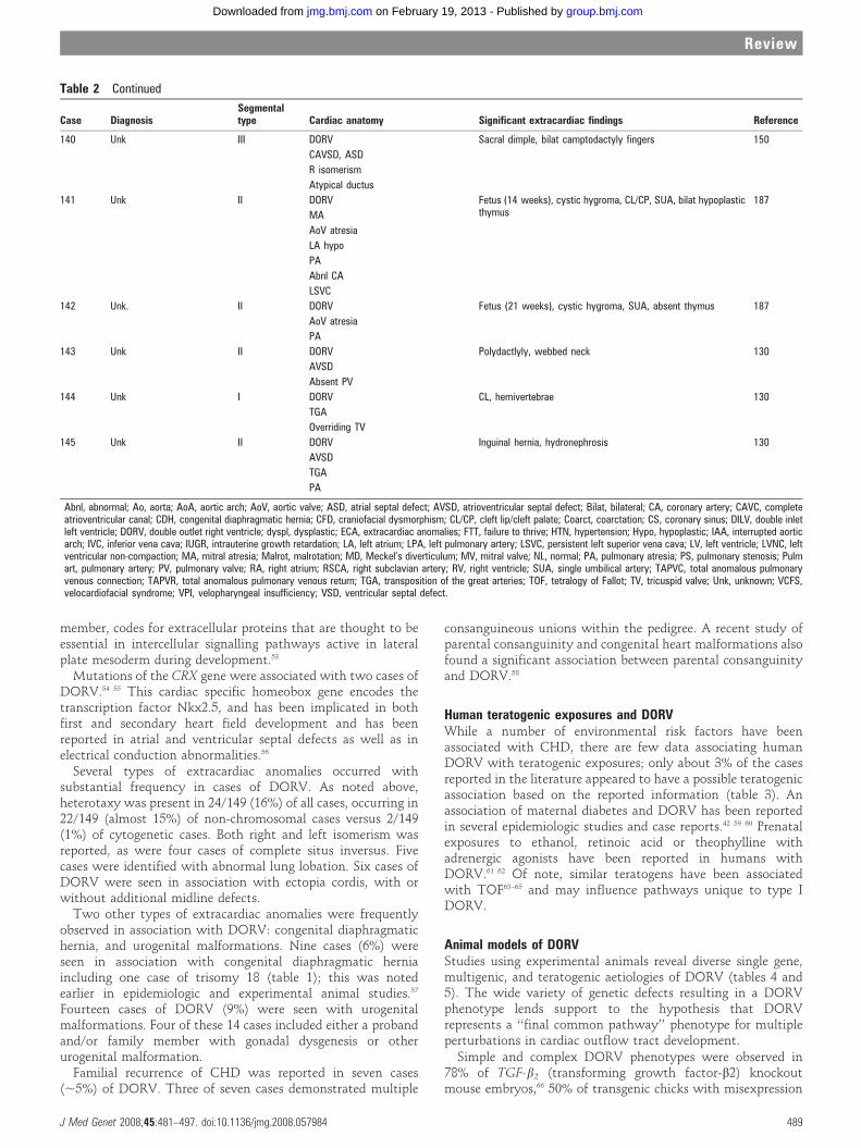
Several types of extracardiac anomalies occurred withsubstantial frequency in cases of DORV. As noted above,heterotaxy was present in 24/149 (16%) of all cases, occurring in22/149 (almost 15%) of non-chromosomal cases versus 2/149(1%) of cytogenetic cases. Both right and left isomerism wasreported, as were four cases of complete situs inversus. Fivecases were identified with abnormal lung lobation. Six cases ofDORV were seen in association with ectopia cordis, with orwithout additional midline defects.
Two other types of extracardiac anomalies were frequentlyobserved in association with DORV: congenital diaphragmatichernia, and urogenital malformations. Nine cases (6%) wereseen in association with congenital diaphragmatic herniaincluding one case of trisomy 18 (table 1); this was notedearlier in epidemiologic and experimental animal studies.57
Fourteen cases of DORV (9%) were seen with urogenitalmalformations. Four of these 14 cases included either a probandand/or family member with gonadal dysgenesis or otherurogenital malformation.
Familial recurrence of CHD was reported in seven cases(,5%) of DORV. Three of seven cases demonstrated multiple
consanguineous unions within the pedigree. A recent study ofparental consanguinity and congenital heart malformations alsofound a significant association between parental consanguinityand DORV.58
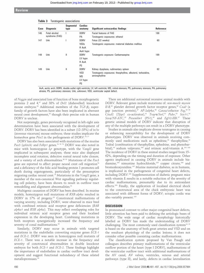
Human teratogenic exposures and DORVWhile a number of environmental risk factors have beenassociated with CHD, there are few data associating humanDORV with teratogenic exposures; only about 3% of the casesreported in the literature appeared to have a possible teratogenicassociation based on the reported information (table 3). Anassociation of maternal diabetes and DORV has been reportedin several epidemiologic studies and case reports.42 59 60 Prenatalexposures to ethanol, retinoic acid or theophylline withadrenergic agonists have been reported in humans withDORV.61 62 Of note, similar teratogens have been associatedwith TOF63–65 and may influence pathways unique to type IDORV.
Animal models of DORVStudies using experimental animals reveal diverse single gene,multigenic, and teratogenic aetiologies of DORV (tables 4 and5). The wide variety of genetic defects resulting in a DORVphenotype lends support to the hypothesis that DORVrepresents a ‘‘final common pathway’’ phenotype for multipleperturbations in cardiac outflow tract development.
Simple and complex DORV phenotypes were observed in78% of TGF-b2 (transforming growth factor-b2) knockoutmouse embryos,66 50% of transgenic chicks with misexpression
Table 2 Continued
Case DiagnosisSegmentaltype Cardiac anatomy Significant extracardiac findings Reference
140 Unk III DORV Sacral dimple, bilat camptodactyly fingers 150
CAVSD, ASD
R isomerism
Atypical ductus
141 Unk II DORV Fetus (14 weeks), cystic hygroma, CL/CP, SUA, bilat hypoplasticthymus
187
MA
AoV atresia
LA hypo
PA
Abnl CA
LSVC
142 Unk. II DORV Fetus (21 weeks), cystic hygroma, SUA, absent thymus 187
AoV atresia
PA
143 Unk II DORV Polydactlyly, webbed neck 130
AVSD
Absent PV
144 Unk I DORV CL, hemivertebrae 130
TGA
Overriding TV
145 Unk II DORV Inguinal hernia, hydronephrosis 130
AVSD
TGA
PA
Abnl, abnormal; Ao, aorta; AoA, aortic arch; AoV, aortic valve; ASD, atrial septal defect; AVSD, atrioventricular septal defect; Bilat, bilateral; CA, coronary artery; CAVC, completeatrioventricular canal; CDH, congenital diaphragmatic hernia; CFD, craniofacial dysmorphism; CL/CP, cleft lip/cleft palate; Coarct, coarctation; CS, coronary sinus; DILV, double inletleft ventricle; DORV, double outlet right ventricle; dyspl, dysplastic; ECA, extracardiac anomalies; FTT, failure to thrive; HTN, hypertension; Hypo, hypoplastic; IAA, interrupted aorticarch; IVC, inferior vena cava; IUGR, intrauterine growth retardation; LA, left atrium; LPA, left pulmonary artery; LSVC, persistent left superior vena cava; LV, left ventricle; LVNC, leftventricular non-compaction; MA, mitral atresia; Malrot, malrotation; MD, Meckel’s diverticulum; MV, mitral valve; NL, normal; PA, pulmonary atresia; PS, pulmonary stenosis; Pulmart, pulmonary artery; PV, pulmonary valve; RA, right atrium; RSCA, right subclavian artery; RV, right ventricle; SUA, single umbilical artery; TAPVC, total anomalous pulmonaryvenous connection; TAPVR, total anomalous pulmonary venous return; TGA, transposition of the great arteries; TOF, tetralogy of Fallot; TV, tricuspid valve; Unk, unknown; VCFS,velocardiofacial syndrome; VPI, velopharyngeal insufficiency; VSD, ventricular septal defect.
Review
J Med Genet 2008;45:481–497. doi:10.1136/jmg.2008.057984 489
group.bmj.com on February 19, 2013 - Published by jmg.bmj.comDownloaded from
of Noggin and associated loss of function of bone morphogeneticproteins 2 and 4,67 and 38% of Dvl2 (dishevelled) knockoutmouse embryos.68 Additional members of the TGF-b2 super-family of growth factors have also been implicated in aberrantneural crest development,69 though their precise role in humanDORV is unclear.
Not surprisingly, genes previously recognised in left-right axisdetermination have been associated with the development ofDORV. DORV has been identified in a subset (12–35%) of iv/iv(inversus viscerum) mouse embryos; these studies implicate thehomeobox gene Pitx2 in the pathogenesis of DORV.70–77
DORV has also been associated with mutations of the murinePax3 (splotch) and Folbp1 genes.70 78 79 DORV was also noted inmice with homozygous Lp genotype, with the Vangl2 geneimplicated in subsequent analyses; these mice also displayedincomplete axial rotation, defective rostral neural tube closure,and a variety of arch abnormalities.80 81 Mutations of the Pax3gene are reported to affect proper neural crest cell migration70
and a deficiency of the folic acid binding protein 1 promotes celldeath during organogenesis, particularly of the presumptivemigrating cardiac neural crest.79 Mutations in the Vangl2 gene, amember of the non-canonical Wnt signalling pathway regulat-ing cell polarity, have been shown to result in outflow tractremodelling and alignment abnormalities.81
Multigenic causation of DORV has been described. In murinemodels, homozygous null mutations of RXR-alpha resulted inDORV in 17% of progeny.82 Conotruncal malformations ofvarying severity, including DORV, were observed in mice bredwith combined retinoic acid receptor gene deficiencies (RARalpha1 and RXR alpha). This may reflect overlapping roles forindividual retinoic acid receptor genes and their localisedexpression in the developing heart. Combining mutations inboth receptors synergistically increased the frequency andseverity of conal malformations.83
Similarly, DORV may occur in animals with targetedmutations in the endothelin converting enzyme genes ECE-1and ECE-2. DORV was seen in homozygous null mutants ofECE-1. There was a notable increase in the frequency andseverity of conotruncal abnormalities in double knockoutembryos for both ECE-1 and ECE-2. These findings highlightthe importance of endothelins in cardiac outflow tract devel-opment and suggest functional redundancy of these relatedmetalloproteases.84
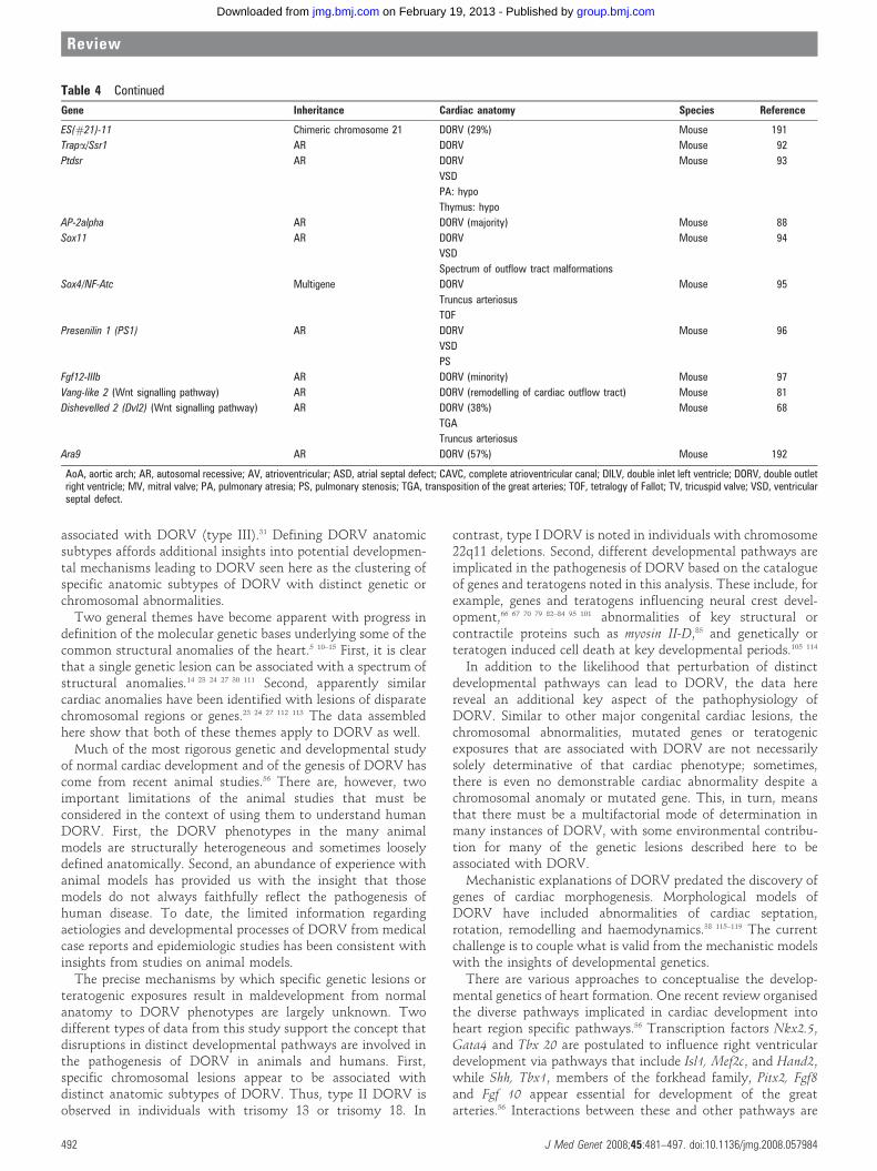
There are additional autosomal recessive animal models withDORV. Relevant genes include mutations of: non-muscle myosinII-B,85 platelet derived growth factor receptor genes,86 Cx40 (agap junction protein),87 AP-2alpha,88 Gata4/cofactor Fog,89 90
Cited2 (Tfap2 co-activator),91 Trapa/Ssrl,92 Ptdsr,93 Sox11,94
Sox4/NF-ATC,95 Presenilin1 (PS1),96 and Fgf12-IIIb.97 Thesegenetic animal models of DORV indicate that disruption ofany of the multiple pathways can result in a DORV phenotype.
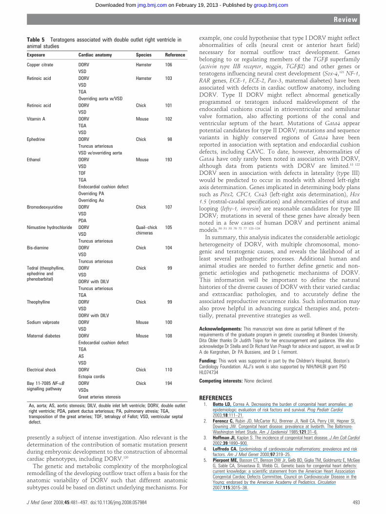
Studies in animals also implicate diverse teratogens in causingor enhancing susceptibility for the development of DORVphenotypes. DORV was observed in animals receiving com-monly used medications such as ephedrine,98 theophylline,99
Tedral (combination of theophylline, ephedrine, and phenobar-bital),99 sodium valproate,100 and retinoic acid/vitamin A.101–103
The incidence of DORV in these animal studies ranged from 15–62%, depending on the timing and duration of exposure. Otheragents implicated in causing DORV in animals include bis-diamine,104 nimustine hydrochloride,105 copper citrate,106 andbromodeoxyuridine.107 Murine maternal diabetes, as in humans,is implicated in the pathogenesis of congenital heart defects,including DORV.108 Supplementation of diabetic pregnant micewith vitamin E results in a notable reduction of the severity ofcardiac malformations, presumably due to its antioxidanteffects.109 Finally, the application of localised electrical shockto the conotruncal area of the chick embryonic heart wasassociated with different forms of DORV; ectopia cordis wasalso variably present.110
DISCUSSIONIn pronounced contrast to other major congenital heart defects,little attention has been paid to defining the aetiologic bases ofDORV. The wide range of cardiac morphology historicallylabelled as DORV has made the understanding of DORVchallenging. The most commonly used classification system34 35
is based on the anatomy of both great arteries and VSD and onthe resultant physiology of the cardiac lesions; it does notconsider other possible coexisting cardiac malformations.
The classification system proposed by Van Praagh andcolleagues describes primary malformations of the ventricularoutflow portion of the heart (type I DORV), malformations ofthe ventricular outflow tract with additional malformations ofthe AV canal, AV valves, ventricles, venous and arterialpathways (type II), and lastly, defects in cardiac lateralisation
Table 3 Teratogenic associations
Case DiagnosisSegmentaltype
Cardiacanatomy Significant extracardiac findings Reference
146 Fetal alcoholsyndrome (FAS)
I DORV Facial features of FAS 188
PA Teratogenic exposures: ethanol
147 Unk I DORV Fetus (22 weeks) 60
VSDs Teratogenic exposures: maternal diabetes mellitus
R AoA
AoA hypo
148 Unk II DORV Teratogenic exposure: Carbamazepine 189
LV hypo
R AoA
PS
149 Unk II DORV Kidney dysplasia, rudimentary spleen 62
VSD Teratogenic exposures: theophylline, albuterol, terbutaline,aminophyllineMS
LV hypo
PS
AoA, aortic arch; DORV, double outlet right ventricle; LV, left ventricle; MS, mitral stenosis; PS, pulmonary stenosis; PA, pulmonaryatresia; PS, pulmonary stenosis; Unk, unknown; VSD, ventricular septal defect.
Review
490 J Med Genet 2008;45:481–497. doi:10.1136/jmg.2008.057984
group.bmj.com on February 19, 2013 - Published by jmg.bmj.comDownloaded from
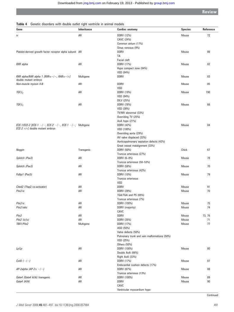
Table 4 Genetic disorders with double outlet right ventricle in animal models
Gene Inheritance Cardiac anatomy Species Reference
iv AR DORV (12%) Mouse 72
CAVC (24%)
Common atrium (17%)
Sinus venosus (9%)
Platelet-derived growth factor receptor alpha subunit AR DORV Mouse 86
TA
Facial cleft
RXR alpha AR DORV (17%) Mouse 82
Hypo compact zone (94%)
VSD (94%)
RXR alpha/RAR alpha 1 [RXRa2/2, RARa2/+]double mutant embryo
Multigene DORV Mouse 83
Non-muscle myosin II-B AR DORV Mouse 85
VSD
TGFb2 AR DORV (19%) Mouse 190
VSD (94%)
DILV (25%)
TGFb2 AR DORV (78%) Mouse 66
VSD (38%)
TV/MV abnormal (33%)
Overriding TV (25%)
AoA hypo (21%)
ECE-1/ECE-2 [ECE-1 2/2; ECE-2 2/2, ECE-1 2/2;ECE-2 +/+] double mutant embryo
Multigene DORV (42%) Mouse 84
VSD (100%)
Overriding aorta (29%)
AV valve displaced (33%)
Aorticopulmonary septation defects (42%)
Great vessel malalignment (33%)
Noggin Transgenic DORV (50%) Chick 67
Truncus arteriosus (27%)
Splotch (Pax3) AR DORV (6–8%) Mouse 78
Truncus arteriosus (50–53%)
Splotch (Pax3) AR DORV (58%) Mouse 70
Truncus arteriosus (42%)
Folbp1 (Pax3) AR DORV (10%) Mouse 79
Truncus arteriosus
VSD
Cited2 (Tfap2 co-activator) AR DORV Mouse 91
Pitx2dc AR DORV (28%) Mouse 70
TGA/TGA and PS (65%)
Truncus arteriosus (7%)
Pitx2dc AR DORV (100%) Mouse 75
Pitx2dabc AR DORV (majority) Mouse 74
CAVC
Pitx2 AR DORV Mouse 73, 76
Pitx2 (iv/iv) AR DORV (35%) Mouse 71
TBX1/Pitx2 Multigene DORV (17%) Mouse 77
ASD (50%)
Valve defects (50%)
Pulmonary trunk and vein malformations (50%)
VSD (25%)
Others (50%)
Lp/Lp AR DORV (100%) Mouse 80
Double AoA (66%)
Right AoA (33%)
Cx40 (2/2) AR DORV (17%) Mouse 87
Endocardial cushion defects (17%)
AP-2alpha (AP-2a 2/2) AR DORV (87%) Mouse 88
Truncus arteriosus (13%)
Gata4 (Gata4 ki/ki) transgenic AR DORV (100%) Mouse 89
Gata4 (H/H) AR DORV Mouse 90
CAVC
Ventricular myocardium hypo
Continued
Review
J Med Genet 2008;45:481–497. doi:10.1136/jmg.2008.057984 491
group.bmj.com on February 19, 2013 - Published by jmg.bmj.comDownloaded from
associated with DORV (type III).31 Defining DORV anatomicsubtypes affords additional insights into potential developmen-tal mechanisms leading to DORV seen here as the clustering ofspecific anatomic subtypes of DORV with distinct genetic orchromosomal abnormalities.
Two general themes have become apparent with progress indefinition of the molecular genetic bases underlying some of thecommon structural anomalies of the heart.5 10–15 First, it is clearthat a single genetic lesion can be associated with a spectrum ofstructural anomalies.14 23 24 27 30 111 Second, apparently similarcardiac anomalies have been identified with lesions of disparatechromosomal regions or genes.23 24 27 112 113 The data assembledhere show that both of these themes apply to DORV as well.
Much of the most rigorous genetic and developmental studyof normal cardiac development and of the genesis of DORV hascome from recent animal studies.56 There are, however, twoimportant limitations of the animal studies that must beconsidered in the context of using them to understand humanDORV. First, the DORV phenotypes in the many animalmodels are structurally heterogeneous and sometimes looselydefined anatomically. Second, an abundance of experience withanimal models has provided us with the insight that thosemodels do not always faithfully reflect the pathogenesis ofhuman disease. To date, the limited information regardingaetiologies and developmental processes of DORV from medicalcase reports and epidemiologic studies has been consistent withinsights from studies on animal models.
The precise mechanisms by which specific genetic lesions orteratogenic exposures result in maldevelopment from normalanatomy to DORV phenotypes are largely unknown. Twodifferent types of data from this study support the concept thatdisruptions in distinct developmental pathways are involved inthe pathogenesis of DORV in animals and humans. First,specific chromosomal lesions appear to be associated withdistinct anatomic subtypes of DORV. Thus, type II DORV isobserved in individuals with trisomy 13 or trisomy 18. In
contrast, type I DORV is noted in individuals with chromosome22q11 deletions. Second, different developmental pathways areimplicated in the pathogenesis of DORV based on the catalogueof genes and teratogens noted in this analysis. These include, forexample, genes and teratogens influencing neural crest devel-opment,66 67 70 79 82–84 95 101 abnormalities of key structural orcontractile proteins such as myosin II-D,85 and genetically orteratogen induced cell death at key developmental periods.105 114
In addition to the likelihood that perturbation of distinctdevelopmental pathways can lead to DORV, the data herereveal an additional key aspect of the pathophysiology ofDORV. Similar to other major congenital cardiac lesions, thechromosomal abnormalities, mutated genes or teratogenicexposures that are associated with DORV are not necessarilysolely determinative of that cardiac phenotype; sometimes,there is even no demonstrable cardiac abnormality despite achromosomal anomaly or mutated gene. This, in turn, meansthat there must be a multifactorial mode of determination inmany instances of DORV, with some environmental contribu-tion for many of the genetic lesions described here to beassociated with DORV.
Mechanistic explanations of DORV predated the discovery ofgenes of cardiac morphogenesis. Morphological models ofDORV have included abnormalities of cardiac septation,rotation, remodelling and haemodynamics.38 115–119 The currentchallenge is to couple what is valid from the mechanistic modelswith the insights of developmental genetics.
There are various approaches to conceptualise the develop-mental genetics of heart formation. One recent review organisedthe diverse pathways implicated in cardiac development intoheart region specific pathways.56 Transcription factors Nkx2.5,Gata4 and Tbx 20 are postulated to influence right ventriculardevelopment via pathways that include Isl1, Mef2c, and Hand2,while Shh, Tbx1, members of the forkhead family, Pitx2, Fgf8and Fgf 10 appear essential for development of the greatarteries.56 Interactions between these and other pathways are
Table 4 Continued
Gene Inheritance Cardiac anatomy Species Reference
ES(#21)-11 Chimeric chromosome 21 DORV (29%) Mouse 191
Trapa/Ssr1 AR DORV Mouse 92
Ptdsr AR DORV Mouse 93
VSD
PA: hypo
Thymus: hypo
AP-2alpha AR DORV (majority) Mouse 88
Sox11 AR DORV Mouse 94
VSD
Spectrum of outflow tract malformations
Sox4/NF-Atc Multigene DORV Mouse 95
Truncus arteriosus
TOF
Presenilin 1 (PS1) AR DORV Mouse 96
VSD
PS
Fgf12-IIIb AR DORV (minority) Mouse 97
Vang-like 2 (Wnt signalling pathway) AR DORV (remodelling of cardiac outflow tract) Mouse 81
Dishevelled 2 (Dvl2) (Wnt signalling pathway) AR DORV (38%) Mouse 68
TGA
Truncus arteriosus
Ara9 AR DORV (57%) Mouse 192
AoA, aortic arch; AR, autosomal recessive; AV, atrioventricular; ASD, atrial septal defect; CAVC, complete atrioventricular canal; DILV, double inlet left ventricle; DORV, double outletright ventricle; MV, mitral valve; PA, pulmonary atresia; PS, pulmonary stenosis; TGA, transposition of the great arteries; TOF, tetralogy of Fallot; TV, tricuspid valve; VSD, ventricularseptal defect.
Review
492 J Med Genet 2008;45:481–497. doi:10.1136/jmg.2008.057984
group.bmj.com on February 19, 2013 - Published by jmg.bmj.comDownloaded from
presently a subject of intense investigation. Also relevant is thedetermination of the contribution of somatic mutation presentduring embryonic development to the construction of abnormalcardiac phenotypes, including DORV.120
The genetic and metabolic complexity of the morphologicalremodelling of the developing outflow tract offers a basis for theanatomic variability of DORV such that different anatomicsubtypes could be based on distinct underlying mechanisms. For
example, one could hypothesise that type I DORV might reflectabnormalities of cells (neural crest or anterior heart field)necessary for normal outflow tract development. Genesbelonging to or regulating members of the TGFb superfamily(activin type IIB receptor, noggin, TGFb2) and other genes orteratogens influencing neural crest development (Sox-4,121 NF-1,RAR genes, ECE-1, ECE-2, Pax-3, maternal diabetes) have beenassociated with defects in cardiac outflow anatomy, includingDORV. Type II DORV might reflect abnormal geneticallyprogrammed or teratogen induced maldevelopment of theendocardial cushions crucial in atrioventricular and semilunarvalve formation, also affecting portions of the conal andventricular septum of the heart. Mutations of Gata4 appearpotential candidates for type II DORV; mutations and sequencevariants in highly conserved regions of Gata4 have beenreported in association with septation and endocardial cushiondefects, including CAVC. To date, however, abnormalities ofGata4 have only rarely been noted in association with DORV,although data from patients with DORV are limited.18 122
DORV seen in association with defects in laterality (type III)would be predicted to occur in models with altered left-rightaxis determination. Genes implicated in determining body planssuch as Pitx2, CFC1, Cx43 (left-right axis determination), Hox1.5 (rostral-caudal specification) and abnormalities of situs andlooping (lefty-1, inversin) are reasonable candidates for type IIIDORV; mutations in several of these genes have already beennoted in a few cases of human DORV and pertinent animalmodels.50 51 53 70 72 77 123–126
In summary, this analysis indicates the considerable aetiologicheterogeneity of DORV, with multiple chromosomal, mono-genic and teratogenic causes, and reveals the likelihood of atleast several pathogenetic processes. Additional human andanimal studies are needed to further define genetic and non-genetic aetiologies and pathogenetic mechanisms of DORV.This information will be important to define the naturalhistories of the diverse causes of DORV with their varied cardiacand extracardiac pathologies, and to accurately define theassociated reproductive recurrence risks. Such information mayalso prove helpful in advancing surgical therapies and, poten-tially, prenatal preventive strategies as well.
Acknowledgements: This manuscript was done as partial fulfilment of therequirements of the graduate program in genetic counselling at Brandeis University.Dita Obler thanks Dr Judith Tsipis for her encouragement and guidance. We alsoacknowledge Dr Stella and Dr Richard Van Praagh for advice and support, as well as DrA de Kergrohen, Dr PA Bussiere, and Dr L Fermont.
Funding: This work was supported in part by the Children’s Hospital, Boston’sCardiology Foundation. ALJ’s work is also supported by NIH/NHLBI grant P50HL074734
Competing interests: None declared.
REFERENCES1. Botto LD, Correa A. Decreasing the burden of congenital heart anomalies: an
epidemiologic evaluation of risk factors and survival. Prog Pediatr Cardiol2003;18:111–21.
2. Ferencz C, Rubin JD, McCarter RJ, Brenner JI, Neill CA, Perry LW, Hepner SI,Downing JW. Congenital heart disease: prevalence at livebirth. The Baltimore-Washington Infant Study. Am J Epidemiol 1985;121:31–6.
3. Hoffman JI, Kaplan S. The incidence of congenital heart disease. J Am Coll Cardiol2002;39:1890–900.
4. Loffredo CA. Epidemiology of cardiovascular malformations: prevalence and riskfactors. Am J Med Genet 2000;97:319–25.
5. Pierpont ME, Basson CT, Benson DW Jr, Gelb BD, Giglia TM, Goldmuntz E, McGeeG, Sable CA, Srivastava D, Webb CL. Genetic basis for congenital heart defects:current knowledge: a scientific statement from the American Heart AssociationCongenital Cardiac Defects Committee, Council on Cardiovascular Disease in theYoung: endorsed by the American Academy of Pediatrics. Circulation2007;115:3015–38.
Table 5 Teratogens associated with double outlet right ventricle inanimal studies
Exposure Cardiac anatomy Species Reference
Copper citrate DORV Hamster 106
VSD
Retinoic acid DORV Hamster 103
VSD
TGA
Overriding aorta w/VSD
Retinoic acid DORV Chick 101
VSD
Vitamin A DORV Mouse 102
TGA
VSD
Ephedrine DORV Chick 98
Truncus arteriosus
VSD w/overriding aorta
Ethanol DORV Mouse 193
VSD
TOF
TGA
Endocardial cushion defect
Overriding PA
Overriding Ao
Bromodeoxyuridine DORV Chick 107
VSD
PDA
Nimustine hydrochloride DORV Quail–chickchimeras
105
VSD
Truncus arteriosus
Bis-diamine DORV Chick 104
VSD
Truncus arteriosus
Tedral (theophylline,ephedrine andphenobarbital)
DORV Chick 99
VSD
DORV with DILV
Truncus arteriosus
TGA
Theophylline DORV Chick 99
VSD
DORV with DILV
Sodium valproate DORV Mouse 100
VSD
Maternal diabetes DORV Mouse 108
Endocardial cushion defect
TGA
AS
VSD
Electrical shock DORV Chick 110
Ectopia cordis
Bay 11-7085 NF-kBsignalling pathway
DORV Chick 194
VSDs
Great arteries stenosis
Ao, aorta; AS, aortic stenosis; DILV, double inlet left ventricle; DORV, double outletright ventricle; PDA, patent ductus arteriosus; PA, pulmonary atresia; TGA,transposition of the great arteries; TOF, tetralogy of Fallot; VSD, ventricular septaldefect.
Review
J Med Genet 2008;45:481–497. doi:10.1136/jmg.2008.057984 493
group.bmj.com on February 19, 2013 - Published by jmg.bmj.comDownloaded from
6. Hoffman JI. Incidence of congenital heart disease: I. Postnatal incidence. PediatrCardiol 1995;16:103–13.
7. Botto LD, Correa A, Erickson JD. Racial and temporal variations in the prevalence ofheart defects. Pediatrics 2001;107:E32.
8. Fixler DE. Epidemiology of congenital heart disease. Oski’s pediatrics: principlesand practice. Philadelphia: Lippincott, Williams, and Wilkins, 1999:277–81.
9. Greenwood RD. Cardiovascular malformations associated with extracardiacanomalies and malformation syndromes. Patterns for diagnosis. Clin Pediatr1984;23:145–51.
10. Ferencz C, Neill CA, Boughman JA, Rubin JD, Brenner JI, Perry LW. Congenitalcardiovascular malformations associated with chromosome abnormalities: anepidemiologic study. J Pediatr 1989;114:79–86.
11. Marino B, Digilio MC. Congenital heart disease and genetic syndromes: specificcorrelation between cardiac phenotype and genotype. Cardiovasc Pathol2000;9:303–15.
12. Nora JJ, Berg K, Nora AH. Congenital heart disease: genetics. Cardiovasculardiseases: genetics, epidemiology and prevention. New York: Oxford University Press,1991:53–80.
13. Opitz JM, Clark EB. Heart development: an introduction. Am J Med Genet2000;97:238–47.
14. Srivastava D. Genetic assembly of the heart: implications for congenital heartdisease. Annu Rev Physiol 2001;63:451–69.
15. Weismann CG, Gelb BD. The genetics of congenital heart disease: a review ofrecent developments. Curr Opin Cardiol 2007;22:200–6.
16. Kirk EP, Sunde M, Costa MW, Rankin SA, Wolstein O, Castro ML, Butler TL, HyunC, Guo G, Otway R, Mackay JP, Waddell LB, Cole AD, Hayward C, Keogh A,Macdonald P, Griffiths L, Fatkin D, Sholler GF, Zorn AM, Feneley MP, Winlaw DS,Harvey RP. Mutations in cardiac T-box factor gene TBX20 are associated withdiverse cardiac pathologies, including defects of septation and valvulogenesis andcardiomyopathy. Am J Hum Genet 2007;81:280–91.
17. Konig K, Will JC, Berger F, Muller D, Benson DW. Familial congenital heart disease,progressive atrioventricular block and the cardiac homeobox transcription factorgene NKX2.5: identification of a novel mutation. Clin Res Cardiol 2006;95:499–503.
18. Rajagopal SK, Ma Q, Obler D, Shen J, Manichaikul A, Tomita-Mitchell A, BoardmanK, Briggs C, Garg V, Srivastava D, Goldmuntz E, Broman KW, Woodrow Benson D,Smoot LB, Pu WT. Spectrum of heart disease associated with murine and humanGATA4 mutation. J Mol Cell Cardiol 2007;43:677–85.
19. Sarkozy A, Conti E, Neri C, D’Agostino R, Digilio MC, Esposito G, Toscano A, MarinoB, Pizzuti A, Dallapiccola B. Spectrum of atrial septal defects associated withmutations of NKX2.5 and GATA4 transcription factors. J Med Genet 2005;42:e16.
20. Schluterman MK, Krysiak AE, Kathiriya IS, Abate N, Chandalia M, Srivastava D,Garg V. Screening and biochemical analysis of GATA4 sequence variations identifiedin patients with congenital heart disease. Am J Med Genet A 2007;143:817–23.
21. Sun G, Lewis LE, Huang X, Nguyen Q, Price C, Huang T. TBX5, a gene mutated inHolt-Oram syndrome, is regulated through a GC box and T-box binding elements(TBEs). J Cell Biochem 2004;92:189–99.
22. Xu H, Morishima M, Wylie JN, Schwartz RJ, Bruneau BG, Lindsay EA, Baldini A.Tbx1 has a dual role in the morphogenesis of the cardiac outflow tract. Development2004;131:3217–27.
23. Brewer C, Holloway S, Zawalnyski P, Schinzel A, FitzPatrick D. A chromosomaldeletion map of human malformations. Am J Hum Genet 1998;63:1153–9.
24. Brewer C, Holloway S, Zawalnyski P, Schinzel A, FitzPatrick D. A chromosomalduplication map of malformations: regions of suspected haplo- and triplolethality –and tolerance of segmental aneuploidy – in humans. Am J Hum Genet1999;64:1702–8.
25. Johnson MC, Hing A, Wood MK, Watson MS. Chromosome abnormalities incongenital heart disease. Am J Med Genet 1997;70:292–8.
26. Storch TG, Mannick EE. Epidemiology of congenital heart disease in Louisiana: anassociation between race and sex and the prevalence of specific cardiacmalformations. Teratology 1992;46:271–6.
27. van Karnebeek CD, Hennekam RC. Associations between chromosomal anomaliesand congenital heart defects: a database search. Am J Med Genet 1999;84:158–66.
28. Gelb BD. Molecular genetics of congenital heart disease. Curr Opin Cardiol1997;12:321–8.
29. Gelb BD. Genetic basis of congenital heart disease. Curr Opin Cardiol2004;19:110–5.
30. Mah CS, Vaughan CJ, Basson CT. Advances in the molecular genetics of congenitalstructural heart disease. Genetic Testing 1999;3:157–72.
31. Van Praagh S, Davidoff A, Chin A, Shiel FS, Reynolds J, Van Praagh R. Doubleoutlet right ventricle: anatomic types and developmental implications based on astudy of 101 autopsied cases. Coeur 1982;XIII:389–440.
32. Braun K, De Vries A, Feingold DS, Ehrenfeld NE, Feldman J, Schorr S. Completedextroposition of the aorta, pulmonary stenosis, interventricular septal defect, andpatent foramen ovale. Am Heart J 1952;43:773–80.
33. Witham AC. Double outlet right ventricle; a partial transposition complex. AmHeart J 1957;53:928–39.
34. Lev M, Bharati S, Meng CC, Liberthson RR, Paul MH, Idriss F. A concept of double-outlet right ventricle. J Thorac Cardiovasc Surg 1972;64:271–81.
35. Walters HL, Mavroudis C, Tchervenkov CI, Jacobs JP, Lacour-Gayet F, Jacobs ML.Congenital Heart Surgery Nomenclature and Database Project: double outlet rightventricle. Ann Thorac Surg 2000;69:249–63.
36. Baciewicz FA Jr, Melvin WS, Basilius D, Davis JT. Congenital heart disease inDown’s syndrome patients: a decade of surgical experience. Thorac Cardiovasc Surg1989;37:369–71.
37. Brown DL, Emerson DS, Shulman LP, Doubilet PM, Felker RE, Van Praagh S.Predicting aneuploidy in fetuses with cardiac anomalies: significance of visceral situsand noncardiac anomalies. J Ultrasound Med 1993;12:153–61.
38. Bruyere HJ, Kargas SA, Levy JM. The causes and underlying developmentalmechanisms of congenital cardiovascular malformations: a critical review. Am J MedGenet Suppl 1987;3:411–31.
39. Fyler DC. Double-outlet right ventricle. Nadas’ pediatric cardiology. Philadelphia:Hanley and Belfus, 1992:643–8.
40. Hagler DJ. Double outlet right ventricle. In: Allen, Driscoll, Shaddy, Feltes, eds.Moss and Adams heart disease in infants, children, and adolescents including thefetus and young adult, 7 ed. Philadelphia: Lippincott, Williams and Wilkins,2008:1100–27.
41. Jones KL. Smith’s recognizable patterns of human malformations, 5 ed.Philadelphia: WB Saunders Co, 1997.
42. Lin AE. Congenital heart defects in malformation syndromes. Clin Perinatol1990;17:641–73.
43. Pradat P, Francannet C, Harris JA, Robert E. The epidemiology of cardiovasculardefects, part I: a study based on data from three large registries of congenitalmalformations. Pediatr Cardiol 2003;24:195–221.
44. Silka MJ. Double-outlet right ventricle. Oski’s pediatrics: principles and practice.Philadelphia: Lippincott, Williams, and Wilkins, 1999:1332–4.
45. Tennstedt C, Chaoui R, Korner H, Dietel M. Spectrum of congenital heart defectsand extracardiac malformations associated with chromosomal abnormalities: resultsof a seven year necropsy study. Heart 1999;82:34–9.
46. Harris JA, Francannet C, Pradat P, Robert E. The epidemiology of cardiovasculardefects, part 2: a study based on data from three large registries of congenitalmalformations. Pediatr Cardiol 2003;24:222–35.
47. Goldmuntz E, Clark BJ, Mitchell LE, Jawad AF, Cuneo BF, Reed L, McDonald-McGinn D, Chien P, Feuer J, Zackai EH, Emanuel BS, Driscoll DA. Frequency of22q11 deletions in patients with conotruncal defects. J Am Coll Cardiol1998;32:492–8.
48. Takahashi K, Kido S, Hoshino K, Ogawa K, Ohashi H, Fukushima Y. Frequency of a22q11 deletion in patients with conotruncal cardiac malformations: a prospectivestudy. Eur J Pediatr 1995;154:878–81.
49. Voigt R, Maier-Weidmann M, Lange PE, Haaf T. Chromosome 10p13-14 and 22q11deletion screening in 100 patients with isolated and syndromic conotruncal heartdefects. J Med Genet 2002;39:e16.
50. Bamford RN, Roessler E, Burdine RD, Saplakoglu U, dela Cruz J, Splitt M, GoodshipJA, Towbin J, Bowers P, Ferrero GB, Marino B, Schier AF, Shen MM, Muenke M,Casey B. Loss-of-function mutations in the EGF-CFC gene CFC1 are associated withhuman left-right laterality defects. Nat Genet 2000;26:365–9.
51. Goldmuntz E, Bamford R, Karkera JD, dela Cruz J, Roessler E, Muenke M. CFC1mutations in patients with transposition of the great arteries and double-outlet rightventricle. Am J Hum Genet 2002;70:776–80.
52. Selamet Tierney ES, Marans Z, Rutkin MB, Chung WK. Variants of the CFC1 genein patients with laterality defects associated with congenital cardiac disease. CardiolYoung 2007;17:268–74.
53. Saijoh Y, Adachi H, Sakuma R, Yeo CY, Yashiro K, Watanabe M, Hashiguchi H,Mochida K, Ohishi S, Kawabata M, Miyazono K, Whitman M, Hamada H. Left-rightasymmetric expression of lefty2 and nodal is induced by a signaling pathway thatincludes the transcription factor FAST2. Mol Cell 2000;5:35–47.
54. Benson DW, Silberbach GM, Kavanaugh-McHugh A, Cottrill C, Zhang Y, Riggs S,Smalls O, Johnson MC, Watson MS, Seidman JG, Seidman CE, Plowden J, KuglerJD. Mutations in the cardiac transcription factor NKX2.5 affect diverse cardiacdevelopmental pathways. J Clin Invest 1999;104:1567–73.
55. McElhinney DB, Geiger E, Blinder J, Benson DW, Goldmuntz E. NKX2.5 mutationsin patients with congenital heart disease. J Am Coll Cardiol 2003;42:1650–5.
56. Srivastava D. Making or breaking the heart: from lineage determination tomorphogenesis. Cell 2006;126:1037–48.
57. Migliazza L, Otten C, Xia H, Rodriguez JI, Diez-Pardo JA, Tovar JA. Cardiovascularmalformations in congenital diaphragmatic hernia: human and experimental studies.J Pediatr Surg 1999;34:1352–8.
58. Nabulsi MM, Tamim H, Sabbagh M, Obeid MY, Yunis KA, Bitar FF. Parentalconsanguinity and congenital heart malformations in a developing country. Am J MedGenet 2003;116A:342–7.
59. Ferencz C, Rubin JD, McCarter RJ, Clark EB. Maternal diabetes and cardiovascularmalformations: predominance of double outlet right ventricle and truncus arteriosus.Teratology 1990;41:319–26.
60. Stewart PA, Wladimiroff JW, Becker AE. Early prenatal detection of double outletright ventricle by echocardiography. Br Heart J 1985;54:340–2.
61. Lammer EJ, Chen DT, Hoar RM, Agnish ND, Benke PJ, Braun JT, Curry CJ, FernhoffPM, Grix AW Jr, Lott IT. Retinoic acid embryopathy. N Engl J Med 1985;313:837–41.
62. Park JM, Schmer V, Myers TL. Cardiovascular anomalies associated with prenatalexposure to theophylline. South Med J 1990;83:1487–8.
63. Adams MM, Mulinare J, Dooley K. Risk factors for conotruncal cardiac defects inAtlanta. J Am Coll Cardiol 1989;14:432–42.
64. Tikkanen J, Heinonen OP. Risk factors for conal malformations of the heart.Eur J Epidemiol 1992;8:48–57.
65. Vergara P, Digilio MC, Zorzi AD, Carlo DD, Capolino R, Rimini A, Pelegrini M,Calabro R, Marino B. Genetic heterogeneity and phenotypic anomalies in children
Review
494 J Med Genet 2008;45:481–497. doi:10.1136/jmg.2008.057984
group.bmj.com on February 19, 2013 - Published by jmg.bmj.comDownloaded from
with atrioventricular canal defect and tetralogy of Fallot. Clin Dysmorphol2006;15:65–70.
66. Bartram U, Molin DG, Wisse LJ, Mohamad A, Sanford LP, Doetschman T, SpeerCP, Poelmann RE, Gittenberger-de Groot AC. Double-outlet right ventricle andoverriding tricuspid valve reflect disturbances of looping, myocardialization,endocardial cushion differentiation, and apoptosis in TGF-beta(2)-knockout mice.Circulation 2001;103:2745–52.
67. Allen SP, Bogardi JP, Barlow AJ, Mir SA, Qayyum SR, Verbeek FJ, Anderson RH,Francis-West PH, Brown NA, Richardson MK. Misexpression of noggin leads toseptal defects in the outflow tract of the chick heart. Dev Biol 2001;235:98–109.
68. Hamblet NS, Lijam N, Ruiz-Lozano P, Wang J, Yang Y, Luo Z, Mei L, Chien KR,Sussman DJ, Wynshaw-Boris A. Dishevelled 2 is essential for cardiac outflow tractdevelopment, somite segmentation and neural tube closure. Development2002;129:5827–38.
69. Stoller JZ, Epstein JA. Cardiac neural crest. Semin Cell Dev Biol 2005;16:704–15.70. Bajolle F, Zaffran S, Kelly RG, Hadchouel J, Bonnet D, Brown NA, Buckingham ME.
Rotation of the myocardial wall of the outflow tract is implicated in the normalpositioning of the great arteries. Circ Res 2006;98:421–8.
71. Campione M, Ros MA, Icardo JM, Piedra E, Christoffels VM, Schweickert A, BlumM, Franco D, Moorman AF. Pitx2 expression defines a left cardiac lineage of cells:evidence for atrial and ventricular molecular isomerism in the iv/iv mice. Dev Biol2001;231:252–64.
72. Icardo JM, Sanchez de Vega MJ. Spectrum of heart malformations in mice withsitus solitus, situs inversus, and associated visceral heterotaxy. Circulation1991;84:2547–58.
73. Kitamura K, Miura H, Miyagawa-Tomita S, Yanazawa M, Katoh-Fukui Y, Suzuki R,Ohuchi H, Suehiro A, Motegi Y, Nakahara Y, Kondo S, Yokoyama M. Mouse Pitx2deficiency leads to anomalies of the ventral body wall, heart, extra- and periocularmesoderm and right pulmonary isomerism. Development 1999;126:5749–58.
74. Liu C, Liu W, Lu MF, Brown NA, Martin JF. Regulation of left-right asymmetry bythresholds of Pitx2c activity. Development 2001;128:2039–48.
75. Liu C, Liu W, Palie J, Lu MF, Brown NA, Martin JF. Pitx2c patterns anteriormyocardium and aortic arch vessels and is required for local cell movement intoatrioventricular cushions. Development 2002;129:5081–91.
76. Lu MF, Pressman C, Dyer R, Johnson RL, Martin JF. Function of Rieger syndromegene in left-right asymmetry and craniofacial development. Nature 1999;401:276–8.
77. Nowotschin S, Liao J, Gage PJ, Epstein JA, Campione M, Morrow BE. Tbx1affects asymmetric cardiac morphogenesis by regulating Pitx2 in the secondaryheart field. Development 2006;133:1565–73.
78. Conway SJ, Henderson DJ, Kirby ML, Anderson RH, Copp AJ. Development of alethal congenital heart defect in the splotch (Pax3) mutant mouse. Cardiovasc Res1997;36:163–73.
79. Tang LS, Wlodarczyk BJ, Santillano DR, Miranda RC, Finnell RH. Developmentalconsequences of abnormal folate transport during murine heart morphogenesis.Birth Defects Res A Clin Mol Teratol 2004;70:449–58.
80. Henderson DJ, Conway SJ, Greene ND, Gerrelli D, Murdoch JN, Anderson RH,Copp AJ. Cardiovascular defects associated with abnormalities in midlinedevelopment in the loop-tail mouse mutant. Circ Res 2001;89:6–12.
81. Henderson DJ, Phillips HM, Chaudhry B. Vang-like 2 and noncanonical Wntsignaling in outflow tract development. Trends Cardiovasc Med 2006;16:38–45.
82. Gruber PJ, Kubalak SW, Pexieder T, Sucov HM, Evans RM, Chien KR. RXR alphadeficiency confers genetic susceptibility for aortic sac, conotruncal, atrioventricularcushion, and ventricular muscle defects in mice. J Clin Invest 1996;98:1332–43.
83. Lee RY, Luo J, Evans RM, Giguere V, Sucov HM. Compartment-selective sensitivityof cardiovascular morphogenesis to combinations of retinoic acid receptor genemutations. Circ Res 1997;80:757–64.
84. Yanagisawa H, Hammer RE, Richardson JA, Emoto N, Williams SC, Takeda S,Clouthier DE, Yanagisawa M. Disruption of ECE-1 and ECE-2 reveals a role forendothelin-converting enzyme-2 in murine cardiac development. J Clin Invest2000;105:1373–82.
85. Tullio AN, Accili D, Ferrans VJ, Yu ZX, Takeda K, Grinberg A, Westphal H, PrestonYA, Adelstein RS. Nonmuscle myosin II-B is required for normal development of themouse heart. Proc Natl Acad Sci U S A 1997;94:12407–12.
86. Schatteman GC, Motley ST, Effmann EL, Bowen-Pope DF. Platelet-derived growthfactor receptor alpha subunit deleted Patch mouse exhibits severe cardiovasculardysmorphogenesis. Teratology 1995;51:351–66.
87. Gu H, Smith FC, Taffet SM, Delmar M. High incidence of cardiac malformations inconnexin40-deficient mice. Circ Res 2003;93:201–6.
88. Brewer S, Jiang X, Donaldson S, Williams T, Sucov HM. Requirement for AP-2alpha in cardiac outflow tract morphogenesis. Mech Dev 2002;110:139–49.
89. Crispino JD, Lodish MB, Thurberg BL, Litovsky SH, Collins T, Molkentin JD, OrkinSH. Proper coronary vascular development and heart morphogenesis depend oninteraction of GATA-4 with FOG cofactors. Genes Dev 2001;15:839–44.
90. Pu WT, Ishiwata T, Juraszek AL, Ma Q, Izumo S. GATA4 is a dosage-sensitiveregulator of cardiac morphogenesis. Dev Biol 2004;275:235–44.
91. Bamforth SD, Braganca J, Eloranta JJ, Murdoch JN, Marques FI, Kranc KR, FarzaH, Henderson DJ, Hurst HC, Bhattacharya S. Cardiac malformations, adrenalagenesis, neural crest defects and exencephaly in mice lacking Cited2, a new Tfap2co-activator. Nat Genet 2001;29:469–74.
92. Mesbah K, Camus A, Babinet C, Barra J. Mutation in the Trapalpha/Ssr1 gene,encoding translocon-associated protein alpha, results in outflow tractmorphogenetic defects. Mol Cell Biol 2006;26:7760–71.
93. Schneider JE, Bose J, Bamforth SD, Gruber AD, Broadbent C, Clarke K, NeubauerS, Lengeling A, Bhattacharya S. Identification of cardiac malformations in micelacking Ptdsr using a novel high-throughput magnetic resonance imaging technique.BMC Dev Biol 2004;4:16.
94. Sock E, Rettig SD, Enderich J, Bosl MR, Tamm ER, Wegner M. Gene targetingreveals a widespread role for the high-mobility-group transcription factor Sox11 intissue remodeling. Mol Cell Biol 2004;24:6635–44.
95. Restivo A, Piacentini G, Placidi S, Saffirio C, Marino B. Cardiac outflow tract: areview of some embryogenetic aspects of the conotruncal region of the heart. AnatRec A Discov Mol Cell Evol Biol 2006;288:936–43.
96. Nakajima M, Moriizumi E, Koseki H, Shirasawa T. Presenilin 1 is essential forcardiac morphogenesis. Dev Dyn 2004;230:795–9.
97. Marguerie A, Bajolle F, Zaffran S, Brown NA, Dickson C, Buckingham ME, Kelly RG.Congenital heart defects in Fgfr2-IIIb and Fgf10 mutant mice. Cardiovasc Res2006;71:50–60.
98. Nishikawa T, Bruyere HJ Jr, Takagi Y, Gilbert EF, Uno H. Cardiovascularteratogenicity of ephedrine in chick embryos. Toxicol Lett 1985;29:59–63.
99. Matsuoka R, Nishikawa T, Bruyere HJ Jr, Gilbert EF. Teratogenic effect of tedral(theophylline, ephedrine, and phenobarbital) on cardiac development in chickembryos. Congenit Anom 1992;31:65–75.
100. Sonoda T, Ohdo S, Ohba K, Okishima T, Hayakawa K. Sodium valproate-inducedcardiovascular abnormalities in the Jcl:ICR mouse fetus: peak sensitivity ofgestational day and dose-dependent effect. Teratology 1993;48:127–32.
101. Bouman HG, Broekhuizen ML, Baasten AM, Gittenberger-de Groot AC, Wenink AC.Spectrum of looping disturbances in stage 34 chicken hearts after retinoic acidtreatment. Anat Rec 1995;243:101–8.
102. Davis LA, Sadler TW. Effects of vitamin A on endocardial cushion development inthe mouse heart. Teratology 1981;24:139–48.
103. Taylor IM, Wiley MJ, Agur A. Retinoic acid-induced heart malformations in thehamster. Teratology 1980;21:193–7.
104. Okishima T, Takamura K, Matsuoka Y, Ohdo S, Hayakawa K. Cardiovascularanomalies in chick embryos produced by bis-diamine in dimethylsulfoxide. Teratology1992;45:155–62.
105. Miyagawa S, Kirby ML. Pathogenesis of persistent truncus arteriosus induced bynimustine hydrochloride in chick embryos. Teratology 1989;39:287–94.
106. DiCarlo FJ Jr. Syndromes of cardiovascular malformations induced by coppercitrate in hamsters. Teratology 1980;21:89–101.
107. Nishibatake M, Kargas SA, Bruyere HJ Jr, Gilbert EF. Cardiovascularmalformations induced by bromodeoxyuridine in the chick embryo. Teratology1987;36:125–32.
108. Morishima M, Yasui H, Ando M, Nakazawa M, Takao A. Influence of genetic andmaternal diabetes in the pathogenesis of visceroatrial heterotaxy in mice. Teratology1996;54:183–90.
109. Siman CM, Gittenberger-de Groot AC, Wisse B, Eriksson UJ. Malformations inoffspring of diabetic rats: morphometric analysis of neural crest-derived organs andeffects of maternal vitamin E treatment. Teratology 2000;61:355–67.
110. Ishikawa S, Inaba Y, Masuda H, Okuyama K, Park DS, Kanda M. Spectrum ofdouble outlet right ventricle induced by electrical shock at the conotruncus of theembryonic chick heart: emphasis on cellular changes and evaluation ofhemodynamics using Doppler. Am J Med Genet Suppl 1987;3:445–58.
111. Rosenthal N, Harvey RP. Single allele mutations at the heart of congenital disease.J Clin Invest 1999;104:1483–4.
112. Budarf ML, Emanuel BS. Progress in the autosomal segmental aneusomy syndromes(SASs): single or multi-locus disorders? Hum Mol Genet 1997;6:1657–65.
113. Roberts AE, Araki T, Swanson KD, Montgomery KT, Schiripo TA, Joshi VA, Li L,Yassin Y, Tamburino AM, Neel BG, Kucherlapati RS. Germline gain-of-functionmutations in SOS1 cause Noonan syndrome. Nat Genet 2007;39:70–4.
114. Sugishita Y, Watanabe M, Fisher SA. The development of the embryonic outflowtract provides novel insights into cardiac differentiation and remodeling. TrendsCardiovasc Med 2004;14:235–41.
115. Anderson RH, McCarthy K, Cook AC. Double outlet right ventricle. Cardiol Young2001;11:329–44.
116. Bartelings MM, Gittenberger-de Groot AC. Morphologic considerations oncongenital malformations of the outflow tract part 2: complete transposition of thegreat ateries and double outlet right ventricle. Int J Cardiol 1991;33:5–26.
117. Bostrom MP, Hutchins GM. Arrested rotation of the outflow tract may explaindouble-outlet right ventricle. Circulation 1988;77:1258–65.
118. Clark EB. Pathogenetic mechanisms of congenital cardiovascular malformationsrevisited. Semin Perinatol 1996;20:465–72.
119. Hartwig NG, Vermeij-Keers C, De Vries HE, Gittenberger-de Groot AC. Aplasia ofsemilunar valve leaflets: two case reports and developmental aspects. PediatrCardiol 1991;12:114–7.
120. Reamon-Buettner SM, Borlak J. Somatic NKX2-5 mutations as a novelmechanism of disease in complex congenital heart disease. J Med Genet2004;41:684–90.
121. Schilham MW, Oosterwegel MA, Moerer P, Ya J, de Boer PA, van de Wetering M,Verbeek S, Lamers WH, Kruisbeek AM, Cumano A, Clevers H. Defects in cardiacoutflow tract formation and pro-B-lymphocyte expansion in mice lacking Sox-4.Nature 1996;380:711–4.
122. Tomita-Mitchell A, Maslen CL, Morris CD, Garg V, Goldmuntz E. GATA4 sequencevariants in patients with congenital heart disease. J Med Genet 2007;44:779–83.
Review
J Med Genet 2008;45:481–497. doi:10.1136/jmg.2008.057984 495
group.bmj.com on February 19, 2013 - Published by jmg.bmj.comDownloaded from
123. Chisaka O, Capecchi MR. Regionally restricted developmental defects resultingfrom targeted disruption of the mouse homeobox gene hox-1.5. Nature1991;350:473–9.
124. Huang GY, Wessels A, Smith BR, Linask KK, Ewart JL, Lo CW. Alteration inconnexin 43 gap junction gene dosage impairs conotruncal heart development. DevBiol 1998;198:32–44.
125. Kathiriya IS, Srivastava D. Left-right asymmetry and cardiac looping: implicationsfor cardiac development and congenital heart disease. Am J Med Genet2000;97:271–9.
126. Schneider H, Brueckner M. Of mice and men: dissecting the genetic pathway thatcontrols left- right asymmetry in mice and humans. Am J Med Genet 2000;97:258–70.
127. Martlew RA, Sharples A. Anaesthesia in a child with Patau’s syndrome.Anaesthesia 1995;50:980–2.
128. Miyabara S, Sugihara H, Maehara N, Shouno H, Tasaki H, Yoshida K, Saito N,Kayama F, Ibara S, Suzumori K. Significance of cardiovascular malformations incystic hygroma: a new interpretation of the pathogenesis. Am J Med Genet1989;34:489–501.
129. Wladimiroff JW, Stewart PA, Reuss A, Sachs ES. Cardiac and extra-cardiacanomalies as indicators for trisomies 13 and 18: a prenatal ultrasound study. PrenatDiagn 1989;9:515–20.
130. Kim N, Friedberg MK, Silverman NH. Diagnosis and prognosis of fetuses withdouble outlet right ventricle. Prenat Diagn 2006;26:740–5.
131. Van Praagh S, Truman T, Firpo A, Bano-Rodrigo A, Fried R, McManus B, Engle MA,Van Praagh R. Cardiac malformations in trisomy-18: a study of 41 postmortemcases. J Am Coll Cardiol 1989;13:1586–97.
132. Duncan WJ, George D, Ezzat W, Wallace K, Van den Beuken B. Double-outletsingle ventricle and an abdominal vascular mass: in utero diagnosis withpathological confirmation. Pediatr Cardiol 1993;14:37–9.
133. Patel CR, Muise KL, Redline RW. Double-outlet right ventricle with intact ventricularseptum in a foetus with trisomy-18. Cardiol Young 1999;9:419–22.
134. Parvathy U, Balakrishnan KR, Ranjith MS, Saldanha R, Sai S, Vakamudi M. Surgicalexperience with congenital heart disease in Down’s syndrome. Indian Heart J2000;52:438–41.
135. Oshima Y, Yamaguchi M, Yoshimura N, Oka S, Ootaki Y. Anatomically correctiverepair of complete atrioventricular septal defects and major cardiac anomalies. AnnThorac Surg 2001;72:424–9.
136. Roskes EJ, Boughman JA, Schwartz S, Cohen MM. Congenital cardiovascularmalformations (CCVM) and structural chromosome abnormalities: a report of 9cases and literature review. Clin Genet 1990;38:198–210.
137. Gelb BD, Towbin JA, McCabe ER, Sujansky E. San Luis Valley recombinantchromosome 8 and tetralogy of Fallot: a review of chromosome 8 anomalies andcongenital heart disease. Am J Med Genet 1991;40:471–6.
138. Hutchinson R, Wilson M, Voullaire L. Distal 8p deletion (8p23.1----8pter): acommon deletion? J Med Genet 1992;29:407–11.
139. Napoleone RM, Varela M, Andersson HC. Complex congenital heart malformationsin mosaic tetrasomy 8p: case report and review of the literature. Am J Med Genet1997;73:330–3.
140. Devriendt K, Matthijs G, Van Dael R, Gewillig M, Eyskens B, Hjalgrim H, Dolmer B,McGaughran J, Brondum-Nielsen K, Marynen P, Fryns JP, Vermeesch JR.Delineation of the critical deletion region for congenital heart defects, onchromosome 8p23.1. Am J Hum Genet 1999;64:1119–26
141. Devriendt K, Van Schoubroeck D, Eyskens B, Gewillig M, Vandenberghe K, FrynsJP. Prenatal diagnosis of a terminal short arm deletion of chromosome 8 in a fetuswith an atrioventricular septal defect. Prenat Diagn 1998;18:65–7.
142. Greenberg F, Courtney KB, Wessels RA, Huhta J, Carpenter RJ, Rich DC, LedbetterDH. Prenatal diagnosis of deletion 17p13 associated with DiGeorge anomaly.Am J Med Genet 1988;31:1–4.
143. van Essen AJ, Schoots CJ, van Lingen RA, Mourits MJ, Tuerlings JH, Leegte B.Isochromosome 18q in a girl with holoprosencephaly, DiGeorge anomaly, and streakovaries. Am J Med Genet 1993;47:85–8.
144. Momma K, Kondo C, Matsuoka R, Takao A. Cardiac anomalies associated with achromosome 22q11 deletion in patients with conotruncal anomaly face syndrome.Am J Cardiol 1996;78:591–4.
145. Goodship J, Robson SC, Sturgiss S, Cross IE, Wright C. Renal abnormalities onobstetric ultrasound as a presentation of DiGeorge syndrome. Prenat Diagn1997;17:867–70.
146. Mehraein Y, Wippermann CF, Michel-Behnke I, Nhan Ngo TK, Hillig U, GiersbergM, Aulepp U, Barth H, Fritz B, Rehder H. Microdeletion 22q11 in complexcardiovascular malformations. Hum Genet 1997;99:433–42.
147. Raymond FL, Simpson JM, Mackie CM, Sharland GK. Prenatal diagnosis of 22q11deletions: a series of five cases with congenital heart defects. J Med Genet1997;34:679–82.
148. Matsuoka R, Kimura M, Scambler PJ, Morrow BE, Imamura S, Minoshima S,Shimizu N, Yamagishi H, Joh-o K, Watanabe S, Oyama K, Saji T, Ando M, Takao A,Momma K. Molecular and clinical study of 183 patients with conotruncal anomalyface syndrome. Hum Genet 1998;103:70–80.
149. Sergi C, Serpi M, Muller-Navia J, Schnabel PA, Hagl S, Otto HF, Ulmer HE. CATCH22 syndrome: report of 7 infants with follow-up data and review of the recentadvancements in the genetic knowledge of the locus 22q11. Pathologica1999;91:166–72.
150. Derbent M, Yilmaz Z, Baltaci V, Saygili A, Varan B, Tokel K. Chromosome 22q11.2deletion and phenotypic features in 30 patients with conotruncal heart defects.Am J Med Genet 2003;116A:129–35.
151. Braga S, Schmidt A. Clinical and cytogenetic spectrum of duplication 3p.Eur J Pediatr 1982;138:195–7.
152. ter Haar B, Hamel B, Hendriks J, de Jager J. Melnick-Needles syndrome: indicationfor an autosomal recessive form. Am J Med Genet 1982;13:469–77.
153. Hamel BC, Draaisma JM, Pinckers AJ, Boetes C, Hoppe RL, Ropers HH, BrunnerHG. Autosomal recessive Melnick-Needles syndrome or ter Haar syndrome? Reportof a patient and reappraisal of an earlier report. Am J Med Genet 1995;56:312–6.
154. Greenberg F, Gresik MV, Carpenter RJ, Law SW, Hoffman LP, Ledbetter DH. TheGardner-Silengo-Wachtel or genito-palato-cardiac syndrome: malepseudohermaphroditism with micrognathia, cleft palate, and conotruncal cardiacdefect. Am J Med Genet 1987;26:59–64.
155. Antonelli D, Antonelli J, Rosenfeld T. Noonan’s syndrome associated withhypoplastic left heart. Cardiology 1990;77:62–5.
156. Cortez AB, Galindo A, Arensman FW, Van Dop C. Congenital heart diseaseassociated with sporadic Kallmann syndrome. Am J Med Genet 1993;46:551–4.
157. Marles SL, Chodirker BN, Greenberg CR, Chudley AE. Evidence for Ritscher-Schinzel syndrome in Canadian Native Indians. Am J Med Genet 1995;56:343–50.
158. Sabry MA, Ismail EA, al-Naggar RL, al-Torki NA, Farah S, al-Awadi SA,Obenbergerova D, Bastaki L. Unusual traits associated with Robinow syndrome.J Med Genet 1997;34:736–40.
159. Jacobson Z, Glickstein J, Hensle T, Marion RW. Further delineation of the Opitz G/BBB syndrome: report of an infant with complex congenital heart disease andbladder exstrophy, and review of the literature. Am J Med Genet 1998;78:294–9.
160. Digilio MC, Marino B, Ammirati A, Borzaga U, Giannotti A, Dallapiccola B. Cardiacmalformations in patients with oral-facial-skeletal syndromes: clinical similaritieswith heterotaxia. Am J Med Genet 1999;84:350–6.
161. Swartz EN, Sanatani S, Sandor GG, Schreiber RA. Vascular abnormalities inAdams-Oliver syndrome: cause or effect? Am J Med Genet 1999;82:49–52.
162. Digilio MC, Marino B, Toscano A, Giannotti A, Dallapiccola B. Congenital heartdefects in Kabuki syndrome. Am J Med Genet 2001;100:269–74.
163. Ohdo S, Madokoro H, Sonoda T, Nishiguchi T, Kawaguchi K, Hayakawa K. Kabukimake-up syndrome (Niikawa-Kuroki syndrome) associated with congenital heartdisease. J Med Genet 1985;22:126–7.
164. Wang CM, Chen SJ, Lu JH, Hwang BT. Tracheal agenesis with multiple congenitalanomalies: a case report. Chung Hua i Hsueh Tsa Chih - Chinese Medical Journal1998;61:48–52.
165. Lienicke U, Hammer H, Schneider M, Heling K, Wauer RR, Mau H, Vogel M.Rhabdomyomatous dysplasia of the newborn lung associated with multiplecongenital malformations of the heart and great vessels. Pediatr Pulmonol2002;34:222–5.
166. Chen P, Xie LJ, Huang GY, Zhao XQ, Chang C. Mutations of connexin43 in fetuseswith congenital heart malformations. Chin Med J (Engl) 2005;118:971–6.
167. Pacifico AD, Ricchi A, Bargeron LM Jr, Colvin EC, Kirklin JW, Kirklin JK. Correctiverepair of complete atrioventricular canal defects and major associated cardiacanomalies. Ann Thorac Surg 1988;46:645–51.
168. Ando F, Shirotani H, Kawai J, Kanzaki Y, Setsuie N. Successful total repair ofcomplicated cardiac anomalies with asplenia syndrome. J Thorac Cardiovasc Surg1976;72:33–8.
169. Smith RS, Comstock CH, Kirk JS, Lee W, Riggs T, Weinhouse E. Double-outlet rightventricle: an antenatal diagnostic dilemma. Ultrasound Obstet Gynecol1999;14:315–9.
170. Bollmann VR, Schilling H, Abet L, Reinhold-Richter L, Zienert A. Ultrasonographicdiagnosis of severe fetal malformation. Zentralblatt fur Gynakologic 1989;111:1185–8.
171. Toews WH, Lortscher RH, Kelminson LL. Double outlet right ventricle with absentaortic valve. Chest 1975;68:381–2.
172. Bierman FZ, Yeh MN, Swersky S, Martin E, Wigger JH, Fox H. Absence of theaortic valve: antenatal and postnatal two-dimensional and Dopplerechocardiographic features. J Am Coll Cardiol 1984;3:833–7.
173. Gunal N, Bilgic A, Lenk MK, Yurdakul Y, Sarigul A, Ispir S. Abnormal connection ofthe inferior vena cava to the left atrium with double outlet right ventricle andheterotaxia: a case report. Cathet Cardiovasc Diagn 1996;37:287–9.
174. Alphonso N, Venugopal PS, Deshpande R, Anderson D. Complete thoracic ectopiacordis. Eur J Cardiothorac Surg 2003;23:426–8.
175. Morales JM, Patel SG, Duff JA, Villareal RL, Simpson JW. Ectopia cordis and othermidline defects. Ann Thorac Surg 2000;70:111–4.
176. Takaya J, Kitamura N, Tsuji K, Watanabe K, Kinoshita Y, Hattori Y, Teraguchi M,Taniuchi S, Takada K, Hamada Y, Kaneko K. Pentalogy of Cantrell with a double-outlet right ventricle: 3.5-year follow-up in a prenatally diagnosed patient.Eur J Pediatr 2008;167:103–5.
177. Leca F, Thibert M, Khoury W, Fermont L, Laborde F, Dumez Y. Extrathoracic heart(ectopia cordis). Report of two cases and review of the literature. Int J Cardiol1989;22:221–8.
178. Reyes C, Chang LK, Waffarn F, Mir H, Warden MJ, Sills J. Delayed repair ofcongenital diaphragmatic hernia with early high-frequency oscillatory ventilationduring preoperative stabilization. J Pediatr Surg 1998;33:1010–4; discussion 4–6.
179. Benjamin DR, Juul S, Siebert JR. Congenital posterolateral diaphragmatic hernia:associated malformations. J Pediatr Surg 1988;23:899–903.
180. Fauza DO, Wilson JM. Congenital diaphragmatic hernia and associated anomalies:their incidence, identification, and impact on prognosis. J Pediatr Surg1994;29:1113–7.
181. Graziano JN. Cardiac anomalies in patients with congenital diaphragmatic herniaand their prognosis: a report from the Congenital Diaphragmatic Hernia Study Group.J Pediatr Surg 2005;40:1045–9; discussion 9–50.
Review
496 J Med Genet 2008;45:481–497. doi:10.1136/jmg.2008.057984
group.bmj.com on February 19, 2013 - Published by jmg.bmj.comDownloaded from
182. Rein AJ, Dollberg S, Gale R. Genetics of conotruncal malformations: review of theliterature and report of a consanguineous kindred with various conotruncalmalformations. Am J Med Genet 1990;36:353–5.
183. Bindewald B, Ulmer H, Muller U. Fallot complex, severe mental, and growthretardation: a new autosomal recessive syndrome? Am J Med Genet 1994;50:173–6.
184. Beemer FA, von Ertbruggen I. Peculiar facial appearance, hydrocephalus, double-outlet right ventricle, genital anomalies and dense bones with lethal outcome.Am J Med Genet 1984;19:391–4.
185. Marek J, Skovranek J, Povysilova V. Congenital absence ofaortic and pulmonary valve in a fetus with severe heart failure. Heart 1996;75:98–100.
186. Kumar A, Friedman JM, Taylor GP, Patterson MW. Pattern of cardiacmalformation in oculoauriculovertebral spectrum. Am J Med Genet 1993;46:423–6.
187. Miyabara S, Ando M, Yoshida K, Saito N, Sugihara H. Absent aortic and pulmonaryvalves: investigation of three fetal cases with cystic hygroma and review of theliterature. Heart Vessels 1994;9:49–55.
188. Smith DF, Sandor GG, MacLeod PM, Tredwell S, Wood B, Newman DE. Intrinsicdefects in the fetal alcohol syndrome: studies on 76 cases from British Columbia andthe Yukon Territory. Neurobehav Toxicol Teratol 1981;3:145–52.
189. Nir A, Weintraub Z, Oliven A, Kelener J, Lurie M. Anatomic evidence ofspontaneous intrauterine closure of a ventricular septal defect. Pediatr Cardiol1990;11:208–10.
190. Sanford LP, Ormsby I, Gittenberger-de Groot AC, Sariola H, Friedman R, Boivin GP,Cardell EL, Doetschman T. TGFbeta2 knockout mice have multiple developmentaldefects that are non-overlapping with other TGFbeta knockout phenotypes.Development 1997;124:2659–70.
191. Shinohara T, Tomizuka K, Miyabara S, Takehara S, Kazuki Y, Inoue J, Katoh M,Nakane H, Iino A, Ohguma A, Ikegami S, Inokuchi K, Ishida I, Reeves RH, OshimuraM. Mice containing a human chromosome 21 model behavioral impairment andcardiac anomalies of Down’s syndrome. Hum Mol Genet 2001;10:1163–75.
192. Lin BC, Sullivan R, Lee Y, Moran S, Glover E, Bradfield CA. Deletion of the arylhydrocarbon receptor-associated protein 9 leads to cardiac malformation andembryonic lethality. J Biol Chem 2007;282:35924–32.
193. Daft PA, Johnston MC, Sulik KK. Abnormal heart and great vessel developmentfollowing acute ethanol exposure in mice. Teratology 1986;33:93–104.
194. Hernandez-Gutierrez S, Garcia-Pelaez I, Zentella-Dehesa A, Ramos-Kuri M,Hernandez-Franco P, Hernandez-Sanchez F, Rojas E. NF-kappaB signaling blockadeby Bay 11-7085 during early cardiac morphogenesis induces alterations of theoutflow tract in chicken heart. Apoptosis 2006;11:1101–9.
BMJ Careers online re-launches
BMJ Careers online has re-launched to give you an even better online experience. You’ll still find ouronline services such as jobs, courses and careers advice, but now they’re even easier to navigate andquicker to find.
New features include:c Job alerts – you tell us how often you want to hear from us with either daily or weekly alertsc Refined keyword searching making it easier to find exactly what you wantc Contextual display – when you search for articles or courses we’ll automatically display job adverts
relevant to your searchc Recruiter logos linked directly to their organisation homepage – find out more about the company
before you applyc RSS feeds now even easier to set up
Visit careers.bmj.com to find out more.
Review
J Med Genet 2008;45:481–497. doi:10.1136/jmg.2008.057984 497
group.bmj.com on February 19, 2013 - Published by jmg.bmj.comDownloaded from
doi: 10.1136/jmg.2008.057984 2008 45: 481-497 originally published online May 2, 2008J Med Genet
D Obler, A L Juraszek, L B Smoot, et al. associationsDouble outlet right ventricle: aetiologies and
http://jmg.bmj.com/content/45/8/481.full.htmlUpdated information and services can be found at:
These include:
References
http://jmg.bmj.com/content/45/8/481.full.html#related-urlsArticle cited in:
http://jmg.bmj.com/content/45/8/481.full.html#ref-list-1This article cites 187 articles, 43 of which can be accessed free at:
serviceEmail alerting
the box at the top right corner of the online article.Receive free email alerts when new articles cite this article. Sign up in
CollectionsTopic
(71 articles)Editor's choice � (469 articles)Reproductive medicine �
(98 articles)Diabetes � (542 articles)Epidemiology �
(72 articles)Congenital heart disease � Articles on similar topics can be found in the following collections
Notes
http://group.bmj.com/group/rights-licensing/permissionsTo request permissions go to:
http://journals.bmj.com/cgi/reprintformTo order reprints go to:
http://group.bmj.com/subscribe/To subscribe to BMJ go to:
group.bmj.com on February 19, 2013 - Published by jmg.bmj.comDownloaded from