dnajb6b is downregulated in synucleinopathies - ios press
TRANSCRIPT

Journal of Parkinson’s Disease 11 (2021) 1791–1803DOI 10.3233/JPD-202512IOS Press
1791
Research Report
DNAJB6b is Downregulated inSynucleinopathies
Jonas Folkea,1, Sertan Arkanb,1, Isak Martinssonc, Susana Aznara, Gunnar Gourasc,Tomasz Brudeka,2 and Christian Hansenb,d,∗,2
aResearch Laboratory for Stereology and Neuroscience, Bispebjerg-Frederiksberg Hospital, University Hospitalof Copenhagen, Copenhagen, DenmarkbMolecular Neurobiology, Department of Experimental Medical Science, Lund, SwedencExperimental Dementia Research Unit, Department of Experimental Medical Science, Lund University, Lund,SwedendDepartment of Technology, University College Copenhagen, Copenhagen, Denmark
Accepted 22 June 2021Pre-press 26 July 2021
Abstract.Background: �-synuclein (�-syn) aggregation contributes to the progression of multiple neurodegenerative diseases. Werecently found that the isoform b of the co-chaperone DNAJB6 is a strong suppressor of �-syn aggregation in vivo and in vitro.However, nothing is known about the role of the endogenous isoform b of DNAJB6 (DNAJB6b) in health and disease, dueto lack of specific antibodies.Objective: Here we generated a novel anti-DNAJB6b antibody to analyze the localization and expression of this isoform incells, in tissue and in clinical material.Methods: To address this we used immunocytochemistry, immunohistochemistry, as well as a novel quantitative DNAJB6specific ELISA method.Results: The endogenous protein is mainly expressed in the cytoplasm and in neurites in vitro, where it is found more indendrites than in axons. We further verified in vivo that DNAJB6b is expressed in the dopaminergic neurons of the substantianigra pars compacta (SNpc), which is a neuronal subpopulation highly sensitive to �-syn aggregation, that degenerate to alarge extend in patients with Parkinson’s disease (PD) and multiple system atrophy (MSA). When we analyzed the expressionlevels of DNAJB6b in brain material from PD and MSA patients, we found a downregulation of DNAJB6b by use of ELISAbased quantification. Interestingly, this was also true when analyzing tissue from patients with progressive supranuclear palsy,a taupathic atypical parkinsonian disorder. However, the total level of DNAJB6 was upregulated in these three diseases, whichmay indicate an upregulation of the other major isoform of DNAJB6, DNAJB6a.Conclusion: This study shows that DNAJB6b is downregulated in several different neurodegenerative diseases, which makesit an interesting target to further investigate in relation to amyloid protein aggregation and disease progression.
Keywords: Alpha-synuclein, clinical samples, DNAJB6, neurodegeneration, synucleinopathy
1These authors contributed equally to this work.2Shared last authorship.∗Correspondence to: Christian Hansen, Molecular Neurobiol-
ogy, Lund University, 221 00 Lund, Sweden. Tel.: +46 462220526;E-mails: [email protected], [email protected].
INTRODUCTION
Synucleinopathies refer to neurodegenerative dis-eases characterized by accumulation of misfoldedalpha-synuclein (�-syn) in neurons, nerve fibers orglial cells. These include Parkinson’s disease (PD),
ISSN 1877-7171 © 2021 – The authors. Published by IOS Press. This is an Open Access article distributed under the termsof the Creative Commons Attribution-NonCommercial License (CC BY-NC 4.0).

1792 J. Folke et al. / DNAJB6b is Downregulated in Synucleinopathies
dementia with Lewy bodies (DLB), multiple sys-tem atrophy (MSA), and other, multiple rare formsof synucleinopathies [1]. The most common synu-cleinopathy is PD which affects more than 2% ofpeople above the age of 65 years old in the west-ern world. The cardinal motor symptoms of PDare primarily associated with the selective loss ofdopaminergic neurons in the substantia nigra parscompacta (SNpc) region of the brain. This neurode-generative process correlates with the formation oflarge protein-rich cytoplasmic inclusions, known asLewy bodies (LBs), in which aggregated �-syn isthe main protein component [2–5]. �-syn is widelyexpressed in brain cells and mainly localized at thepresynaptic terminals, where it is suggested to playa role in synaptic vesicle dynamics and dopaminer-gic neurotransmission [6, 7]. The SNCA gene, whichencodes �-syn, is also linked to familial forms of PDcaused by gene duplication/triplication and missensemutations that result in increased aggregation of theprotein. It is therefore believed that �-syn aggregationplays a key role in PD pathogenesis [3, 8, 9].
The misfolding and aggregation of �-syn may bethe result of an age-dependent impairment of the cel-lular mechanisms maintaining proteostasis in whichmolecular chaperones play a crucial role [10, 11].70 kDa heat shock proteins (HSP70) are protectiveagainst protein aggregation and a series of in vitro andin vivo studies have indeed demonstrated that Hsp70can ameliorate �-syn aggregation and �-syn-inducedneurotoxicity [12–14]. Hsp70 chaperones promotefolding of unfolded or misfolded proteins, in whichco-chaperones such as the DNAJ/Hsp40 family ofproteins (hereafter named the DNAJ proteins), playa crucial role [15, 16]. The DNAJ proteins can bindto misfolded or unfolded polypeptides, and transferthese to the Hsp70 chaperones, which in turn promoteeither folding of the polypeptide into its native struc-ture or degradation [17, 18]. There are more than 40DNAJ proteins encoded in the human genome [15,19]. Three of these DNAJ genes have been linked torare forms of PD. These are the genes DNAJB2 [20],DNAJC6 [21, 22], and DNAJC13 [23, 24]. The mech-anisms behind their roles in PD are still unknown.In addition, DNAJB1 has been shown to disaggre-gate �-syn fibrils in vitro [25]. One additional DNAJmember, DNAJB6, is expressed in neurons, and hasbeen found to be present in LBs of PD patients[26]. In our previous cell-based studies we foundthat DNAJB6 is important for suppression of �-synaggregation. In addition, we showed that DNAJB6prevents �-syn aggregation in a HSP70 dependent
manner [27]. Importantly, we found that DNAJB6isoform “b” (hereafter DNAJB6b) was responsiblefor suppressing �-syn aggregation and that the “a”isoform (hereafter DNAJB6a) was not involved [27].The a and b isoforms of DNAJB6 share the first7 exons of the DNAJB6 gene (encoding 231 aa),but has 3 and 1 unique exons, in addition. Thisresults in that DNAJB6a and DNAJB6b are 326 and241 aa long, respectively. In support of the role ofDNAJB6 as a suppressor of �-syn aggregation, usingan unbiased �-syn FRET system to quantify �-synaggregation, we also found that DNAJB6 prevents�-syn preformed fibrils (PFFs) induced �-syn aggre-gation [28]. This is important, as �-syn seeding byPFFs in cellular and animal models of the diseasereplicates many of the features seen in PD [29, 30]. Ithas been demonstrated by multiple research labs, thatDNAJB6 suppresses aggregation of multiple amy-loid proteins in cells apart from �-syn [31–33] andrecently it was demonstrated that it suppresses diseaseprogression in a Huntington’s disease (HD) mousemodel [34]. These data suggest that DNAJB6 maybe a dominant suppressor of amyloid protein aggre-gation in the brain. Since we had observed that it isonly DNAJB6b, and not DNAJB6a, that suppresses�-syn aggregation, we wanted to explore specificallyif DNAJB6b is dysregulated in PD and other neu-rodegenerative movement disorders. Here we showthat DNAJB6b is downregulated in PD as well as theatypical movement disorders, MSA and progressivesupranuclear palsy (PSP), whereas total DNAJB6was found to be upregulated in those diseases.
MATERIALS AND METHODS
DNAJB6b antibody generation
Polyclonal rabbit anti-DNAJB6b antibody wasgenerated by innovagen (Lund; Sweden) in thefollowing manner: Synthetic peptide DNAJB6(232–240): (NH2-) CKEQLLRLDNK (-COOH) wascoupled to KLH protein and agarose bead matrix bySH-group of N-terminal cysteine. The peptide con-jugate was used for immunizing one rabbit with 5doses over a 12-week period. Serum was collected inweek 6, week 9, and week 12. Serum sample 3 wasused for antibody purification. The serum was puri-fied on Protein G followed by affinity purification onthe peptide-linked agarose matrix. Bound antibodywas eluted by Glycine pH 2.7 into Tris-containingvials. Antibody fraction was then buffer exchangedinto phosphate buffered saline (PBS) by gel filtration.
J. Folke et al. / DNAJB6b is Downregulated in Synucleinopathies 1793
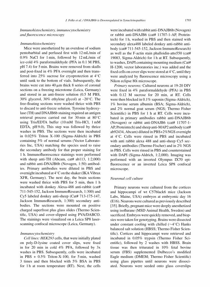
Immunohistochemistry, immunocytochemistryand fluorescence microscopy
ImmunohistochemistryMice were anesthetized by an overdose of sodium
pentobarbital and perfused first with 12 mL/min of0.9% NaCl for 1 min, followed by 12 mL/min ofice-cold 4% paraformaldehyde (PFA in 0.1 M PBS,pH 7.4) for 5 min. Brains were removed from skullsand post-fixed in 4% PFA overnight and then trans-ferred into 25% sucrose for cryoprotection at 4◦Cuntil sunk to the bottom of vials. Subsequently, thebrains were cut into 40 �m thick 8 series of coronalsections on a freezing microtome (Leica, Germany)and stored in an anti-freeze solution (0.5 M PBS,30% glycerol, 30% ethylene glycol) at –20◦C. Thefree-floating sections were washed thrice with PBSto discard to anti-freeze solution. Tyrosine hydroxy-lase (TH) and DNAJB6b staining required an antigenretrieval process carried out for 30 min at 80◦Cusing Tris/EDTA buffer (10 mM Tris-HCl, 1 mMEDTA, pH 9.0), This step was followed by thricewashes in PBS. The sections were then incubatedin 0.025% Triton X-100 (Sigma-Aldrich) in PBScontaining 5% of normal serums (Vector Laborato-ries Inc, USA) matching the species used to raisethe secondary antibody for that proper staining for1 h. Immunofluorescence stainings were performedwith sheep anti-TH (Abcam, cat# ab113, 1:2,000)and rabbit anti-DNAJB6b (Novagen, 1:50) antibod-ies. Primary antibodies were diluted in PBS andovernight incubated at 4◦C on the shaker (IKA VibraxXFR, Germany). The next day, the brain sectionswere washed thrice with PBS for 5 min, then 1 hincubated with donkey Alexa-488 anti-rabbit (cat#711-545-152, Jackson ImmunoResearch, 1:300) andCy5 labeled donkey anti-sheep (Cat# 713-175-147,Jackson ImmunoResearch, 1:300) secondary anti-bodies. The sections were mounted on positivecharged superfrost plus glass slides (Thermo Scien-tific, USA) and cover-slipped using PVA/DABCO.The stainings were visualized on a Leica SP8 laser-scanning confocal microscope (Leica, Germany).
ImmunocytochemistryCell lines: HEK293 cells, that were initially plated
on poly-D-lysine coated cover slips, were fixedin for 20 min in cold 4% PFA, followed by 3xwashes in PBS. Subsequently, cells were incubatedin PBS + 0.5% Triton-X-100, for 5 min, washed3 times and then blocked with 5% BSA in PBSfor 1 h at room temperature (RT). Next, the cells
were incubated with rabbit anti-DNAJB6b (Novagen)or rabbit anti-DNAJB6 (cat# 11707-1-AP, Protein-tech) for 1 h, washed in PBS and then stained withsecondary alexa488 labeled donkey anti-rabbit anti-body (cat# 711-545-152, Jackson ImmunoResearch)as well as the F-actin stain phalloidin-atto550 (cat#19083, Sigma-Aldrich) for 1 h at RT. Subsequently,to washes, DAPI containing mounting medium (Cat#H-1200, vector laboratories inc.) was added and thefixed cells on cover slips were stored at 4◦C, until theywere analyzed by fluorescence microscopy using aNikon eclipse 80i microscope.
Primary neurons: Cultured neurons at 19-20 DIVwere fixed in 4% paraformaldehyde (PFA) in PBSwith 0.12 M sucrose for 20 min, at RT. Cellswere then blocked in 0.1% saponin (Sigma-Aldrich),1% bovine serum albumin (BSA; Sigma-Aldrich),and 2% normal goat serum (NGS; Thermo FisherScientific) in PBS for 1 h at RT. Cells were incu-bated in primary antibodies rabbit anti-DNAJB6b(Novagen) or rabbit anti-DNAJB6 (cat# 11707-1-AP, Proteintech) and sheep anti-MAP2 antibody (cat#ab92434, Abcam) diluted in PBS+2%NGS overnightat 4◦C. Cells were rinsed in PBS and incubatedwith anti rabbit alexa 488 and anti-sheep Cy5 sec-ondary antibodies (Thermo Fischer) and in 2% NGSin PBS. Cells were rinsed in PBS and counterstainedwith DAPI (Sigma-Aldrich, 1:2,000). Imaging wasperformed with an inverted Olympus IX70 epi-fluorescence or an inverted Leica SP8 confocalmicroscope.
Neuronal cell culture
Primary neurons were cultured from the corticesand hippocampi of wt C57black6 mice (JacksonLabs, Maine, USA) embryos at embryonic day 16(E16). Neurons were cultured as previously described[35]. Briefly, pregnant mice were deeply anesthetizedusing isoflurane (MSD Animal Health, Sweden) andsacrificed. Embryos were quickly removed, and biop-sies were taken for genotyping. Brains were dissectedunder constant cooling with chilled (∼4◦C) Hanksbalanced salt solution (HBSS; Thermo Fisher Scien-tific). Cortices and hippocampi were retrieved andincubated in 0.05% trypsin (Thermo Fisher Sci-entific), followed by 2 washes with HBSS. Braintissue was then triturated in 10% fetal bovineserum (FBS) supplemented Dulbecco’s modifiedEagle medium (DMEM; Thermo Fisher Scientific)using glass pipettes until neurons were dissoci-ated. Neurons were seeded onto glass coverslips
1794 J. Folke et al. / DNAJB6b is Downregulated in Synucleinopathies
in 24-well plates (Sarstedt, Germany), coated withPoly-D-lysine (Sigma-Aldrich). Neurons were platedwith 10% FBS and 1% penicillin-streptomycin inDMEM; following 3–5 h incubation, media wasexchanged for complete Neurobasal solution, con-sisting of Neurobasal medium, B27 supplement,penicillin-streptomycin and L-glutamine (ThermoFisher Scientific). All animal experiments were per-formed in accordance with the ethical guidelines andwere approved by the Animal Ethical Committeeat Lund University ethical permit number 5.8.18-05983/2019.
Western blot
Wild type C57BL/6J mice brain was dissected tocortex, striatum, olfactory bulbs, hippocampus andstored at –80◦C until they were used. The brainsamples were sonicated (Branson SLPe, USA) inbuffer (1% SDS, 0.1 M Tris-HCl pH 7.4, and 1mM Ethylenediaminetetraacetic acid (EDTA), with1:100 protease inhibitor cocktail (cat# P8340, Sigma-Aldrich). The tissue debris was spun down at 20,000rcf for 30 min at 4◦C, and the supernatant was col-lected. The protein concentration was measured byBCA assay (Pierce BCA Protein Assy Kit, ThermoScientific, USA). Tissue lysates mixed with 25% 4x Laemmli buffer containing 10% of 0.1 M Dithio-threitol (DTT) and boiled for 5 min at 96◦C. 5 �gof proteins from brain lysate samples were sepa-rated on 10% SDS-PAGE polyacrylamide gels, thenthe samples transferred onto Polyvinylidene fluo-rid (PVDF) membranes using the Trans-Blot TurboTransfer System (Bio-Rad, USA). The membranewas blocked with 5% skimmed milk powder dis-solved in PBS containing 0.05% Tween (PBS-T) for1 h at RT, after which they were washed thrice for5 min with PBS-T at RT. Subsequently, the membraneincubated in 2% skimmed milk in PBS-T with eitherrabbit anti-DNAJB6 primary antibody (cat# 11707-1-AP, Proteintech, 1:1,000) or rabbit anti-DNAJB6b(Novagen, 1:200) overnight shaking in 4◦C. The nextday, the membrane was washed thrice in PBS-T for5 min and then incubated shaking at RT for 1 h in3% skimmed milk in PBS-T with anti-rabbit sec-ondary antibody (cat# P0448, Dako, 1:5,000). Themembrane was washed then thrice for 5 min in PBS-T. Western Blotting Luminol Reagent (Santa CruzBiotechnology, USA) was used for chemilumines-cence reactions and protein bands were developedusing a ChemiDoc™ XRS+Molecular Imager (Bio-Rad, USA) and the Image Lab software (Bio-Rad,
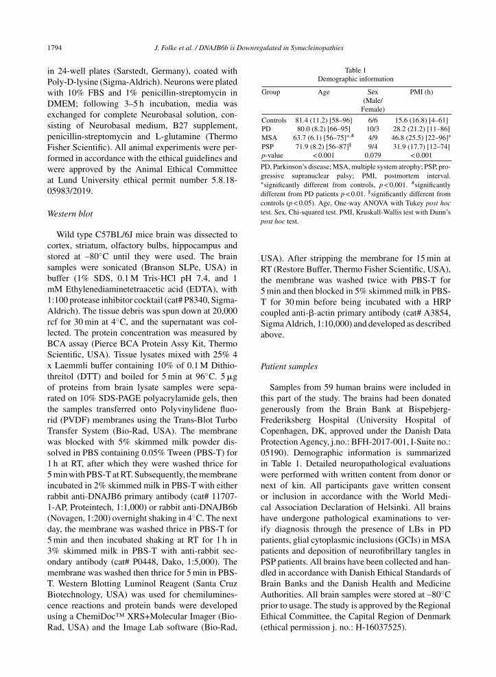
Table 1Demographic information
Group Age Sex PMI (h)(Male/
Female)
Controls 81.4 (11.2) [58–96] 6/6 15.6 (16.8) [4–61]PD 80.0 (8.2) [66–95] 10/3 28.2 (21.2) [11–86]MSA 63.7 (6.1) [56–75]∗,# 4/9 46.8 (25.5) [22–96]∗PSP 71.9 (8.2) [56–87]§ 9/4 31.9 (17.7) [12–74]p-value < 0.001 0.079 < 0.001
PD, Parkinson’s disease; MSA, multiple system atrophy; PSP, pro-gressive supranuclear palsy; PMI, postmortem interval.∗significantly different from controls, p < 0.001. #significantlydifferent from PD patients p < 0.01. §significantly different fromcontrols (p < 0.05). Age, One-way ANOVA with Tukey post hoctest. Sex, Chi-squared test. PMI, Kruskall-Wallis test with Dunn’spost hoc test.
USA). After stripping the membrane for 15 min atRT (Restore Buffer, Thermo Fisher Scientific, USA),the membrane was washed twice with PBS-T for5 min and then blocked in 5% skimmed milk in PBS-T for 30 min before being incubated with a HRPcoupled anti-�-actin primary antibody (cat# A3854,Sigma Aldrich, 1:10,000) and developed as describedabove.
Patient samples
Samples from 59 human brains were included inthis part of the study. The brains had been donatedgenerously from the Brain Bank at Bispebjerg-Frederiksberg Hospital (University Hospital ofCopenhagen, DK, approved under the Danish DataProtection Agency, j.no.: BFH-2017-001, I-Suite no.:05190). Demographic information is summarizedin Table 1. Detailed neuropathological evaluationswere performed with written content from donor ornext of kin. All participants gave written consentor inclusion in accordance with the World Medi-cal Association Declaration of Helsinki. All brainshave undergone pathological examinations to ver-ify diagnosis through the presence of LBs in PDpatients, glial cytoplasmic inclusions (GCIs) in MSApatients and deposition of neurofibrillary tangles inPSP patients. All brains have been collected and han-dled in accordance with Danish Ethical Standards ofBrain Banks and the Danish Health and MedicineAuthorities. All brain samples were stored at –80◦Cprior to usage. The study is approved by the RegionalEthical Committee, the Capital Region of Denmark(ethical permission j. no.: H-16037525).

J. Folke et al. / DNAJB6b is Downregulated in Synucleinopathies 1795
Tissue handling and protein extraction frompatient material
Approximately 100 mg of brain tissue from thedorsomedial prefrontal cortex (dPFC) was added to1 mL N-PER™ Neuronal Protein Extraction Reagent(cat# 87792, Life Technologies) with pre-addedHALT™ Phosphatase Inhibitor Cocktail mix (cat#788420, Life Technologies, 1:100) and HALT™
Protease Inhibitor Cocktail mix (cat# 87786, LifeTechnologies, 1:100). Brain tissue samples werehomogenized using MagNA Lyser Green Beads tubes(cat# 03358941001, Roche Diagnostics) and homog-enized twice on MagNA Lyser instrument (RocheDiagnostics, CH) for 25 s at 6,000 RPM followedby instant cooling using the MagNA Lyser RotorCooling Block (cat# 03358976001, Roche Diagnos-tics) for 90 s. Homogenates were spun for 1 min at10,000 × g at 4◦C to reduce foam. Samples werealiquoted at stored at –80◦C.
Measurement of DNAJB6 and DNAJB6b usingELISA
In-house sandwich ELISA was used to evaluate theabsolute amounts of DNAJB6 and DNAJB6b frombrain homogenates of PD, MSA, and PSP patients,compared to normal controls. In details: 96-wellpolystyrene MaxiSorp plates (cat# 144531, Nunc)were coated in 4◦C Carbonate Buffer 0.1 M, pH 9.4(cat# C3041, Sigma-Aldrich) overnight with rabbitanti-human DNAJB6 (cat# 11707-1-AP, Protein-tech, [1 �g/mL]) or DNAJB6b (Novagen, 7 �g/mL).Recombinant DNAJB6b was a kind gift from Pro-fessor Cecilia Emanuelsson (Center for MolecularProtein Science, Division of Biochemistry and Struc-tural biology, Lund University), which was producedas described in [36, 37]. The plates were emp-tied and blocked with PBS pH 7.4 containing 3%BSA fraction V (cat# 10735094001, Sigma-Aldrich)and 0.1% Tergitol™ solution (cat# NP40S, Sigma-Aldrich) for 2 h at RT. Plates were then washed fiveconsecutive times in 300 �l/well PBS + 0.1%-Tween-20 (cat# P1379, Sigma-Aldrich) using a WellWashinstrument (Thermo Scientific, USA). Human brainhomogenates from PD, MSA, and PSP patients aswell as controls were diluted in PBS + 0.1%BSAsolution (DNAJB6 1:20; DNAJB6b 1:1) and addedto the plates in duplicates and incubated for 1 hat RT. After a washing step, the plates were incu-bated at RT for 2 h with 50 �l of mouse anti-human DNAJB6 antibody (cat# H00010049-M01,
Novus Biologicals, DNAJB6: 500 ng/mL, DNAJB6b: 2,000 ng/mL). After an additional washingstep, 50 �l of goat anti-mouse HRP-conjugated anti-body (cat# ab98717, Abcam, DNAJB6: 1:10,000,DNAJB6b: 1:5,000) was added to each well and incu-bated for 2 h at RT. After a last washing step, 50 �Lof tetramethylbenzidine (TMB) Liquid PeroxidaseSubstrate (cat# T8665, Sigma-Aldrich) was addedand stopped after 30 min incubation in dark at RTwith 50 �L 0.5 N H2SO4. The absorbance at 450 nmwas measured on a Fisher Scientific MultiskanTMFC Microplate Reader (Thermo Fischer Scientific,USA). Standard curves were applied using under thesame conditions as described above using the pep-tide DNAJB6b starting at 2,000 ng/mL and serialdiluted at -3x. The ODs were interpolated using sig-moidal, 4PL, curves. For each assay standard curveassessment and spike-and-recovery was performed(Supplementary Figure 1). Demonstrating the pre-ciseness and accuracy of our in-house ELISA setup,the coefficient of variabilities (CVs) in percentageswere calculated to 3.01% for DNAJB6 and 2.97%for DNAJB6b, which was concluded sufficientlyprecise.
Immunohistochemistry on patient material
Samples of putamen from 6 human brains (2healthy controls, 2 PD patients, 2 MSA patients) wereincluded in this study. Tissue blocks that includedputamen were excised from the fresh brains. Sam-ples were fixed for minimum 48 h in 10% bufferedformalin (cat# 1000.5000, CellPath). Samples werethen embedded in paraffin on a Leica ASP300 Stissue processor (Leica, DE) before sectioning ona sliding microtome at 5–7 �m. On the day ofimmunoreactions, slides were deparaffinized throughheating for 45 min at 60◦C followed by washingin xylene (#28973.294, VWR) for 2 × 5 min, 99%EtOH for 2 × 2.5 min, 96% EtOH for 2 min, 70%EtOH for 2 min, and running water for 5 min. Sam-ples were demasked in boiling pH 9 TEG-buffer(cat# 862338, RegionH Apoteket) for 15 min fol-lowed by cooling for 20 min and washed in pH 7 PBS2 × 5 min before blocking in 10% FCS in PBS. Sliceswere incubated overnight at RT, respectively, witheither rabbit anti-human DNAJB6 (cat# 11707-1-AP,Proteintech, 0.02 �g/mL) or DNAJB6b (Novagen,0.28 �g/mL). Endogenous peroxidases were blockedusing 3% H2O2 (cat# 212892, Apotekernes A.m.b.a,DNK) for 8 min. After washing, samples wereincubated with EnVision + System anti-rabbit (cat#

1796 J. Folke et al. / DNAJB6b is Downregulated in Synucleinopathies
K003, Dako) for 1 h. Reactions were developed for10–15 min using a solution consisting 0.25% 3,3′-diaminobenzidine tetrahydrochloride hydrate (cat#D5637, Sigma-Aldrich) and 0.15% H2O2 in PBS.Following washing in dH2O for 5 min, nucleiwere stained in Mayer’s hematoxylin (cat# 860213,RegionH Apoteket) for 40 s, and then washed inrunning dH2O for 10 min. Finally, samples weredehydrated in 70% EtOH, 96% EtOH for 2 min,99% EtOH for 2 × 2.5 min, and xylene for 2 × 5 min.Cover slides were mounted using Pertex Mount-ing Medium (cat# SEA-0100-00A, CellPath). Slideswere studied using an Olympus BX60 microscope.
Statistics
Data analyses were performed using GraphPadPrism v. 8.01 (GraphPad Software Inc., USA).Demographic differences were tested using one-wayANOVA with Tukey post hoc test, Kruskal-Walliswith Dunn’s post hoc test, and the chi-squared test.Normality was assessed using D’Agostino Omnibustest. Data was log10-transformed if not passed fornormality. Outliers were identified using ROUT testat 1% discovery rate. For group comparisons, one-way ANOVA with Tukey post hoc test for multipletesting. Differences were significant at p < 0.05.
RESULTS
In order to assess the localization and expressionof the DNAJB6b protein in cells and in tissues, wegenerated an antibody specific for DNAJB6b, whichonly has 10 amino acid residues at its C-terminusthat are unique compared to the a-isoform (Fig. 1A).We tested this antibody in multiple cell lines usingDNAJB6 KO cells previously generated, as a negativecontrol [27] and found that the antibody was indeedspecific for the DNAJB6b isoform (Fig. 1B). In addi-tion, we also stained HEK293 wt cells or DNAJB6KO HEK cells as a negative control and observed thatthe DNAJB6b isoform mainly localizes to the cyto-plasm of cells, whereas as expected no staining couldbe seen in DNAJB6 KO cells, by immunocytochem-istry (Fig. 1C). In comparison, staining of HEK293wt cells with anti-(total) DNAJB6 antibody revealeda strong nuclear staining, which was expected as theDNAJB6a isoform carries a nucleus localization sig-nal (Fig. 1A, D). Having determined the specificityof the antibody and the localization of the protein, wewanted to see how the DNAJB6b protein is localized
in primary neurons, due to its potential important roleas a suppressor of amyloid protein aggregation inneurons. We found that DNAJB6b was found to alarge extend in the soma of neurons, but not in thenucleus (Fig. 2A). In addition, we observed that withregards to the projections from the primary neurons,DNAJB6b was found more in the dendrites than in theaxons, as evaluated by staining with the anti-MAP2antibody for dendritic localization (Fig. 2A, C).Indeed, the �-syn staining in proximity to DNAJB6bstaining is presumably due to presynaptic terminalsin proximity to dendrites (Fig. 2C), as it is wellknown that most of non-aggregated �-syn localizeat the presynaptic terminals. In line with these resultsa staining for anti-(total)-DNAJB6 revealed similarlocalization results, albeit with slightly more stain-ing of the nucleus, which we assume may be causedby the staining of DNAJB6a (Fig. 2B). The mostcommon synucleinopathy, PD, is characterized bya loss of the dopaminergic neurons in the SNpc.Therefore, in order to know if DNAJB6b could poten-tially be protective in these neurons against �-synaggregation, we wanted to know if the protein isexpressed in these cells. Indeed, we observed thatboth DNAJB6b (Fig. 3A) and total DNAJB6 (Fig. 3B)is expressed in these cells as well as we observed thatslightly more DNAJB6 was found in the nucleus,when we stained for total DNAJB6 (Fig. 3A, B).DNAJB6b and total DNAJB6 was furthermore foundto be widely expressed in mouse brain, as revealedby western blotting analysis of mouse brain lysates(Fig. 3C).
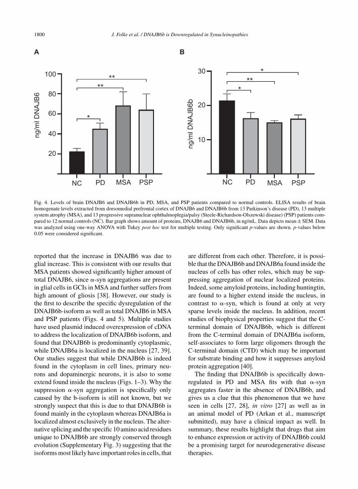
Due to the semi-quantitative nature of immunohis-tochemical staining we wanted to establish a moresensitive way to address if this protein could be dys-regulated in a more disease environment. By use ofrecombinant DNAJB6b and commercially availableanti-DNAJB6 antibodies, we established an ELISAbased set up to measure DNAJB6 (SupplementaryFigure 1A, C) and DNAJB6b (Supplementary Fig-ure 1B, D) concentrations. We tested a cohort ofpostmortem brain material from patients with PD,MSA, PSP, and control subjects. Total levels ofDNAJB6 in putamen samples were significantly dif-ferent between the groups (F(3) = 6.2; p = 0.001)and found to be increased in PD (p = 0.048), MSA(p = 0.001), and PSP (p = 0.009) patients comparedto normal controls (Fig. 4A). Inversely, the totallevels of DNAJB6b were significantly differentbetween groups (F(3) = 4.3; p = 0.009) and foundto be reduced in PD (p = 0.048), MSA (p = 0.009),and PSP (p = 0.041) patients compared to normal

J. Folke et al. / DNAJB6b is Downregulated in Synucleinopathies 1797
Fig. 1. Expression of endogenous DNAJB6b as evaluated by western blot and immunocytochemistry. A) Illustration depicting the two majorisoforms of DNAJB6 and their domains. nls, Nucleus Localization Signal. B) Expression of DNAJB6b in lysates from cell lines, analyzedby probing membranes with anti-DNAJB6b and HRP conjugated anti-rabbit antibodies. The membrane was probed with anti-total DNAJB6and anti-actin as a control. C) DNAJB6 KO HEK 293 cells or wt HEK293 cells were probed with anti-DNAJB6b antibody, phalloidin-Alexa547 as well as secondary anti-rabbit Alexa 488 coupled antibody. D) wt HEK293 cells were probed with anti- total DNAJB6 antibody,phalloidin-alexa 547 as well as secondary anti-rabbit alexa 488 coupled antibody.
controls (Fig. 4B). Further, representative immuno-histochemical stainings from 2 normal controls, 2PD, and 2 MSA patients (Fig. 5A-5C respectively)with anti-(total) DNAJB6 or anti-DNAJB6b antibod-ies, suggests that this dysregulation may largely beoccurring in neurons rather than glial cells of patients.Western blot analysis of brain samples from controlsubjects (NC), PD, MSA, or PSP patients showedthat the antibodies are specific for DNAJB6b andtotal DNAJB6 in human brain tissue samples as well(Supplementary Figure 2).
DISCUSSION
Here we demonstrate that DNAJB6b is downreg-ulated in PD, MSA, and PSP, whereas the total levelof DNAJB6 protein was found to be upregulated inthese diseases. One study has prior to this investigatedthe dysregulation of DNAJB6 in neurodegenerativediseases. In this study by Durrenberger et al., thetotal level of DNAJB6 was found to be upregu-lated in PD patients [26], which is in line with theresults presented here. Durrenberger et al. further

1798 J. Folke et al. / DNAJB6b is Downregulated in Synucleinopathies
Fig. 2. DNAJB6b and total DNAJB6 are expressed in primary neurons. Fixed primary mouse neurons stained with anti-DNAJB6b (A)or anti-total DNAJB6 (B) as well as anti-MAP2, anti-�-syn and dapi and fluorescently labeled anti rabbit Alexa 488 and anti sheep Cy5antibodies. C) Depicts a close up picture of a dendrite stained with antibodies against �-syn, MAP2, and DNAJB6b as well as secondaryfluorescently labeled anti-rabbit and anti-sheep antibodies.

J. Folke et al. / DNAJB6b is Downregulated in Synucleinopathies 1799
Fig. 3. DNAJB6b and total DNAJB6 are expressed in SNpc. 40 uM coronal sections from wt C57black6 mice containing SNpc, were probedwith anti-DNAJB6b (A) or anti-total DNAJB6 (B) as well as anti-rabbit alexa 488 and anti-TH and well as anti sheep Cy5. C) Expression ofDNAJB6b in brain lysates from different regions of mouse brain, as analyzed by probing membranes with anti-DNAJB6b, anti-total-DNAJB6and HRP conjugated anti-rabbit antibody as well as HRP conjugated anti-actin antibody.
1800 J. Folke et al. / DNAJB6b is Downregulated in Synucleinopathies
Fig. 4. Levels of brain DNAJB6 and DNAJB6b in PD, MSA, and PSP patients compared to normal controls. ELISA results of brainhomogenate levels extracted from dorsomedial prefrontal cortex of DNAJB6 and DNAJB6b from 13 Parkinson’s disease (PD), 13 multiplesystem atrophy (MSA), and 13 progressive supranuclear ophthalmoplegia/palsy (Steele-Richardson-Olszewski disease) (PSP) patients com-pared to 12 normal controls (NC). Bar graph shows amount of proteins, DNAJB6 and DNAJB6b, in ng/mL. Data depicts mean ± SEM. Datawas analyzed using one-way ANOVA with Tukey post hoc test for multiple testing. Only significant p-values are shown. p-values below0.05 were considered significant.
reported that the increase in DNAJB6 was due toglial increase. This is consistent with our results thatMSA patients showed significantly higher amount oftotal DNAJB6, since �-syn aggregations are presentin glial cells in GCIs in MSA and further suffers fromhigh amount of gliosis [38]. However, our study isthe first to describe the specific dysregulation of theDNAJB6b-isoform as well as total DNAJB6 in MSAand PSP patients (Figs. 4 and 5). Multiple studieshave used plasmid induced overexpression of cDNAto address the localization of DNAJB6b isoform, andfound that DNAJB6b is predominantly cytoplasmic,while DNAJB6a is localized in the nucleus [27, 39].Our studies suggest that while DNAJB6b is indeedfound in the cytoplasm in cell lines, primary neu-rons and dopaminergic neurons, it is also to someextend found inside the nucleus (Figs. 1–3). Why thesuppression �-syn aggregation is specifically onlycaused by the b-isoform is still not known, but westrongly suspect that this is due to that DNAJB6b isfound mainly in the cytoplasm whereas DNAJB6a islocalized almost exclusively in the nucleus. The alter-native splicing and the specific 10 amino acid residuesunique to DNAJB6b are strongly conserved throughevolution (Supplementary Fig. 3) suggesting that theisoforms most likely have important roles in cells, that
are different from each other. Therefore, it is possi-ble that the DNAJB6b and DNAJB6a found inside thenucleus of cells has other roles, which may be sup-pressing aggregation of nuclear localized proteins.Indeed, some amyloid proteins, including huntingtin,are found to a higher extend inside the nucleus, incontrast to �-syn, which is found at only at verysparse levels inside the nucleus. In addition, recentstudies of biophysical properties suggest that the C-terminal domain of DNAJB6b, which is differentfrom the C-terminal domain of DNAJB6a isoform,self-associates to form large oligomers through theC-terminal domain (CTD) which may be importantfor substrate binding and how it suppresses amyloidprotein aggregation [40].
The finding that DNAJB6b is specifically down-regulated in PD and MSA fits with that �-synaggregates faster in the absence of DNAJB6b, andgives us a clue that this phenomenon that we haveseen in cells [27, 28], in vitro [27] as well as inan animal model of PD (Arkan et al., manuscriptsubmitted), may have a clinical impact as well. Insummary, these results highlight that drugs that aimto enhance expression or activity of DNAJB6b couldbe a promising target for neurodegenerative diseasetherapies.

J. Folke et al. / DNAJB6b is Downregulated in Synucleinopathies 1801
Fig. 5. Micrographs showing DNAJB6 and DNJB6B stained sections of the putamen from (A) 2 normal controls, (B) 2 PD patients, and(C) 2 MSA patients at ×10 (large pictures, scale bar = 200 �m) and ×60 (small pictures, scale bar = 50 �m) magnification.
1802 J. Folke et al. / DNAJB6b is Downregulated in Synucleinopathies
ACKNOWLEDGMENTS
This study was supported by the Parkinson Foun-dation (Sweden), CH as well as the Stiftelsen OlleEnqvist Byggmastare (Sweden), SA and CH.
CONFLICT OF INTEREST
The authors have no conflict of interest to report.
SUPPLEMENTARY MATERIAL
The supplementary material is available in theelectronic version of this article: https://dx.doi.org/10.3233/JPD-202512.
REFERENCES
[1] Goedert M, Jakes R, Spillantini MG (2017) The synucle-inopathies: twenty years on. J Parkinsons Dis 7, S51-S69.
[2] Spillantini MG, Schmidt ML, Lee VM, Trojanowski JQ,Jakes R, Goedert M (1997) Alpha-synuclein in Lewy bodies.Nature 388, 839-840.
[3] Vekrellis K, Xilouri M, Emmanouilidou E, Rideout HJ,Stefanis L (2011) Pathological roles of alpha-synuclein inneurological disorders. Lancet Neurol 10, 1015-1025.
[4] Angot E, Steiner JA, Hansen C, Li JY, Brundin P (2010)Are synucleinopathies prion-like disorders? Lancet Neurol9, 1128-1138.
[5] Hansen C, Li JY (2012) Beyond alpha-synuclein transfer:pathology propagation in Parkinson’s disease. Trends MolMed 18, 248-255.
[6] Logan T, Bendor J, Toupin C, Thorn K, Edwards RH (2017)alpha-Synuclein promotes dilation of the exocytotic fusionpore. Nat Neurosci 20, 681-689.
[7] Burre J, Sharma M, Tsetsenis T, Buchman V, EthertonMR, Sudhof TC (2010) Alpha-synuclein promotes SNARE-complex assembly in vivo and in vitro. Science 329,1663-1667.
[8] Polymeropoulos MH, Lavedan C, Leroy E, Ide SE, Dehe-jia A, Dutra A, Pike B, Root H, Rubenstein J, BoyerR, Stenroos ES, Chandrasekharappa S, Athanassiadou A,Papapetropoulos T, Johnson WG, Lazzarini AM, DuvoisinRC, Di Iorio G, Golbe LI, Nussbaum RL (1997) Muta-tion in the alpha-synuclein gene identified in families withParkinson’s disease. Science 276, 2045-2047.
[9] Goedert M, Spillantini MG, Del Tredici K, Braak H (2013)100 years of Lewy pathology. Nat Rev Neurol 9, 13-24.
[10] Witt SN (2013) Molecular chaperones, alpha-synuclein, andneurodegeneration. Mol Neurobiol 47, 552-560.
[11] De Mattos EP, Wentink A, Nussbaum-Krammer C, HansenC, Bergink S, Melki R, Kampinga HH (2020) Protein qualitycontrol pathways at the crossroad of synucleinopathies. JParkinsons Dis 10, 369-382.
[12] Aprile FA, Sormanni P, Vendruscolo M (2015) A rationaldesign strategy for the selective activity enhancement of amolecular chaperone toward a target substrate. Biochemistry54, 5103-5112.
[13] Gao X, Carroni M, Nussbaum-Krammer C, Mogk A, Nil-legoda NB, Szlachcic A, Guilbride DL, Saibil HR, Mayer
MP, Bukau B (2015) Human Hsp70 disaggregase reversesParkinson’s-linked alpha-synuclein amyloid fibrils. MolCell 59, 781-793.
[14] McLean PJ, Klucken J, Shin Y, Hyman BT (2004) Gel-danamycin induces Hsp70 and prevents alpha-synucleinaggregation and toxicity in vitro. Biochem Biophys ResCommun 321, 665-669.
[15] Qiu XB, Shao YM, Miao S, Wang L (2006) The diversityof the DnaJ/Hsp40 family, the crucial partners for Hsp70chaperones. Cell Mol Life Sci 63, 2560-2570.
[16] Mogk A, Bukau B, Kampinga HH (2018) Cellular handlingof protein aggregates by disaggregation machines. Mol Cell69, 214-226.
[17] Summers DW, Douglas PM, Ramos CH, Cyr DM (2009)Polypeptide transfer from Hsp40 to Hsp70 molecular chap-erones. Trends Biochem Sci 34, 230-233.
[18] Summers DW, Wolfe KJ, Ren HY, Cyr DM (2013) The TypeII Hsp40 Sis1 cooperates with Hsp70 and the E3 ligase Ubr1to promote degradation of terminally misfolded cytosolicprotein. PLoS One 8, e52099.
[19] Zarouchlioti C, Parfitt DA, Li W, Gittings LM, CheethamME (2018) DNAJ proteins in neurodegeneration: essentialand protective factors. Philos Trans R Soc Lond B Biol Sci373, 20160534.
[20] Sanchez E, Darvish H, Mesias R, Taghavi S, FirouzabadiSG, Walker RH, Tafakhori A, Paisan-Ruiz C (2016) Identi-fication of a large DNAJB2 deletion in a family with spinalmuscular atrophy and parkinsonism. Hum Mutat 37, 1180-1189.
[21] Olgiati S, Quadri M, Fang M, Rood JP, Saute JA, ChienHF, Bouwkamp CG, Graafland J, Minneboo M, BreedveldGJ, Zhang J, International Parkinsonism Genetics Network,Verheijen FW, Boon AJ, Kievit AJ, Jardim LB, Mande-makers W, Barbosa ER, Rieder CR, Leenders KL, WangJ, Bonifati V (2016) DNAJC6 mutations associated withearly-onset Parkinson’s disease. Ann Neurol 79, 244-256.
[22] Elsayed LE, Drouet V, Usenko T, Mohammed IN, HamedAA, Elseed MA, Salih MA, Koko ME, Mohamed AY, SiddigRA, Elbashir MI, Ibrahim ME, Durr A, Stevanin G, LesageS, Ahmed AE, Brice A (2016) A novel nonsense mutationin DNAJC6 expands the phenotype of autosomal-recessivejuvenile-onset Parkinson’s disease. Ann Neurol 79, 335-337.
[23] Yoshida S, Hasegawa T, Suzuki M, Sugeno N, KobayashiJ, Ueyama M, Fukuda M, Ido-Fujibayashi A, Sekiguchi K,Ezura M, Kikuchi A, Baba T, Takeda A, Mochizuki H, NagaiY, Aoki M (2018) Parkinson’s disease-linked DNAJC13mutation aggravates alpha-synuclein-induced neurotoxicitythrough perturbation of endosomal trafficking. Hum MolGenet 27, 823-836.
[24] Vilarino-Guell C, Rajput A, Milnerwood AJ, Shah B, Szu-Tu C, Trinh J, Yu I, Encarnacion M, Munsie LN, Tapia L,Gustavsson EK, Chou P, Tatarnikov I, Evans DM, PishottaFT, Volta M, Beccano-Kelly D, Thompson C, Lin MK, Sher-man HE, Han HJ, Guenther BL, Wasserman WW, BernardV, Ross CJ, Appel-Cresswell S, Stoessl AJ, Robinson CA,Dickson DW, Ross OA, Wszolek ZK, Aasly JO, Wu RM,Hentati F, Gibson RA, McPherson PS, Girard M, RajputM, Rajput AH, Farrer MJ (2014) DNAJC13 mutations inParkinson disease. Hum Mol Genet 23, 1794-1801.
[25] Wentink AS, Nillegoda NB, Feufel J, Ubartaite G, SchneiderCP, De Los Rios P, Hennig J, Barducci A, Bukau B (2020)Molecular dissection of amyloid disaggregation by humanHSP70. Nature 587, 483-488.
[26] Durrenberger PF, Filiou MD, Moran LB, Michael GJ,Novoselov S, Cheetham ME, Clark P, Pearce RK, Graeber

J. Folke et al. / DNAJB6b is Downregulated in Synucleinopathies 1803
MB (2009) DnaJB6 is present in the core of Lewy bod-ies and is highly up-regulated in parkinsonian astrocytes. JNeurosci Res 87, 238-245.
[27] Aprile FA, Kallstig E, Limorenko G, Vendruscolo M, Ron D,Hansen C (2017) The molecular chaperones DNAJB6 andHsp70 cooperate to suppress alpha-synuclein aggregation.Sci Rep 7, 9039.
[28] Deshayes N, Arkan S, Hansen C (2019) The molecular chap-erone DNAJB6, but not DNAJB1, suppresses the seededaggregation of alpha-synuclein in cells. Int J Mol Sci 20,4495.
[29] Luk KC, Kehm V, Carroll J, Zhang B, O’Brien P, Tro-janowski JQ, Lee VM (2012) Pathological alpha-synucleintransmission initiates Parkinson-like neurodegeneration innontransgenic mice. Science 338, 949-953.
[30] Rey NL, Steiner JA, Maroof N, Luk KC, Madaj Z, Tro-janowski JQ, Lee VM, Brundin P (2016) Widespreadtransneuronal propagation of alpha-synucleinopathy trig-gered in olfactory bulb mimics prodromal Parkinson’sdisease. J Exp Med 213, 1759-1778.
[31] Rodrıguez-Gonzalez C, Lin S, Arkan S, Hansen C (2020)Co-chaperones DNAJA1 and DNAJB6 are critical forregulation of polyglutamine aggregation. Sci Rep 10,8130.
[32] Gillis J, Schipper-Krom S, Juenemann K, Gruber A, CoolenS, van den Nieuwendijk R, van Veen H, Overkleeft H,Goedhart J, Kampinga HH, Reits EA (2013) The DNAJB6and DNAJB8 protein chaperones prevent intracellularaggregation of polyglutamine peptides. J Biol Chem 288,17225-17237.
[33] Hussein RM, Hashem RM, Rashed LA (2015) Evaluation ofthe amyloid beta-GFP fusion protein as a model of amyloidbeta peptides-mediated aggregation: a study of DNAJB6chaperone. Front Mol Neurosci 8, 40.
[34] Kakkar V, Mansson C, de Mattos EP, Bergink S, van derZwaag M, van Waarde MA, Kloosterhuis NJ, Melki R,
van Cruchten RT, Al-Karadaghi S, Arosio P, Dobson CM,Knowles TP, Bates GP, van Deursen JM, Linse S, van deSluis B, Emanuelsson C, Kampinga HH (2016) The S/T-rich motif in the DNAJB6 chaperone delays polyglutamineaggregation and the onset of disease in a mouse model. MolCell 62, 272-283.
[35] Willen K, Sroka A, Takahashi RH, Gouras GK (2017)Heterogeneous association of Alzheimer’s disease-linkedamyloid-beta and amyloid-beta protein precursor withsynapses. J Alzheimers Dis 60, 511-524.
[36] Mansson C, Arosio P, Hussein R, Kampinga HH, HashemRM, Boelens WC, Dobson CM, Knowles TP, Linse S,Emanuelsson C (2014) Interaction of the molecular chap-erone DNAJB6 with growing amyloid-beta 42 (Abeta42)aggregates leads to sub-stoichiometric inhibition of amyloidformation. J Biol Chem 289, 31066-31076.
[37] Mansson C, van Cruchten RTP, Weininger U, Yang X,Cukalevski R, Arosio P, Dobson CM, Knowles T, Akke M,Linse S, Emanuelsson C (2018) Conserved S/T residues ofthe human chaperone DNAJB6 are required for effectiveinhibition of Abeta42 amyloid fibril formation. Biochem-istry 57, 4891-4902.
[38] Salvesen L, Winge K, Brudek T, Agander TK, LokkegaardA, Pakkenberg B (2017) Neocortical neuronal loss inpatients with multiple system atrophy: a stereological study.Cereb Cortex 27, 400-410.
[39] Pei Y, Fu W, Yang E, Shen A, Chen YC, Gong H, Chen J,Huang J, Xiao G, Liu F (2012) A Hsp40 chaperone proteininteracts with and modulates the cellular distribution of theprimase protein of human cytomegalovirus. PLoS Pathog 8,e1002968.
[40] Karamanos TK, Tugarinov V, Clore GM (2019) Unravel-ing the structure and dynamics of the human DNAJB6bchaperone by NMR reveals insights into Hsp40-mediatedproteostasis. Proc Natl Acad Sci U S A 116, 21529-21538.