dna extractions -the past, present and future approaches
TRANSCRIPT

DNA Extractions - the Past, Present and Future Approaches
Nur Munirah Bt Majid, Uda Hashim*, Subash C.B. Gopinath, and
xxxxxxxxx
Biomedical Nano Diagnostics Research Group, Institute of NanoElectronic Engineering (INEE), Universiti Malaysia Perlis
(UniMAP), 01000 Kangar, Perlis, Malaysia.
Correspondence to:
Prof. Uda HashimBiomedical Nano Diagnostics Research Group,
1
Institute of Nano Electronic Engineering (INEE), University Malaysia Perlis (UniMAP), 01000 Kangar, Perlis, Malaysia.Email: [email protected]
Abstract
DNA, RNA and protein are the major macromolecules play important
roles in the functional aspects of every living thing. As stated
by ‘central dogma’ DNA is the primary molecules not only as
building block of human genetics but also towards medical
diagnosis and forensic applications, due to high specificity of
DNA and they varies from one individual to another. DNA
molecules in native condition are in the complex form with other
macromolecules in most of the cases to fulfill functions. This
complex formation makes the necessity of separation (extraction)
of DNA molecules from others before being applied for sensing or
forensic applications. Moreover, as technologies develop, there
is an urge toward method to extract macromolecules that specific
to particular species. The processes of extraction and
purification of DNA used previously were complicated, time-
consuming, labor-intensive, and limited in terms of overall
throughput, but now-a-days there are many specialized and
sophisticated methods that can be used to extract DNA in pure
form. Current technologies should allow a high-throughput of
samples to be extracted for DNA, however the crucial
developmental process of DNA extraction during transitional
stages of developments were not explored properly. In this2
overview, we gleaned the extraction methods demonstrated from
past to present and for a future with their critics.
Key words: DNA, Extraction, Purification, Macromolecule,
Nucleases
1. Introduction
Nucleic acid extraction and purification is one of the
essential steps should follow the workflow of genetic analyses
and considered to be a most crucial method used in molecular
biology. Biological macromolecules including deoxyribonucleic
acid (DNA), can be isolated from biological materials such as
living or conserved tissues, cells, virus particles, plant
material or other samples. Thus, extractions of macromolecules
becoming the basic method that are commonly used in molecular
studies. Further, DNAs should be isolated from other complexed
macromolecules, as DNAs are carrying basic genetic information
for subsequent downstream processes and analytical or preparative
purposes. DNA purification involves two major categories that
are isolation of recombinant DNA such as plasmids or
bacteriophage and the isolation of chromosomal or genomic DNA
from prokaryotic or eukaryotic organisms (Doyle, 1996). A
successful use of available downstream applications will gain
with high-quantity and quality DNA output (Pyzowski & Tan, 2007).
There are three important steps that will lead to successful
nucleic acid extraction, firstly the effective disruption of
3
cells or tissue, secondly denaturation of nucleoprotein complexes
using appropriate reagents to inactivate the nucleases, for
example, RNase for RNA removal and DNase for DNA removal and
lastly away from contamination (Wink, 2006). The target nucleic
acid should be free of contaminants including protein,
carbohydrate, lipids, or other factors that will lead to impure
output. So, it is essential to choose a suitable extraction
method, and thus, few considerations have to be made when
evaluating the available options. These may include technical
requirements, time efficiency, cost-effectiveness, as well as
biological specimens to be used and their collection and storage
requirements.
Extraction methods involve in DNA isolation becoming more
effective and efficient as extraction kits being developed which
containing most of the components needed to isolate nucleic acid,
but still additional steps are needed to remove other components
depending on the type of specimen, which are time consuming and
tedious. To overcome these problems, automated systems being
designed for medium-to-large laboratory scales have grown in
demand over recent years. The prime issues with DNA extraction is
to increase the yield and purity of desired product,
reproducibility, and scalability of the molecules as well as the
speed, accuracy, and reliability of the assay, while minimizing
the risk of cross-contamination. Overall, to generate an
efficient DNA extraction method, even though proposed methods are
seems to be simple, there are critics and rationales with the
4
methods in respect to the purpose. To pin-point and understand
more about the crucial steps involved in different methods will
make the way to generate a finely tuned method, will be a common
strategy suitable to the laboratories and industries. In this
overview, the important steps involved in the DNA extraction and
purification methods formulated in the past, present and a
direction towards future developments are narrated. This study
is necessary due to the active participation of DNA molecules in
several instances in wide range of studies.
DNA is a long polymer of repeating sub-unit called
nucleotides, and each nucleotide consists of three sub-units of
pentose or five-carbon sugar, a phosphate group and an organic
nitrogenous (nitrogen-containing) group (Cseke et al, 2004). DNA is
a complex molecule that contains all of the information necessary
to build and maintain an organism. In fact, nearly every cell in
a multicellular organism possesses the full set of DNA required
for that organism. However, DNA does more than specify the
structure and function of living things (Mitnik et al., 2012).
Whenever organisms reproduce, a portion of their DNA is passed
along to their offspring, this transmission of all or part of an
organism's DNA helps to ensure a certain level of continuity from
one generation to the next, while still allowing for slight
changes that contribute to the diversity of life (Buckingham &
Flaw, 2007). DNA encodes the genetic information used to assemble
proteins in similar to the way the letters on books encode
information (Blin & Stafford, 1976). Unique among macromolecules,
5

nucleic acid are able to serve as templates to produce precisely
copies of them.
2. Discovery of DNA as main building block
Even though, initially the first nucleic acid discovery was
realized by a few people, nucleic acid was first identified in
the year 1869 by the Swiss physiological chemist, Friedrich
Miescher that was called as "nuclein" resides in the nuclei of
human white blood cells. The term "nuclein" was later changed to
"nucleic acid" and eventually to "deoxyribonucleic acid," or "DNA
(Whitford, 2005). At first, Miescher's plan was to isolate and
characterize the protein components of leukocytes or white blood
cells. For that, he made arrangements for local surgical clinics
to send him the used, pus-coated patient bandages for his
research. Once he received the bandages, he planned to wash them,
filter out the leukocytes, extract and identify the various
proteins within the white blood cells until he came across a
substance from the cell nuclei that had chemical properties
unlike any proteins, including a much higher phosphorous content
and resistance to proteolysis, Miescher realized that he had
discovered a new substance (Dahm, 2008).
Other scientists, Phoebus Levene from Russian biochemist
have continued to investigate the chemical nature of the molecule
formerly known as ‘nuclein’ and publishing more than 700 papers
on the chemistry of biological molecules over the course of their
career (Haines et al, 2005). Levene was the first person to
6
discover the order of three major components (phosphate-sugar-
base) of a single nucleotide and the first discovered the
carbohydrate component of DNA (Brent, 1998). Levene also proposed
that nucleic acids were composed of a series of nucleotides, and
that each nucleotide was in turn composed of just one of four
nitrogen-containing bases, a sugar molecule, and a phosphate
group. Levene made his initial proposal in 1919, discrediting
other suggestions that had been put forth about the structure of
nucleic acids (Kojima & Ozawa, 2002).
Erwin Chargaff, an Australian biochemist was one of a
handful of scientists who expanded Levene's work by uncovering
additional details of the structure of DNA, thus further paving
the way for Watson and Crick model, which demonstrated that
hereditary units, or genes, are composed of DNA. As his first
step in this search, Chargaff set out to see whether there were
any differences in DNA among different species (Dahm, 2004).
After developing a new paper chromatography method for separating
and identifying small amounts of organic material, Chargaff
reached two major conclusions. First, he noted that the
nucleotide composition of DNA varies among species. In other
words, the same nucleotides do not repeat in the same order, as
proposed by Levene. Second, Chargaff concluded that almost all
DNA, no matter what organism or tissue type it comes from,
maintains certain properties, even its composition varies (Kojima
& Ozawa, 2002). In particular, the amount of adenine (A) is
usually similar to the amount of thymine (T), and the amount of
7
guanine (G) usually approximates the amount of cytosine (C). This
second major conclusion is now known as "Chargaff's rule."
Chargaff's research was vital to the later work of Watson and
Crick model, but Chargaff himself could not imagine the
explanation of these relationships and specifically, that A bound
to T and C bound to G within the molecular structure of DNA
(Brooks, 1998).
Chargaff's realization that A = T and C = G, combined with
some crucially important X-ray crystallography work by English
researchers Rosalind Franklin and Maurice Wilkins, contributed to
Watson and Crick's derivation of three-dimensional, double-
helical model for the structure of DNA. Watson and Crick's
discovery was also made possible by recent advances in model
building, or the assembly of possible three-dimensional
structures based on known molecular distances and bond angles.
(Sambrook & Russel, 2001).
3. DNA extraction discovery
As stated above, Friedrich Miescher, the first scientist
attempted to isolate DNA while studying the chemical composition
of cells. Firstly, he was aimed in solving the fundamental issue
or principles in life by determine the composition of the cell.
In 1869, he tried to isolate cells in lymph nodes but the purity
of lymphocyte was impossible to obtain, thus he switch to use
leukocytes that he has collected from the samples on fresh
surgical bandages and conducted experiments to purify and
8
classify proteins in these cells. He was mainly concentrating in
various proteins that make leukocyte, but during his experiments
he accidently identified a novel substance in the nuclei, and he
called “nuclein” (Dahm, 2004).
He has developed two protocols to separate cell’s nuclei
from cytoplasm and to isolate this novel compound, a so-called
DNA, which is differed from proteins and other cellular
substances. He noticed that a substance precipitated from the
solution, when acid was added and dissolved, when alkali was
added. Thus, for the first time he had obtained a crude
precipitate of DNA (Brooks, 2002). However, his first protocol
was believed to be failed to yield enough material to continue
with further analysis. Then, he had developed a second protocol
to obtain larger quantities of purified nuclein, which had been
named as ‘nucleic acid’ later by his student, Richard Altman.
This scientific finding, together with the isolation protocols
being standardize and was published in 1871 in collaboration with
his mentor, Felix Hoppe-Seyler. However, in the year 1958,
Meselson and Stahl have developed a routine laboratory procedure
for DNA extraction. They performed DNA extraction from bacterial
samples of Escherichia coli using a salt density gradient
centrifugation, resulting in DNA extraction techniques that can
perform on various types of biological sources (Melkonyan et al.,
2008).
These developments formed the basis for DNA extraction
methods developed in the later stages, which follow the important
9
facts that mentioned earlier, as they need effective disruption
of cells, denaturation of nucleoprotein complexes, inactivation
of nucleases and other enzymes, removal of biological and
chemical contaminants, and finally obtained the pure DNA as
precipitants. Most of the conventional methods or other modified
methods follow similar basic steps and included the use of
organic and non-organic reagents and centrifugation. These basic
steps finally entered into varieties of automated procedures and
commercially available kits.
4. Conventional extraction methods
After the Miescher achievement to obtain DNA from cell, many
scientists interested and followed the lead, eventually to
further advancements in the DNA isolation and purification
protocol were attained. The first laboratory procedures developed
for DNA extraction were from density gradient centrifugation
strategies. This was proposed by Meselson and Stahl in the year
of 1958 to demonstrate semi-conservative replication of DNA
(Buckingham & Flaws, 2007). Later protocol was mainly based on
the use of differences in solubility of large chromosomal DNA,
plasmids, and proteins in alkaline buffer, now-a-days there are
many specialized methods of extracting pure DNA. General
extraction protocols are divided into solution-based and column-
based and most of these protocols have been implemented in
commercial kits that ease the DNA extraction processes.
4.1. Phenol-chloroform extraction
10
Phenol-chloroform is a liquid-liquid extraction technique
in biochemistry and molecular biology studies to purify nucleic
acids and eliminate the proteins. In brief, aqueous samples are
mixed with equal volumes of phenol:chloroform mixture. After
mixing, the mixture is centrifuged and two distinct phases are
formed, because phenol:chloroform mixture is immiscible with
water. The aqueous phase is on the top, due to its less density
than the organic phase (phenol:chloroform). Proteins will
partition into the lower organic phase while the nucleic acids
(as well as other contaminants such as salts, sugars, etc.)
remain in the upper aqueous phase. The upper aqueous phase is
pipetted off and this procedure is often performed multiple times
to increase the purity of the DNA (Chomczynski & Sacchi, 2006).
By mixing two phases, and allowing the phases to be
separated by centrifugation, chloroform and phenol mixture is
more efficient, as it denature the proteins than either reagent
is alone. The phenol-chloroform combination reduces the
partitioning of poly (A)+ mRNA into the organic phase and reduces
the formation of insoluble nucleic acid and protein complexes at
the interphase (Dederich et al., 2002). Moreover, phenol retains
about 10-15% of the aqueous phase, which results in a similar
loss of nucleic acid. Chloroform also prevents this retention of
water and thus improves the yield (Massart, 1981). Typical
mixtures of phenol to chloroform are 1:1 and 5:1 (v/v). At acidic
pH, 5:1 ratio results in the absence of DNA from the upper
aqueous phase; whereas 1:1 ratio, providing maximal recovery,
11
will maintain some DNA present in the upper aqueous phase (Gjerse
et al, 2009). For example, a very polar solute such as urea is
soluble in highly polar water, less soluble in fairly polar
methanol, and almost insoluble in non-polar solvents, such as
chloroform and ether (Esser et al., 2006). Nucleic acids are polar,
because of their negatively charged phosphate backbone, and
therefore nucleic acids are soluble in the upper aqueous phase
instead of the lower organic phase (water is more polar than
phenol) (Woodard et al., 1994). As contrast to protein, contain
varying proportions of charged and uncharged domains, producing
hydrophobic and hydrophilic regions (Buckingham & Flaws, 2007).
In the presence of phenol, the hydrophobic cores interact with
phenol, causing precipitation of proteins and polymers (including
carbohydrates) to collect at the interface between two phases
(often as a white flocculent) or for lipids to dissolve in the
lower organic phase (Massart, 1981).
The pH of phenol determines the partitioning of DNA and RNA
between the organic phase and the aqueous phase (Arnold et al,
2005). At neutral or slightly alkaline pH (pH 7-8), the phosphate
diesters in nucleic acids are negatively charged, and thus DNA
and RNA both partition into the aqueous phase. DNA is removed
from the aqueous layer by lowering the pH to 4.8. At this acidic
pH, most proteins and small DNA fragments (<10 kb) fractionate
into the organic phase and large DNA fragments and some proteins
remain at the interphase (Arnold et al, 2005; Whitford, 2005).
Acidic phenol retains RNA in the aqueous phase, but moves DNA
12
into the phenol phase, because the phosphate groups on the DNA
are more easily neutralized than those in RNA and an acid pH also
minimizes RNase activity (Watson et al, 2004). Isoamyl alcohol is
sometimes added to prevent foaming (typically in a ratio of 24
parts chloroform to 1 part isoamyl alcohol). Guanidinium salts
are also used to reduce the effect of nucleases (Puissant &
Houdebine, 1990).
4.2. Alkaline lysis
Alkaline lysis is the method of choice for isolating
circular plasmid DNA, from bacterial cells. It was first
described by Birnboim and Doly in 1979 and with a few
modifications, been the preferred method for plasmid DNA
extraction from bacteria. It has been proven to work well with
all strains of E. coli and with bacterial cultures ranging in size
from small scale (1 mL) to large scale (500 mL) in the presence
of Sodium Dodecyl Sulfate (SDS) (Cui et al, 2003).
The principle in the alkaline lysis procedure, bacterial
cells is exposed to Sodium hydroxide (NaOH) and SDS which are
strong detergents. Eventually, this will cause the cell walls and
membranes to burst and the contents of the bacteria are spilled
out (Feng et al., 2004). An acidic solution of sodium acetate is
then added to neutralize the solution. At this point, most of the
cell membrane material and the genomic DNA precipitate to form a
phlegm-like mass. The cell contents (including plasmids) can be
separated from this material by centrifugation. The resultant
13
supernatant is then extracted to purify the plasmid DNA (Tinay et
al., 1998; Dai et al., 2001; Cui et al., 2006). It is one of the most
general useful techniques as it is a fast, reliable and
relatively clean way to obtain DNA from cells. In this method,
rapid anneal the following denaturation, allows the plasmid DNA
to be separated from the bacterial chromosome.
4.3. CTAB extraction method
This method has been shown to give intact genomic DNA from
plant tissue (Simon, 1996). The initial step that needs to
extract the plant genomic DNA is to grind the sample by freezing
it with liquid nitrogen to break down the cell wall material and
allow access to DNA while harmful cellular enzymes and chemical
remain inactivated. After grinding the sample, it can be re-
suspended in a Cetyltrimethylammonium bromide (CTAB) buffer. CTAB
is a non-ionic detergent that can precipitate nucleic acids and
acidic polysaccharides from low ionic strength solutions
(Sambrook & Russel, 2001). In order to purify DNA, insoluble
particulates are removed through centrifugation while soluble
proteins and other material are separated through mixing with
chloroform and centrifugation. DNA must then be precipitated from
the aqueous phase and washed thoroughly to remove contaminating
salts. The purified DNA is then re-suspended and stored in TE
buffer or sterile distilled water (Schott & Arnold, 1994). This
method has been shown to give intact genomic DNA from plant and
other tissues. To check the quality of the extracted DNA, a
14

sample is run on an agarose gel, stained with ethidium bromide
and visualized under UV light.
4.4. Ethidium bromide (EtBr)-Cesium Chloride (CsCl) gradient
centrifugation
The CsCl gradient centrifugation is a complicated,
expensive, and time-consuming method compared to other
purification protocols and requires large scale bacterial culture
to perform. Therefore, this protocol is not suitable for the
mini-preparation of plasmid DNA (Cseke et al., 2004). The desired
nucleic acids can be concentrated by centrifugation in an EtBr-
CsCl gradient after alcohol precipitation and re-suspension. The
main principle is the intercalation of EtBr that will alters the
density of the molecule in high molar CsCl. Thus, the closed
circular molecules will accumulate at lower densities in the CsCl
gradient, because they incorporate less EtBr per base pair
compared to linear molecules. The hydrophobic EtBr is then
removed with appropriate hydrophobic solvents after extraction.
The purified nucleic acid then will be re-precipitated with
alcohol (Wink, 2006).
5. Solid-phase nucleic acid extraction
Solid-phase nucleic acid purification is an alternative
toward a conventional method and can be found in most of the
commercial extraction kits in the market. This system also
15
promises for quick and efficient purification compared to
conventional methods (Esser et al, 2005). There are many drawbacks
with liquid-liquid extraction such as incomplete phase
separation, repeated centrifugation step and others. Via solid
phase system such as silica, glass bead, it can absorb nucleic
acid in the extraction process depending on the pH and salt
content of the buffer (Guarerro et al., 2010). The absorption
process is based on the hydrogen-binding interaction with a
hydrophilic matrix under chaotropic conditions.
In this system, there are four key steps involved such as
cell lysis, nucleic acids adsorption, washing, and elution
(Kojima & Ozawa, 2002). The initial step in a solid phase
extraction process is to condition the column for sample
adsorption. This can be done by using certain type of buffer at a
particular pH to convert the surface or functional groups on the
solid into a particular chemical form (Gjerse et al., 2009). Next,
the sample then being degraded by using lysis buffer and the
desired nucleic acid will absorb to the column with the aid of
high pH and salt concentration of the binding solution (Smith et
al., 2002). Other compounds, such as protein being removed from
the desired product by using the washing buffer containing a
competitive agent (Padhye et al., 1997). Finally, for the elution
step, TE buffer or distilled water is introduced to release the
desired nucleic acid from the column by breaking the bond of DNA
backbone from the surface of the solid compound, so that it can
16
be collected in a purified state (Smith et al., 2002). Normally,
rapid centrifugation, vacuum filtration, or column separation is
required during the washing and elution steps of purification
process.
7.1. Silica matrices-based nucleic acid purification
The basis for most of the products related to nucleic acid
purification is the unique properties of silica matrices for
selective DNA binding. There are many types of silica materials
can be used including glass particles, such as glass powder,
silica particles, and glass microfibers prepared by grinding
glass fiber filter papers, and including diatomaceous earth
(Padhye et al., 1997). Hydrated silica matrix, which was prepared
by refluxing silicon dioxide in sodium hydroxide or potassium
hydroxide at a molar ratio of about 2:1 to 10:1 for at least
about 48 hours, had been introduced in DNA purification (Woodard
et al, 1994).
The principle of silica matrices purification is based on
high affinity of negatively charged DNA backbone towards
positively charged silica particles (Esser et al., 2006). Sodium
plays a role as a cation bridge that attracts the negatively
charged oxygen in the phosphate backbone of nucleic acid (Feng et
al., 2004). Sodium cations break the hydrogen bonds between the
hydrogen in water and the negatively charged oxygen ions in
silica under high salt conditions (pH ≤ 7). In the presence of
17
high salt buffer, DNA will binds to silica particles (Arnold et
al., 2005). Then the silica with adsorbed DNA will be washed using
washing buffer to remove salt and impurities from the original
sample, and the clean DNA will eluted under low ionic strength
(pH ≥ 7) either in water or TE buffer (Chen et al., 2007). This
isolation method of DNA using silica is said to be faster
(approximately 20 min) and easier to perform than the other
organic-based extraction method. This method also replaces the
Potassium Iodide (KI) based procedure, where free Iodine may
modify the purified DNA. When using silica adsorption method for
isolating DNA from agarose gels, it is important to note that use
of TBE buffer (Tris-borate-EDTA) can inhibit the ability of DNA
to bind silica, thus lowering recovery efficiency.
Silica extraction works well with a wide size range of DNA
and allows efficient recovery (90%) of product. By using silica,
it can purify DNA as small as 100 bp (Salgueiri˜no-Maceira et al,
2006). There is no upper size limit on recovery efficiency of
DNA. However, precautions should be taken during purification of
longer DNA fragments (20 kb) to avoid shearing (Woodard et al,
1994). Proteins and RNA do not bind to silica and are eliminated
during washes and for this reason it is also an ideal tool to
purify and concentrate DNA directly from various reaction
mixtures. Other advantage is, because silica does not bind to
oligonucleotides with high efficiency. This method can also be
used to remove low molecular weight oligonucleotides or
nucleotides from DNA. Silica can also be used to remove RNA from
18
DNA, because RNA does not bind to silica and will yield only the
pure DNA as a final result (Esser et al., 2006). The purified DNA
is suitable for any molecular biology procedures such as
restriction digestion, cloning, sequencing and etc. Small amounts
of silica do not inhibit enzymatic reactions, therefore silica
bound DNA can be used directly for PCR or enzymatic cleavages
without prior elution of DNA (Woodard et al., 1994).
7.2. Magnetic bead-based nucleic acid purification
Magnetic bead technology is recognized as combining high
quality genomic DNA (gDNA) production with compatibility for
high-throughput processing. Furthermore, automation of magnetic
bead methodology reduces user to be exposed to bio-hazardous
human materials. Magnetic separation is known to be a very
simple and efficient way which used in purification of any
desired nucleic acid. Currently, many magnetic carriers are now
commercially available. Particles having a magnetic charge may be
removed by using a permanent magnet in the application of a
magnetic field. Usually, magnetic carriers often immobilized with
affinity ligands or can be prepared from biopolymer tend to show
affinity toward the target nucleic acid, are commonly used for
the isolation process (Dederich et al., 2002).
For example, magnetic particles that are produced from
different synthetic polymers, biopolymers, porous glass or
magnetic particles based on inorganic magnetic materials, such as
surface modified iron oxide are preferred to be used in the
19

binding of nucleic acids, but magnetic particulate materials such
as beads are more preferable to be a support in isolation
process, because of their larger binding capacity. The nucleic
acid binding process may be assisted by the nucleic acid
surrounding support and a permanent magnet can be applied to the
side of the vessel to aggregate the particles near the wall of
the vessel and the sample or desired DNA can be eluted using TE
buffer or distilled water (Berensmeier, 2006).
Particles having magnetic or paramagnetic properties are
employed in an invention where they are encapsulated in a polymer
such as magnetizable cellulose (Nargessi, 2005). In the presence
of certain concentrations of salt and polyalkylene glycol,
magnetizable cellulose can bind to nucleic acids. Small nucleic
acid required higher salt concentrations for strong binding to
the magnetizable cellulose particles. Therefore, salt
concentration can be selectively manipulated to release nucleic
acid bound to magnetizable cellulose on the basis of size. The
magnetizable cellulose which bound with nucleic acid will be
washed with suitable wash buffer before they contact with a
suitable elution buffer to separate out the desired nucleic acid
with cellulose. Separation of magnetizable cellulose from
supernatant during all the purification steps can be done by
applying a magnetic field to draw down or draw them to the side
of the vessel (Nargessi, 2005). The magnetizable cellulose used
in this invention has an iron oxide content of up to around 90%
20
by weight of the total mass of the cellulose. The magnetic
component of cellulose can also be substituted by other magnetic
compounds such as ferrous oxide or nickel oxide (Berensmeier,
2006).
There are many extraction kits that are currently implies
this technology, available commercially in the market (Sasso et
al., 2012). The special part of this kit is that the reagents
provided are intended for use with magnetic tools, such as in
GeneCatcher™ extraction kits and other kits that currently
available in the market. Magnetic beads are a simple and reliable
method of purifying genomic, plasmid and mitochondrial DNA. Under
optimized conditions, DNA selectively binds to the surface of
magnetic beads, while other contaminants stay in solution (Keeley
et al., 2013). Purified DNA can then be used directly (recovered by
using elution buffer) for many applications, such as sequencing
or restriction digestion. The major advantage of this method is
that there is no need for centrifugation or vacuum manifolds,
which can be a bottle-neck in many automated processes.
Separation can be done manually, semi-automated or fully
automated in 24, 96 and 384 well plates. Equipment necessary for
this technology includes the bead purification kits, magnetic
particle processing systems, magnetic separators, tubes, columns
and flasks. Features to be looked are narrow size distribution of
the beads and an adequate magnetic mass susceptibility (Sasso et
al, 2012).
21
There is also another extraction kit that has the same
principle as the extraction described above, which used the
magnetic-particle technology for nucleic acid purification that
combines the speed and efficiency of silica-based DNA
purification with the convenient handling of magnetic particles
(QIAGEN Inc., QAsymphony_ DNA Handbook, QIAGEN,Alameda, Calif,
USA, 2008). A magnetic rod protected by a rod cover is used for
the capture of magnetic particles. It enters a vessel containing
the samples and attracts the magnetic particles. Then, the
magnetic rod cover is positioned above another vessel and the
magnetic particles are released (Applied Biosystems, MagMAXTM
Total Nucleic Acid Isolation Kit, Applied Biosystems, Foster City, Calif,
USA, 2008).
Other than magnetic bead, zirconia bead also can be used in
nucleic acid purification. These micro-spherical paramagnetic
beads have a large available binding surface and can be dispersed
in solution. This characteristic allowed thorough nucleic acid
binding, washing, and elution. The extraction kits that use
zirconia beads mainly adapt the guanidinium thiocyanate-based
solution protocol that not only releases nucleic acid but also
inactivate nuclease in the sample matrix (Ma et al., 2013). After
the lysis step, dilution of samples is done by isopropanol.
Paramagenetic beads are added to the samples for the nucleic acid
binding purpose. The mixture of beads and nucleic acid are
immobilized on magnets and washed to remove protein and other
22
contaminants. Removal of residual binding solution is done with a
second wash solution and finally the nucleic acid is eluted in
low-salt buffer (Ma et al., 2013).
7.3. Glass Particle-based nucleic acid purification
Glass particles, powder and beads are useful for nucleic
acid purification. The adsorption of nucleic acid on the glass
substrate occurs most likely based on the mechanism and principle
that similar to adsorption chromatography (Dederich et al, 2002).
Nucleic acid purification can also be done on silica gel and
glass mixture. This invention has discovered that a mixture of
silica gel and glass particles can be used to separate nucleic
acid from other substances in the presence of chaotropic salts
solution (Padye et al., 1997).
8. Diatomaceous earth
Diatomaceous earth, which is also known as kieselguhr or
diatomite, has silica content as high as 94% (Little, 1991). It
has been useful for the purification of plasmid and other DNA by
immobilizing DNA onto its particles in the presence of a
chaotropic agent. There are several methods regarding
purification of plasmid DNA using this technique has been
described in detail. The principle behind this purification
technique is almost similar as other purification techniques. The
23
desired DNA will binds to the silica dioxide, which is the major
component of the diatomaceous earth. In the presence of
chaotrophic salts such as guanidine hydrochloride, NAL or
guanidine isothiocyanate, the chaotropic salts will denature the
protein and will be washed out using washing buffer. The resin-
bound DNA will be collected in plastic column using vaccum
manifold and eluted using low salt buffer or in distilled water
(Vrancken et al, 1995).
9. Anion-exchange materials
Ion exchange chromatography has emerged as a reliable
alternative to classic CsCl-ethidium bromide gradients for
isolating nucleic acids with the highest purity. The principle is
based on the interaction between positively charged
diethylaminoethyl cellulose (DEAE) groups on the resin’s surface
and negatively charged phosphates of the DNA backbone. Most of
the anion-exchange resin consists of defined silica beads with a
large pore size and some of the anion-exchange might contain a
porous silica that modified with diethylaminoethanol that give a
hydrophilic surface coating with a high charge density (Endres et
al., 2003). The resin works over a wide range of pH conditions (pH
6–9) and/or salt concentration (0.1–1.6 M) which can optimize the
separation of DNA from RNA and other impurities (Knudsen et al,
2001).
24
In this technique, salt concentration and pH conditions of
the buffers play the main role that determine whether nucleic
acid is bound or eluted out from the column. DNA can bind to the
DEAE group over a wide range of salt concentrations. Other
unwanted impurities such as protein and RNA usually washed from
the resin by using medium-salt buffers, while DNA remains bound
until eluted with a high salt buffer (Endres et al., 2003). There
are lots of commercially available strong or weak positively
charged anion exchanger materials that can be used with selected
solutions of known ionic strength for adsorption and elution of
desired DNA. The ionic strength for elution is generated by using
known salt concentration, which mixed with a buffer to control pH
strength, ideally corresponding to the lowest ionic strength at
which the nucleic acids will completely elute (Selingson and
Shrawder, 1990).
Based on the research, plasmid DNA usually isolated well
using this technique. Plasmid purification method based on a
unique anion exchange membrane (IEXM) was developed for the
production of superior quality plasmids (Marion & Warren, 1989).
This method was simpler and more efficient as compared to the
conventional bead-based methods. Plasmids were extracted from
bacterial cells through alkaline lysis and the crude lysate was
clarified by a sequential filtration device that not only removed
cell debris but micellar aggregates as well (Prazeres et al, 1998).
The clarified lysate was mixed with an extraction solution and
25
loaded into a spin column containing IEXM. The binding, washing,
and elution conditions were optimized to achieve efficient
isolation of plasmids from the impurities. IEXM had an
exceedingly highly dynamic binding capacity, excellent
selectivity, and a near 100% recovery for plasmids (Prazeres et
al., 1998). These products are thought to be cost-effective,
disposable, minimize cross contamination and sophisticated
chromatographic instrument is not required.
10. All-in-one biomolecules extraction
Generally, the extraction or purification techniques or
kits available in the market can only allow the extraction of one
type of nucleic acid, either DNA or RNA, or protein from a
targeted organism. When the sample material is limiting, it is
desirable to extract DNA, RNA and protein from the same source.
For example, the clinical sample, such as bone tissue of fluids
that are painful to collect from the patient should be used
wisely. A variation on the single-step isolation method of
Chomczynski and Sacchi in 1987 shows that the guanidinium
thyicyanate homogenate is extracted with phenol:chloroform at
reduced pH, allows the preparation of DNA, RNA and protein from
tissue or cells. This method involves the lysis of cells with
guanidine isothiocyanate and phenol in a single phase solution
and the second phase forms after the addition of chloroform that
result in DNA and proteins extracted and leaving RNA in the
26
aqueous supernatant. The DNA and proteins can be isolated from
the organic phase by precipitation with ethanol or isopropanol
and the RNA precipitated from aqueous phase with isopropanol
(Sambrook & Russel, 2001).
Currently, as the technologies develop, extraction process
also becoming easier, formulated as all-in-one extraction kits
have been introduced in the market. For example, a column-based
extraction kit that designed to purify genomic DNA, total RNA and
total protein from a single biological sample simultaneously,
without the usage of toxic substances such as phenol: chloroform
and alcohol precipitation and usage of small sample size. The
targeted sample does not need to be separated before the
purification step (Prazeres et al., 1998). Other than that,
solution-based 3-in-1 extraction kit is another example of non-
organic solutions kit that can extract and purify DNA, RNA and
protein, from different organisms in any types and sizes. The
principle is the same as three simple steps protocol, less time
consuming (15-30 min) and thus, provides a fast and easier way to
do the extraction of different biomolecules and gives higher
yield of desired results (DeRiPRO, DNA, RNA and Proteins
Extraction Technology).
11. Automated Extraction System
27

When come across automated extraction system, people will
think about large, expensive and complex instrumentation designed
for high-throughput sample processing. To handle such instrument
does need for professional expertise to handle. To overcome the
drawbacks from complicated to user friendly instruments,
researchers tried to developed many system to simplify the
isolation of nucleic acids (Loeffler et al., 2004). Thus, now-a-
days with high technology, this system was designed for medium to
large laboratories which has grown in presence over recent years
(Boyd, 2002).
There are many extraction system that is available in the
market has met the requirements stated above. Some of the
applications, such as forensic department required lot of sample
to be process at once. Thus, forensic laboratories need to be
fast and reliable sample processing along with high-quality
automated DNA purification (Mijatovic et al, 2005). Paramagnetic-
particle handling system usually used to process the sample in
large amount and provide consistent yield and purity as there is
no detectable cross-contamination between samples. The whole
extraction process reported takes about 20 min. There are three
steps are needed in extraction using paramagnetic-particles. It
start with the addition of liquid samples to reagent cartridge,
next, place the reagent cartridges into the machine and lastly to
press the start button on the machine. At the last step, the
28
desired DNA is then being eluted into elution buffer (Okamoto et
al., 2007).
Another example of automated system that is flexible and
efficient for extraction of nucleic acids and proteins has been
introduced by using various starting materials or sample and
those sample can be processed by using this system at once using
the same machine and extraction protocol. This system was
designed not only for small scale sample and also for medium
scale sample. In this system, the purification step using the
benefit of the surface functionalized paramagnetic particles to
adsorb the isolated nucleic acid (Okamoto et al., 2007). It is
reported that the flexibility of this system allows the
extraction of nucleic acid up to twelve samples simultaneously
and only takes about 20 to 40 min to complete, depending on the
application. These kits useful in extracting not only genomic DNA
but also cellular RNA, viral and bacterial nucleic acids. As
conclusion, automating nucleic acid extraction processes are
potentially beneficial for a number of reasons, including reduced
working time, decreased labor costs, increasing worker safety and
in the midst provides opportunity in increasing reproducibility
and quality of results (Boyd, 2002). Besides, it is a key
solution to increase the laboratory efficiency.
12. Future directions
29
Now-a-days, the extraction protocol has been standardized
and can be easily performed by everyone that gives great benefit
and save time. Even though, there were lots of extraction kits
are available that does not require tedious lab-based extraction,
still there are lots of drawbacks to be improved. Biomolecules
extraction for example is the first step that needs to be
performed for the downstream analysis or manipulation processes.
The liquid handling requirement is the most challenging aspect in
extraction process. Therefore, an automatic system must include
not only automatic equipment for each extraction step, but also
equipment for automating the transfer of liquid between machines.
This might reduce the consequent of doing mistakes that will lead
to low purity result (Wallace, 1987). Automation is currently
highly aided in increasing the throughput and improving the
reliability of the process, but these systems are still designed
for the use in laboratory environments, which mean in a small
scale. Some of the nucleic acid extraction systems that are
available in the market, suitable for large scale and to process
the higher number of samples, currently require manual pre-
processing stages by laboratory staff with technical expertise
(Goedecke et al., 2004). Therefore, the urge for robotic
workstations in large scale nucleic acid extraction should be
fulfill with a fully automated process. A combination of all-in-
one biomolecules extraction solution and method with fully
automated extraction system can be a prospective invention in the
future.
30

There is also need that the purification of DNA, RNA or
protein from various organisms can be performed simultaneously
using the same type of extraction system with just a single
extraction method. It is often inconvenient that targeted
biomolecules sample from an animal, plant or even a clinical
sample must be sent to the laboratory for it to be extracted and
analyzed separately. Current technique requires different
protocols that lead time consuming and will eventually destroyed
the sample. For some precious sample, such as clinical specimen
need to be refrigerated and transfer to the expertise laboratory
to be processed, will sometimes reduce the yield of final result.
Hence, a portable biomolecules extraction system that could be
perform anywhere without the need of expertise really in demand
as its brings several advantages such as reduced labor, reduced
waste and increased speed of extracting process (Thomsin, 2007).
The combination of portable extraction system with DNA, RNA, or
protein analyzer can be build up in the future to help
researchers in reducing working time and increasing the work
efficiency. Thus, continued improvement in miniaturization will
be the future trend of robotic automation in the laboratory
(Thomsin, 2007). Besides, this automation system can be
implemented at relatively low cost, improving the turnaround
times and also reduce the labor costs.
Introduction to microfluidic
31
Other than that, the manipulation of fluids in channels with
dimensions of tens of micrometers, microfluidics has emerged as a
distinct new field. Microfluidics has the potential to influence
subject areas from chemical synthesis and biological analysis to
optics and information technology. Microfluidic is the science
and technology of systems that process or manipulate small (10–9
to 10–18 liters) amounts of fluids, using channels with dimensions
of tens to hundreds of micrometers. The main applications of
microfluidic technologies have been in analysis as they offer a
number of useful capabilities such as the ability to use very
small quantities of samples and reagents, carry out separations
and detections with high resolution and sensitivity, low cost,
short times for analysis and small footprints for the analytical
devices (Cho et al., 2007).
Now-a-days, microfluidics technology not only based on
photolithography, but also association of photolithography in
silicon microelectronics and in micro electromechanical systems
(MEMS) that would be directly applicable to microfluidics.
Beginning work in fluidic microsystems uses silicon and glass,
but these materials have largely been displaced by plastics. This
is due to unnecessary or inappropriate fabricated device in glass
and silicon used for analysis of biological samples in water.
Silicon, on the other hand, is expensive and opaque to visible
and ultraviolet light, thus cannot be used with conventional
optical methods of detection. It is easier to fabricate the
components required for micro analytical systems, especially
32
pumps and valves in elastomers than in rigid materials. Thus,
microfluidic devices in exploratory research have been carried
out in a polymer which is called as poly (dimethylsiloxane), or
PDMS. The properties of PDMS are entirely distinct from those of
silicon (Sia & Kricka, 2008). PDMS is an optically transparent
and soft elastomer. Microelectronic technologies however, been
indispensable for the development of microfluidics, and as the
field has developed, glass, steel and silicon have again emerged
as materials which to build specialized systems that require
chemical and thermal stability (Daar et al., 2002).
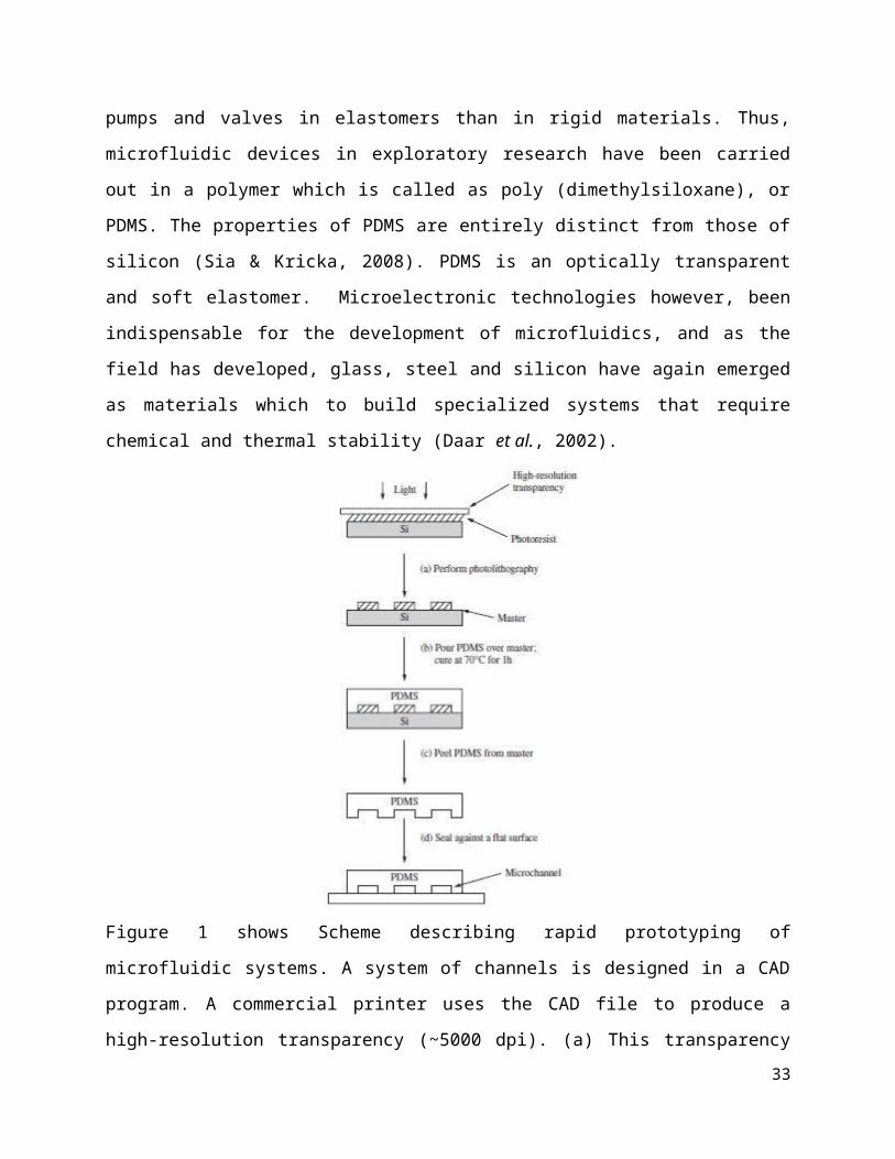
Figure 1 shows Scheme describing rapid prototyping of
microfluidic systems. A system of channels is designed in a CAD
program. A commercial printer uses the CAD file to produce a
high-resolution transparency (~5000 dpi). (a) This transparency33
is used as a photomask in contact photolithography to produce a
master. A master consists of a positive relief of photoresist on
a silicon wafer and serves as a mold for PDMS. (b) Liquid PDMS
pre-polymer is poured over the master and cured for 1 h at 70°C.
(c) The PDMS replica is peeled from the master. (d) The replica
is sealed to a flat surface to enclose the channels. The overall
process takes ~24 h. (McDonald & Whitesides, 2002).
Microfluidic for DNA extraction
A microfluidic system must have a series of generic
components from a method of introducing reagents and samples,
methods for moving the fluids around on the chip, combining and
mixing the fluids until various other devices such as detectors
for most micro analytical work, and components for purification
of products for systems used in synthesis. The field has been
centered on demonstrating concepts for these components. Two
particularly important contributions have been the development of
soft lithography in PDMS as a method for fabricating prototype
devices, and the development of a simple method of fabricating
34
pneumatically activated valves, mixers and pumps on the basis of
soft-lithographic procedures (Gravesen et al., 1993; Dolnik &
Jovanovich, 2000). These methods have made it possible to
fabricate prototype devices that test new ideas in a time period
much shorter than that which could be achieved using silicon
technology which is non specialized specialists that would
consume longer period of time.
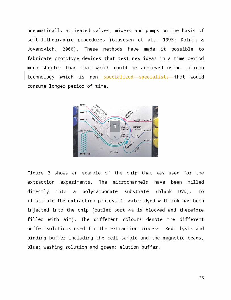
Figure 2 shows an example of the chip that was used for the
extraction experiments. The microchannels have been milled
directly into a polycarbonate substrate (blank DVD). To
illustrate the extraction process DI water dyed with ink has been
injected into the chip (outlet port 4a is blocked and therefore
filled with air). The different colours denote the different
buffer solutions used for the extraction process. Red: lysis and
binding buffer including the cell sample and the magnetic beads,
blue: washing solution and green: elution buffer.
35
Since in the last century, the miniaturization of electronic
devices or microelectronics has been regarded as the most
significant enabling technology in human history. With the
integrated circuits and progress in information processing,
microelectronics has revolutionized the way we work, live and
play. Miniaturization concepts have recently been brought into
the fluidics since the introduction by Manz et al., at the 5th
International conference on Solid-State Sensors and Actuators
(Transducers ‘89): microfluidics, which appeared as the name for
the new research discipline dealing with transport phenomena and
fluid-based devices at microscopic length scales (Manz et al,
1990).
One of the most impressive developments of microfluidics in
life sciences is ‘Bed-side analyses’ or ‘Point-of-Care testing
(POCT)’ such as glucose test meter, pregnancy strip test and
others, which is defined as diagnostic testing at or near the
site of patient care to make the test convenient and immediate
(Sia & Kricka, 2008). Patients can now receive the testing result
within minutes. Such devices can be used in hospitals, at a
doctor office or simply by patients themselves at home without
any professional knowledge or particular skill. Thus, currently,
miniaturized devices are likely to impact economy and improve
public health significantly (Daar et al, 2002).
Other than contributed in biomedicine application,
microfluidic also being applied in biotechnology application such
as in continuous DNA extraction. The detection of DNA and its
36
variation is critical for many fields, including clinical and
veterinary diagnostics, industrial and environmental testing,
agricultural researches and forensic science. Disease diagnosis
and prognosis are based on effective detection of chronic disease
conditions, such as in cancer or contagious disease such as HIV
and genetic markers. However, DNA analysis from original
specimens is a complex process involving multiple chemical
compositions as well as multistep reactions. Conventionally this
procedure is time consuming, labor intensive and contamination
prone, it is not compatible with high throughput or field testing
requirement. To integrate the automatic sample pretreatment
functions will reduce reagent consuming, assay time and
contamination risk. More importantly, it will not only affect the
efficiency and reliability but also determine the feasibility of
a final product. In the past decade, about 3,000 papers published
about on chip DNA tests. Almost all of them need off-chip sample
preparation and reagent handling. A full function system with
sample-in–answer-out capability is still rare (Easley et al.,
2006). By using microfluidic DNA extraction can be performed
anywhere and by anyone without the need of professional
assistance. The concentration of DNA analyte in the test samples
is usually not yet high enough for direct detection. Therefore,
DNA amplification is a required step to raise the concentration
of the target sequence. There is many articles and review paper
regarding combining the continuous DNA extraction using
microfluidic together with DNA amplification. The integration of
37
extraction and polymerase chain reaction (PCR) in the same device
becoming the future revolution in POTC (Dineva et al, 2007).
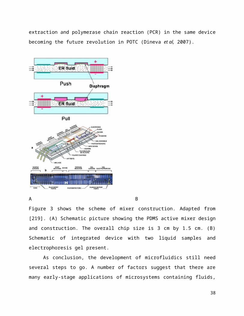
A B
Figure 3 shows the scheme of mixer construction. Adapted from
[219]. (A) Schematic picture showing the PDMS active mixer design
and construction. The overall chip size is 3 cm by 1.5 cm. (B)
Schematic of integrated device with two liquid samples and
electrophoresis gel present.
As conclusion, the development of microfluidics still need
several steps to go. A number of factors suggest that there are
many early-stage applications of microsystems containing fluids,
38
including the exploration of fluidic optics and cells, the
development of new types of organic synthesis in small-channel
systems, the continuing development of technologies based on
large arrays of detectors and on high-throughput screening, the
fabrication of microrobotic systems using hydraulic systems based
on microfluidics, other fluidic versions of MEMS, and work on
biomimetic systems with microfluidic components. Different types
of micro/nano-fluidic technologies have facilitated DNA
purification, amplification and detection to be integrated into
one chip which combine the advantages of small sizes, much
shorter reaction times, less manual operation and reduced cost.
Successful DNA amplification and detection on chip depends on the
optimization of several parameters, which is a cumbersome task
due to many variables (conditions and components) typically
involved and requires comprehensive knowledge of multi-subject
intersecting molecular biology, chemistry, physics, mechanics and
micro/nano-fabrication technologies.
13. Conclusion
DNA plays many roles now-a-days, not only for the interest
of the researcher but it is also important for the phylogenic
tree to trace the species of new things. For forensic
applications, to trace criminals and also for medical purposes
with hereditary and unknown diseases. Thus, DNA extraction
process has attract the biggest interest as re-cap back when the
39
first DNA isolation was successfully done by Friedrich Miescher
in 1869 and the initial DNA extraction developed from density
gradient centrifugation strategies by Meselson and Stahl in 1958,
many techniques for biomolecules purification has been developed
afterwards. From the phenol-chloroform extraction to the column-
technology has widely used in nucleic acids extraction.
Biomolecules extraction has helped researchers and scientists in
manipulating subsequent molecular biology analysis in order to
have a better understanding about the biological materials. The
automated nucleic acid extraction system has been developed due
to the influence of rapid growth of automation technology.
Automating nucleic acid extraction process is potentially
beneficial for a number of reasons including to reduce working
time, decrease labor costs, increase worker safety and at the
same time provides opportunity in increasing reproducibility and
quality of results. However, improvement with the currently
available instruments needs to be conducted all the time. In the
meantime, an all-in-one biomolecules extraction system, or the
invention of a miniature and portable extraction system can
become a prospective development in the future.
References
Arnold, T. E., Meyering, M. T., and Chesterson, R.S. (2005).
“Nucleic acid binding matrix,” United States patent US 6869532
B2, CUNO Incorporated.
40
Buckingham, L., and Flaws, M. L. (2007). Molecular Diagnostics:
Fundamentals, Methods, & Clinical Applications, F.A. Davis,
Philadelphia, Pa, USA.
Blin, N., and Stafford, D. W. (1976). A general method for
isolation of high molecular weight DNA from eukaryotes. Nucleic
Acids Res 3(9): 2303-8.
Brent, J. (1998). Breaking Up Isn’t Hard To Do: A cacophony of
sonicators, cell bombs, and grindes. The Scientist 12(22): 23.
Brooks, G. (1998). Biotechnology in Healthcare: An Introduction
to Biopharmaceuticals, Pharmaceutical Press, London, UK.
Berensmeier, S. (2006). “Magnetic particles for the separation
and purification of nucleic acids,” Applied Microbiology and
Biotechnology, vol. 73, no. 3, pp. 495–504.
Boyd, J. (2002). “Robotic laboratory automation,” Science, vol.
295, no. 5554, pp. 517–518.
Chomczynski, P., and Sacchi, N. (2006). "The single-step method
of RNA isolation by acid guanidinium thiocyanate-phenol-
chloroform extraction: twenty-something years on".Nature
Protocols 1(2): 581-585.
Cui, Z.Q., Qian, Y.Y., and Shi, M.Y. (2003). Protein extraction
from cottonseed meal produced by two-phase solvent extraction.
China oils and fats, 28(11):43-45.
41
Chen, X., Cui, D. F., Liu C. C., and Li, H. (2007). Fabrication
of DNA purification microchip integrated with mesoporous matrix
based on MEMS technology. Microsystem Technologies 14: 51-57.
Cseke, L. J., Kaufman, P.B., Podila, G. K., and Tsai, C. J.
(2004). Handbook of Molecular and Cellular Methods in Biology and
Medicine, CRC Press, Boca Raton, Fla, USA, 2nd edition.
Dederich,D. E., Okwuonu, G., Garner, T. (2002). “Glass bead
purification of plasmid template DNA for high throughput
sequencing of mammalian genomes,” Nucleic Acids Research, vol.
30, no. 7, article e32.
Doyle, K. (1996). The Source of Discovery: Protocols and
Applications Guide, PROMEGA, Madison,Wis, USA.
Dai, S..J., Zhao, G.R. (2001). Development and application of
cottonseed protein isolates. Shihezi science and technology,
(1):30-32.
Dahm, R. (2004). Friedrich Miescher and the Discovery of DNA,
Elsevier, Amsterdam, the Netherland.
Endres, H. N., Johnson, J. A., Ross, C. A., Welp, J. K., and
Etzel, M. R. (2003). Evaluation of an ion-exchange membrane for
the purification of plasmid DNA. Biotechnol Appl Biochem; 37:259–
266.
Esser, K. H., Marx, W. H., and Lisowsky, T. (2006). “MaxXbond:
first regeneration system for DNA binding silica matrices,”
Nature Methods, vol. 3, no. 1, pp. 1–2.
42
Feng, Z.L., Yang, Z.J., and Yuan, B.L. (2004). Study of
extracting technology of isolating soybean protein. China oils
and fats, 29(11):29-30.
Goedecke, N., McKenna, B., El-Difrawy, S., Carey, L., Matsudaira,
P., and Ehrlich, D. (2004). A high-performance multilane
microdevice system designed for the DNA forensics laboratory.
Electrophoresis 25(10-11): 1678-86.
Guerrero-Mart´ınez, A., P´erez-Juste, J., and Liz-Marz´an, L. M.
(2010). “Recent progress on silica coating of nanoparticles and
related nanomaterials,” Advanced Materials, vol. 22, no. 11, pp.
1182– 1195.
Gjerse, D. T., Hoang, L., and Hornby, D. (2009). RNA Purification
and Analysis: Sample Preparation, Extraction, Chromatography,
Wiley-VCH,Weinheim, Germany, 1st edition.
Haines, J. L., Korf, B. R., Moir, D. T., Morton, C. C. (2005).
Current Protocols in Human Genetics Vol 2, Copyright © 2005 by
John Wiley and Sons, Inc.
Knudsen, H. L., Fahrner, R. L., Xu, Y., Norling, L. A., and
Blank, G. S. (2001). Membrane ion-exchange chromatography for
process-scale antibody purification. J Chromatogr A.907:145–154.
Keeley, B., Stark, A., Pisanic, T. R. (2013). Extraction and
processing of circulating DNA from large sample volumes using
methylation on beads for the detection of rare epigenetic events.
Clin Chim Acta.;425:169–175.
43
Kojima, K., and Ozawa, S. (2002). “Method for isolating and
purifying nucleic acids,” United State patent US 2002/0192667 A1.
Loeffler, J., Schmidt, K. D., Hebart, H., and Einsele, H. (2004).
“Automated nucleic acid extraction,” Encyclopedia of Genomics and
Proteomics, pp. 93–96.
Little, M. C. (1991). “Process for the purification of DNA on
diatomaceous earth,” United States patent US 5075430, Bio-Rad
Laboratories Inc.
Mitnik, L., Carey, L., Burger, R., Desmarais, S., Koutny, L.,
Wernet, O., Matsudaira, P., and Ehrlich, D. (2002). High-speed
analysis of multiplexed short tandem repeats with an
electrophoretic microdevice. Electrophoresis 23(5): 719-26.
Mijatovic, D., Eijkel, J. C. T., and van den Berg, A. (2005).
Technologies for nanofluidic systems: topdown vs. bottom-up — a
review. Lab Chip 5, 492–500.
Merion, M., Warren, W. (1989). Purification of supercoiled
plasmids from crude cell lysates using high performance anion
exchange chromatography. Biotechniques;7:60–67.
Ma, C., Li, C., Wang, F. (2013). Magnetic nanoparticles-based
extraction and verification of nucleic acids from different
sources. J Biomed Nanotechnol;9:703–709.
Massart, R. (1981). Preparation of aqueous magnetic liquids in
alkaline and acidic media, IEEE Trans.Magn.17(1981)1247–1248.
44

Nargessi, R. D. (2005).“Magnetic isolation and purification of
nucleic acids,” United States patent US 6855499 B1, Cortex
Biochem, Inc.
Okamoto, Y., Kitagawa, F., Otsuka, K. (2007). Online
concentration and affinity separation of biomolecules using
multifunctional particles in capillary electrophoresis under
magnetic field, Anal.Chem.79.3041–3047.
Padhye, V. V., York, C., and Burkiewiez, A. (1997). “Nucleic acid
purification on silica gel and glass mixture,” United States
patent US 5658548, Promega Corporation.
Pyzowski, P., and Tan, E. (2007). Advances in Biochip-Based
Analysis: A Rapid Field-Based Approach 59th Annual Meeting of the
American Academy of Forensic Sciences San Antonio, TX, February
19-24.
Prazeres, D. M., Schluep, T., Cooney, C. (1998). Preparative
purification of supercoiled plasmid DNA using anion-exchange
chromatography. J Chromatogr A;806:31–45.
Puissant, C., and Houdebine. L. M. (1990). "An Improvement of the
Single-Step Method of RNA Isolation by Acid Guanidinium
Thiocyanate-Phenol-Chloroform Extraction". Bio Techniques 8: 148
- 149.
Qiagen Inc. EndoFree Plasmid Purification Handbook 2005:38–41.
45

Sambrook, J., and Russel, D. (2001). Molecular Cloning: A
Laboratory Manual, vol. 3, Cold Spring Harbor Laboratory Press,
New York, NY, USA, 3rd edition.
Selingson, D. B., and Shrawder, E. J. (1990). “Method of
isolating and purifying nucleic acids from biological samples,”
United State patent US 4935342, Syngene Inc.
Smith, C. E., Holmes, D. L., Simpson, D. J., Kayzhendler, J.,
Bitner,R. H., and Groseh, J. C. (2002). “Mixed-bed solid phase
and its use in the isolation of nucleic acids,” United State
patent US 2002/0001812 A1, Promega Corporation.
Scott, O., Rogers and Arnold J. B. (1994). Extraction of total
cellular DNA from plant, algae and fungi. Plant Molecular Biology
Manual D1: 1-8
Simon, A. J., and Warner. (1996). Genomic DNA Isolation and
Lambda Library Construction. In: Gary D. Foster and David Twell
(ed) Plant Gene Isolation, John Wiley & Sons, England, pp 56-58
Salgueiri˜no-Maceira, V., Correa-Duarte, M. A., Spasova, M., Liz-
Marz´an, L. M., and Farle, M. (2006). “Composite silica spheres
with magnetic and luminescent functionalities,” Advanced
Functional Materials, vol. 16, no. 4, pp. 509–514.
Sasso, L. A., Johnston, I. H., Zheng, M., Gupte, R. K., Undar, A.
(2012). Automated microfluidic processing platform for
multiplexed magnetic bead immunoassays. Microfluidics and
Nanofluidics 13: 603–612.
46
Tinay, E. L., Nour, A. H., and Abdel-Karim, S.H. (1988). Aqueous
protein and gossypol extraction from glanded cottonseed flour:
Factors affecting protein coagulation and gossypol content. Food
Chemistry, 30(1):19-27.
Thomsin, A. (2007) “Insights into lab automation’s future,” IVD
Technology.
Vrancken, K. C., Possemiers, K., Van Der Voort, P., and Vansant,
E. F. (1995). “Surface modification of silica gels with amino
organo-silanes,” Colloids and Surfaces A, vol. 98, no. 3, pp.
235–241.
Wink, M. (2006). An Introduction to Molecular Biotechnology:
Molecular Fundamentals,Methods and Application inModern
Biotechnology, Wiley-VCH,Weinheim, Germany.
Whitford, D. (2005). Proteins: Structure and Function, John Wiley
& Sons, London, UK.
Woodard, D. L., Howard, A. J., and Down, J. A. (1994). “Process
for purifying DNA on hydrated silica,” United State patent US
5342931, Becton, Dickinson and Company.
Wallace, D. M. (1987). Large-and small-scale phenol extractions.
Methods Enzymol.152: 33-41.
Watson, J. D., Baker, T. A., Bell, S. P., Gann, A., Lecine, M.,
and Losick, R. (2004). Molecular Biology of the Gene, Benjamin
Cummings, San Francisco, Calif, USA, 5th edition.
47