distinct viral and mutational spectrum of endemic burkitt lymphoma
TRANSCRIPT

RESEARCH ARTICLE
Distinct Viral and Mutational Spectrum ofEndemic Burkitt LymphomaFrancesco Abate1,2☯, Maria Raffaella Ambrosio3☯, Lucia Mundo3, MariaAntonella Laginestra4, Fabio Fuligni4, Maura Rossi4, Sakellarios Zairis1, Sara Gazaneo3,Giulia De Falco3,5, Stefano Lazzi3, Cristiana Bellan3, Bruno Jim Rocca3, Teresa Amato3,Elena Marasco4, Maryam Etebari4, Martin Ogwang6, Valeria Calbi6, Isaac Ndede7,Kirtika Patel7, David Chumba7, Pier Paolo Piccaluga4, Stefano Pileri4,8*,Lorenzo Leoncini3,4*, Raul Rabadan1,2*
1 Department of Systems Biology, Columbia University College of Physicians and Surgeons, New York,New York, United States of America, 2 Department of Biomedical Informatics, Columbia University Collegeof Physicians and Surgeons, New York, New York, United States of America, 3 Department of MedicalBiotechnologies, Section of Pathology, University of Siena, Siena, Italy, 4 Department of Experimental,Diagnostic, and Specialty Medicine (DIMES), S. Orsola-Malpighi Hospital, Bologna University School ofMedicine, Bologna, Italy, 5 School of Biological and Chemical Sciences, Queen Mary University of London,London, United Kingdom, 6 Lacor Hospital, Gulu, Uganda, 7 Moi University, Eldoret, Kenya, 8 Unit ofHaematopathology, European Institute of Oncology, Milan and Bologna University School of Medicine,Bologna, Italy
☯ These authors contributed equally to this work.* [email protected], [email protected] (SP); [email protected] (LL); [email protected](RR)
AbstractEndemic Burkitt lymphoma (eBL) is primarily found in children in equatorial regions and rep-
resents the first historical example of a virus-associated human malignancy. Although
Epstein-Barr virus (EBV) infection andMYC translocations are hallmarks of the disease, it is
unclear whether other factors may contribute to its development. We performed RNA-Seq
on 20 eBL cases from Uganda and showed that the mutational and viral landscape of
eBL is more complex than previously reported. First, we found the presence of other her-
pesviridae family members in 8 cases (40%), in particular human herpesvirus 5 and human
herpesvirus 8 and confirmed their presence by immunohistochemistry in the adjacent non-
neoplastic tissue. Second, we identified a distinct latency program in EBV involving lytic
genes in association with TCF3 activity. Third, by comparing the eBL mutational landscape
with published data on sporadic Burkitt lymphoma (sBL), we detected lower frequencies of
mutations inMYC, ID3, TCF3 and TP53, and a higher frequency of mutation in ARID1A in
eBL samples. Recurrent mutations in two genes not previously associated with eBL were
identified in 20% of tumors: RHOA and cyclin F (CCNF). We also observed that polyviral
samples showed lower numbers of somatic mutations in common altered genes in compari-
son to sBL specimens, suggesting dual mechanisms of transformation, mutation versus
virus driven in sBL and eBL respectively.
PLOS Pathogens | DOI:10.1371/journal.ppat.1005158 October 15, 2015 1 / 21
OPEN ACCESS
Citation: Abate F, Ambrosio MR, Mundo L,Laginestra MA, Fuligni F, Rossi M, et al. (2015)Distinct Viral and Mutational Spectrum of EndemicBurkitt Lymphoma. PLoS Pathog 11(10): e1005158.doi:10.1371/journal.ppat.1005158
Editor: Paul M Lieberman, Wistar Institute, UNITEDSTATES
Received: March 30, 2015
Accepted: August 19, 2015
Published: October 15, 2015
Copyright: © 2015 Abate et al. This is an openaccess article distributed under the terms of theCreative Commons Attribution License, which permitsunrestricted use, distribution, and reproduction in anymedium, provided the original author and source arecredited.
Data Availability Statement: RNA-Seq data havebeen deposited at the NCBI SRA service (accessionnumber PRJNA292327).
Funding: This work was supported by grants fromthe Italian Association for Cancer Research (AIRC5x1000, n. 10007) and Programma Strategico“Innovative approaches to the diagnosis andpharmacogenetic based therapies of primary hepatictumours, peripheral B and T – cell lymphomas andlymphoblastic leukaemias”, Programma di RicercaRegione-Università 2010–2012: Area 1 “RicercaInnovativa” (SP); MIUR 2012 (LL); StewardFoundation and NIH 1 U54 CA121852-05 (RR). The
Author Summary
Burkitt lymphoma is endemic in sub-Saharan Africa and affects primarily children of age4–7 years. Historically, it was one of the first tumors associated with a virus (EBV) andbearing a translocation involving an oncogene, i.e.MYC. There are three distinct clinicalvariants of Burkitt lymphoma according to the World Health Organization: sporadic,endemic and immunodeficiency-related. Although there has been some recent work onthe molecular characterization of sporadic Burkitt lymphomas, little is known about thepathogenesis of endemic cases. In this work, we analyzed 20 samples of RNASeq from Bur-kitt lymphoma collected in Lacor Hospital (Uganda, Africa) and validated in an extensionpanel of 73 samples from Uganda and Kenya. We identify the presence in the adjacentnon-neoplastic tissue of other herpesviridae family members in 53% of the cases, namelycytomegalovirus (CMV) and Kaposi sarcoma herpesvirus (KSHV). We also demonstrateexpression of EBV lytic genes in primary tumor samples and find an inverse associationbetween EBV lytic expression and TCF3 activity. When studying the mutational profile ofendemic Burkitt tumors, we find recurrent alterations in genes rarely mutated in sporadicBurkitt lymphomas, i.e. ARID1A, CCNF and RHOA, and lower numbers of mutations ingenes previously reported to be commonly mutated in sporadic cases, i.e.MYC, ID3,TCF3, TP53. Together, these results illustrate a distinct genetic and viral profile of endemicBurkitt lymphoma, suggesting a dual mechanism of transformation (mutation versus virusdriven in sBL and eBL respectively).
IntroductionBurkitt lymphoma (BL) is the first human cancer to be associated with the Epstein-Barr virus(EBV), the first tumor to exhibit a chromosomal translocation activating an oncogene (MYC),and the first lymphoma to be associated with human immunodeficiency virus (HIV) infection.The World Health Organization[1] classification describes three clinical variants of BL:endemic, sporadic, and immunodeficiency-related. These variants are similar in morphology,immunophenotype, and genetics. While the sporadic variant (sBL) occurs outside of Africaand is rarely associated with EBV infection, the endemic variant (eBL) arises mainly in Africaand is associated with malaria endemicity and EBV infection in almost all cases. Epidemiologi-cal studies have shown that malaria and EBV combined do not fully explain the distribution ofeBL in high risk regions[2]. Malaria and EBV are in fact ubiquitous within the lymphoma beltof Africa, suggesting that other etiologic agents may be involved[3]. However, it is unclearwhat other epidemiological factors could play a role in the genesis of eBLs.
Three types of EBV latency have been described in EBV-related lymphomas according tothe pattern of EBV nuclear antigen (EBNA) and the latent membrane protein (LMP) expres-sion, namely latency I, II, and III[4]. Specifically, latency I is usually associated with eBL and itdenotes a transcriptional program in which an EBV infection does not produce virions andexpresses a single protein, EBNA-1. While the latency I program has been extensively charac-terized in vitro, a different form of latency has been recently reported in 15% of eBL that uses adifferent set of promoters. Termed Wp-restricted latency[5], this program shows a homoge-neous host expression signature[6] characterized by down-regulation of BCL-6 and up-regula-tion of IRF-4 and BLIMP-1. Other reports have described latency program heterogeneity atsingle cell level[7] and low expression of LMP genes in a fraction of cases[8,9]. HeterogeneousEBV transcription profiles with LMP expression have been recently reported in some cases ofAIDS-related and sporadic BL[10], but extensive data on endemic cases are not available yet.
Distinct Viral and Mutational Spectrum of Endemic Burkitt Lymphoma
PLOS Pathogens | DOI:10.1371/journal.ppat.1005158 October 15, 2015 2 / 21
funders had no role in study design, data collectionand analysis, decision to publish, or preparation ofthe manuscript.
Competing Interests: The authors have declaredthat no competing interests exist.
These studies indicate that the transcriptional EBV programs of primary eBL could be morecomplex than expected across cases and within individuals. Therefore, the exact role of EBVhas remained elusive and further investigation is required.
The genetic hallmark of all three clinical variants of BL is the t(8;14) translocation involvingthe juxtaposition of the immunoglobulin heavy chain locus (IGH) with theMYC oncogene[11]. However, although transgenic mice expressingMYC under the control of the intronicIGH enhancer (Eμ) develop B cell lymphomas[12], successive molecular characterization dem-onstrated that this model does not fully recapitulate the human disease. The comparisonbetween the gene expression profile (GEP) of BL and diffuse large B-cell lymphoma (DLBCL)highlighted a distinct signature of BL characterized by the expression of bothMYC targets andgerminal-center B-cell genes[13]. Furthermore, hypermutation and different breakpoint pat-terns of IGH/MYC translocation[14,15] suggests that the origin of human BL derives fromaberrant class switching in the germinal center (GC), while transgenic IGH/MYCmice typicallyarise from precursor/naive B-cells. The more accurate PI3K/MYC transgenic mouse model bySander et al[16] better recapitulates the human phenotype of BL and highlights the importanceof the PI3K pathway in the disease. Moreover, GEP analysis has demonstrated that the tran-scriptional profile of eBL is different from that observed in sBL[17]. Recent studies haveunveiled the genetic landscape of sBL characterized by mutations affecting the B-cell receptor(BCR) pathway and in particular the transcription factor TCF3, its negative regulator ID3, thecell-cycle G1/S regulator CCND3[18,19], and the chromatin-remodeling gene ARID1A[20].On the contrary, very little is known about the spectrum of alterations in eBL, how it might dif-fer from that of sBL, the correlations between host mutation and viral infection, and the spe-cific viral/host transcriptional programs.
In this study, we aim to characterize the presence of other potential agents, to define theEBV transcriptional profile and to link these profiles to the mutational status of new and previ-ously reported genes. We provide a characterization of the mutational and viral landscape ofeBL using 20 cases from Uganda. RNA-Seq, in combination with targeted sequencing technol-ogy on a larger cohort of cases, allows the identification, validation and assessment of the recur-rence of new somatic mutations. In addition, in contrast with earlier microarray-basedexpression studies, RNA-Seq provides the opportunity to identify and associate microbial andtumor mutational and expression profiles.
Results
Endemic BL is associated with multiple viral infectionsTo identify new pathogens in eBL, we applied Pandora, a new pipeline for the characterizationof tumor microbiomes, to a discovery cohort of 20 RNA-Seq samples. We established a readcutoff on the basis of those samples that tested positive for RNA in situ hybridization (ISH) ofthe EBER transcript. Since ISH validated all the RNA-Seq samples as positive, we establishedthe threshold to call a virus present in a particular sample as the minimal number of readsdetecting EBV (S1 Fig). Next, we established the EBV subtype by aligning RNA-Seq reads tothe genomes of both EBV type I and type II and deduced type I as the closest genotype. In addi-tion to EBV, RNA-Seq revealed the presence of other viruses. In particular, 5/20 cases con-tained human herpesvirus 5 (HHV5, cytomegalovirus, CMV), 4/20 human herpesvirus 8(HHV8, Kaposi sarcoma herpes virus, KSHV), and 1/20 human T-lymphotropic virus 1(HTLV-1) (Fig 1A, S2 Fig and S3 Fig). Human immunodeficiency virus (HIV) was notdetected in any case, confirming that pediatric eBL is rarely associated with the immunodefi-ciency syndrome[21]. Nested PCR and immunohistochemical (IHC) analysis performed on all20 original samples confirmed the presence of all the viruses in the discovery cohort (S1A
Distinct Viral and Mutational Spectrum of Endemic Burkitt Lymphoma
PLOS Pathogens | DOI:10.1371/journal.ppat.1005158 October 15, 2015 3 / 21

Table). To assess whether RNA-Seq findings generalize for EBV, CMV, KSHV, and HTLV-1,we assayed for the presence of these four viruses in 20 additional cases from western Kenya byIHC (S1B Table). In this Kenyan cohort, EBV was detected in 20/20 samples, CMV in 8/20samples (Fig 1B and S4 Fig), KSHV in 7/20 samples (Fig 1C and 1D, and S5 Fig), and HTLV-1in 0/20 samples. Therefore, over the 40 cases, we report the overall viral infection frequenciesof 40/40 (100%) for EBV, 13/40 (32.5%) for CMV, 11/40 (27.5%) for KSHV, and 1/40 (2.5%)for HTLV-1. IHC analysis demonstrated the presence of CMV in the stromal cells andmacrophages localized within the tumors and in the adjacent reactive lymphoid tissue (Fig 1B,
Fig 1. (A) RNA-Seq technology reveals the presence of EBV and of other viruses. In particular, 5/20 cases contain human herpesvirus 5 (CMV), 4/20 human herpesvirus 8 (KSHV), and 1/20 human T-lymphotropic virus 1 (HTLV-1). (B) Immunohistochemical evaluation demonstrates thepresence of CMV in the stromal cells in the adjacent reactive lymphoid tissue. CMV stain, Original Magnification (O.M.): 40x. (C) KSHV positivity isshown, respectively in few neoplastic cells and in the endothelial cells within the neoplastic proliferation. LANA-1 (LN53 antibody), O.M.: 40x; (D)LANA-1 (AT4C11 antibody) O.M.: 40x.
doi:10.1371/journal.ppat.1005158.g001
Distinct Viral and Mutational Spectrum of Endemic Burkitt Lymphoma
PLOS Pathogens | DOI:10.1371/journal.ppat.1005158 October 15, 2015 4 / 21

S2 Fig and S4 Fig). KSHV was identified not only in normal B-lymphocytes and endothelialcells from the adjacent reactive lymphoid tissue (S3 Fig and S5 Fig), but also in one case inabout 5–10% of neoplastic cells (Fig 1C and 1D). HTLV-1 was detected in reactive T-lympho-cytes in the only positive case of the discovery cohort. Sections of the samples incubated withthe secondary antibody alone and sections of reactive lymphoid tissue were used as negativecontrols. Sections of lymph nodes with infectious mononucleosis were used as positive controlfor EBV. Next, we compared the viral landscape of endemic and sporadic cases by analyzing 27RNA-Seq sBL samples from Schmitz et al.[19] with Pandora. The analysis showed the presenceof EBV and HIV respectively in 4/27 (15%) and in 1/27 (4%) cases, consistent with several liter-ature sources[22].
Epstein-Barr virus latent and lytic infection in BLBeyond identification of EBV presence, RNA-Seq enabled us to quantitatively analyze the viraltranscriptional program. In addition to EBER-1 and EBER-2 transcripts, expression analysis ofthe viral genes showed the expression of EBNA-1, a gene associated to latency I type, in 18/20cases (Fig 2A). We also detected either LMP-1 or LMP-2A, characterizing the latency II type, in13/20 samples (65%), and also EBNA-2 in 1/20 cases (5%). Interestingly, 2/20 cases (10%) werecharacterized by the expression of EBNA-3A/B/C/LP, together with the lytic gene BHRF-1, sug-gesting a Wp-restricted program[23]. However, the specific analysis of EBV isoforms showedthe presence of H2-HF splicing event, which is hallmark of lytic BHRF-1 expression[24–26](S6Fig). Unsupervised hierarchical clustering of expressed EBV genes demonstrated two mainclusters distinguished largely by gene products involved in EBV replication (BALF-2, BCRF-1,BHRF-1, BILF-1, BMRF-1, BNLF-2a, BZLF-1). The expression of these genes suggests a non-canonical latency program of the virus with a subset of viral episomes initiating lytic reactiva-tion[23].
Due to the heterogeneity of the viral transcriptional programs, we aimed to validate thelatency type by performing RT-qPCR for the EBNA-1, LMP-1, LMP-2A, EBNA-2, EBNA-3C,and BHRF-1 transcripts across an additional series of 26 cases from an extended cohort of sam-ples from Kenya. EBNA-1 was detected in 26/26 (100%), LMP-1 and LMP-2 in respectively 5/26 (20%) and 20/26 (75%) cases (S3 Table and S7A Fig), EBNA-2 in 0/26 (0%), and the combi-nation of EBNA-3C and BHRF-1 in 4/26 (15%). These results are largely consistent with theRNA-Seq data with the exception of LMP-1 that has been detected at higher frequency inRNA-Seq (S2 Table). Next, we evaluated the lytic cycle activation and found BILF-1, BALF-4,and LF-2 in all 26 cases, whereas we observed the expression of BALF-2 in 23/26 (90%), BHRF-1 in 20/26 (80%), BZLF-1 and BMRF-1 in 15/26 (60%), BNLF-2a in 13/26 (50%), and BCRF-1in 11/26 (45%) of the cases (S3 Table and S7B–S7D Fig).
We then validated the expression of all the available encoded-proteins by IHC using strin-gent positive and negative controls as reported in Materials and Methods. Overall, IHC evalua-tion confirmed a non-canonical latency associated program with the expression of someproteins characterizing latency II (i.e. LMP-1 in 2/26 and LMP-2A in 17/26 of the cases);however, there was heterogeneity in the intensity of protein staining and in the proportion ofpositive tumor cells. LMP-1 was detected in few cells, whereas LMP-2A was identified in a pro-portion of cells ranging from 25% to 50% (Fig 2B and 2C). EBV replication was assessed bynuclear expression of the immediate-early BZLF-1/ZEBRA and early BMRF-1/Ea-D, BHRF-1/Ea-R lytic proteins (Fig 2D–2I). There was positive staining in the neoplastic cells for BZLF1,BHRF-1/Ea-R and BMRF-1/Ea-D, respectively in 11/26 (40%), 16/26 (60%), and 13/26 (50%)of the cases (S3 Table).
Distinct Viral and Mutational Spectrum of Endemic Burkitt Lymphoma
PLOS Pathogens | DOI:10.1371/journal.ppat.1005158 October 15, 2015 5 / 21

Fig 2. (A) Unsupervised hierarchical clustering of expressed EBV genes demonstrates a diversity of non-canonical latency-associated geneexpression programs with a subset of viral episome initiating lytic reactivation as indicated by expression of genes corresponding to the lyticprogram. (B) LMP-2A is expressed by 40 to 50% of neoplastic cells. LMP-2A stain, O.M.: 40x; (C) LMP-2A expression is identified in a proportion ofneoplastic cells ranging from 20 to 30%. LMP-2A stain, O.M.: 40x; (D) BZLF1/ZEBRA positivity is expressed by 5 to 10% of neoplastic cells. BZLF1/ZEBRA stain, O.M.: 40x; (E) BZLF1/ZEBRA expression is detected in few neoplastic cells. BZLF1/ZEBRA stain, O.M.: 40x; (F) BMRF-1/Ea-D
Distinct Viral and Mutational Spectrum of Endemic Burkitt Lymphoma
PLOS Pathogens | DOI:10.1371/journal.ppat.1005158 October 15, 2015 6 / 21

Finally, we compared the patterns of latent and lytic gene expression between endemic andsporadic BLs using the 4 EBV-positive sBLs of the 27 RNA-Seq samples from Schmitz et al.16
We observed the expression of BHRF-1 and BMRF-1 in 1 case; BZLF-1 was present in 2 casesand LMP-2A in 4 cases.
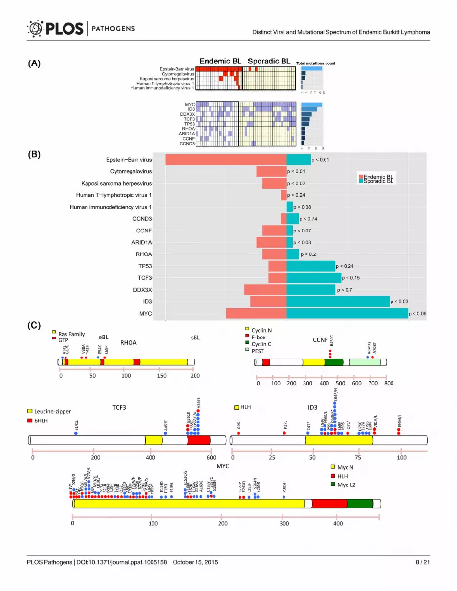
Mutational landscape of eBL and correlation with EBV presenceTo identify the genes that are somatically mutated in eBL, we applied the SAVI algorithm[27]to the cohort of 20 RNA-Seq samples (see Material and Methods for gene selection criteria).Our analysis identified 13 genes recurrently mutated in more than 4 samples. We confirmedthe presence of mutations in genes previously reported in BL literature[18,19,28] (Fig 3A–3Cand S4 Table), includingMYC in 10/20 (50%), DDX3X in 7/20 (35%), ID3 in 6/20 (30%),ARID1A in 5/20 (25%), RHOA in 4/20 (20%), TCF3 and TP53 in 3/20 (15%), CCND3 in 1/20(5%) of the cases. In addition, we found recurrent mutations in one gene not reported so far:CCNF, detected in 4 out of the 20 cases (20%). Since RHOAmutations have not been previ-ously detected in eBL and CCNFmutation was a new discovery, their prevalence as specificmutations was further assessed using Sequenom technology on an extended panel of 66 neo-plastic samples plus 7 cases with matched normal controls (S8A and S8B Fig). Recurrent muta-tions in RHOA were found in 6/73 eBL cases (8%), and in 0/7 normal samples. Two of the 6RHOAmutations occurred in paired eBL/normal cases, confirming that the alterations aresomatic (S5 Table). Recurrent mutations in codon 451 of CCNF were found in 14/73 eBL cases(19%), and in 0/7 normal samples. One of the 14 CCNFmutations occurred in a paired eBL/normal case, showing that also CCNF alteration is somatic. Direct sequencing of genomicDNA confirmed all the mutations identified by Sequenom tecnnology and RNAseq (S9 Fig andS10 Fig).
The distribution of somatic mutations and viral presence across both eBL and sBL samplesexhibit two interesting features (Fig 3B). First, in eBL samples we observed lower mutationalfrequencies in the genesMYC, ID3, TCF3, DDX3X, CCND3 and TP53, as compared to theirreported recurrence in sBL, and higher mutational frequencies in ARID1A, RHOA, and CCNF[18,19]. Second, in sBL cases an almost mutual exclusivity can be seen between EBV presenceand mutations in TCF3/ID3 both known to be driver genes in sBL (p-value< 0.02, Fisher exacttest). To explore this hypothesis, we performed a hierarchical clustering of both endemic andsporadic cases on TCF3 target genes (previously reported in Schmitz et al.[19]) and we demon-strated that the first bifurcation of the dendrogram classifies the samples into EBV-positiveand EBV-negative BL independently on the specific subtype with an accuracy of 96% (45/47).(Fig 4A). The results show that the TCF3 pathway is more activated in EBV-negative cases, asindicated by the significant negative enrichment of TCF3 target genes in EBV-positive samples.Furthermore, we observe that when considering the overall panel of both endemic and sporadicBL samples, the mutually exclusivity between TCF3/ID3mutations and EBV infection yields amore significant effect (p-value< 0.0008, Fisher exact test). To further investigate the hosttranscriptional programming related to EBV presence, we performed GSEA C2 analysis ongenes differentially expressed between EBV-positive and EBV-negative cases. Interestingly, wedetected a significant enrichment for the LMP-1 gene set signature, reported by Sengupta et al.[29] in nasopharyngeal carcinoma, which is consistent with the detected LMP-1 expression in
expression is observed in 50% of neoplastic cells. BMRF-1/Ea-D stain, O.M.: 40x; (G) BMRF-1/Ea-D protein expression in 5% to 10% of neoplasticcells is shown. BMRF-1/Ea-D stain, O.M.: 40x; (H) BHRF-1/Ea-R staining is found in 60% of neoplastic cells. BHRF-1/Ea-R stain, O.M.: 40x; (I)BHRF-1/Ea-R is expressed in 10% of neoplastic cells. BHRF-1/Ea-R stain, O.M.: 40x.
doi:10.1371/journal.ppat.1005158.g002
Distinct Viral and Mutational Spectrum of Endemic Burkitt Lymphoma
PLOS Pathogens | DOI:10.1371/journal.ppat.1005158 October 15, 2015 7 / 21
Distinct Viral and Mutational Spectrum of Endemic Burkitt Lymphoma
PLOS Pathogens | DOI:10.1371/journal.ppat.1005158 October 15, 2015 8 / 21
RNA-Seq data (Fig 4B). Moreover, since 13/20 RNA-Seq cases were positive for LMP2A, weinvestigated the role of this viral gene in the context of eBL and GSEA C2 analysis has been per-formed on gene differentially expressed between LMP-2A positive and LMP-2A negative sam-ples. Interestingly, the E2F, E2F3 and cell cycle G1/S gene sets presented the highest significantenrichment score (see S11 Fig), together with the down-regulation of retinoblastoma pathway.These results can be explained as an effect of the interaction betweenMYC and LMP2A. Infact, previous studies showed that LMP-2A promotesMYC-induced lymphomagenesis[30],and E2F is a know target ofMYC during cell division and proliferation[31]. Moreover, severalworks associate LMP-2A expression to the PI3K/Akt pathway activation[32–35] and the studyfrom Brennan et al. (Oncogene, 2002 [36]) shows that the activation of PI3K pathway in lym-phoblastoid cell lines can promote E2F transcription activity to affect cell cycle and cellularproliferation.
DiscussionOver the past few years, the concept that many diseases can be etiologically linked to infectionby more than one pathogen has drawn increased attention[37–40]. Whether endemic Burkittlymphoma should also be considered a polymicrobial disease and what role genetic alterationsplay in the tumor are still open questions. In this paper, we analyzed the presence of pathogensother than EBV in 40 eBL primary tumors by RNA sequencing, PCR, and immunohistochem-istry, and found the presence of CMV and KSHV. We detected these viruses, which are fre-quently reported in the African population[41], primarily in the surrounding non-neoplastictissue. Their prevalence in areas endemic for EBV, along with their absence in the sporadiccases, suggests that CMV or KSHV could contribute to the chronic antigenic stimulation inwhich eBL occurs.
The presence of these additional cofactors may also induce EBV lytic cycle through B-cellreactivation and spreading EBV infection out of its natural niche of memory B-cells, character-ized by a latency 0/I program[42,43]. In fact, in our samples we showed a non-canonicallatency program of the virus characterized by a large number of cases expressing LMP-1/-2A/-2B in a significant proportion of cells along with lytic reactivation. Our results are in agreementwith recent studies showing more complex EBV protein expression in Akata and Mutu celllines, commonly used to study the role of EBV in Burkitt lymphoma[44]. By using an alterna-tive approach based on RT-QPCR array platform, Tierney et al. report a quantitative character-ization of EBV transcripts in different experimental infection models that were validated inendemic Burkitt lymphoma samples[24]. Interestingly, in this study a significant expression ofLMP-2 gene was revealed. Moreover, our results are in accordance with a previously publishedstudy in primary AIDS-related lymphomas (ARL) by Arvey and colleagues[10], although arigorous comparison is limited by the small number of ARL BLs. All together, our findingsconfirm recent evidence that LMP-2A cooperates in reprogramming the function of normal B-lymphocytes and enhanceMYC driven lymphomagenesis through the activation of PI3K-path-way[45,46]. This pathway is a crucial toMYCmediated transformation as shown by PI3K/MYC transgenic mouse that produces a model that represent a phenocopy of human tumors interms of histology, gene and protein marker expression, and somatic hypermutations[47]. This
Fig 3. (A) The presence of mutations in genes previously described in BL is reported, includingMYC (50%),DDX3X (35%), ID3 (30%), ARID1A(25%),RHOA (20%), TCF3 and TP53 (15%), andCCND3 1/20 (5%). In addition, a newmutation is shown, involvingCCNF and detected in 20% of thecases. (B) Bar plot showing the frequency comparison of virus presence and driver mutations between endemic and sporadic BL. For eachcomparison we report the p-value associated with rejecting the null hypothesis of equal eBL and sBL prevalences. (C) Distribution of mutations in5 driver genes. Red points indicate endemic BL, while blue points the sporadic ones.
doi:10.1371/journal.ppat.1005158.g003
Distinct Viral and Mutational Spectrum of Endemic Burkitt Lymphoma
PLOS Pathogens | DOI:10.1371/journal.ppat.1005158 October 15, 2015 9 / 21

Fig 4. (A) the dendrogram classifies the samples into EBV-positive and EBV-negative BL independently on the specific subtype with an accuracyof 96% (45/47). (B) GSEA C2 analysis on genes differentially expressed between EBV-positive and EBV-negative cases detects a significantenrichment for the LMP-1 gene set signature. GSEA: gene set enrichment analysis.
doi:10.1371/journal.ppat.1005158.g004
Distinct Viral and Mutational Spectrum of Endemic Burkitt Lymphoma
PLOS Pathogens | DOI:10.1371/journal.ppat.1005158 October 15, 2015 10 / 21

scenario suggests that LMP-2A activation of PI3K is an alternative/convergent mechanism tothe one driven by TCF3/ID3mutations.
The expression of genes characterizing the lytic phase of EBV found by RNA-Seq was con-firmed by IHC staining for the three main genes involved in the initiation of the lytic phase,BZLF-1/ZEBRA, BMRF-1/Ea-D and BHRF-1/Ea-R. In early latent infection, EBV can beinduced to enter the lytic cycle by a variety of causes including B-cell receptor stimulation,Toll-like receptor-9 activation, hypoxia, and growth factors[48,49]. Although lytic infectionkills the host cell, it also allows horizontal spread of EBV from cell to cell and may increase thepool of latently infected B-lymphocytes from which transformed cells arise. Additionally, lyti-cally infected B-cells secrete factors that may promote tumorigenesis, including growth andangiogenesis factors and immunosuppressive cytokines. Recent evidence has challenged theview that only the latency phase of EBV infection is significant for the development of EBV-associated malignancies, proposing that lytic EBV replication may be of pathogenic relevance.[50] Humanized mice infected with lytic active viral strains develop more lymphomas than ani-mals infected with replication-defective strains[39], suggesting that lytic EBV infection may beof importance also in the context of an active immune response. In the present study we gaveevidence for the first time that this occurs in vivo in the neoplastic cells of the primary tumors.Physiologically, lytic gene products are expressed in three consecutive stages: immediate-early,early, and late. Immediate-early lytic gene products initiate the process by inducing the activa-tion of transcription of the other genes. Early genes control replication and metabolism of neo-plastic cells[51]. Fatty acid synthase expression is induced by the BRLF-1 immediate–earlyprotein, and interestingly BL tumors are characterized by altered lipid metabolism[52]. Lategene products code for viral capsid antigens and proteins involved in immune evasion. BNLF-2A, detected in a significant number of our cases, may protect infected B-cells from immunerecognition and elimination[53]. Finally, the EBV transcriptome during the reactivation mayinvolve the contribution of a wide array of other virus-encoded RNAs, such as BARF-0, BARF-1, BcLF-1, and RPMSI-1[54], that are not translated and may function as non-coding RNAmolecules which could participate in regulating gene expression[55]. Heterogeneity in lytic/latent expression programs can be observed not only between patients but also within individ-ual tumors, on a cell-to-cell basis. Intra-patient heterogeneity might be related to the activationof the immune response following the expression of the viral genes. Therefore, the tumor isunder selective pressure and needs alternative mechanisms to survive and proliferate[56].
Our data on the mutational landscape of eBL seems to support this hypothesis. In fact, eBLsamples were characterized by a lower number of point mutations in genes previously foundaltered in sBL, includingMYC, ID3, TCF3, DDX3X, CCND3, and TP53. These results are con-sistent with previous studies by Schmitz et al. [19]in which TCF3/ID3mutations were morecommon in sBL (70% of the cases) than eBL (40%). In particular, we observed a near mutualexclusivity between TCF3/ID3mutations and the presence of EBV, indicating that TCF3 path-way is more significantly activated in EBV-negative cases.
The inverse correlation we observed between the presence and expression of EBV and thenumber of cellular mutations in the different BL cases, may represent an in vivo picture of thedynamic process by which a neoplastic cell, initially dependent upon EBV, switches-off viralgenes and switches-on cellular mutated genes to survive and proliferate. These results are con-sistent with previous analysis of pediatric BL[20]. Based on our findings, one should infer thateBL may arise from pathogenic pathways that are partially distinct from those driving sBL, sug-gesting dual mechanisms of transformation in BL, mutationally versus virally driven. On theother hand, ARID1A and RHOA were more often mutated in eBL than in sBL. ARID1A is oneof the subunits of the Switch/Sucrose Non-Fermentable (SWI/SNF) chromatin remodelingcomplex and is currently thought to behave like a tumor suppressor gene. Consistently,
Distinct Viral and Mutational Spectrum of Endemic Burkitt Lymphoma
PLOS Pathogens | DOI:10.1371/journal.ppat.1005158 October 15, 2015 11 / 21

ARID1Amutations frequently occur as insertion/deletion, and in most of our cases involvedthe amino acid G1630. This gene has been reported as frequently mutated in the context ofpediatric BL, with a significant association to EBV negative cases[20], suggesting that the highprevalence in eBL compared to the sBL may be due to the pediatric nature of the endemic case.However, other EBV-associated cancer types show frequent deregulation of ARID1A[57–61].In particular, in EBV-associated gastric cancer a strong correlation between ARID1A deactiva-tion and EBV presence has been reported[62–64]. RHOA, which belongs to the Ras homologfamily, is a small GTP-ase protein recently found to be mutated in three tumors associatedwith EBV infection, namely peripheral T-cell lymphoma (where it relates to follicular helper T-cells[65–67]), diffuse gastric carcinoma[68] and paediatric sBL[28]. The distribution of RHOAmutations in our cohort overlaps with the already reported mutations (codons 5, 17, 42 and69) suggesting a similar functional role.
Finally we identified recurrent mutations involving the amino acid R451C in one gene notpreviously detected in endemic or sporadic BLs, CCNF, altered in 20% of our cases. CCNFencodes a member of the cyclin family belonging to the F-box protein family; it acts as aninhibitor of centrosome reduplication during G2 phase and protects the cell from genomeinstability[69]. Therefore, it is reasonable that CCNFmutations may cooperate in inducinglymphomagenesis by promoting chromosome instability and a hypermutator phenotype[70,71].
Understanding the mechanisms regulating EBV lymphomagenesis will hopefully lead to thedevelopment of highly specific therapies. To avoid the tumor evasion from the already availabletherapies, we need to identify and target the multiplicity of pathways that are deregulated inthe neoplastic cells and decrease tumor survival and proliferation.
Materials and Methods
Cases selectionA total of 20 BL samples preserved in RNAlater (RNA stabilization Reagent-QIAGEN, Valen-cia, CA) were collected from the Department of Human Pathology of the Lacor Hospital(Uganda, Africa), in endemic areas. For all of them, formalin-fixed and paraffin-embedded(FFPE) samples have been available. All diagnoses were reviewed by 2 expert hematopatholo-gists and were formulated according to the 2008 WHO classification. The clinical and histo-pathologic characteristics of the 20 BL cases are summarized in S6 Table. Briefly, all cases weret(8;14)-positive, and the immunophenotype was consistent with the diagnosis of BL (CD20positive, CD10 positive, BCL-6 positive, Ki67> 98%, BCL-2 negative). Epstein-Barr virus wasdetected by using in situ hybridization with EBER probes (INFORM EBER, Roche Diagnostics,Basel, Switzerland). EBV infection in tumor cells was observed in 100% of the samples, assessedby strong nuclear expression of small EBV-encoded RNA genes, EBER-1 and -2. These caseshave been previously studied for gene expression profile analysis and showed a molecular pro-file consistent with molecular BL[17].
We used two distinct series of cases for validation of RNA-Seq results. The first included 26primary tumors collected at the Moi University, Eldoret (Western Kenya). Of these, 20 wereused for virus data validation and 26 for EBV latency validation. The second was comprised of66 neoplastic samples plus 7 cases for which matched normal controls were available (1 liver, 6lymph nodes) collected from endemic area in Africa, and was used for Sequenom validation.
RNA extractionTotal RNA extraction was perfomed by RNeasy Plus Mini Kit(QIAGEN, Valencia, CA)according to the manufacture instructions. The amount and quality of RNA were evaluated by
Distinct Viral and Mutational Spectrum of Endemic Burkitt Lymphoma
PLOS Pathogens | DOI:10.1371/journal.ppat.1005158 October 15, 2015 12 / 21
measuring the optical density (OD) at 260 nm, the 260/230 and the 260/280 ratios using aNanodrop spectrophotometer (ND-100, Nanodrop, Thermo Scientific, Celbio, Italy).
RNA sequencingPaired-end libraries (2x75 base pair) were prepared according to the TruSeq RNA sample prep-aration v2 protocol (Illumina, San Diego, USA). Briefly, 2 μg of Poly(A)+ RNA was purifiedfrom total RNA using poly-T oligo attached magnetic beads and then used for fragmentationinto 130–290 bp fragments. First, single stranded cDNA was synthesized using reverse tran-scriptase (SuperScript II, Invitrogen, Life Technologies,USA) and random hexamer priming,followed by generation of double-stranded cDNA. AmpureXP beads (Beckman Coulter, BreaCA) were used to purify the ds cDNA and end repair step was performed to convert the over-hangs, resulting from fragmentation, into blunt ends by 3’ to 5’ exonuclease activity. A single“A” nucleotide was added to the 3’ ends of the blunt fragments to prevent them from ligatingto one another during the adapter ligation reaction. This approach was adopted to ensure a lowrate of chimera (concatenated template) formation. Subsequently, sequencing adapters wereadded to the ends of the ds cDNA fragment and a PCR reaction was used to selectively enrichthose ds cDNA fragments that had adapter molecules on both ends, amplifying the amount ofds cDNA in the final libraries. Lastly, PCR library products were purified by AmpureXP beadsand quality control analysis was assessed using a DNA-1000 (Agilent, USA). The quantificationwas performed by Quant-it PicoGreendsDNA Assay Kit according to manufacturer’s protocol(Invitrogen, Life Technologies,USA). The resulting libraries were sequenced on an IlluminaHiScan SQ (Illumina, San Diego, USA) following the manufacturer's instructions.
Point mutation identification using RNASeqSequence variants were obtained using the SAVI (Statistical Algorithm for Variant Identifica-tion)[72,73] algorithm independently for each sample. Candidate somatic mutations wereobtained by eliminating common germline variants (dbSNP 132 and variants from 10 reactivelymph nodes). Genes recurrently mutated in more than 4 samples and expressing the corre-sponding transcript with RPKM>3 were selected. Mutations occurring in the exact same posi-tion in more than 4 samples have been discarded. Conversely, genes previously reported in BL[19,74,75] were selected, even at low recurrence, to allow the comparison between endemic andsporadic subtypes (S3 Table). Sanger sequencing was used for technical validation.
Pandora: A pipeline for pathogen discovery using RNASeqCharacterization of the tumor microbiome is accomplished with Pandora, a new RNA-Seqpipeline for pathogen identification and discovery (S12 Fig). The algorithm takes raw RNA-Seqdata as input and outputs annotated microbial spectra present in the tumor sample. Pandoraimplements a subtractive algorithm consisting of discrete modules. First, theHost Removalphase sequentially aligns the input reads to the host reference using bowtie2[76], blastN[77]and Megablast,[78] and filters out the data originating from the host. Second, the unaligned(non-host) reads are passed as input to theMicrobe Identification phase where the reads arealigned to curated sets of NCBI microbial sequences representing viruses/viroids, bacteria,fungi, and select taxa of eukaryotic parasites. Third, the NCBI records matching each non-hostread are input to the Reporting phase where microbial load, gene expression, and relevantclinical parameters are computed as the final output. The microbial load is computed as thenumber of reads mapping to the organism or virus normalized by the genome length. Geneexpression quantification is computed as transcript per million (TPM)[79], which provides a
Distinct Viral and Mutational Spectrum of Endemic Burkitt Lymphoma
PLOS Pathogens | DOI:10.1371/journal.ppat.1005158 October 15, 2015 13 / 21

more accurate relative quantification of mRNA abundance compared to other normalizationmethods such as RPKM.
EBV genome analysis and expressionRNA-Seq reads were aligned to the GRCh37/hg19 reference genome using Bowtie2[76], Blastn[77] and Megablast[78]. Reads not aligning to homo sapiens (non-host reads) were mapped tohuman herpesvirus 4, type I (NCBI accession number NC_007605.1) using TopHat, a splicingaware alignment program [80] (S9 Table and S10 Table). EBV viral gene expression was nor-malized as transcripts per million (TPM)[79]. For each viral product the TPM expression wasnormalized by the expression of A73 genes, which is consistently expressed in all the HHV4positive BL samples. Hierarchical clustering was computed with Pearson distance and Ward’slinkage method.
Expression analysisGene expression analysis was performed on both endemic and sporadic[19] RNA-Seq samplesof Burkitt Lymphoma. All the reads were aligned to human reference genome (GRCh37/hg19)by means of TopHat version 1.3.3. Transcript abundance quantification was computed asFPKM using Cufflinks, Cuffquant and Cuffnorm version 2.2.1[81]. Hierarchical clustering wasperformed with Pearson distance and Ward’s linkage method. Gene set enrichment analysiswas obtained by running GSEA software on pre-ranked list of log2 ratio of the FPKMmeanfold change between two conditions[82].
DNA extractionThe DNA was extracted from formalin-fixed paraffin embedded (FFPE) of the original neo-plastic samples using NucleoSpin Tissue (Machery-Nagel, Italy) following manufacture’sinstructions. The amount and quality of DNA were evaluated by measuring the optic density(OD) at 260 nm, the 260/230 and the 260/280 ratios using a Nanodrop spectrophotometer(ND-100, Nanodrop, Thermo Scientific, Celbio, Italy).
PCR amplificationTo detect the presence of HTLV-1, CMV and KSHV, a nested PCR assay was performed onDNA of original tumor samples as previously reported[83],[84] (S7 Table and S8 Table). DNAfrom HTLV-1-positive cells, CMV-positive cells, and KSHV-positive cells were used as positivecontrols, whereas DNA from HeLa293 cells was used as negative control. Several precautionshave been taken to prevent false-positive PCR results: (a) rooms for pre- and post-PCR proce-dures were physically separated; (b) reagents were prepared in large batches and stored insmall aliquots; (c) equipment such as the microcentrifuge, water baths, pipettes, tube racks,and other small equipment was designated for PCR work only; (d) gloves were changed fre-quently; and (e) aerosol-barrier pipette tips, PCR tubes, and autoclaved, diethylpyrocarbonate-treated water were sterilized by UV irradiation prior to PCR. Finally, 15 μl aliquots of the PCRmixture were electrophoresed on a 2% agarose gel and directly visualized by ethidium bromidestaining under ultraviolet light[85].
Validation in an extended panelThe MassARRAY Assay Design Suite software was used to design 8 different multiplex reac-tions for investigating 115 SNPs. Genotyping was performed using iPLEX Gold technology 57MassARRAY high-throughput DNA analysis with matrix-assisted laser desorption/ionization
Distinct Viral and Mutational Spectrum of Endemic Burkitt Lymphoma
PLOS Pathogens | DOI:10.1371/journal.ppat.1005158 October 15, 2015 14 / 21

time-of-flight mass spectrometry (Sequenom), according to the manufacturer’s protocol. 66neoplastic cases plus 7 samples with matched normal controls (1 liver, 6 lymph nodes) wereanalysed.
Real-time quantitative reverse transcription PCR (RT-qPCR)The expression of EBV-encoded genes (EBNA-1, EBNA-2, EBNA-3c, BALF-2, BALF-4, BCRF-1,BHRF-1, BILF-1, BNLF-2a, BMRF-1, BZLF-1, LMP-1, LMP-2A LF-2), which characterize the dif-ferent latency programs, has been investigated on an additional series of 26 samples by RT-qPCRusing the QuantiTect SYBR Green PCR Kit (Qiagen, CA) as previously reported (S7 Table). Allsamples were run in triplicate. The stably expressed housekeeping gene hypoxanthine-guaninephosphoribosyltransferase (HPRT) was used as an endogenous control and reference gene forrelative quantification of each target gene. The relative expression is expressed as 2ΔCt, where ΔCtis defined as the difference in mean cycle thresholds of the gene of interest and HPRT[86]. Thesamples were defined as “not expressed” if the ΔCt value exceeded 50 cycles[87].
ImmunohistochemistryTo further validate the presence of HTLV-1 and HHV-8, immunohistochemistry for viralproducts (HTLV-1-TAX 1: 70, Abcam, Cambridge, United Kingdom; HHV8-LANA 1: 50,Leica Biosystems, Newcastle Lid, United Kingdom; HHV8 clone AT4C11 1:50, Abnova, TaipeyCity, Taiwan) was performed on formalin-fixed paraffed-embedded (FFPE) sections of theoriginal samples and in an additional series of 20 cases. CMV was detected using in situ hybrid-ization (ISH) with Bond ISH Probe.
The protein expression of EBNA-1 (1:150, AbCam, Italy), EBNA-2 (1:100, AbCam, Italy),LMP-1 (1:100, Novus Biologicals, Italy), LMP-2A (1:100, AbCam, Italy), BZLF-1/ZEBRA(1:100, Novus Biologicals, Italy), BMRF-1/Ea-D (1:150, AbCam, Italy), BHRF-1/Ea-R (1:150,Novus Biologicals, Italy), was assessed by immunohistochemistry on FFPE sections of the orig-inal samples and on an additional series of 26 primary tumors. Sections of the samples incu-bated with the secondary antibody alone and sections of reactive lymphoid tissue were used asnegative controls. Sections of lymph nodes with infectious mononucleosis were used as positivecontrol. Immunoreactivity was performed on Bond Max automated immunostainer (LeicaMicrosystem, Bannockburn, IL, USA), with controls in parallel. No epitope retrieval was used.Ultravision Detection System using anti-Polyvalent HRP (LabVision, Fremont, CA, USA) anddiaminobenzidine (DAB, Dako, Milan-Italy) as chromogen was used. Two independent inves-tigators assessed immunoreactivity. Case were considered positive when more than 20% of thecells were stained for latent gene products and when more than 5% of the cells were stained forlytic gene products.
Ethics statementEthics approval for this study was obtained from the Institutional Review Board at the Univer-sity of Siena (Italy), from the Ethics and Research Committee at the Lacor Hospital (Uganda)and from the Ethics and Research Committee at Moi University, Eldoret (Kenya). Written per-mission and informed consent have been obtained before sample collection in accordance withthe Declaration of Helsinki.
Supporting InformationS1 Fig. Number of viral reads per million of human reads across the 20 eBL of the discoverycohort. The red line indicates the minimal number of viral reads to detect any of the viruses in
Distinct Viral and Mutational Spectrum of Endemic Burkitt Lymphoma
PLOS Pathogens | DOI:10.1371/journal.ppat.1005158 October 15, 2015 15 / 21

the corresponding sample.(TIFF)
S2 Fig. CMV detection on discovery cohort samples by IHC.(TIFF)
S3 Fig. KSHV detection on discovery cohort samples by IHC.(TIFF)
S4 Fig. CMV detection on Kenyan cohort samples by IHC.(TIFF)
S5 Fig. KSHV detection on Kenyan cohort samples by IHC.(TIFF)
S6 Fig. The specific analysis of EBV isoforms showed the presence of H2-HF splicing event.(TIFF)
S7 Fig. Validation of latency and lytic genes expression by RT-qPCR for LMP-2A (A),BZLF-1 (B), BMRF-1 (C), BHRF-1 (D) in additional series of 26 cases is shown.(TIFF)
S8 Fig. A-B Results of Sequenom analysis on an extended panel of 66 neoplastic samplesplus 7 cases with matched normal controls are demonstrated.(TIFF)
S9 Fig. A-C Examples of mutations in RHOA (A-B) and in CCNF (C) detected by Sequenomtechnology and validated by Sanger sequencing are shown.(TIFF)
S10 Fig. A-F Examples of mutations in ARID1A (A), DDX3X (B), CCNF (C), RHOA (D),ID3 (E), TCF3 (F) detected by RNA-Seq technique and validated by Sanger sequencing areshown.(TIFF)
S11 Fig. A-B The E2F, E2F3 and cell cycle G1/S gene sets presented the highest significantenrichment score together with the down-regulation of retinoblastoma pathway.(TIFF)
S12 Fig. Characterization of the tumor microbiome by Pandora.(TIFF)
S1 Table. (A) PCR Nested and IHC validation results on original samples. (B) IHC valida-tion results on extended cohort from Kenya.(TIFF)
S2 Table. Validation of latency type by RT-qPCR in comparison with RNA-sequencingresults.(TIFF)
S3 Table. Immunohistochemical results in single case.(TIFF)
S4 Table. List of selected mutations detected in RNA-Seq.(XLSX)
Distinct Viral and Mutational Spectrum of Endemic Burkitt Lymphoma
PLOS Pathogens | DOI:10.1371/journal.ppat.1005158 October 15, 2015 16 / 21
S5 Table. List of variants detected through Sequenom technology.(XLS)
S6 Table. Clinical and histopathologic features of discovery cohort BL patients.(TIFF)
S7 Table. Primers sequences for detection and typing of human lymphotropic herpesvi-ruses.(TIFF)
S8 Table. Primers sequences for detection of EBV-encoded genes.(PDF)
S9 Table. Complete list of mutations detected in RNA-Seq.(XLSX)
S10 Table. Table of read counts of all the human genes and transcripts detected in RNA-Seq.(XLSX)
AcknowledgmentsThe authors would like to acknowledge Laura Pasqualucci, Riccardo Dalla Favera, and AdolfoFerrando for helpful discussions.
Author ContributionsConceived and designed the experiments: FA RR SZ LL SP PPP. Performed the experiments:LMMRA SG GDF CB TA SL BJR ME. Analyzed the data: FA SZ RR. Contributed reagents/materials/analysis tools: SP LL. Wrote the paper: FA RR LL SP SZ. Contributed clinical sam-ples: MO VC IN KP DC. Analyzed Sequenom data: PPP MALMR FF EM.
References1. World Health O (2008) Report of the ninth meeting of the WHO Technical Advisory Group on Leprosy
Control: Cairo, Egypt, 6–7 March 2008. Lepr Rev 79: 452–470. PMID: 19274996
2. Ogwang MD, Bhatia K, Biggar RJ, Mbulaiteye SM (2008) Incidence and geographic distribution ofendemic Burkitt lymphoma in northern Uganda revisited. Int J Cancer 123: 2658–2663. doi: 10.1002/ijc.23800 PMID: 18767045
3. van den Bosch C (2012) A Role for RNA Viruses in the Pathogenesis of Burkitt's Lymphoma: The Needfor Reappraisal. Advances in hematology 2012: 494758. doi: 10.1155/2012/494758 PMID: 22550493
4. Thorley-Lawson DA, Hawkins JB, Tracy SI, Shapiro M (2013) The pathogenesis of Epstein-Barr viruspersistent infection. Curr Opin Virol 3: 227–232. doi: 10.1016/j.coviro.2013.04.005 PMID: 23683686
5. Kelly G, Bell A, Rickinson A (2002) Epstein-Barr virus-associated Burkitt lymphomagenesis selects fordownregulation of the nuclear antigen EBNA2. Nature medicine 8: 1098–1104. PMID: 12219084
6. Kelly GL, Stylianou J, Rasaiyaah J, Wei W, ThomasW, et al. (2013) Different patterns of Epstein-Barrvirus latency in endemic Burkitt lymphoma (BL) lead to distinct variants within the BL-associated geneexpression signature. Journal of virology 87: 2882–2894. doi: 10.1128/JVI.03003-12 PMID: 23269792
7. Kelly GL, Milner AE, Baldwin GS, Bell AI, Rickinson AB (2006) Three restricted forms of Epstein-Barrvirus latency counteracting apoptosis in c-myc-expressing Burkitt lymphoma cells. Proceedings of theNational Academy of Sciences of the United States of America 103: 14935–14940. PMID: 17001014
8. Bell AI, Groves K, Kelly GL, Croom-Carter D, Hui E, et al. (2006) Analysis of Epstein-Barr virus latentgene expression in endemic Burkitt's lymphoma and nasopharyngeal carcinoma tumour cells by usingquantitative real-time PCR assays. The Journal of general virology 87: 2885–2890. PMID: 16963746
9. Niedobitek G, Agathanggelou A, Rowe M, Jones EL, Jones DB, et al. (1995) Heterogeneous expres-sion of Epstein-Barr virus latent proteins in endemic Burkitt's lymphoma. Blood 86: 659–665. PMID:7605996
Distinct Viral and Mutational Spectrum of Endemic Burkitt Lymphoma
PLOS Pathogens | DOI:10.1371/journal.ppat.1005158 October 15, 2015 17 / 21

10. Arvey A, Ojesina AI, Pedamallu CS, Ballon G, Jung J, et al. (2015) The tumor virus landscape of AIDS-related lymphomas. Blood 125: e14–e22. doi: 10.1182/blood-2014-11-599951 PMID: 25827832
11. Dalla-Favera R, Bregni M, Erikson J, Patterson D, Gallo RC, et al. (1982) Human c-myc onc gene islocated on the region of chromosome 8 that is translocated in Burkitt lymphoma cells. Proc Natl AcadSci U S A 79: 7824–7827. PMID: 6961453
12. Adams JM, Harris AW, Pinkert CA, Corcoran LM, Alexander WS, et al. (1985) The c-myc oncogenedriven by immunoglobulin enhancers induces lymphoid malignancy in transgenic mice. Nature 318:533–538. PMID: 3906410
13. Dave SS, Fu K, Wright GW, Lam LT, Kluin P, et al. (2006) Molecular diagnosis of Burkitt's lymphoma. NEngl J Med 354: 2431–2442. PMID: 16760443
14. Neri A, Barriga F, Knowles DM, Magrath IT, Dalla-Favera R (1988) Different regions of the immunoglob-ulin heavy-chain locus are involved in chromosomal translocations in distinct pathogenetic forms ofBurkitt lymphoma. Proc Natl Acad Sci U S A 85: 2748–2752. PMID: 2833750
15. Cario G, Stadt UZ, Reiter A, Welte K, Sykora KW (2000) Variant translocations in sporadic Burkitt's lym-phoma detected in fresh tumour material: analysis of three cases. Br J Haematol 110: 537–546. PMID:10997962
16. Sander S, Calado DP, Srinivasan L, Kochert K, Zhang B, et al. (2012) Synergy between PI3K signalingand MYC in Burkitt lymphomagenesis. Cancer Cell 22: 167–179. doi: 10.1016/j.ccr.2012.06.012PMID: 22897848
17. Piccaluga PP, De Falco G, Kustagi M, Gazzola A, Agostinelli C, et al. (2011) Gene expression analysisuncovers similarity and differences among Burkitt lymphoma subtypes. Blood 117: 3596–3608. doi:10.1182/blood-2010-08-301556 PMID: 21245480
18. Schmitz R, Ceribelli M, Pittaluga S, Wright G, Staudt LM (2014) Oncogenic mechanisms in Burkitt lym-phoma. Cold Spring Harb Perspect Med 4. doi: 10.1101/cshperspect.a014282 PMID: 24492847
19. Schmitz R, Young RM, Ceribelli M, Jhavar S, XiaoW, et al. (2012) Burkitt lymphoma pathogenesis andtherapeutic targets from structural and functional genomics. Nature 490: 116–120. doi: 10.1038/nature11378 PMID: 22885699
20. Giulino-Roth L, Wang K, MacDonald TY, Mathew S, Tam Y, et al. (2012) Targeted genomic sequencingof pediatric Burkitt lymphoma identifies recurrent alterations in antiapoptotic and chromatin-remodelinggenes. Blood 120: 5181–5184. doi: 10.1182/blood-2012-06-437624 PMID: 23091298
21. Naresh KN, Raphael M, Ayers L, Hurwitz N, Calbi V, et al. (2011) Lymphomas in sub-Saharan Africa—what can we learn and how can we help in improving diagnosis, managing patients and fostering trans-lational research? Br J Haematol 154: 696–703. doi: 10.1111/j.1365-2141.2011.08772.x PMID:21707579
22. Gutierrez MI, Bhatia K, Barriga F, Diez B, Muriel FS, et al. (1992) Molecular epidemiology of Burkitt'slymphoma from South America: differences in breakpoint location and Epstein-Barr virus associationfrom tumors in other world regions. Blood 79: 3261–3266. PMID: 1317726
23. Kelly GL, Long HM, Stylianou J, ThomasWA, Leese A, et al. (2009) An Epstein-Barr virus anti-apopto-tic protein constitutively expressed in transformed cells and implicated in burkitt lymphomagenesis: theWp/BHRF1 link. PLoS pathogens 5: e1000341. doi: 10.1371/journal.ppat.1000341 PMID: 19283066
24. Tierney RJ, Shannon-Lowe CD, Fitzsimmons L, Bell AI, Rowe M (2015) Unexpected patterns ofEpstein-Barr virus transcription revealed by a high throughput PCR array for absolute quantification ofviral mRNA. Virology 474: 117–130. doi: 10.1016/j.virol.2014.10.030 PMID: 25463610
25. Oudejans JJ, van den Brule AJ, Jiwa NM, de Bruin PC, Ossenkoppele GJ, et al. (1995) BHRF1, theEpstein-Barr virus (EBV) homologue of the BCL-2 protooncogene, is transcribed in EBV-associated B-cell lymphomas and in reactive lymphocytes. Blood 86: 1893–1902. PMID: 7655018
26. Lear AL, Rowe M, Kurilla MG, Lee S, Henderson S, et al. (1992) The Epstein-Barr virus (EBV) nuclearantigen 1 BamHI F promoter is activated on entry of EBV-transformed B cells into the lytic cycle. J Virol66: 7461–7468. PMID: 1331531
27. Vladimir Trifonov LP, Enrico Tiacci, Brunangelo Falini, Raul Rabadan (2013) Statistical Algorithm forVariant Frequency Identification. BMC Systems Biology In Press.
28. Rohde M, Richter J, Schlesner M, Betts MJ, Claviez A, et al. (2014) Recurrent RHOAmutations in pedi-atric Burkitt lymphoma treated according to the NHL-BFM protocols. Genes Chromosomes Cancer 53:911–916. doi: 10.1002/gcc.22202 PMID: 25044415
29. Sengupta S, den Boon JA, Chen IH, Newton MA, Dahl DB, et al. (2006) Genome-wide expression pro-filing reveals EBV-associated inhibition of MHC class I expression in nasopharyngeal carcinoma. Can-cer Res 66: 7999–8006. PMID: 16912175
Distinct Viral and Mutational Spectrum of Endemic Burkitt Lymphoma
PLOS Pathogens | DOI:10.1371/journal.ppat.1005158 October 15, 2015 18 / 21

30. Bultema R, Longnecker R, Swanson-Mungerson M (2009) Epstein-Barr virus LMP2A acceleratesMYC-induced lymphomagenesis. Oncogene 28: 1471–1476. doi: 10.1038/onc.2008.492 PMID:19182823
31. Bouchard C, Staller P, Eilers M (1998) Control of cell proliferation by Myc. Trends in Cell Biology 8:202–206. PMID: 9695840
32. Portis T, Longnecker R (2004) Epstein-Barr virus (EBV) LMP2Amediates B-lymphocyte survivalthrough constitutive activation of the Ras/PI3K/Akt pathway. Oncogene 23: 8619–8628. PMID:15361852
33. Scholle F, Bendt KM, Raab-Traub N (2000) Epstein-Barr virus LMP2A transforms epithelial cells, inhib-its cell differentiation, and activates Akt. J Virol 74: 10681–10689. PMID: 11044112
34. Swart R, Ruf IK, Sample J, Longnecker R (2000) Latent membrane protein 2A-mediated effects on thephosphatidylinositol 3-Kinase/Akt pathway. J Virol 74: 10838–10845. PMID: 11044134
35. Fukuda M, Longnecker R (2004) Latent membrane protein 2A inhibits transforming growth factor-beta1-induced apoptosis through the phosphatidylinositol 3-kinase/Akt pathway. J Virol 78: 1697–1705.PMID: 14747535
36. Brennan P, Mehl AM, Jones M, RoweM (2002) Phosphatidylinositol 3-kinase is essential for the prolif-eration of lymphoblastoid cells. Oncogene 21: 1263–1271. PMID: 11850846
37. Hatton OL, Harris-Arnold A, Schaffert S, Krams SM, Martinez OM (2014) The interplay betweenEpstein-Barr virus and B lymphocytes: implications for infection, immunity, and disease. Immunologicresearch 58: 268–276. doi: 10.1007/s12026-014-8496-1 PMID: 24619311
38. Chene A, Donati D, Orem J, Mbidde ER, Kironde F, et al. (2009) Endemic Burkitt's lymphoma as a poly-microbial disease: new insights on the interaction between Plasmodium falciparum and Epstein-Barrvirus. Seminars in cancer biology 19: 411–420. doi: 10.1016/j.semcancer.2009.10.002 PMID:19897039
39. Moormann AM, Snider CJ, Chelimo K (2011) The company malaria keeps: how co-infection withEpstein-Barr virus leads to endemic Burkitt lymphoma. Current opinion in infectious diseases 24: 435–441. doi: 10.1097/QCO.0b013e328349ac4f PMID: 21885920
40. Dolcetti R, Dal Col J, Martorelli D, Carbone A, Klein E (2013) Interplay among viral antigens, cellularpathways and tumor microenvironment in the pathogenesis of EBV-driven lymphomas. Seminars incancer biology 23: 441–456. doi: 10.1016/j.semcancer.2013.07.005 PMID: 23917255
41. Biryahwaho B, Dollard SC, Pfeiffer RM, Shebl FM, Munuo S, et al. (2010) Sex and geographic patternsof human herpesvirus 8 infection in a nationally representative population-based sample in Uganda. JInfect Dis 202: 1347–1353. doi: 10.1086/656525 PMID: 20863232
42. Kenney SC, Mertz JE (2014) Regulation of the latent-lytic switch in Epstein-Barr virus. Semin CancerBiol 26: 60–68. doi: 10.1016/j.semcancer.2014.01.002 PMID: 24457012
43. Chene A, Donati D, Guerreiro-Cacais AO, Levitsky V, Chen Q, et al. (2007) A molecular link betweenmalaria and Epstein-Barr virus reactivation. PLoS Pathog 3: e80. PMID: 17559303
44. Lin Z, Wang X, Strong MJ, Concha M, Baddoo M, et al. (2013) Whole-genome sequencing of the Akataand Mutu Epstein-Barr virus strains. J Virol 87: 1172–1182. doi: 10.1128/JVI.02517-12 PMID:23152513
45. Fish K, Chen J, Longnecker R (2014) Epstein-Barr virus latent membrane protein 2A enhances MYC-driven cell cycle progression in a mouse model of B lymphoma. Blood 123: 530–540. doi: 10.1182/blood-2013-07-517649 PMID: 24174629
46. Dittmer DP (2014) Not like a wrecking ball: EBV fine-tunes MYC lymphomagenesis. Blood 123: 460–461. doi: 10.1182/blood-2013-11-537076 PMID: 24458272
47. Sander S, Calado DP, Srinivasan L, Kochert K, Zhang BC, et al. (2012) Synergy between PI3K Signal-ing and MYC in Burkitt Lymphomagenesis. Cancer Cell 22: 167–179. doi: 10.1016/j.ccr.2012.06.012PMID: 22897848
48. Kenney SC, Mertz JE (2014) Regulation of the latent-lytic switch in Epstein-Barr virus. Seminars in can-cer biology 26: 60–68. doi: 10.1016/j.semcancer.2014.01.002 PMID: 24457012
49. Sivachandran N, Wang X, Frappier L (2012) Functions of the Epstein-Barr virus EBNA1 protein in viralreactivation and lytic infection. Journal of virology 86: 6146–6158. doi: 10.1128/JVI.00013-12 PMID:22491455
50. Xue SA, Labrecque LG, Lu QL, Ong SK, Lampert IA, et al. (2002) Promiscuous expression of Epstein-Barr virus genes in Burkitt's lymphoma from the central African country Malawi. International journal ofcancer Journal international du cancer 99: 635–643. PMID: 12115495
51. Li Y, Webster-Cyriaque J, Tomlinson CC, Yohe M, Kenney S (2004) Fatty acid synthase expression isinduced by the Epstein-Barr virus immediate-early protein BRLF1 and is required for lytic viral geneexpression. Journal of virology 78: 4197–4206. PMID: 15047835
Distinct Viral and Mutational Spectrum of Endemic Burkitt Lymphoma
PLOS Pathogens | DOI:10.1371/journal.ppat.1005158 October 15, 2015 19 / 21

52. Ambrosio MR, Piccaluga PP, Ponzoni M, Rocca BJ, Malagnino V, et al. (2012) The alteration of lipidmetabolism in Burkitt lymphoma identifies a novel marker: adipophilin. PloS one 7: e44315. doi: 10.1371/journal.pone.0044315 PMID: 22952953
53. Jochum S, Moosmann A, Lang S, Hammerschmidt W, Zeidler R (2012) The EBV immunoevasins vIL-10 and BNLF2a protect newly infected B cells from immune recognition and elimination. PLoS patho-gens 8: e1002704. doi: 10.1371/journal.ppat.1002704 PMID: 22615564
54. Jang BG, Jung EJ, KimWH (2011) Expression of BamHI-A Rightward Transcripts in Epstein-BarrVirus-Associated Gastric Cancers. Cancer research and treatment: official journal of Korean CancerAssociation 43: 250–254.
55. O'Grady T, Cao S, Strong MJ, Concha M, Wang X, et al. (2014) Global bidirectional transcription of theEpstein-Barr virus genome during reactivation. Journal of virology 88: 1604–1616. doi: 10.1128/JVI.02989-13 PMID: 24257595
56. Vereide DT, Sugden B (2011) Lymphomas differ in their dependence on Epstein-Barr virus. Blood 117:1977–1985. doi: 10.1182/blood-2010-05-285791 PMID: 21088132
57. Shain AH, Giacomini CP, Matsukuma K, Karikari CA, BashyamMD, et al. (2012) Convergent structuralalterations define SWItch/Sucrose NonFermentable (SWI/SNF) chromatin remodeler as a centraltumor suppressive complex in pancreatic cancer. Proc Natl Acad Sci U S A 109: E252–259. doi: 10.1073/pnas.1114817109 PMID: 22233809
58. Trifonov V, Pasqualucci L, Dalla Favera R, Rabadan R (2013) MutComFocal: an integrative approachto identifying recurrent and focal genomic alterations in tumor samples. BMC systems biology 7: 25.doi: 10.1186/1752-0509-7-25 PMID: 23531283
59. Wang K, Kan J, Yuen ST, Shi ST, Chu KM, et al. (2011) Exome sequencing identifies frequent mutationof ARID1A in molecular subtypes of gastric cancer. Nature genetics 43: 1219–1223. doi: 10.1038/ng.982 PMID: 22037554
60. Wiegand KC, Shah SP, Al-Agha OM, Zhao Y, Tse K, et al. (2010) ARID1Amutations in endometriosis-associated ovarian carcinomas. The New England journal of medicine 363: 1532–1543. doi: 10.1056/NEJMoa1008433 PMID: 20942669
61. Abe H, Maeda D, Hino R, Otake Y, Isogai M, et al. (2012) ARID1A expression loss in gastric cancer:pathway-dependent roles with and without Epstein-Barr virus infection and microsatellite instability.Virchows Arch 461: 367–377. doi: 10.1007/s00428-012-1303-2 PMID: 22915242
62. Wang K, Kan J, Yuen ST, Shi ST, Chu KM, et al. (2011) Exome sequencing identifies frequent mutationof ARID1A in molecular subtypes of gastric cancer. Nat Genet 43: 1219–1223. doi: 10.1038/ng.982PMID: 22037554
63. Gulley ML (2015) Genomic assays for Epstein-Barr virus-positive gastric adenocarcinoma. Exp MolMed 47: e134. doi: 10.1038/emm.2014.93 PMID: 25613731
64. Wang K, Yuen ST, Xu J, Lee SP, Yan HH, et al. (2014) Whole-genome sequencing and comprehensivemolecular profiling identify new driver mutations in gastric cancer. Nat Genet 46: 573–582. doi: 10.1038/ng.2983 PMID: 24816253
65. Palomero T, Couronne L, Khiabanian H, Kim MY, Ambesi-Impiombato A, et al. (2014) Recurrent muta-tions in epigenetic regulators, RHOA and FYN kinase in peripheral T cell lymphomas. Nat Genet 46:166–170. doi: 10.1038/ng.2873 PMID: 24413734
66. Sakata-Yanagimoto M, Enami T, Yoshida K, Shiraishi Y, Ishii R, et al. (2014) Somatic RHOAmutationin angioimmunoblastic T cell lymphoma. Nat Genet 46: 171–175. doi: 10.1038/ng.2872 PMID:24413737
67. Yoo HY, Sung MK, Lee SH, Kim S, Lee H, et al. (2014) A recurrent inactivating mutation in RHOAGTPase in angioimmunoblastic T cell lymphoma. Nat Genet 46: 371–375. doi: 10.1038/ng.2916 PMID:24584070
68. Kakiuchi M, Nishizawa T, Ueda H, Gotoh K, Tanaka A, et al. (2014) Recurrent gain-of-function muta-tions of RHOA in diffuse-type gastric carcinoma. Nat Genet 46: 583–587. doi: 10.1038/ng.2984 PMID:24816255
69. D'Angiolella V, Esencay M, Pagano M (2013) A cyclin without cyclin-dependent kinases: cyclin F con-trols genome stability through ubiquitin-mediated proteolysis. Trends Cell Biol 23: 135–140. doi: 10.1016/j.tcb.2012.10.011 PMID: 23182110
70. D'Angiolella V, Donato V, Vijayakumar S, Saraf A, Florens L, et al. (2010) SCF(Cyclin F) controls cen-trosome homeostasis and mitotic fidelity through CP110 degradation. Nature 466: 138–142. doi: 10.1038/nature09140 PMID: 20596027
71. D'Angiolella V, Donato V, Forrester FM, Jeong YT, Pellacani C, et al. (2012) Cyclin F-mediated degra-dation of ribonucleotide reductase M2 controls genome integrity and DNA repair. Cell 149: 1023–1034.doi: 10.1016/j.cell.2012.03.043 PMID: 22632967
Distinct Viral and Mutational Spectrum of Endemic Burkitt Lymphoma
PLOS Pathogens | DOI:10.1371/journal.ppat.1005158 October 15, 2015 20 / 21

72. Tiacci E, Trifonov V, Schiavoni G, Holmes A, Kern W, et al. (2011) BRAFmutations in hairy-cell leuke-mia. The New England journal of medicine 364: 2305–2315. doi: 10.1056/NEJMoa1014209 PMID:21663470
73. Trifonov V, Pasqualucci L, Tiacci E, Falini B, Rabadan R (2013) SAVI: a statistical algorithm for variantfrequency identification. BMC systems biology 7 Suppl 2: S2. doi: 10.1186/1752-0509-7-S2-S2 PMID:24564980
74. Love C, Sun Z, Jima D, Li G, Zhang J, et al. (2012) The genetic landscape of mutations in Burkitt lym-phoma. Nat Genet 44: 1321–1325. doi: 10.1038/ng.2468 PMID: 23143597
75. Richter J, Schlesner M, Hoffmann S, Kreuz M, Leich E, et al. (2012) Recurrent mutation of the ID3 genein Burkitt lymphoma identified by integrated genome, exome and transcriptome sequencing. Nat Genet44: 1316–1320. doi: 10.1038/ng.2469 PMID: 23143595
76. Langmead B, Salzberg SL (2012) Fast gapped-read alignment with Bowtie 2. Nat Methods 9: 357–359. doi: 10.1038/nmeth.1923 PMID: 22388286
77. Altschul SF, GishW, Miller W, Myers EW, Lipman DJ (1990) Basic local alignment search tool. J MolBiol 215: 403–410. PMID: 2231712
78. Morgulis A, Coulouris G, Raytselis Y, Madden TL, Agarwala R, et al. (2008) Database indexing for pro-duction MegaBLAST searches. Bioinformatics 24: 1757–1764. doi: 10.1093/bioinformatics/btn322PMID: 18567917
79. Wagner GP, Kin K, Lynch VJ (2012) Measurement of mRNA abundance using RNA-seq data: RPKMmeasure is inconsistent among samples. Theory in Biosciences 131: 281–285. doi: 10.1007/s12064-012-0162-3 PMID: 22872506
80. Trapnell C, Pachter L, Salzberg SL (2009) TopHat: discovering splice junctions with RNA-Seq. Bioinfor-matics 25: 1105–1111. doi: 10.1093/bioinformatics/btp120 PMID: 19289445
81. Trapnell C, Williams BA, Pertea G, Mortazavi A, Kwan G, et al. (2010) Transcript assembly and quantifi-cation by RNA-Seq reveals unannotated transcripts and isoform switching during cell differentiation.Nat Biotechnol 28: 511–515. doi: 10.1038/nbt.1621 PMID: 20436464
82. Subramanian A, Tamayo P, Mootha VK, Mukherjee S, Ebert BL, et al. (2005) Gene set enrichmentanalysis: a knowledge-based approach for interpreting genome-wide expression profiles. Proc NatlAcad Sci U S A 102: 15545–15550. PMID: 16199517
83. Pozo F, Tenorio A (1999) Detection and typing of lymphotropic herpesviruses by multiplex polymerasechain reaction. J Virol Methods 79: 9–19. PMID: 10328531
84. Laurentino RV, Lopes IG, Azevedo VN, Machado LF, Moreira MR, et al. (2005) Molecular characteriza-tion of human T-cell lymphotropic virus coinfecting human immunodeficiency virus 1 infected patients inthe Amazon region of Brazil. Mem Inst Oswaldo Cruz 100: 371–376. PMID: 16113884
85. Pellett PE, Spira TJ, Bagasra O, Boshoff C, Corey L, et al. (1999) Multicenter comparison of PCRassays for detection of human herpesvirus 8 DNA in semen. J Clin Microbiol 37: 1298–1301. PMID:10203474
86. Qiu J, Smith P, Leahy L, Thorley-Lawson DA (2015) The Epstein-Barr virus encoded BARTmiRNAspotentiate tumor growth in vivo. PLoS Pathog 11: e1004561. doi: 10.1371/journal.ppat.1004561 PMID:25590614
87. Bell AI, Groves K, Kelly GL, Croom-Carter D, Hui E, et al. (2006) Analysis of Epstein-Barr virus latentgene expression in endemic Burkitt's lymphoma and nasopharyngeal carcinoma tumour cells by usingquantitative real-time PCR assays. J Gen Virol 87: 2885–2890. PMID: 16963746
Distinct Viral and Mutational Spectrum of Endemic Burkitt Lymphoma
PLOS Pathogens | DOI:10.1371/journal.ppat.1005158 October 15, 2015 21 / 21