disruption of mdck cell tight junctions by the free-living amoeba naegleria fowleri
TRANSCRIPT
Disruption of MDCK cell tight junctions by thefree-living amoeba Naegleria fowleri
Mineko Shibayama,1 Moises Martınez-Castillo,1 Angelica Silva-Olivares,1
Silvia Galindo-Gomez,1 Fernando Navarro-Garcıa,2
Jaime Escobar-Herrera,2 Myrna Sabanero,3 Vıctor Tsutsumi1
and Jesus Serrano-Luna2
Correspondence
Jesus Serrano-Luna
Received 31 August 2012
Revised 27 November 2012
Accepted 19 December 2012
1Department of Infectomics and Molecular Pathogenesis, Center for Research and AdvancedStudies of the National Polytechnic Institute, Av. IPN 2508, Mexico City 07360, Mexico
2Department of Cell Biology, Center for Research and Advanced Studies of the NationalPolytechnic Institute, Av. IPN 2508, Mexico City 07360, Mexico
3Department of Biology, University of Guanajuato, 36050 Guanajuato, Mexico
Naegleria fowleri is the aetiological agent of primary amoebic meningoencephalitis. This parasite
invades its host by penetrating the olfactory mucosa. However, the mechanism of epithelium
penetration is not well understood. In the present study, we evaluated the effect of N. fowleri
trophozoites and the non-pathogenic Naegleria gruberi on Madin–Darby canine kidney (MDCK)
tight junction proteins, including claudin-1, occludin and ZO-1, as well as on the actin
cytoskeleton. Trophozoites from each of the free-living amoeba species were co-cultured with
MDCK cells in a 1 : 1 ratio for 1, 3, 6 or 10 h. Light microscopy revealed that N. fowleri caused
morphological changes as early as 3 h post-infection in an epithelial MDCK monolayer. Confocal
microscopy analysis revealed that after 10 h of co-culture, N. fowleri trophozoites induced
epithelial cell damage, which was characterized by changes in the actin apical ring and disruption
of the ZO-1 and claudin-1 proteins but not occludin. Western blot assays revealed gradual
degradation of ZO-1 and claudin-1 as early as 3 h post-infection. Likewise, there was a drop in
transepithelial electrical resistance that resulted in increased epithelial permeability and facilitated
the invasion of N. fowleri trophozoites by a paracellular route. In contrast, N. gruberi did not induce
alterations in MDCK cells even at 10 h post-infection. Based on these results, we suggest that N.
fowleri trophozoites disrupt epithelial monolayers, which could enable their penetration of the
olfactory epithelium and subsequent invasion of the central nervous system.
INTRODUCTION
Primary amoebic meningoencephalitis (PAM) in humansis caused by the protozoan parasite Naegleria fowleri. PAMis an acute and rapidly fatal infection that is characterizedby parasitic invasion of the central nervous system (CNS).The parasite gains access to the CNS by penetrating theolfactory neuroepithelium and migrating through olfactorynerves until it reaches the olfactory bulbs (Carter, 1970;Jaroli et al., 2002; Jarolim et al., 2000; Rojas-Hernandezet al., 2004; Shibayama et al., 2003). Immunohistochemicalstudies of the early events of infection using a murine
model have shown that the amoebae induce intense mucussecretion and an inflammatory reaction in the nasal cavity(Cervantes-Sandoval et al., 2008a; Rojas-Hernandez et al.,2004). However, N. fowleri is able to evade host innatedefence mechanisms by mucous degradation that occursvia a 37 kDa cysteine protease (Cervantes-Sandoval et al.,2008b), allowing the amoebae to adhere to the neuro-olfactory epithelium. A striking observation is that N.fowleri transits the epithelium (Cervantes-Sandoval et al.,2008a; Rojas-Hernandez et al., 2004) by traversing celljunctions without causing any apparent damage.
Tight junctions (TJs) constitute one of the first barriersagainst invasion by various micro-organisms. The expres-sion of tissue-specific transport proteins and channels iscrucial for substance exchange between the internal andexternal cellular environment that occurs at TJs (Ohland &Macnaughton, 2010). The transmembrane proteins thatconstitute TJs are attached to the cytoskeleton, thereby
Abbreviations: CNS, central nervous system; E-64, trans-epoxysuccinyl-leucylamide(4-guanidine)butane; FBS, fetal bovine serum; HBMEC,human brain microepithelial cells; MDCK, Madin–Darby canine kidney;PAM, primary amoebic meningoencephalitis; ROD, relative opticaldensity; TEER, transepithelial electrical resistance; TJ, tight junction;TLCK, N-tosyl-L-lysine chloromethyl ketone; TPCK, N-tosyl-L-phenylala-nine chloromethyl ketone.
Microbiology (2013), 159, 392–401 DOI 10.1099/mic.0.063255-0
392 063255 G 2013 SGM Printed in Great Britain

linking one cell to another (Wittchen et al., 2000). TJs arecomposed of the transmembrane proteins claudin, occludinand junctional adhesion molecule (JAM) (Tsukita et al.,2001). Claudin and occludin span the plasma membranefour times; each protein contains two extracellular loops,one intracellular loop and two cytosolic termini. Theextracellular loops of adjacent cells bind to each other andgenerate close membrane proximity (Gonzalez-Mariscalet al., 2003; Turner, 2006). The carboxyl termini of bothoccludin and claudin are associated with the guanylatekinase homologues ZO-1, ZO-2 and ZO-3.
Some micro-organisms, such as bacteria, viruses, nema-todes, and even protozoa like Entamoeba histolytica andAcanthamoeba castellanii, are able to disassemble TJ proteinsand thus invade the host and cause serious diseases (Forster,2008; Khan, 2008; Leroy et al., 2000). E. histolytica degradesZO-1 by a mechanism involving phosphorylation anddephosphorylation (Leroy et al., 2000; Que & Reed, 1997).Recently, it was reported that A. castellanii, a free-livingamoeba, affects TJs at the blood–brain barrier (Khan, 2008).In contrast, there is no information regarding the penetra-tion mechanism for N. fowleri into the olfactory epithelium.In the present study, we show the degradation of ZO-1 andclaudin-1 in TJs and actin apical ring alteration in Madin–Darby canine kidney (MDCK) monolayers by N. fowleritrophozoites.
METHODS
Amoebic and cell cultures. The pathogenic strain N. fowleri (ATCC
30808) and the non-pathogenic strain Naegleria gruberi (kindly
provided by Dr G. Visvesvara, CDC, Atlanta GA, USA) were used inall experiments. Trophozoites were axenically cultured in 2 % (w/v)
bactocasitone medium supplemented with 10 % (v/v) fetal bovine
serum (FBS; Equitech-bio) at 37 uC for N. fowleri and at roomtemperature for N. gruberi. Trophozoites were harvested during the
exponential growth phase (48 h). MDCK cells were grown in minimal
essential medium (MEM; Gibco Invitrogen) with 10 % (v/v) FBS(Equitech-bio) in a 5 % CO2 atmosphere at 37 uC.
Interactions of MDCK cells and Naegleria strains. Trophozoites
of N. fowleri and N. gruberi were removed from the culture flask
surface by incubating in an ice bath for 10 min, centrifuging at 2100 gfor 10 min, and washing twice with PBS (pH 7.2). Trophozoites of
each strain were adjusted to 66106 cells in 5 ml FBS-free MEM and
added to confluent monolayers of MDCK cells (ratio 1 : 1).Conditioned medium (CM) was obtained as previously reported by
Serrano-Luna et al. (2007). Co-cultures with N. fowleri trophozoites
or CM were incubated at 37 uC in a 5 % CO2 atmosphere, and co-
cultures with N. gruberi trophozoites or CM were incubated at roomtemperature for 1, 3, 6 or 10 h. Control MDCK cells were grown
without amoebae for the same incubation times. The samples were
prepared for light microscopy or fixed for confocal microscopyanalysis.
Transepithelial electrical resistance (TEER) in MDCK cells after
co-incubation with free-living amoebae. TEER of culturedmonolayers was measured directly using an epithelial voltmeter
(EVOM). Briefly, 66105 MDCK cells were co-incubated in a 1 : 1
ratio with each amoebic strain, which was seeded in 12 mm2
permeable transwell supports, containing the MDCK cell monolayer
(0.4 mm pore size; Costar, Corning). The electrodes were cleaned with70 % ethanol and rinsed with sterile PBS before measurements weretaken. One electrode was immersed in the medium covering theMDCK monolayers, while the other electrode was immersed in themedium on the outside of the transwell. The resistance was registeredin V cm22. When the resistance was stable (approx. 400 V cm22), themonolayers were washed with PBS and co-incubated with theamoebae in MEM. In order to avoid interference in TEERquantification by trophozoites, we removed them from cell mono-layers by chilling in an ice bath for 20 min. Then TEER was quantifiedfor periods of 1, 3, 6 or 10 h. The resistance data were obtained fromthree independent experiments. The results were compared with anexperimental control (cells incubated without amoebae) and arereported as relative percentages. The data were analysed with theSystat software Sigma Plot 12 (http://www.sigmaplot.com).
Confocal microscopy analysis of the MDCK cell TJs during co-
culture with N. fowleri or with N. gruberi. MDCK monolayers usedfor fluorescent staining were grown on coverslips pre-treated with 3 %Sylane (Aldrich) and 56105 cells were incubated with N. fowleri or N.gruberi in a 1 : 1 cell ratio. After 1, 3, 6 or 10 h of incubation, MDCKcells were washed twice in cold PBS (4 uC) and fixed with 2 %paraformaldehyde in PBS for 20 min at room temperature. Sampleswere permeabilized with 0.2 % (w/v) Triton X-100 in PBS for 15 minat room temperature and washed three times with cold PBS.Coverslips were blocked with 1 % albumin for 1 h at roomtemperature. Cells were labelled with a polyclonal mouse anti-occludin antibody (1 : 200), rabbit anti-claudin-1 antibody (1 : 200) orrabbit anti-ZO-1 antibody (1 : 400) (Zymed Laboratories). Cells wereincubated for 2 h at 37 uC with the primary antibody, washed threetimes with PBS, and incubated with the appropriate fluorescein-labelled secondary antibody [FITC-goat-anti-rabbit (1 : 25) for ZO-1and claudin-1, FITC-goat-anti-mouse (1 : 25) for occludin; SantaCruz Biotechnology]. Actin filaments were immunostained withrhodamine phalloidin (1 : 150) for 20 min at room temperature. Cellnuclei were stained with propidium iodide (0.001 %) for 5 min.Amoebae were immunolabelled with a polyclonal amoebic antibody(1 : 50) and a secondary antibody coupled to CY5 or FITC (1 : 50).Cells were incubated for 1 h at 37 uC and washed three times withcold PBS. Finally, the coverslips were mounted using Vectashield(Vector Laboratory). Images were obtained with an Olympus confocalmicroscope (model FV-500).
MDCK cell lysates. MDCK cell lysates were obtained using lysisbuffer [150 mM NaCl, 1 % (v/v) Triton X-100, 50 mM Trizma base]
with a protease inhibitor cocktail. The protease inhibitors and theirfinal concentrations were as follows: cysteine proteases, 10 mMp-hydroxymercuribenzoate, 5 mM N-ethylmaleimide and 10 mMtrans-epoxysuccinyl-leucylamide(4-guanidine)butane (E-64) (RocheApplied Science); metalloproteases, 2 mM EDTA; serine proteases,5 mM PMSF; and cysteine and serine proteases, 3 mM N-tosyl-L-lysinechloromethyl ketone (TLCK), 3 mM N-tosyl-L-phenylalanine chlor-omethyl ketone (TPCK) and 5 mM iodoacetic acid (Sigma-Aldrich).Cell protein concentrations were quantified by the Bradford method.
Detection of TJ degradation by Western blot assays. MDCK cellswere recovered for electrophoresis and Western blot assays using lysisbuffer with a protease inhibitor cocktail. Protein samples of MDCKcells were processed in sample buffer (Laemmli 56) with 5 % b-mercaptoethanol (Sigma). Protein was loaded at 40 mg per well.MDCK cell proteins were separated using 7.5 %, 10 %, 12 % or 15 %SDS-PAGE. Electrophoresis was performed at 4 uC in an ice bath at aconstant voltage (100 V) for 1 h, and the proteins were transferred toPVDF membranes (Immobilon-P, Millipore). The transfer wasperformed at 4 uC for 45 min at 400 mA. PVDF membranes wereblocked at room temperature with 5 % skim milk (Difco) dissolved inPBS and incubated overnight at 4 uC. For protein immunodetection,
Naegleria fowleri disrupts MDCK cell tight junctions
http://mic.sgmjournals.org 393

the membranes were incubated with a polyclonal mouse anti-
occludin antibody (1 : 200), anti-actin antibody (1 : 100), polyclonal
rabbit anti-claudin-1 antibody (1 : 200) or anti-ZO-1 antibody(1 : 400) (Zymed) for 2 h at 37 uC. Membranes were washed three
times with 0.05 % Tween 20 in PBS (PBS-T) and incubated with
secondary antibodies for 1 h at 37 uC. The secondary antibodies were
used at the following dilutions: goat-anti-mouse-IgG peroxidase-
conjugated, 1 : 500 for occludin and 1 : 2500 for actin; goat anti-rabbitIgG peroxidase-conjugated, 1 : 1000 for ZO-1 and 1 : 500 for claudin-1
(Santa Cruz Biotechnology). Finally, the membranes were washed six
times in PBS-T and revealed with luminol kit reagent (Santa Cruz
Biotechnology) using photographic Kodak film. Densitometry
analysis was performed using the ImageJ program (http://rsb.info.nih.gov/nih-image), and the results are expressed as relative optical
densities (RODs).
Prevention of TJ damage by a cysteine protease inhibitor.Pathogenic trophozoites were adjusted to 66106 cells in 5 ml FBS-free bactocasitone medium and incubated with 2 mM E-64 for 45 min
(trophozoite viability was evaluated by trypan blue staining and
indicated that 99 % of the trophozoites were viable). The trophozoites
were washed three times with PBS (pH 7.2) and added to confluent
monolayers of MDCK cells (ratio 1 : 1). Co-cultures were incubated at37 uC in a 5 % CO2 atmosphere for up to 10 h. Finally MDCK cells
were observed by light microscopy; duplicate samples were processed
to assess TJ proteins by Western blot. Densitometry analysis was
performed using ImageJ and results expressed as RODs. Additionally,
TEER analysis was performed.
RESULTS
Changes in epithelial cell morphology induced byN. fowleri trophozoites
N. fowleri and N. gruberi trophozoites were incubated withMDCK cells for 1, 3, 6 or 10 h. Morphological analysesshowed that N. fowleri adhered to the MDCK cells beginningafter the first hour of incubation, and 3 h following the startof co-cultivation, structural changes were observed in themonolayer, which were characterized by monolayer discon-tinuity (Fig. 1a). At 6 and 10 h post-infection, the integrityof the monolayer showed significant areas of damage (Fig.1b, c), which decreased the cell viability by about 25 % at10 h post-infection (data not shown). In contrast, N. gruberi(a non-pathogenic strain) was unable to cause damage atany time after its infection of the MDCK cell monolayer(Fig. 1d). As a negative control, MDCK cells were cultured inthe absence of amoebae, and the control cells displayedcomplete monolayer integrity (data not shown).
N. fowleri disrupts the TEER of MDCK cells
To characterize the morphological cell damage, the initialfunctional damage was determined by measuring the TEERof MDCK monolayers that were exposed to trophozoites.N. fowleri or N. gruberi trophozoites were attached to theapical side of the MDCK cell monolayers. N. fowleri causeda 30 % decrease in the TEER after the first hour of co-culture. Moreover, the TEER further decreased until itreached 40 % of the initial value at 3 h post-infection, and10 % of the initial value at 10 h (Fig. 2). These data
correlate with those presented in Fig. 1, which illustrate thediscontinuity of the epithelial monolayer. In the case of theN. gruberi strain, the TEERs were unaltered following allincubation times, similar to the TEERs measured forMDCK cells that were not exposed to free-living amoebae(Fig. 2).
(a) (b)
(c)
N.f. 3 h N.f. 6 h
N.f. 10 h N.g. 10 h
(d)
Fig. 1. Light microscopy analysis of MDCK cells co-cultured withamoebic trophozoites. Co-culture of MDCK cells with N. fowleri at(a) 3 h, (b) 6 h and (c) 10 h post-infection and with N. gruberi at10 h post-infection (d). Arrows highlight areas of monolayerdamage. Images were obtained with an inverted Nikon microscope(TMS-F) at �40.
Fig. 2. TEER in MDCK cells after co-culture with free-livingamoebae. N. fowleri ($) abolished TEER, and N. gruberi (#) hadno effect. The percentage of TEER is compared with MDCKcontrol values (.). The data are the means±SEM of threeindependent experiments.
M. Shibayama and others
394 Microbiology 159

N. fowleri trophozoites induce actin cytoskeletonalterations in MDCK cells
To further understand the interaction of N. fowleritrophozoites with intercellular adhesion proteins inMDCK cells, actin apical ring integrity was investigated.Because actin fibres form a ring that circumscribes theplasma membrane and defines the apical pole, MDCK F-actin was detected with rhodamine phalloidin during theinteraction of MDCK cells with N. fowleri or N. gruberifree-living amoebae. At 3 h post-infection, the F-actincytoskeleton of MDCK monolayers that were exposed to N.fowleri trophozoites displayed an altered morphology,which included actin reorganization and apical ringdisassembly into individual actin stress fibres (Fig. 3b).At later times post-infection, actin disruption was moreevident, including a loss of continuity in the actin apicalring (Fig. 3b–d) and the presence of F-actin tufts at 6 and10 h post-infection (Fig. 3c, d). Interestingly, N. gruberitrophozoites were unable to alter the actin cytoskeleton ofMDCK cells at any time following infection (Fig. 3e–h).MDCK cells cultured in the absence of trophozoitesshowed normal actin cytoskeleton distribution and organ-ization (Fig. 3i–l). Interestingly, confocal XZ stacks of actinorganization showed that treated cells incubated with N.
fowleri for 1 h preserved their actin apical ring organization(Fig. 3m) like the control of untreated cells at 10 h (datanot shown); thereafter, N. fowleri trophozoites causeddisorganization of this actin apical ring. The effect on actinorganization was time-dependent (Fig. 3n–p); at 10 h theactin apical ring was completed disorganized and processesof cell sloughing were observed (Fig. 3p). Notably, theamoebic trophozoites from both strains exhibited atropism for cell junction regions (Fig. 3a–h).
N. fowleri trophozoites induce the rearrangementof TJ proteins
The tropism of trophozoites to the intercellular boundariesand the disruption of the actin apical ring led us to furtherinvestigate the disruption of the TJ proteins. Because theapical ring and the TJs are located at the same plane incells, claudin, occludin and ZO-1 were examined duringtrophozoite–MDCK cell interactions. The interaction of N.fowleri trophozoites with MDCK cells decreased the level ofimmunolabelling of ZO-1 and claudin-1, and led to apartial decrease in the immunolabelling of occludin. Thedecrease in ZO-1 and claudin-1 was time-dependent (Fig.4a–f). The alteration in ZO-1 was most evident at 6 and10 h post-infection, at which point the label was lost in
1 h
MD
CK
/N.fo
wle
riM
DC
K/N
.gru
beri
MD
CK
(actin)
X Z (m)
6 h3 h 10 h
(n) (o) (p)
(i) (j) (k) (l)
(e) (f) (g) (h)
(a) (b) (c) (d)
Fig. 3. Actin cytoskeleton alterations during the interaction of N. fowleri trophozoites with MDCK cells. N. fowleri (a–d) and N.
gruberi (e–h) trophozoites were incubated with MDCK cells for different times. Alteration in actin apical ring by N. fowleri wasevident at 3, 6 and 10 h (arrows); XZ stacks confirm these alterations in actin protein (m–p). MDCK cells were cultured withoutamoebae as a control (i–l). Actin was stained with rhodamine phalloidin, and the trophozoites of both strains wereimmunolabelled with an FITC-conjugated polyclonal antibody against free-living amoebae (arrowheads). Images were obtainedvia confocal microscopy (Olympus FV-500) at �60. Scale bars, 50 mm.
Naegleria fowleri disrupts MDCK cell tight junctions
http://mic.sgmjournals.org 395

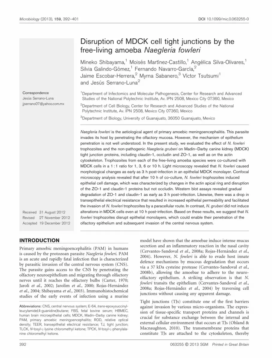
some cells (Fig. 4b, c). Claudin-1 behaved similarly to ZO-1, with diffuse and discontinuous immunolabelling mainlyat 6 and 10 h post-infection (Fig. 4e–f). In contrast, thechanges in occludin levels were less evident than those inclaudin-1 and ZO-1 at any time that was examined (Fig.4g–i), with the most obvious changes occurring at 10 hpost-infection. Co-cultures of non-pathogenic N. gruberitrophozoites with MDCK cells showed no alterations in thepattern of immunolabelling of ZO-1, claudin-1 or occludinat any time post-infection (Fig. 5a, c, e). Finally, asexpected for the negative control cells, MDCK cells thatwere cultured in the absence of amoebae displayedconstant protein levels throughout the cell culture period(Fig. 5b, d, f).
Degradation of TJ proteins by N. fowleritrophozoites
To analyse the possible degradation of MDCK TJ proteinsduring co-culture with N. fowleri or N. gruberi trophozoitesfor 1, 3, 6 or 10 h, we removed the trophozoites at eachpost-infection time and processed the MDCK monolayersto obtain crude cell extracts for analysis by SDS-PAGE atdifferent acrylamide concentrations. Immunoblotting wasperformed on PVDF membranes with antibodies againstactin, ZO-1, claudin-1 and occludin. We found that co-culture of MDCK cells with N. fowleri resulted in thedegradation of ZO-1 and claudin-1, which was evident at3 h post-infection and was time dependent (Fig. 6a). Therewas no significant degradation of occludin at any time
post-infection (Fig. 6a) (P.0.05). In contrast, co-culturingthe non-pathogenic N. gruberi strain with MDCK cells didnot lead to the degradation of ZO-1, claudin-1 or occludin(Fig. 6a). We also assessed the integrity of actin in responseto trophozoite co-culture. Actin was not degraded after anylength of time of co-culture with N. fowleri or N. gruberi(Fig. 6a). These differences in proteolysis were evidentwhen protein levels of each TJ protein were analysed withImageJ and expressed as relative optical densities (RODs)(Fig. 6b).
Cysteine protease inhibition with E-64 preventsMDCK TJ damage
To explore the participation of the proteases secreted by N.fowleri (Serrano-Luna et al., 2007) in the interaction of thisamoeba with MDCK cells, these cells were incubated withconditioned medium from N. fowleri or N. gruberi atdifferent times. Only conditioned medium from N. fowleriwas able to cause cell damage, as observed when livingtrophozoites were used. The highest effect was detected at10 h of incubation (Fig. 7a) and this effect was not detectedwhen conditioned medium from N. gruberi was used (Fig.7b). In order to know if the proteases secreted by N. fowleriare implicated in this cell damage, the effect of a permeablecysteine protease inhibitor on TJ damage was determined.The cysteine protease inhibitor E-64 was used to pre-treatN. fowleri trophozoites for 45 min, followed by incubationof the trophozoites with MDCK cells for up to 10 h. Usinglight microscopy, we observed that the E-64 inhibitor per
1 h
ZO
-1
10 h6 h
(e) (f)
(g) (h) (i)
(a) (b) (c)
(d)
Cla
ud
in-1
Occlu
din
Fig. 4. MDCK cell TJ proteins are damaged byN. fowleri trophozoites. N. fowleri trophozoiteswere co-cultured with MDCK cells, and ZO-1(a–c), claudin-1 (d–f) and occludin (g–i) wereanalysed at various time points. ZO-1, claudin-1 and occludin were immunolabelled withFITC, cell nuclei were labelled with propidiumiodide, and the trophozoites were labelled withCY5 (blue). Arrows show the loss of immuno-labelling. Images were obtained via confocalmicroscopy (Olympus FV-500) at �60. Scalebars, 50 mm.
M. Shibayama and others
396 Microbiology 159
se did not cause cell damage (Fig. 7d) and the cellsresembled the untreated cells (Fig. 7c). In contrast, E-64 wasable to block cell damage induced by N. fowleri trophozoites(Fig. 7f). The absence of cell damage correlated with a lackof proteolysis of TJ proteins, as Western blot analysisdemonstrated no decrease in ZO-1, claudin-1 or occludinlevels following the co-culture of MDCK cells with E-64 pre-treated N. fowleri trophozoites (Fig. 7g). E-64 also causedinhibition of the TEER decrement induced by N. fowleritrophozoites (Fig. 7i). As expected, MDCK cells without E-64 pre-treatment showed cell damage and TJ disruption at10 h of interaction with the amoebae (Fig. 7e, g). Thedifferences in proteolysis for each TJ protein were quantifiedby measuring the protein levels with ImageJ (Fig. 7f).Previously, our group showed that cysteine proteases are themain group secreted by N. fowleri and lesser quantities ofserine proteases are found (Serrano-Luna et al., 2007).Consistent with this observation, we found that aprotinin, anon-toxic serine protease inhibitor, did not protect againstTJ disruption induced by N. fowleri (data not shown).
DISCUSSION
N. fowleri is the causal agent of PAM, and the acquisition ofthis entity is commonly associated with a recent history ofaquatic activity by the patient (Schuster & Visvesvara,2004). The route of invasion of N. fowleri has been analysedby histopathological studies using a mouse model (Carter,1970; Cervantes-Sandoval et al., 2008a; Jaroli et al., 2002).These studies revealed that the amoeba infects the host byfirst penetrating the olfactory mucosa and subsequentlycrossing the cribriform plate of the ethmoid bone,eventually reaching the olfactory bulbs of the CNS. In theearly stages of infection, N. fowleri trophozoites areembedded in a large amount of mucus and surroundedby an inflammatory infiltrate composed mainly ofneutrophils (Cervantes-Sandoval et al., 2008a; Rojas-Hernandez et al., 2004). However, N. fowleri is able todegrade the mucus by secreting a 37 kDa cysteine protease(Cervantes-Sandoval et al., 2008b), which most likelyallows the amoeba to evade the mucus and adhere to theolfactory epithelium (Cervantes-Sandoval et al., 2008a).For many micro-organisms, adherence is one of the mostimportant features observed during the early invasion ofdifferent tissues or organs (Kucknoor et al., 2005; McCoy etal., 1994; Panjwani, 2010). N. fowleri is known to adhere todifferent cell types in a process that involves glycoconju-gates as well as extracellular matrix proteins, such asfibronectin and collagen (Cervantes-Sandoval et al., 2010;Han et al., 2004; Shibayama et al., 2003). During the initialadherence step, N. fowleri may activate intracellular signalsthat could be important for traversing epithelial cells. Inthe present study, we focused our analysis on the amoeba–epithelium interaction to understand how this amoebamay cross the epithelium before invading the CNS. In aprevious paper, we reported that N. fowleri trophozoitesare capable of migrating through the neuroepitheliumwithout producing any apparent damage, suggesting thatthis migration could occur via a paracellular route(Cervantes-Sandoval et al., 2008a). During evolution,pathogens acquire or adapt different mechanisms tointrude or penetrate various defence mechanisms of thehost, such as mucus, inflammatory cells and epithelialbarriers. At the cellular level, one of the strategies ofpathogenic protozoa is to traverse the epithelium using aparacellular route by disrupting TJs. Some parasites, suchas Giardia intestinalis and Trichomonas vaginalis, do notinvade host cells but rather decrease the cellular permeab-ility of CaCo-2 epithelial cells. This effect was determinedby examining TEER upon modifying the junctionalcomplex in epithelial cells because these parasites alterthe distribution of the ZO-1 and ZO-2 proteins (da Costaet al., 2005; Maia-Brigagao et al., 2012). In contrast, co-culture of E. histolytica with an enteric T-84 cell layerdecreases the TEER by disrupting the TJ proteins via thedephosphorylation and degradation of ZO-1 (Leroy et al.,2000). More recently, Khan & Siddiqui (2009) reportedthat A. castellanii, a pathogenic free-living amoeba, releasessecretory products into the culture medium, which results
ZO
-1
MDCK+ N. gruberi 10 h Control MDCK cell 10 h
(e) (f)
(a) (b)
(c) (d)
Cla
ud
in-1
Occlu
din
Fig. 5. N. gruberi trophozoites are unable to damage MDCK cell TJproteins. Analysis of TJ protein levels in MDCK cells infected withN. gruberi trophozoites for 10 h infection (a, c, e). As a control,MDCK cells were cultured without amoebae for 10 h (b, d, f). ZO-1 (a, b), claudin-1 (c, d) and occludin (e, f) were immunolabelledwith FITC-conjugated antibodies. Cell nuclei were labelled withpropidium iodide. Images were obtained via confocal microscopy(Olympus FV-500) at �60. Scale bars, 50 mm.
Naegleria fowleri disrupts MDCK cell tight junctions
http://mic.sgmjournals.org 397

in the degradation of ZO-1 and occludin in human brainmicrovascular endothelial cells (HBMEC) (Khan &Siddiqui, 2009). In the present study, we analysed theinteraction of epithelial MDCK cells with N. fowleri and N.gruberi trophozoites. Similar to E. histolytica trophozoites,we found that N. fowleri causes an important decrease in theTEER, but this decrease occurs at a slower rate than thatcaused by E. histolytica (Leroy et al., 2000). In addition to thedegradation of ZO-1, we also observed claudin-1 degrada-tion. However, as reported for A. castellanii trophozoites,occludin was not degraded (Khan & Siddiqui, 2009). N.fowleri trophozoites are known to produce cysteine andserine proteases that are considered to be importantvirulence factors and these proteases could participate inTJ disruption (Aldape et al., 1994; Ferrante et al., 1988;Serrano-Luna et al., 2007). To test this possibility, we pre-treated N. fowleri trophozoites with E-64, a cysteine proteaseinhibitor, and found that E-64 abrogated MDCK TJdegradation, but this inhibition was not observed whenaprotinin was used, a serine protease inhibitor. Similarly,Lauwaet et al. (2004) revealed that TPCK and TLCK, twocysteine and serine protease inhibitors, are able to inhibit E.histolytica cysteine proteases and prevent the proteolysis ofZO-1, ZO-2 and villin in Caco-2 cells (Lauwaet et al., 2004).Nikolskaia et al. (2006) found that the crossing of HBMEC
by Trypanosoma brucei gambiense was abrogated byN-methylpiperazine-urea-Phe-homophenylalanine-vinylsulfone-benzene (K11777), an irreversible inhibitor ofcathepsin L-like cysteine proteases (Nikolskaia et al.,2006). Finally, Sissons et al. (2006) used an isolate ofAcanthamoeba sp. to evaluate the ability of PMSF (aserine protease inhibitor) to inhibit TJ degradation inHBMEC cells (Sissons et al., 2006). In other micro-organisms, such as Bacteroides fragilis and Helicobacterpylori, it has been shown that the release of zinc-dependent metalloproteases causes the destabilization ofTJs (Wu et al., 1998). Here we report, we believe for thefirst time, the disruption and degradation by which N.fowleri alters TJ proteins. This phenomenon may occurduring the process of tissue invasion, both at the olfactoryneuroepithelium and at the blood–brain barrier.However, it will be necessary to perform in vivo assaysto validate the hypothesis that amoebic migration occursthrough a paracellular route to provide further evidencefor this mechanism of N. fowleri pathogenicity.Importantly, the non-pathogenic strain N. gruberi didnot disrupt MDCK cell TJs, although it is known that thisstrain also releases some cysteine proteases as well asother types of proteases (Serrano-Luna et al., 2007).Recently, it has been reported that there are differences
Fig. 6. Degradation of TJ proteins by N. fowleri trophozoites. (a) Western blot analysis of the MDCK proteins ZO-1, claudin-1,occludin and actin after co-culturing with N. fowleri or N. gruberi for different times. Controls were MDCK cells that werecultured without amoebae for different times. (b) Densitometric analyses were performed with GraphPad Prism. Control, blackbars; N. fowleri or N. gruberi co-culture samples, white bars. All bars show the mean±SEM of three independent assays(*P,0.05).
M. Shibayama and others
398 Microbiology 159

between pathogenic and non-pathogenic strains, includingthe differential expression of recognition moleculesimportant for the adhesion and invasion steps ofinfection, such as lectins and extracellular matrix proteins(Jamerson et al., 2012). These and other differencesbetween N. fowleri and N. gruberi amoebae may explainthe lack of virulence of N. gruberi. In summary, secretedcysteine proteases from adhered N. fowleri trophozoites
are able to degrade ZO-1 and claudin-1 but not occludin;actin is not degraded, but the actin apical ring isdisorganized and long exposure (10 h) can lead to cellsloughing. It is interesting to note that any of thesedegraded components causes TJ disruption. However,occludin can be endocytosed since it is not degraded byN. fowleri. Occludin could be cleaved in its long carboxyltail ending in a coiled-coil domain made of three alpha
0.75
0.50
DO
R
0.25
1.00
h)
i)
g)
(c)
(a)
(e)
(d)
10 x 10 x
10 x 10 x
20 x 10 x
(b)
(f)
0.00
120
TE
ER
(%
of in
itia
l va
lues) 100
80
60
N. fowleriN. fowleri +E-64MDCK cells without E-64
40
20
0864
Time (h)
20 10
N.f +e-
64 (1h)
N.f +e-
64 (3h)
N.f +e-
64 (6h)
N.f +e-
64 (10h)
N.f (10h)
E-64 (1
0h)
N.f +e-
64 (1h)
N.f +e-
64 (3h)
N.f +e-
64 (6h)
N.f +e-
64 (10h)
N.f (10h)
E-64 (1
0h)
N.f +e-
64 (1h)
N.f +e-
64 (3h)
N.f +e-
64 (6h)
N.f +e-
64 (10h)
N.f (10h)
E-64 (1
0h)
N.f +e-
64 (1h)
N.f +e-
64 (3h)
N.f +e-
64 (6h)
N.f +e-
64 (10h)
N.f (10h)
E-64 (1
0h)
ZO-1
ZO-1
claudin-1
claudin-1
**
occludin
occludin
actin
Fig. 7. E-64 prevents the degradation of MDCK TJ proteins during co-culture with N. fowleri. (a–f) Light microscopy of MDCKepithelial cells, co-cultured (a) with conditioned medium of N. fowleri; (b) with conditioned medium of N. gruberi; (c) in MEM; (d)in MEM with E-64; (e) with N. fowleri trophozoites; (f) with N. fowleri trophozoites pre-treated with E-64 (2 mM) for 45 min. Allsamples were cultured for 10 h. Images were obtained with an inverted Nikon microscope (TMS-F) at �10 (a, b, c, d, f) or �20(e). (g) Western blots of ZO-1, claudin-1, occludin and actin proteins, after co-culturing with N. fowleri pre-treated with E-64.(h) Densitometric analyses were performed with GraphPad Prism. N. fowleri in the presence of E-64, white bars; control MDCKcells with E-64, light grey bars; N. fowleri without E-64, dark grey bars. All bars show the mean±SEM of three independentassays (*P,0.05). (i) TEER assays. MDCK cells were co-incubated with N. fowleri (.) or N. fowleri pre-treated with E-64 (&).The percentage of TEER is compared with MDCK control values (X) . The data were analysed with the Systat software SigmaPlot 12. All data are the mean±SEM of three independent assays (P,0.05).
Naegleria fowleri disrupts MDCK cell tight junctions
http://mic.sgmjournals.org 399

helices. This coiled-coil interacts with ZO-1. In fact in theabsence of proteases, using Lantrunculin A (a drug) it ispossible to disassemble the actin apical ring, which leadsto TJ disassembly and endocytosis of the transmembranecomponents such as claudins and occludin, withoutproteolysis (Shen & Turner, 2005). Thus, various eventsindicate that N. fowleri can use the paracellular route tocause PAM: (1) the amoeba has been seen invading theolfactory epithelium without evidence of cellular damageor ulceration (in animal models); (2) as shown here,using cultured cells, N. fowleri causes loss of cell viabilityof about 25 % after 10 h of interaction, which is not areally strong cytolytic effect; (3) amoebal conditionedmedium is able to cause TJ protein disruption withoutdestruction of MDCK cells. This TJ disruption led to cellsloughing, whereby the amoebae can move to invade theneuro-olfactory epithelium.
In conclusion, our study demonstrates that pathogenic N.fowleri trophozoites can disrupt MDCK TJs by degradingZO-1 and claudin-1 using amoebic cysteine proteases.These results suggest that N. fowleri could use thismechanism to invade the neuro-olfactory epithelial cellsand/or the blood–brain barrier of the host.
ACKNOWLEDGEMENTS
We are grateful to Juana Narvaez-Morales for her valuable technicalhelp with the confocal microscope and Claudıa Galindo for hertechnical assistance. This work was supported by a CONACyT grant(number 128317) and a student fellowship from CONACyT (number290093) to Moises Martınez-Castillo.
REFERENCES
Aldape, K., Huizinga, H., Bouvier, J. & McKerrow, J. (1994). Naegleriafowleri: characterization of a secreted histolytic cysteine protease. ExpParasitol 78, 230–241.
Carter, R. F. (1970). Description of a Naegleria sp. isolated from twocases of primary amoebic meningo-encephalitis, and of the experi-mental pathological changes induced by it. J Pathol 100, 217–244.
Cervantes-Sandoval, I., Serrano-Luna, J. J., Garcıa-Latorre, E.,Tsutsumi, V. & Shibayama, M. (2008a). Characterization of braininflammation during primary amoebic meningoencephalitis. ParasitolInt 57, 307–313.
Cervantes-Sandoval, I., Serrano-Luna, J. J., Garcıa-Latorre, E.,Tsutsumi, V. & Shibayama, M. (2008b). Mucins in the host defenceagainst Naegleria fowleri and mucinolytic activity as a possible meansof evasion. Microbiology 154, 3895–3904.
Cervantes-Sandoval, I., Jesus Serrano-Luna, J., Pacheco-Yepez, J.,Silva-Olivares, A., Tsutsumi, V. & Shibayama, M. (2010). Differencesbetween Naegleria fowleri and Naegleria gruberi in expression ofmannose and fucose glycoconjugates. Parasitol Res 106, 695–701.
da Costa, R. F. M., de Souza, W., Benchimol, M., Alderete, J. F. &Morgado-Dıaz, J. A. (2005). Trichomonas vaginalis perturbs thejunctional complex in epithelial cells. Cell Res 15, 704–716.
Ferrante, A., Carter, R. F., Lopez, A. F., Rowan-Kelly, B., Hill, N. L. &Vadas, M. A. (1988). Depression of immunity to Naegleria fowleri inmice by selective depletion of neutrophils with a monoclonalantibody. Infect Immun 56, 2286–2291.
Forster, C. (2008). Tight junctions and the modulation of barrier
function in disease. Histochem Cell Biol 130, 55–70.
Gonzalez-Mariscal, L., Betanzos, A., Nava, P. & Jaramillo, B. E.(2003). Tight junction proteins. Prog Biophys Mol Biol 81, 1–44.
Han, K. L., Lee, H. J., Shin, M. H., Shin, H. J., Im, K. I. & Park, S. J.
(2004). The involvement of an integrin-like protein and protein
kinase C in amoebic adhesion to fibronectin and amoebic
cytotoxicity. Parasitol Res 94, 53–60.
Jamerson, M., da Rocha-Azevedo, B., Cabral, G. A. & Marciano-Cabral, F. (2012). Pathogenic Naegleria fowleri and non-pathogenic
Naegleria lovaniensis exhibit differential adhesion to, and invasion of,
extracellular matrix proteins. Microbiology 158, 791–803.
Jaroli, K. L., McCosh, J. K. & Howard, M. J. (2002). The role of blood
vessels and lungs in the dissemination of Naegleria fowleri following
intranasal inoculation in mice. Folia Parasitol (Praha) 49, 183–188.
Jarolim, K. L., McCosh, J. K., Howard, M. J. & John, D. T. (2000). A
light microscopy study of the migration of Naegleria fowleri from the
nasal submucosa to the central nervous system during the early stage
of primary amebic meningoencephalitis in mice. J Parasitol 86, 50–55.
Khan, N. A. (2008). Acanthamoeba and the blood-brain barrier: the
breakthrough. J Med Microbiol 57, 1051–1057.
Khan, N. A. & Siddiqui, R. (2009). Acanthamoeba affects the integrity
of human brain microvascular endothelial cells and degrades the tight
junction proteins. Int J Parasitol 39, 1611–1616.
Kucknoor, A. S., Mundodi, V. & Alderete, J. F. (2005). Adherence to
human vaginal epithelial cells signals for increased expression of
Trichomonas vaginalis genes. Infect Immun 73, 6472–6478.
Lauwaet, T., Oliveira, M. J., Callewaert, B., De Bruyne, G., Mareel, M.& Leroy, A. (2004). Proteinase inhibitors TPCK and TLCK prevent
Entamoeba histolytica induced disturbance of tight junctions and
microvilli in enteric cell layers in vitro. Int J Parasitol 34, 785–
794.
Leroy, A., Lauwaet, T., De Bruyne, G., Cornelissen, M. & Mareel, M.(2000). Entamoeba histolytica disturbs the tight junction complex in
human enteric T84 cell layers. FASEB J 14, 1139–1146.
Maia-Brigagao, C., Morgado-Dıaz, J. A. & De Souza, W. (2012).Giardia disrupts the arrangement of tight, adherens and desmosomal
junction proteins of intestinal cells. Parasitol Int 61, 280–287.
McCoy, J. J., Mann, B. J. & Petri, W. A., Jr (1994). Adherence and
cytotoxicity of Entamoeba histolytica or how lectins let parasites stick
around. Infect Immun 62, 3045–3050.
Nikolskaia, O. V., de A Lima, A. P., Kim, Y. V., Lonsdale-Eccles, J. D.,Fukuma, T., Scharfstein, J. & Grab, D. J. (2006). Blood-brain barrier
traversal by African trypanosomes requires calcium signaling induced
by parasite cysteine protease. J Clin Invest 116, 2739–2747.
Ohland, C. L. & Macnaughton, W. K. (2010). Probiotic bacteria and
intestinal epithelial barrier function. Am J Physiol Gastrointest Liver
Physiol 298, G807–G819.
Panjwani, N. (2010). Pathogenesis of acanthamoeba keratitis. Ocul
Surf 8, 70–79.
Que, X. & Reed, S. L. (1997). The role of extracellular cysteine
proteinases in pathogenesis of Entamoeba histolytica invasion.
Parasitol Today 13, 190–194.
Rojas-Hernandez, S., Jarillo-Luna, A., Rodrıguez-Monroy, M.,Moreno-Fierros, L. & Campos-Rodrıguez, R. (2004). Immunohisto-
chemical characterization of the initial stages of Naegleria fowleri
meningoencephalitis in mice. Parasitol Res 94, 31–36.
Schuster, F. L. & Visvesvara, G. S. (2004). Free-living amoebae as
opportunistic and non-opportunistic pathogens of humans and
animals. Int J Parasitol 34, 1001–1027.
M. Shibayama and others
400 Microbiology 159

Serrano-Luna, J., Cervantes-Sandoval, I., Tsutsumi, V. &Shibayama, M. (2007). A biochemical comparison of proteases frompathogenic Naegleria fowleri and non-pathogenic Naegleria gruberi.J Eukaryot Microbiol 54, 411–417.
Shen, L. & Turner, J. R. (2005). Actin depolymerization disrupts tightjunctions via caveolae-mediated endocytosis. Mol Biol Cell 16, 3919–3936.
Shibayama, M., Serrano-Luna, J. J., Rojas-Hernandez, S., Campos-Rodrıguez, R. & Tsutsumi, V. (2003). Interaction of secretoryimmunoglobulin A antibodies with Naegleria fowleri trophozoitesand collagen type I. Can J Microbiol 49, 164–170.
Sissons, J., Alsam, S., Goldsworthy, G., Lightfoot, M., Jarroll, E. L. &Khan, N. A. (2006). Identification and properties of proteases from anAcanthamoeba isolate capable of producing granulomatous enceph-alitis. BMC Microbiol 6, 42.
Tsukita, S., Furuse, M. & Itoh, M. (2001). Multifunctional strands intight junctions. Nat Rev Mol Cell Biol 2, 285–293.
Turner, J. R. (2006). Molecular basis of epithelial barrier regulation:from basic mechanisms to clinical application. Am J Pathol 169,1901–1909.
Wittchen, E. S., Haskins, J. & Stevenson, B. R. (2000). Exogenousexpression of the amino-terminal half of the tight junction proteinZO-3 perturbs junctional complex assembly. J Cell Biol 151, 825–836.
Wu, S., Lim, K. C., Huang, J., Saidi, R. F. & Sears, C. L. (1998).Bacteroides fragilis enterotoxin cleaves the zonula adherens protein, E-cadherin. Proc Natl Acad Sci U S A 95, 14979–14984.
Edited by: L. Knoll
Naegleria fowleri disrupts MDCK cell tight junctions
http://mic.sgmjournals.org 401