dishevelled phosphorylation, subcellular localization and multimerization regulate its role in early...
TRANSCRIPT
The EMBO Journal Vol.19 No.5 pp.1010–1022, 2000
Dishevelled phosphorylation, subcellular localizationand multimerization regulate its role in earlyembryogenesis
Ute Rothbacher1,2,3, Micheline N.Laurent4,Matthew A.Deardorff5, Peter S.Klein5,Ken W.Y.Cho4 and Scott E.Fraser1,3
1Division of Biology and Beckman Institute, California Institute ofTechnology, Pasadena, CA 91125, 4Department of Developmental andCell Biology, University of California at Irvine, Irvine, CA 92697 and5Cell and Molecular Biology, Howard Hughes Medical Institute andDepartment of Medicine, University of Pennsylvania School ofMedicine, Philadelphia, PA, USA
2Present address: Laboratoire de Genetique et Physiologie duDeveloppement, IBDM, Campus de Luminy, Marseille, France3Corresponding authorse-mail: [email protected] or [email protected]
K.W.Y.Cho and S.E.Fraser contributed equally to this work
Dishevelled (Dsh) induces a secondary axis and cantranslocate to the membrane when activated byFrizzleds; however, dominant-negative approacheshave not supported a role for Dsh in primary axisformation. We demonstrate that the Dsh protein ispost-translationally modified at the dorsal side of theembryo: timing and position of this regulation suggestsa role of Dsh in dorsal–ventral patterning in Xenopus.To create functional links between these properties ofDsh we analyzed the influence of endogenous Frizzledsand the Dsh domain dependency for these character-istics. Xenopus Frizzleds phosphorylate and translocateXdsh to the membrane irrespective of their differentialectopic axes inducing abilities, showing that transloca-tion is insufficient for axis induction. Dsh deletionanalysis revealed that axis inducing abilities did notsegregate with Xdsh membrane association. The DIXregion and a short stretch at the N-terminus of theDEP domain are necessary for axis induction whilethe DEP region is required for Dsh membrane associ-ation and its phosphorylation. In addition, Dsh formshomomeric complexes in embryos suggesting thatmultimerization is important for its proper function.Keywords: axis formation/DEP/Dishevelled/Frizzled/Wntsignaling
Introduction
The differential activation of members of the Wnt signalingpathway (β-catenin in particular) is the earliest knownmolecular manifestation of dorsal–ventral patterning inamphibians (for a review see Moon and Kimelman, 1998).How this pathway is activated regionally in the embryoremains one of the key questions in developmental biology.Recently, understanding of the Wnt pathway has pro-gressed significantly by studies in various model systems.Wnt molecules belong to an increasingly large family of
1010 © European Molecular Biology Organization
growth factors (19 members to date) and are thought tobind to the Frizzled family of seven-transmembranedomain molecules (Bhanot et al., 1996). In many systems,binding of Wnts to these putative receptors may inducethe activation of intracellular Dishevelled (Dsh), which inturn regulates the activity of the serine-threonine kinaseGSK-3/Zw-3/Sgg (for a review see Cadigan and Nusse,1997). In the absence of signal, GSK-3 phosphorylatesβ-catenin/Armadillo, resulting in its high turnover rate;however, activation of Dsh inhibits GSK-3 and leads tothe stabilization and accumulation of β-catenin in thecytoplasm, promoting its interaction with members of theTcf/Lef-1/pangolin family of DNA-binding molecules toinfluence target gene expression.
In Xenopus, the Wnt pathway is crucial for properdorso-ventral axis specification (for reviews see Millerand Moon, 1996; Heasman, 1997; Moon and Kimelman,1998). First, depletion of maternal β-catenin preventsthe formation of the embryonic axis. Secondly, ectopicexpression of Xwnt8, Xdsh, β-catenin or dominant-negative GSK-3 leads to secondary axis formation. Thirdly,Wnt signaling directly affects the transcription machineryin the Spemann’s organizer (Rothbacher et al., 1995;Watabe et al., 1995), and organizer-specific homeoboxgenes Xsiamois and Xtwin are direct transcriptional targetsof the β-catenin–Tcf complex (Brannon et al., 1997;Laurent et al., 1997). While these pieces of evidencedemonstrate the importance of the Wnt pathway, it is stillnot clear how this pathway is first activated in embryos.One possibility is that an endogenous maternal Wntregulates this process. However, thus far, no Wnt moleculehas been demonstrated to display a spatio-temporalpattern of expression compatible with a role during axisformation. An alternative model favors activation of mem-bers of the pathway downstream of Dsh. In support ofthis hypothesis is the observation that β-catenin becomesactivated at the site of the future dorsal organizer, whereasdominant-negative versions of Wnt-8, Frizzled (or wild-type FRP/FrzB) and Xdsh have been reported to fail atblocking the endogenous axis formation of embryos.However, such negative results must be interpreted cau-tiously, as dominant-negative approaches can fail to abolishinteractions of endogenous proteins. For instance, ifendogenous molecules are already pre-engaged in stablecomplexes, the complex might be unaltered by the laterexpression of the dominant-negative protein (Wittbrodtand Rosa, 1994). Thus, the involvement of the Wntpathway upstream of GSK-3 in the specification of theendogenous axis must still be considered an open question.
The role of Dsh is not well understood. In addition tomediating the classical Wnt pathway, Dsh affects cellpolarity (Theisen et al., 1994; Heslip et al., 1997) andinteracts with Notch signaling (Ruel et al., 1993; Cousoand Martinez Arias, 1994; Axelrod et al., 1996). The
Regulation of Xenopus Dishevelled function
presence of multiple domains in Dsh suggests that it mayinteract with different signaling pathways via differentdomains. Several structural motifs are conserved in Dshof various species, ranging from Caenorhabditis elegansto humans (our observations, Figure 3; and Sussman et al.,1994; Sokol et al., 1995; Klingensmith et al., 1996; Tsanget al., 1996; Yang et al., 1996; Semenov and Snyder,1997). The N-terminal DIX domain (DIX named afterDishevelled and axin) can interact physically with andhas homologies to the C-terminal region of axin, a negativeregulator of Wnt signaling (Zeng et al., 1997; Hamadaet al., 1999; Kishida et al., 1999, Li et al., 1999a; Smalleyet al., 1999). The medial PDZ domain of Dsh representsa globular protein–protein interaction domain containedin many adaptor molecules found in cellular junctionalcomplexes. PDZ domains bind C-terminal ends ofmembrane receptors and/or interact with other PDZdomains (Kennedy, 1995; Ponting et al., 1997). Finally,the C-terminal DEP domain (named after Dishevelled,Egl-10 and plekstrin) is found in several molecules thatregulate G-protein functions (Ponting and Bork, 1996).Although the high conservation of the Dsh domains islikely to reflect their conserved properties in embryo-genesis (Rothbacher et al., 1995), much of their functionalsignificance has yet to be determined. Only recently, theDEP region of Drosophila Dsh has been shown to play arole in tissue polarity in Drosophila (Axelrod et al., 1998;Boutros et al., 1998). Thus, understanding how Dshmediates differential cellular responses in a given bio-logical context is central to elucidating how Wnt signalingpathways can be activated in the embryo.
In order to elucidate the function of Dsh, we haveanalyzed various properties of the Dsh protein in earlyXenopus embryogenesis. We show that Dsh is regulatedpost-translationally by phosphorylation at the right timeand place to be involved in early dorso-ventral axisspecification of the embryo. Xenopus Frizzleds present inearly embryos all influence the intracellular localizationof Dsh, suggesting their endogenous interaction with Dsh.To understand the mechanism of action of Dsh and toassign roles to the conserved domains of Dsh in Xenopus,we generated various deletion constructs within the con-served regions of Dsh and tested their ability to accumulateat the membrane, to induce ectopic axes and to bephosphorylated in Xenopus embryos. Additionally, we findthat Xdsh forms homomeric complexes irrespective of thepresence or absence of Frizzled, raising the possibilitythat dimerization is important for the proper functions ofDsh. If so, dominant-negative approaches may have limitedutility for disrupting endogenous Dsh functions. We pro-pose a model where Dsh is a homomeric, multi-modularprotein that mediates different functions by distinctdomains at different subcellular locations and may particip-ate in setting up dorsal–ventral differences in the embryo.
Results
Dynamic post-translational modification of
endogenous Xdsh
Xenopus Dsh (Xdsh) mRNA is expressed abundantly inearly embryos (Rothbacher et al., 1995; Sokol et al., 1995)and Xdsh protein is present throughout early Xenopusdevelopment (Figure 1B). Xdsh protein migrates as two
1011
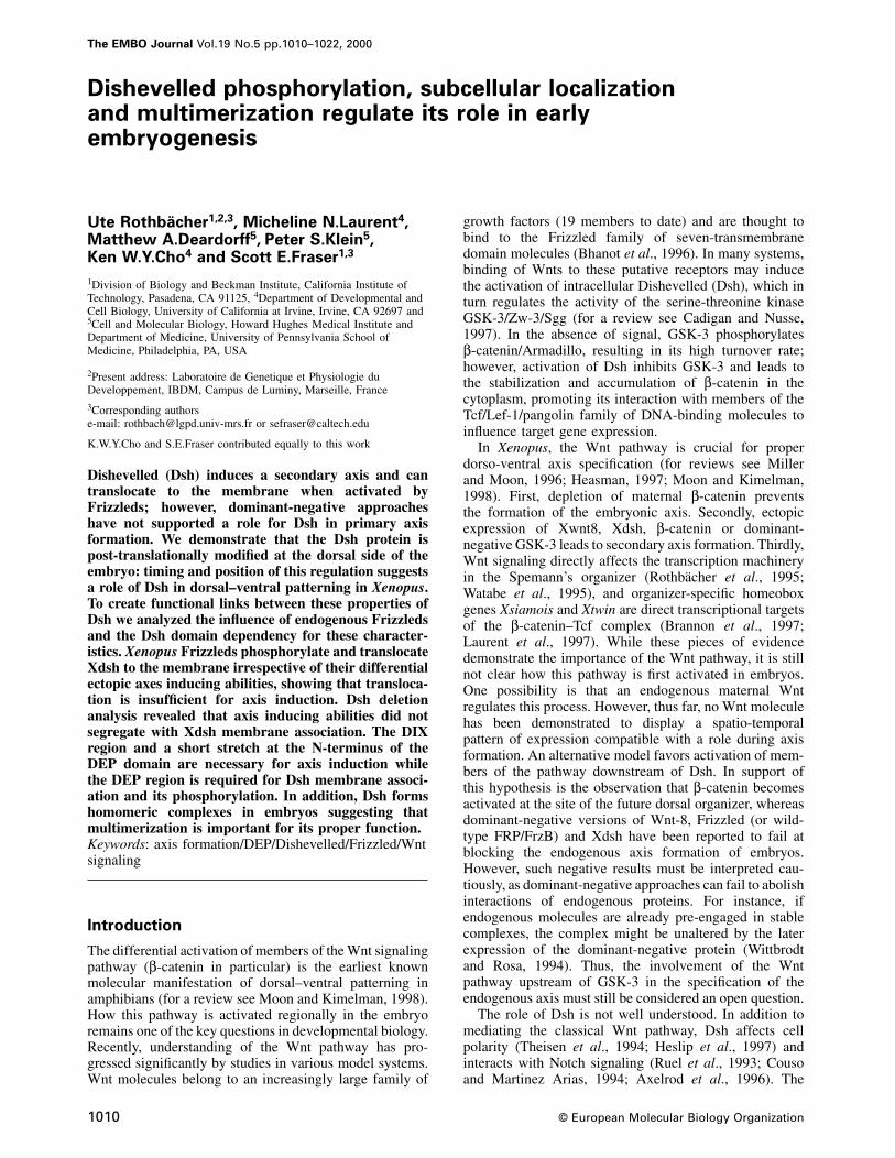
major bands, as revealed by analysis using crude extractsof embryos isolated from various stages of development(Figure 1B and C). Likewise, immunoprecipitations ofectopically expressed epitope-tagged Xdsh (Xdsh-HA)identified two similarly migrating bands (Figure 1A andD). As previous work suggested that Drosophila andmouse Dishevelled are phosphorylated (Yanagawa et al.,1995; Semenov and Snyder, 1997), we examined thispossibility in Xenopus. Immunoprecipitated Xdsh-HAtreated with acid phosphatase migrates as a single band of80 kDa (Figure 1A), suggesting that the higher molecularweight band represents mobility shifted, phosphorylatedXdsh and the lower molecular weight band is non- orpoorly phosphorylated Xdsh.
The phosphorylation profile of Xdsh varies at develop-mental stages (Figure 1B). Initially, Xdsh isolated fromoocytes (not shown) and from early (post-fertilized)embryos migrates as a single band. During early cleavagestages, the mobility-shifted (phosphorylated) form appearsas early as the 16-cell stage, continues to accumulateuntil blastula stage and then decreases slightly duringgastrulation.
The timing of the developmental changes in Xdshphosphorylation is suggestive of its early role in develop-ment. Shortly after fertilization, cytoplasmic rearrange-ments occur to translocate vegetal components to thedorsal side of the embryo. As development proceeds,dorsally located cells show accumulation of β-catenin(Schneider et al., 1996; Larabell et al., 1997; Rowninget al., 1997). To determine whether the phosphorylationof Xdsh correlates with the onset of dorsalizing events,we analyzed the degree of Xdsh phosphorylation in dorsal(D) and ventral (V) explants. At the 64- to 128-cell stage,when phosphorylation reaches a peak, phosphorylatedXdsh is detected more abundantly in the dorsal region(Figure 1B, compare D with V). Densitometric quantitationof panels D and V shown in Figure 1B shows a 1.35-folddifference of lower migrating, underphosphorylated bandsand a 3.85-fold difference in higher migrating, phosphoryl-ated bands. Thus, a 2.8-fold higher level of phosphorylationis detected in dorsal explants even after normalizing theamount of underphosphorylated Xdsh protein in dorsal andventral samples (for details, see Materials and methods).β-catenin levels (not shown) were 2.2-fold higher in dorsalsamples. The experiment was performed twice, withsimilar results. No significant differences in Xdsh phos-phorylation were detected between dorsal and ventralhalves at late blastula and early gastrula stages (data notshown). Thus, phosphorylation of Xdsh parallels theactivation of dorsal determinants in time and place.Together with β-catenin nuclear accumulation, the differ-ential phosphorylation of Xdsh is the earliest molecularmanifestation of events that break the radial symmetry inthe embryo.
The above results may suggest that phosphorylation ofXdsh is involved in axis formation. Two classes of findingsindicate that the role of Xdsh phosphorylation should beexamined carefully. First, doses of Xdsh mRNA requiredto induce ectopic axes do not correlate well with the levelsof Xdsh phosphorylation (Figure 1C). The extent ofthe induced ectopic axis is concentration dependent, i.e.increasing concentrations of injected RNA induce pro-gressively stronger secondary axis (data not shown, see
U.Rothbacher et al.
Fig. 1. Xdsh is phosphorylated differentially during development. (A) Slower migrating forms of Xdsh are caused by phosphorylation. Phosphatasetreatment of Xdsh-HA immunoprecipitates from blastula embryos injected with 1 ng of mRNA at the 4-cell stage. Precipitates were incubated with(�) or without (–) potato acid phosphatase (PAP). The blot is stained with anti-HA antibody. (B) Developmentally regulated phosphorylation ofendogenous Xdsh in post-fertilization stages. Left panel: endogenous Xdsh phosphorylation in embryonic extracts at various early stages ofdevelopment. Right panel: extracts of dorsal (D) and ventral (V) halves at the 64- to 128-cell stage, stained with anti-Drosophila Ddsh antiserum. Anequivalent of two embryos is shown per lane. At the 64- to 128-cell stage, Xdsh phosphorylation is significantly higher in the dorsal half ofembryos. (C) Increased phosphorylation following Xdsh overexpression. Upon injection of 0.05 ng (1), 0.5 ng (2) or 1 ng (3) of Xdsh-HA mRNA in4-cell embryos and collection of extracts at blastula stages, a higher ratio of phosphorylated versus non-phosphorylated forms is observed in ectopicXdsh (upper panel, anti-HA stain) when compared with endogenous Xdsh (lower panel, anti-Dvl-1 stain) in the same samples. Higher amounts ofectopic Xdsh only slightly increase the phosphorylation of endogenous Xdsh (compare lanes 1 and 3). (D) Xdsh is hyperphosphorylated upon ratfrizzled-1 co-expression. Co-injection of 0.5 ng of Xdsh-HA and 1 ng of rat Frizzled-1 (Rfz-1) mRNA (�) leads to a shift of slower migrating,phosphorylated bands when compared with injection of Xdsh-HA mRNA only (–). Blots of anti-HA precipitations were stained with anti-HAantibody. The two Xdsh bands are not well separated due to a shorter electrophoresis run in (D).
also Figure 6 and Materials and methods). In contrast,phosphorylation levels are not proportional to the amountof injected RNA, and even low concentrations of ectopicXdsh are highly phosphorylated (Figure 1C, comparelanes 1 and 3). It is possible, however, that phosphorylatedXdsh accumulates at high ectopic Xdsh concentrations tothreshold levels necessary for ectopic axis induction.Secondly, we find that overexpression of rat frizzled-1alone does not induce secondary axis formation in Xenopus(data not shown) but can induce the translocation ofectopic Xdsh to the membrane (Yang-Snyder et al.,1996) and causes a marked increase in mobility-shifted,hyperphosphorylated Dsh (compared with injection ofXdsh alone; Figure 1D). Taken together, these findingsindicate that Dsh phosphorylation may be correlated betterwith its translocation to the membrane; variations inother components, such as binding of Xdsh-interactingmolecules and/or activity changes in Xdsh, might thenresult in the axis-inducing dose–response curve.
1012
Membrane association and hyperphosphorylation
of Xdsh by Xenopus Frizzleds are independent of
their axis-inducing abilities
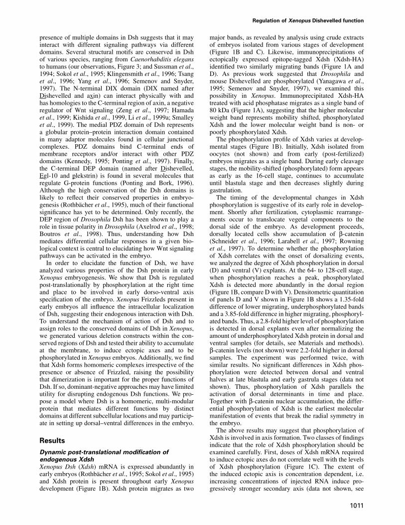
Our results suggest that Xdsh phosphorylation in earlyembryos could be linked to a membrane localization event.To explore whether a Xdsh membrane association eventcould take place during early Xenopus development,we tested several Xenopus Frizzleds for their ability toaccumulate Xdsh at the membrane. Three recently isolatedXenopus homologs of the frizzled gene family (Xfz-3,Xfz-7 and Xfz-8) are expressed during early stages ofXenopus development (Deardorff et al., 1998; Itoh et al.,1998; Shi et al., 1998). Overexpression of Xfz-8 in theventral side of early cleavage embryos induces a secondaryaxis; in contrast, Xfz-3 and -7 fail to form an ectopic axis(Deardorff et al., 1998). While Xdsh–green fluorescentprotein (GFP) is evenly distributed in animal cap cells(Figure 2A), co-expression with rat Frizzled-1 butnot rat Frizzled-2 accumulates Xdsh at the membrane
Regulation of Xenopus Dishevelled function
Fig. 2. Xenopus Frizzleds translocate and phosphorylate Xdshindependently of their axis-inducing abilities. Xenopus Frizzled-3, -7and -8 can translocate Xdsh equally to the membrane; a comparisonwith rat Frizzled-1 and -2 is shown. (A) Ectopic Xdsh–GFP isdistributed in a punctuate fashion in animal cap cells. (B) Co-expression of Xdsh–GFP with rat Frizzled-2 does not change itsdistribution. (C) Co-expression with rat Frizzled-1 leads to theaccumulation of Xdsh–GFP near the cell membrane. Co-expression of(D) Xfz-3, (E) Xfz-7 and (F) Xfz-8 equally enriches Xdsh–GFP at thecell membrane. (G) Xfz-3, -7 and -8 can hyperphosphorylate Xdsh.Anti-myc staining of embryonic extracts is shown following injectionof myc-Xdsh alone (WT) or after co-injection with Xfz-3 (3),Xfz-7 (7) or Xfz-8 (8).
(Yang-Snyder et al., 1996; and Figure 2B and C). Incontrast, all three Xenopus Frizzleds tested accumulateXdsh at the membrane (Figure 2D–F), irrespective of theirability to induce ectopic axes.
We further find that all three Xenopus Frizzleds causeXdsh hyperphosphorylation (Figure 2G).
From these experiments, we conclude that the abilityof Frizzled molecules to recruit Xdsh to the membraneand to cause its hyperphosphorylation is distinct fromtheir axis-inducing ability, and that membrane associationalone is not sufficient to activate the nuclear (β-catenin-mediated) signaling pathway. Because these Frizzleds areexpressed early in development and can mediate Xdshmembrane accumulation, such an event may be importantduring early Xenopus development. The exact functionalinterplay between Frizzled-mediated membrane accumula-tion and phosphorylation of Xdsh during embryogenesisremains to be resolved.
1013
The C-terminal region containing DEP is necessary
and sufficient for Xdsh membrane accumulation
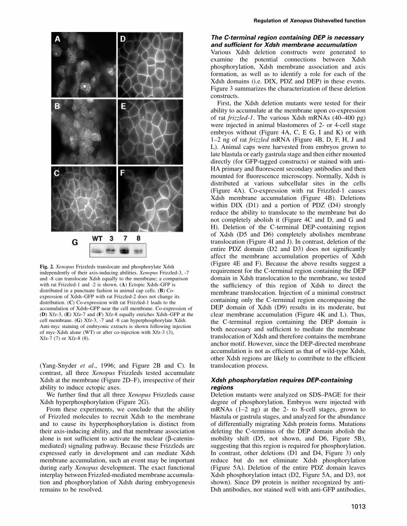
Various Xdsh deletion constructs were generated toexamine the potential connections between Xdshphosphorylation, Xdsh membrane association and axisformation, as well as to identify a role for each of theXdsh domains (i.e. DIX, PDZ and DEP) in these events.Figure 3 summarizes the characterization of these deletionconstructs.
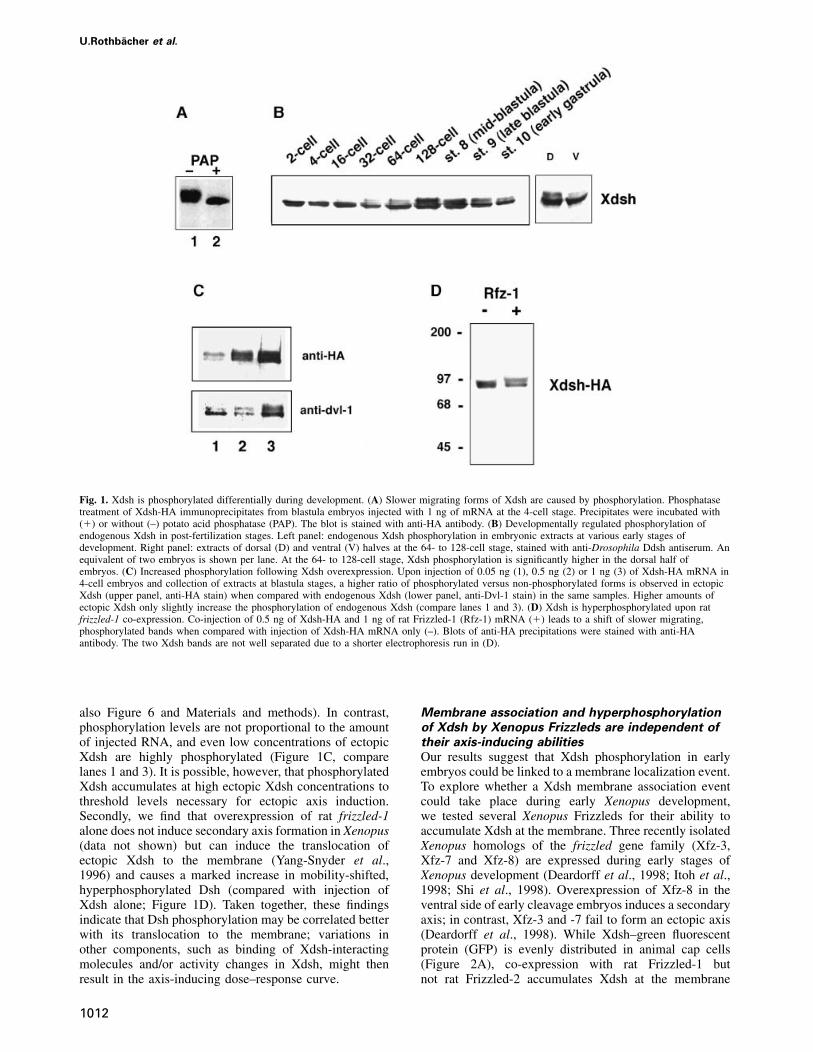
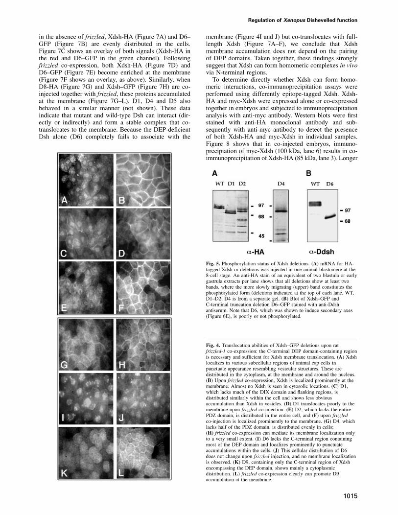
First, the Xdsh deletion mutants were tested for theirability to accumulate at the membrane upon co-expressionof rat frizzled-1. The various Xdsh mRNAs (40–400 pg)were injected in animal blastomeres of 2- or 4-cell stageembryos without (Figure 4A, C, E G, I and K) or with1–2 ng of rat frizzled mRNA (Figure 4B, D, F, H, J andL). Animal caps were harvested from embryos grown tolate blastula or early gastrula stage and then either mounteddirectly (for GFP-tagged constructs) or stained with anti-HA primary and fluorescent secondary antibodies and thenmounted for fluorescence microscopy. Normally, Xdsh isdistributed at various subcellular sites in the cells(Figure 4A). Co-expression with rat Frizzled-1 causesXdsh membrane accumulation (Figure 4B). Deletionswithin DIX (D1) and a portion of PDZ (D4) stronglyreduce the ability to translocate to the membrane but donot completely abolish it (Figure 4C and D, and G andH). Deletion of the C-terminal DEP-containing regionof Xdsh (D5 and D6) completely abolishes membranetranslocation (Figure 4I and J). In contrast, deletion of theentire PDZ domain (D2 and D3) does not significantlyaffect the membrane accumulation properties of Xdsh(Figure 4E and F). Because the above results suggest arequirement for the C-terminal region containing the DEPdomain in Xdsh translocation to the membrane, we testedthe sufficiency of this region of Xdsh to direct themembrane translocation. Injection of a minimal constructcontaining only the C-terminal region encompassing theDEP domain of Xdsh (D9) results in its moderate, butclear membrane accumulation (Figure 4K and L). Thus,the C-terminal region containing the DEP domain isboth necessary and sufficient to mediate the membranetranslocation of Xdsh and therefore contains the membraneanchor motif. However, since the DEP-directed membraneaccumulation is not as efficient as that of wild-type Xdsh,other Xdsh regions are likely to contribute to the efficienttranslocation process.
Xdsh phosphorylation requires DEP-containing
regions
Deletion mutants were analyzed on SDS–PAGE for theirdegree of phosphorylation. Embryos were injected withmRNAs (1–2 ng) at the 2- to 8-cell stages, grown toblastula or gastrula stages, and analyzed for the abundanceof differentially migrating Xdsh protein forms. Mutationsdeleting the C-terminus of the DEP domain abolish themobility shift (D5, not shown, and D6, Figure 5B),suggesting that this region is required for phosphorylation.In contrast, other deletions (D1 and D4, Figure 3) onlyreduce but do not eliminate Xdsh phosphorylation(Figure 5A). Deletion of the entire PDZ domain leavesXdsh phosphorylation intact (D2, Figure 5A, and D3, notshown). Since D9 protein is neither recognized by anti-Dsh antibodies, nor stained well with anti-GFP antibodies,
U.Rothbacher et al.
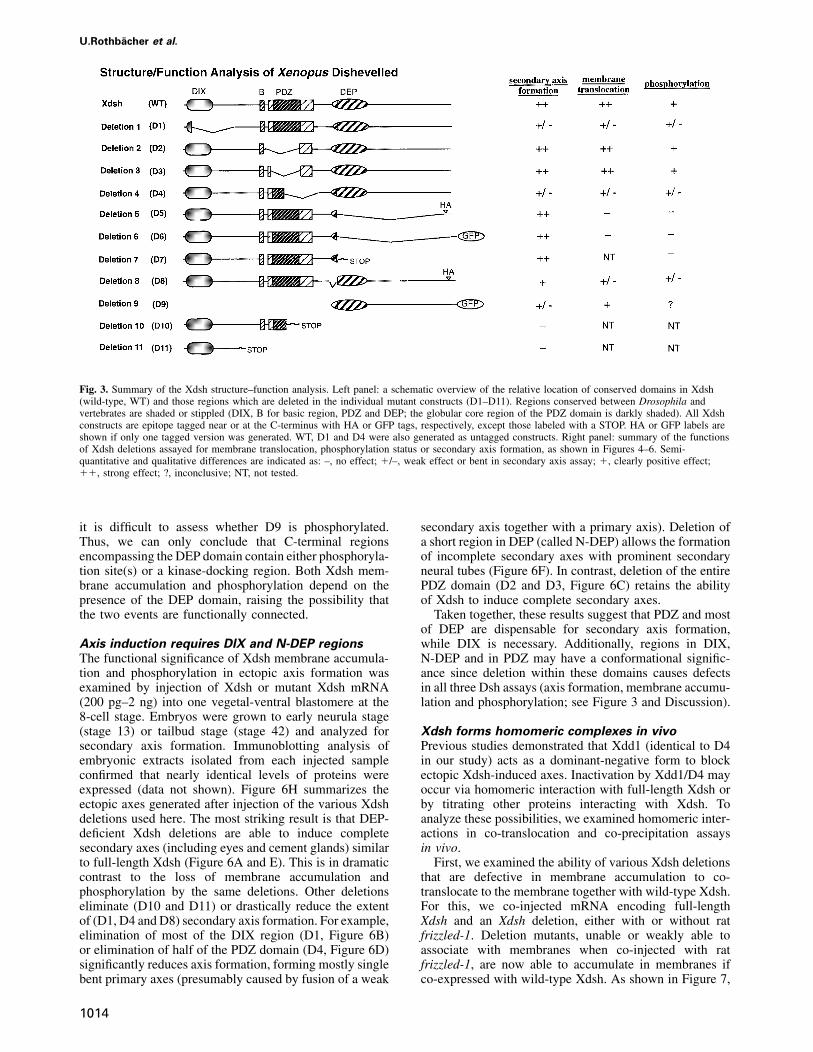
Fig. 3. Summary of the Xdsh structure–function analysis. Left panel: a schematic overview of the relative location of conserved domains in Xdsh(wild-type, WT) and those regions which are deleted in the individual mutant constructs (D1–D11). Regions conserved between Drosophila andvertebrates are shaded or stippled (DIX, B for basic region, PDZ and DEP; the globular core region of the PDZ domain is darkly shaded). All Xdshconstructs are epitope tagged near or at the C-terminus with HA or GFP tags, respectively, except those labeled with a STOP. HA or GFP labels areshown if only one tagged version was generated. WT, D1 and D4 were also generated as untagged constructs. Right panel: summary of the functionsof Xdsh deletions assayed for membrane translocation, phosphorylation status or secondary axis formation, as shown in Figures 4–6. Semi-quantitative and qualitative differences are indicated as: –, no effect; �/–, weak effect or bent in secondary axis assay; �, clearly positive effect;��, strong effect; ?, inconclusive; NT, not tested.
it is difficult to assess whether D9 is phosphorylated.Thus, we can only conclude that C-terminal regionsencompassing the DEP domain contain either phosphoryla-tion site(s) or a kinase-docking region. Both Xdsh mem-brane accumulation and phosphorylation depend on thepresence of the DEP domain, raising the possibility thatthe two events are functionally connected.
Axis induction requires DIX and N-DEP regions
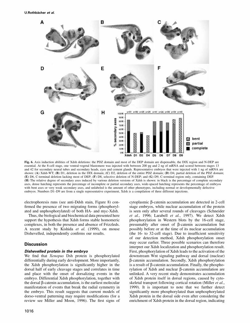
The functional significance of Xdsh membrane accumula-tion and phosphorylation in ectopic axis formation wasexamined by injection of Xdsh or mutant Xdsh mRNA(200 pg–2 ng) into one vegetal-ventral blastomere at the8-cell stage. Embryos were grown to early neurula stage(stage 13) or tailbud stage (stage 42) and analyzed forsecondary axis formation. Immunoblotting analysis ofembryonic extracts isolated from each injected sampleconfirmed that nearly identical levels of proteins wereexpressed (data not shown). Figure 6H summarizes theectopic axes generated after injection of the various Xdshdeletions used here. The most striking result is that DEP-deficient Xdsh deletions are able to induce completesecondary axes (including eyes and cement glands) similarto full-length Xdsh (Figure 6A and E). This is in dramaticcontrast to the loss of membrane accumulation andphosphorylation by the same deletions. Other deletionseliminate (D10 and D11) or drastically reduce the extentof (D1, D4 and D8) secondary axis formation. For example,elimination of most of the DIX region (D1, Figure 6B)or elimination of half of the PDZ domain (D4, Figure 6D)significantly reduces axis formation, forming mostly singlebent primary axes (presumably caused by fusion of a weak
1014
secondary axis together with a primary axis). Deletion ofa short region in DEP (called N-DEP) allows the formationof incomplete secondary axes with prominent secondaryneural tubes (Figure 6F). In contrast, deletion of the entirePDZ domain (D2 and D3, Figure 6C) retains the abilityof Xdsh to induce complete secondary axes.
Taken together, these results suggest that PDZ and mostof DEP are dispensable for secondary axis formation,while DIX is necessary. Additionally, regions in DIX,N-DEP and in PDZ may have a conformational signific-ance since deletion within these domains causes defectsin all three Dsh assays (axis formation, membrane accumu-lation and phosphorylation; see Figure 3 and Discussion).
Xdsh forms homomeric complexes in vivo
Previous studies demonstrated that Xdd1 (identical to D4in our study) acts as a dominant-negative form to blockectopic Xdsh-induced axes. Inactivation by Xdd1/D4 mayoccur via homomeric interaction with full-length Xdsh orby titrating other proteins interacting with Xdsh. Toanalyze these possibilities, we examined homomeric inter-actions in co-translocation and co-precipitation assaysin vivo.
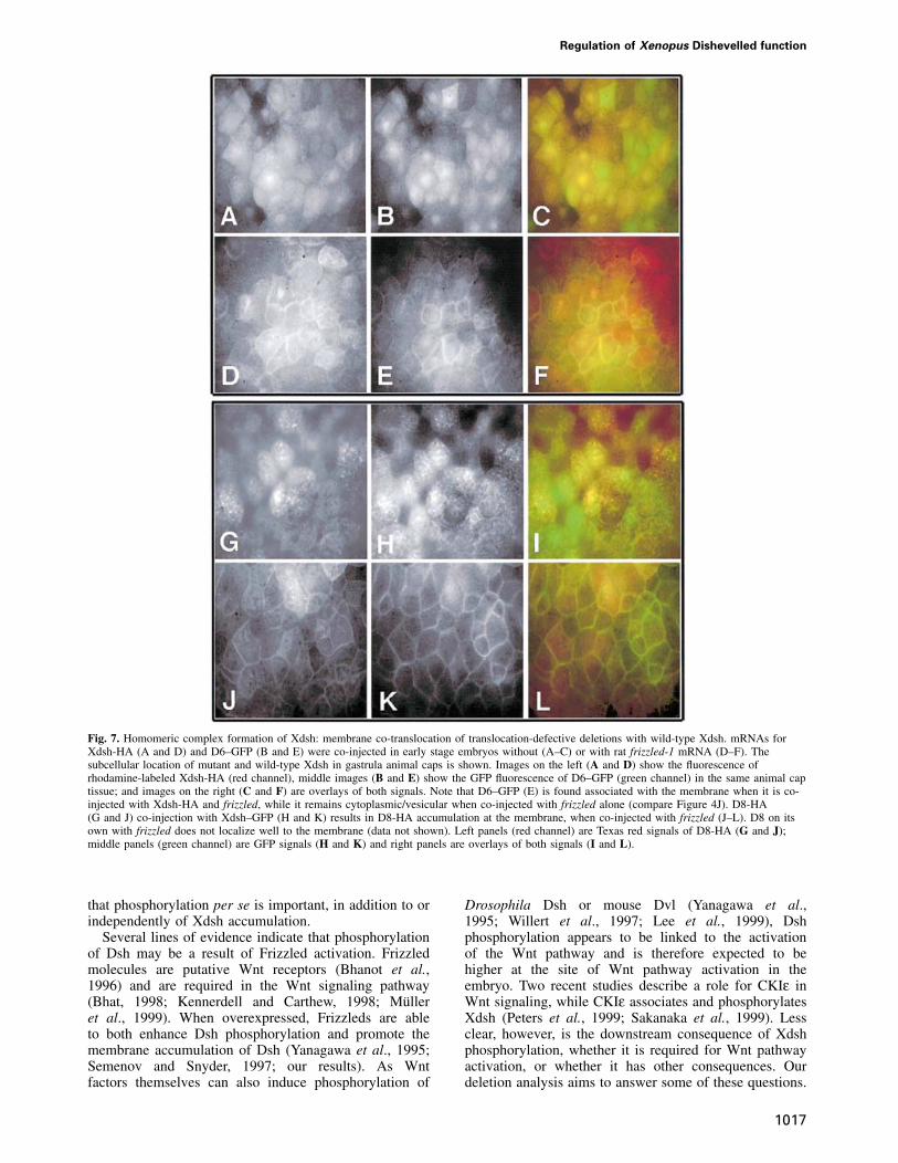
First, we examined the ability of various Xdsh deletionsthat are defective in membrane accumulation to co-translocate to the membrane together with wild-type Xdsh.For this, we co-injected mRNA encoding full-lengthXdsh and an Xdsh deletion, either with or without ratfrizzled-1. Deletion mutants, unable or weakly able toassociate with membranes when co-injected with ratfrizzled-1, are now able to accumulate in membranes ifco-expressed with wild-type Xdsh. As shown in Figure 7,
Regulation of Xenopus Dishevelled function
in the absence of frizzled, Xdsh-HA (Figure 7A) and D6–GFP (Figure 7B) are evenly distributed in the cells.Figure 7C shows an overlay of both signals (Xdsh-HA inthe red and D6–GFP in the green channel). Followingfrizzled co-expression, both Xdsh-HA (Figure 7D) andD6–GFP (Figure 7E) become enriched at the membrane(Figure 7F shows an overlay, as above). Similarly, whenD8-HA (Figure 7G) and Xdsh–GFP (Figure 7H) are co-injected together with frizzled, these proteins accumulatedat the membrane (Figure 7G–L). D1, D4 and D5 alsobehaved in a similar manner (not shown). These dataindicate that mutant and wild-type Dsh can interact (dir-ectly or indirectly) and form a stable complex that co-translocates to the membrane. Because the DEP-deficientDsh alone (D6) completely fails to associate with the
1015
membrane (Figure 4I and J) but co-translocates with full-length Xdsh (Figure 7A–F), we conclude that Xdshmembrane accumulation does not depend on the pairingof DEP domains. Taken together, these findings stronglysuggest that Xdsh can form homomeric complexes in vivovia N-terminal regions.
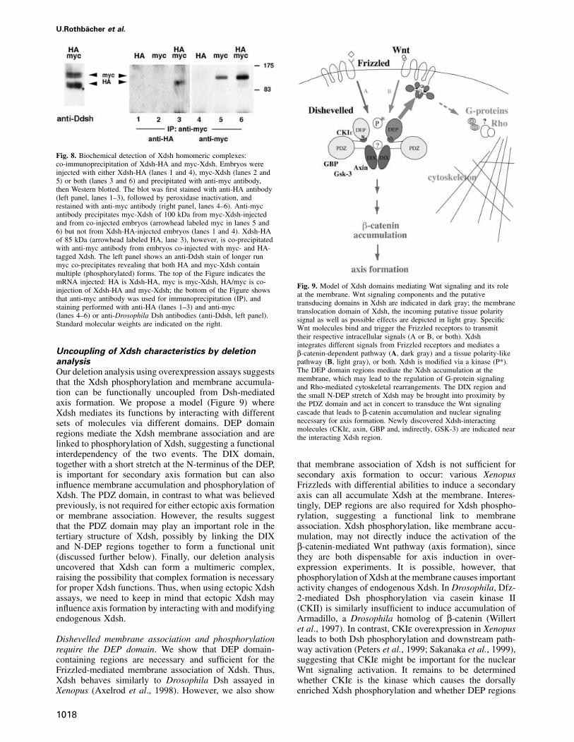
To determine directly whether Xdsh can form homo-meric interactions, co-immunoprecipitation assays wereperformed using differently epitope-tagged Xdsh. Xdsh-HA and myc-Xdsh were expressed alone or co-expressedtogether in embryos and subjected to immunoprecipitationanalysis with anti-myc antibody. Western blots were firststained with anti-HA monoclonal antibody and sub-sequently with anti-myc antibody to detect the presenceof both Xdsh-HA and myc-Xdsh in individual samples.Figure 8 shows that in co-injected embryos, immuno-precipiation of myc-Xdsh (100 kDa, lane 6) results in co-immunoprecipitation of Xdsh-HA (85 kDa, lane 3). Longer
Fig. 5. Phosphorylation status of Xdsh deletions. (A) mRNA for HA-tagged Xdsh or deletions was injected in one animal blastomere at the8-cell stage. An anti-HA stain of an equivalent of two blastula or earlygastrula extracts per lane shows that all deletions show at least twobands, where the more slowly migrating (upper) band constitutes thephosphorylated form (deletions indicated at the top of each lane, WT,D1–D2; D4 is from a separate gel. (B) Blot of Xdsh–GFP andC-terminal truncation deletion D6–GFP stained with anti-Ddshantiserum. Note that D6, which was shown to induce secondary axes(Figure 6E), is poorly or not phosphorylated.
Fig. 4. Translocation abilities of Xdsh–GFP deletions upon ratfrizzled-1 co-expression: the C-terminal DEP domain-containing regionis necessary and sufficient for Xdsh membrane translocation. (A) Xdshlocalizes in various subcellular regions of animal cap cells inpunctuate appearance resembling vesicular structures. These aredistributed in the cytoplasm, at the membrane and around the nucleus.(B) Upon frizzled co-expression, Xdsh is localized prominently at themembrane. Almost no Xdsh is seen in cytosolic locations. (C) D1,which lacks much of the DIX domain and flanking regions, isdistributed similarly within the cell and shows less obviousaccumulation than Xdsh in vesicles. (D) D1 translocates poorly to themembrane upon frizzled co-injection. (E) D2, which lacks the entirePDZ domain, is distributed in the entire cell, and (F) upon frizzledco-injection is localized prominently to the membrane. (G) D4, whichlacks half of the PDZ domain, is distributed evenly in cells;(H) frizzled co-expression can mediate its membrane localization onlyto a very small extent. (I) D6 lacks the C-terminal region containingmost of the DEP domain and localizes prominently to punctuateaccumulations within the cells. (J) This cellular distribution of D6does not change upon frizzled injection, and no membrane localizationis observed. (K) D9, containing only the C-terminal region of Xdshencompassing the DEP domain, shows mainly a cytoplasmicdistribution. (L) frizzled co-expression clearly can promote D9accumulation at the membrane.
U.Rothbacher et al.
Fig. 6. Axis induction abilities of Xdsh deletions: the PDZ domain and most of the DEP domain are dispensable, the DIX region and N-DEP areessential. At the 8-cell stage, one ventral-vegetal blastomere was injected with between 200 pg and 2 ng of mRNA and scored between stages 13and 42 for secondary neural tubes and secondary heads, eyes and cement glands. Representative embryos that were injected with 1 ng of mRNA areshown: (A) Xdsh-WT; (B) D1, deletion in the DIX domain; (C) D2, deletion of the entire PDZ domain; (D) D4, partial deletion of the PDZ domain;(E) D6, C-terminal deletion lacking most of DEP; (F) D8, selective deletion of N-DEP; and (G) D9, C-terminal region only, containing DEP.(H) The relative degree of secondary axes induced by various deletion versions of Xdsh is shown: in black is the percentage of complete secondaryaxes, dense hatching represents the percentage of incomplete or partial secondary axes, wide-spaced hatching represents the percentage of embryoswith bent axes or very weak secondary axes, and unlabeled is the amount of other phenotypes, including normal or developmentally defectiveembryos. Numbers D1–D9 are from a single representative experiment, Xdsh is a compilation of three different injections.
electrophoresis runs (see anti-Ddsh stain, Figure 8) con-firmed the presence of two migrating forms (phosphoryl-ated and unphosphorylated) of both HA- and myc-Xdsh.
Thus, the biological and biochemical data presented heresupport the hypothesis that Xdsh forms stable homomericcomplexes, in both the presence and absence of Frizzleds.A recent study by Kishida et al. (1999), on mouseDishevelled, independently confirms our results.
Discussion
Dishevelled protein in the embryo
We find that Xenopus Dsh protein is phosphorylateddifferentially during early development. More importantly,the Xdsh phosphorylation is significantly higher in thedorsal half of early cleavage stages and correlates in timeand place with the onset of dorsalizing events in theembryo. Differential Xdsh phosphorylation, together withthe dorsal β-catenin accumulation, is the earliest molecularmanifestation of events that break the radial symmetry inthe embryo. This result suggests that current models ofdorso-ventral patterning may require modifications (for areview see Miller and Moon, 1996). The first signs of
1016
cytoplasmic β-catenin accumulation are detected in 2-cellstage embryos, while nuclear accumulation of the proteinis seen only after several rounds of cleavages (Schneideret al., 1996; Larabell et al., 1997). We detect Xdshphosphorylation in Western blots by the 16-cell stage,presumably after onset of β-catenin accumulation butpossibly before or at the time of its nuclear accumulation(the 16- to 32-cell stage). Due to insufficient sensitivityof our detection method, Xdsh phosphorylation onsetmay occur earlier. Three possible scenarios can thereforeinterpret our Xdsh localization and phosphorylation result.First, phosphorylation of Xdsh leads to the activation of thedownstream Wnt signaling pathway and dorsal (nuclear)β-catenin accumulation. Secondly, Xdsh phosphorylationis a result of β-catenin accumulation. Finally, the phospho-rylation of Xdsh and nuclear β-catenin accumulation areunlinked. A very recent study demonstrates accumulationof Xdsh protein itself in dorsal regions, caused by cyto-skeletal transport following cortical rotation (Miller et al.,1999). It is important to note that we further detectsignificantly more phosphorylated than unphosphorylatedXdsh protein in the dorsal side even after considering theenrichment of Xdsh protein in the dorsal region, indicating
Regulation of Xenopus Dishevelled function
Fig. 7. Homomeric complex formation of Xdsh: membrane co-translocation of translocation-defective deletions with wild-type Xdsh. mRNAs forXdsh-HA (A and D) and D6–GFP (B and E) were co-injected in early stage embryos without (A–C) or with rat frizzled-1 mRNA (D–F). Thesubcellular location of mutant and wild-type Xdsh in gastrula animal caps is shown. Images on the left (A and D) show the fluorescence ofrhodamine-labeled Xdsh-HA (red channel), middle images (B and E) show the GFP fluorescence of D6–GFP (green channel) in the same animal captissue; and images on the right (C and F) are overlays of both signals. Note that D6–GFP (E) is found associated with the membrane when it is co-injected with Xdsh-HA and frizzled, while it remains cytoplasmic/vesicular when co-injected with frizzled alone (compare Figure 4J). D8-HA(G and J) co-injection with Xdsh–GFP (H and K) results in D8-HA accumulation at the membrane, when co-injected with frizzled (J–L). D8 on itsown with frizzled does not localize well to the membrane (data not shown). Left panels (red channel) are Texas red signals of D8-HA (G and J);middle panels (green channel) are GFP signals (H and K) and right panels are overlays of both signals (I and L).
that phosphorylation per se is important, in addition to orindependently of Xdsh accumulation.
Several lines of evidence indicate that phosphorylationof Dsh may be a result of Frizzled activation. Frizzledmolecules are putative Wnt receptors (Bhanot et al.,1996) and are required in the Wnt signaling pathway(Bhat, 1998; Kennerdell and Carthew, 1998; Mulleret al., 1999). When overexpressed, Frizzleds are ableto both enhance Dsh phosphorylation and promote themembrane accumulation of Dsh (Yanagawa et al., 1995;Semenov and Snyder, 1997; our results). As Wntfactors themselves can also induce phosphorylation of
1017
Drosophila Dsh or mouse Dvl (Yanagawa et al.,1995; Willert et al., 1997; Lee et al., 1999), Dshphosphorylation appears to be linked to the activationof the Wnt pathway and is therefore expected to behigher at the site of Wnt pathway activation in theembryo. Two recent studies describe a role for CKIε inWnt signaling, while CKIε associates and phosphorylatesXdsh (Peters et al., 1999; Sakanaka et al., 1999). Lessclear, however, is the downstream consequence of Xdshphosphorylation, whether it is required for Wnt pathwayactivation, or whether it has other consequences. Ourdeletion analysis aims to answer some of these questions.
U.Rothbacher et al.
Fig. 8. Biochemical detection of Xdsh homomeric complexes:co-immunoprecipitation of Xdsh-HA and myc-Xdsh. Embryos wereinjected with either Xdsh-HA (lanes 1 and 4), myc-Xdsh (lanes 2 and5) or both (lanes 3 and 6) and precipitated with anti-myc antibody,then Western blotted. The blot was first stained with anti-HA antibody(left panel, lanes 1–3), followed by peroxidase inactivation, andrestained with anti-myc antibody (right panel, lanes 4–6). Anti-mycantibody precipitates myc-Xdsh of 100 kDa from myc-Xdsh-injectedand from co-injected embryos (arrowhead labeled myc in lanes 5 and6) but not from Xdsh-HA-injected embryos (lanes 1 and 4). Xdsh-HAof 85 kDa (arrowhead labeled HA, lane 3), however, is co-precipitatedwith anti-myc antibody from embryos co-injected with myc- and HA-tagged Xdsh. The left panel shows an anti-Ddsh stain of longer runmyc co-precipitates revealing that both HA and myc-Xdsh containmultiple (phosphorylated) forms. The top of the Figure indicates themRNA injected: HA is Xdsh-HA, myc is myc-Xdsh, HA/myc is co-injection of Xdsh-HA and myc-Xdsh; the bottom of the Figure showsthat anti-myc antibody was used for immunoprecipitation (IP), andstaining performed with anti-HA (lanes 1–3) and anti-myc(lanes 4–6) or anti-Drosophila Dsh antibodies (anti-Ddsh, left panel).Standard molecular weights are indicated on the right.
Uncoupling of Xdsh characteristics by deletion
analysis
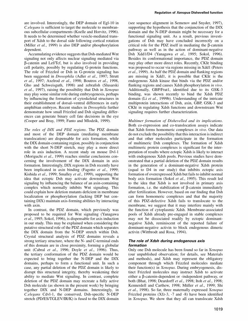
Our deletion analysis using overexpression assays suggeststhat the Xdsh phosphorylation and membrane accumula-tion can be functionally uncoupled from Dsh-mediatedaxis formation. We propose a model (Figure 9) whereXdsh mediates its functions by interacting with differentsets of molecules via different domains. DEP domainregions mediate the Xdsh membrane association and arelinked to phosphorylation of Xdsh, suggesting a functionalinterdependency of the two events. The DIX domain,together with a short stretch at the N-terminus of the DEP,is important for secondary axis formation but can alsoinfluence membrane accumulation and phosphorylation ofXdsh. The PDZ domain, in contrast to what was believedpreviously, is not required for either ectopic axis formationor membrane association. However, the results suggestthat the PDZ domain may play an important role in thetertiary structure of Xdsh, possibly by linking the DIXand N-DEP regions together to form a functional unit(discussed further below). Finally, our deletion analysisuncovered that Xdsh can form a multimeric complex,raising the possibility that complex formation is necessaryfor proper Xdsh functions. Thus, when using ectopic Xdshassays, we need to keep in mind that ectopic Xdsh mayinfluence axis formation by interacting with and modifyingendogenous Xdsh.
Dishevelled membrane association and phosphorylationrequire the DEP domain. We show that DEP domain-containing regions are necessary and sufficient for theFrizzled-mediated membrane association of Xdsh. Thus,Xdsh behaves similarly to Drosophila Dsh assayed inXenopus (Axelrod et al., 1998). However, we also show
1018
Fig. 9. Model of Xdsh domains mediating Wnt signaling and its roleat the membrane. Wnt signaling components and the putativetransducing domains in Xdsh are indicated in dark gray; the membranetranslocation domain of Xdsh, the incoming putative tissue polaritysignal as well as possible effects are depicted in light gray. SpecificWnt molecules bind and trigger the Frizzled receptors to transmittheir respective intracellular signals (A or B, or both). Xdshintegrates different signals from Frizzled receptors and mediates aβ-catenin-dependent pathway (A, dark gray) and a tissue polarity-likepathway (B, light gray), or both. Xdsh is modified via a kinase (P*).The DEP domain regions mediate the Xdsh accumulation at themembrane, which may lead to the regulation of G-protein signalingand Rho-mediated cytoskeletal rearrangements. The DIX region andthe small N-DEP stretch of Xdsh may be brought into proximity bythe PDZ domain and act in concert to transduce the Wnt signalingcascade that leads to β-catenin accumulation and nuclear signalingnecessary for axis formation. Newly discovered Xdsh-interactingmolecules (CKIε, axin, GBP and, indirectly, GSK-3) are indicated nearthe interacting Xdsh region.
that membrane association of Xdsh is not sufficient forsecondary axis formation to occur: various XenopusFrizzleds with differential abilities to induce a secondaryaxis can all accumulate Xdsh at the membrane. Interes-tingly, DEP regions are also required for Xdsh phospho-rylation, suggesting a functional link to membraneassociation. Xdsh phosphorylation, like membrane accu-mulation, may not directly induce the activation of theβ-catenin-mediated Wnt pathway (axis formation), sincethey are both dispensable for axis induction in over-expression experiments. It is possible, however, thatphosphorylation of Xdsh at the membrane causes importantactivity changes of endogenous Xdsh. In Drosophila, Dfz-2-mediated Dsh phosphorylation via casein kinase II(CKII) is similarly insufficient to induce accumulation ofArmadillo, a Drosophila homolog of β-catenin (Willertet al., 1997). In contrast, CKIε overexpression in Xenopusleads to both Dsh phosphorylation and downstream path-way activation (Peters et al., 1999; Sakanaka et al., 1999),suggesting that CKIε might be important for the nuclearWnt signaling activation. It remains to be determinedwhether CKIε is the kinase which causes the dorsallyenriched Xdsh phosphorylation and whether DEP regions
Regulation of Xenopus Dishevelled function
are involved. Interestingly, the DEP domain of Egl-10 inC.elegans is sufficient to target the molecule to membran-ous subcellular compartments (Koelle and Horvitz, 1996).It needs to be determined whether vesicle-mediated trans-port of Xdsh to the dorsal side following cortical rotation(Miller et al., 1999) is also DEP and/or phosphorylationdependent.
Accumulating evidence suggests that Dsh-mediated Wntsignaling not only affects nuclear signaling mediated viaβ-catenin and Lef/Tcf, but is also involved in providingcell polarity, possibly by regulating G-protein functions.The role of Frizzled or Dsh in G-protein signaling hasbeen suggested in Drosophila (Adler et al., 1997; Struttet al., 1997; Axelrod et al., 1998; Boutros et al., 1998,Gho and Schweisguth, 1998) and zebrafish (Slusarskiet al., 1997), raising the possibility that Dsh in Xenopusmay play some similar role during embryogenesis, perhapsby influencing the formation of cytoskeletal structures intheir establishment of dorsal–ventral differences in earlyamphibian embryos. Recent studies in Drosophila furtherdemonstrate how small Frizzled and Dsh signaling differ-ences can generate binary cell fate decisions in the eye(Cooper and Bray, 1999; Fanto and Mlodzik, 1999).
The roles of DIX and PDZ regions. The PDZ domainand most of the DEP domain (mediating membranetranslocation) are dispensable for axis formation, whilethe DIX domain-containing region, possibly in conjunctionwith the short N-DEP stretch, may play a more directrole in axis induction. A recent study on mouse Dvl-1(Moriguchi et al., 1999) reaches similar conclusions con-cerning the involvement of the DIX domain in axisformation. Interestingly, DIX regions in Dsh recently havebeen implicated in axin binding (Fagotto et al., 1999;Kishida et al., 1999; Smalley et al., 1999), supporting theidea that ectopic Dsh may activate downstream Wntsignaling by influencing the balance of an axin-containingcomplex which normally inhibits Wnt signaling. Thiscould explain how deletion mutants deficient in membranelocalization or phosphorylation (lacking DEP but con-taining DIX) maintain axis-forming abilities by interactingwith axin.
In contrast, the PDZ domain, which previously wasproposed to be required for Wnt signaling (Yanagawaet al., 1995; Sokol, 1996), is dispensable for axis inductionin our study. This may be explained when considering theputative structural role of the PDZ domain which separatesthe DIX domain from the N-DEP stretch within Dsh.Crystal structural analysis of PDZ domains reveals astrong tertiary structure, where the N- and C-terminal endsof this domain are in close proximity, forming a globularunit (Cabral et al., 1996; Doyle et al., 1996). Thus,the tertiary conformation of the PDZ domain would beexpected to bring together the N-DEP and the DIXdomains, perhaps to form a functional unit. In such acase, any partial deletion of the PDZ domain is likely todisrupt this structural integrity, thereby weakening theirability to mediate Wnt signaling. In contrast, completedeletion of the PDZ domain may recreate a fully activeDsh molecule (as shown in the present work) by bringingtogether DIX and N-DEP domains. Interestingly, inC.elegans Cdvl-1, the conserved, Dsh-specific N-DEPstretch (PD/ES/TGLEV/IR/K) is fused to the DIX domain
1019
(see sequence alignment in Semenov and Snyder, 1997),supporting the hypothesis that the conjunction of the DIXdomain and the N-DEP domain might be necessary for afunctional signaling unit. As a result, previous investi-gations of Dsh may have concluded incorrectly on acritical role for the PDZ itself in mediating the β-cateninpathway as well as in the action of dominant-negativeDsh Xdd1/D4 (Yanagawa et al., 1995; Sokol, 1996).Besides its conformational importance, the PDZ domainmay play other more direct roles. Recently, CKIε bindingwas proposed to occur via regions missing in Xdd1 (Peterset al., 1999). As half the PDZ domain and flanking regionsare missing in Xdd1, it is possible that CKIε is theendogenous Xdsh kinase that binds via the PDZ and/orflanking regions and causes Xdsh phosphorylation in vivo.Additionally, GBP/Frat1, identified due to its GSK-3binding, was shown recently to bind the Xdsh PDZdomain (Li et al., 1999b). Understanding of the intricatemultiprotein interactions of Dsh, axin, GBP, GSK-3 andCKIε in regulating Xdsh functions and downstream Wntsignaling requires further characterization.
Multimer formation of Dishevelled and its implications.Both co-expression and co-translocation assays indicatethat Xdsh forms homomeric complexes in vivo. Our datado not exclude the possibility that this interaction is indirectand that other molecules participate in the formationof multimeric Dsh complexes. The formation of Xdshmultimeric protein complexes is significant for the inter-pretation of results where ectopic Xdsh is likely to interactwith endogenous Xdsh pools. Previous studies have dem-onstrated that a partial deletion of the PDZ domain resultsin the generation of a dominant-negative Xdsh protein(equal to D4 in our study) that inhibits ectopic axisformation of overexpressed Xdsh but fails to inhibit normalbody axis formation (Sokol et al., 1995). This result ledto the idea that Xdsh is not involved in primary axisformation, i.e. the stabilization of β-catenin immediatelyafter fertilization. However, based on our finding that Dshcan form homomeric complexes and that the majorityof this PDZ-defective Xdsh fails to translocate to themembrane, we suggest that it may interfere mainly withthe function of cytoplasmic Xdsh. Membrane-associatedpools of Xdsh already pre-engaged in stable complexesmay not be dissociated readily by ectopic dominant-negative Xdsh, reminiscent of the reported failure ofdominant-negative activin to block endogenous dimericactivin (Wittbrodt and Rosa, 1994).
The role of Xdsh during endogenous axis
formation
Only one Dsh molecule has been found so far in Xenopus(our unpublished observation; for details, see Materialsand methods), and Xdsh may represent the obligatorycomponent through which Frizzled molecules mediatetheir function(s) in Xenopus. During embryogenesis, dis-tinct Frizzled molecules may instruct Xdsh to activateeither a β-catenin-dependent or -independent pathway, orboth (Bhat, 1998; Deardorff et al., 1998; Itoh et al., 1998;Kennerdell and Carthew, 1998; Muller et al., 1999; Shiet al., 1998). So far, three maternally expressed XenopusFrizzled proteins (Xfz-3, -7 and -8) have been identifiedin Xenopus. We show that they all can translocate Xdsh
U.Rothbacher et al.
to the membrane; in contrast, only Xfz-8 can induce asecondary axis (Deardorff et al., 1998). Xfz-7 is expressedmost abundantly in early cleavage stages (M.A.Deardoffand P.S.Klein, unpublished) and thus may function as aprimary anchor to recruit endogenous Xdsh to the mem-brane. Initial phosphorylation of Xdsh soon after fertiliza-tion may reflect such membrane association events. Atpresent, it is unclear how differential Xdsh phosphorylationis achieved in the embryo One possibility is that as yetunidentified Wnt-like growth factors activate the Frizzledreceptor system. A recent study in zebrafish supportsthis hypothesis, since a dominant-negative Fz blocksendogenous D/V signaling (Nasevicius et al., 1998).Alternatively, Xdsh in early embryos might be activatedde novo by a dorsally activated kinase in a Frizzled-independent manner. While our deletion analysis showsthat different domains are involved in phosphorylationand membrane association versus axis formation, the exactmechanisms by which the activity of Xdsh is regulated inthe embryo remain to be determined. In particular, the wayin which phosphorylation may influence axis formation orother early events, including polarity of cells in thedeveloping embryo, is still unclear. Two independentpieces of evidence clearly support our idea that endogenousXdsh and possibly its phosphorylation are actually import-ant in dorso-ventral patterning. First, CKIε causes Xdshphosphorylation while being involved in Wnt signalingand is thus a good candidate for the endogenous Xdshphosphorylation (Peters et al., 1999; Sakanaka et al.,1999). Interactions of Dsh and CKIε with axin and theGBP–GSK-3 complex suggest that the regulation of Dshvia phosphorylation or segregation to the membrane maybe important factors in the activation of axis formation.Thus, while membrane association and phosphorylationnormally may be required to regulate endogenous Dshactivities, in overexpression, a membrane association-andphosphorylation-deficient Dsh may cause axis formationby influencing the balance of this multiprotein complex.Secondly, the demonstration of active transport of Xdshto the dorsal side (Miller et al., 1999) additionally supportsour hypothesis that Xdsh is involved in dorsal–ventralpatterning.
For Dsh to be clearly implicated in activating thedownstream Wnt signaling pathway in the embryo, it hasto meet several criteria: Dsh protein should be presentand active at the correct time and place in the embryo.In addition, activation of Dsh function should inducesecondary axes, while blocking its activity should blockprimary axis formation. Here we present new evidencethat maternal Dsh RNA is translated into a protein product,which is present throughout early cleavage stage embryos.Furthermore, Dsh protein becomes phosphorylated in adorsal location at the appropriate time and place, correlat-ing well with the initiation of dorso-ventral axis specifica-tion and the translocation of β-catenin to nuclei. Suchactivation of members of the Wnt signaling pathway wasshown to lead to axis formation, and ectopic expressionof Dsh itself induces secondary axis formation. Our resultssuggest that endogenous Xdsh is present in multimericcomplexes. Because such pre-arranged multimers maynot be destabilized and inactivated by dominant-negativeforms, the evidence previously offered for lack of needfor Xdsh is inconclusive. The evidence presented here
1020
therefore revives the possibility that Xdsh is indeedinvolved in endogenous axial patterning.
Materials and methods
Xdsh expression constructsCloning of Xdsh was performed using primers annealing to regionsconserved between Drosophila and mouse Dsh (encoding amino acidsPCFNGR and AKCWDP, primers were: 5�TGCCGTGCTTTAATGG/ACCG3� and 5�GGTCCCAGCATTTGGCCAC3�). A 800 bp PCR frag-ment obtained by RT–PCR from Xenopus gastrula stage RNA was usedfor screening Xenopus oocyte, egg, gastrula and head phage cDNAlibraries. The Xdsh cDNA sequence obtained is largely identical to thesubsequently published Xdsh sequence, DDBJ/EMBL/GenBank acces-sion No. U31552, and differs in 13 positions, therefore probably reflectingeither sequencing errors or tetraploidity variations of Xdsh. For expressionin Xenopus embryos, a blunted 2190 bp Xdsh BfaI fragment (encodingall except eight amino acids at the C-terminus of Xdsh) was cloned intothe blunted BalI–BstEII sites of the Sp64T/Xβm vector (obtained byBalI–BspEII excision of noggin-coding regions from the Xβnog vectorobtained from R.Harland), called Xdsh-Xβm. An HA tag was introducedinto an NcoI site close to the C-terminus. GFP tagging was performedusing a modified pCS2-GFP vector (D.Turned, R.Rupp and H.Weintraub,Fred Hutchinson Cancer Research Center, Seattle, WA) encoding anenhanced version of GFP (obtained from C.LaBonne) and blunt cloningof a HindIII–blunt XhoI fragment and an XhoI–StuI-cut PCR fragmentinto the blunted EcoRI site in-frame with the GFP-coding sequence,thus giving rise to full-length Xdsh with GFP fused to its C-terminalend. The BspEI–StuI-cut PCR fragment was generated using the primers5�GGAGGCCTCATGACATCCACAAAGAAC3� and 5�GGGCGGCC-GTTCTCAAAATTAATGTC3�. Xdsh and deletion constructs (below)were sequenced to confirm correct cloning sites and sequences withinPCR fragments. The following deletion constructs were generated: D1(missing DIX and flanking regions) was generated by excision of anSspI–FspI fragment (bp 124–442 deleted), fusing amino acids ASHAKto (C)RDRVR. D2 (missing the PDZ domain) was generated by cloninga HindIII–ApaI-cut PCR fragment in HindIII–ApaI-cut Xdsh-HA vectorusing primer 5�ATGGGCCCCGTCC GTTCCAGGCGC3� (bp 707–990deleted), thus fusing amino acids RLERT to GPIVL. D3 (missing mostof the PDZ domain) was generated by cloning a HindIII–ApaI-cut PCRfragment in HindIII–ApaI-cut Xdsh-HA vector using primer5�ATGGCCCCACGATGGAGATG3� (bp 806–990 deleted), fusingamino acids GISIV to GPIVL. D4 (missing half the PDZ domain) wasgenerated by excision of a SmaI fragment (bp 894–1134 deleted), fusingamino acids GRIEP to GSASM. D5 (missing most DEP and C-terminalregions, HA tagged) was generated by excision of an XhoI–NcoI fragment(bp 1287–2164 deleted); the five most C-terminal amino acids of N-DEP are PESGL(D) followed by the HA tag and amino acids MGNP,vector sequences provide the STOP codon. D6 (missing most DEP andC-terminal regions, GFP tagged) was generated by ApaI–EcoRI digestionof a PCR fragment generated with primers SP6 and 5�GGGAATTCTCT-CGGACCTCGAGGC3� ligated in ApaI–EcoRI sites of Xdsh–GFP(bp 1287–2202 deleted, bp 1–1286 maintained); the C-terminal Xdsh-coding amino acids are LEVRD, followed by GFP-coding sequences.D7 (missing most DEP and C-terminal regions, out-of-frame), is an out-of-frame construct which maintains bp 1–1286 of Xdsh (the C-terminalcoding amino acids are PESGL) with a STOP codon further downstream,and contains 16 additional amino acids (DRSSGSGWGRLEVGIW)generated by frameshift. D8 (missing the N-terminal region within theDEP domain) was generated by ApaI–XhoI digestion of a PCR fragmentgenerated by primers SP6 and 5�ACCTCGAGAGCCATGACCTT-CACAAC3� and cloning into ApaI–XhoI-cut Xdsh-HA vector, resultingin a five amino acid deletion (amino acids SPESG, bp 1271–1286). D9(missing all Xdsh regions upstream of the DEP domain, therefore missingDIX and PDZ but maintaining DEP) was generated by ClaI–BspEIdigestion of a PCR fragment generated with primers 5�GGATCGAT-CCATGGCTTCTGTTGTGAAGG3� and 5�GGGAATTCTCTCGG-ACCTCGAGGC3� and cloning into ClaI–BspEI-cut Xdsh–GFP vector.Therefore, the most N-terminal coding amino acids are (M)ASVVK.D10 (truncation missing half of PDZ and all C-terminal regions, alsoDEP) is an out-of-frame construct, which maintains bp 1–893 of Xdsh;the most C-terminal coding amino acids are GRIEP followed by antisensesequences. D11 (truncation missing all middle and C-terminal regions,DIX maintained) is an out-of-frame construct which maintainsbp 1–450 of Xdsh; the most C-terminal coding amino acids are SMRRDfollowed by out-of-frame amino acids.
Regulation of Xenopus Dishevelled function
Microinjection of synthetic RNA into fertilized eggsSynthetic RNA was transcribed using the SP6 mMessage Machine Kit(Ambion) according to the manufacturer’s recommendations. Injectionsand handling of embryos were as described previously (Blitz and Cho,1995). For the secondary axis assay, 1 ng of Xdsh or mutant XdshmRNA was injected in one vegetal-ventral blastomere at the 8-cell stageand scored for phenotypes after stage 15. The induced phenotypes werescored by the presence of secondary neural tubes, heads and cement,and termed weakly dorsalized/bent when embryos were bent and some-times had short secondary neural tubes (�/– in Figures 3 and 6B, D andG), partial secondary axis when a secondary neural tube but no eyes orcement gland were visible (� in Figures 3 and 6F), complete secondaryaxis when a secondary neural tube and head including eyes and cementgland were present (�� in Figures 3 and 6A, C and E) or hyperdorsalizedwhen supernumerary eyes and cement glands were visible. For immuno-precipitations and immunoblotting, 1–2 ng of mRNA was injected perembryo. Phosphatase treatment was performed (Yanagawa et al., 1995)using potato acid phosphatase (PAP) from Sigma. Full color documenta-tion of embryos was performed using a Zeiss Axiophot dissectingmicroscope equipped with a Leica camera or a ProgRes 3012 digital colorcamera and ImageManager software (Roche Image Analysis Systems).
Immunoblotting and antibodiesTen Xenopus embryos were homogenized in 200 μl of RIPA buffer(150 mM NaCl, 50 mM Tris pH 8, 1% NP-40, 0.5% sodium deoxycholate,0.1% SDS) or NP-40 buffer (150 mM NaCl, 50 mM Tris pH 8, 1% NP-40). Cell debris was removed by centrifugation at 15 000 g for 10 min.Immunoprecipitations were performed using protein A–Sepharose beadsor GammaBind G–Sepharose (Pharmacia) as described previously(Hawley et al., 1995) using anti-HA antibody (12CA5, Berkeley AntibodyCompany) or anti-myc antibody (Oncogene Science). Alternatively, acrush method was used to detect both endogenous and overexpressedXdsh from embryonic extracts (Watabe et al., 1995). Briefly, 3–20embryos in 25 μl of 0.1� MMR between stages 0 and 11 werecentrifuged for 3 min at 15 000 g to remove yolk platelets. Yolk plateletsand Xdsh protein migrate at a similar molecular weight in SDS–PAGE.Comparable amounts of overexpressed Xdsh protein were recovered bythe crush method and immunoprecipitation technique. A slight loss inhigher molecular weight forms of Xdsh was observed by the crushmethod, most probably due to a small loss in membranous compartments.This method, however, makes the separation of endogenous Xdshextracts possible and therefore renders Xdsh proteins in semi-quantitativeamounts. Separation of immunoprecipitates or extracts by SDS–PAGEand immunoblotting was followed by antibody incubation. Antiserum toDrosophila (anti-Ddsh region I; Yanagawa et al., 1995) or mouseDsh (anti-Dvl-1; R.Nusse and K.Willert, Stanford University, personalcommunication) was used to detect either endogenous and ectopic orendogenous Xdsh only, respectively. The anti-mouse Dvl-1/2 antibody,which is directed against a C-terminal portion of the molecule, does notrecognize the HA-tagged Xdsh. Anti-HA monoclonal antibody (12CA5,Berkeley Antibody Company), anti-myc monoclonal (Oncogene Science)and anti-myc polyclonal (Santa Cruz Sciences) antibodies were used todetect ectopic Xdsh on Western blots. Secondary antibodies werehorseradish peroxidase (HRP)-conjugated goat anti-rat (Amersham), goatanti-mouse (Jackson Laboratories or KPL) or goat anti-rabbit (KPL)antisera. HRP signals were visualized by ECL (Amersham). In consecut-ive antibody staining of the same blot, HRP activity of the first stainwas inactivated in 0.1% sodium azide, washed in Tris-buffered saline(TBS) and restained with a different set of antibodies. Densitometricevaluation of X-ray films was performed using a Gretag D200 densito-meter. Inverse log data of a 0.3 � 3 mm area were collected andnormalized towards each other to establish relative intensities.
Fluorescence microscopyMembrane translocation of Xdsh was monitored using an assay previouslydescribed (Yang-Snyder et al., 1996). mRNAs (40–400 pg) of GFP- orHA-tagged Xdsh or Xdsh deletions were injected or co-injected with1–2 ng of frizzled mRNAs in each blastomere at the 2- to 4-cell stagesin animal regions of Xenopus embryos. At blastula to early gastrulastages, animal caps were dissected and fixed in 2% paraformaldehydein phosphate-buffered saline (PBS) for 1 h, rinsed in PBS and directlymounted. Alternatively, they were washed afterwards in TBS/Tween andstained with anti-HA and fluorescein isothiocyanate (FITC)-labeled(Jackson Laboratories) or rhodamine-labeled (Cappel) goat anti-mousesecondary antiserum. Additionally, Texas red–streptavidin (Amersham)sandwich labeling was performed using a biotin-labeled secondary sheepanti-mouse antibody (Amersham). Fluorescence was viewed using a
1021
Zeiss Axioplan epifluorescence microscope equipped with a light-intensifying camera (Hamamatsu SIT C4200) and an image processor(Imaging Technology 151) controlled by a VidIm software package(Belford, Stollberg and Fraser, unpublished data) to import images of16 frames averaged. Adobe Photoshop was used to enhance contrast.
Acknowledgements
We thank R.Nusse for anti-Ddsh and anti-Dvl-1 antibodies and discussionprior to publication, R.Moon for pCS2-rat frizzled-1 and -2 constructsand for anti-β-catenin antiserum, I.L.Blitz for HA epitope oligos and forhelpful discussion and friendship throughout the course of this work,S.Sokol for myc-Xdsh, C.LaBonne and R.Davis for the CS2P–GFPconstruct, J.Axelrod and M.Mlodzik for communication of resultsprior to publication, C.Marcelle, C.LaBonne, I.L.Blitz, A.Knecht andM.Dickinson for initial comments on the manuscript, and Jacques Pradeland Patrick Lemaire for support. This work was supported by grantsfrom Boehringer Ingelheim Fonds (to U.R.), NIH HD29507 and PewScholars Program in Biomedical Sciences (to K.W.Y.C.) and NIMHMH49176, HD25390 and DA/MH08944-05 (to S.E.F.).
References
Adler,P.N., Krasnow,R.E. and Liu,J. (1997) Tissue polarity points fromcells that have higher Frizzled levels towards cells that have lowerFrizzled levels. Curr. Biol., 7, 940–949.
Axelrod,J.D., Matsuno,K., Artavanis-Tsakonas,S. and Perrimon,N.(1996) Interaction between Wingless and Notch signaling pathwaysmediated by dishevelled. Science, 271, 1826–1832.
Axelrod,J.D., Miller,J.R., Shulman,J.M., Moon,R.T. and Perrimon,N.(1998) Differential recruitment of Dishevelled provides signalingspecificity in the planar cell polarity and Wingless signaling pathways.Genes Dev., 12, 2610–2622.
Bhanot,P., Brink,M., Samos,C.H., Hsieh,J.C., Wang,Y.S., Macke,J.P.,Andrew,D., Nathans,J. and Nusse,R. (1996) A new member of thefrizzled family from Drosophila functions as a wingless receptor.Nature, 382, 225–230.
Bhat,K.M. (1998) frizzled and frizzled 2 play a partially redundant rolein wingless signaling and have similar requirements to wingless inneurogenesis. Cell, 95, 1027–1036.
Blitz,I.L. and Cho,K.W. (1995) Anterior neurectoderm is progressivelyinduced during gastrulation: the role of the Xenopus homeobox geneorthodenticle. Development, 121, 993-1004.
Boutros,M., Paricio,N., Strutt,D.I. and Mlodzik,M. (1998) Dishevelledactivates JNK and discriminates between JNK pathways in planarpolarity and wingless signaling. Cell, 94, 109–118.
Brannon,M., Gomperts,M., Sumoy,L., Moon,R.T. and Kimelman,D.(1997) A β-catenin/XTcf-3 complex binds to the siamois promoterto regulate dorsal axis specification in Xenopus. Genes Dev., 11,2359–2370.
Cabral,J.H., Petosa,C., Sutcliffe,M.J., Raza,S., Byron,O., Poy,F.,Marfatia,S.M., Chishti,A.H. and Liddington,R.C. (1996) Crystalstructure of a PDZ domain. Nature, 382, 649–652.
Cadigan,K.M. and Nusse,R. (1997) Wnt signaling: a common theme inanimal development. Genes Dev., 11, 3286–3305.
Cooper,M.T. and Bray,S.J. (1999) Frizzled regulation of Notch signallingpolarizes cell fate in the Drosophila eye. Nature, 397, 526–530.
Couso,J.P. and Martinez Arias,A. (1994) Notch is required for winglesssignaling in the epidermis of Drosophila. Cell, 79, 259–272.
Deardorff,M.A., Tan,C., Conrad,L.J. and Klein,P.S. (1998) Frizzled-8 isexpressed in the Spemann organizer and plays a role in earlymorphogenesis. Development, 125, 2687–2700.
Doyle,D.A., Lee,A., Lewis,J., Kim,E., Sheng,M. and MacKinnon,R.(1996) Crystal structures of a complexed and peptide-free membraneprotein-binding domain: molecular basis of peptide recognition byPDZ. Cell, 85, 1067–1076.
Fagotto,F., Jho,E., Zeng,L., Kurth,T., Joos,T., Kaufmann,C. andCostantini,F. (1999) Domains of axin involved in protein–proteininteractions, wnt pathway inhibition and intracellular localization.J. Cell Biol., 145, 741–756.
Fanto,M. and Mlodzik,M. (1999) Asymmetric Notch activation specifiesphotoreceptors R3 and R4 and planar polarity in the Drosophila eye.Nature, 397, 523–526.
Gho,M. and Schweisguth,F. (1998) Frizzled signalling controlsorientation of asymmetric sense organ precursor cell divisions inDrosophila. Nature, 393, 178–181.
U.Rothbacher et al.
Hamada,F. et al. (1999) Negative regulation of Wingless signaling byD-axin, a Drosophila homolog of axin. Science, 283, 1739–1742.
Hawley,S.H., Wunnenberg-Stapleton,K., Hashimoto,C., Laurent,M.N.,Watabe,T., Blumberg,B.W. and Cho,K.W. (1995) Disruption of BMPsignals in embryonic Xenopus ectoderm leads to direct neuralinduction. Genes Dev., 9, 2923–2935.
Heasman,J. (1997) Patterning the Xenopus blastula. Development, 124,4179–4191.
Heslip,T.R., Theisen,H., Walker,H. and Marsh,J.L. (1997) Shaggy anddishevelled exert opposite effects on Wingless and Decapentaplegicexpression and on positional identity in imaginal discs. Development,124, 1069–1078.
Itoh,K., Jacob,J. and Sokol,S.Y. (1998) A role for Xenopus Frizzled 8in dorsal development. Mech. Dev., 74, 145–157.
Kennedy,M.B. (1995) Origin of PDZ (DHR, GLGF) domains. TrendsBiochem. Sci., 20, 350.
Kennerdell,J.R. and Carthew,R.W. (1998) Use of dsRNA-mediatedgenetic interference to demonstrate that frizzled and frizzled 2 act inthe wingless pathway. Cell, 95, 1017–1026.
Kishida,S., Yamamoto,H., Hino,S., Ikeda,S., Kishida,M. and Kikuchi,A.(1999) DIX domains of Dvl and axin are necessary for proteininteractions and their ability to regulate β-catenin stability. Mol. Cell.Biol., 19, 4414–4422.
Klingensmith,J., Yang,Y., Axelrod,J.D., Beier,D.R., Perrimon,N. andSussman,D.J. (1996) Conservation of dishevelled structure andfunction between flies and mice–isolation and characterization of dvl2.Mech. Dev., 58, 15–26.
Koelle,M.R. and Horvitz,H.R. (1996) EGL-10 regulates G proteinsignaling in the C.elegans nervous system and shares a conserveddomain with many mammalian proteins. Cell, 84, 115–125.
Larabell,C.A., Torres,M., Rowning,B.A., Yost,C., Miller,J.R., Wu,M.,Kimelman,D. and Moon,R.T. (1997) Establishment of the dorso-ventral axis in Xenopus embryos is presaged by early asymmetries inβ-catenin that are modulated by the Wnt signaling pathway. J. CellBiol., 136, 1123–1136.
Laurent,M.N., Blitz,I.L., Hashimoto,C., Rothbacher,U. and Cho,K.W.(1997) The Xenopus homeobox gene twin mediates Wnt induction ofgoosecoid in establishment of Spemann’s organizer. Development,124, 4905–4916.
Lee,J.S., Ishimoto,A. and Yanagawa,S. (1999) Characterization of mousedishevelled (Dvl) proteins in Wnt/Wingless signaling pathway. J. Biol.Chem., 274, 21464–21470.
Li,L., Yuan,H., Weaver,C.D., Mao,J., Farr,G.H.,3rd, Sussman,D.J.,Jonkers,J., Kimelman,D. and Wu,D. (1999a) Axin and Frat1 interactwith dvl and GSK, bridging Dvl to GSK in Wnt-mediated regulationof LEF-1. EMBO J., 18, 4233–4240.
Li,L., Yuan,H., Xie,W., Mao,J., Caruso,A.M., McMahon,A.,Sussman,D.J. and Wu,D. (1999b) Dishevelled proteins lead to twosignaling pathways. Regulation of LEF-1 and c-Jun N-terminal kinasein mammalian cells. J. Biol. Chem., 274, 129–134.
Miller,J.R. and Moon,R.T. (1996) Signal transduction through β-cateninand specification of cell fate during embryogenesis. Genes Dev., 10,2527–2539.
Miller,J.R., Rowning,B.A., Larabell,C.A., Yang-Snyder,J.A., Bates,R.L.and Moon,R.T. (1999) Establishment of the dorsal–ventral axis inXenopus embryos coincides with the dorsal enrichment of dishevelledthat is dependent on cortical rotation. J. Cell Biol., 146, 427–437.
Moon,R.T. and Kimelman,D. (1998) From cortical rotation to organizergene expression: toward a molecular explanation of axis specificationin Xenopus. BioEssays, 20, 536–545.
Moriguchi,T., Kawachi,K., Kamakura,S., Masuyama,N., Yamanaka,H.,Matsumoto,K., Kikuchi,A. and Nishida,E. (1999) Distinct domains ofmouse dishevelled are responsible for the c-Jun N-terminal kinase/stress-activated protein kinase activation and the axis formation invertebrates. J. Biol. Chem., 274, 30957–30962.
Muller,H., Samanta,R. and Wieschaus,E. (1999) Wingless signaling inthe Drosophila embryo: zygotic requirements and the role of thefrizzled genes. Development, 126, 577–586.
Nasevicius,A., Hyatt,T., Kim,H., Guttman,J., Walsh,E., Sumanas,S.,Wang,Y. and Ekker,S.C. (1998) Evidence for a frizzled-mediatedwnt pathway required for zebrafish dorsal mesoderm formation.Development, 125, 4283–4292.
Peters,J.M., McKay,R.M., McKay,J.P. and Graff,J.M. (1999) Caseinkinase I transduces Wnt signals. Nature, 401, 345–350.
Polakis,P. (1999) The oncogenic activation of β-catenin. Curr. Opin.Genet. Dev., 9, 15–21.
1022
Ponting,C.P. and Bork,P. (1996) Pleckstrin’s repeat performance: a noveldomain in G-protein signaling? Trends Biochem. Sci., 21, 245–246.
Ponting,C.P., Phillips,C., Davies,K.E. and Blake,D.J. (1997) PDZdomains: targeting signalling molecules to sub-membranous sites.BioEssays, 19, 469–479.
Rothbacher,U., Laurent,M.N., Blitz,I.L., Watabe,T., Marsh,J.L. andCho,K.W.Y. (1995) Functional conservation of the wnt signalingpathway revealed by ectopic expression of Drosophila dishevelled inXenopus. Dev. Biol., 170, 717–721.
Rowning,B.A., Wells,J., Wu,M., Gerhart,J.C., Moon,R.T. andLarabell,C.A. (1997) Microtubule-mediated transport of organellesand localization of β-catenin to the future dorsal side of Xenopuseggs. Proc. Natl Acad. Sci. USA, 94, 1224–1229.
Ruel,L., Bourouis,M., Heitzler,P., Pantesco,V. and Simpson,P. (1993)Drosophila shaggy kinase and rat glycogen synthase kinase-3 haveconserved activities and act downstream of Notch. Nature, 362,557–560.
Sakanaka,C., Leong,P., Xu,L., Harrison,S.D. and Williams,L.T. (1999)Casein kinase Iε in the Wnt pathway: regulation of β-catenin function.Proc. Natl Acad. Sci. USA, 96, 12548–12552.
Schneider,S., Steinbeisser,H., Warga,R.M. and Hausen,P. (1996)β-catenin translocation into nuclei demarcates the dorsalizing centersin frog and fish embryos. Mech. Dev., 57, 191–198.
Semenov,M.V. and Snyder,M. (1997) Human dishevelled genes constitutea DHR-containing multigene family. Genomics, 42, 302–310.
Shi,D.L., Goisset,C. and Boucaut,J.C. (1998) Expression of Xfz3, aXenopus frizzled family member, is restricted to the early nervoussystem. Mech. Dev., 70, 35–47.
Slusarski,D.C., Corces,V.G. and Moon,R.T. (1997) Interaction of Wnt anda Frizzled homologue triggers G-protein-linked phosphatidylinositolsignalling. Nature, 390, 410–413.
Smalley,M.J. et al. (1999) Interaction of axin and dvl-2 proteinsregulates dvl-2-stimulated TCF-dependent transcription. EMBO J., 18,2823–2835.
Sokol,S.Y. (1996) Analysis of Dishevelled signalling pathways duringXenopus development. Curr. Biol., 6, 1456–1467.
Sokol,S.Y., Klingensmith,J., Perrimon,N. and Itoh,K. (1995) Dorsalizingand neuralizing properties of xdsh, a maternally expressed Xenopushomolog of dishevelled. Development, 121, 1637–1647.
Strutt,D.I., Weber,U. and Mlodzik,M. (1997) The role of RhoA in tissuepolarity and Frizzled signalling. Nature, 387, 292–295.
Sussman,D.J., Klingensmith,J., Salinas,P., Adams,P.S., Nusse,R. andPerrimon,N. (1994) Isolation and characterization of a mouse homologof the Drosophila segment polarity gene dishevelled. Dev. Biol., 166,73–86.
Theisen,H., Purcell,J., Bennett,M., Kansagara,D., Syed,A. and Marsh,J.L.(1994) dishevelled is required during wingless signaling to establishboth cell polarity and cell identity. Development, 120, 347–360.
Tsang,M., Lijam,N., Yang,Y., Beier,D.R., Wynshaw-Boris,A. andSussman,D.J. (1996) Isolation and characterization of mousedishevelled-3. Dev. Dynam., 207, 253–262.
Watabe,T., Kim,S., Candia,A., Rothbacher,U., Hashimoto,C., Inoue,K.and Cho,K.W.Y. (1995) Molecular mechanisms of Spemanns organizerformation–conserved growth-factor synergy between Xenopus andmouse. Genes Dev., 9, 3038–3050.
Willert,K., Brink,M., Wodarz,A., Varmus,H. and Nusse,R. (1997) Caseinkinase-2 associates with and phosphorylates dishevelled. EMBO J.,16, 3089–3096.
Wittbrodt,J. and Rosa,F.M. (1994) Disruption of mesoderm and axisformation in fish by ectopic expression of activin variants–the role ofmaternal activin. Genes Dev., 8, 1448–1462.
Yanagawa,S., van Leeuwen,F., Wodarz,A., Klingensmith,J. and Nusse,R.(1995) The dishevelled protein is modified by wingless signaling inDrosophila. Genes Dev., 9, 1087–1097.
Yang,Y.S., Lijam,N., Sussman,D.J. and Tsang,M. (1996) Genomicorganization of mouse dishevelled genes. Gene, 180, 121–123.
Yang-Snyder,J., Miller,J.R., Brown,J.D., Lai,C.J. and Moon,R.T. (1996)A frizzled homolog functions in a vertebrate wnt signaling pathway.Curr. Biol., 6, 1302–1306.
Zeng,L. et al. (1997) The mouse Fused locus encodes Axin, an inhibitorof the Wnt signaling pathway that regulates embryonic axis formation.Cell, 90, 181–192.
Received July 14, 1999; revised and accepted December 21, 1999