differentiation of oxidized low density lipoproteins by nanosensors
TRANSCRIPT

Sensors and Actuators B 114 (2006) 788–798
Differentiation of oxidized low density lipoproteins by nanosensors
Mahsa Rouhanizadeh a, Tao Tang b, Chao Li b, Juliana Hwang c,Chongwu Zhou b, Tzung K. Hsiai a,∗
a Department of Biomedical Engineering and Division of Cardiovascular Medicine, University of Southern California,School of Engineering and School of Medicine, Los Angeles, CA 90089-1451, USA
b Department of Electrical Engineering, Electro-Physics, University of Southern California, School of Engineering,Los Angeles, CA 90089-1451, USA
c Department of Molecular Pharmacology and Toxicology and the Atherosclerosis Research Unit,School of Pharmacy, University of Southern California, USA
Received 25 March 2005; received in revised form 24 June 2005; accepted 29 June 2005Available online 15 August 2005
Abstract
LWdwatnan©
K
1
idieactp
0d
Oxidized low density lipoprotein (oxLDL) is considered a biomarker for acute heart attack in patients with coronary artery disease (CAD).DL cholesterol in the circulatory system can undergo oxidative modification to oxidized LDL (oxLDL), leading to the development of CAD.e tested whether indium oxide (In2O3) nanowires network- and carbon nanotube network-based field effect transistors (FETs) were able to
ifferentiate the LDL cholesterol between the reduced (native LDL) and the oxidized state (oxLDL). LDL samples isolated from human plasmaere exposed to In2O3 FETs, and conductivities and gating characteristics were obtained as current versus drain-source voltage (ID–VDS),
nd current versus gate-source voltage (ID–VGS). A higher conductivity was observed in the LDL sample containing 15.1% oxLDL relative tohe sample containing 4.4% oxLDL. The results were validated by high performance liquid chromatography (HPLC). Next, carbon nanotubeetwork-based FETs conjugated with anti-copper oxLDL antibody were exposed to the LDL samples. Distinct conductivities between nLDLnd oxLDL were also observed from the ID versus time domain curve in the presence of bovine serum albumin (BSA), demonstratingano-scale sensors as potential lab-on-a-chip devices for detection of oxLDL cholesterol.
2005 Elsevier B.V. All rights reserved.
eywords: Oxidized low density lipoprotein (LDL); Indium oxide (In2O3) nanowires; Field effect transistor; Carbon nanotubes
. Introduction
Cardiovascular diseases are the emergent global healthssue largely owing to the accelerating prevalence in theeveloping nations [1]. Oxidized LDL cholesterol (oxLDL)s known to initiate the development of coronary artery dis-ase [2]. Elevated serum level of oxLDL predicts acute heartttack or coronary syndromes [3–5]. High performance liquidhromatography (HPLC) has been the mainstay to fractionatehe percentage of oxLDL in LDL samples from the humanlasma [6]. However, the development of nano-scale sensors
∗ Corresponding author. Tel.: +1 213 740 7236; fax: +1 213 740 0343.E-mail address: [email protected] (T.K. Hsiai).
provides a potential lab-on-chip capability to detect oxLDLcholesterol in a small amount of LDL samples.
Nanowire-based field effect transistors (FETs) have beendemonstrated in biochemical applications. The developmentof nanowires has been based on a family of oxides harbor-ing interesting optical and electrical properties. These binaryoxide nanowires include GeO2, Ga2O3, MgO4 and SiO2 [7].Indium oxide (In2O3) represents a wide band gap transparentconductor with a direct and indirect band gaps at ∼3.6 andat ∼2.6 eV, respectively [8,9]. This unique property providesa broad spectrum of applications from solar cells to liquidcrystal displays [10]. In addition to the application of chem-ical sensors [11], boron-doped silicon nanowire-based FETshave also been capable of detecting calcium ion (Ca2+) [12].Also, the tin-doped indium oxide thin film (In2O3: Sn, ITO)
925-4005/$ – see front matter © 2005 Elsevier B.V. All rights reserved.oi:10.1016/j.snb.2005.06.067

M. Rouhanizadeh et al. / Sensors and Actuators B 114 (2006) 788–798 789
has been applied for flat panel displays in biomedical instru-ments by virtue of its high electrical conductivity and opticaltransparency [13].
Recently, In2O3 nanowires-based FETs have been demon-strated as a sensitive chemical sensor for NO2 and NH3 [14].In2O3 functions as the n-type semiconductor as a result ofoxygen vacancy doping [15]. The level of doping is inverselyrelated to oxygen concentration. When an oxygen vacancy ispresent, a vacancy level appears in the band gap. The vacancylevel is composed of In-5 sp orbital hybridized with O-2porbital which exhibits a strong In–In interaction. Occupationof electrons to the oxygen vacancy results in a stronger In–Inbonding strength. Thus, the change in In2O3 conductance isdue to the electron transfer between the nanowire and thetarget molecules.
In this context, the redox property of LDL cholesterolpermits the nanosensors to accumulate or deplete electrons.The individual LDL cholesterol is approximately 22 nm indiameter with a molecular weight of 2300 kDa (kilo-Dalton).The LDL cholesterol contains an Apo B-100 lipoprotein(550 kDa) [16] which is distinct from high density lipoprotein(HDL). The Apo B-100 lipoprotein contains lysine residueswhich undergo oxidation in blood or arterial walls; thereby,converting reduced state of LDL (nLDL) to the oxidate state(oxLDL) (Fig. 1). These oxLDL particles trigger vascularoxidative stress and recruitment of inflammatory cells intotcetss
onintdvlesdtpcasapptaab
Fig. 1. Native vs. oxidized LDL particles. (a) Native LDL represents ∼95%of total LDL particles. Oxidative modification of LDL involves alterationsin both the protein and lipid components of the LDL particles. Progressiveoxidation of the apoprotein is associated with the loss of specific amino acidresidues sensitive to oxidation, such as lysine, tyrosine and cysteine [28].(b) OxLDL or LDL−, which is found in plasma in vivo, is characterizedby its electronegativity and oxidative status [6]. OxLDL represents 0.2–8%of total LDL particles, and is strongly associated with an increased risk ofatherosclerosis [29]. Three possible sources of oxLDL are: (1) oxidation ofLDL trapped in the arterial wall; (2) ingestion of oxidants or generation frompostprandial lipoprotein remnants [30]; and (3) oxidation in plasma [31].
cific for oxLDL enhanced the selective detection of oxLDLcholesterol.
2. Materials and methods
Bovine serum albumin and methoxypolyethylene glycolimidazolyl carbonyl were purchased from Sigma–Aldrich.Copper-induced oxLDL antibodies were purchased fromBiodesign Co, MA. LDL samples were obtained from fast-ing adult human volunteers under institutional review boardapproval at the University of Southern California Atheroscle-rosis Research Unit, Department of Medicine.
he vessel walls. The transmigration of these cells, specifi-ally, monocytes, from blood into arterial walls is a crucialvent in initiating coronary artery disease [17]. Therefore,he level of circulating oxLDL in human plasma is con-idered as an emergent marker to predict acute coronaryyndromes.
We hereby investigated the effects of reduced nLDL andxidized LDL cholesterol on the conductivities of In2O3anowires and carbon nanotubes. The LDL sample contain-ng 15.1% oxLDL particles increased the conductance of the-type In2O3 nanowire-based FET relative to the sample con-aining 4.4% oxLDL as demonstrated by the current versusrain-source voltage (ID–VDS) and current versus gate-sourceoltage (ID–VGS) plots and validated by high performanceiquid chromatography. The identical LDL samples werexposed to the p-type carbon nanotube network. The LDLample containing 15.1% oxLDL particles decreased the con-uctance of the p-type carbon nanotube network relative tohe sample containing 4.4% oxLDL, demonstrating a com-lementary response by the ID − VDS [18]. Furthermore, thearbon nanotube network-based FETs were conjugated withnti-copper oxLDL antibodies and were exposed to the LDLamples in the presence of bovine serum albumin (BSA),ubiquitous protein carrier in the blood. The LDL sam-
le containing 15.1% oxLDL particles demonstrated higheaks in the ID versus time plot. Despite the high sensi-ivity of nanowire-based FETs [19]; device-to-device vari-tions exist among individual transistors. The developmentnd application of high density carbon nanotube network-ased FETs which were conjugated with antibodies spe-

790 M. Rouhanizadeh et al. / Sensors and Actuators B 114 (2006) 788–798
2.1. Preparation of LDL
Plasma was obtained from the venous blood, and waspooled and immediately separated by centrifugation at1500 × g for 10 min at 4 ◦C. LDL (δ = 1.019–1.063 g/mL)was isolated from freshly separated plasma by preparativeultracentrifugation using a Beckman L8-55 ultracentrifugeand a SW-41 rotor. The technique used for separating LDLwas similar to that described previously [20]. The isolatedLDL was extensively dialyzed against argon-sparged 0.01 Mtris–buffer, pH 7.2, containing 100 �M EDTA, sterilized byfiltration (0.2 �m Millipore membrane) and stored at 4 ◦Cunder nitrogen. The LDL fraction was dialyzed and concen-trated using a centrifugal filter device (Millipore Corporation,MA) with a molecular weight cut off at 30 kDa. LDL (1 mgprotein/mL) in phosphate buffered saline (PBS) containing100 �M EDTA which was sterilized by filtration through0.2 �m syringe filter (Corning Corporation, NY) and storedat 4 ◦C until used for various experiments. Protein concen-trations were measured by the Bio-Rad protein assay reagent(Bio-Rad, CA) using bovine serum albumin as a standard.Analysis of oxidized LDL in terms of LDL− and LDL2−was used as a measurement of LDL modification accordingto the previously described methods [21].
LDL− and LDL2− are found in plasma in vivo. LDL−particles are minimally oxidized subspecies of LDL, char-a[daah
2
aaaleaicL
2e
lemoDc
Fig. 2. Scanning electron microscopy (SEM) of In2O3 nanowires.Nanowires were synthesized by laser ablation on a Si/SiO2 substrate usingmonodispersed 10 nm gold clusters as the catalyst. The insert is a tunnelingelectron microscopy (TEM) of an In2O3 nanowire with a catalyst particle atthe tip. The scale bar corresponds to 10 nm.
O2 was introduced into the chamber. Indium vapor, madefrom laser ablation, alloyed with gold particles throughvapour–liquid–solid mechanism. At 770 ◦C in the quartztube, super saturation developed and single crystalline In2O3were grown in the presence of the gold catalyst by reactingwith oxygen (Fig. 2). To make In2O3-nanowire FETs, we son-icated these nanowires from the silicon substrate suspended inisopropyl alcohol, followed by depositing them onto a degen-erately doped silicon wafer coated with 500 nm SiO2. Pho-tolithography and successive Ti/Au deposition were appliedto pattern the source and drain electrodes and to establishcontact with the individual nanowires (Fig. 3).
2.4. Fabricating the microfluidic testing channels
Polydimethylsiloxane (PDMS) elastomer is widely usedin microfluidic applications to form components such aschannels, valves and diaphragms [23,24] (Sylgard 184kit; Dow Corning). By using SU8-50 negative photoresistas a molding master, we fabricated the PDMS channelsat 360 �m × 100 �m × 2 mm. The silicon wafers with thenanowires or nanotubes formed the floor of the channels. Tar-get molecules were injected into the channels via a syringepump (Harvard Apparatus) while the conductivities weremeasured via the source and drain electrodes patterned ata
2
tvnaN
cterized by their electro-negativity and oxidative status21]. LDL− particles are predominantly found in the smallense LDL fraction, and they are strongly associated withn increased risk of atherosclerosis [21,22]. LDL2− particlesre more electronegative than LDL−, and they appear to beighly oxidized sub-fractions of LDL [21].
.2. Separation of LDL subspecies by HPLC
Native versus oxidized LDL particles were analyzedccording to the previously described methods [17]. Briefly,liquots of LDL (in PBS) isolated from the culture mediumt 4 h were subjected to an anion exchange high performanceiquid chromatography (Perkin-Elmer). The total LDL wasluted through a UNO-Q1 anion exchange column (BioRad)t 1.0 mL/min and the eluent monitored at 280 nm. Accord-ngly, the ratios of oxidized LDL relative to total LDL wereompared. The proportion of oxidized LDL relative to totalDL was a measure of LDL modification.
.3. Fabrication of indium oxide nanowire-based fieldffect transistors
Indium oxide nanowires were synthesized by using theaser ablation process [22]. To achieve a controllable diam-ter nanowire, a Si/SiO2 substrate, decorated with 10 nmonodispersed gold nanoparticles, was placed downstream
f the furnace and the target, InAs, was placed upstream.uring laser ablation, a constant flow of 150 standard cubic
entimeters per minute (sccm) of argon mixed with 0.02%
distance from fluid exposure (Fig. 4).
.5. Nanotube network transistors
Despite superior sensitivity to detect individual electronransfer [19]; device-to-device variations exist among indi-idual nanowire-based FETs. High density carbon nanotubeetwork-based FETs were fabricated by increasing the cat-lyst concentration (monodispersed 10 nm iron clusters).ano-scale iron particles were deposited onto a patterned

M. Rouhanizadeh et al. / Sensors and Actuators B 114 (2006) 788–798 791
Fig. 3. In2O3 nanowires. (a) After laser ablation synthesis, the In2O3
nanowires were deposited onto a Si/SiO2 substrate by spin coating froma suspension. E-beam lithography or photolithography was used to patternthe source/drain electrodes, followed by deposition and lift-off of Ti/Au tocontact individual nanowires. The Si substrate was used as a back gate. (b)I–VDS curves of the device at room temperature: by varying the gate voltagefrom −5 to 25 V, the conductance of the nanowire was gradually suppressed.This behavior was in agreement with the well-known fact that In2O3 is ann-type semiconductor due to O2 deficiency [15,32].
Si/SiO2 substrate, which allowed single wall nanotubes togrow at 900 ◦C in the presence of CH4 as the feeding gas[25]. The individual transistors contained multiple nanotubesfunctioning as the conductive channel between the drains andsources at 500 �m apart from each others. As a result, theoverall transistor and sensing characteristics were averagedover an ensemble of nanotubes to reduce device-to-devicevariations.
2.6. Nanotube network transistors with anti-oxLDLantibodies
The surface of nanotube transistors was coated with alayer of methoxypolyethylene glycol imidazolyl carbonyl(PEG-PEI) (Sigma–Aldrich), a linker layer between the sil-icon substrate and anti-oxLDL. This coating provided anincrease in the binding affinity of carboxyl terminals at the Fcregions of the anti-oxLDL antibodies with the substrate andreduced non-specific binding with non-anti-oxLDL antibod-ies (Fig. 5a and b). Then, the surface of a network of carbonnanotubes was conjugated with the polyclonal rabbit anti-bodies specific for copper-induced oxLDL (Biodesign, ME).
Fig. 4. Electrodes were chosen as source and drain wherever their connec-tion through the nanowire was established. A PDMS channel was designedto cover the area where the nanowires exist to provide the exposure ofbiomolecules with the nanowires.
This anti-oxLDL antibody provided selectivity for oxLDLparticles in the presence of bovine serum albumin. These tran-sistors on the silicon wafers formed the floor of the PDMSchannel (Fig. 5c).
3. Experimental protocols
3.1. Individual nanowire-based FETs
The presence of oxLDL in the LDL samples was analyzedby high performance liquid chromatography. The first sam-ple contained 15.1% of oxidized LDL suspended in DI water.The second sample contained 4.4% of oxLDL. We pipetted0.02 �L of the LDL particles at 0.1 M to the nanowire-basedFETs. FET transistor characteristics were analyzed in termsof I–VDS and I–VGS of the FETs (Agilent 4156B Semicon-ductor parameter analyzer). The baseline I–VDS and I–VGSwere calibrated in the presence of DI water. The conductiv-ities of FET depended on whether the energy level of waterwas above or below the FET’s Fermi level. The LDL samplesexposed to two individual nanowire-based FETs.
3.2. Nanotube network-based FETs
The copper-induced oxLDL particles at 200 �g/mL wereintroduced to the PDMS channel which housed the nanotubesct(ndC
onjugated with copper-induced oxLDL antibodies. Conduc-ivity curve was constructed by using a lock-in amplifierSR810, Stanford Research Systems) to improve the signal tooise ratio. BSA (Sigma–Aldrich) at 200 �g/mL was intro-uced, followed by introduction of native LDL and water.hanges in conductivity were monitored.

792 M. Rouhanizadeh et al. / Sensors and Actuators B 114 (2006) 788–798
Fig. 5. Surface functionalization. (a) Surface chemistry modification was performed to the nanowire for selective detection of oxLDL. The copper-inducedanti-oxLDL antibodies were conjugated to the nanowires via a linker layer of methoxypolyethylene glycol imidazolyl carbonyl. (b) A three-dimensionalconfiguration of a functionalized FET. (c) A PDMS channel houses the nanowire network FETs.
4. Results
4.1. The extent of LDL oxidation by HPLC
The LDL samples from human plasma were analyzed forthe extent of LDL oxidization by HPLC (Fig. 6). The majorfraction of the LDL particles was nLDL as shown in Fig. 6a.The fraction of oxLDL was shown as two smaller peaks interms of LDL− and LDL2−, respectively. In sample 1, thetotal oxidized LDL was at 4.4%, consisting of LDL− at 2.9%
and LDL2− at 1.5% (Fig. 6a). In sample 2, the total oxidizedLDL was at 15.1%, consisting of LDL− at 12.9% and LDL2−at 2.2% (Fig. 6b).
4.2. The extent of LDL oxidation corresponded todistinct conductivity by nanowire-based FETs
The FET conductivities characterized by I–VDS and I–VGScurves were compared between the two samples. DI waterwas pippetted to the nano device to establish baseline. A

M. Rouhanizadeh et al. / Sensors and Actuators B 114 (2006) 788–798 793
Fig. 6. HPLC chromatograms of LDL particles isolated from human plasma. (a) Three peaks corresponded to nLDL, the most abundant fraction, followedby LDL− and LDL2−. The LDL sample contained 4.4% of oxLDL, including 2.9% of LDL− and 1.5% of LDL2−. (b) The LDL sample contained 15.1% ofoxLDL; including 12.9% of LDL−1 and 2.2% of LDL2−.
very small change in conductivity was observed in the pres-ence of DI water; however, exposing nanowires to LDLparticles at different degrees of oxidation resulted in a sig-nificant change in conductivities. The LDL sample con-taining 15.1% oxidized LDL induced a higher level ofconductivity than the one containing 4.4%, suggesting anincrease in the FET carrier concentration (Fig. 7). I–VDScurve showed the conductivity at 3 �S in the presence of15.1% oxLDL and at 0.45 �S in the presence of 4.4%oxLDL.
4.3. The extent of LDL oxidation induced a differentlevel of threshold voltage for the nanowire-based FET
Threshold voltage represented the voltage at which theFET was switched from a blocking (off) state to a conducting(on) state. In response to the LDL particles, FET thresholdvoltage was reduced to a more negative value as demonstratedin the I–VGS curve. The nanowire-based FET was switchedto a conducting state earlier than the initial state prior to LDLexposure (Fig. 8). We observed that the sample containing4.4% oxLDL induced a more negative shift in the thresholdvoltage compared with the sample containing 15.1% oxLDL(Fig. 8a and b).
LDL particles induced a greater extent of hysteresis thanthe initial state (Fig. 8a and b) as evidenced by the double-sweeping I–VGS curve at 1 V/s. This observation suggestsa substantially weakened gate effect as a result of the LDLparticles attached to the nanowires forming charge traps. Asimilar phenomenon has also been reported in detecting NH3by using In2O3 nanowires [26].
4.4. Time-dependent responses
The nanowire-based conductivity modulation over timewas performed in terms of current versus time curves. Intro-duction of the LDL particles immediately increased the FETconductivity that remained for 2000 s (Fig. 9). The samplecontaining 15.1% of oxLDL gave rise to a constant currentat 0.6 �A (Fig. 9a) while the sample containing 4.4% of oxi-dized LDL gave rise to a lower current level (Fig. 9b). Thisobservation was consistent with the trends in terms of I–VDScurves (Fig. 7a and b).
4.5. I–VDS curves between nLDL and oxLDL
I–VDS curve of FET showed a distinct difference in theconductivity between samples containing 15.1% of oxidized

794 M. Rouhanizadeh et al. / Sensors and Actuators B 114 (2006) 788–798
Fig. 7. I–VDS curve. (a) OxLDL gave rise to an increase in conductivity compared to the initial state and water. I–VDS curve was biased at zero gate voltage.(b) Native LDL generated a smaller degree of increase in conductivity compared to oxLDL.
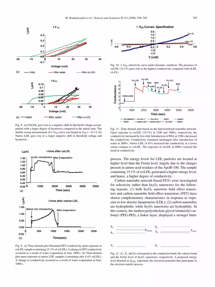
LDL and 4.4% of oxidized LDL (Fig. 10). The initial conduc-tivity (in black) was lower than that of native LDL (sample 2:4.4% of oxLDL, 95.5% of nLDL) (in blue). Oxidized LDL(sample 1: 15.1% oxLDL) significantly increased the con-ductivity by five-fold.
4.6. Detection of oxLDL by the nanotube network-basedFETs
Carbon nanotube network FETs functionalized withmethoxypolyethylene glycol imidazolyl carbonyl andcopper-induced anti-oxLDL antibodies were used to detectconductivities upon exposure to LDL particles in the pres-ence of BSA. OxLDL increased the conductivity by five-fold(Fig. 11). However, BSA decreased the conductivity. Re-exposure to oxLDL restored the conductivity. This trend wasreproducible with repeated trials, suggesting that nanotubenetwork-based FETs were able to selectively detect oxLDLfrom the non-redox protein, BSA.
5. Discussion
In2O3 nanowire- and carbon nanotube-based FETs pro-vide a new venue to differentiate LDL cholesterol betweenthe reduced and oxidized states. To our best knowledge, appli-cations of nanosensors to detect oxLDL have not previouslybeen demonstrated. The results were validated by HPLC asthe gold standard to fractionate LDL particles. The nanowire-and nanotube-based FETs usher in a potential lab-on-a-chipplatform to predict acute coronary syndromes at a small quan-tity of samples from the patients’ plasma.
Electron transfer from oxLDL particles to the In2O3nanowire changed the level of FET’s conductivity. This phe-nomenon can be described by the concept of energy bands(Fig. 12). Two energy band diagrams are illustrated with Ec,Ev and Ef, corresponding to the conduction band, the valenceband and the Fermi level of In2O3 nanowires. A proposedenergy level denoted as ELDL represents the electrical poten-tial of the electrons that participate in the electron transfer
M. Rouhanizadeh et al. / Sensors and Actuators B 114 (2006) 788–798 795
Fig. 8. (a) OxLDL gave rise to a negative shift in threshold voltage accom-panied with a larger degree of hysteresis compared to the initial state. Thedouble sweep measurement of I–VGS curve was biased at VDS = −0.1 V. (b)Native LDL gave rise to a larger negative shift in threshold voltage andhysteresis.
Fig. 9. (a) Time-domain plot illustrated FET conductivity upon exposure tooxLDL sample (containing 15.1% of oxLDL). A change in FET conductivityoccurred as a result of water evaporation at time 1800 s. (b) Time-domainplot upon exposure to native LDL samples (containing only 4.4% oxLDL).A change in conductivity occurred as a result of water evaporation at time:1000 s.
Fig. 10. I–VDS selectivity curve under dynamic condition. The presence ofoxLDL (15.1%) gave rise to the highest conductivity compared with nLDL(4.4%).
Fig. 11. Time-domain plot based on the functionalized nanotube network.Upon exposure to oxLDL (15.1%) at 1200 and 1800 s, respectively, theconductivity increased by five-fold. Introduction of BSA at 2300 s decreasedthe conductivity. Conductivity remained unchanged after introduction ofwater at 2600 s. Native LDL (4.4%) increased the conductivity at a lowerextent compare to oxLDL. The exposure of oxLDL at 4000 s restored thetrend in conductivity.
process. The energy levels for LDL particles are located athigher level than the Fermi level, largely due to the chargespresent in amino acid residues of the ApoB-100. The samplecontaining 15.1% of oxLDL generated a higher energy level,and hence, a higher degree of conductivity.
Carbon nanotube network-based FETs were investigatedfor selectivity rather than In2O3 nanowires for the follow-ing reasons: (1) both In2O3 nanowire field effect transis-tors and carbon nanotube field effect transistors (FET) haveshown complementary characteristics in response to expo-sure to low density lipoproteins (LDL); (2) carbon nanotubesare hydrophobic while In2O3 nanowires are hydrophilic. Inthis context, the methoxypolyethylene glycol imidazolyl car-bonyl (PEG-PEI), a linker layer, displayed a stronger bind-
Fig. 12. Ec, Ev and Ef correspond to the conduction band, the valence bandand the Fermi level of In2O3 nanowires, respectively. A proposed energylevel denoted as ELDL represents the electrical potential that participate inthe electron transfer process.

796 M. Rouhanizadeh et al. / Sensors and Actuators B 114 (2006) 788–798
Fig. 13. (a) Nanowire network devices consist of a matrix of synthesizednanotubes contacted by source (S), drain (D) electrodes and gate (G). (b)SEM photo of carbon nanotube network (drain and source electrodes are notshown in this range).
ing affinity to the nanotube-based FETs than to the In2O3nanowires; (3) carbon nanotubes were fabricated in a networkfashion (Fig. 13). Selectivity was achieved by functionaliza-tion of individual sensors in the entire network. This enhancedthe signal to noise ratio as well as the selectivity. The individ-ual transistors contained multiple nanotubes functioning asthe conductive channel between the drains and sources. As aresult, the overall transistor and sensing characteristics wereaveraged over an ensemble of nanotubes to reduce device-to-device variations. Despite the trade off in sensitivity, thecombination of nanotube network and surface functionaliza-tion with specific anti-oxLDL favored the investigation ofselectivity using carbon nanotube network.
Two possible factors accounted for the increased electronconcentration in the nanowires: (1) the amino groups car-ried by the ApoB-100 protein in LDL particles functionedas reductive species by donating electrons to the nanowires;and (2) positive charges carried by the amino groups func-tioned as a positive gate bias to the nanowires, leading to anenhanced carrier concentration. In both samples, the aminogroups in the Apo-B100 portion of the native LDL parti-cles carried positive charges (Fig. 1). The positive chargesinfluenced FETs to the same degree as applying positive gatevoltage to turn on the n-type transistor at a lower threshold
voltage. In the case of oxLDL, the positive charges in theamino groups were replaced by the hydroxyl group [27].
Individual nanowire transistors exhibit device-to-devicevariations. To gain the fabrication consistency, we have testedcarbon nanotube networks as shown in Fig. 13. Such a net-work allowed for a multitude of conducting channels betweensource and drain in the FET configurations. The network-based FETs provided two advantages: (1) the architecture isfault tolerant; that is, the dysfunction of one channel stillallows for other channels open for the conduction path-way between source and drain; and (2) the large number ofnanowires provides statistical averaging and identical overallnanowire density. Using the carbon nanotubes network conju-gated with anti-oxLDL antibodies, we were able to reproducethe conductivities upon exposure to oxLDL samples in thepresence of BSA. Future efforts will be to optimize the In2O3nanowire network by balancing the fabrication consistencyand the device performance.
While the majority of literatures for nano-sensing havebeen conducted in the vacuum condition to achieve a highsignal to noise ratio, we demonstrate the possibilities ofdetecting oxLDL under the liquid condition. Furthermore,functionalization of the nanotube network provided an entrypoint toward detection of oxLDL in the presence of non-redoxBSA.
6
radoapot
A
oosDUCTAt(dHcg
. Conclusion
Our study demonstrates that In2O3 nanowire-based FETepresents a potential sensor for detecting redox proteins suchs oxLDL particles. Our data supported the possibility ofistinguishing the LDL sample containing oxLDL from thatf nLDL via the changes in nanowire-based FET conductivitynd threshold voltages. Using both the ID–VDS and ID–VGSlots, we showed that the sample containing higher level ofxLDL particles increased the conductivity to a greater extenthan the one containing lower level of oxLDL.
cknowledgements
We are grateful for the advices from Dr. Alex Sevanianf the Department of Molecular Pharmacology and Toxicol-gy and the University of Southern California Atherosclero-is Research Unit. We also appreciate the assistance fromr. Howard Hodis, Director of Atherosclerosis Researchnit, Department of Preventive Medicine and Division ofardiovascular Medicine, for providing the LDL samples..K.H. is supported by the following grants: American Heartssociation (AHA BGIA 0265166U), The National Insti-
utes of Health Physician Scientist Career Research AwardK08 HL068689-01A1), and The National Heart Foun-ation/American Health Assistance Foundation (AHAF:2003-028). C.Z. is supported by USC Powell Award, NASA
ontract NAS2-99092, NSF CAREER award, NSF NER pro-ram and a Zumberger Award.

M. Rouhanizadeh et al. / Sensors and Actuators B 114 (2006) 788–798 797
References
[1] C.J. Murray, A.D. Lopez, Alternative projections of mortality anddisability by cause 1990–2020: Global Burden of Disease StudyLancet. 1997 May 24 349 (9064) 1498–1504.
[2] M. Navab, A.M. Fogelman, J.A. Berliner, M.C. Territo, L.L. Demer,J.S. Frank, A.D. Watson, et al., Pathogenesis of atherosclerosis, Am.J. Cardiol. 76 (1995) 18C–23C.
[3] M.S. Penn, M.Z. Cui, A.L. Winokur, et al., Smooth muscle cellsurface tissue factor pathway activation by oxidized low-densitylipoprotein requires cellular lipid peroxidation, Blood 96 (9) (2000)3056–3063.
[4] M.A. Austin, Small, dense low-density lipoprotein as a risk factorfor coronary heart disease, Int. J. Clin. Lab. Res. 24 (4) (1994) 187–192.
[5] S. Ehara, M. Ueda, T. Naruko, K. Haze, A. Itoh, M. Otsuka,R. Komatsu, T. Matsuo, et al., Elevated levels of oxidized lowdensity lipoprotein show a positive relationship with the severityof acute coronary syndromes, Circulation 103 (15) (2001) 1955–1960.
[6] A. Sevanian, H. Hodis, Antioxidants and atherosclerosis: anoverview, Biofactors 6 (1997) 385–390.
[7] Supriyo Bandyopadhyay, Hari Singh Nalwa, Quantum Dots andnanowires, American Scientific publishers, p. 193.
[8] R.L. Weiher, R.P. Ley, J. Appl. Phys. 37 (1966) 299.[9] G. Rupprecht, Z. Phys. 139 (1954) 504.
[10] Zu Rong Dai, Zhong Lin Wang, Nanobelts of Semiconcting Oxides,Science, vol. 291, 2001, p. 1947.
[11] J. Kong, N. Franklin, C. Zhou, S. Peng, K. Cho, H. Dai, Nanotubemolecular wires as chemical sensors, Science 287 (2000) 622.
[12] Q. Yi Cui, W. Hongkun Park, M. Charles, Lieber, Nanowire nanosen-
[[
[
[
[
[
[
[
[
[
[
[24] A. Unger, H.-P. Chou, T. Thorsen, A. Scherer, S.R. Quake, Mono-lithic microfabricated valves and pumps by multilayer soft lithogra-phy, Science 288 (2000) 113–116.
[25] X. Liu, C. Lee, C. Zhou, J. Han, Carbon nanotube field effect invert-ers, Appl. Phys. Lett. 79 (20) (2001) 3329–3331, November 12.
[26] D. Zhang, C. Li, X. Liu, S. Han, T. Tang, C. Zhou, Doping-dependentammonia sensing of indium oxide nanowires, Appl. Phys. Lett. 84(No. 9) (2003) 1845.
[27] A. Sevanian, L. Asatryan, O. Ziouzenkova, Low density lipoprotein(LDL) modification: basic concepts and relationship to atherosclero-sis, Blood Purif. 17 (1999) 66–78.
[28] A. Sevanian, G. Bittolo-Bon, G. Cazzolato, H. Hodis, J. Hwang, A.Zamburlini, M. Maiorino, F. Ursini, LDL is a lipid hydroperoxide-enriched circulating lipoprotein, J. Lipid Res. 38 (3) (1997) 419–428.
[29] M.A. Austin, Small, dense low-density lipoprotein as a risk factorfor coronary heart disease, Int. J. Clin. Lab. Res. 24 (4) (1994) 187–192.
[30] R.B. Wesley Jr, X. Meng, D. Godin, Z.S. Galis, Extracellular matrixmodulates macrophage functions characteristic to atheroma: colla-gen type I enhances acquisition of resident macrophage traits byhuman peripheral blood monocytes in vitro, Arterioscler. Thromb.Vasc. Biol. 18 (1998) 432–440.
[31] W.L. Stone, M. Heimberg, R.L. Scott, I. LeClair, H.G. Wilcox,Altered hepatic catabolism of low-density lipoprotein subjected tolipid peroxidation in vitro, Biochem. J. 297 (Part 3) (1994) 573–579.
[32] J. Tamaki, C. Naruo, Y. Yamamoto, M. Mastuoka, Sensing proper-ties to dilute chlorine gas of indium oxide based thin film sensorsprepared by electron beam evaporation, Sens. Actuators B 83 (190)(2002).
B
MStagscaaa
TCit
Cmara
JatPemca
Cr
sors for highly sensitive and selective detection of biological andchemical species, Science 293 (2001) 1289–1292.
13] D.S. Ginley, C. Bright, Bull. Mater. Res. Soc. 25 (2000) 15.14] C. Li, D. Zhang, X. Liu, S. Han, T. Tang, J. Han, C. Zhou, In2O3
nanowires as chemical sensors, Appl. Phys. Lett. 82 (2003) 1613.15] J.R. Bellingham, A.P. Mackenzie, W.A. Philips, Transparent con-
ducting thin films: precise measurement of the oxygen content, Appl.Phys. Lett. 58 (1991) 2506.
16] Lubert Stryer, Biochemistry, third edition, W.H. Freeman and com-pany, p. 561.
17] T. Henriksen, E.M. Mahoney, D. Steinberg, Enhanced macrophagedegradation of low density lipoprotein previously incubated with cul-tured endothelial cells: recognition by receptors for acetylated lowdensity lipoproteins, Proc. Natl. Acad. Sci. U.S.A. 78 (10) (1981)6499–6503.
18] T. Tang, X. Liu, C. Li, B. Lei, D. Zhang, M. Rouhanizadeh, T. Hsiai,C. Zhou, Complementary response of In2O3 nanowires and carbonnanotubes to low-density lipoprotein chemical gating, Appl. Phys.Lett. (2005) 86.
19] C. Li, B. lei, D. Zhang, X. Liu, S. Han, T. Tang, M. Rouhanizadeh,T. Hsiai, C. Zhou, Chemical gating of In2O3 nanowires by organicand biomolecules, Appl. Phys. Lett. (2003) 4014–4016.
20] H.N. Hodis, D.M. Kramsch, P. Avogaro, G. Bittolo-Bon, G. Cazzo-lato, J. Hwang, H. Peterson, A. Sevanian, Biochemical and cytotoxiccharacteristics of an in vivo circulating oxidized low density lipopro-tein (LDL-), J. Lipid Res. 35 (1994) 669–677.
21] J. Hwang, M. Ing, A. Salazar, M. Navab, A. Sevanian, T. Hsiai,Pulsatlile versus oscillatory shear stress regulates NADPH oxidasesubunit: implication for native LDL oxidation, Circ. Res. 93 (12)(2003) 1225–1232, Epub 2003 October 30, December 12.
22] C. Li, D. Zhang, S. Han, X. Liu, T. Tang, C. Zhou, Diameter-controlled growth of single-crystalline In2O3 nanowires and theirelectronic properties, Adv. Mater. 15 (2003) 143.
23] B.-H. Jo, L.M. Van Lerberghe, K.M. Motsegood, D.J. Beebe, Threedimensional micro-channel fabrication in PDMS elastomer, J. MEMS9 (2000) 76–81.
iographies
ahsa Rouhanizadeh received her B.S. in Electrical Engineering fromharif University of Technology, Tehran, Iran in February 1999. She
hen obtained a DEA in “Information processing for electrical systems”t University of Paris, France in September 2000. Then she continuedraduate studies at University of Southern California, Los Angeles wherehe received M.S. degree in Electrical Engineering in May 2002. She isurrently a Ph.D. candidate in the Department of Biomedical Engineeringnd Division of Cardiovascular Medicine. Her research interests are micrond nano-sensors for diagnosis of cardiovascular diseases. She has beenmember of IEEE since 2001.
ao Tang received B.Sc. degree from Beijing Institute of Technology,hina, in 2001, and M.Sc. degree from University of Southern California
n 2004. He is currently pursuing Ph.D. degree from Department of Elec-rical Engineering – Electrophysics at University of Southern California.
hao Li received her Ph.D. degree from Electrical Engineering depart-ent of University of Southern California in 2005. She received her M.Sc.
nd B.Sc. degrees from Lanzhou University, China, in 2001 and 1998,espectively. Her research interests are the synthesis, electronic studiesnd applications of nanoscale materials.
uliana Hwang received Pharm.D. degree from the School of Pharmacyt the University of Sao Paulo, Brazil in 1986. She currently holds a posi-ion of Research Assistant Professor at School of Pharmacy at Molecularharmacology and Toxicology Department at the University of South-rn California. Her research interests are in the area of investigating theanner by which estrogen facilitates the antioxidant capacity of vascular
ells and mechanisms by which estrogen decrease the development oftherogenesis in the vessel wall.
hongwu Zhou received his Ph.D. from Yale University in 1999. Heeceived his B.Sc. from the University of Science and Technology of

798 M. Rouhanizadeh et al. / Sensors and Actuators B 114 (2006) 788–798
China in 1993. He worked as a Postdoctoral Research Assistant at Stan-ford University from 1998 to 2000. He is currently an Assistant Professorat the Department of Electrical Engineering – Electrophysics of Universityof Southern California. His research interests are in the areas of carbonnanotubes, semiconductive nanowires, transition metal oxide nanowires,molecular electronics, and chemical and bio sensing. He has won a num-ber of awards including the NSF CAREER Award and the NASA TGiRAward.
Dr. Tzung K. Hsiai received his B.S. from Columbia University andM.D. from the University of Chicago. Dr. Hsiai underwent his post-graduate medical training and NIH NRSA for cardiology fellowship atUCLA School of Medicine, where he obtained a Ph.D. in BiomedicalEngineering in 2002. He is recipient of NIH Physician Scientist CareerDevelopment Award, and American Heart Association John J. SimpsonOutstanding Research Achievement Award. T.K. Hsiai was elected as aFellow of American College of Cardiology in 2005.